Chapter 1: Biology and Behavior
Chapter 1: Biology and Behavior
SCIENCE MASTERY ASSESSMENT
Every pre-med knows this feeling: there is so much content I have to know for the MCAT! How do I know what to do first or what’s important?
While the high-yield badges throughout this book will help you identify the most important topics, this Science Mastery Assessment is another tool in your MCAT prep arsenal. This quiz (which can also be taken in your online resources) and the guidance below will help ensure that you are spending the appropriate amount of time on this chapter based on your personal strengths and weaknesses. Don’t worry though—skipping something now does not mean you’ll never study it. Later on in your prep, as you complete full-length tests, you’ll uncover specific pieces of content that you need to review and can come back to these chapters as appropriate.
How to Use This Assessment
If you answer 0–7 questions correctly:
Spend about 1 hour to read this chapter in full and take limited notes throughout. Follow up by reviewing all quiz questions to ensure that you now understand how to solve each one.
If you answer 8–11 questions correctly:
Spend 20–40 minutes reviewing the quiz questions. Beginning with the questions you missed, read and take notes on the corresponding subchapters. For questions you answered correctly, ensure your thinking matches that of the explanation and you understand why each choice was correct or incorrect.
If you answer 12–15 questions correctly:
Spend less than 20 minutes reviewing all questions from the quiz. If you missed any, then include a quick read-through of the corresponding subchapters, or even just the relevant content within a subchapter, as part of your question review. For questions you answered correctly, ensure your thinking matches that of the explanation and review the Concept Summary at the end of the chapter.
- A researcher deletes a gene from an organism to determine the gene’s function. This approach is most analogous to the work of which of the following scientists?
- Paul Broca
- Pierre Flourens
- Franz Gall
- Sir Charles Sherrington
- Which component of the nervous system is NOT involved in the initial reflexive response to pain?
- Spinal cord
- Cerebral cortex
- Interneuron
- Motor neuron
- A child has experienced nervous system damage and can no longer coordinate the movements to dribble a basketball, although the child can still walk in an uncoordinated fashion. Which region of the central nervous system was most likely affected?
- Forebrain
- Midbrain
- Hindbrain
- Spinal cord
- The temporal lobe deals with all of the following EXCEPT:
- language comprehension.
- memory.
- emotion.
- motor skills.
- Which part of the brain deals with both homeostasis and emotions?
- Cerebellum
- Pons
- Hypothalamus
- Thalamus
- Which of the following activities would most likely be completed by the right hemisphere of a left-handed person?
- Finding a car in a parking lot
- Learning a new language
- Reading a book for pleasure
- Jumping rope with friends
- Which of the following is/are true with regard to neurulation?
- The neural tube differentiates from endoderm.
- The neural tube becomes the peripheral nervous system.
- Neural crest cells migrate from their original site.
- I only
- III only
- II and III only
- I, II, and III
- Which of the following neurotransmitters is NOT classified as a catecholamine?
- Epinephrine
- Norepinephrine
- Dopamine
- Acetylcholine
- If the amount of acetylcholinesterase, an enzyme that breaks down acetylcholine, is increased, which of the following would likely be the result?
- Weakness of muscle movements
- Excessive pain or discomfort
- Mood swings and mood instability
- Auditory and visual hallucinations
- The adrenal glands do all of the following EXCEPT:
- promote the fight-or-flight response via estrogen.
- produce stress responses via cortisol.
- produce both hormones and neurotransmitters.
- release estrogen in males and testosterone in females.
- A disorder of the pineal gland would most likely result in which of the following disorders?
- High blood pressure
- Diabetes
- Insomnia
- Hyperthyroidism
- Which of the following conclusions would William James most likely support?
- Mental processes help individuals adapt to their environments.
- Psychological attributes could be measured by feeling the skull.
- Specific functional impairments can be linked to specific lesions in the brain.
- Synaptic transmission is an electrical process.
- A scientist designs a study to determine if different regions of the brain are activated when a person speaks their native language vs. a second language. Which of the following methods would the scientist most likely choose?
- MRI
- CT scan
- fMRI
- EEG
- During a physical examination, a physician brushes the bottom of the foot of a patient who is fifty years old with multiple sclerosis. The patient’s toes are observed to curl toward the bottom of the foot, with no fanning of the toes. This response is:
- abnormal, and evidence that the patient is exhibiting a primitive reflex.
- normal, and evidence that the patient is exhibiting a primitive reflex.
- abnormal, and evidence that the patient is not exhibiting a primitive reflex.
- normal, and evidence that the patient is not exhibiting a primitive reflex.
- Which of the following fine motor tasks would one expect to see first in an infant?
- Grasping for objects with two fingers
- Following objects with the eyes
- Scribbling with a crayon
- Moving a toy from one hand to the other
Answer Key
- B
- B
- C
- D
- C
- A
- B
- D
- A
- A
- C
- A
- C
- D
- B
Chapter 1: Biology and Behavior
CHAPTER 1
BIOLOGY AND BEHAVIOR
In This Chapter
1.1 A Brief History of Neuropsychology 1.2 Organization of the Human Nervous System
Central and Peripheral Nervous Systems
The Autonomic Nervous System
1.3 Organization of the Brain
Hindbrain
Midbrain
Forebrain
Methods of Mapping the Brain
1.4 Parts of the Forebrain
Thalamus
Hypothalamus
Other Parts of the Diencephalon
Basal Ganglia
Limbic System
Cerebral Cortex
1.5 Influences on Behavior
Neurotransmitters
The Endocrine System
Genetics and Behavior
1.6 Development
Prenatal
Motor
Social
Concept Summary
CHAPTER PROFILE
The content in this chapter should be relevant to about 8% of all questions about the behavioral sciences on the MCAT.
This chapter covers material from the following AAMC content categories:
3A: Structure and functions of the nervous and endocrine systems and ways in which these systems coordinate the organ systems
7A: Individual influences on behavior
Introduction
When you woke up this morning and got ready to start reading MCAT Behavioral Sciences Review, you almost certainly had specific feelings about it—perhaps you were excited to crack open the book and start learning some of the material that will get you that top score on the MCAT; perhaps you dreaded the size and rich detail of the information in the book. Either way, your body began to respond to these impulses from your mind: increasing heart rate, increasing breathing rate, dilating the eyes, and slowing down digestion. This link between the mind and the body is still a hot topic in medicine, although we’ve been exploring the effects of psychology on well-being for almost two centuries now.
In this chapter, we’ll begin our exploration of psychology and sociology by looking at the biological side of psychology. After a quick survey of the history of neuropsychology, we’ll look at the structure and organization of the human nervous system, communication between the nervous and endocrine systems, the effects of genes and environment on behavior, and some aspects of psychological development.
1.1 A Brief History of Neuropsychology
LEARNING OBJECTIVES
After Chapter 1.1, you will be able to:
- Recall the major contributions to early neuropsychology
- Connect the major contributors of early neuropsychology to their contributions
Researchers in the 19th century began to think about behavior from a physiological perspective. Many of these early thinkers formed the foundation of current knowledge about neuroanatomy, linking the functions of specific areas of the brain with thought and behavior.
Franz Gall (1758–1828) had one of the earliest theories that behavior, intellect, and even personality might be linked to brain anatomy. He developed the doctrine of phrenology. The basic idea was that if a particular trait was well-developed, then the part of the brain responsible for that trait would expand. This expansion, according to Gall, would push the area of the skull that covered that part of the brain outward and therefore cause a bulge on the head. Gall believed that one could thus measure psychological attributes by feeling or measuring the skull. Although phrenology was shown to be false, it did generate serious research on brain functions and was the impetus for the work of other psychologists through the remainder of the 19th century.
Pierre Flourens (1794–1867) was the first person to study the functions of the major sections of the brain. He did this by extirpation, also known as ablation, on rabbits and pigeons. In extirpation, various parts of the brain are surgically removed and the behavioral consequences are observed. Flourens’s work led to his assertion that specific parts of the brain had specific functions, and that the removal of one part weakens the whole brain.
William James (1842–1910), known as the founder of American psychology, studied how the mind adapts to the environment. His views formed the foundation for the system of thought in psychology known as functionalism, which studies how mental processes help individuals adapt to their environments.
John Dewey (1859–1952) is another important name in functionalism because his 1896 article is seen as its inception. This article criticized the concept of the reflex arc, which breaks the process of reacting to a stimulus into discrete parts. Dewey believed that psychology should focus on the study of the organism as a whole as it functioned to adapt to the environment.
Around 1860, Paul Broca (1824–1880) added to the knowledge of physiology by examining the behavioral deficits of people with brain damage. He was the first person to demonstrate that specific functional impairments could be linked with specific brain lesions. Broca studied a person who was unable to speak and discovered that the person’s disability was due to a lesion in a specific area on the left side of the person’s brain. This area of the brain is now referred to as Broca’s area.
Hermann von Helmholtz (1821–1894) was the first to measure the speed of a nerve impulse. He also related the measured speed of such impulses to reaction time, providing an important early link between behavior and underlying nervous system activity. Because Helmholtz provided one of the earliest measurable links between psychology and physiology, he is often credited with the transition of psychology out of the realm of philosophy and into the realm of quantifiable natural science.
Around the turn of the century, Sir Charles Sherrington (1857–1952) first inferred the existence of synapses. Many of his conclusions have held over time—except for one. He thought that synaptic transmission was an electrical process, but we now know that it is primarily a chemical process.
BRIDGE
Solutions to concept checks for a given chapter in MCAT Behavioral Sciences Review can be found near the end of the chapter in which the concept check is located, following the Concept Summary for that chapter.
MCAT CONCEPT CHECK 1.1
Before you move on, assess your understanding of the material with this question.
- Briefly list the main contributions of each of the following scientists to neuropsychology.
_________________________
- Franz Gall:
_________________________
- Pierre Flourens:
_________________________
- William James:
_________________________
- John Dewey:
_________________________
- Paul Broca:
_________________________
- Hermann von Helmholtz:
_________________________
- Sir Charles Sherrington:
1.2 Organization of the Human Nervous System
LEARNING OBJECTIVES
After Chapter 1.2, you will be able to:
- Correctly associate regions of the nervous system with the CNS or PNS
- Distinguish between afferent and efferent neurons
- Describe the functions of the somatic and autonomic nervous systems, as well as the sympathetic and parasympathetic nervous systems:
MCAT EXPERTISE
The “High-Yield” badge on this section indicates that the content is frequently tested on the MCAT.
The human nervous system is a complex web of over 100 billion cells that communicate, coordinate, and regulate signals for the rest of the body. Mental and physical action occurs when the body can react to external stimuli using the nervous system. In this section, we will look at the nervous system and its basic organization.
Note: Much of the information contained in this section is also discussed in Chapter 4 of MCAT Biology Review.
Central and Peripheral Nervous Systems
There are three kinds of nerve cells in the nervous system: sensory neurons, motor neurons, and interneurons. Sensory neurons (also known as afferent neurons) transmit sensory information from receptors to the spinal cord and brain. Motor neurons (also known as efferent neurons) transmit motor information from the brain and spinal cord to muscles and glands. Interneurons are found between other neurons and are the most numerous of the three types of neurons. Interneurons are located predominantly in the brain and spinal cord and are often linked to reflexive behavior. Neural circuits called reflex arcs control this type of reflexive behavior. For example, consider what occurs when someone steps on a nail. Receptors in the foot detect pain and the pain signal is transmitted by sensory neurons up to the spinal cord. At that point, the sensory neurons connect with interneurons, which then relay pain impulses up to the brain. Rather However, rather than waiting for the brain to send out a signal, interneurons in the spinal cord send signals to the muscles of both legs directly, causing the individual to reflexively withdraw the foot in pain while simultaneously reflexively transferring weight to the other foot. The original sensory information still makes its way up to the brain; however, by that time, the muscles have already responded to the pain, thanks to the cooperation of these several reflex arcs.
MNEMONIC
Afferent neurons ascend in the cord toward the brain; efferent neurons exit the cord on their way to the rest of the body.
Let’s turn to the overall structure of the human nervous system, which is diagrammed in Figure 1.1. The nervous system can be broadly divided into two primary components: the central and peripheral nervous systems. The central nervous system (CNS) is composed of the brain and spinal cord. The peripheral nervous system (PNS), in contrast, is made up of nerve tissue and fibers outside the brain and spinal cord. Note that the peripheral nervous system includes all 31 pairs of nerves emanating from the spinal cord, which are called spinal nerves, and 12 pairs of nerves emanating directly from the brain, called cranial nerves. The olfactory and optic nerves (cranial nerves I and II) are structurally outgrowths of the central nervous system, but are still considered components of the peripheral nervous system. The PNS thus connects the CNS to the rest of the body.
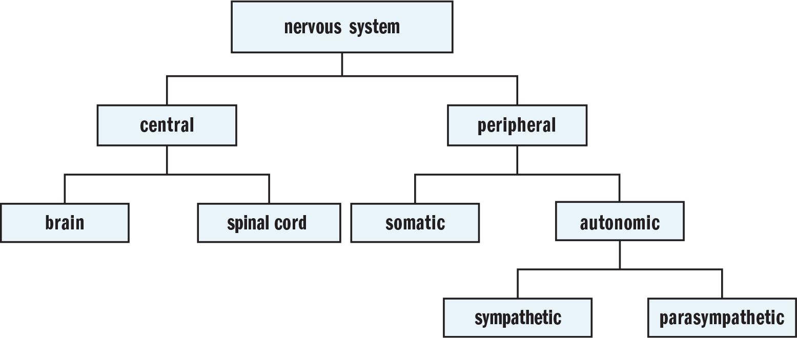
Figure 1.1. Major Divisions of the Nervous System
The peripheral nervous system is further subdivided into the somatic and autonomic nervous systems. The somatic nervous system consists of sensory and motor neurons distributed throughout the skin, joints, and muscles. Sensory neurons transmit information toward the CNS through afferent fibers. Motor impulses, in contrast, travel from the CNS back to the body along efferent fibers.
The autonomic nervous system (ANS) generally regulates heartbeat, respiration, digestion, and glandular secretions. In other words, the ANS manages the involuntary muscles associated with many internal organs and glands. The ANS also helps regulate body temperature by activating sweating or piloerection, depending on whether the body is too hot or too cold. The main thing to understand about all of these functions is that they are automatic, or independent of conscious control. Note the similarity between the words autonomic and automatic. This association makes it easy to remember that the autonomic nervous system manages automatic functions such as heartbeat, respiration, digestion, and temperature control.
The Autonomic Nervous System
The ANS has two subdivisions: the sympathetic nervous system and the parasympathetic nervous system. These two branches often act in opposition to one another, meaning they are antagonistic. For example, the sympathetic nervous system acts to accelerate heart rate and inhibit digestion, while the parasympathetic nervous system decelerates heart rate and increases digestion.
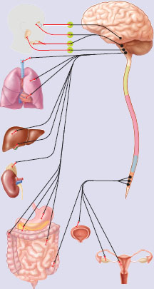
The main role of the parasympathetic nervous system is to conserve energy. It is associated with resting and sleeping states, and acts to reduce heart rate and constrict the bronchi. The parasympathetic nervous system is also responsible for managing digestion by increasing peristalsis and exocrine secretions. Acetylcholine is the neurotransmitter responsible for parasympathetic responses in the body. The functions of the parasympathetic nervous system are summarized in Figure 1.2.

Figure 1.2. Functions of the Parasympathetic Nervous System
In contrast, the sympathetic nervous system is activated by stress. This can include everything from a mild stressor, such as keeping up with schoolwork, to emergencies that mean the difference between life and death. The sympathetic nervous system is closely associated with rage and fear reactions, also known as “fight-or-flight” reactions.
When activated, the sympathetic nervous system:
- Increases heart rate
- Redistributes blood to muscles of locomotion
- Increases blood glucose concentration
- Relaxes the bronchi
- Decreases digestion and peristalsis
- Dilates the eyes to maximize light intake
- Releases epinephrine into the bloodstream
MNEMONIC
Sympathetic and parasympathetic nervous systems:
- Sympathetic: “fight-or-flight”
- Parasympathetic: “rest-and-digest”
The functions of the sympathetic nervous system are summarized in Figure 1.3.

Figure 1.3. Functions of the Sympathetic Nervous System
MCAT CONCEPT CHECK 1.2
Before you move on, assess your understanding of the material with these questions.
- What parts of the nervous system are in the central nervous system (CNS)? Peripheral nervous system (PNS)?
_________________________
- CNS:
_________________________
- PNS:
- What do afferent neurons do? Efferent neurons?
_________________________
- Afferent:
_________________________
- Efferent:
- What functions are accomplished by the somatic nervous system? The autonomic nervous system?
___________________________
- Somatic:
_________________________
- Autonomic:
- What are the effects of the sympathetic nervous system? The parasympathetic nervous system?
_________________________
- Sympathetic:
_________________________
- Parasympathetic:
1.3 Organization of the Brain
LEARNING OBJECTIVES
After Chapter 1.3, you will be able to:
- Describe the major functions of the hindbrain, midbrain, and forebrain
- Recognize the most commonly used methods for mapping the brain
- Identify the structures protecting and surrounding the brain
Throughout this section, refer to Figure 1.4, which identifies various anatomical structures inside the human brain. As we discuss different parts of the brain, it’s important to remember the functions of these brain structures. Different parts of the brain perform remarkably different functions. For instance, one part of the brain processes sensory information while an entirely different part of the brain maintains activities of the internal organs. For complex functions such as playing a musical instrument, several brain regions work together. For the MCAT, you will need to know some of the basics about how the brain integrates input from different regions.
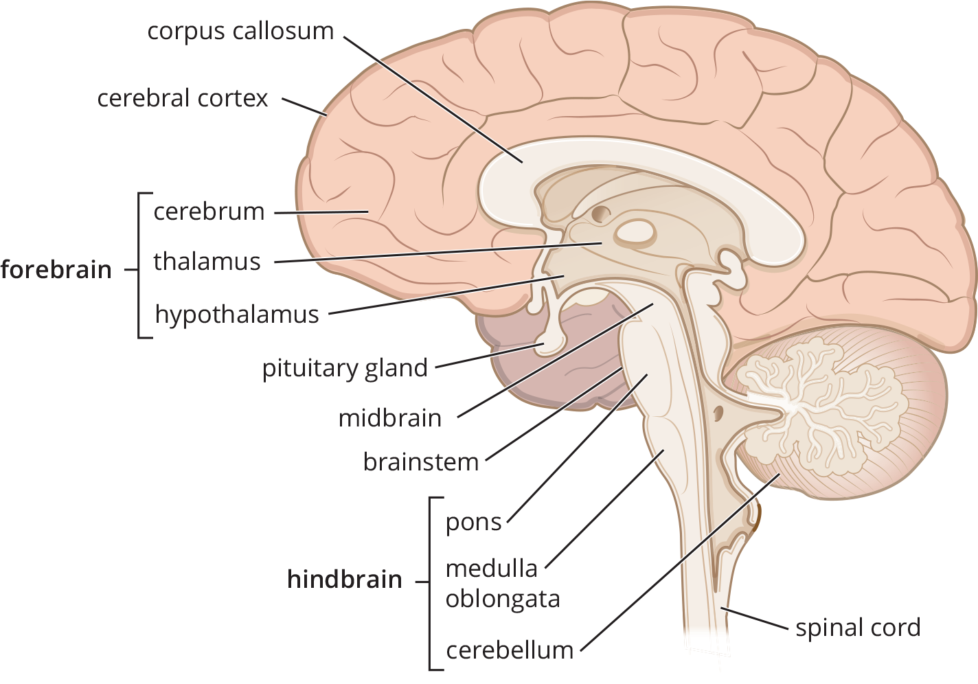
Figure 1.4. Anatomical Structures Inside the Human Brain
The brain is covered with a thick, three-layered sheath of connective tissue collectively called the meninges. The outer layer of connective tissue is the dura mater, and is connected directly to the skull. The middle layer is a fibrous, weblike structure called arachnoid mater. And the inner layer, connected directly to the brain, is known as the pia mater. These three layers of connective tissue are shown in Figure 1.5. The meninges help protect the brain by keeping it anchored within the skull, and the meninges also resorb cerebrospinal fluid, which is the aqueous solution that nourishes the brain and spinal cord and provides a protective cushion. Cerebrospinal fluid is produced by specialized cells that line the ventricles (internal cavities) of the brain.
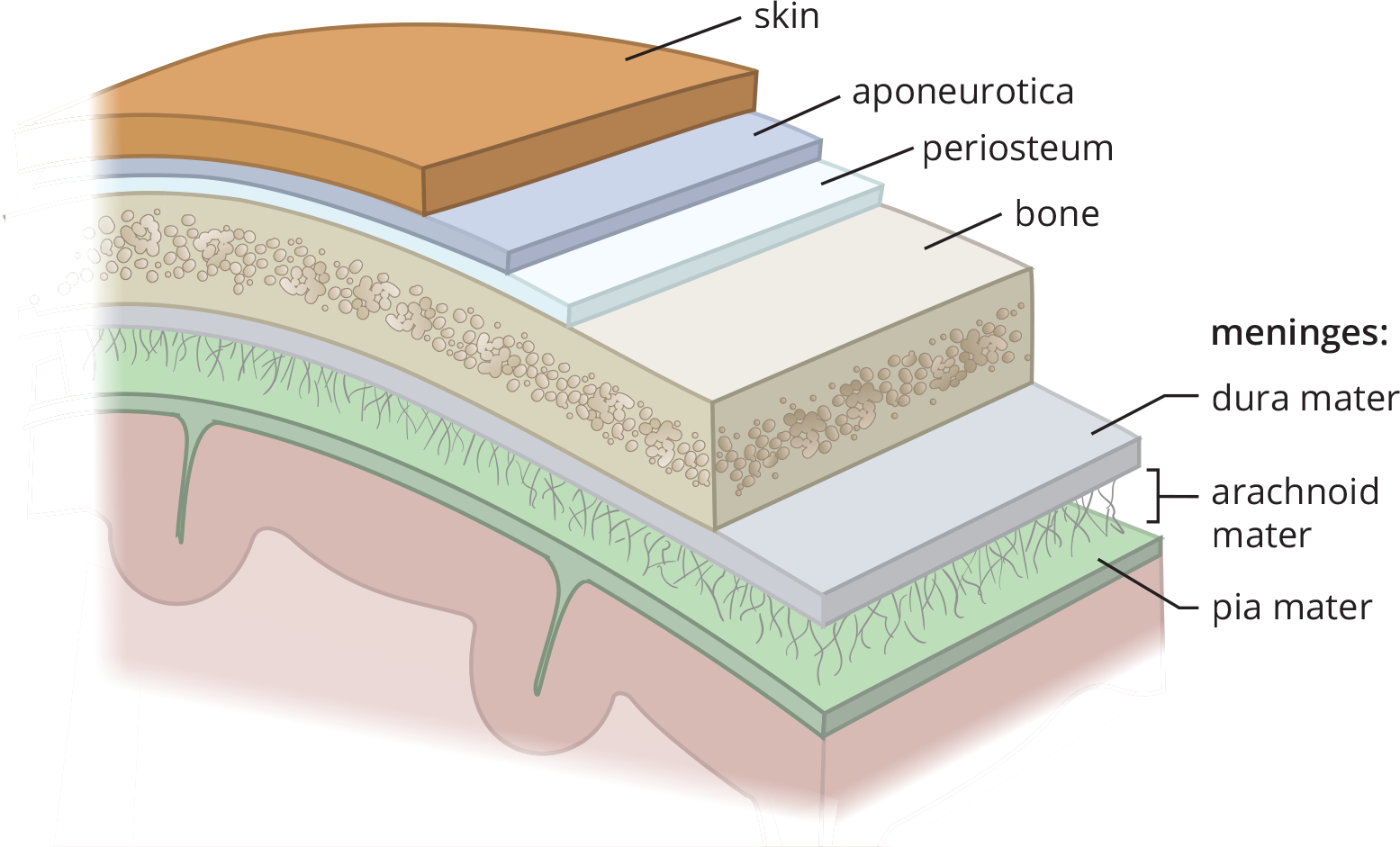
Figure 1.5. Layers of the Meninges
The human brain can be divided into three basic parts: the hindbrain, the midbrain, and the forebrain. Notice that brain structures associated with basic survival are located at the base of the brain and brain structures with more complex functions are located higher up. The meaningful connection between brain location and functional complexity is no accident. In evolutionary terms, the hindbrain and midbrain were brain structures that developed earlier. Together they form the brainstem, which is the most primitive region of the brain. The forebrain developed later, including the limbic system, a group of neural structures primarily associated with emotion and memory. Aggression, fear, pleasure, and pain are all related to the limbic system. The most recent evolutionary development of the human brain is the cerebral cortex, which is the outer covering of the cerebral hemispheres. In humans, the cerebral cortex is associated with everything from language processing to problem solving, and from impulse control to long-term planning. Most of the key brain regions described in the following sections are summarized in Table 1.1.
Table 1.1. Anatomical Subdivisions of the Brain
MAJOR DIVISIONS AND PRINCIPAL STRUCTURES FUNCTIONS
- Forebrain
- Cerebral cortex
- Basal ganglia
- Limbic system
- Thalamus
- Hypothalamus
-
- Complex perceptual, cognitive, and behavioral processes
- Movement
- Emotion and memory
- Sensory relay station
- Hunger and thirst; emotion
- Midbrain
- Inferior and superior colliculi
-
- Sensorimotor reflexes
- Hindbrain
- Cerebellum
- Medulla oblongata
- Reticular formation
- Pons
-
- Refined motor movements
- Heart, vital reflexes (vomiting, coughing)
- Arousal and alertness
- Communication within the brain, breathing
In prenatal life, the brain develops from the neural tube. At first, the tube is composed of three swellings, which correspond to the hindbrain, midbrain, and forebrain. Both the hindbrain and forebrain later divide into two swellings, creating five total swellings in the mature neural tube. The embryonic brain is diagrammed in Figure 1.6, and its subdivisions are described further in the following sections. Understanding the relationship between the structures of the developing brain and the fully developed brain is important. So the following sections describe both the structures of the developing brain and what those structures develop into.
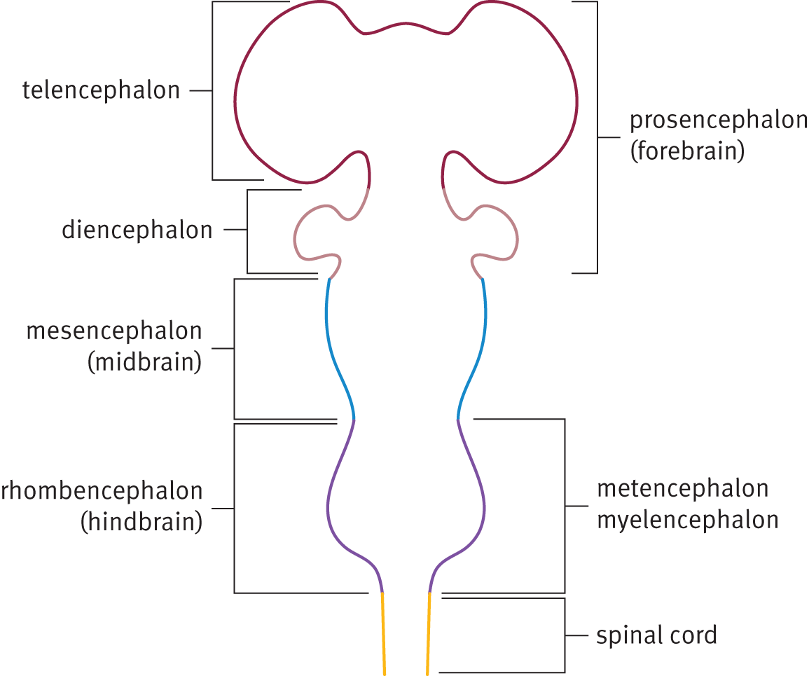
Figure 1.6. Subdivisions of the Embryonic Brain
Hindbrain
Located where the brain meets the spinal cord, the hindbrain (rhombencephalon) controls balance, motor coordination, breathing, digestion, and general arousal processes such as sleeping and waking. In short, the hindbrain manages vital functioning necessary for survival. During embryonic development, the rhombencephalon divides to form the myelencephalon (which becomes the medulla oblongata) and the metencephalon (which becomes the pons and cerebellum). In the developed brain, the medulla oblongata is a lower brain structure that is responsible for regulating vital functions such as breathing, heart rate, and digestion. The pons lies above the medulla and contains sensory and motor pathways between the cortex and the medulla. At the top of the hindbrain, mushrooming out of the back of the pons, is the cerebellum, a structure that helps maintain posture and balance and coordinates body movements. Damage to the cerebellum causes clumsiness, slurred speech, and loss of balance. Notably, alcohol impairs the functioning of the cerebellum, and consequently affects speech and balance.
Midbrain
Just above the hindbrain is the midbrain (mesencephalon), which receives sensory and motor information from the rest of the body. The midbrain is associated with involuntary reflex responses triggered by visual or auditory stimuli. There are several prominent nuclei in the midbrain, two of which are collectively called colliculi. The superior colliculus receives visual sensory input, and the inferior colliculus receives sensory information from the auditory system. The inferior colliculus has a role in reflexive reactions to sudden loud noises.
Forebrain
Above the midbrain is the forebrain (prosencephalon), which is associated with complex perceptual, cognitive, and behavioral processes. Among its other functions, the forebrain is associated with emotion and memory; it is the forebrain that has the greatest influence on human behavior. Its functions are not absolutely necessary for survival, but are associated instead with the intellectual and emotional capacities most characteristic of humans. During prenatal development, the prosencephalon divides to form the telencephalon (which forms the cerebral cortex, basal ganglia, and limbic system) and the diencephalon (which forms the thalamus, hypothalamus, posterior pituitary gland, and pineal gland).
Methods of Mapping the Brain
Neuropsychology refers to the study of functions and behaviors associated with specific regions of the brain. It is most often applied in research settings, where researchers attempt to associate very specific areas in the brain to behavior. Neuropsychology is also applied in clinical settings with evaluations of patient cognitive and behavioral functioning, as well as the diagnosis and treatment of brain disorders. Neuropsychology has its own experimental methodology and technology.
Studying human patients with brain lesions is one way that researchers have determined the functions of the brain. In order to conclude that a specific structure of the brain is responsible for a specific function, researchers look for patients that exhibit damage to that structure coupled with a loss of the function. One problem in studying human brain lesions, however, is that such lesions are rarely isolated to specific brain structures. When several brain structures are damaged, the impairment could be attributed to any of the damaged structures, and pinpointing a specific link between brain structure and function becomes difficult.
One method for studying the relationship of brain regions and behaviors is to study brain lesions in lab animals. The advantage of this approach is that precisely defined brain lesions can be created in animals by extirpation. Researchers can also produce lesions by inserting tiny electrodes inside the brain and then selectively applying intense heat, cold, or electricity to specific brain regions. Such electrodes can be placed with great precision by using stereotactic instruments, which provide high-resolution, three-coordinate images of the brain. Ethical or cruelty concerns notwithstanding, such studies have greatly increased our understanding of comparable neural structures in humans.
Another neuropsychology method involves electrically stimulating the brain and recording consequent brain activity. While operating on the brain, a surgeon can stimulate a patient’s cortex with a small electrode. This stimulation causes groups of neurons to fire, thereby activating the behavioral or perceptual processes associated with those neurons. For instance, if the electrode stimulates neurons in the motor cortex, the stimulation can lead to specific muscle movements. If the electrode stimulates the visual cortex, the patient may “see” flashes of light that are not really there. By using electrical stimulation, neurosurgeons can thus create cortical maps. This method relies on the assistance of the patient, who is awake and alert. Because there are no pain receptors in the brain, only local anesthesia is required. Electrodes have also been used in lab animals to study deeper regions of the brain. Depending on where these electrodes are implanted, they can elicit sleep, sexual arousal, rage, or terror. Once the electrode is turned off, these behaviors cease.
Electrodes can also be used to record electrical activity produced by the brain itself. In some studies, individual neurons are recorded by inserting ultrasensitive microelectrodes into individual brain cells and recording their electrical activity. Electrical activity generated by larger groups of neurons can be studied using an electroencephalogram (EEG), which involves placing several electrodes on the scalp. Broad patterns of electrical activity can thus be detected and recorded. Because this procedure is noninvasive (it does not cause any damage), electroencephalograms are commonly used with human subjects. In fact, research on sleep, seizures, and brain lesions relies heavily on EEGs, like the one shown in Figure 1.7.

Figure 1.7. Electroencephalogram (EEG) during REM Sleep
Another noninvasive mapping procedure is regional cerebral blood flow (rCBF), which detects broad patterns of neural activity based on increased blood flow to different parts of the brain. rCBF relies on the assumption that blood flow increases to regions of the brain that are engaged in cognitive function. For example, listening to music may increase blood flow to the right auditory cortex because music is processed in that region in most individuals’ brains. To measure blood flow, the patient inhales a harmless radioactive gas; a special device that can detect radioactivity in the bloodstream can then correlate radioactivity levels with regional cerebral blood flow. This research method uses noninvasive computerized scanning devices.
Some of the other common scanning devices and methods of visualization used for brain imaging include:
- CT (computed tomography), also known as CAT (computed axial tomography) scan, in which multiple X-rays are taken at different angles and processed by a computer to produce cross-sectional images of the tissue.
- PET (positron emission tomography) scan, in which a radioactive sugar is injected and absorbed into the body, and its dispersion and uptake throughout the target tissue is imaged.
- MRI (magnetic resonance imaging), in which a magnetic field that interacts with hydrogen atoms is used to map out hydrogen dense regions of the body.
- fMRI (functional magnetic resonance imaging), which uses the same base technique as MRI, but specifically measures changes associated with blood flow. fMRI is especially useful for monitoring neural activity, since increased blood flow to a region of the brain is typically coupled with its neuronal activation.
BRIDGE
MRI techniques are dependent on the reaction of hydrogen to a magnetic field, and the scientific principles behind MRI scans are also applied in NMR techniques, which can be found in Chapter 11 of MCAT Organic Chemistry Review.
MCAT CONCEPT CHECK 1.3
Before you move on, assess your understanding of the material with these questions.
- What are the main functions of the hindbrain? Midbrain? Forebrain?
Subdivision Functions Hindbrain Midbrain Forebrain
- What are some of the methods used for mapping the brain? _________________________
- What structures surround and protect the brain? _________________________
BEHAVIORAL SCIENCES GUIDED EXAMPLE WITH EXPERT THINKING
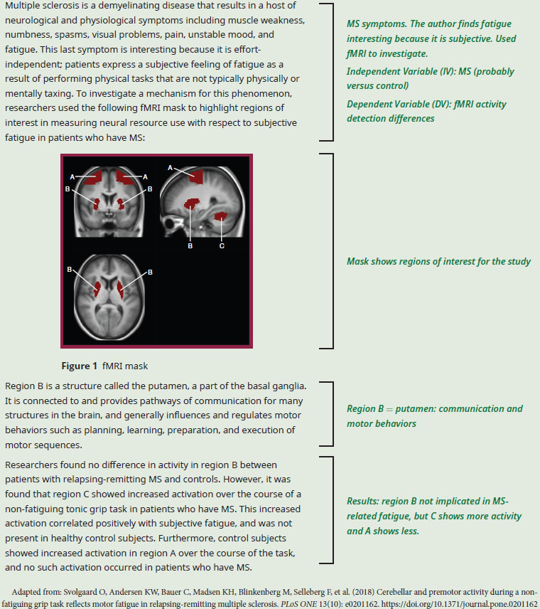
Based on the functions of the regions studied, what do these results suggest about the nature of subjective fatigue in patients who have MS compared to healthy participants?
Our first step in answering this question is to identify the regions presented in the study that are referenced in the question stem. For this particular question, we don’t have to worry too much about the structure of the experiment, as most of the information we need is in the results and the description of the regions. The authors give us the name and function of region B: the putamen, described in paragraph 2. This information is helpful because the actual brain isn’t color coded and labeled, and due to the low structural resolution of the image, it’s tough to tell exactly what region B is from the shape of the mask. On Test Day, using the image alone, we might be able to infer that region B is part of the midbrain and therefore, like other structures of the midbrain, it is probably involved in some kind of relay system. But the additional information in the passage text gives us insight that the picture alone just cannot provide. The passage also says that activity in region B is the same in patients who have MS and controls, allowing us to infer that difficulties in relaying motor signals are probably not the cause of subjective fatigue.
Based on our outside content knowledge, region C is the cerebellum, which we know is responsible for coordinating movement and for maintaining posture and balance. A differential increase in activity here implies that patients with MS may need more resources to perform motor tasks the longer these tasks are maintained.
Region A is in the forebrain. If we’ve studied the regions of the cerebrum, we might recognize this region as the premotor cortex, which is responsible for higher-level motor control and motivation. However, even without the specifics, we can guess that this region of the forebrain has something to do with executive motor control because of its general location. From the noted activity pattern in the final paragraph, we can guess that increased activity in region A helps to prevent subjective fatigue; thus, for patients who have MS, a lack of activation in this region may contribute to their experience of increased subjective fatigue.
We now have enough information to form a general picture of events here. In patients who have MS, the cerebellum is more active, presumably consuming more resources during maintained motor movements than the cerebellum of their healthy counterparts. This increase in resource consumption could be the patient’s brain attempting to accommodate for functions from other regions that have been lost as a result of disease, or could indicate a greater overall demand on cognitive processes involving movement. This overtaxing of cerebellar resources is most likely related to the increase in subjective fatigue experienced by patients who have MS.
1.4 Parts of the Forebrain
LEARNING OBJECTIVES
After Chapter 1.4, you will be able to:
- Link the regions of the forebrain to their functions
- Describe the laterality of brain–body communication and the role of hemispheric dominance
- Describe the functions of the four lobes of the cerebral cortex:
The forebrain is the most “modern” portion of the brain, and—in humans—forms the largest portion of the brain by weight and volume. The forebrain contains regions derived from the diencephalon, such as the thalamus, hypothalamus, posterior pituitary, and pineal gland; it also includes derivatives of the telencephalon, such as the cerebral cortex, basal ganglia, and limbic system.
Thalamus
The thalamus is a structure within the forebrain that serves as an important relay station for incoming sensory information, including all senses except for smell. After receiving incoming sensory impulses, the thalamus sorts and transmits them to the appropriate areas of the cerebral cortex. The thalamus is therefore a sensory “way station.”
Hypothalamus
The hypothalamus, subdivided into the lateral hypothalamus, ventromedial hypothalamus, and anterior hypothalamus, serves homeostatic functions, and is a key player in emotional experiences during high arousal states, aggressive behavior, and sexual behavior. The hypothalamus also helps control some endocrine functions, as well as the autonomic nervous system. The hypothalamus serves many homeostatic functions, which are self-regulatory processes that maintain a stable balance within the body. Receptors in the hypothalamus regulate metabolism, temperature, and water balance. When any of these functions are out of balance, the hypothalamus detects the problem and signals the body to correct the imbalance; for example, osmoreceptors in the hypothalamus may trigger the release of antidiuretic hormone to increase water reabsorption as part of fluid balance. The hypothalamus is also the primary regulator of the autonomic nervous system and is important in drive behaviors: hunger, thirst, and sexual behavior.
MNEMONIC
Functions of the Hypothalamus—The Four Fs:
- Feeding
- Fighting
- Flighting
- (Sexual) Functioning
The lateral hypothalamus (LH) is referred to as the hunger center because it has special receptors thought to detect when the body needs more food or fluids. In other words, the LH triggers eating and drinking. When this part of the hypothalamus is removed in lab rats, they refuse to eat and drink and must be force-fed with tubes to survive.
MNEMONIC
When the Lateral Hypothalamus (LH) is removed, one Lacks Hunger.
The ventromedial hypothalamus (VMH) is identified as the “satiety center,” and provides signals to stop eating. Brain lesions to this area usually lead to obesity.
MNEMONIC
When the VentroMedial Hypothalamus (VMH) is destroyed, one is Very Much Hungry.
The anterior hypothalamus controls sexual behavior. When the anterior hypothalamus is stimulated, lab animals will mount just about anything (including inanimate objects). In many species, damage to the anterior hypothalamus leads to permanent inhibition of sexual activity. The anterior hypothalamus also regulates sleep and body temperature.
REAL WORLD
In the early 1920s, researchers first discovered the hypothalamus’s role in rage and fighting through classic experiments conducted with cats. When researchers removed the cat’s cerebral cortex but left the hypothalamus in place, the cat displayed a pattern of pseudoaggressive behavior that was called “sham rage”—lashing of the tail, arching of the back, clawing, and biting—except that rage was spontaneous or triggered by the mildest touch. The researchers concluded that the cortex typically inhibits this type of response. When the researchers removed the cat’s cortex and hypothalamus together, the outcome was very different. The cat no longer showed any signs of sham rage, and much rougher stimulation was required before the cats showed any defensive behavior at all.
Other Parts of the Diencephalon
The diencephalon also differentiates to form the posterior pituitary gland, pineal gland, and connecting pathways to other brain regions. The posterior pituitary is comprised of axonal projections from the hypothalamus and is the site of release for the hypothalamic hormones antidiuretic hormone (ADH, also called vasopressin) and oxytocin. The functions of these hormones are described in Chapter 5 of MCAT Biology Review. The pineal gland is the key player in several biological rhythms. Most notably, the pineal gland secretes a hormone called melatonin, which regulates circadian rhythms. The pineal gland receives direct signals from the retina for coordination with sunlight.
Basal Ganglia
In the middle of the brain are a group of structures known as the basal ganglia. The basal ganglia coordinate muscle movement as they receive information from the cortex and relay this information (via the extrapyramidal motor system) to the brain and the spinal cord. The extrapyramidal system gathers information about body position and carries this information to the central nervous system, but does not function directly through motor neurons. Essentially, the basal ganglia help make our movements smooth and our posture steady. Parkinson’s disease is one chronic illness associated with destruction of portions of the basal ganglia. This disease is characterized by jerky movements and uncontrolled resting tremors. The basal ganglia may also play a role in schizophrenia and obsessive–compulsive disorder.
Limbic System
The limbic system, diagrammed in Figure 1.8, comprises a group of interconnected structures looping around the central portion of the brain and is primarily associated with emotion and memory. Its primary components include the septal nuclei, amygdala, hippocampus, and anterior cingulate cortex. In Chapter 5 of MCAT Behavioral Sciences Review, we will also explore the roles of the thalamus, hypothalamus, and cortex in the limbic system.
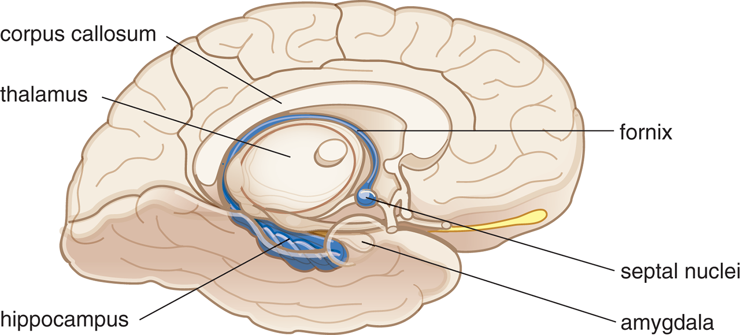
Figure 1.8. The Limbic System
Septal Nuclei
The septal nuclei contain one of the primary pleasure centers in the brain. Mild stimulation of the septal nuclei is reported to be intensely pleasurable; there is an association between these nuclei and addictive behavior.
REAL WORLD
James Olds and Peter Milner discovered the association between the septal nuclei and addictive behavior in the 1950s. They demonstrated that when rats could stimulate their septal regions at will by pushing a lever, they found it so pleasurable that they preferred it to eating or any other activities, even after going 24 hours without food or sleep.
Amygdala
The amygdala is a structure that plays an important role in defensive and aggressive behaviors, including fear and rage. Researchers base this observation on studies of animals and humans with brain lesions. When the amygdala is damaged, aggression and fear reactions are markedly reduced. Lesions to the amygdala result in docility and hypersexual states.
REAL WORLD
Heinrich Klüver and Paul Bucy performed studies that linked the amygdala with defensive and aggressive behavior in monkeys. When the amygdala of the Rhesus monkey was removed, they noted increased sexual behavior, decreased fear responses, and hyperorality, or the examination of inanimate or animate objects by mouth. These symptoms are now referred to as Klüver–Bucy syndrome.
Hippocampus
The hippocampus plays a vital role in learning and memory processes; specifically, the hippocampus helps consolidate information to form long-term memories, and can redistribute remote memories to the cerebral cortex. The hippocampus communicates with other portions of the limbic system through a long projection called the fornix. Researchers originally discovered the connection between memory and the hippocampus through a famous patient named Henry Molaison (known as H.M. in the scientific literature until his death in 2008). Parts of H.M.’s temporal lobes—including the amygdala and hippocampus—were removed in an effort to control epileptic seizures. After surgery, H.M.’s intelligence was largely intact but he suffered a drastic and irreversible loss of memory for any new information. This kind of memory loss is called anterograde amnesia and is characterized by not being able to establish new long-term memories, whereas memory for events that occurred before brain injury is usually intact. The opposite kind of memory loss, retrograde amnesia, refers to memory loss of events that transpired before brain injury.
BRIDGE
Learning and memory are discussed thoroughly in Chapter 3 of MCAT Behavioral Sciences Review.
MNEMONIC
Lobes of the brain: F-POT
- Frontal
- Parietal
- Occipital
- Temporal
Anterior Cingulate Cortex
Due to the connection with the frontal and parietal lobes, the anterior cingulate cortex functions in higher order cognitive processes, including regulation of impulse control and decision-making. It also maintains connections to other parts of the limbic system, and thus plays a role in emotion and motivation.
Cerebral Cortex
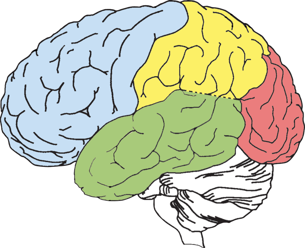
The outer surface of the brain is called the cerebral cortex. The cortex is sometimes called the neocortex, a reminder that the cortex is the most recent brain region to evolve. Rather than having a smooth surface, the cortex has numerous bumps and folds called gyri and sulci, respectively. The convoluted structure of the brain provides increased surface area. The cerebrum is divided into two halves, called cerebral hemispheres. The surface of the cortex is divided into four lobes—the frontal lobe, parietal lobe, occipital lobe, and temporal lobe. These lobes are identified in Figure 1.9, which shows a side view of the left cerebral hemisphere.
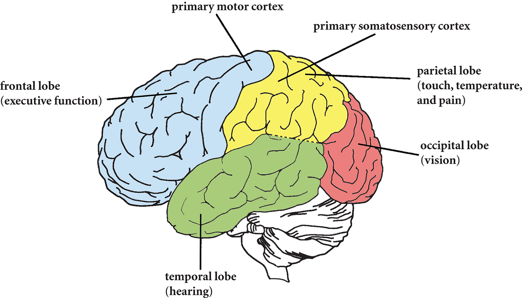
Figure 1.9. Lobes of the Brain
Frontal Lobe
The frontal lobe is comprised of two basic regions: the prefrontal cortex and the motor cortex. The prefrontal cortex manages executive function by supervising and directing the operations of other brain regions. To regulate attention and alertness, the prefrontal cortext communicates with the reticular formation in the brainstem, telling an individual either to wake up or to relax, depending on the situation. This region also supervises processes associated with perception, memory, emotion, impulse control, and long-term planning. In memory, for instance, the role of the prefrontal cortex is not to store any memory traces, but rather to remind individuals that they have something to remember at all.
Damage to the prefrontal cortex impairs its overall supervisory functions. People with prefrontal lesions may be more impulsive and generally less in control of their behavior. As a result, these individuals can have an increased tendency towards angry outbursts, as well as a higher predisposition to crying. Additionally, it is not unusual for someone with a prefrontal lesion to make vulgar and inappropriate sexual remarks, or to be apathetic to the emotional responses of others.
Because the prefrontal cortex integrates information from different cortical regions, the prefrontal cortex is a good example of an association area, which is an area that integrates input from diverse regions of the brain. For example, multiple inputs may be necessary to solve a complex puzzle, to plan ahead for the future, or to reach a difficult decision.
Association areas are generally contrasted with projection areas, which perform more rudimentary perceptual and motor tasks. An example of a projection area is the primary motor cortex, which is located on the precentral gyrus, just in front of the central sulcus that divides the frontal and parietal lobes. The function of the primary motor cortex is to initiate voluntary motor movements by sending neural impulses down the spinal cord toward the muscles. As such, it is considered a projection area in the brain. The neurons in the motor cortex are arranged systematically according to the parts of the body to which they are connected. This organizational pattern can be visualized through the motor homunculus, as shown in Figure 1.10. Because certain sets of muscles require finer motor control than others, they take up additional space in the cortex relative to their size in the body.
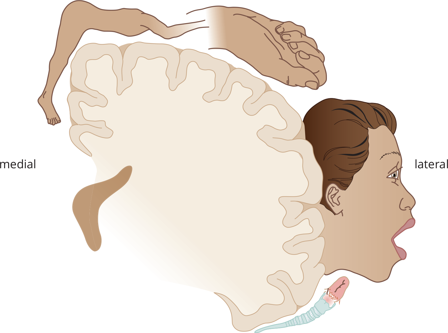
Figure 1.10. Motor Homunculus on the Precentral Gyrus of the Frontal Lobe
A third important part of the frontal lobe is Broca’s area, which is vitally important for speech production. Broca’s area is usually found in only one hemisphere, the so-called “dominant” hemisphere; for most people—both right- and left-handed—this is the left hemisphere.
Parietal Lobe
The parietal lobe is located to the rear of the frontal lobe. The somatosensory cortex is located on the postcentral gyrus (just behind the central sulcus) and is involved in somatosensory information processing. This projection area is the destination for all incoming sensory signals for touch, pressure, temperature, and pain. Despite certain differences, the somatosensory cortex and motor cortex are very closely related. In fact, they are so interrelated they sometimes are described as a single unit: the sensorimotor cortex. The somatosensory homunculus is shown in Figure 1.11. The central region of the parietal lobe is associated with spatial processing and manipulation. This region makes it possible to orient oneself and other objects in three-dimensional space, to do spatial manipulation of objects, and to apply spatial orientation skills such as those required for map reading.
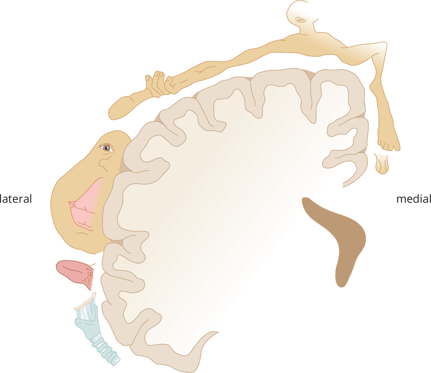
Figure 1.11. Somatosensory Homunculus on the Postcentral Gyrus of the Parietal Lobe
Occipital Lobe
The occipital lobes, at the very rear of the brain, contain the visual cortex, which is sometimes called the striate cortex. Striate means furrowed or striped, which is how the visual cortex appears when examined under a microscope. The visual cortex is one of the best-understood brain regions, owing to the large amount of research that has been done on visual processing. Sensation and perception of visual information are discussed thoroughly in Chapter 2 of MCAT Behavioral Sciences Review. Areas in the occipital lobe have also been implicated in learning and motor control.
Temporal Lobe
The temporal lobes are associated with a number of functions. The auditory cortex and Wernicke’s area are located in the temporal lobe. The auditory cortex is the primary site of most sound processing, including speech, music, and other sound information. Wernicke’s area is associated with language reception and comprehension. The temporal lobe also functions in memory processing, emotion, and language. Studies have shown that electrical stimulation of the temporal lobe can evoke memories for past events. This makes sense because the hippocampus is located deep inside the temporal lobe. It is important to note that the lobes, although having seemingly independent functions, are not truly independent of one another. Often, a sensory modality may be represented in more than one area.
REAL WORLD
Several techniques have been developed to assess the function of brain regions, especially those associated with language and auditory processing. One such technique is called speech shadowing, which is a technique used to research both stuttering and speech perception. Speech shadowing involves participants reciting along with auditory inputs, which can be presented to one or both ears. If different messages are presented to each ear, as in a dichotic listening test, speech shadowing can ensure that the participant is paying attention to the auditory input to the correct ear. This seemingly simple experimental task requires successful functioning of the temporal lobe, parietal lobe, and frontal cortex!
Cerebral Hemispheres and Laterality
In most cases, one side of the brain communicates with the opposite side of the body. In such cases, we say a cerebral hemisphere communicates contralaterally. For example, the motor neurons on the left side of the brain activate movements on the right side of the body. In other cases (for instance, hearing), cerebral hemispheres communicate with the same side of the body. In such cases, the hemispheres communicate ipsilaterally.
We distinguish between dominant and nondominant hemispheres. The dominant hemisphere is typically defined as the one that is more heavily stimulated during language reception and production. In the past, hand dominance was used as a proxy for hemispheric dominance; that is, right-handed individuals were assumed to have left-dominant brains and left-handed individuals were assumed to have right-dominant brains (because the brain communicates contralaterally with the hand). However, this correlation has not held up under scrutiny; 95 percent of right-handed individuals are indeed left brain dominant, but only 18 percent of left-handed individuals are right brain dominant.
The dominant hemisphere (usually the left) is primarily analytic in function, making it well-suited for managing details. For instance, language, logic, and math skills are all located in the dominant hemisphere. Again, language production (Broca’s area) and language comprehension (Wernicke’s area) are primarily driven by the dominant hemisphere.
REAL WORLD
The corpus callosum connects and shares information between the two cerebral hemispheres; its function was discovered in patients who have epilepsy whose corpora callosa were severed in a last-ditch effort to limit their convulsive seizures. In these “split-brain” patients, in whom the corpus callosum has been severed, each hemisphere has its own function and specialization that is no longer accessible by the other. As an example of the result: an object felt only by the left hand (which projects to the right hemisphere) could not be named (because language function is usually in the left hemisphere).
The nondominant hemisphere (usually the right) is associated with intuition, creativity, music cognition, and spatial processing. The nondominant hemisphere simultaneously processes the pieces of a stimulus and assembles them into a holistic image. The nondominant hemisphere serves a less prominent role in language. It is more sensitive to the emotional tone of spoken language, and permits us to recognize others’ moods based on visual and auditory cues, which adds to communication. The dominant hemisphere thus screens incoming language to analyze its content, and the nondominant hemisphere interprets it according to its emotional tone. The roles of the dominant and nondominant hemispheres are summarized in Table 1.2; remember that the left hemisphere is the dominant hemisphere in most individuals, regardless of handedness.
Table 1.2. Comparison of Dominant and Nondominant Hemispheres’ Functions
FUNCTION DOMINANT HEMISPHERE NONDOMINANT HEMISPHERE
Visual system Letters, words Faces
Auditory system Language-related sounds Music
Language Speech, reading, writing, arithmetic Emotions
Movement Complex voluntary movement –
Spatial processes – Geometry, sense of direction
MCAT CONCEPT CHECK 1.4
Before you move on, assess your understanding of the material with these questions.
- Match the parts of the brain below to their functions:
- Basal ganglia
- Cerebellum
- Cerebral cortex
- Hypothalamus
- Inferior and superior colliculi
- Limbic system
- Medulla oblongata
- Reticular formation
- Thalamus
- Smooth movement
- Sensory relay station
- Sensorimotor reflexes
- Arousal and alertness
- Hunger and thirst; emotion
- Complex perceptual, cognitive, and behavioral processes
- Vital function (breathing, digestion)
- Coordinated movement
- Emotion and memory
- What are the four lobes of the cerebral cortex, and what is the function of each?
Lobe Function
_________________________
- What is the difference between ipsilateral and contralateral communication between the brain and the body?
- How is the dominant hemisphere typically defined? _________________________
1.5 Influences on Behavior
LEARNING OBJECTIVES
After Chapter 1.5, you will be able to:
- Associate major neurotransmitters with their common functions
- Detail the links between the endocrine system and the brain
- Explain the nature vs. nurture debate and the different study types used to explore this question
Merely describing the functions of brain regions does not fully explain the wide variety of human behaviors that are possible. Other influences on behavior include chemical controls (neurotransmitters, hormones in the endocrine system), heredity, and the environment.
Neurotransmitters
A neurotransmitter is a chemical used by neurons to send signals to other neurons; more than 100 neurotransmitters have been identified. Several of the most important are described in this section and are summarized in Table 1.3. Some drugs mimic the action of neurotransmitters by binding to the same receptor to produce the same biological response. A drug that mimics the action of some neurotransmitter is called an agonist. Drugs can also act by blocking the action of neurotransmitters, and such drugs are called antagonists.
Acetylcholine
Acetylcholine is a neurotransmitter found in both the central and peripheral nervous systems. In the peripheral nervous system, acetylcholine is used to transmit nerve impulses to the muscles. It is the neurotransmitter used by the parasympathetic nervous system and a small portion of the sympathetic nervous system (in ganglia and for innervating sweat glands). In the central nervous system, acetylcholine has been linked to attention and arousal. In fact, loss of cholinergic neurons connecting with the hippocampus is associated with Alzheimer’s disease, an illness resulting in progressive and incurable memory loss.
KEY CONCEPT
Acetylcholine is the neurotransmitter used by the efferent limb of the somatic nervous system and the parasympathetic nervous system. Acetylcholine can act as an excitatory or inhibitory neurotransmitter in muscle cells, dependent on the type of receptor found on the cell. For example, acetylcholine will transmit an inhibitory response in cardiac muscle cells, but it can also transmit an excitatory response if acting on skeletal muscle cells. Acetylcholine within the central nervous system largely functions as an excitatory neurotransmitter.
Epinephrine and Norepinephrine
Epinephrine, norepinephrine, and dopamine are three closely related neurotransmitters known as catecholamines. Due to similarities in their molecular composition, these three transmitters are also classified as monoamines or biogenic amines. The most important thing to know about the catecholamines is that they all play important roles in the experience of emotions.
Epinephrine (adrenaline) and norepinephrine (noradrenaline) are involved in controlling alertness and wakefulness. As the primary neurotransmitter of the sympathetic nervous system, they promote the fight-or-flight response. Whereas norepinephrine more commonly acts at a local level as a neurotransmitter, epinephrine is more often secreted from the adrenal medulla to act systemically as a hormone. Low levels of norepinephrine are associated with depression; high levels are associated with anxiety and mania.
Dopamine
Dopamine is another catecholamine, created by the enzyme dopamine decarboxylase from L-dihydroxyphenylalanine (L-DOPA), that plays an important role in movement and posture. High concentrations of dopamine are normally found in the basal ganglia, which help smooth movements and maintain postural stability.
Imbalances in dopamine transmission have been found to play a role in schizophrenia. An important theory about the origin of this mental illness is called the dopamine hypothesis of schizophrenia. The dopamine hypothesis argues that delusions, hallucinations, and agitation associated with schizophrenia arise from either too much dopamine or from an oversensitivity to dopamine in the brain. Although the dopamine hypothesis of schizophrenia is an important theory, it does not account for all of the findings of the disease.
Parkinson’s disease is associated with a loss of dopaminergic neurons in the basal ganglia. These disruptions of dopamine transmission lead to resting tremors and jerky movements, as well as to postural instability.
REAL WORLD
The role of dopamine in both schizophrenia and Parkinson’s disease can be seen in their treatment. Antipsychotic medications used in schizophrenia are dopamine blockers, and can cause motor disturbances (“extrapyramidal symptoms”) as a side effect. Parkinson’s disease can be treated withL-DOPA, which increases dopamine levels in the brain; an overdose ofL-DOPA can lead to psychotic symptoms similar to schizophrenia.
Serotonin
Along with the catecholamines, serotonin is classified as a monoamine or biogenic amine neurotransmitter. Serotonin is generally thought to play roles in regulating mood, eating, sleeping, and dreaming. Like norepinephrine, serotonin is thought to play a role in depression and mania. An oversupply of serotonin is thought to produce manic states; an undersupply is thought to produce depression.
GABA, Glycine, and Glutamate
The neurotransmitter ***γ*-aminobutyric acid(GABA**) produces inhibitory postsynaptic potentials and is thought to play an important role in stabilizing neural activity in the brain. GABA exerts its effects by causing hyperpolarization of the postsynaptic membrane.
Glycine may be better known as one of the twenty proteinogenic amino acids, but it also serves as an inhibitory neurotransmitter in the central nervous system by increasing chloride influx into the neuron. This hyperpolarizes the postsynaptic membrane, similar to the function of GABA.
Finally, glutamate, another of the twenty proteinogenic amino acids, also acts as a neurotransmitter in the central nervous system. In contrast to glycine, however, it is an excitatory neurotransmitter.
Peptide Neurotransmitters
Studies suggest that peptides are also involved in neurotransmission. The synaptic action of these neuromodulators (also called neuropeptides) involves a more complicated chain of events in the postsynaptic cell than that of regular neurotransmitters. Neuromodulators are therefore relatively slow and have longer effects on the postsynaptic cell than neurotransmitters. The endorphins, which are natural painkillers produced in the brain, are the most important peptides to know. Endorphins (and their relatives, enkephalins) have actions similar to morphine or other opioids in the body.
Table 1.3. Neurotransmitters and Their Functions
NEUROTRANSMITTER BEHAVIOR
Acetylcholine Voluntary muscle control, parasympathetic nervous system, attention, alertness
Epinephrine and Norepinephrine Fight-or-flight responses, wakefulness, alertness
Dopamine Smooth movements, postural stability
Serotonin Mood, sleep, eating, dreaming
GABA and Glycine Brain “stabilization”
Glutamate Brain excitation
Endorphins Natural painkillers
The Endocrine System
We’ve already discussed the relatively fast communication network—the nervous system—that uses chemical messages called neurotransmitters. The endocrine system is the other internal communication network in the body, and it uses chemical messengers called hormones. The endocrine system is somewhat slower than the nervous system because hormones travel to their target destinations through the bloodstream. The endocrine system is covered extensively in Chapter 5 of MCAT Biology Review, so our focus here will be on the role of certain endocrine organs on behavior.
BRIDGE
The entire endocrine system is covered in Chapter 5 of MCAT Biology Review.
The hypothalamus links the endocrine and nervous systems and, in addition to the roles described earlier, regulates the hormonal function of the pituitary gland. The hypothalamus and pituitary gland are spatially close to each other, and control is maintained through endocrine release of hormones into the hypophyseal portal system that directly connects the two organs, as shown in Figure 1.12.
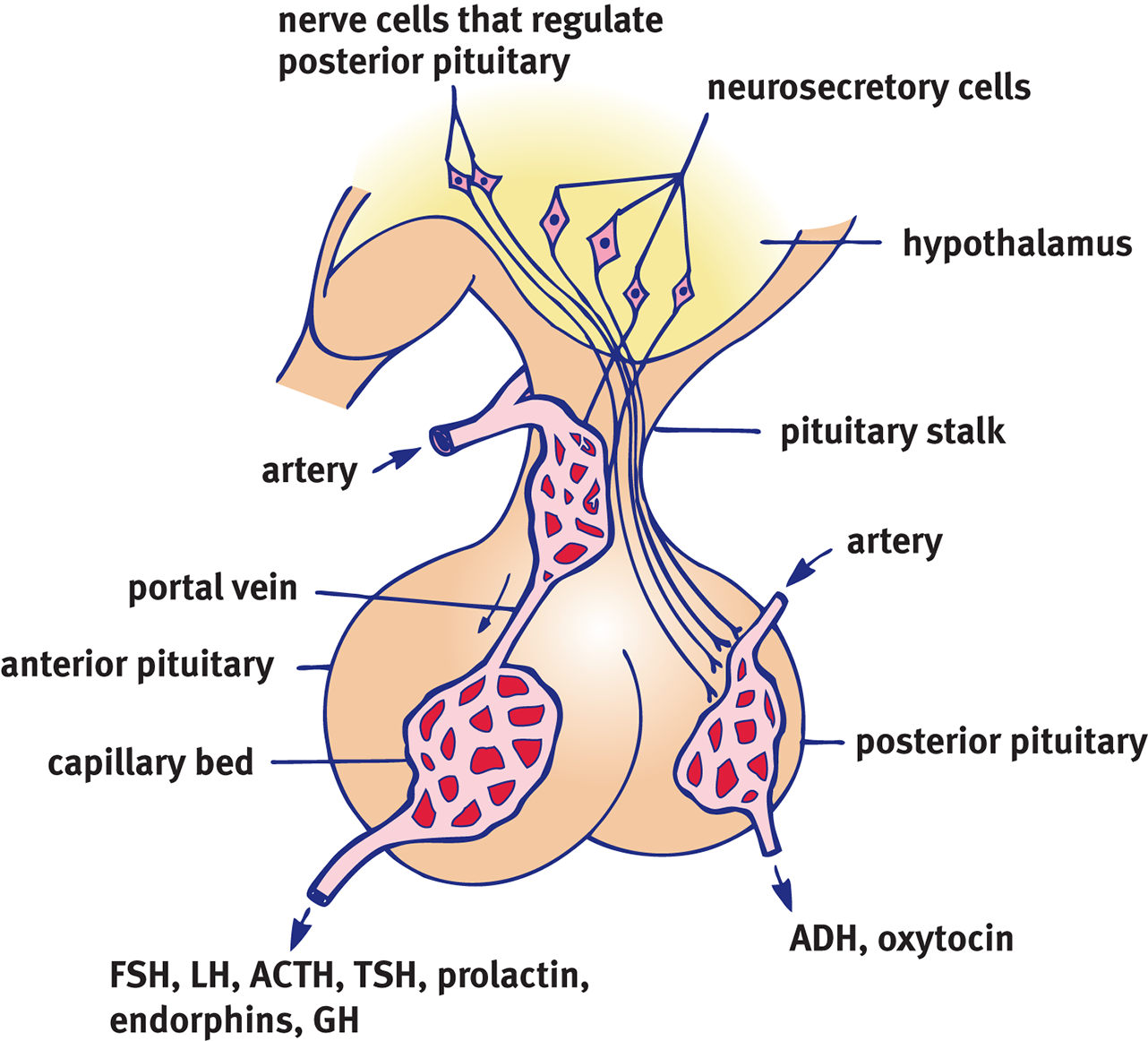
Figure 1.12. The Hypophyseal Portal System
The pituitary gland, sometimes referred to as the “master” gland, is located at the base of the brain and is divided into two parts: anterior and posterior. It is the anterior pituitary that is the “master” because it releases hormones that regulate activities of endocrine glands elsewhere in the body. However, the anterior pituitary itself is controlled by the hypothalamus. The pituitary secretes various hormones into the bloodstream that travel to other endocrine glands located elsewhere in the body to activate them. Once activated by the pituitary, a given endocrine gland manufactures and secretes its own characteristic hormone into the bloodstream.
The adrenal glands are located on top of the kidneys and are divided into two parts: the adrenal medulla and adrenal cortex. The adrenal medulla releases epinephrine and norepinephrine as part of the sympathetic nervous system. The adrenal cortex produces many hormones called corticosteroids, including the stress hormone cortisol. The adrenal cortex also contributes to sexual functioning by producing sex hormones, such as testosterone and estrogen.
The gonads are the sex glands of the body—ovaries in females and testes in males. These glands produce sex hormones in higher concentrations, leading to increased levels of testosterone in males and increased levels of estrogen in females. These sex hormones increase libido and contribute to mating behavior and sexual function. Higher levels of testosterone also increase aggressive behavior.
Genetics and Behavior
Just as physical traits are inherited from parents, behavioral traits can be inherited as well. Evidence for the inherited nature of behavior comes from the fact that many behaviors are species specific. For example, many animals exhibit mating behaviors only seen within their species. Behaviors can also be bred into a species; many breeds of dog have been bred for certain traits and behaviors. Behaviors are also seen to run in families. Often times, violence and aggression are observed passing along a family line, as are mental illnesses.
REAL WORLD
Bipolar disorder is considered one of the most heritable disorders, including medical illnesses. In one study, having a monozygotic (identical) twin with bipolar disorder was associated with a 43% risk of being diagnosed with the same disorder.
Innate behavior is genetically programmed as a result of evolution and is seen in all individuals regardless of environment or experience. In contrast, other behaviors are considered learned. Learned behaviors are not based on heredity but instead are based on experience and environment. Adaptive value is the extent to which a trait or behavior positively benefits a species by influencing the evolutionary fitness of the species, thus leading to adaptation through natural selection.
BRIDGE
Natural selection is discussed in greater detail in Chapter 12 of MCAT Biology Review.
How much of an individual’s behavior is based on genetic makeup and how much is based on environment and experiences? This controversial question is often referred to as the **nature vs. nurturequestion. Here,natureis the influence of inherited characteristics on behavior.Nurture** refers to the influence of environment and physical surroundings on behavior. There is no easy answer to this long-debated question. An individual’s behavior is not only influenced by both genetics and environment, but also by how these two factors may influence each other. For example, hereditary traits may make a certain person more likely to have an addictive personality. But, the individual would still have to be exposed to drugs, alcohol, or gambling to develop an addiction.
To determine the degree of genetic influence on behavior, researchers often use one of three methods: family studies, twin studies, and adoption studies. Family studies rely on the fact that genetically related individuals are more similar genotypically than unrelated individuals. Researchers may compare rates of a given trait among family members to rates of that trait among unrelated individuals. For example, family studies have determined that the risk of developing schizophrenia for children of a patient who has schizophrenia is 13 times higher than in the general population. For siblings of a patient who has schizophrenia, the rate is 9 times higher. Observations such as these have led psychologists to conclude that schizophrenia has a hereditary component. Family studies are limited, however, because families share both genetics and environment. Family studies cannot distinguish shared environmental factors from shared genetic factors. For example, what if the increased rates of schizophrenia in families are a result of experiencing the same emotional climate in the home rather than genetically shared characteristics?
Twin studies, comparing concordance rates for a trait between monozygotic (MZ; identical) and dizygotic (DZ; fraternal) twins, are better able to distinguish the relative effects of shared environment and genetics. Concordance rates refer to the likelihood that both twins exhibit the same trait. MZ twins are genetically identical, sharing 100 percent of their genes, whereas DZ twins share approximately 50 percent of their genes. The assumption made by twin studies is that the two individuals in each MZ or DZ twin pair share the same environment; thus, differences between MZ and DZ twins are thought to reflect hereditary factors. Twin studies can also be used to measure genetic effects relative to environmental effects. In this version of the twin study, researchers compare traits in twins raised together versus twins raised apart. For example, one study of personality characteristics showed that MZ (identical) twins raised in separate families were still more similar than DZ (fraternal) twins raised together. Such a result offers convincing evidence for a strong genetic component to personality.
Finally, adoption studies also help us understand environmental and genetic influences on behavior. These studies compare the similarities between biological relatives and the child who was adopted to similarities between adoptive relatives and the child. For example, researchers have found that the IQ of children who were adopted is more similar to their biological parents’ IQ than to their adoptive parents’ IQ. This research suggests that IQ is heritable. Criminal behavior among teenagers shows a similar pattern of heritability.
MCAT CONCEPT CHECK 1.5
Before you move on, assess your understanding of the material with these questions.
- Match the neurotransmitters below to their functions:
- Acetylcholine
- Dopamine
- Endorphins
- Epinephrine/norepinephrine
- GABA/glycine
- Glutamate
- Serotonin
- Wakefulness and alertness, fight-or-flight responses
- Brain “stabilizer”
- Mood, sleep, eating, dreaming
- Natural painkiller
- Smooth movements and steady posture
- Voluntary muscle control
- Brain excitation
- Which endocrine organs influence behavior? What hormones do they use, and what do they accomplish?
____________________________
- Briefly discuss the influence of nature vs. nurture on behavior.
____________________________
- In each of the study types below, what is the sample group? The control group?
Study Sample Group Control Group Family study Twin study Adoption study
1.6 Development
LEARNING OBJECTIVES
After Chapter 1.6, you will be able to:
- Describe the process of neurulation
- Link the primitive reflexes with the behaviors to which they correspond
- Identify the main themes that dictate stages of motor development in children
The developmental process begins at the moment of conception. Physiological changes are rapid from embryonic to fetal stages, and well into infancy. Children exhibit surprisingly consistent patterns of motor abilities, as well as physiological changes based on age. Understanding these changes and when they occur is important in the discussion of developmental psychology. There are specific periods in development where children are particularly susceptible to environmental factors, called critical periods. Absence of the appropriate environmental factors may result in failure to learn a given skill or trait during the critical period, which may also mean learning that skill later on is difficult or even impossible.
Prenatal
The development of the nervous system starts with neurulation, at three to four weeks’ gestational age. Neurulation occurs when the ectoderm overlying the notochord begins to furrow, forming a neural groove surrounded by two neural folds, as shown in Figure 1.13. Cells at the leading edge of the neural fold are called the neural crest, and will migrate throughout the body to form disparate tissues, including dorsal root ganglia, melanocytes (pigment-producing cells), and calcitonin-producing cells of the thyroid. The remainder of the furrow closes to form the neural tube, which will ultimately form the central nervous system (CNS). The neural tube has an alar plate, which differentiates into sensory neurons, and a basal plate, which differentiates into motor neurons. Over time, the neural tube invaginates and folds on itself many times; the embryonic brain begins as three swellings (prosencephalon, mesencephalon, rhombencephalon) that become five swellings (telencephalon, diencephalon, mesencephalon, metencephalon, myelencephalon) as it becomes the mature brain, as demonstrated in Figure 1.6 earlier in this chapter.
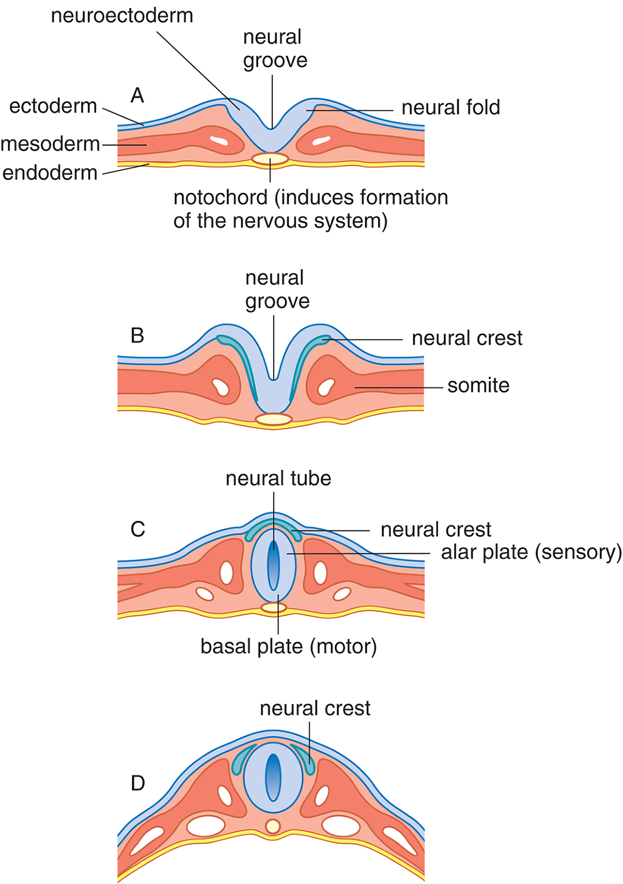
Figure 1.13. Development of the Nervous System
Prenatal development does not occur in a vacuum, of course, but in the mother’s uterus. Within this environment, temperature, chemical balance, orientation of the fetus with respect to gravity, and atmospheric pressure are all carefully controlled and remain relatively constant. The fetus is attached to the uterine wall and placenta by the umbilical cord. The placenta transmits food, oxygen, and water to the fetus while returning water and waste to the mother. Maternal blood supplies many of the proteins and amino acids needed for growth, although the embryo begins to produce its own proteins and amino acids as well.
A variety of external influences can have deleterious effects on the development of the fetus. A number of viruses or bacteria can cross the placenta and cause damage to the developing fetus, including rubella (German measles), which may cause cataracts, deafness, heart defects, and intellectual disability. Other viral infections—such as measles, mumps, hepatitis, influenza, varicella (chickenpox), and herpes—have been linked to various birth defects.
An unfortunate side effect of the revolution in pharmaceutical development is that many drugs that help the mother can have damaging effects on the fetus she carries. The most notorious of these drugs is thalidomide, which was prescribed in the late 1950s and early 1960s to reduce morning sickness. Mothers who took this drug while pregnant often gave birth to babies with missing and malformed limbs and defects of the heart, eyes, ears, digestive tract, and kidneys. Antiepileptic medications are associated with neural tube defects, in which the neural tube fails to close completely, leading to malformations such as spina bifida or anencephaly.
A host of environmental factors and exposures may also affect maturation. Maternal malnutrition is considered to be a leading cause of abnormal development. Protein deficiency can slow growth, lead to intellectual disability, and reduce immunity to disease. Maternal narcotic addiction produces chemically dependent infants who must undergo severe withdrawal after birth. Regular cigarette smoking can lead to slowed growth, increased fetal heart rate, and a greater chance of premature birth. Daily use of alcohol also leads to slowed growth, both physically and psychologically. Finally, prenatal exposure to X-rays has been strongly linked to intellectual disabilities; defects of the skull, spinal cord, and eyes; cleft palate; and limb deformities.
Motor
Although they may seem helpless, infants are equipped with well-developed somatic structures and a broad array of reflexes that help ensure survival. A reflex is a behavior that occurs in response to a given stimulus without higher cognitive input. While motor and startle reflexes exist in adults, infants have a number of primitive reflexes that disappear with age. For example, the rooting reflex is the automatic turning of the head in the direction of a stimulus that touches the cheek—such as a nipple during feeding. Sucking and swallowing when an object is placed in the mouth are also examples of reflexes related to feeding.
Other primitive reflexes may have served an adaptive purpose in earlier stages of human evolution, but are currently used mainly in assessing infant neurological development. By comparing the point in time at which each of these reflexes disappears relative to the established norms, it is possible to tell whether neurological development is taking place in a normal fashion. One such reflex is the Moro reflex, illustrated in Figure 1.14. Infants react to abrupt movements of their heads by flinging out their arms, then slowly retracting their arms and crying. It has been speculated that this reflex may have developed during a time when our prehuman ancestors lived in trees and falling could have been prevented by instinctive clutching. The Moro reflex usually disappears after four months and its continuation at one year is a strong suggestion of developmental difficulties. Asymmetry of the Moro reflex may hint at underlying neuromuscular problems.
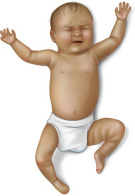
Figure 1.14. The Moro Reflex The infant extends the arms, then slowly withdraws them and cries.
The Babinski reflex causes the toes to spread apart automatically when the sole of the foot is stimulated, as seen in Figure 1.15. The grasping reflex occurs when infants close their fingers around an object placed in the hand. Adults with neurological diseases may exhibit these primitive reflexes, especially in illnesses that cause demyelination (loss of the myelin sheath).
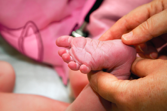
Figure 1.15. The Babinski Reflex The big toe extends while the other toes fan outward.
Although reflexive behavior dominates the repertoire of the neonate, other behaviors occur as well. Newborn infants also kick, turn, and wave their arms. These uncoordinated, unconnected behaviors form the basis for later, more coordinated movements.
Infants typically develop motor skills at about the same age, in the same order. Due to this pattern, most psychologists and doctors agree that these are innately programmed abilities for human infants. However, the educational richness of the environment has been observed to affect the rate of learning, with more enriched environments promoting quicker development.
Motor skills are broken down into two classes: gross and fine motor skills. Gross motor skills incorporate movement from large muscle groups and whole body motion, such as sitting, crawling, and walking. Fine motor skills involve the smaller muscles of the fingers, toes, and eyes, providing more specific and delicate movement. Fine motor abilities include tracking motion, drawing, catching, and waving.
Social
In addition to motor skills, social development occurs in infancy and through adolescence. At birth, the parental figure becomes the center of the infant’s world, and as the infant ages, stranger anxiety (a fear and apprehension of unfamiliar individuals) and separation anxiety (a fear of being separated from the parental figure) develop at approximately seven months and one year, respectively. During this time, play style progresses from solitary to onlooker, and at two years develops into parallel play, in which children will play alongside each other without influencing each other’s behavior. At age three, children have an awareness of their assigned gender at birth, engage in gender-stereotyped play, and know their full name.
By age five, conformity to peers and romantic feelings for others begin to develop. From ages six through twelve, friend circles tend to be of the same gender without expression of romantic feelings. In the teenage years, children become more self-sufficient, and often express their desire for independence by rebelling against their parents. Inter-gender friendships become more common. Individuals also become more aware of their gender identity and sexual orientation and may begin sexual relationships.
In this chapter we have described several abilities and behaviors that are expected to emerge at particular times in a person’s development. These skills are known as developmental milestones. The developmental milestones of the first three years of life are listed in Table 1.4. While this is a general timetable based on averages, most children fall within plus or minus two months of the chart. The goal is not to memorize this chart, but to recognize some themes. For example, gross motor skills progress in a head-to-toe order: children first develop the ability to lift the head, then to stabilize the trunk, and finally to walk. There is also a correlation between the development of motor skills and proximity to the center of the body, with skills being developed at the core prior to extremities. Social skills move from being parent-oriented to self-oriented to other-oriented. Language skills, discussed in Chapter 4 of MCAT Behavioral Sciences Review, become more complex and structured.
Table 1.4. Child Development Milestones
AGE PHYSICAL AND MOTOR DEVELOPMENTS SOCIAL DEVELOPMENTS LANGUAGE DEVELOPMENTS
1st year of life
- Puts everything in mouth
- Sits with support (4 mo)
- Stands with help (8 mo)
- Crawls, fear of falling (9 mo)
- Pincer grasp (10 mo)
- Follows objects to midline (4 wk)
- One-handed approach/grasp of toy
- Feet in mouth (5 mo)
- Bang and rattle stage
- Changes hands with toy (6 mo)
- Parental figure central
- Issues of trust are key
- Stranger anxiety (7 mo)
- Play is solitary and exploratory
- Pat-a-cake, peek-a-boo (10 mo)
- Laughs aloud (4 mo)
- Repetitive responding (8 mo)
- “mama, dada” (10 mo)
Age 1
- Walks alone (13 mo)
- Climbs stairs alone (18 mo)
- Emergence of hand preference (18 mo)
- Kicks ball, throws ball
- Pats pictures in book
- Stacks three cubes (18 mo)
- Separation anxiety (12 mo)
- Dependency on parental figure
- Onlooker play
- Great variation in timing of language development
- Uses 10 words
Age 2
- High activity level
- Walks backward
- Can turn doorknob, unscrew jar lid
- Scribbles with crayon
- Stacks six cubes (24 mo)
- Stands on tiptoes (30 mo)
- Able to aim thrown ball
- Selfish and self-centered
- Imitates mannerisms and activities
- May be aggressive
- Recognizes self in mirror
- “No” is favorite word
- Parallel play
- Use of pronouns
- Parents understand most
- Two-word sentences
- Uses 250 words
- Identifies body parts by pointing
Age 3
- Rides tricycle
- Stacks 9 cubes (36 mo)
- Alternates feet going up stairs
- Bowel and bladder control (toilet training)
- Draws recognizable figures
- Catches ball with arms
- Cuts paper with scissors
- Unbuttons buttons
- Awareness of assigned gender
- Gender-stereotyped play
- Understands “taking turns”
- Knows full name
- Complete sentences
- Uses 900 words
- Understands 3600 words
- Strangers can understand
- Recognizes common objects in pictures
- Can answer, “Tell me what we wear on our feet?” “Which block is bigger?”
MCAT CONCEPT CHECK 1.6
Before you move on, assess your understanding of the material with these questions.
- Describe the process of neurulation.
___________________________
- For each of the primitive reflexes below, briefly describe the observed behavior.
Primitive Reflex Behavior Rooting Moro Babinski Grasping
- What are the two main themes that dictate the stages of motor development in early childhood?
- _________________________
- _________________________
Conclusion
Behavioral psychology is the study of all physical and mental actions based on the response of the body to external stimuli, specifically the activity of the nervous and endocrine systems. The nervous system is a complex organization of structures and neurons that communicate and coordinate information. The endocrine system, in conjunction with the nervous system, controls human behavior. Aside from neurotransmitter and hormonal control of behavior, certain behaviors are genetically passed from generation to generation, as are many other physical traits. The genetic aspects of behavior are thought to interact with the learned components of behavior. Human behavior is also studied as it correlates to the development from embryo to fetus to infant and well into adolescence and adulthood. The development of motor skills and social behavior is seen to progress at a consistent rate across the species.
In the next chapter, our focus will be on the neurological systems used to interact with the world—most notably, those systems that exist for sensation and perception of the environment. These include vision, hearing, smell and taste, somatosensation, and others.
GO ONLINE!
You’ve reviewed the content, now test your knowledge and critical thinking skills by completing a test-like passage set in your online resources!
CONCEPT SUMMARY
A Brief History of Neuropsychology
- Neuropsychology is the study of the connection between the nervous system and behavior. It most often focuses on the functions of various brain regions.
Organization of the Human Nervous System
- There are three types of neurons in the nervous system: sensory (afferent) neurons, motor (efferent) neurons, and interneurons.
- Reflex arcs use the ability of interneurons in the spinal cord to relay information to the source of stimuli while simultaneously routing it to the brain.
- The nervous system is made up of the central nervous system (CNS; brain and spinal cord) and peripheral nervous system (PNS; most cranial and spinal nerves).
- The PNS is divided into the somatic (voluntary) and autonomic (automatic) divisions.
- The autonomic system is further divided into the parasympathetic (rest-and-digest) and sympathetic (fight-or-flight) branches.
Organization of the Brain
- The brain has three subdivisions: hindbrain, midbrain, and forebrain.
- The hindbrain contains the cerebellum, medulla oblongata, and reticular formation.
- The midbrain contains the inferior and superior colliculi.
- The forebrain contains the thalamus, hypothalamus, basal ganglia, limbic system, and cerebral cortex.
- Methods of studying the brain include studying humans and animals with lesions, electrical stimulation and activity recording (including electroencephalography [EEG]), and regional cerebral blood flow.
Parts of the Forebrain
- The thalamus is a relay station for sensory information.
- The hypothalamus maintains homeostasis and integrates with the endocrine system through the hypophyseal portal system that connects it to the anterior pituitary.
- The basal ganglia smoothen movements and help maintain postural stability.
- The limbic system, which contains the septal nuclei, amygdala, and hippocampus, controls emotion and memory.
- The septal nuclei are involved with feelings of pleasure, pleasure-seeking behavior, and addiction.
- The amygdala controls fear and aggression.
- The hippocampus consolidates memories and communicates with other parts of the limbic system through an extension called the fornix.
- The cerebral cortex is divided into four lobes: frontal, parietal, occipital, and temporal.
- The frontal lobe controls executive function, impulse control, long-term planning, motor function, and speech production.
- The parietal lobe controls sensations of touch, pressure, temperature, and pain; spatial processing; orientation; and manipulation.
- The occipital lobe controls visual processing.
- The temporal lobe controls sound processing, speech perception, memory, and emotion.
- The brain is divided into two cerebral hemispheres, left and right. In most individuals, the left hemisphere is the dominant hemisphere for language.
Influences on Behavior
- Neurotransmitters are released by neurons and carry a signal to another neuron or effector (a muscle fiber or a gland).
- Acetylcholine is used by the somatic nervous system (to move muscles), the parasympathetic nervous system, and the central nervous system (for alertness).
- Dopamine maintains smooth movements and steady posture.
- Endorphins and enkephalins act as natural painkillers.
- Epinephrine and norepinephrine maintain wakefulness and alertness and mediate fight-or-flight responses. Epinephrine tends to act as a hormone, and norepinephrine tends to act more classically as a neurotransmitter.
- ***γ*-Aminobutyric acid(GABA) andglycine** act as brain “stabilizers.”
- Glutamate acts as an excitatory neurotransmitter in the brain.
- Serotonin modulates mood, sleep patterns, eating patterns, and dreaming.
- The endocrine system is tied to the nervous system through the hypothalamus and the anterior pituitary, as well as a few other hormones.
- Cortisol is a stress hormone released by the adrenal cortex.
- Testosterone and estrogen mediate libido; testosterone also increases aggressive behavior. Both are released by the adrenal cortex. In males, the testes also produce testosterone. In females, the ovaries also produce estrogen.
- Epinephrine and norepinephrine are released by the adrenal medulla and cause physiological changes associated with the sympathetic nervous system.
- **Nature vs. nurture** is a classic debate regarding the relative contributions of genetics (nature) and environment (nurture) to an individual’s traits. For most traits, both nature and nurture play a role. The relative effects of each can be studied.
- Family studies look at the relative frequency of a trait within a family compared to the general population.
- Twin studies compare concordance rates between monozygotic (identical) and dizygotic (fraternal) twins.
- Adoption studies compare similarities between children who were adopted and their adoptive parents, relative to similarities with their biological parents.
Development
- The nervous system develops through neurulation, in which the notochord stimulates overlying ectoderm to fold over, creating a neural tube topped with neural crest cells.
- The neural tube becomes the central nervous system (CNS).
- The neural crest cells spread out throughout the body, differentiating into many different tissues.
- Primitive reflexes exist in infants and should disappear with age. Most primitive reflexes serve (or served, in earlier times) a protective role. They can reappear in certain nervous system disorders.
- In the rooting reflex, infants turn their heads toward anything that brushes the cheek.
- In the Moro reflex, the infant extends the arms, then slowly retracts them and cries in response to a sensation of falling.
- In the Babinski reflex, the big toe is extended and the other toes fan in response to the brushing of the sole of the foot.
- In the grasping reflex, infants grab anything put into their hands.
- Developmental milestones give an indication of what skills and abilities a child should have at a given age. Most children adhere closely to these milestones, deviating by only one or two months.
- Gross and fine motor abilities progress head to toe and core to periphery.
- Social skills shift from parent-oriented to self-oriented to other-oriented.
- Language skills become increasingly complex.
ANSWERS TO CONCEPT CHECKS
**1.1**
-
- Franz Gall: phrenology; associated development of a trait with growth of its relevant part of the brain.
- Pierre Flourens: extirpation/ablation; concluded that different brain regions have specific functions.
- William James: “founder of American psychology”; pushed for importance of studying adaptations of the individual to the environment.
- John Dewey: credited with the landmark article on functionalism; argued for studying the entire organism as a whole.
- Paul Broca: correlated pathology with specific brain regions, such as speech production from Broca’s area.
- Hermann von Helmholtz: measured speed of a nerve impulse.
- Sir Charles Sherrington: inferred the existence of synapses.
**1.2**
- The central nervous system includes the brain and spinal cord. The peripheral nervous system includes most of the cranial and spinal nerves and sensors.
- Afferent (sensory) neurons bring signals from a sensor to the central nervous system. Efferent (motor) neurons bring signals from the central nervous system to an effector.
- The somatic nervous system is responsible for voluntary actions; most notably, moving muscles. The autonomic nervous system is responsible for involuntary actions, like heart rate, bronchial dilation, dilation of the eyes, exocrine gland function, and peristalsis.
- The sympathetic nervous system promotes a fight-or-flight response, with increased heart rate and bronchial dilation, redistribution of blood to locomotor muscles, dilation of the eyes, and slowing of digestive and urinary functions. The parasympathetic nervous system promotes rest-and-digest functions, slowing heart rate and constricting the bronchi, redistributing blood to the gut, promoting exocrine secretions, constricting the pupils, and promoting peristalsis and urinary function.
**1.3**
-
Subdivision Functions Hindbrain Balance, motor coordination, breathing, digestion, general arousal processes (sleeping and waking); “vital functioning”
Midbrain Receives sensory and motor information from the rest of the body; reflexes to auditory and visual stimuli
Forebrain Complex perceptual, cognitive, and behavioral processes; emotion and memory
- Methods used for mapping the brain include studying humans with brain lesions, extirpation, stimulation or recording with electrodes (cortical mapping, single-cell electrode recordings, electroencephalogram [EEG]), and regional cerebral blood flow (rCBF).
- From most deep to most superficial, the structures surrounding the brain are the meninges, bone, periosteum, and skin.
**1.4**
- 1–A, 2–H, 3–F, 4–E, 5–C, 6–I, 7–G, 8–D, 9–B
-
Lobe Function Frontal Executive function, impulse control, long-term planning (prefrontal cortex), motor function (primary motor cortex), speech production (Broca’s area)
Parietal Sensation of touch, pressure, temperature, and pain (somatosensory cortex); spatial processing, orientation, and manipulation
Occipital Visual processing
Temporal Sound processing (auditory cortex), speech perception (Wernicke’s area), memory, and emotion (limbic system)
- Ipsilateral communication occurs when cerebral hemispheres communicate with the same side of the body. Contralateral communication occurs when cerebral hemispheres communicate with the opposite side of the body.
- The dominant hemisphere is typically defined as the one that is more heavily stimulated during language reception and production.
**1.5**
- 1–F, 2–E, 3–D, 4–A, 5–B, 6–G, 7–C
- The hypothalamus controls release of pituitary hormones; the pituitary is the “master gland” that triggers hormone secretion in many other endocrine glands. The adrenal medulla produces adrenaline (epinephrine), which causes sympathetic nervous system effects throughout the body. The adrenal cortex produces cortisol, a stress hormone. The adrenal cortex and testes produce testosterone, which is associated with libido.
- Nature is defined as heredity, or the influence of inherited characteristics on behavior. Nurture refers to the influence of environment and physical surroundings on behavior. It has long been debated whether nature or nurture has the larger influence; it is a complicated situation, but for most traits, both exert some influence.
-
Study Sample Group Control Group Family study Family of genetically related individuals Unrelated individuals (general population)
Twin study Monozygotic (MZ, identical) twins Dizygotic (DZ, fraternal) twins
Adoption study Adoptive family (relative to child who was adopted) Biological family (relative to child who was adopted)
**1.6**
- Neurulation occurs when a furrow is produced from ectoderm overlying the notochord and consists of the neural groove and two neural folds. As the neural folds grow, the cells at their leading edge are called neural crest cells. When the neural folds fuse, this creates the neural tube, which will form the CNS.
-
Primitive Reflex Behavior Rooting Turns head toward direction of any object touching the cheek
Moro In response to sudden head movement, arms extend and slowly retract; baby usually cries
Babinski Extension of big toe and fanning of other toes in response to brushing the sole of the foot
Grasping Holding onto any object placed in the hand
- Gross motor development proceeds from head to toe, and from the core to the periphery.
SCIENCE MASTERY ASSESSMENT EXPLANATIONS
1. B
Creating knockouts to study gene function is similar to ablation, or extirpation, in which specific parts of an organism’s anatomy are removed. This approach was used by Pierre Flourens to show that specific parts of the brain served particular functions, supporting (B) as the correct answer. Unlike Pierre, Paul Broca, choice (A), did not ablate regions of the brain; rather he studied the functional impairments in individuals with different brain lesions.
2. B
The cerebral cortex is not involved in the initial reflexive response to pain. Instead, the sensory receptors send information to the interneurons in the spinal cord, which stimulate a motor neuron to allow quick withdrawal. While the brain does ultimately get the signal, the reflexive withdrawal has already occurred by that time.
3. C
The hindbrain is responsible for balance and motor coordination, which would be necessary for dribbling a basketball. The midbrain, (B), manages sensorimotor reflexes that also promote survival. The forebrain, (A), is associated with emotion, memory, and higher-order cognition. The spinal cord, (D), is likely not damaged as the child can still walk.
4. D
The temporal lobes have many functions, but motor skills are not associated with this area. The temporal lobes contain Wernicke’s area, which is responsible for language comprehension, (A). The temporal lobes also function in memory and emotion, (B) and (C), because they contain the hippocampus and amygdala. Motor skills are associated with the frontal lobe (primary motor cortex), basal ganglia (smooth movements), and cerebellum (coordination).
5. C
The hypothalamus is responsible for homeostatic and emotional functions. The cerebellum, (A), is responsible for maintaining posture and balance while the pons, (B), is above the medulla and contains sensory and motor tracts between the cortex and the medulla. The thalamus, (D), acts as a relay station for sensory information.
6. A
The right hemisphere is usually the nondominant hemisphere, even in left-handed individuals. Sense of direction is an ability of the nondominant hemisphere. The other answer choices are all abilities attributed to the dominant hemisphere.
7. B
Neurulation occurs when the notochord causes differentiation of overlying ectoderm into the neural tube and neural crest cells. The neural tube ultimately becomes the central nervous system (brain and spinal cord), and neural crest cells migrate to other sites in the body to differentiate into a number of different tissues. Thus, only statement III is true.
8. D
Catecholamines are the hormones produced by the adrenal glands during the fight-or-flight response, and include epinephrine, norepinephrine, and dopamine. Acetylcholine is produced by cholinergic neurons and is, thus, not a catecholamine.
9. A
If there were increased amounts of acetylcholinesterase, more acetylcholine would be degraded, lowering acetylcholine levels in the body. Low levels of acetylcholine would result in weakness or paralysis of muscles. Pain, (B), could result if one were injured and endorphins were found in low levels. Mood swings, (C), could be a result of varying levels of serotonin. Hallucinations, (D), have been seen to result from high levels of dopamine.
10. A
The adrenal glands do promote the fight-or-flight response, but through epinephrine and norepinephrine, not estrogen. The adrenal cortex produces both estrogen and testosterone in both sexes, as mentioned in (D), thus serving as a source of estrogen in males and testosterone in females.
11. C
The pineal gland is responsible for producing melatonin, which controls the body’s circadian rhythm. Insomnia would be a disturbance of this circadian rhythm, and may be attributable to a pineal gland disorder in some cases. Hypertension, diabetes, and hyperthyroidism would be unrelated to issues with the pineal gland.
12. A
William James studied how the mind adapts to the environment and formed the foundation of functionalism. Thus, (A) is correct. The remaining wrong answer choices can be ascribed to other scientists. Answer (B) describes phrenology, the theory of Franz Gall. Answer (C) describes the conclusions of Paul Broca. Answer (D) describes one of the conclusions of Sir Charles Sherrington (although we now know that his conclusion was false).
13. C
Activity in different regions of the brain is assumed to positively correlate with blood flow. Thus, regions that are more active should have increased blood flow. Changes in blood flow can be measured via fMRI, supporting (C) as the preferred methodology. On the other hand, MRI and CT scans are used to image tissues, ruling out (A) and (B), and EEGs are useful in measuring broad brain activity and not specific brain regions, eliminating (D).
14. D
The Babinski reflex is a primitive reflex that refers to an extension of the big toe accompanied by fanning of the other toes. It is normal in infants, but should disappear with time—certainly by the time a child begins to walk. In a person who is fifty years old, the Babinski reflex would be abnormal. However, despite having a neurological illness, this patient is exhibiting a normal response to the brushing of the foot; that is, the patient is not showing the Babinski reflex.
15. B
Motor skills tend to develop from the core toward the periphery. Following objects with the eyes occurs around four weeks of age. The other actions all require movements of the hand, which do not occur in an organized fashion until later.
GO ONLINE
Consult your online resources for additional practice.
SHARED CONCEPTS
Behavioral Sciences Chapter 2
Sensation and Perception
Behavioral Sciences Chapter 3
Learning and Memory
Behavioral Sciences Chapter 4
Cognition, Consciousness, and Language
Biology Review Chapter 3
Embryogenesis and Development
Biology Review Chapter 4
The Nervous System
Biology Review Chapter 5
The Endocrine System