Chapter 2: Sensation and Perception
Chapter 2: Sensation and Perception
SCIENCE MASTERY ASSESSMENT
Every pre-med knows this feeling: there is so much content I have to know for the MCAT! How do I know what to do first or what’s important?
While the high-yield badges throughout this book will help you identify the most important topics, this Science Mastery Assessment is another tool in your MCAT prep arsenal. This quiz (which can also be taken in your online resources) and the guidance below will help ensure that you are spending the appropriate amount of time on this chapter based on your personal strengths and weaknesses. Don’t worry though—skipping something now does not mean you’ll never study it. Later on in your prep, as you complete full-length tests, you’ll uncover specific pieces of content that you need to review and can come back to these chapters as appropriate.
How to Use This Assessment
If you answer 0–7 questions correctly:
Spend about 1 hour to read this chapter in full and take limited notes throughout. Follow up by reviewing all quiz questions to ensure that you now understand how to solve each one.
If you answer 8–11 questions correctly:
Spend 20–40 minutes reviewing the quiz questions. Beginning with the questions you missed, read and take notes on the corresponding subchapters. For questions you answered correctly, ensure your thinking matches that of the explanation and you understand why each choice was correct or incorrect.
If you answer 12–15 questions correctly:
Spend less than 20 minutes reviewing all questions from the quiz. If you missed any, then include a quick read-through of the corresponding subchapters, or even just the relevant content within a subchapter, as part of your question review. For questions you answered correctly, ensure your thinking matches that of the explanation and review the Concept Summary at the end of the chapter.
- A weight lifter is just able to tell the difference between 100 and 125 pounds. According to Weber’s law, the lifter would notice a difference between:
- 125 and 150 pounds.
- 5 and 6 pounds.
- 25 and 35 pounds.
- 225 and 275 pounds.
- A person is at a party with a friend. There is loud music in the background and the location is crowded. While listening to the music, the person hears what seems like the friend’s laughter and turns around to investigate. The person is exhibiting:
- feature detection.
- bottom-up processing.
- vestibular sense.
- signal detection.
- A person is at a restaurant and orders a spicy entrée. After the first bite, the person experiences burning in the mouth and becomes concerned that the food is too hot. The next few bites are similarly uncomfortable, but after a while the spiciness seems to subside somewhat, and by the end of the meal, the person doesn’t notice the spice level. The end of the meal experience is best described as:
- adaptation.
- signal detection.
- a difference threshold.
- pain perception.
- Which sensory receptors send signals in response to tissue damage?
- Chemoreceptors
- Nociceptors
- Osmoreceptors
- Photoreceptors
- Which part of the eye is responsible for gathering and focusing light?
- Cornea
- Pupil
- Iris
- Retina
- A person is looking for change to do laundry and decides to look under the seats of the household car. Even using a flashlight allows no more than an obscured look at the space below. There are various items such as wrappers and papers, but the person sees the glint of silver from an object laying flat and determines it to be a coin. To make this determination, this person used:
- signal detection.
- sensory adaptation.
- feature detection.
- kinesthetic sense.
- Upon which part of the eye are images projected and transduced into electrical signals?
- Cornea
- Pupil
- Retina
- Lens
- The ability to sense stimuli against one’s own skin is known as:
- somatosensation.
- kinesthetic sense.
- vestibular sense.
- chemoreception.
- Which of the following is NOT a taste modality?
- Sweet
- Floral
- Savory
- Bitter
- Which of the following best describes the difference between endolymph and perilymph?
- Endolymph is found in the vestibule, while perilymph is found in the cochlea.
- Endolymph is found in the cochlea, while perilymph is found in the vestibule.
- Endolymph is found in the membranous labyrinth, while perilymph is found in the bony labyrinth.
- Endolymph is found in the bony labyrinth, while perilymph is found in the membranous labyrinth.
- Chemicals that compel behavior after binding with chemoreceptors are known as:
- pheromones.
- olfactory receptors.
- somatostimuli.
- papillae.
- Prolonged vitamin B12 deficiency can be associated with subacute combined degeneration of the spinal cord. Patients with this disease have difficulty walking because they lose the ability to feel where their feet are in space. This represents a loss of:
- vestibular sense.
- kinesthetic sense.
- parallel processing.
- feature detection.
- A person proofreading a paper reads over a long, misspelled word in which an “e” is replaced with an “o.” The person does not recognize the error and reads the word as correct. Which of the following could explain why the proofreader read the word as correct?
- Parallel processing
- Feature detection
- Top-down processing
- Bottom-up processing
- A corporate logo uses five unconnected angles equally spaced in a circular fashion. When viewed, it appears to be a star. Which of the following is the logo artist using to create a complete pattern to viewers?
- Bottom-up processing
- Top-down processing
- Gate theory
- Gestalt principles
- A patient comes in with a tumor of the pituitary gland which has grown upward into the optic chiasm and caused a visual field defect. The most likely defect from compression of the optic chiasm is:
- complete blindness in one eye.
- loss of the upper visual fields in both eyes.
- loss of the nasal visual fields in both eyes.
- loss of the temporal visual fields in both eyes.
Answer Key
- C
- D
- A
- B
- A
- C
- C
- A
- B
- C
- A
- B
- C
- D
- D
Chapter 2: Sensation and Perception
CHAPTER 2
SENSATION AND PERCEPTION
In This Chapter
**2.1 Sensation vs. Perception**
Sensory Receptors
Thresholds
Signal Detection Theory
Adaptation
2.2 Vision
Structure and Function of the Eye
Visual Pathways
Processing
2.3 Hearing and Vestibular Sense
Structure and Function of the Ear
Auditory Pathways
Hair Cells
2.4 Other Senses
Smell
Taste
Somatosensation
Kinesthetic Sense
2.5 Object Recognition
Gestalt Principles
Concept Summary
CHAPTER PROFILE
The content in this chapter should be relevant to about 6% of all questions about the behavioral sciences on the MCAT.
This chapter covers material from the following AAMC content category:
6A: Sensing the environment
Introduction
It’s your first time visiting a new city. You can’t wait to explore the area, try out a new café or restaurant, and see famous landmarks there. You want to take in the sights and sounds of this new place—you want to have a sensory experience. To truly experience any location, your sensory receptors—for vision, hearing, taste, smell, and somatosensation—gather all of the information from the world around you, and your brain filters and processes that information to focus on the most salient details. This activity involves a complex interplay between sensory processes, neural tracts, and the brain itself.
You finally arrive in the new city and begin to explore. You turn the corner on one street and are suddenly overwhelmed with an odd feeling of familiarity. But . . . I’ve never been here before! you think as the strange sensation of déjà vu sets in. Everything just seems “right”: the signs are in the proper place, the cars look familiar, and everything is bizarrely where you expect it to be. Déjà vu (French for “already seen”) comes from many sources, including processing information faster than expected. When you process an image (or other sensory input) for the first time, it actually takes longer than the next time you are exposed to that same stimulus. Thus, an exposure to the same scenery at an earlier time through a movie or television show may have primed you for déjà vu.
But we don’t feel déjà vu every time we see an image again; that’s where memory comes in—a topic we’ll discuss in Chapter 3 of MCAT Behavioral Sciences Review. Indeed, this phenomenon of déjà vu comes from the brain’s sensory receptors saying, Yes, you have seen this before! in tandem with the memory system saying, But I don’t know when or where!
In this chapter, we will focus on the concept of sensation and its associated receptors, including the eyes and hair cells in the ear, as well as perception and the complex brain functions associated with processing sensory information. We’ll briefly touch on the other sensory modalities, including vestibular sense, taste, smell, somatosensation, and kinesthetic sense, and consider the roles these senses play in helping us interact with the world.
2.1 Sensation *vs*. Perception
LEARNING OBJECTIVES
After Chapter 2.1, you will be able to:
- Explain the pathway for a stimulus to reach conscious perception
- Connect the common sensory receptors to their functions
- Describe absolute threshold, threshold of conscious perception, and difference threshold
- Explain Weber’s law and signal detection theory
- Describe how sensory adaptation affects a difference threshold
In common parlance, we often use the terms “sensation” and “perception” interchangeably, as synonyms. However, in the field of psychology, these two terms have very specific definitions and are commonly contrasted. Sensation more appropriately aligns with transduction, which means taking the physical, electromagnetic, auditory, and other information from our internal and external environment and converting this information into electrical signals in the nervous system. Sensation is performed by receptors in the peripheral nervous system, which forward the stimuli to the central nervous system in the form of action potentials and neurotransmitters. Sensation can therefore be thought of as a raw signal, which is unfiltered and unprocessed until it enters the central nervous system.
Perception, on the other hand, refers to processing this information within the central nervous system in order to make sense of the information’s significance. The complex manipulations involved in perception include both the external sensory experience and the internal activities of the brain and spinal cord. Perception thus helps us make sense of the world. The difference between sensation and perception is key to the challenge of creating artificial intelligence: we can easily create sensors for robots to pick up information from their environment, but teaching them how to comprehend and respond to that information is far more challenging.
Sensory processing is a common topic on the MCAT; you should not only understand the definitions of these terms, but also be able to apply the concepts herein to your own day-to-day sensory experiences.
Sensory Receptors
Sensory receptors are neurons that respond to stimuli by triggering electrical signals that carry information to the central nervous system. Physical objects outside of the body are referred to as distal stimuli. These objects often produce photons, sound waves, heat, pressure, or other stimuli that directly interact with sensory receptors; these sensory-stimulating byproducts are called proximal stimuli. For example, a campfire is a distal stimulus. The photons that are emitted by the fire, the sounds of crackling and popping, and the energetic gas particles that transfer heat energy are all proximal stimuli. So, proximal stimuli directly interact with and affect the sensory receptors, and thereby inform the observer about the presence of distal stimuli. Sensory receptors may encode multiple aspects of a stimulus. For example, photoreceptors respond to light and can encode not only the brightness of the light, but also its color and shape. The relationship between the physical nature of stimuli and the sensations and perceptions these stimuli evoke is studied in the field of psychophysics.
MNEMONIC
Distal = in the distance Proximal = in close proximity
In order to inform the central nervous system, the signals from these stimuli must pass through specific sensory pathways. In each case, different types of receptors—generally nerve endings or specific sensory cells—receive the stimulus, transduce the stimulus into electrical signals, and transmit the data to the central nervous system through sensory ganglia. Ganglia are collections of neuron cell bodies found outside the central nervous system. Once transduction from these sensory ganglia occurs, the electrochemical energy is sent along neural pathways to various projection areas in the brain, which further analyze the sensory input.
Sensory receptors differ from one sense to another. There are over a dozen recognized sensory receptors, but the MCAT is unlikely to test even half of those. The most heavily tested receptors include:
- Photoreceptors: respond to electromagnetic waves in the visible spectrum (sight)
- Mechanoreceptors: respond to pressure or movement. Hair cells, for example, respond to movement of fluid in the inner ear structures (movement, vibration, hearing, rotational and linear acceleration)
- Nociceptors: respond to painful or noxious stimuli (somatosensation)
- Thermoreceptors: respond to changes in temperature (thermosensation)
- Osmoreceptors: respond to the osmolarity of the blood (water homeostasis)
- Olfactory receptors: respond to volatile compounds (smell)
- Taste receptors: respond to dissolved compounds (taste)
Thresholds
Perception, like sensation, is closely tied to the biology and physiology of interpreting the world around us. However, unlike sensation, perception is inextricably linked to experience as well as to internal and external biases. Sensations are relayed to the brain, which perceives the significance of the stimulus. To illustrate the significance of perception, keep in mind that all sensory information is sent to the central nervous system in the form of action potentials, which the central nervous system must then interpret and act upon. For example, the central nervous system must determine whether incoming action potentials from thermoreceptors are indicating whether an object is hot or is cold, and whether that temperature difference is enough to cause us harm. Moreover, the same sensation can produce radically different perceptions in different people, and because these variations must be explained by central nervous system activity, perception is considered a part of psychology.
A good example of the psychological element of perception is a threshold—the minimum amount of a stimulus that renders a difference in perception. For example, the temperature may noticeably change from warm to cool when the sun sets, but subtle fluctuations in temperature throughout the day are generally unnoticeable because they are below the difference threshold. If sound volume increases 10 dB (ten times the sound intensity), the change is usually very obvious; but, if volume increases only 0.1 dB, the change might be too small to detect. There are three main types of thresholds: the absolute threshold, the threshold of conscious perception, and the difference threshold.
MCAT EXPERTISE
On the MCAT, thresholds will frequently be used in conjunction with subjects in studies. Be on the lookout for experimental design questions when thresholds appear in a passage.
Absolute Threshold
The absolute threshold is the minimum of stimulus energy that is needed to activate a sensory system. This threshold is therefore a threshold in sensation, not in perception. While most human sensory systems are extremely sensitive, all systems also have an absolute threshold below which the stimulus will not be transduced into action potentials, and the information will therefore never be sent to the central nervous system. For example, sounds of extremely low intensity may still cause slight vibrations in the sensory receptors of the inner ear, but these vibrations might not be significant enough to open ion channels linked to these sensory receptors. The absolute threshold for sweet taste is a teaspoon of sucrose dissolved in two gallons of water. On a clear, dark night with no other lights shining, the eye can just detect the light of one candle burning thirty miles away. When we are talking about an absolute threshold, we’re talking about how bright, loud, or intense a stimulus must be before it is sensed.
KEY CONCEPT
The absolute threshold is the minimum intensity at which a stimulus will be transduced (converted into action potentials).
BRIDGE
You may already know one of the absolute thresholds from the discussion of sound in Chapter 7 of MCAT Physics and Math Review. Remember that

in the equation for sound level is the absolute threshold of normal human hearing.
Threshold of Conscious Perception
It is possible for sensory systems to send signals to the central nervous system without a person perceiving these signals. This lack of conscious perception may be because the stimulus is too subtle to demand our attention, or may last for too brief a duration for the brain to fully process the information. The level of intensity that a stimulus must pass in order to be consciously perceived by the brain is the threshold of conscious perception. By way of contrast, information that is received by the central nervous system but that does not cross this threshold is called subliminal perception. Note the difference between the absolute threshold and the threshold for conscious perception: a stimulus below the absolute threshold will not be transduced, and thus never reaches the central nervous system. A stimulus below the threshold of conscious perception arrives at the central nervous system, but does not reach the higher-order brain regions that control attention and consciousness. Contrary to common thinking, there is actually little practical value to using subliminal perception to sell products.
REAL WORLD
The Latin word for “thresholds” is limina. Hence, something that is “subliminal” is literally “below threshold.” The threshold referred to in the term “subliminal perception” is the threshold of conscious perception. So, signals that are “subliminal” are strong enough to pass the absolute threshold, but not strong enough to pass the threshold of conscious perception.
Difference Threshold
A third commonly studied threshold is the difference threshold, sometimes called the just-noticeable difference (jnd) between two stimuli. The difference threshold refers to the minimum change in magnitude required for an observer to perceive that two different stimuli are, in fact, different. If the difference between stimuli is below the difference threshold, the two stimuli will seem to the observer to be the same. For example, imagine two sound waves are played one after the other, the first having frequency 440 Hz and then the second having frequency 441 Hz. These sounds are different. But without formal ear training, most individuals cannot hear the difference. In this range of sound frequencies, the just-noticeable difference for most listeners is about 3 Hz. So, for the average person to hear a difference in pitch, the sound waves need to be 440 Hz and 443 Hz. Below this difference threshold, the two pitches will sound the same.
The previous example illustrates one common experimental technique researchers use to explore the difference threshold. The technique is called psychophysical discrimination testing, or sometimes just discrimination testing. In a common discrimination testing experiment, a participant is presented with a stimulus. The stimulus is then varied slightly and researchers ask the participant to report whether they perceive a change. Often, the difference continues to be increased until the participant reports they notice the change, and this interval is recorded as the just noticeable difference.
Returning to the example of two sounds: The difference between a 440 Hz sound and a 443 Hz sound is just noticeable for most people. But, by using discrimination testing, researchers have discovered that the absolute difference (3 Hz, in this case) is far less important than the percent difference. For this reason, the just noticeable difference is usually reported as a fraction or a percent. To compute this percent, divide the change in stimulus by the magnitude of the original stimulus. In our example, we would compute 3 Hz / 440 Hz = 0.0068 = 0.68%. To illustrate why percentages are used, consider a 1000 Hz sound. An increase of 0.68% results in a sound of frequency 1007 Hz. So, to the average person, the difference in pitch from 1000 Hz to 1007 Hz would be just noticeable. By contrast, the difference from 1000 Hz to 1003 Hz would not be noticeable. While a 3 Hz difference was noticeable in the lower frequency range, that same 3 Hz difference is not noticeable in the higher frequency range.
Ernst Heinrich Weber (1795–1878) is often credited with the observation that difference thresholds are proportional and must be computed as percentages. This idea is therefore often called Weber’s law. Weber’s law applies to the perception of a number of senses, including the perception of loudness and pitch of sounds, the perception of brightness of light, and the perception of weight of objects.
MCAT EXPERTISE
When the MCAT brings up Weber’s law, questions will usually give a numerical relationship and then ask for it to be applied; typically, the solution simply amounts to applying a ratio.
Signal Detection Theory
Perception of stimuli can also be affected by nonsensory factors, such as experiences (memory), motives, and expectations. Signal detection theory studies how internal (psychological) and external (environmental) factors influence thresholds of sensation and perception. For example, how loud would someone need to yell your name in a crowd to get your attention? The answer depends on environmental factors, like the size of the crowd; social factors, like the makeup of the crowd and your comfort with the individuals around you; psychological factors, like whether or not you are expecting to have your name called; and personality factors, like your level of introversion or extroversion. In signal detection theory, these factors are treated like independent variables. For example, researchers can measure how likely a person is to hear their name called when the person is informed that at some point their name will be called, versus when the person is left uninformed.
A basic signal detection experiment consists of many trials; during each trial, a stimulus (signal) may or may not be presented. Trials in which the signal is presented are called noise trials, whereas those in which the signal is not presented are called catch trials. After each trial, the subject is asked to indicate whether or not a signal was presented. There are therefore four possible outcomes for each trial, as illustrated in Figure 2.1. A hit is a trial in which the signal is presented and the subject correctly perceives the signal; a miss is a trial in which the subject fails to perceive the presented signal. A false alarm is a trial in which the subject indicates perceiving the signal, even though the signal was not presented; a correct negative is a trial in which the subject correctly identifies that no signal was presented. By tracking the rates of these various outcomes, researchers are able to identify factors that influence perception.
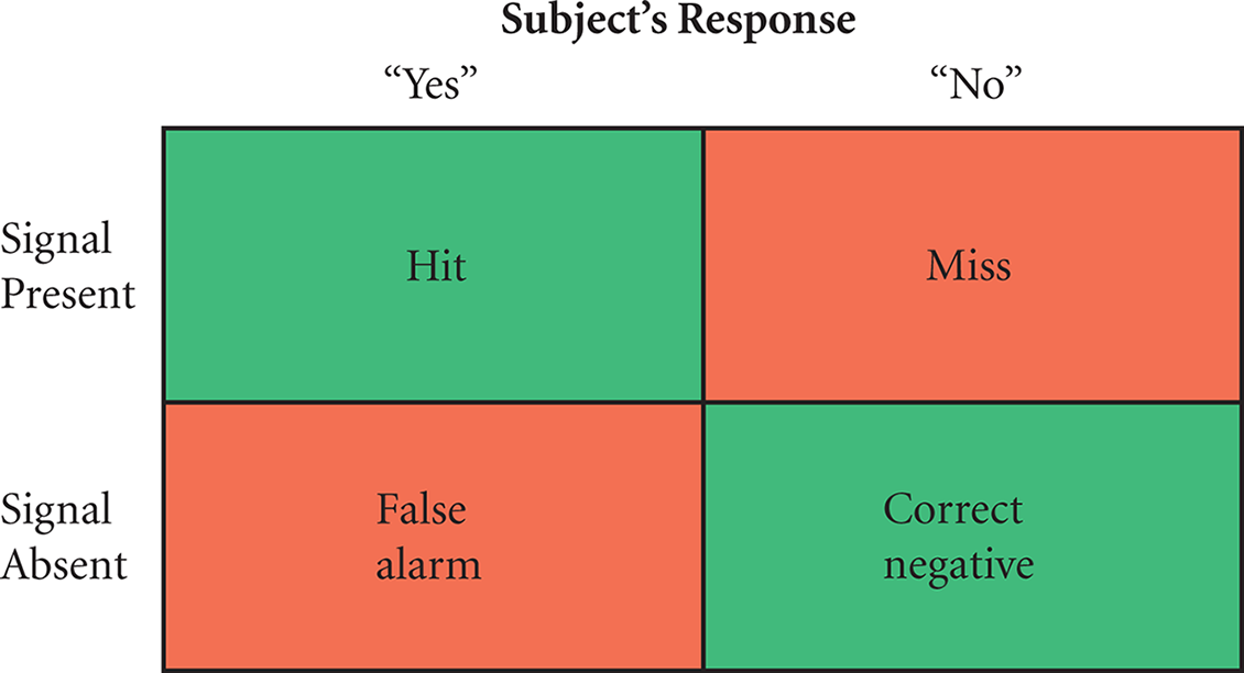
Figure 2.1. Possible Outcomes from a Signal Detection Experiment Trial
REAL WORLD
On the surface, signal detection experiments would appear to be easy tasks—shouldn’t individuals easily be able to tell if they perceived something or not? However, consider the thought processes that occur when you’re quietly studying in the library with your phone on silent and you suddenly think you may have heard a buzz. Is my phone ringing? you wonder. You freeze in place and wait for another buzz; even if it doesn’t come, you may still be so convinced you heard a signal that you still check your phone. Perception is not a passive matter!
Adaptation
Our ability to detect a stimulus can change over time through adaptation. Adaptation can have both a physiological (sensory) component and a psychological (perceptual) component. For example, the pupils of the eyes will dilate in the dark and constrict in the light, which illustrates of physiological adaptation. Similarly, in loud environments, small muscles in the middle ear will reflexively contract in order to dampen the vibration of the ossicles, reducing sound intensity. We also adapt to somatosensory stimuli; cold water no longer seems so cold once our bodies “get used to it.” Once we’re dressed, we stop feeling the clothes on our bodies until we have a reason to think about them. Adaptation is one way the mind and body try to focus attention on only the most relevant stimuli, which are usually changes in the environment around us.
MCAT CONCEPT CHECK 2.1
Before you move on, assess your understanding of the material with these questions.
_____________________________
- What is the pathway for a stimulus to reach conscious perception?
- Match each sensory receptor to its function:
- Hair cell
- Nociceptor
- Olfactory receptor
- Osmoreceptor
- Photoreceptor
- Taste receptor
- Thermoreceptor
- Sense painful or bothersome physical stimuli
- Sense changes in temperature
- Sense electromagnetic radiation in the visible range
- Sense changes in blood concentration
- Sense volatile chemicals
- Sense motion of fluid in the inner ear
- Sense dissolved chemicals
- For each of the thresholds below, provide a brief description:
_____________________________
- Absolute threshold:
_____________________________
- Threshold of conscious perception:
_________________________
- Difference threshold:
- What aspect of thresholds do Weber’s law and signal detection theory focus on?
____________________________
- Weber’s law:
____________________________
- Signal detection theory:
- How does sensory adaptation affect a difference threshold? ___________________________
2.2 Vision
LEARNING OBJECTIVES
After Chapter 2.2, you will be able to:
- List the functions of the parts of the eye, including the cornea, pupil, iris, ciliary body, canal of Schlemm, lens, retina, and sclera
- Describe parallel processing
- Identify the cell types responsible for color, shape, and motion detection
- Recall the structures in the visual pathway:
Vision is a highly adapted sense in human beings. With the ability to sense brightness, color, shape, and movement, and then to integrate this information to create a cohesive three-dimensional model of the world, the visual pathways are extremely important to everyday life. In fact, vision is the only sense to which an entire lobe of the brain is devoted: the occipital lobe. While some individuals experience vision differently, such as those who are color deficient or partially sighted, the MCAT focuses primarily on the details of unimpaired vision.
Structure and Function of the Eye
The anatomy of the eye is shown in Figure 2.2.
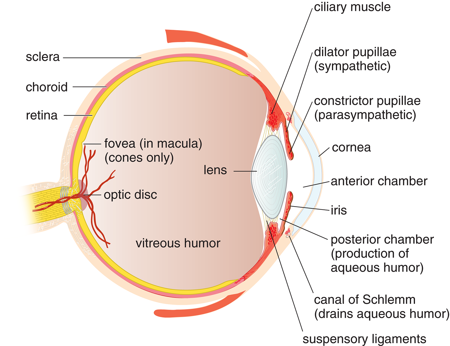
Figure 2.2. Anatomy of the Eye
The eye is a specialized organ used to detect light in the form of photons. Most of the exposed portion of the eye is covered by a thick structural layer known as the sclera, or the white of the eye. The sclera does not cover the frontmost portion of the eye, the cornea. The eye is supplied with nutrients by two sets of blood vessels: the choroidal vessels, a complex intermingling of blood vessels between the sclera and the retina, and the retinal vessels. The innermost layer of the eye is the retina, which contains the actual photoreceptors that transduce light into electrical information the brain can process.
When entering the eye, light passes first through the cornea, a clear, domelike window in the front of the eye, which gathers and focuses the incoming light. The front of the eye is divided into the anterior chamber, which lies in front of the iris, and the posterior chamber between the iris and the lens. The iris, which is the colored part of the eye, is composed of two muscles: the dilator pupillae, which opens the pupil under sympathetic stimulation; and the constrictor pupillae, which constricts the pupil under parasympathetic stimulation. The iris is continuous with the choroid, which is a vascular layer of connective tissue that surrounds and provides nourishment to the retina. The iris is also continuous with the the ciliary body, which produces the aqueous humor that bathes the front part of the eye before draining into the canal of Schlemm. The lens lies right behind the iris and helps control the refraction of the incoming light. Contraction of the ciliary muscle, a component of the ciliary body, is under parasympathetic control. As the muscle contracts, it pulls on the suspensory ligaments and changes the shape of the lens to focus on an image as the distance varies, a phenomenon known as accommodation. Behind the lens lies the vitreous humor, a transparent gel that supports the retina.
The retina is in the back of the eye and is like a screen consisting of neural elements and blood vessels. Its function is to convert incoming photons of light to electrical signals. It is actually considered part of the central nervous system and develops as an outgrowth of brain tissue. The duplexity or duplicity theory of vision states that the retina contains two kinds of photoreceptors: those specialized for light-and-dark detection and those specialized for color detection.
The retina is made up of approximately 6 million cones and 120 million rods. Cones are used for color vision and to sense fine details. Cones are most effective in bright light and come in three forms, which are named for the wavelengths of light they best absorb, as shown in Figure 2.3.
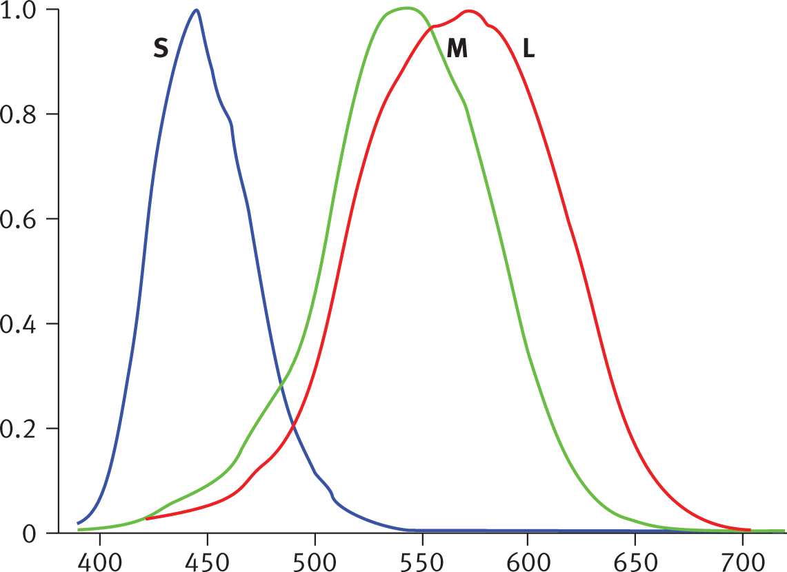
Figure 2.3. Relative Absorption of the Three Types of Cones at Different Wavelengths The cones are named for the wavelengths at which they have highest light absorption: short (S, also called blue), medium (M, green), and long (L, red).
In reduced illumination, rods are more functional than cones because each rod cell is highly sensitive to photons and is somewhat easier to stimulate than a cone cell. In part, the sensitivity of rods has to do with the fact that all rods contain only a single pigment type called rhodopsin. In general, color vision requires far more light because each cone responds only to certain wavelengths of light. By contrast, a rod can be stimulated by light of any color. However, while rods permit vision in reduced light, the tradeoff is that rods only allow sensation of light and dark. Also, even though individual rods are highly sensitive to light, as a whole they are less useful for detecting fine details because rods are spread over a much larger area of the retina.
MNEMONIC
Cones are for color vision. Rods function best in “roduced” light.
While there are many more rods than cones in the human eye, the central section of the retina, called the macula, has a high concentration of cones; in fact, the centermost region of the macula, called the fovea, contains only cones. As one moves further away from the fovea towards the peripheral retina, the concentration of rods increases while the concentration of cones decreases. Therefore, visual acuity is best at the fovea, and the fovea is most sensitive in normal daylight vision. Some distance away from the center of the retina, the optic nerve leaves the eye. This region of the retina, which is devoid of photoreceptors, is called the optic disk, and gives rise to a blind spot, as shown in Figure 2.4.
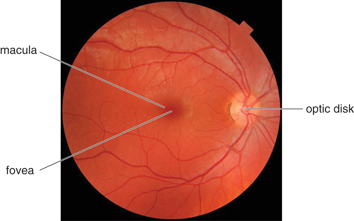
Figure 2.4. Specialized Regions of the Retina
Rods and cones are specialized neurons and, like most neurons, connect with other neurons through synapses. However, rods and cones do not connect directly to the optic nerve. Rather, there are several layers of neurons in between, as shown in Figure 2.5. Rods and cones synapse directly with bipolar cells, which highlight gradients between adjacent rods or cones. Bipolar cells then synapse with ganglion cells, the axons of which group together to form the optic nerve. These bipolar and ganglion cells not only fall “in between” the rods and cones and the optic nerve, but also the bipolar and ganglion cells are actually located in front of the rods and cones, closer to the front of the eye. This arrangement means that a photon must actually navigate past several layers of cells to reach the rods and cones at the “back” of the retina; the information is then transmitted “forward” (in the form of action potentials) from the rod and cone cells until the signal reaches the ganglion cells. Observe in Figure 2.5 that there are significantly more photoreceptor cells than ganglion cells, so the output from each ganglion cell represents the combined activity of many rods and cones. The result is a pruning of details as information from the photoreceptors is combined. As the number of receptors that converge through the bipolar neurons onto one ganglion cell increases, the resolution decreases. On average, the number of cones converging onto an individual ganglion cell is smaller than for rods. This arrangement helps explain why color vision has a greater sensitivity to fine detail than black-and-white vision does.
Also shown in Figure 2.5 are amacrine and horizontal cells, which receive input from multiple retinal cells in the same area before the information is passed on to ganglion cells. Amacrine and horizontal cells can thereby accentuate slight differences between the visual information in each bipolar cell. For example, these cells are important for edge detection, as they increase our perception of contrasts.
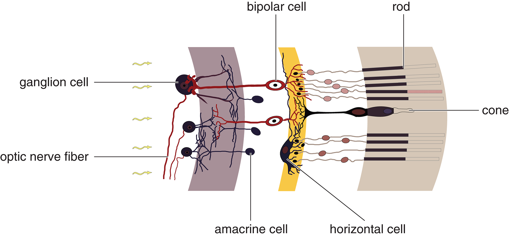
Figure 2.5. Cells of the Retina
Visual Pathways
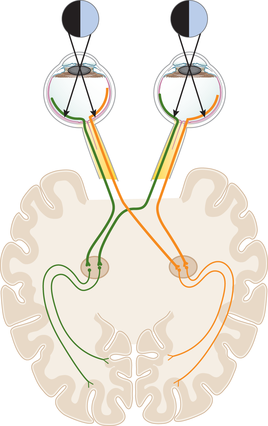
Visual pathways refer to both the anatomical connections between the eyes and brain and to the flow of visual information along these connections. These pathways are illustrated in Figure 2.6. The terminology for these pathways can seem confusing. So here is a simple rule of thumb to start: If an object is to your left, then photons from that object stimulate the right side of the retina in each eye. To help visualize this result, look at Figure 2.6. In this figure, an object to your left is represented by the black patches. Photons from that object enter each eye and stimulate the retinal fibers that are illustrated using the color orange. This result is due to simple geometry! If an object is to your left, then photons from that object must travel to the right to reach your eyes, and in so doing, these photons enter your eyes and continue travelling to the right, thereby stimulating the right side of each of your eyes’ retinas. From there, retinal fibers from each eye project to the right side of your brain. All of this leads to a famous result: Visual information from objects to your left is processed by the right side of your brain. Similarly, if an object is to your right (the blue patches in the figure), then photons from this object stimulate the left side of each retina (the green nerves), and this visual data is sent to the left side of your brain.
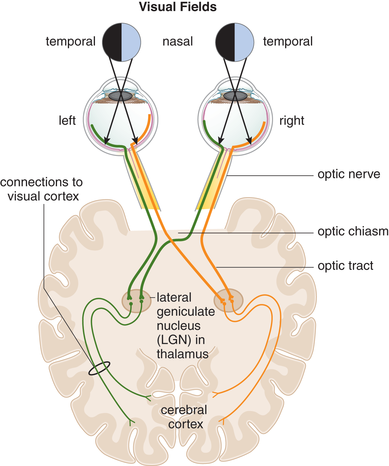
Figure 2.6. Visual Pathways
To help describe these phenomena with more specificity, scientists use the terms temporal and nasal retinal fibers. Here’s an example. Consider an object to your left, which, again, is represented in Figure 2.6 by the black patches. In each eye, photons from this object strike the retinal fibers that are illustrated in Figure 2.6 using the color orange. But notice: In the right eye, those orange fibers are on the lateral side of the retina, closer to your temple, and so these fibers are called the temporal fibers of the right eye. In the left eye, the orange fibers are medial, closer to your nose, and so are called the nasal fibers of the left eye.
Scientists also describe the placement of the object in space, relative to your eyes, using the terms "temporal" and "nasal," but now referring to the temporal and nasal visual fields. Again, this terminology is best explained by example. Imagine an object is a little to your left. Then photons from that object must cross in front of your nose in order to reach your right eye. So, this object is described as being in the nasal field of your right eye. Think carefully about what happens to these photons next. They enter your right eye, continue heading to the right, and ultimately strike the temporal fibers on the right side of your right eye. Pay close attention to the terminology: We’re saying that an object in the nasal field of the right eye stimulates the temporal fibers of that eye! This mixing of terminology—nasal field stimulates temporal fibers—illustrates the crossing over of visual information. By contrast, for that same object, which, remember, is spatially to your left, photons can enter your left eye directly. Therefore, this object is described as being in the temporal field of your left eye. Then, these photons strike the nasal fibers of the left eye. So again, there is mixing of the terminology—the object is in the temporal field of the left eye, and photons strike the nasal fibers of the left eye.
MNEMONIC
If photons from an object would need to cross your nose to reach your eye (or if the object is right in front of your nose) then the object is said to be in the nasal visual field for that eye.
As the retinal fibers from each eye travel through the optic nerves toward the brain, a significant event occurs at the optic chiasm. Here, nasal fibers from the left and right eyes cross paths. Notice that only the nasal fibers cross at the optic chiasm. Again, an example helps illustrate: Visual information from an object to your left is ultimately processed by the right side of your brain. So if photons from that object are captured by the left eye, then this visual information needs to be routed to the opposite side of your brain. That routing is done using the optic chiasm. To use the "nasal" and "temporal" terminology: An object to your left will stimulate the nasal fibers of your left eye, and then these nasal fibers are routed through the optic chiasm to the right side of your brain. By contrast, that same object, off to your left, will stimulate the temporal fibers of your right eye. Because these temporal fibers are already on the right side of your body, there is no need to cross them. Therefore, only nasal fibers cross at the optic chiasm. These reorganized pathways are called optic tracts after leaving the optic chiasm. And the end result is that visual information emanating from an object to your left is ultimately processed on the right side of your brain.
To summarize, and to gain practice with this terminology, now consider an object off to your right, represented in Figure 2.6 by the blue patches and stimulating the fibers illustrated using the color green. Notice that the terms used depend on which eye is considered. Let’s start with the left eye. An object to your right would be in the nasal field of your left eye, because photons would have to cross your nose to reach your left eye. These photons would stimulate the temporal fibers of the left eye—nasal field stimulates temporal fibers. Then, because these temporal fibers are already on the left side of your body, they would be routed directly back to the left side of your brain. Now consider the right eye. An object spatially to your right is in the temporal field of your right eye, and stimulates the nasal fibers of that eye. Then, in order to bring this visual information to the left side of your brain, these nasal fibers are routed through the optic chiasm. Through all of this, keep these two trends in mind: First, the temporal field of each eye stimulates the nasal fibers of each eye, and vice versa. Second, the nasal fibers cross at the optic chiasm.
MNEMONIC
For each eye, the temporal visual field stimulates the nasal retinal fibers. The nasal visual field stimulates the temporal retinal fibers.
From the optic chiasm, the information goes to several different places in the brain: some nerve fibers pass to the lateral geniculate nucleus (LGN) of the thalamus where they synapse with nerves that then pass through radiations in the temporal and parietal lobes to the visual cortex in the occipital lobe. This connectivity makes sense because the thalamus is a well-known connecting and routing center of the forebrain. Other nerve fibers branch off from the optic tracts, skip the thalamus, and head directly to the superior colliculi in the midbrain, which control some reflexive responses to visual stimuli and reflexive eye movements.
REAL WORLD
When there is a sudden, bright flash of light, the superior colliculus aligns the eyes with the likely stimulus. In other words, it’s the superior colliculus (as well as the sympathetic nervous system) that gives us the “deer in the headlights” appearance during the startle response.
Processing
The ability to sense light information in the environment around us is useful in its own right. But, to effectively interact with the environment, we must also be able to make sense of visual stimuli. The connections between optic tract, LGN, and visual cortex help create a cohesive image of the world through a phenomenon known as parallel processing. Visual parallel processing is the brain’s ability to analyze information regarding color, form, motion, and depth simultaneously, i.e. “in parallel,” using independent pathways in the brain. For example, most people can quickly and easily recognize a moving car from a distance. The speed of recognition is facilitated, in part, by the fact that the form of the car (i.e. its shape) and the motion of the car are processed simultaneously in separate, parallel pathways in the brain. In contrast, serial processing occurs when cognitive processes are executed in a sequential, step-by-step manner. While slower than parallel processing, serial processing allows for the processing of tasks that require a high level of focus and attention, leading to greater accuracy.
Now let’s explore where each of these four aspects of vision is processed and the specialized cells that contribute to their detection. As described previously, cones are responsible for our perception of color. Form refers not only to the shape of an object, but also our ability to discriminate an object of interest from the background by detecting its boundaries. Neurons carrying information from the fovea and surrounding central portion of the retina synapse with parvocellular cells in the lateral geniculate nucleus. These cells have very high color spatial resolution; that is, these cells permit us to detect very fine detail when thoroughly examining an object. However, parvocellular cells can only work with stationary or slow-moving objects because these cells have very low temporal resolution.
Conversely, magnocellular cells are well-suited for detecting motion because these cells have very high temporal resolution. Reflecting the fact that form and motion are processed in parallel, magnocellular cells and parvocellular cells are located in distinct layers of the lateral geniculate nucleus. Also, magnocellular cells predominantly receive inputs from the periphery of our vision, allowing more rapid detection of objects approaching us from the sides. However, magnocellular cells have low spatial resolution, so much of the rich detail of an object can no longer be seen once the object is in motion. Magnocellular cells therefore provide a blurry but moving image of an object.
Depth perception, our ability to discriminate the three-dimensional shape of our environment and judge the distance of objects within it, is largely based on discrepancies between the inputs the brain receives from our two eyes (more on this to follow in MCAT Behavioral Sciences Review, Section 2.5, Object Recognition). Specialized cells in the visual cortex known as binocular neurons are responsible for comparing the inputs to each hemisphere and detecting these differences.
Finally, our brains wouldn’t be very good at processing visual information if they didn’t learn to associate certain patterns of stimuli with expected behaviors or outcomes. To assist in this, a whole slew of even more specialized cells called feature detectors exist in the visual cortex. Each feature detector cell type detects a very particular, individual feature of an object in the visual field. For example, if we were to look at a stop sign we would activate: a feature detector for the color red, while another feature detector would respond to the white border and letters. Yet another type of feature detector would recognize the horizontal lines, while still others would be activated by the angled lines of the octagon. Rather than needing to individually process each of these features every time, the overall combination of feature detectors become activated in parallel. Finally, our response to the stop sign, i.e. to STOP, also is stored for future retrieval.
MNEMONIC
Magnocellular cells specialize in motion detection.
MCAT CONCEPT CHECK 2.2
Before you move on, assess your understanding of the material with these questions.
- List the functions of the various parts of the eye:
____________________________
- Cornea:
____________________________
- Pupil:
____________________________
- Iris:
____________________________
- Ciliary body:
____________________________
- Canal of Schlemm:
____________________________
- Lens:
____________________________
- Retina:
_________________________
- Sclera:
- List the structures in the visual pathway, from where light enters the cornea to the visual projection areas in the brain. _________________________
- What is parallel processing? ____________________________
- In feature detection, what type of cells are responsible for color? Form? Motion? Depth?
___________________________
- Color:
_________________________
- Form:
__________________________
- Motion:
- Depth:
2.3 Hearing and Vestibular Sense
LEARNING OBJECTIVES
After Chapter 2.3, you will be able to:
- Identify the structures used to detect linear acceleration and rotational acceleration
- Explain how the structural features of the cochlea and the hair cells are able to transmit information about pitch of an incoming sound to the brain
- List the structures in the auditory pathway
The ear is a complex organ responsible not only for our sense of hearing, but also for our vestibular sense, which is our ability to both detect rotational and linear acceleration and to use this information to inform our sense of balance and spatial orientation. These senses are critically important to our ability to get around in the world, and their associated structures are encased in some of the densest bone of the body to protect these structures from damage. While recognizing that individuals can differ in their hearing and vestibular senses, as with vision, the MCAT primarily focuses on the standard healthy operations of these senses.
Structure and Function of the Ear
The ear is divided into three parts, as shown in Figure 2.7: the outer, middle, and inner ear. A sound wave first reaches the cartilaginous outside part of the ear, called the pinna or auricle. The main function of the pinna is to channel sound waves into the external auditory canal, which directs the sound waves to the tympanic membrane (eardrum). The membrane vibrates in phase with the incoming sound waves. The frequency of the sound wave determines the rate at which the tympanic membrane vibrates: it moves back and forth at a high rate for high-frequency sounds and more slowly for low-frequency sounds. Louder sounds have greater intensity, which corresponds to an increased amplitude of vibration.
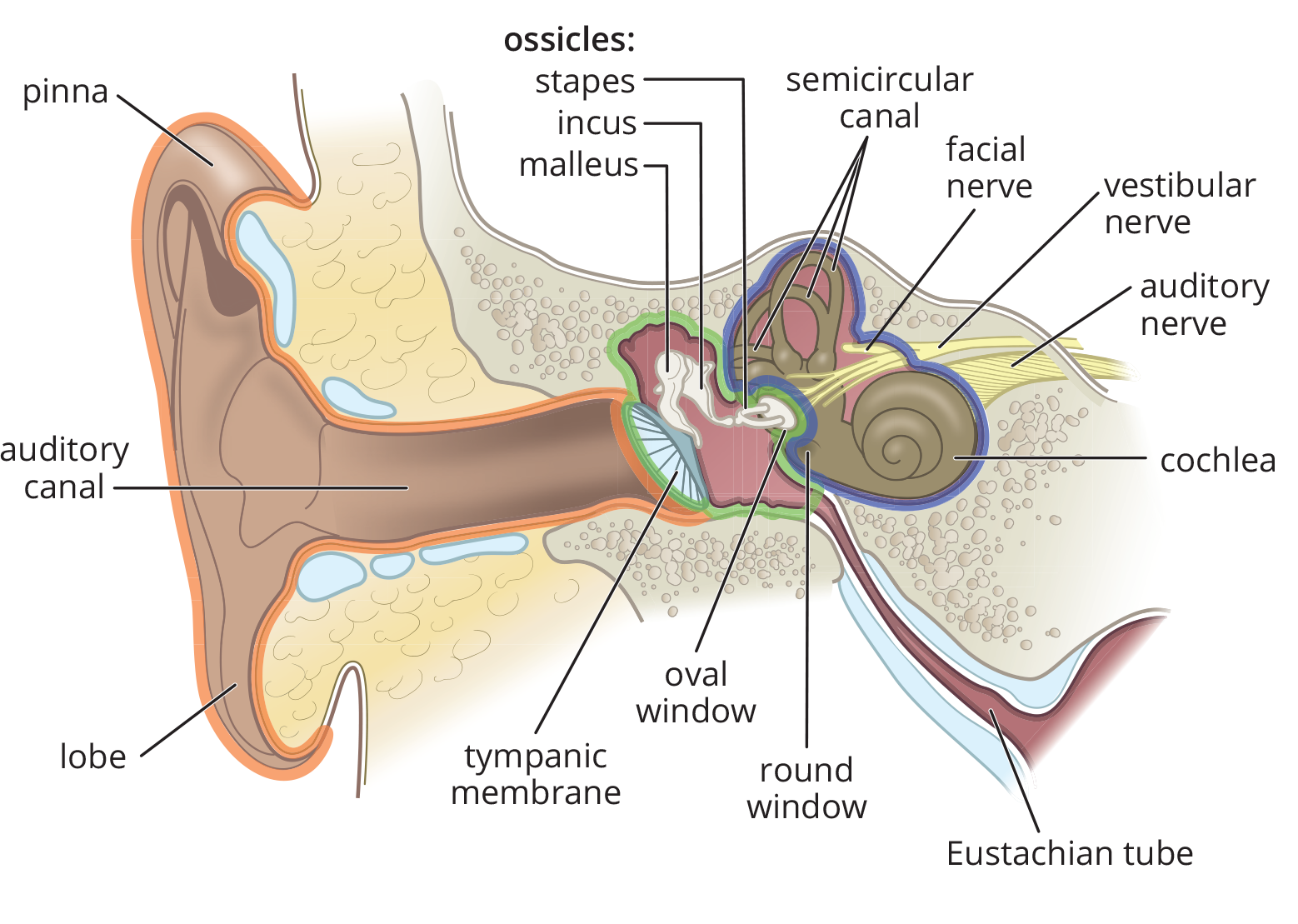
Figure 2.7. Anatomy of the Ear
The tympanic membrane divides the outer ear from the middle ear. The middle ear houses the three smallest bones in the body, called ossicles. The ossicles help transmit and amplify the vibrations from the tympanic membrane to the inner ear. The malleus (hammer) is affixed to the tympanic membrane; it acts on the incus (anvil), which acts on the stapes (stirrup). The baseplate of the stapes rests on the oval window of the cochlea, which is the entrance to the inner ear. The middle ear is connected to the nasal cavity via the Eustachian tube, which helps equalize pressure between the middle ear and the environment.
BRIDGE
Remember that sound is a longitudinal wave carried through air (or another medium), which causes displacement of particles parallel to the axis of sound propagation. In other words, when a sound wave hits your eardrum, it literally causes it to oscillate back and forth because of moving air particles. Sound is discussed in Chapter 7 of MCAT Physics and Math Review.
The inner ear sits within a bony labyrinth, which is a hollow region of the temporal bone containing the cochlea, vestibule, and semicircular canals, as shown in Figure 2.8. Inside the bony labyrinth rests a continuous collection of tubes and chambers called the membranous labyrinth. This collection of structures contains receptors for the sense of equilibrium and hearing. The membranous labyrinth is filled by a potassium-rich fluid called endolymph, and is suspended within the bony labyrinth by a thin layer of another fluid called perilymph. Perilymph simultaneously transmits vibrations from the outside world and cushions the inner ear structures.

Figure 2.8. The Membranous and Bony Labyrinth The membranous labyrinth is filled with endolymph (blue); it is suspended within the bony labyrinth, which is filled with perilymph (purple).
Cochlea
The cochlea is a spiral-shaped organ that contains the receptors for hearing; it is divided into three parts called scalae, as shown in Figure 2.9. All three scalae run the entire length of the cochlea. The middle scala houses the actual hearing apparatus, called the organ of Corti, which rests on a thin, flexible membrane called the basilar membrane. The organ of Corti is composed of thousands of hair cells, which are bathed in endolymph. On top of the organ of Corti is a relatively immobile membrane called the tectorial membrane. The other two scalae, filled with perilymph, surround the hearing apparatus and are continuous with the oval and round windows of the cochlea. Thus, sound entering the cochlea through the oval window causes vibrations in perilymph, which are transmitted to the basilar membrane. Because fluids are essentially incompressible, the round window, a membrane-covered hole in the cochlea, permits the perilymph to actually move within the cochlea. Like the rods and cones of the eye, the hair cells in the organ of Corti transduce the physical stimulus into an electrical signal, which is carried to the central nervous system by the auditory (vestibulocochlear) nerve.
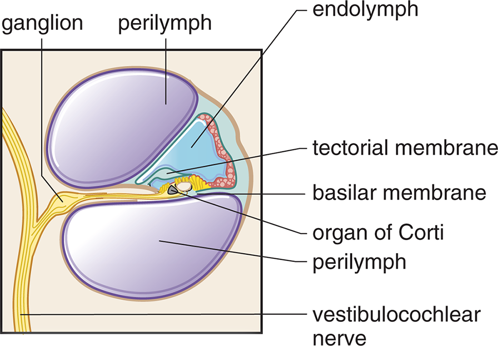
Figure 2.9. Structure of the Cochlea (Cross-Section)
BRIDGE
The junction between the stapes and the oval window is extremely similar to a thermodynamic gas–piston system, as described in Chapter 3 of MCAT Physics and Math Review. However, fluids are not as compressible as gases; therefore, the round window must be present to allow the perilymph in the cochlea to actually move back and forth with the stapedial footplate.
Vestibule
The vestibule refers to the portion of the bony labyrinth that contains the utricle and saccule. These structures are sensitive to linear acceleration, so are used as part of the balancing apparatus and to determine one’s orientation in three-dimensional space. The utricle and saccule contain modified hair cells covered with otoliths. As the body accelerates, these otoliths will resist that motion. This bends and stimulates the underlying hair cells, which send a signal to the brain.
Semicircular Canals
While the utricle and saccule are sensitive to linear acceleration, the three semicircular canals are sensitive to rotational acceleration. The semicircular canals are arranged perpendicularly to each other, and each ends in a swelling called an ampulla, where hair cells are located. When the head rotates, endolymph in the semicircular canal resists this motion, bending the underlying hair cells, which send a signal to the brain.
Auditory Pathways
The auditory pathways in the brain are a bit more complex than the visual pathways. Most sound information passes through the vestibulocochlear nerve to the brainstem, where it ascends to the medial geniculate nucleus (MGN) of the thalamus. From there, nerve fibers project to the auditory cortex in the temporal lobe for sound processing. Some information is also sent to the superior olive, which localizes the sound, and the inferior colliculus, which is involved in the startle reflex and helps keep the eyes fixed on a point while the head is turned (vestibulo–ocular reflex).
MNEMONIC
The lateral geniculate nucleus (LGN) is for light; the medial geniculate nucleus (MGN) is for music.
Hair Cells
Hair cells are named for the long tufts of stereocilia on their top surface, shown in Figure 2.10. As vibrations reach the basilar membrane underlying the organ of Corti, the stereocilia adorning the hair cells begin to sway back and forth within the endolymph. The swaying causes the opening of ion channels, which cause a receptor potential. Certain hair cells are also directly connected to the immobile tectorial membrane; these hair cells are involved in amplifying the incoming sound.
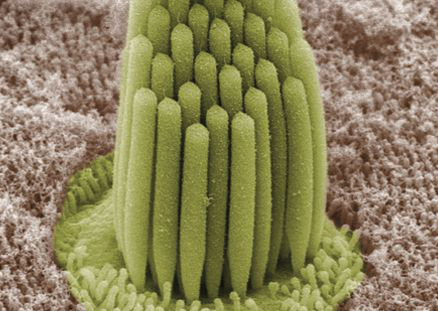
Figure 2.10. Stereocilia of a Hair Cell Movement of fluid inside the cochlea leads to depolarization of the neuron associated with the hair cell.
The basilar membrane changes thickness depending on its location in the cochlea. The accepted theory on sound perception is place theory, which states that the location of a hair cell on the basilar membrane determines the perception of pitch when that hair cell is vibrated. The highest-frequency pitches cause vibrations of the basilar membrane very close to the oval window, whereas low-frequency pitches cause vibrations at the apex, away from the oval window. Thus, the cochlea is tonotopically organized: which hair cells are vibrating gives the brain an indication of the pitch of the sound.
BEHAVIORAL SCIENCES GUIDED EXAMPLE WITH EXPERT THINKING
What results would help to support the researchers’ hypothesis? What effect would these results have on the place theory of sound perception?
When we encounter experimental passages on the MCAT, there are a few things we should always be on the lookout for: what is the hypothesis, and what are the independent and dependent variables? What results are presented, and what effect do they have on the hypothesis? What conclusion did the researchers come to, and is this conclusion reasonable given the results? If any of these pieces are missing, we should anticipate questions asking us to identify possibilities that would fit with the passage and our outside content knowledge. Here, we are given a hypothesis without results, and the question asks us for results that would support the hypothesis. We’ll need to dive into the scenario to identify what is being tested and why, and then use that information in conjunction with our content knowledge to answer this question.
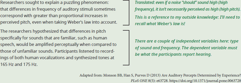
The researchers hypothesized that differences in perceived pitch are amplified for more commonly encountered sounds. Take a moment to consider what the researchers should expect if this hypothesis is correct. It would stand to reason that the difference between perceived pitch for the 165 Hz and 175 Hz samples would be larger for voices (which we hear all the time) than for synthesized tones (which we don’t hear too often). In other words, a result that would help support the researchers’ hypothesis would be experimental evidence that human vocalizations sounded more different in pitch than the synthesized tones, despite the absolute difference in frequency remaining the same.
For the second part of the question, we need to recall that place theory predicts that perceived pitch results directly from the location of the hair cells that are vibrated when exposed to that frequency; higher frequencies vibrate hair cells closer to the oval window, and lower frequencies vibrate hair cells that are farther away. What is important here is that, according to place theory, the type of sound is irrelevant; sounds of the same frequency should be perceived in the same way. However, the hypothesis of this study (and the hypothetical results we’ve just imagined in support of that hypothesis), call place theory into question as, according to the passage, similar changes in vibrations are not perceived as the same change in pitch. According to this study, the perceived pitch instead depends at least in part on the nature of the sound perceived by the listener. Note that this new finding wouldn’t affect a place theory of sound sensation/detection, which could still be accurate: this experiment is specifically about how sounds are perceived, not about how they are sensed.
In sum, to support the researchers, the results must find a greater perceived difference in pitch between the vocal samples as compared to the synthesized audio samples. This finding would contradict the place theory of sound perception.
MCAT CONCEPT CHECK 2.3
Before you move on, assess your understanding of the material with these questions.
- What structures are used to detect linear acceleration? Rotational acceleration?
____________________________
- Linear acceleration:
_________________________
- Rotational acceleration:
- List the structures in the auditory pathway, from where sound enters the pinna to the auditory projection areas in the brain. __________________________
- How does the organization of the cochlea indicate the pitch of an incoming sound? ____________________________
2.4 Other Senses
LEARNING OBJECTIVES
After Chapter 2.4, you will be able to:
- List the structures in the olfactory pathway
- Distinguish between the chemicals detected by the nose and mouth
- Recall the four main modalities of somatosensation
While vision and hearing are, by far, the most heavily tested senses on the MCAT, the other senses are still considered fair game on Test Day. These include the chemical senses of smell and taste; somatosensation, which includes all of the modalities of “touch”; and kinesthetic sense.
Smell
Smell is considered one of the chemical senses, which means that it responds to incoming chemicals from the outside world. Specifically, the sense of smell responds to volatile or aerosolized compounds through the use of chemoreceptors, or receptors that detect sensory information by binding to chemical stimuli. These olfactory chemoreceptors (olfactory nerves) are located in the olfactory epithelium in the upper part of the nasal cavity. There are a tremendous number of specific chemoreceptors, which allows us to recognize subtle differences in similar scents, such as lavender and jasmine.
REAL WORLD
Smell is an impressive motivator for behavior. Food aromas may make a person hungry, a familiar fragrance may remind a person of a significant other from years ago, and an unpleasant smell may signify that an unknown bottle contains a dangerous chemical rather than water. Smell is the only sense that does not pass through the thalamus, but rather travels—unfiltered—into higher-order brain centers.
Smell can also carry interpersonal information through the medium of pheromones, which are chemicals secreted by one animal, and which, once bonded with chemoreceptors, compel or urge another animal to behave in a specific way. Pheromones have debatable effects on humans, but play an enormous role in many animals’ social, foraging, and sexual behaviors.
As is true with all senses, there is a defined olfactory pathway to the brain. Odor molecules are inhaled into the nasal passages and then contact the olfactory nerves in the olfactory epithelium. These receptor cells are activated, sending signals to the olfactory bulb. These signals are then relayed via the olfactory tract to higher regions of the brain, including the limbic system.
Taste
As a sense, taste is often simpler than we imagine. There are five basic tastes: sweet, sour, salty, bitter, and umami (savory). Flavor is not synonymous with taste, but rather refers to the complex interplay between smell and taste, which can be affected by nonchemical stimuli like texture and the individual’s mood.
Similar to smell, tastes are detected using chemoreceptors. These chemoreceptors are sensitive to dissolved compounds. Saltiness, for example, is a reaction to alkali metals, and is generally triggered by the sodium found in table salt. Sourness, on the other hand, is a reaction to acid, such as lemon juice or vinegar. Sweet, bitter, and savory flavors are also triggered by specific molecules binding to receptors. The receptors for taste are groups of cells called taste buds, which are found in little bumps on the tongue called papillae. Taste information travels from taste buds to the brainstem, and then ascends to the taste center in the thalamus before traveling to higher-order brain regions.
Somatosensation
Somatosensation is often reduced to “touch” when listed as a sense, but is actually quite complex. Somatosensation is usually described as having four modalities: pressure, vibration, pain, and temperature. At least five different types of receptor receive tactile information, including:
- Pacinian corpuscles: respond to deep pressure and vibration
- Meissner corpuscles: respond to light touch
- Merkel cells (discs): respond to deep pressure and texture
- Ruffini endings: respond to stretch
- Free nerve endings: respond to pain and temperature
REAL WORLD
Pain and temperature actually use a different pathway than pressure and vibration through the spinal cord. This can be seen in Brown-Séquard syndrome, in which half of the spinal cord is severed. Patients lose pressure and vibration sense on the same side as the lesion, but lose pain and temperature sensation on the opposite side.
Transduction occurs in the receptors, which send the signal to the central nervous system where it eventually travels to the somatosensory cortex in the parietal lobe.
There are three additional concepts related to touch perception that are important to know: two-point thresholds, physiological zero, and gate theory of pain. A two-point threshold refers to the minimum distance necessary between two points of stimulation on the skin such that the points will be felt as two distinct stimuli. Below the two-point threshold, the two stimuli will be felt as one. The size of the two-point threshold depends on the density of nerves in the particular area of skin being tested. Temperature is judged relative to physiological zero, or the normal temperature of the skin (between 86° and 97°F). Thus, an object feels “cold” because it is under physiological zero; an object feels “warm” because it is above physiological zero.
Pain perception is part of the somatosensory system and can result from signals sent from a variety of sensory receptors, most commonly nociceptors. Pain also relies on thresholds, which may vary greatly from person to person. For example, the temperature of water that is perceived to be “so hot it hurts” may vary by several degrees between individuals. The gate theory of pain proposes that a special “gating” mechanism can turn pain signals on or off, affecting whether or not we perceive pain. In this theory, the spinal cord is able to preferentially forward the signals from other touch modalities (pressure, temperature) to the brain, thus reducing the sensation of pain. Gate theory has been superseded by other theories, but is still a useful model for understanding how touch is processed at the spinal cord.
REAL WORLD
The gate theory of pain explains why rubbing an injury (like bumping your knee on a table) seems to reduce the pain of the injury.
Kinesthetic Sense
Kinesthetic sense is also called proprioception and refers to the ability to tell where one’s body is in space. For example, even with your eyes closed, you could still describe the location and position of your hand. The receptors for proprioception, called proprioceptors, are found mostly in muscle and joints, and play critical roles in hand–eye coordination, balance, and mobility.
MCAT CONCEPT CHECK 2.4
Before you move on, assess your understanding of the material with these questions.
- List the structures in the olfactory pathway, from where odor molecules enter the nose to where olfactory signals project in the brain. _____________________________
- Both smell and taste are sensitive to chemicals. What is different about the types of chemicals each one can sense? ________________________
- What are the four main modalities of somatosensation?
- _________________________
- _________________________
- _________________________
- _________________________
2.5 Object Recognition
LEARNING OBJECTIVES
After Chapter 2.5, you will be able to:
- Compare and contrast bottom-up processing and top-down processing
- Describe each of the Gestalt principles: proximity, similarity, good continuation, subjective contours, closure, and prägnanz
Modern theories of object recognition assume at least two major types of psychological processing: bottom-up processing and top-down processing. Bottom-up (data-driven) processing refers to object recognition by parallel processing and feature detection, as described earlier. Essentially, the brain takes the individual sensory stimuli and combines them together to create a cohesive image before determining what the object is. Top-down (conceptually driven) processing is driven by memories and expectations that allow the brain to recognize the whole object and then recognize the components based on these expectations. In other words, top-down processing allows us to quickly recognize objects without needing to analyze their specific parts. Neither system is sufficient by itself: if we only performed bottom-up processing, we would be extremely inefficient at recognizing objects; every time we looked at an object, it would be like looking at the object for the first time. On the other hand, if we only performed top-down processing, we would have difficulty discriminating slight differences between similar objects. This distinction is also partially responsible for the feeling of déjà vu described in the introduction to this chapter: when we believe we are experiencing something for the first time, we expect to rely on bottom-up processing; however, when the mind is able to recognize an experience more quickly than expected (through top-down processing), the mind searches for a reason for this recognition. In other words, déjà vu is often evoked when we have recognition without an obvious reason: I know that person from somewhere . . . but where? The distinction between top-down and bottom-up processing is relevant for all senses, but is most commonly applied in the context of vision.
Perceptual organization refers to the ability to create a complete picture or idea by combining top-down and bottom-up processing with all of the other sensory clues gathered from an object. Most of the images we see in everyday life are incomplete; often, we may only be able to see a part of an object and we must infer what the rest of the object looks like. By using what information is available in terms of depth, form, motion, constancy, and other clues, we can often “fill in the gaps” using Gestalt principles (described below).
Depth perception relies on a number of visual cues that are interpreted by the brain to deduce an object’s distance. These visual cues are separated into monocular and binocular cues. Monocular cues only require one eye and include relative size, interposition, linear perspective, motion parallax, and other minor cues. Relative size refers to the idea that objects appear larger the closer they are. Interposition means that when two objects overlap, the one in front is closer. Linear perspective refers to the convergence of parallel lines at a distance: the greater the convergence, the further the distance. Motion parallax is the perception that objects closer to us seem to move faster when we change our field of vision (look at something else).
REAL WORLD
If you’ve ever looked out the side window of a car, bus, or train on a clear night, you’ve experienced motion parallax. Parallax is the reason why the objects on the side of the road are a blur as you go past, why objects further away move more slowly as you pass them, and why the moon seems to follow you as you ride along.
Binocular cues primarily involve retinal disparity which refers to the slight difference in images projected on the two retinas. This feature of depth perception is exploited in virtual reality (VR) devices: the images supplied to each eye are slightly different, giving the perception of depth even though the VR device displays 2D images. A secondary binocular cue is convergence, in which the brain detects the angle between the two eyes required to bring an object into focus. If a person was looking at a distant object, both of their eyes would stare straight ahead. However if they were looking at something nearby (perhaps their own nose!) the left and right eyes would be held at an extreme angle. This difference in the degree of convergence is used to perceive distance.
The form of an object is usually determined through parvocellular cells and feature detection, and the motion of an object is perceived through magnocellular cells, as described earlier. Constancy refers to our ability to perceive that certain characteristics of objects remain the same, despite changes in the environment. For example, we perceive a white piece of paper as essentially the same color whether the paper is illuminated by fluorescent lights, incandescent bulbs, or sunlight—this type of constancy is called color constancy. We also have constancy for brightness, size, and shape, depending on context.
Gestalt Principles
The brain constantly uses incomplete information to try to create a complete picture of the environment. Gestalt principles are a set of general rules that account for the fact that the brain tends to view incomplete stimuli in organized, patterned ways. There are dozens of Gestalt principles, but the highest-yield are summarized below and can be visualized in Figure 2.11.
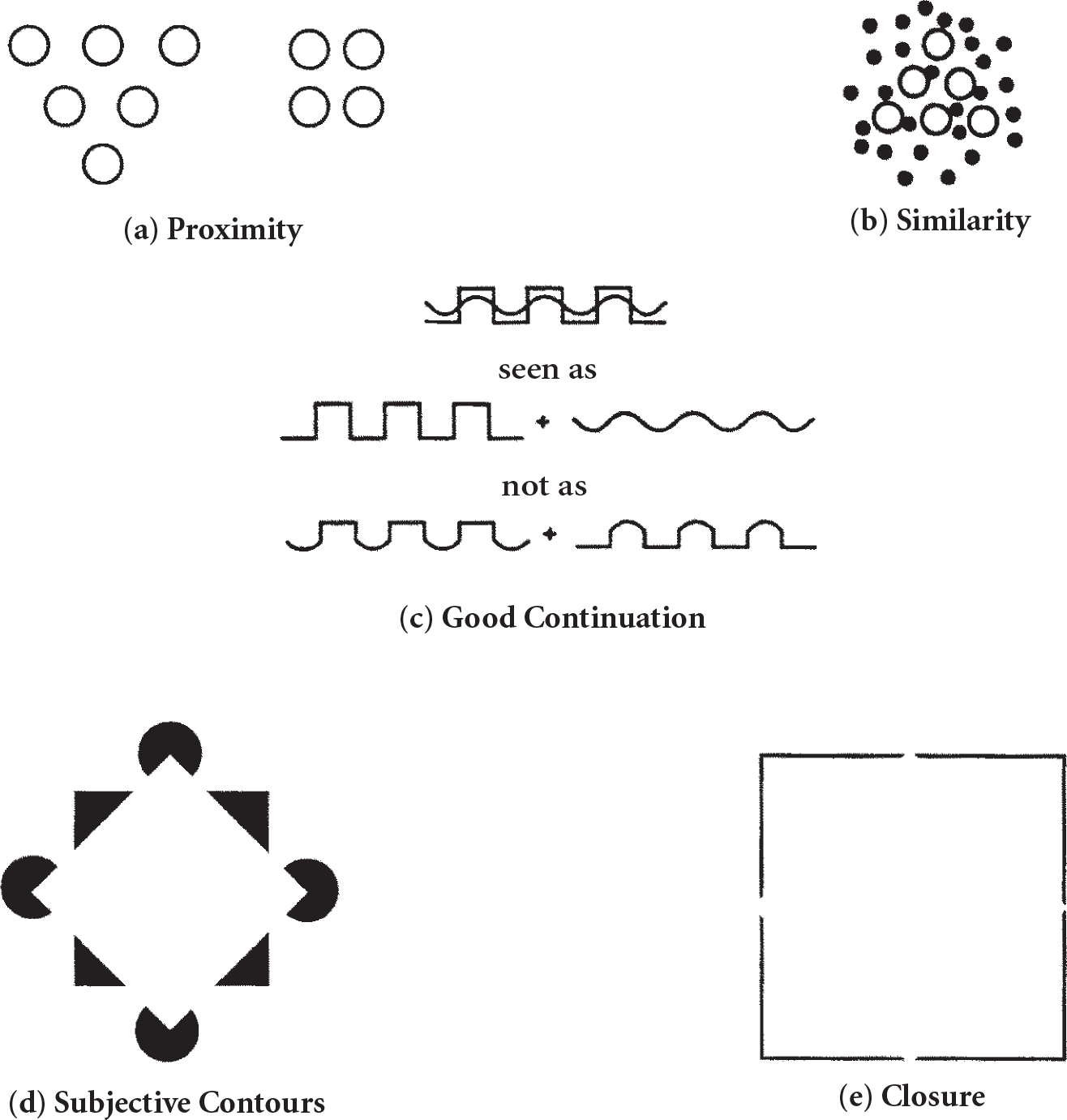
Figure 2.11. Gestalt Principles
The law of proximity says that elements close to one another tend to be perceived as a unit. In Figure 2.11a, we do not see ten unrelated dots; rather, we see a triangle and a square, each composed of a certain number of dots. The law of similarity says that objects that are similar tend to be grouped together. In Figure 2.11b, we see the big hollow dots as being distinct from the others, forming a triangle against a background of small filled-in dots. The law of good continuation says that elements that appear to follow in the same pathway tend to be grouped together. That is, there is a tendency to perceive continuous patterns in stimuli rather than abrupt changes. As seen in Figure 2.11c, our mind tends to break down this complex figure into a sawtooth line and a wavy line, rather than two lines that contain both sawtooth and wavy elements. Some researchers have argued that the phenomena of subjective contours may arise from this law. Subjective contours have to do with perceiving contours and, therefore, shapes that are not actually present in the stimulus. In Figure 2.11d, subjective contours lead to the perception of a white diamond on a black square with its corners lying on the four circles. Finally, the law of closure says that when a space is enclosed by a contour, the space tends to be perceived as a complete figure. Closure also refers to the fact that certain figures tend to be perceived as more complete (or closed) than they really are. In Figure 2.11e, we don’t see four right angles; instead, we see a square, even though the four sides aren’t complete. All these laws operate to create the most stable, consistent, and simplest figures possible within a given visual field. Taken altogether, the Gestalt principles are governed by the law of prägnanz, which says that perceptual organization will always be as regular, simple, and symmetric as possible.
MCAT CONCEPT CHECK 2.5
Before you move on, assess your understanding of the material with these questions.
- How is sensory information integrated in bottom-up processing? Top-down processing?
____________________________
- Bottom-up processing:
_________________________
- Top-down processing:
- Briefly describe each of the Gestalt principles below:
Gestalt Principle DescriptionProximitySimilarityGood continuationSubjective contoursClosurePrägnanz****
Conclusion
The sensory systems described in this chapter are key to your success on Test Day. Not only are the eye, ear, and other senses high-yield in their own right, but connections to topics in physics, biology, research design, and other concepts in the behavioral sciences make these key topics for passages. But sensation is only one part of the system; we must then take this raw information and process it in the brain to truly perceive the world around us. We use complex neurological pathways to integrate and sort sensory information. We then process sensory information through multiple systems, analyzing individual features and components of the environment while building expectations based on our memories and past experiences. We fill in gaps in our sensorium using Gestalt principles. And what reaches our conscious awareness is the final product: a cohesive concept of the world around us.
You’ve completed your visit to the new city. You used your rods and cones to see the sites, your chemoreceptors to taste and smell the local food, your hair cells to listen to the local sounds, and your kinesthetic and vestibular senses to help navigate through physical space. As you get ready to head home, all you’re left with are your memories—a topic we’ll turn to in the next chapter.
GO ONLINE!
You’ve reviewed the content, now test your knowledge and critical thinking skills by completing a test-like passage set in your online resources!
CONCEPT SUMMARY
Sensation *vs*. Perception
- Sensation is the conversion, or transduction, of physical, electromagnetic, auditory, and other information from the internal and external environment into electrical signals in the nervous system.
- Perception is the processing of sensory information to make sense of its significance.
- Sensory receptors are nerves that respond to stimuli and trigger electrical signals.
- Sensory neurons are associated with sensory ganglia: collections of cell bodies outside the central nervous system.
- Sensory stimuli are transmitted to projection areas in the brain, which further analyze the sensory input.
- Common sensory receptors include photoreceptors, hair cells, nociceptors, thermoreceptors, osmoreceptors, olfactory receptors, and taste receptors.
- A threshold is the minimum stimulus that causes a change in signal transduction.
- The absolute threshold is the minimum of stimulus energy that is needed to activate a sensory system.
- The threshold of conscious perception is the minimum of stimulus energy that will create a signal large enough in size and long enough in duration to be brought into awareness.
- The difference threshold or just-noticeable difference (jnd) is the minimum difference in magnitude between two stimuli before one can perceive this difference.
- Weber’s law states that the jnd for a stimulus is proportional to the magnitude of the stimulus, and that this proportion is constant over most of the range of possible stimuli.
- Signal detection theory refers to the effects of nonsensory factors, such as experiences, motives, and expectations, on perception of stimuli.
- Signal detection experiments allow us to look at response bias. In a signal detection experiment, a stimulus may or may not be given, and the subject is asked to state whether or not the stimulus was given. There are four possible outcomes: hits, misses, false alarms, or correct negatives.
- Adaptation refers to a decrease in response to a stimulus over time.
Vision
- The eye is an organ specialized to detect light in the form of photons.
- The cornea gathers and filters incoming light.
- The iris divides the front of the eye into the anterior and posterior chambers. It contains two muscles, the dilator and constrictor pupillae, which open and close the pupil.
- The lens refracts incoming light to focus it on the retina and is held in place by suspensory ligaments connected to the ciliary muscle.
- The ciliary body produces aqueous humor, which drains through the canal of Schlemm.
- The retina contains rods and cones. Rods detect light and dark; cones come in three forms (short-, medium-, and long-wavelength) to detect colors.
- The retina contains mostly cones in the macula, which corresponds to the central visual field. The center of the macula is the fovea, which contains only cones.
- Rods and cones synapse on bipolar cells, which synapse on ganglion cells. Integration of the signals from ganglion cells and edge-sharpening is performed by horizontal and amacrine cells.
- The bulk of the eye is supported by the vitreous on the inside and the sclera and choroid on the outside.
- The visual pathway starts from the eye, and travels through the optic nerves, optic chiasm, optic tracts, lateral geniculate nucleus (LGN) of the thalamus, and visual radiations to get to the visual cortex.
- The optic chiasm contains fibers crossing from the nasal side of the retina (temporal visual fields) of both eyes.
- The visual radiations run through the temporal and parietal lobes.
- The visual cortex is in the occipital lobe.
- Vision, like all senses, is processed through parallel processing: the ability to simultaneously analyze and combine information regarding color, form, motion, and depth.
- Color is detected by cones.
- Form is detected by parvocellular cells, with high spatial resolution and low temporal resolution.
- Motion is detected by magnocellular cells, with low spatial resolution and high temporal resolution.
- Depth is detected by binocular neurons.
Hearing and Vestibular Sense
- The ear is divided into the outer, middle, and inner ear.
- The outer ear consists of the pinna (auricle), external auditory canal, and tympanic membrane.
- The middle ear consists of the ossicles: malleus (hammer), incus (anvil), and stapes (stirrup). The footplate of the stapes rests on the oval window of the cochlea. The middle ear is connected to the nasal cavity by the Eustachian tube.
- The inner ear contains the bony labyrinth, within which is the membranous labyrinth. The bony labyrinth is filled with perilymph; the membranous labyrinth is filled with endolymph. The membranous labyrinth consists of the cochlea, which detects sound; utricle and saccule, which detect linear acceleration; and semicircular canals, which detect rotational acceleration.
- The auditory pathway starts from the cochlea and travels through the vestibulocochlear nerve and medial geniculate nucleus (MGN) of the thalamus to get to the auditory cortex in the temporal lobe.
- Sound information also projects to the superior olive, which localizes the sound, and the inferior colliculus, which is involved in the startle reflex.
Other Senses
- Smell is the detection of volatile or aerosolized chemicals by the olfactory chemoreceptors (olfactory nerves) in the olfactory epithelium.
- The olfactory pathway starts from the olfactory nerves and travels through the olfactory bulb and olfactory tract to get to higher-order brain areas, such as the limbic system.
- Pheromones are chemicals given off by animals that have an effect on social, foraging, and sexual behavior in other members of that species.
- Taste is the detection of dissolved compounds by taste buds in papillae. It comes in five modalities: sweet, sour, salty, bitter, and umami (savory).
- Somatosensation refers to the four touch modalities: pressure, vibration, pain, and temperature.
- A two-point threshold is the minimum distance necessary between two points of stimulation on the skin such that the points will be felt as two distinct stimuli.
- Physiological zero is the normal temperature of the skin to which objects are compared to determine if they feel “warm” or “cold.”
- Nociceptors are responsible for pain perception. The gate theory of pain states that pain sensation is reduced when other somatosensory signals are present.
- Kinesthetic sense (proprioception) refers to the ability to tell where one’s body is in three-dimensional space.
Object Recognition
- Bottom-up (data-driven) processing refers to recognition of objects by parallel processing and feature detection. It is slower, but less prone to mistakes.
- Top-down (conceptually driven) processing refers to recognition of an object by memories and expectations, with little attention to detail. It is faster, but more prone to mistakes.
- Perceptual organization refers to our synthesis of stimuli to make sense of the world, including integration of depth, form, motion, and constancy.
- Gestalt principles are ways that the brain can infer missing parts of a picture when a picture is incomplete.
- The law of proximity says that elements close to one another tend to be perceived as a unit.
- The law of similarity says that objects that are similar appear to be grouped together.
- The law of good continuation says that elements that appear to follow the same pathway tend to be grouped together.
- Subjective contours refer to the perception of nonexistent edges in figures, based on surrounding visual cues.
- The law of closure says that when a space is enclosed by a group of lines, it is perceived as a complete or closed line.
- The law of prägnanz says that perceptual organization will always be as regular, simple, and symmetric as possible.
ANSWERS TO CONCEPT CHECKS
**2.1**
- Sensory receptor → afferent neuron → sensory ganglion → spinal cord → brain (projection areas)
- 1–F, 2–A, 3–E, 4–D, 5–C, 6–G, 7–B
- Absolute threshold is the minimum stimulus that can evoke an action potential in a sensory receptor. Threshold of conscious perception is the minimum stimulus that can evoke enough action potentials for a long enough time that the brain perceives the stimulus. The difference threshold (just-noticeable difference) is the minimum difference between two stimuli that can be detected by the brain.
- Weber’s law explains that just-noticeable differences are best expressed as a ratio, which is constant over most of the range of sensory stimuli. Signal detection theory concerns the threshold to sense a stimulus, given obscuring internal and external stimuli.
- Adaptation generally raises the difference threshold for a sensory response; as one becomes used to small fluctuations in the stimulus, the difference in stimulus required to evoke a response must be larger.
**2.2**
Pupil: allows passage of light from the anterior to posterior chamber Iris: controls the size of the pupil Ciliary body: produces aqueous humor; accommodation of the lens Canal of Schlemm: drains aqueous humor Lens: refracts the incoming light to focus it on the retina Retina: detects images Sclera: provides structural support
- Cornea: gathers and focuses the incoming light
- Cornea → pupil → lens → vitreous → retina (rods and cones → bipolar cells → ganglion cells) → optic nerve → optic chiasm → optic tract → lateral geniculate nucleus (LGN) of thalamus → radiations through parietal and temporal lobes → visual cortex (occipital lobe)
- Parallel processing is the ability to simultaneously analyze color, shape, and motion of an object and to integrate this information to create a cohesive image of the world. Parallel processing also calls on memory systems to compare a visual stimulus to past experiences to help determine the object’s identity.
- Cones are responsible for color. Parvocellular cells are responsible for form. Magnocellular cells are responsible for motion. Binocular neurons are responsible for depth.
**2.3**
- Linear acceleration is detected by the utricle and saccule. Rotational acceleration is detected by the semicircular canals.
- Pinna → external auditory canal → tympanic membrane → malleus → incus → stapes → oval window → perilymph in cochlea → basilar membrane → hair cells → vestibulocochlear nerve → brainstem → medial geniculate nucleus (MGN) of thalamus → auditory cortex (temporal lobe)
- The basilar membrane is tonotopically organized: high-pitched sounds cause vibrations at the base of the cochlea, whereas low-pitched sounds cause vibrations at the apex of the cochlea.
**2.4**
- Nostril → nasal cavity → olfactory chemoreceptors (olfactory nerves) on olfactory epithelium → olfactory bulb → olfactory tract → higher-order brain regions, including limbic system
- Smell is sensitive to volatile or aerosolized compounds; taste is sensitive to dissolved compounds.
- The four main modalities of somatosensation are pressure, vibration, pain, and temperature.
**2.5**
- Bottom-up processing requires each component of an object to be interpreted through parallel processing and then integrated into one cohesive whole. Top-down processing starts with the whole object and, through memory, creates expectations for the components of the object.
-
Gestalt Principle Description Proximity Components close to one another tend to be perceived as a unit.
Similarity Components that are similar (in color, shape, size) tend to be grouped together.
Good continuation Components that appear to follow in the same pathway tend to be grouped together; abrupt changes in form are less likely than continuation of the same pattern.
Subjective contours Edges or shapes that are not actually present can be implied by the surrounding objects (especially if good continuation is present).
Closure A space enclosed by a contour tends to be perceived as a complete figure; such figures tend to be perceived as more complete (or closed) than they really are.
Prägnanz Perceptual organization will always be as regular, simple, and symmetric as possible.
SCIENCE MASTERY ASSESSMENT EXPLANATIONS
1. C
Weber’s law posits that thresholds are proportional. Going from 100 to 125 pounds is a 25 percent increase. (C) is a 40 percent increase while all the rest are all under 25 percent.
2. D
The person is discerning a specific noise within a field of many noises. This is the definition of signal detection. In an experimental setup, the person’s response would be considered a hit if the friend was indeed laughing; it would be considered a false alarm if the friend was not laughing.
3. A
The spicy food can be considered an extreme stimulus because it eclipses what the person believes can be handled in terms of heat. However, after experiencing the stimulus over and over, the experience of spice drops to barely perceptible. This is sensory adaptation: a reduction in response to a stimulus over time.
4. B
Nociceptors are important for pain sensation, which would be expected during tissue damage. Chemoreceptors, (A), respond to chemicals, whether volatile or aerosolized (olfaction) or dissolved (taste). Osmoreceptors, (C), respond to changes in blood osmolarity, and photoreceptors, (D), respond to light.
5. A
The cornea is responsible for gathering and focusing light. The pupil and iris, (B) and (C), are both involved in regulating the amount of light coming into the eye but not in focusing it. The retina, (D), transduces the light into electrical signals that are sent to the brain. The lens serves a similar function to the cornea and would also be a valid answer to this question.
6. C
This person was able to distinguish the coin from other items by recognizing specific features of the coin; in this case, it was the glint of the metal surface and its position in the car. This phenomenon is called feature detection.
7. C
The retina is the part of the eye upon which images are projected. Rods and cones in the retina then convert the electromagnetic radiation into electrical signals.
8. A
Somatosensation refers to the various modalities of touch: pressure, vibration, temperature, and pain. Kinesthetic sense, (B), refers to the ability to tell where one’s body is in space. Vestibular sense, (C), refers to the detection of linear and rotational acceleration in the middle ear. Finally, chemoreception, (D), refers to sensing chemicals in the environment.
9. B
The five tastes are sweet, sour, salty, bitter, and umami. Floral would be related to smell rather than taste.
10. C
Endolymph is the potassium-rich fluid that bathes the hair cells of the inner ear, all of which are found within the membranous labyrinth. Perilymph is found in the space between the membranous labyrinth and the bony labyrinth. Both the membranous labyrinth and bony labyrinth contribute to the cochlea and the vestibule, eliminating (A) and (B).
11. A
Pheromones are the volatile chemicals given off by organisms that bind with olfactory chemoreceptors and influence behavior. It is debatable if pheromones serve a role in humans, but they are known to affect foraging and sexual behavior in some animals.
12. B
Kinesthetic sense, or proprioception, refers to the ability to tell where body parts are in three-dimensional space. The sensors for proprioception are found predominantly in the muscles and joints. Loss of vestibular sense, (A), would also cause difficulty walking, but this would be due to a sense of dizziness or vertigo, not an inability to feel one’s feet.
13. C
The proofreader used a larger pattern to identify the word and expected to see an “e,” thus missing the error. This is related to top-down processing; the proofreader used recognition and expectations, which led to missing a detail. Bottom-up processing, (D), would be the analysis of each detail individually before creating a cohesive image.
14. D
Gestalt principles are the basis for many optical illusions and include the tendency of people to see continuity even when lines are unconnected. Specifically, this logo appears to rely on the law of closure to create one complete star from five nontouching angles.
15. D
The optic chiasm houses the crossing fibers from each optic nerve. Specifically, the fibers coming from the nasal half of the retina in each eye cross in the chiasm to join the optic tract on the opposite side. Remember that the lens of the eye causes inversion, so images on the nasal half of the retina actually originate in the temporal visual field. This condition is called bitemporal hemianopsia.
GO ONLINE
Consult your online resources for additional practice.
SHARED CONCEPTS
Behavioral Sciences Chapter 1
Biology and Behavior
Biology Chapter 4
The Nervous System
Biology Chapter 10
Homeostasis
Physics and Math Chapter 3
Thermodynamics
Physics and Math Chapter 7
Waves and Sound
Physics and Math Chapter 8
Light and Optics