U1 · Neuroscience perspective + brain anatomy
📖 Bear · related chapters

- Levels of analysis
- Molecular → cellular → systems → behavioral → cognitive. Each level constrains the others.
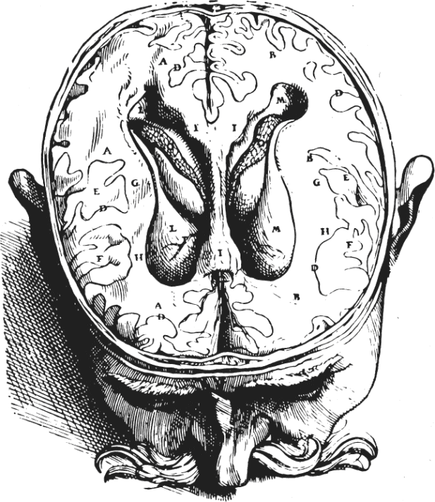
- Central nervous system (CNS)
- Brain + spinal cord. Encased in bone (cranium + vertebral column), bathed in CSF.
- Peripheral nervous system (PNS)
- Cranial + spinal nerves + autonomic ganglia outside CNS.
- Major brain divisions
- Telencephalon (cerebrum, basal ganglia) · diencephalon (thalamus, hypothalamus) · mesencephalon (midbrain) · metencephalon (pons, cerebellum) · myelencephalon (medulla).
- Cerebral cortex lobes
- Frontal (motor + executive), parietal (somatosensation, spatial), temporal (auditory + memory + face), occipital (visual). Insula, cingulate sit deeper.
- Anatomical planes
- Sagittal (left-right), coronal/frontal (front-back), horizontal/axial (top-bottom). Rostral=anterior, caudal=posterior.
- Gray vs white matter
- Gray = cell bodies + dendrites + synapses. White = myelinated axon tracts.
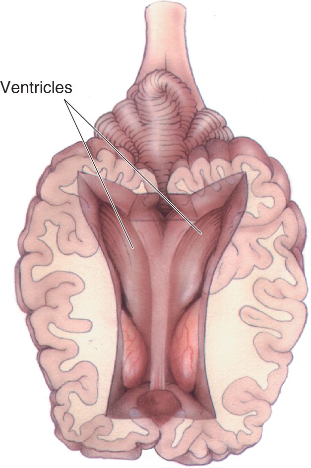
- Ventricles
- Lateral (×2) → third → cerebral aqueduct → fourth → central canal. Choroid plexus produces CSF.
U2 · Neurons + glia
📖 Bear · related chapters

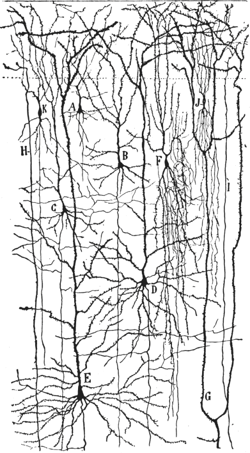
- Neuron doctrine
- Cajal: nervous system = discrete cells communicating across gaps (synapses). Defeated Golgi's reticular theory.
- Soma
- Cell body containing nucleus + organelles. Site of protein synthesis (Nissl bodies = stacks of rough ER).
- Dendrite
- Branched input region; receives synapses; spines on excitatory contacts.
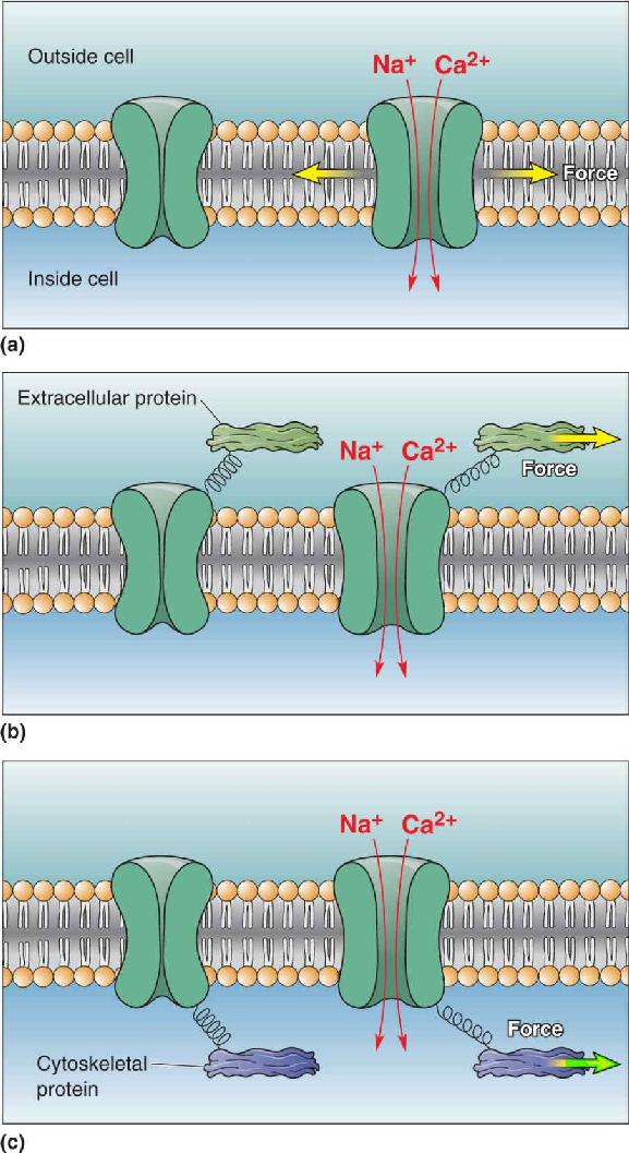
- Axon
- Output process. Single per neuron. Initiates action potential at axon hillock; conducts to terminals.
- Axon hillock / initial segment
- High density of voltage-gated Na⁺ channels — site of AP initiation.
- Neuron classifications
- By shape: unipolar, bipolar, multipolar. By function: sensory (afferent), motor (efferent), interneuron.
- Astrocyte
- Star-shaped glia. K⁺ buffering, glutamate uptake (EAAT), tripartite synapse, BBB end-feet, lactate shuttle to neurons.
- Oligodendrocyte
- CNS myelinator; one cell wraps multiple axons.
- Schwann cell
- PNS myelinator; one cell wraps one axon segment.
- Microglia [Chivero focus]
- CNS-resident immune cells. Phagocytose debris + dead cells; respond to injury + infection. Activated states: M1 pro-inflammatory vs M2 anti-inflammatory. Drive neuroinflammation in HIV, methamphetamine, neurodegeneration.
- Ependymal cells
- Ciliated cells lining ventricles + central canal. Choroid plexus produces CSF.
- Blood-brain barrier (BBB)
- Tight junctions between brain capillary endothelial cells (claudin-5, occludin, ZO-1) + astrocyte end-feet + pericytes. Excludes most polar/large molecules.
U3 · Membrane potential
📖 Bear · related chapters
- Ion gradients (typical mammalian neuron)
- Inside: high K⁺ (~140 mM), low Na⁺ (~10 mM), low Cl⁻ (~10 mM), low Ca²⁺ (~100 nM). Outside: opposite.
- Resting membrane potential
- ~−65 mV (range −60 to −80 mV in different neurons). Set primarily by K⁺ permeability through leak channels.
- Nernst equation
- E_ion = (RT/zF) ln([out]/[in]). At 37°C: E_K ~ −85 mV, E_Na ~ +60 mV, E_Cl ~ −65 mV, E_Ca ~ +120 mV.
- Goldman-Hodgkin-Katz equation
- V_m = (RT/F) ln[(P_K[K]_o + P_Na[Na]_o + P_Cl[Cl]_i) / (P_K[K]_i + P_Na[Na]_i + P_Cl[Cl]_o)]. Weighted by permeabilities.
- Why is V_m close to E_K?
- Resting membrane is most permeable to K⁺ (open leak K⁺ channels). V_m drifts toward whichever ion has the highest permeability.
- Na⁺/K⁺-ATPase
- Maintains gradients: 3 Na⁺ out + 2 K⁺ in per ATP. Electrogenic — contributes ~−5 to −10 mV directly.
- Equilibrium vs steady state
- At E_ion, no net flux for that ion (reversal potential). Resting V_m is steady state — pumps balance leakage.
U4 · Action potential
📖 Bear · related chapters

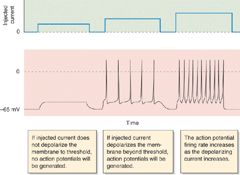
- Action potential definition
- All-or-none rapid depolarization (~100 mV swing) lasting ~1-2 ms; propagates without decrement along axon.
- Threshold
- ~−55 mV. Voltage-gated Na⁺ channel opening exceeds K⁺ leak → positive feedback → AP.
- Voltage-gated Na⁺ channel
- Three states: closed (resting), open (activated), inactivated (ball-and-chain). Inactivation explains absolute refractory period.
- Voltage-gated K⁺ channel
- Slower activation (delayed rectifier). Repolarizes membrane → afterhyperpolarization. No fast inactivation.
- Phases of AP
- (1) Rising: Na⁺ in. (2) Overshoot: peaks ~+30 to +40 mV. (3) Falling: Na⁺ inactivates, K⁺ out. (4) Undershoot/AHP: V_m below rest until K⁺ closes.
- Absolute refractory period
- ~1 ms; Na⁺ channels inactivated, no AP possible regardless of stimulus.
- Relative refractory period
- ~2-4 ms; some Na⁺ channels still inactivated + AHP — stronger stimulus required.
- Saltatory conduction
- AP "jumps" between nodes of Ranvier in myelinated axons. ~10-50× faster than unmyelinated of same diameter.
- Conduction velocity factors
- ↑ axon diameter → ↑ velocity (less internal resistance). Myelin → much faster (saltatory).
- Tetrodotoxin (TTX)
- Pufferfish toxin; blocks voltage-gated Na⁺ channels → no AP. Classic experimental tool.
U5 · Synaptic transmission
📖 Bear · related chapters

- Electrical synapse
- Gap junction (connexons) between cells. Bidirectional, fast, no delay. Coupling for synchronized firing.
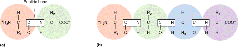
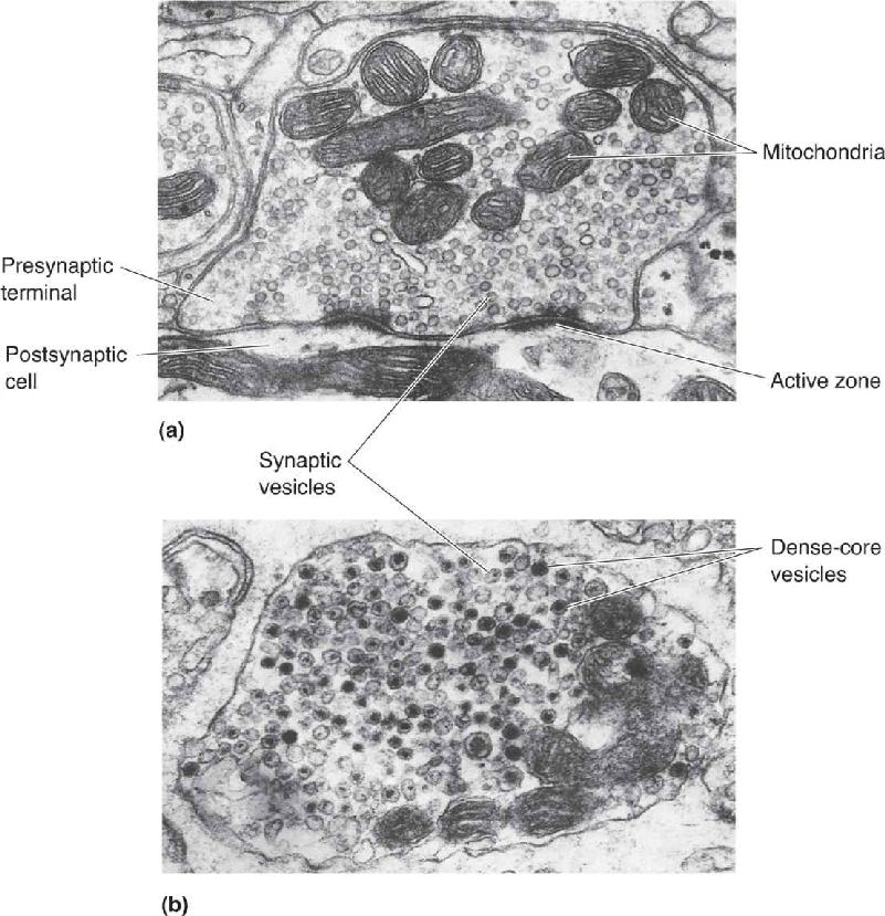
- Chemical synapse — sequence
- (1) AP arrives at terminal. (2) Voltage-gated Ca²⁺ channels open. (3) Ca²⁺ triggers vesicle fusion via SNAREs + synaptotagmin. (4) NT released into cleft. (5) Binds postsynaptic receptors. (6) Termination by reuptake/enzyme/diffusion.
- Vesicle fusion proteins
- v-SNARE synaptobrevin (VAMP) + t-SNAREs syntaxin + SNAP-25 form 4-helix bundle. Ca²⁺ sensor: synaptotagmin.
- EPSP
- Excitatory postsynaptic potential — depolarizing (e.g., glutamate → cation influx through AMPA/NMDA).
- IPSP
- Inhibitory postsynaptic potential — hyperpolarizing (e.g., GABA → Cl⁻ influx through GABA_A; or K⁺ efflux through GABA_B-coupled GIRK).
- Spatial vs temporal summation
- Spatial: multiple synapses simultaneously. Temporal: rapid trains from one synapse. Both bring the soma toward AP threshold.
- Ionotropic vs metabotropic receptor
- Ionotropic = ligand-gated ion channel (fast, ms). Metabotropic = GPCR → 2nd messenger (slow, seconds, modulatory).
- Long-term potentiation (LTP)
- Sustained increase in synaptic strength. Classic NMDA-dependent LTP in hippocampal CA1: Ca²⁺ through NMDA → CaMKII → AMPA insertion. Cellular basis of memory.
- Long-term depression (LTD)
- Sustained decrease in synaptic strength. Modest Ca²⁺ rise → phosphatases → AMPA internalization.
U6 · Neurotransmitters
📖 Bear · related chapters
- Glutamate
- Major excitatory NT in CNS. Receptors: AMPA (fast Na⁺/K⁺), NMDA (Ca²⁺, Mg²⁺ block, voltage-dependent), kainate, mGluRs.
- GABA
- Major inhibitory NT in CNS. Synthesized from glutamate by GAD. Receptors: GABA_A (Cl⁻ ionotropic), GABA_B (GPCR → K⁺ open + Ca²⁺ close).
- Glycine
- Inhibitory in spinal cord + brainstem. Cl⁻ channel. Strychnine antagonist.
- Acetylcholine (ACh)
- NMJ + autonomic + brain (cholinergic basal forebrain). Receptors: nicotinic (ionotropic, Na⁺/K⁺) + muscarinic (GPCR).
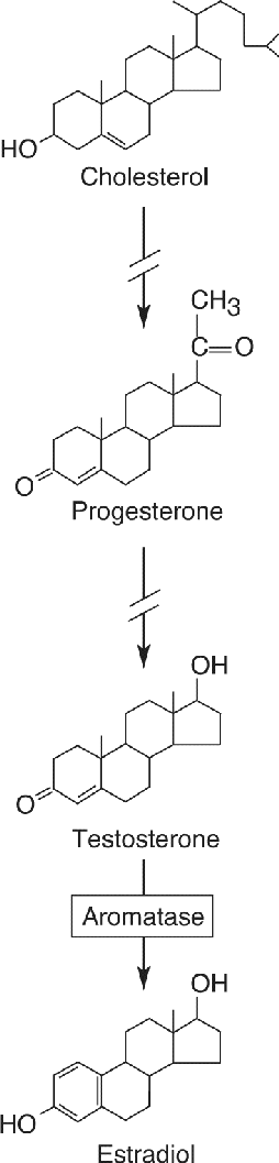
- Dopamine (DA)
- Motivation, reward, motor (substantia nigra → striatum). 5 receptor subtypes (D1-D5), all GPCRs. Implicated in Parkinson, addiction, schizophrenia.
- Norepinephrine (NE)
- Arousal, attention, autonomic. Locus coeruleus (CNS). α + β adrenergic receptors (GPCRs).
- Serotonin (5-HT)
- Mood, sleep, appetite. Raphe nuclei. ~14 receptor subtypes (mostly GPCR; 5-HT3 ionotropic).
- Histamine
- Wakefulness. Tuberomammillary nucleus. H1-H4 receptors.
- Endocannabinoids
- Retrograde messengers (anandamide, 2-AG). Activate presynaptic CB1 → reduce NT release.
- Nitric oxide (NO)
- Gas messenger, diffuses freely. Made by nNOS; activates soluble guanylyl cyclase → cGMP.
- Neuropeptides
- Larger NT (e.g., substance P, enkephalin, oxytocin, neuropeptide Y). Synthesized in soma, transported in dense-core vesicles, GPCR signaling.
U7 · Sensory + Somatosensory system
📖 Bear · related chapters
- Sensory transduction
- Conversion of stimulus energy into electrical signals (receptor potential).
- Receptor classes
- Mechanoreceptors (touch, hearing), thermoreceptors, photoreceptors, chemoreceptors, nociceptors (pain).
- Receptive field
- Region of stimulus space (skin area, retinal location) that affects a sensory neuron's firing.
- Adaptation
- Slowly adapting (SA) receptors fire continuously to maintained stimulus; rapidly adapting (RA) fire on changes.
- Touch receptors of glabrous skin
- Meissner (RA, fluttering touch) · Pacinian (RA, vibration deep) · Merkel (SA, pressure + form) · Ruffini (SA, skin stretch).
- Dorsal column–medial lemniscus pathway
- Fine touch, vibration, proprioception. 1st neuron → ipsilateral dorsal columns → gracile/cuneate nucleus (medulla) → DECUSSATES → medial lemniscus → VPL thalamus → S1 cortex.
- Spinothalamic (anterolateral) pathway
- Pain, temperature, crude touch. 1st neuron synapses in dorsal horn → DECUSSATES at spinal level → ascends contralaterally → VPL thalamus → S1.
- Sensory homunculus
- Distorted body map in S1 with overrepresentation of hands + face. Penfield's stimulation studies.
U8 · Pain & nociception
📖 Bear · related chapters
- Aδ fibers
- Thinly myelinated, fast (5-30 m/s); sharp, well-localized "first" pain.
- C fibers
- Unmyelinated, slow (0.5-2 m/s); dull, throbbing "second" pain; longer-lasting.
- TRPV1
- Capsaicin + heat (>43°C) receptor on nociceptors. Cation channel.
- Gate control theory (Melzack-Wall)
- Aβ touch fibers activate dorsal horn inhibitory interneurons → "gate" partially closes pain transmission. Why rubbing reduces pain.
- Periaqueductal gray (PAG)
- Midbrain center for descending pain modulation; activates raphe + locus coeruleus → spinal inhibition. Endogenous opioid system.
- Endogenous opioids
- Endorphins, enkephalins, dynorphins. Bind μ, δ, κ opioid receptors → presynaptic + postsynaptic inhibition of pain pathway.
- Hyperalgesia vs allodynia
- Hyperalgesia = exaggerated pain to noxious stimulus. Allodynia = pain from normally non-painful stimulus (light touch).
U9 · Vision
📖 Bear · related chapters
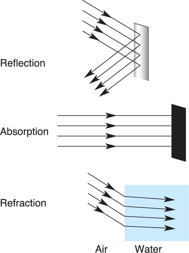

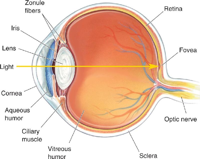
- Eye optics
- Cornea (~⅔ refraction) + lens (variable). Pupil = aperture; iris controls. Retina at back has receptors.
- Photoreceptors
- Rods (high sensitivity, low resolution, peripheral, scotopic) · cones (low sens, high res, central, photopic, color). 3 cone types (S/M/L; "blue/green/red").
- Phototransduction
- Dark: cGMP holds CNG channel open → Na⁺/Ca²⁺ in → photoreceptor depolarized → glutamate released. Light: rhodopsin → transducin → PDE → ↓ cGMP → channel closes → hyperpolarization → ↓ glutamate.
- Retinal cell layers
- Photoreceptor → bipolar → ganglion (output). Horizontal + amacrine = lateral interactions. Light enters from ganglion side.
- Center-surround receptive field
- ON-center: light in center excites, surround inhibits. OFF-center: opposite. Computed by horizontal cell lateral inhibition.
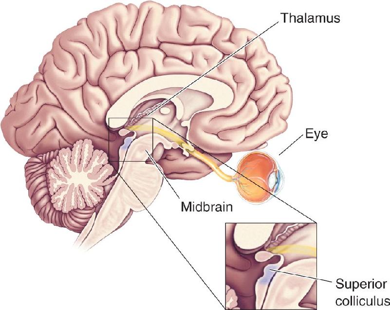
- Retinal ganglion cell axons
- Optic nerve → optic chiasm (decussation of nasal fibers) → optic tract → LGN of thalamus → V1 (primary visual cortex).
- Magnocellular vs parvocellular
- M: large, fast, motion + low contrast. P: small, slow, color + form, high acuity. Parallel processing.
- Dorsal vs ventral stream
- Dorsal "where/how" = parietal, motion + spatial. Ventral "what" = temporal, object + face recognition.
U10 · Audition + vestibular
📖 Bear · related chapters
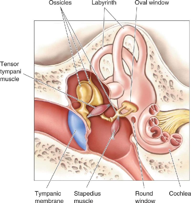

- Outer ear
- Pinna, ear canal → tympanic membrane (eardrum).
- Middle ear
- Ossicles malleus → incus → stapes (oval window). Impedance matching air → fluid (×22 amplification).
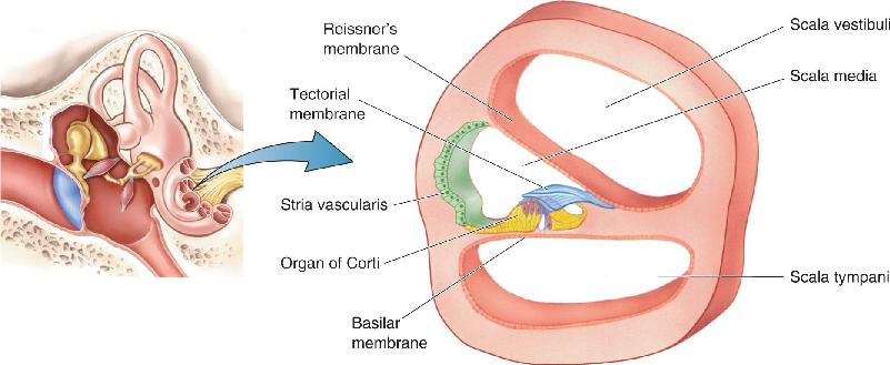
- Cochlea
- Spiral fluid-filled tube (3 chambers: scala vestibuli, scala media, scala tympani). Basilar membrane runs length.
- Organ of Corti
- On basilar membrane. Inner hair cells (sensory; ~3,500) + outer hair cells (motile, amplify; ~12,000). Tectorial membrane on top.
- Tonotopic organization
- Base of cochlea = high frequency; apex = low frequency. Maintained through auditory pathway up to A1 cortex.
- Mechanotransduction
- Stereocilia bending → tip-link tension → mechanically gated cation channel opens → K⁺ + Ca²⁺ in → depolarize hair cell → glutamate to spiral ganglion neurons.
- Auditory pathway
- Hair cell → spiral ganglion → cochlear nucleus → superior olive (sound localization) → inferior colliculus → MGN of thalamus → A1 (Heschl's gyrus).
- Sound localization
- Interaural time difference (ITD; low freq, medial superior olive) + interaural level difference (ILD; high freq, lateral superior olive).
- Vestibular system
- Semicircular canals (3, angular acceleration via cupula + ampulla) + otolith organs (utricle + saccule, linear accel + gravity via otoconia on macula). Hair cells transduce.
U11 · Chemical senses
📖 Bear · related chapters
- Olfactory receptor neurons
- Bipolar neurons in nasal epithelium. Cilia have GPCR olfactory receptors → G_olf → AC → cAMP → CNG channel → depolarization.
- OR gene family
- ~400 functional ORs in humans (largest gene family). Each ORN expresses one OR.
- Olfactory glomerulus
- All ORNs expressing the same OR converge on ~2 glomeruli in olfactory bulb. Mitral cells → piriform cortex (no thalamus relay!).
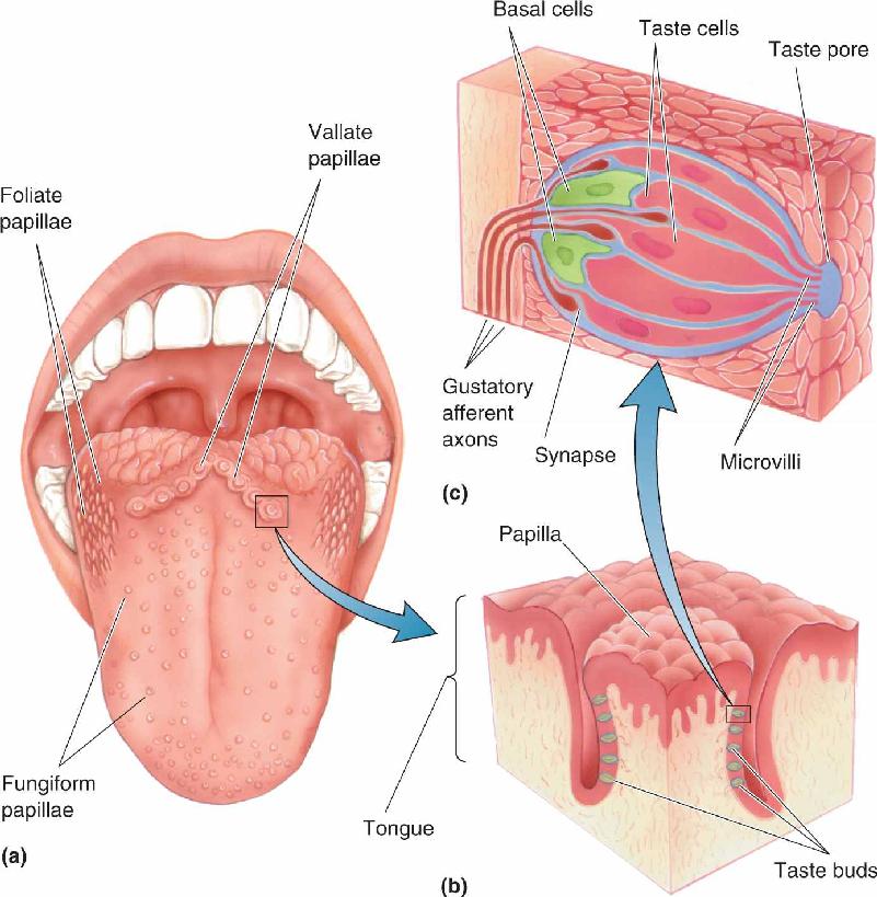
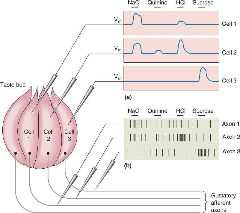
- 5 taste modalities
- Sweet, salty, sour, bitter, umami. Sweet/bitter/umami via GPCRs (T1R, T2R) → α-gustducin. Salty + sour via ion channels (ENaC, TRP).
- Taste pathway
- Taste bud → CN VII (anterior 2/3 tongue), IX (posterior 1/3), X (epiglottis) → solitary nucleus (medulla) → VPM thalamus → gustatory cortex (insula).
U12 · Motor systems intro
📖 Bear · related chapters
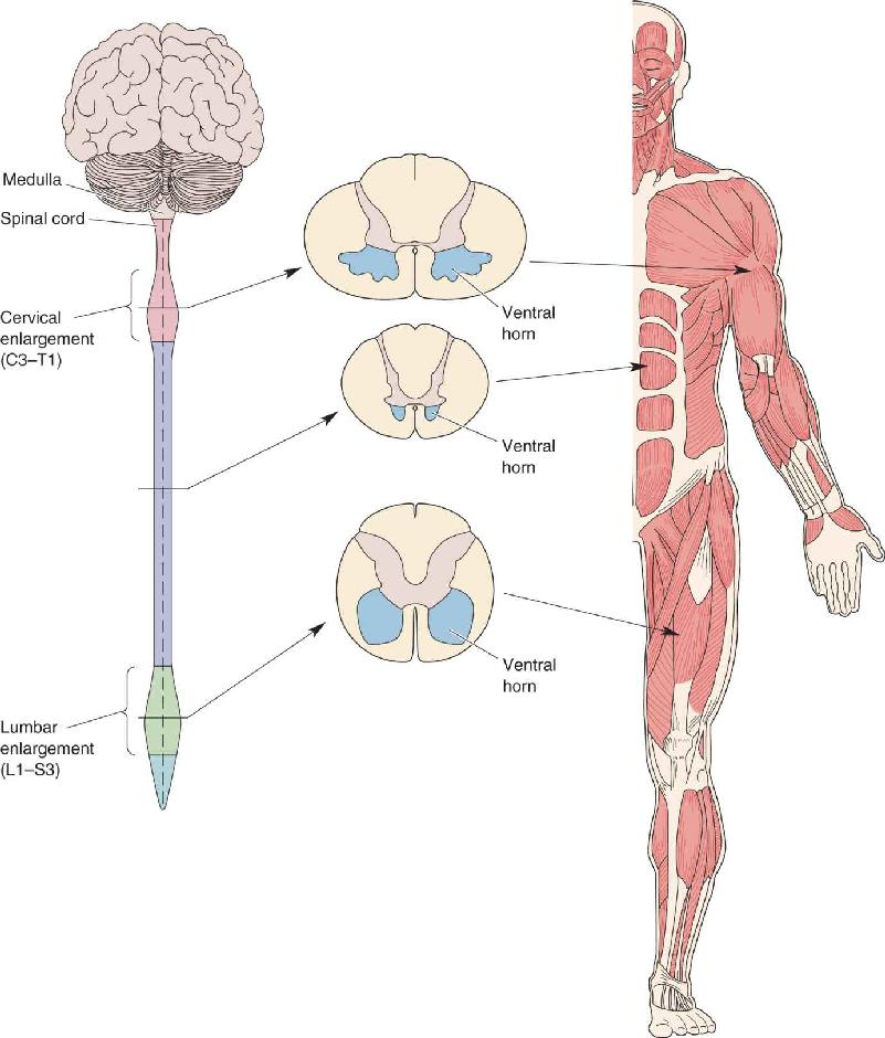

- Lower motor neuron (LMN)
- Final common pathway: cell body in ventral horn or brainstem motor nuclei → axon → muscle. Lesion → flaccid paralysis, atrophy, fasciculations.
- Upper motor neuron (UMN)
- Originates in motor cortex; descends via corticospinal tract; synapses on LMN. Lesion → spastic paralysis, hyperreflexia, Babinski sign.
- Motor unit
- One LMN + all muscle fibers it innervates. Size principle: smaller units recruited first.
- Stretch reflex (myotatic)
- Muscle spindle (Ia afferent) → monosynaptic excitation of homonymous motor neuron + reciprocal inhibition of antagonist via Ia interneuron. Knee jerk.
- Golgi tendon organ reflex
- Ib afferent senses tension → inhibits its own motor neuron. Protects against overload.
- Withdrawal reflex
- Polysynaptic; flexor activation + crossed extension contralateral.
- Corticospinal tract
- M1 → internal capsule → cerebral peduncle → medulla pyramids → DECUSSATES → lateral corticospinal tract → ventral horn LMN. Lateral = limbs; ventral = trunk.
- Basal ganglia
- Caudate, putamen, globus pallidus, subthalamic, substantia nigra. Direct (D1, GO) + indirect (D2, NO-GO) pathways. Parkinson = SNc dopamine loss; Huntington = caudate degeneration.
- Cerebellum
- Coordination, balance, motor learning. Three peduncles. Purkinje cell GABAergic output to deep cerebellar nuclei. Lesions → ataxia, dysmetria.
U13 · Glia & neuroinflammation — Chivero focus
📖 Bear · related chapters
- Microglia origin
- Yolk-sac-derived; resident macrophages of CNS. Distinct from infiltrating monocytes.
- Microglial activation states
- "M1" pro-inflammatory (TNFα, IL-1β, ROS) vs "M2" anti-inflammatory/restorative (IL-10, TGF-β). Spectrum, not binary.
- NLRP3 inflammasome
- Cytosolic multiprotein complex. Two-signal activation: priming (TLR → NF-κB → upregulates components) + activation (DAMPs/PAMPs → assembly). Recruits ASC + procaspase-1 → cleaves IL-1β + IL-18 + gasdermin D → pyroptosis. Chivero's research target.
- HIV-Tat in neuroinflammation
- HIV-1 Trans-activator of transcription crosses BBB; activates microglia; primes NLRP3; contributes to HAND (HIV-associated neurocognitive disorder) even on suppressive ART. Chivero's research.
- Methamphetamine + microglia
- Meth crosses BBB, activates microglia + induces oxidative stress + NLRP3 priming. Synergistic with HIV in dual-exposed individuals. Chivero's research.
- Astrocyte tripartite synapse
- Astrocyte processes ensheath synapses; take up glutamate (EAAT1/2), release gliotransmitters (glutamate, ATP, D-serine). Buffer K⁺.
- Reactive astrocytosis
- Astrocytes upregulate GFAP, hypertrophy, form glial scar after injury. A1 (neurotoxic) vs A2 (neuroprotective) phenotypes.
- Microglial pruning
- Complement-tagged synapses (C1q, C3) phagocytosed by microglia during development + aging + Alzheimer.
- Neurodegeneration + microglia
- Alzheimer (TREM2, complement), Parkinson (α-syn-activated), ALS, MS — chronic microglial activation contributes to pathology.
U14 · Pharmacology & drugs of abuse
📖 Bear · related chapters
- Agonist vs antagonist
- Agonist binds + activates receptor. Antagonist binds + blocks. Inverse agonist reduces constitutive activity.
- Allosteric modulator
- Binds non-orthosteric site → potentiates (PAM) or inhibits (NAM) agonist effect. Benzodiazepines = PAM at GABA_A.
- Mesolimbic dopamine pathway
- VTA → nucleus accumbens (NAc) + PFC. Final common reward circuit; activated by virtually all addictive drugs.
- Cocaine
- Blocks DAT, NET, SERT → ↑ synaptic monoamines. Strong reinforcer via NAc DA.
- Amphetamine + methamphetamine
- Reverses DAT (efflux), enters vesicles displacing DA. Massive ↑ extracellular DA. Neurotoxic at high doses (oxidative stress, microglia).
- Opioids
- μ receptor agonists → presynaptic Ca²⁺ ↓ + postsynaptic K⁺ ↑ → inhibition. Analgesia + euphoria + respiratory depression.
- Alcohol
- Enhances GABA_A + inhibits NMDA → sedation. Chronic → withdrawal hyperexcitability + addiction.
- Nicotine
- Nicotinic ACh receptor agonist; α4β2 on VTA DA neurons → NAc reward.
- Cannabis (THC)
- CB1 agonist; presynaptic; reduces NT release. Affects memory (hippocampus), motor (basal ganglia), reward.
- Tolerance
- Reduced response after repeated exposure. Pharmacodynamic (receptor downregulation) + pharmacokinetic (faster metabolism).
- Sensitization
- Increased response with repeated exposure (especially psychomotor stimulants).
- Addiction circuits
- VTA → NAc + amygdala + PFC; loss of top-down PFC control + amygdala stress dysregulation. Chivero's lab studies SUD-microglia interactions.
U15 · Methods + integration
📖 Bear · related chapters
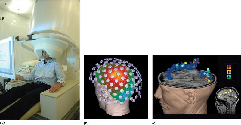
- EEG
- Scalp electrodes record summed dendritic potentials. Excellent temporal (ms), poor spatial. Frequency bands: δ, θ, α, β, γ.
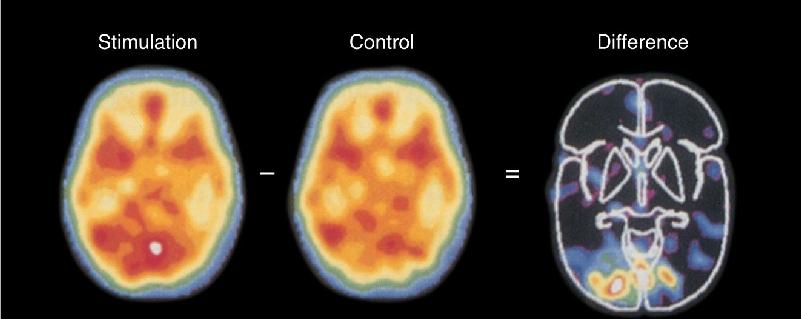
- fMRI
- BOLD signal — blood oxygenation. Good spatial (~mm), poor temporal (~seconds). Indirect measure of activity.
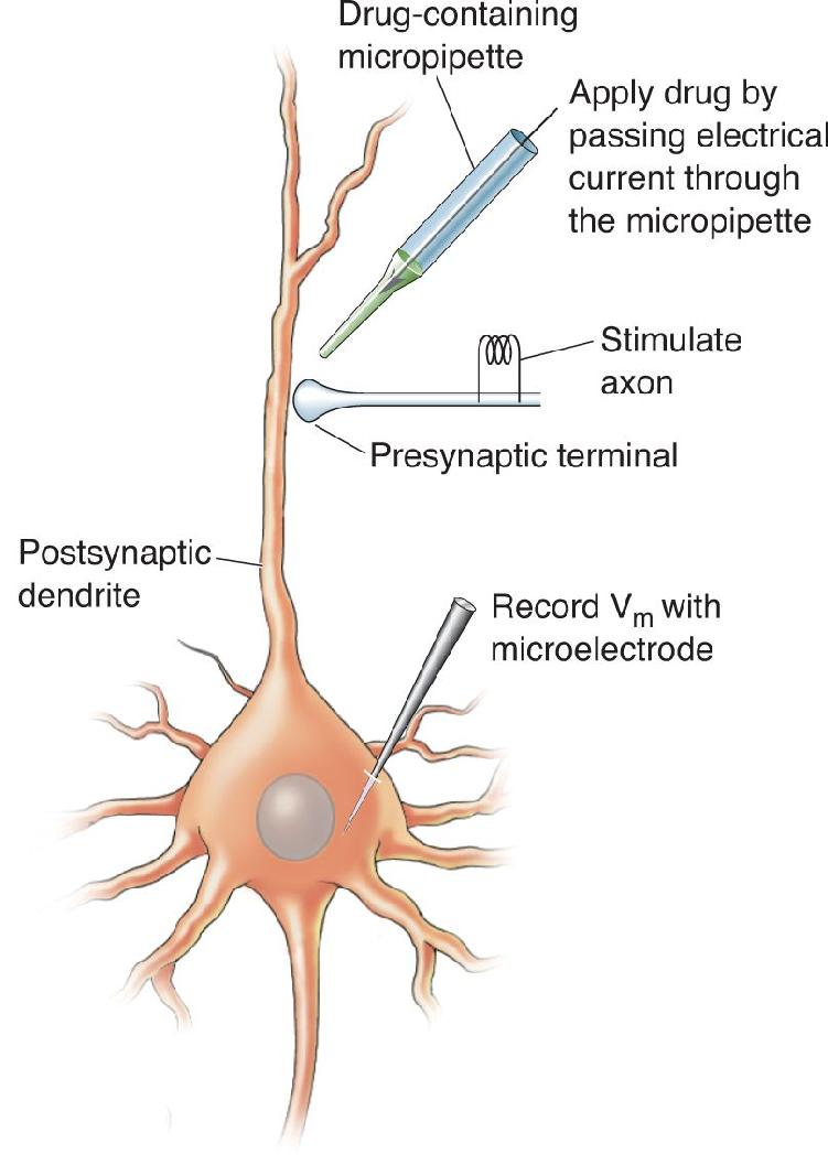
- Patch clamp
- Glass pipette suction onto cell membrane → record currents from individual ion channels (single-channel) or whole cell.
- Intracellular vs extracellular recording
- Intra: pipette inside cell; measures V_m + APs + synaptic potentials. Extra: outside; only spikes resolved.
- Optogenetics
- Express channelrhodopsin (ChR2, Na⁺ in, excitation) or halorhodopsin (Cl⁻ in, inhibition) → light-triggered control of neurons in vivo.
- Chemogenetics (DREADDs)
- Designer GPCRs (hM3Dq, hM4Di) activated by clozapine-N-oxide → minute-scale modulation.
- Calcium imaging
- GCaMP fluoresces with Ca²⁺. Bulk or single-cell readout of activity over time.
- Lesion studies
- Loss of function via surgical, chemical, or pharmacological ablation → infer normal role.
Chivero-targeted exam tips
- Be ready to diagram an action potential with phases + ion movements + channel states.
- Know NLRP3 inflammasome: priming → activation → caspase-1 → IL-1β + gasdermin D → pyroptosis.
- For each NT system: synthesis enzyme, source nucleus, receptor types, behavioral role.
- Sensory pathways — DC-ML vs spinothalamic vs visual — be able to draw decussation point + relay nuclei.
- Watch for Chivero's HIV-Tat / methamphetamine / microglia framing on standard topics.
📚 Textbook companion · Bear Neuroscience Enhanced
Each unit above maps to chapters in the locally-OCR'd Bear Neuroscience Enhanced. Use the cards below as a quick visual jump into the embedded textbook reader — one figure per chapter, click to read the full chapter:
CH 01
Neuroscience: Past, Present, and Future
CH 02
Neurons and Glia
CH 03
The Neuronal Membrane at Rest
CH 04
The Action Potential
CH 05
Synaptic Transmission
CH 06
Neurotransmitter Systems
CH 07
The Structure of the Nervous System
CH 08
The Chemical Senses
CH 09
The Eye
CH 10
The Central Visual System
CH 11
The Auditory and Vestibular Systems
CH 12
The Somatic Sensory System

CH 13
Spinal Control of Movement
CH 14
Brain Control of Movement
CH 15
Chemical Control of the Brain and Behavior
CH 16
Motivation
CH 17
Sex and the Brain
CH 18
Brain Mechanisms of Emotion
CH 19
Brain Rhythms and Sleep
CH 20
Language
CH 21
-One The Resting Brain, Attention, and Consciousness
CH 22
-Two Mental Illness
CH 23
-Three Wiring the Brain
CH 24
-Four Memory Systems
CH 25
-five Molecular Mechanisms of Learning and Memory