U1 · Cells, model organisms & microscopy
📖 Lodish 9e · related chapters

- Cell theory
- All organisms are made of cells; cells are the basic units of life; cells arise from preexisting cells (Schleiden, Schwann, Virchow).
- Prokaryotes vs eukaryotes
- Prokaryotes lack a nucleus and membrane organelles; eukaryotes have both. Eukaryotic cells originated by endosymbiosis (mitochondria from α-proteobacteria, chloroplasts from cyanobacteria).
- Model organisms
- E. coli (bacteria), S. cerevisiae (yeast), C. elegans (worm — Karen Kim Guisbert uses this), Drosophila, zebrafish, mouse, human cell culture lines (HeLa, HEK293).
- Light microscopy
- Resolution limit ~200 nm (Abbe). Bright-field, phase contrast, DIC, fluorescence (matters for Johnson's lab).
- Confocal microscopy
- Pinhole excludes out-of-focus light → optical sections of thick samples.
- Super-resolution microscopy
- STED, STORM, PALM beat the diffraction limit (~20-50 nm).
- Electron microscopy
- TEM (transmission, ~0.1 nm) and SEM (surface, ~1 nm). Requires fixation, sections.
- Immunofluorescence (IF)
- Primary antibody binds antigen; fluorescent secondary antibody binds primary. Used to localize proteins in cells.
U2 · Cell chemistry & biosynthesis
📖 Lodish 9e · related chapters
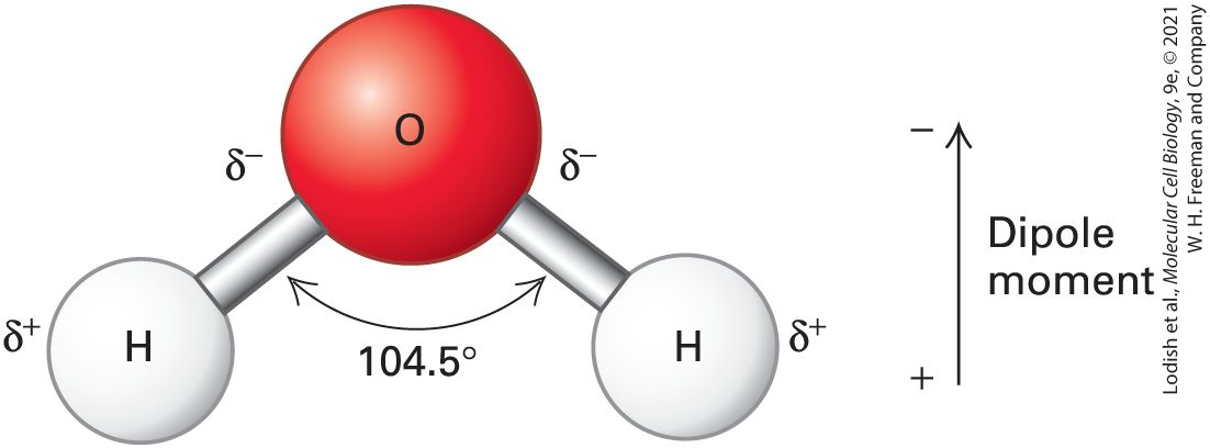
- Water properties
- Polar; H-bonds give high specific heat, surface tension, cohesion. Hydrophobic effect drives folding + membrane assembly.
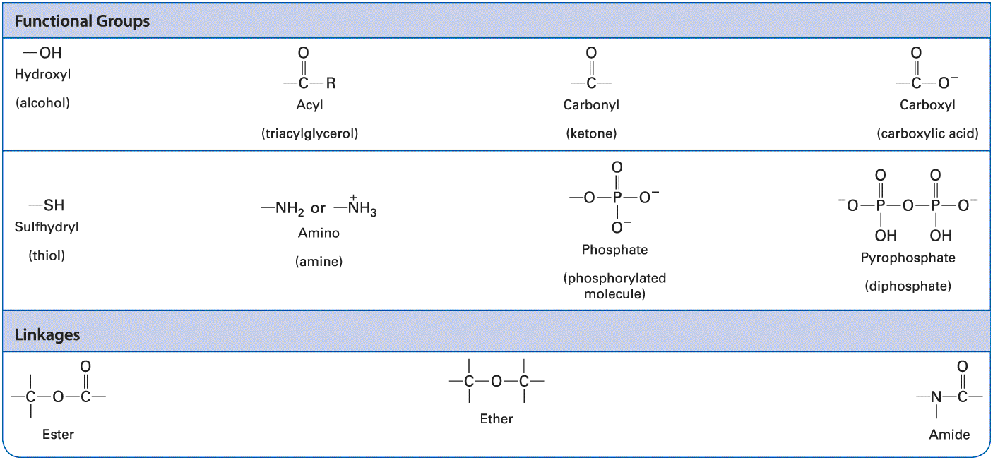
- Macromolecules
- Proteins (AA), nucleic acids (nucleotides), polysaccharides (sugars), lipids (FAs + glycerol/sterol).
- ΔG (free energy change)
- ΔG < 0 = spontaneous (exergonic); ΔG > 0 = nonspontaneous (endergonic). Cells couple endergonic to exergonic via ATP hydrolysis.
- ATP
- Energy currency. Hydrolysis of γ-phosphate releases ~−7.3 kcal/mol under standard conditions; much more in cells.
- Enzyme catalysis
- Lower activation energy. Michaelis-Menten: v = Vmax[S] / (Km + [S]). Km = [S] at half Vmax.
- Allosteric regulation
- Effector binds non-active site, changes conformation + activity. Foundation of cell-signaling switches.
U3 · Proteins
📖 Lodish 9e · related chapters
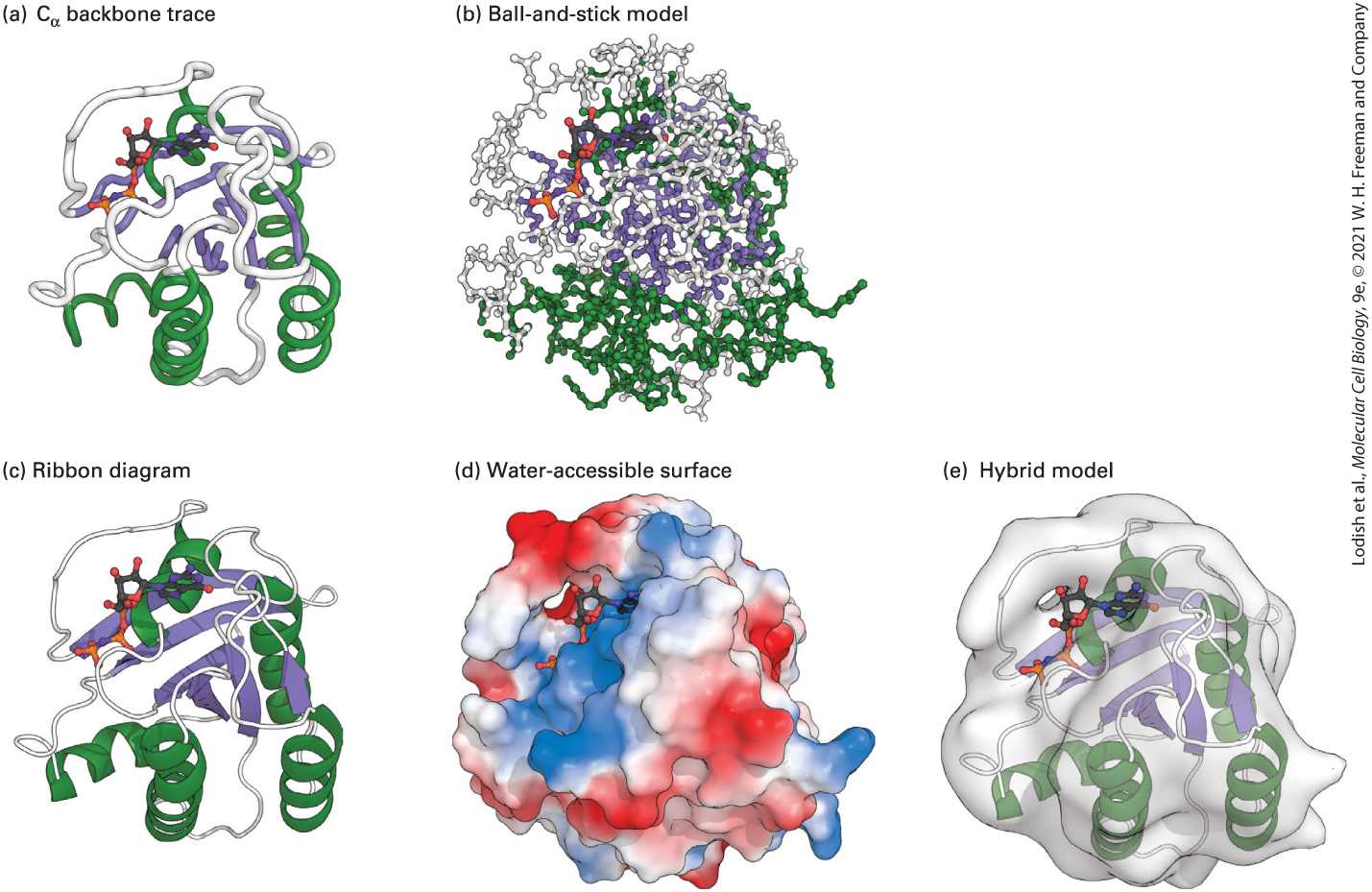
- Primary structure
- Amino acid sequence (peptide bonds).
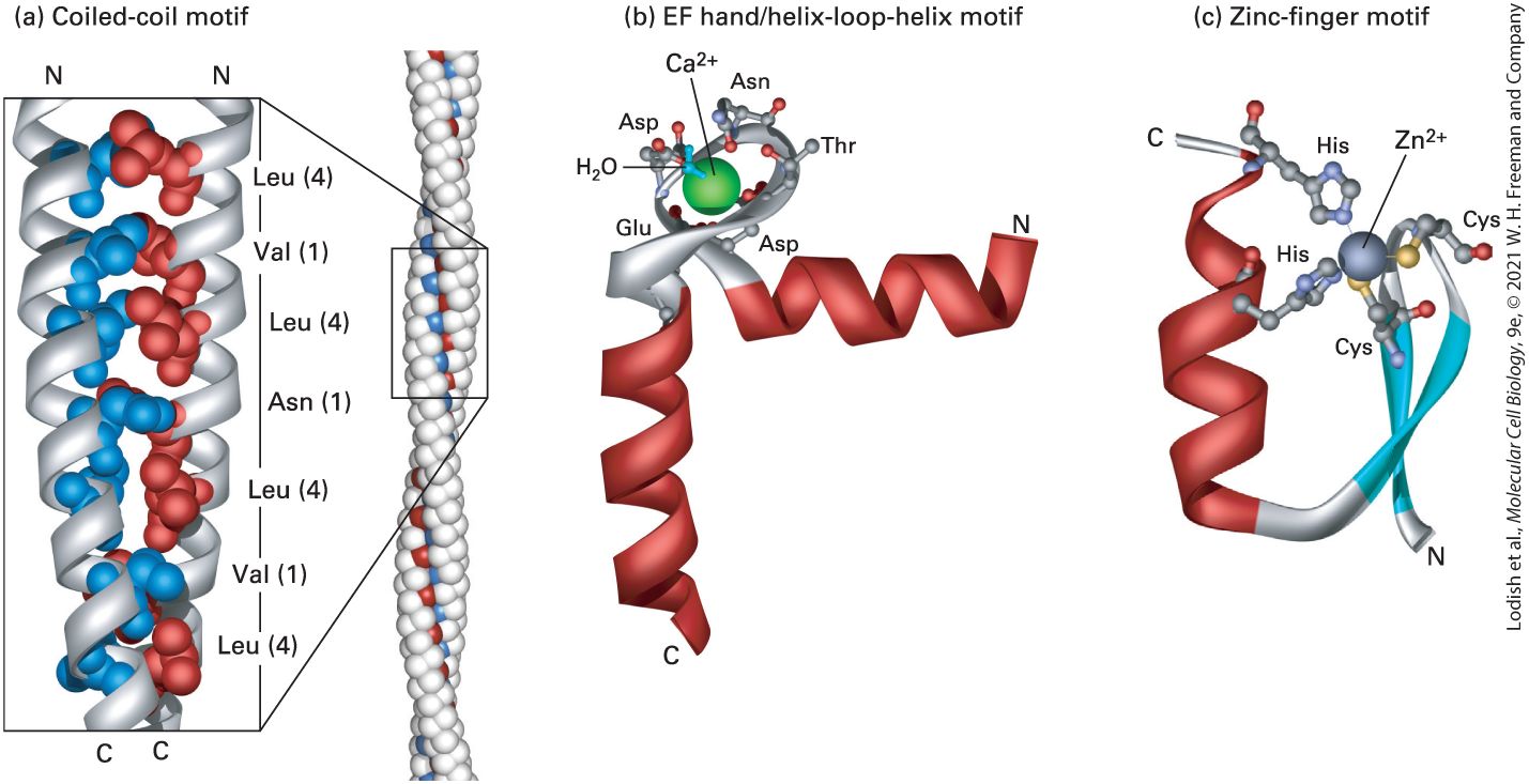
- Secondary structure
- Local folds — α-helix, β-sheet — stabilized by backbone H-bonds.
- Tertiary structure
- 3D fold of one polypeptide; stabilized by H-bonds, ionic, hydrophobic interactions, disulfides.
- Quaternary structure
- Multi-subunit assembly (hemoglobin = 2α + 2β).
- Chaperone
- Hsp70, Hsp90, GroEL/ES — assist folding; rescue misfolded proteins.
- Ubiquitin-proteasome
- Tags damaged/regulated proteins with poly-Ub chain → 26S proteasome degrades.
- Motor proteins
- Myosin (actin), kinesin + dynein (microtubules); ATP-driven directional movement.
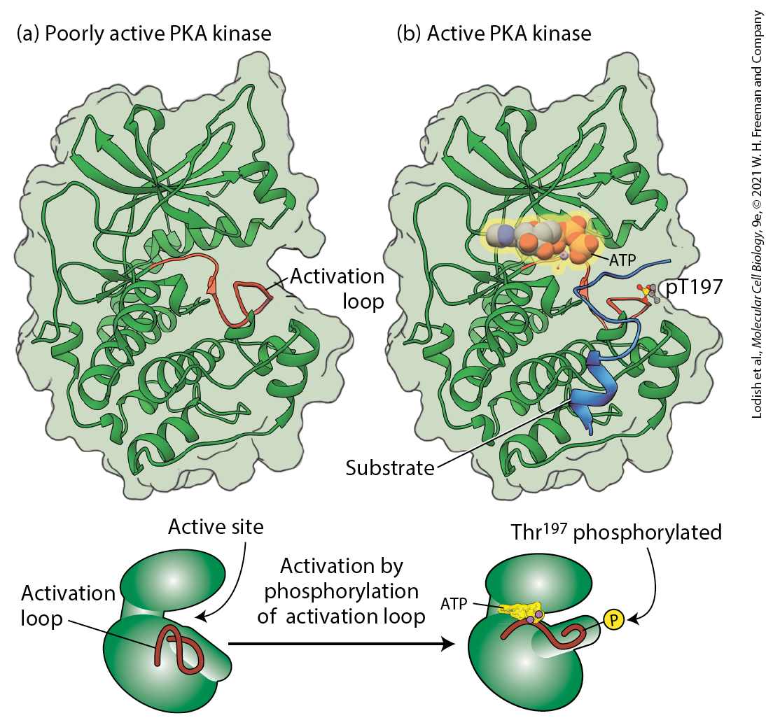
- Phosphorylation
- Kinase adds phosphate (Ser/Thr/Tyr), phosphatase removes. Most common reversible regulatory PTM.
U4 · DNA, chromosomes & replication
📖 Lodish 9e · related chapters

- DNA structure
- Antiparallel double helix; AT (2 H-bonds), GC (3). 10.5 bp/turn, major + minor grooves.
- DNA replication
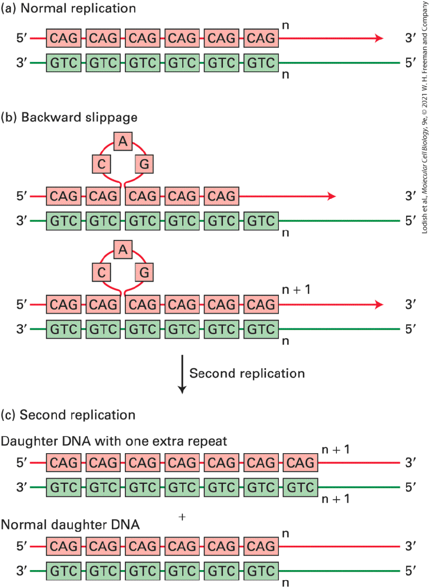
- Semiconservative. Helicase unwinds, primase lays RNA primer, DNA pol III extends 5'→3'. Leading vs lagging strand (Okazaki fragments). Telomerase extends telomeres.
- DNA repair
- Mismatch repair (MMR), base excision (BER), nucleotide excision (NER), homologous recombination (HR), non-homologous end joining (NHEJ).
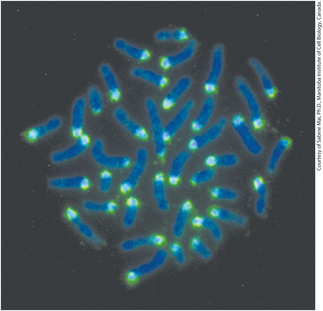
- Chromosome structure
- DNA + histones = nucleosome (~146 bp around 8 histones). 30-nm fiber → loops → chromatid in mitosis.
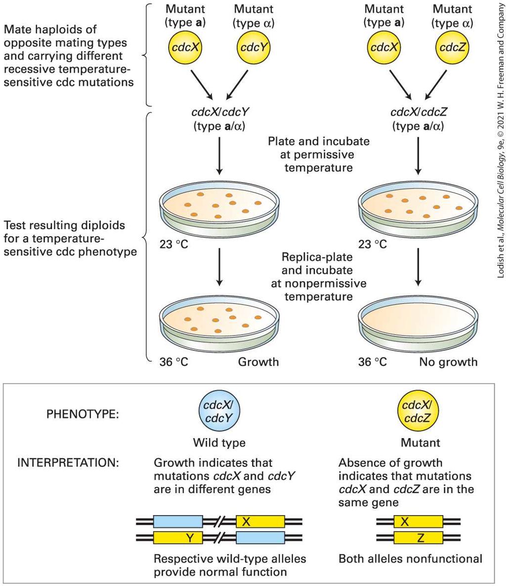
- Centromere / kinetochore
- Heterochromatic region where spindle attaches via kinetochore protein complex.
- Telomere
- (TTAGGG)n caps; shortens with each replication. Telomerase active in germ + stem + many cancer cells.
U5 · Gene expression
📖 Lodish 9e · related chapters
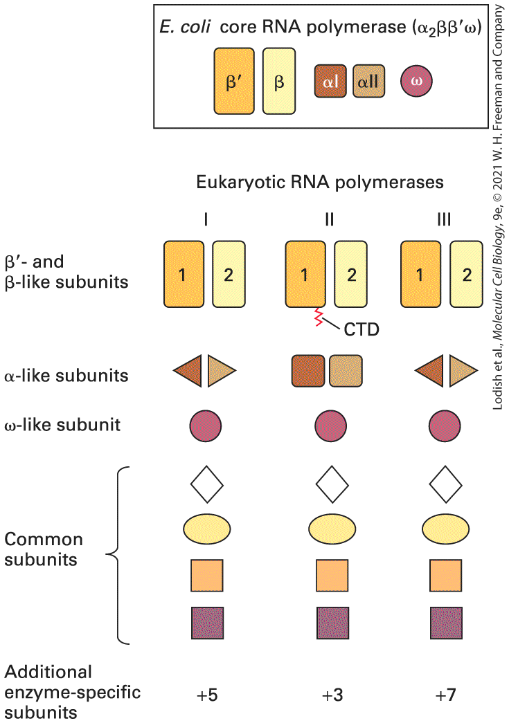
- Transcription
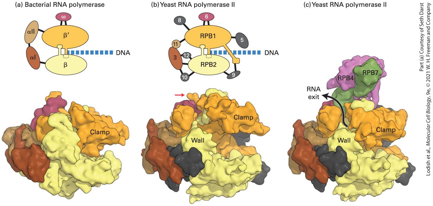
- RNA pol II makes mRNA from DNA template. Promoter (TATA, etc.) + general TFs + enhancers + activators.
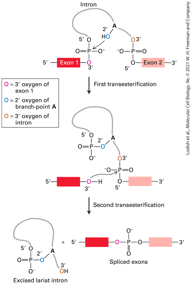
- RNA processing
- 5' cap (m7G), poly-A tail, splicing (spliceosome removes introns; alternative splicing diversifies proteome).
- Translation
- Ribosome reads mRNA codons via tRNA anticodons. Initiation (Met-tRNA, eIFs) → elongation → termination (release factors at stop codon).
- Genetic code
- Triplet, redundant (degenerate), nearly universal. Wobble at 3rd codon position.
- Transcription factor (TF)
- DNA-binding protein that activates/represses transcription (e.g., p53, Myc, NF-κB).
- Epigenetic marks
- DNA methylation (CpG), histone modifications (H3K4me3 active, H3K27me3 repressive, H3K9ac active).
- miRNA
- ~22 nt; binds 3'UTR → translational repression or mRNA decay (RISC complex).
U6 · Membranes & transport
📖 Lodish 9e · related chapters

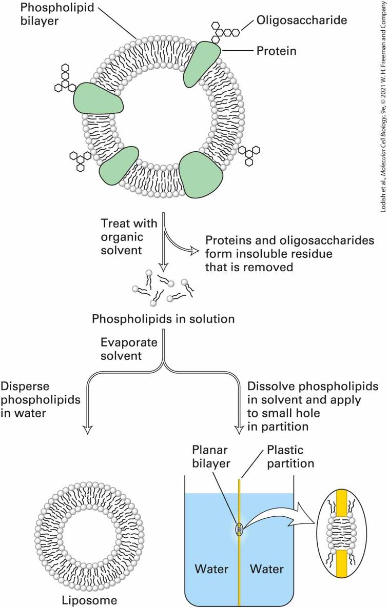
- Lipid bilayer
- Amphipathic phospholipids; fluid mosaic. Cholesterol modulates fluidity; sphingolipids cluster in lipid rafts.
- Membrane proteins
- Integral (transmembrane), peripheral (cytosolic-facing), lipid-anchored (GPI-anchored, prenylated).
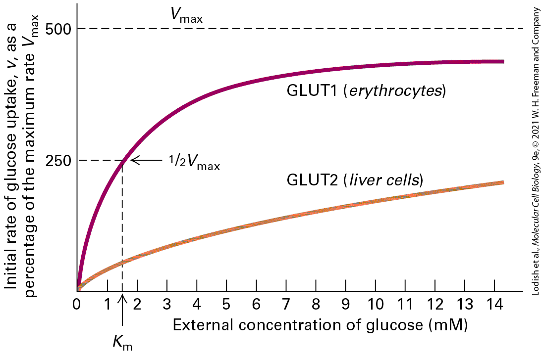
- Passive transport
- Diffusion (down gradient, no ATP); facilitated diffusion through channels or carriers.
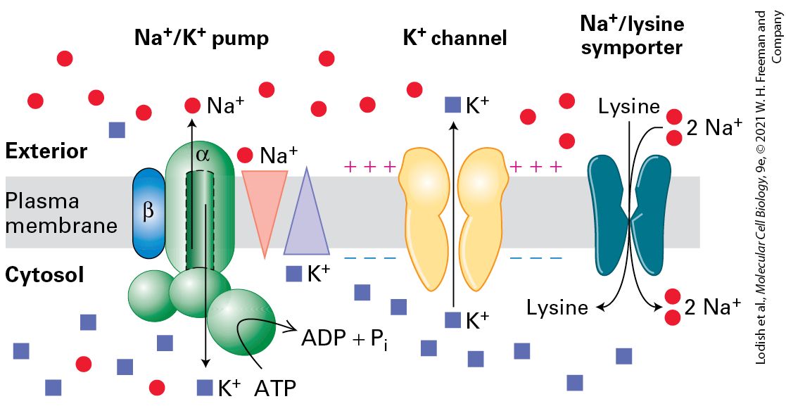
- Active transport
- Against gradient, requires ATP. Primary: Na⁺/K⁺-ATPase, Ca²⁺-ATPase, H⁺-ATPase. Secondary: symport/antiport coupled to ion gradient.
- Na⁺/K⁺-ATPase
- 3 Na⁺ out, 2 K⁺ in per ATP. Sets resting potential, drives secondary transport.
- Aquaporin
- Water-selective channel; key in kidney, RBCs.
- Action potential prep
- Voltage-gated channels open in response to depolarization. (Cross-reference NEUR 1520 U4!)
U7 · Internal compartments & sorting
📖 Lodish 9e · related chapters
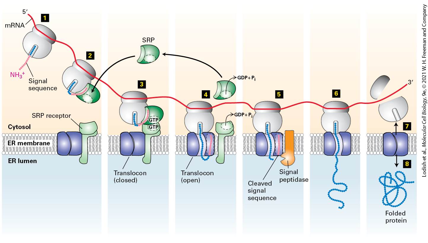
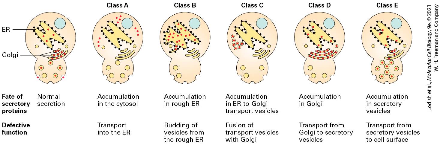
- ER (rough)
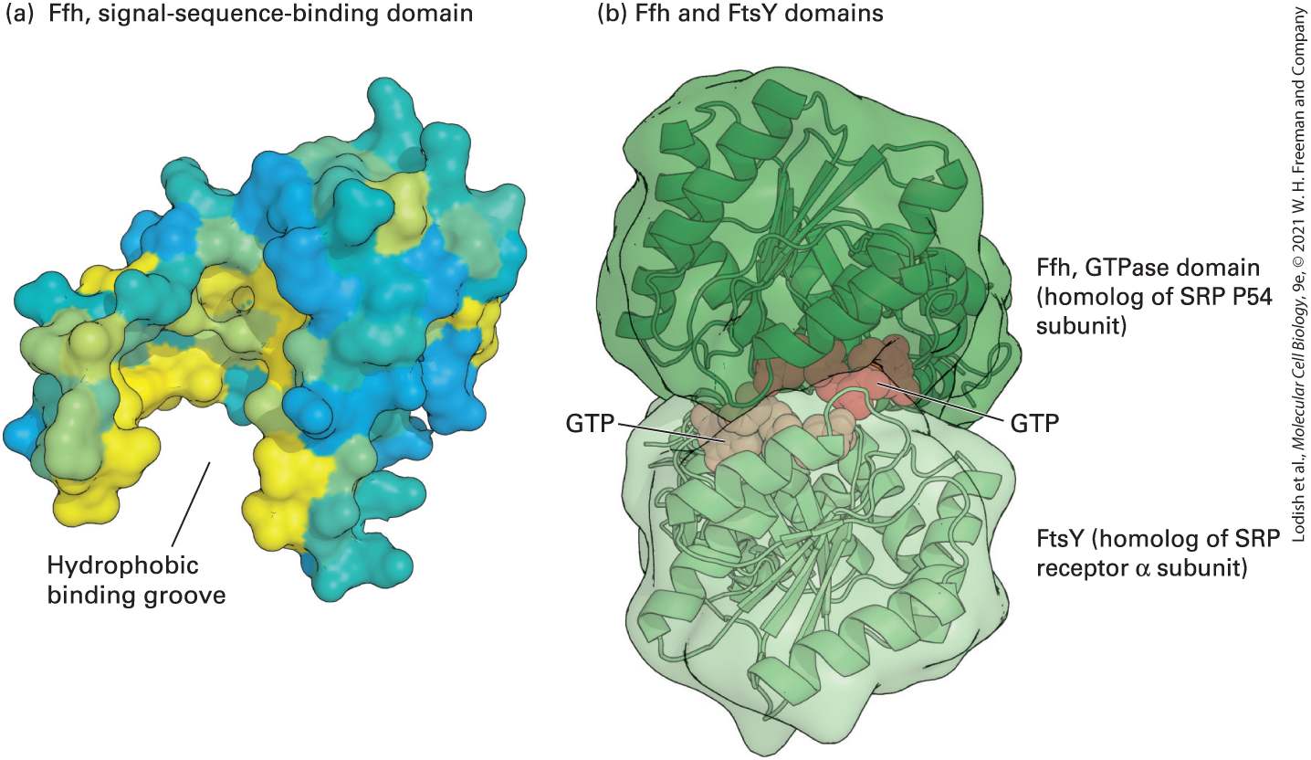
- Studded with ribosomes; site of secretory + membrane protein synthesis. Co-translational import via SRP + signal sequence.
- ER (smooth)
- Lipid synthesis, Ca²⁺ storage, detox (P450 enzymes in liver).
- Golgi apparatus
- Cis → medial → trans cisternae. Modifies glycoproteins, sorts to lysosomes / plasma / secretion.
- Lysosome
- Acidic (pH ~4.5) hydrolytic compartment; hydrolases tagged with mannose-6-phosphate.
- Peroxisome
- β-oxidation of very-long-chain fatty acids; H₂O₂ neutralized by catalase.
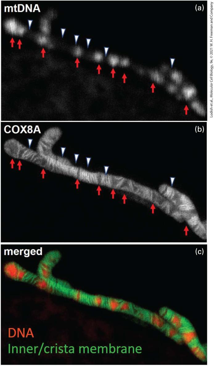
- Mitochondrion
- Double membrane; matrix has TCA cycle + mtDNA; inner membrane houses ETC + ATP synthase. Imports nuclear-encoded proteins post-translationally via TOM/TIM.
- Nuclear localization signal (NLS)
- Lys/Arg-rich sequence recognized by importin α/β; transport through nuclear pore.
U8 · Vesicle traffic
📖 Lodish 9e · related chapters
- COPII
- ER → Golgi anterograde traffic; Sar1 GTPase coordinates assembly.
- COPI
- Golgi → ER (retrograde) and intra-Golgi; Arf1 GTPase.
- Clathrin
- Plasma membrane endocytosis + Golgi → endosome. Adaptor proteins (AP1/2) link cargo to coat.
- SNARE
- v-SNARE (vesicle) + t-SNARE (target) zip into 4-helix bundle → membrane fusion. Specificity for compartment pairing.
- Endocytosis types
- Phagocytosis (large particles, immune cells), pinocytosis (fluid uptake), receptor-mediated endocytosis (LDL, transferrin).
- Exocytosis
- Constitutive (continuous) or regulated (Ca²⁺-triggered, e.g., insulin secretion, neurotransmitters).
U9 · Mitochondrial energy conversion
📖 Lodish 9e · related chapters
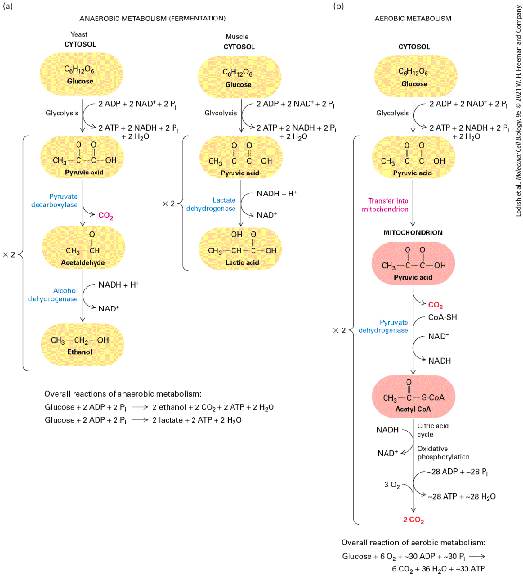

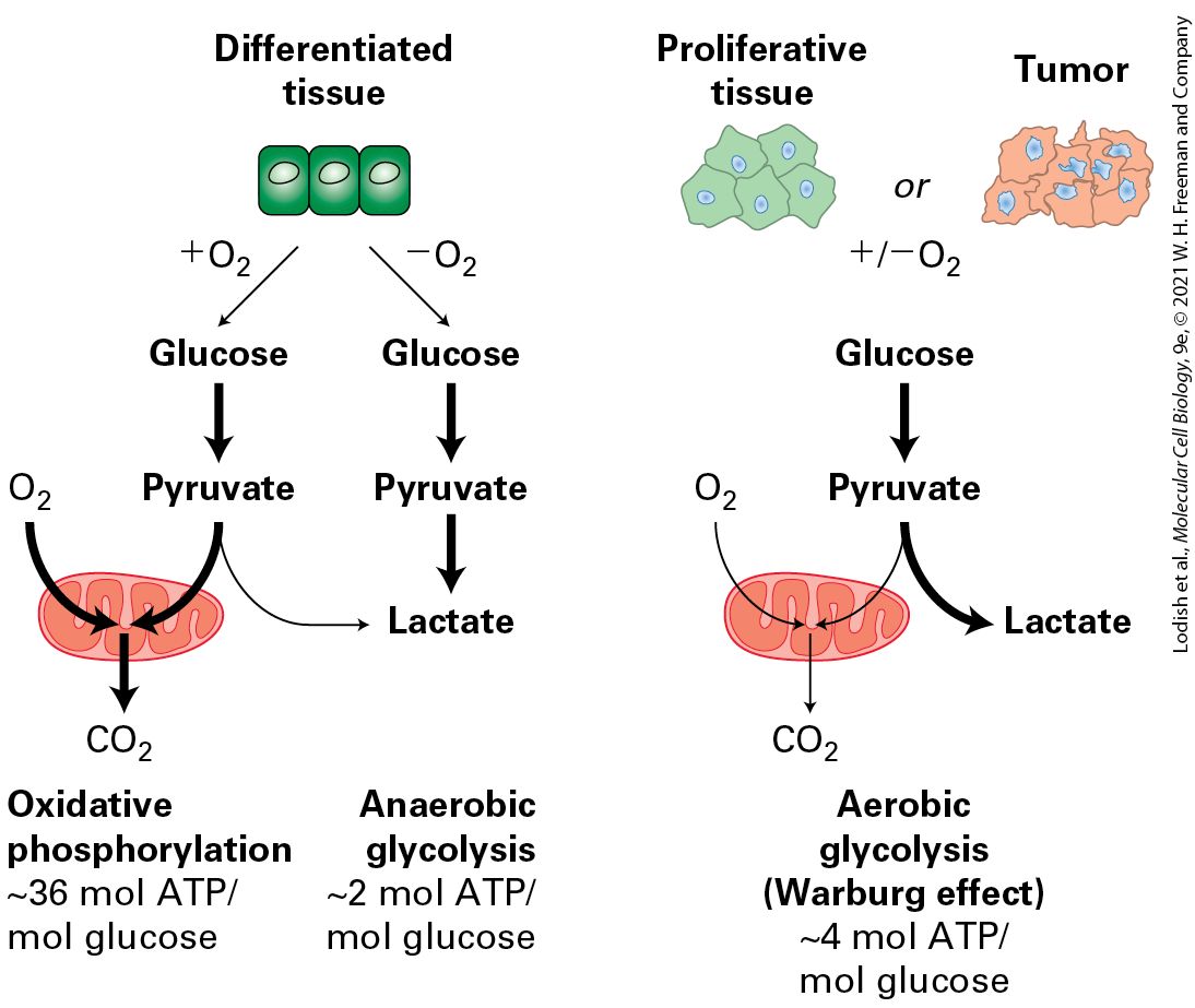
- Glycolysis
- Cytosolic, glucose → 2 pyruvate, net 2 ATP + 2 NADH.
- TCA / Krebs cycle
- Mitochondrial matrix. Acetyl-CoA → CO₂ + 3 NADH + 1 FADH₂ + 1 GTP per acetyl-CoA.
- Electron transport chain
- Complex I (NADH-DH), II (succinate-DH), III (cyt bc1), IV (cyt c oxidase). Electrons from NADH/FADH₂ → O₂ → H₂O. Pumps H⁺ to intermembrane space.
- Chemiosmosis
- Proton gradient drives ATP synthase (Complex V): F₀ rotor + F₁ catalytic head. ~32 ATP per glucose total.
- Uncoupling protein (UCP1)
- Brown adipose; dissipates proton gradient as heat (thermogenesis).
U10 · Cell signaling
📖 Lodish 9e · related chapters
- Receptor classes
- Cell-surface: GPCR, RTK, ion-channel-coupled, integrins. Intracellular: nuclear receptors (steroid, thyroid).
- GPCR signaling
- Ligand → Gα-GTP → effectors (adenylyl cyclase → cAMP → PKA; PLCβ → IP₃/DAG → Ca²⁺/PKC). Desensitization by GRKs + arrestins.
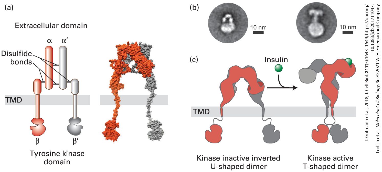
- RTK signaling
- Ligand → dimerization → trans-autophosphorylation → SH2/PTB-domain adaptors (Grb2, Shc, PI3K). Examples: EGFR, insulin receptor, VEGFR.
- MAPK cascade
- Ras → Raf → MEK → ERK → nuclear TFs (Elk1, Fos). Drives proliferation. Mutated in many cancers.
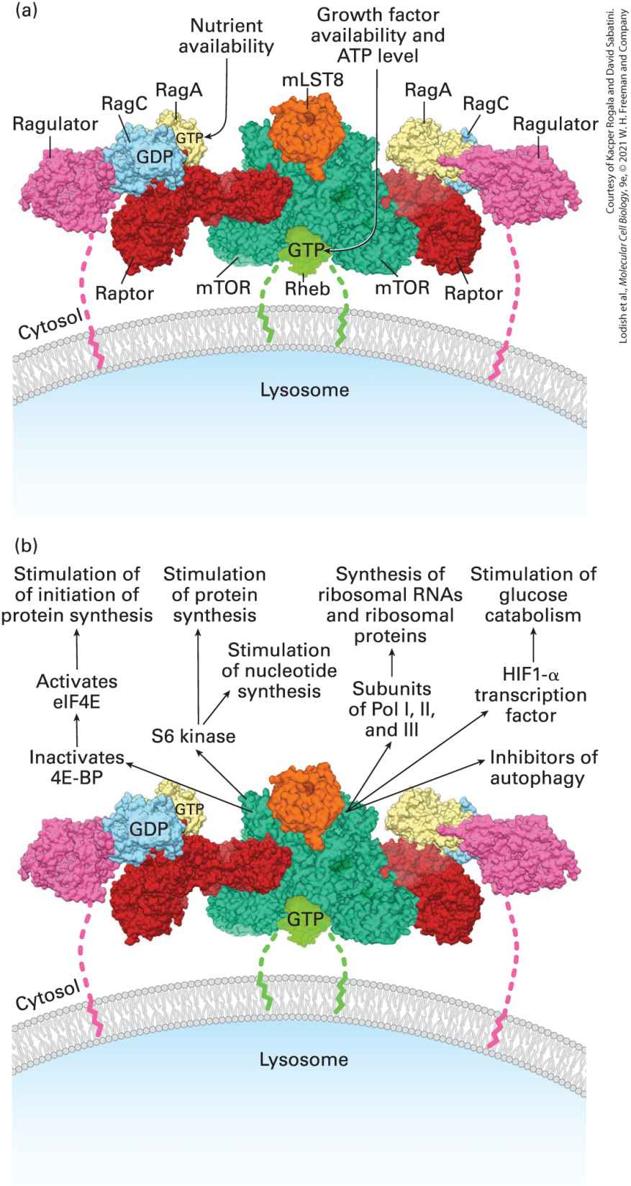
- PI3K-AKT-mTOR
- Survival + growth. PIP₂ → PIP₃ recruits AKT; PTEN reverses. mTORC1 controls translation/autophagy.
- Notch
- Juxtacrine; intramembrane proteolysis releases NICD → nucleus → CSL → HES/HEY targets. Lateral inhibition.
- Wnt/β-catenin
- Wnt → Frizzled/LRP → Dishevelled → inhibits destruction complex (APC, GSK3, axin) → β-catenin to nucleus → TCF/LEF target genes.
- TGF-β / BMP
- Receptor Ser/Thr kinase phosphorylates SMADs → nucleus.
- Second messengers
- cAMP, cGMP, IP₃, DAG, Ca²⁺, NO.
U11 · Cytoskeleton
📖 Lodish 9e · related chapters
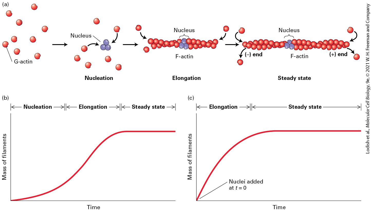
- Actin (microfilaments)
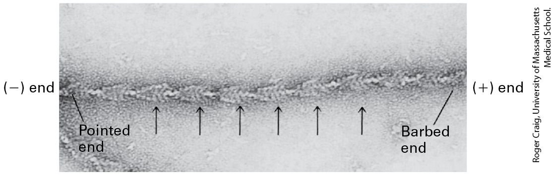
- ~7 nm; G-actin polymerizes (ATP-dependent) to F-actin. Polar (+/-). Regulators: profilin, cofilin, Arp2/3, formins. Cell shape, migration.
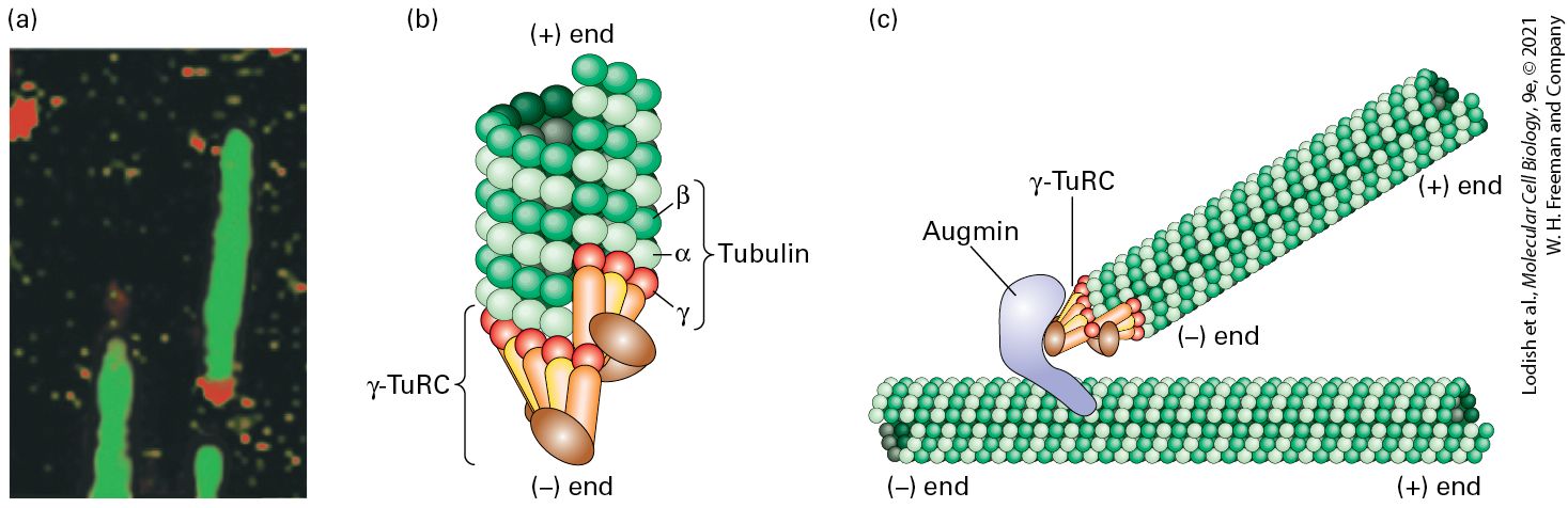
- Microtubules
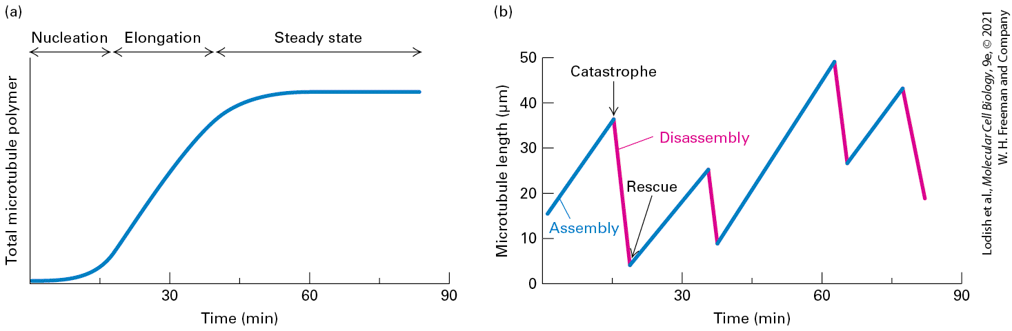
- ~25 nm; α/β-tubulin dimers (GTP-dependent). 13 protofilaments. Polar — minus end at centrosome, plus end outward. Dynamic instability (catastrophe / rescue).
- Intermediate filaments
- ~10 nm; non-polar; coiled-coil dimers → tetramers → filaments. Mechanical support. Examples: keratins (epithelia), vimentin (mesenchymal), nuclear lamins.
- Myosin
- Actin-based motor. Myosin II = muscle + cytokinesis. Myosin V = vesicle transport.
- Kinesin
- MT + end-directed motor (anterograde transport, mitotic spindle).
- Dynein
- MT − end-directed (retrograde), also drives cilia/flagella.
- Cilia/flagella
- 9+2 axoneme. Beat by dynein-driven sliding of doublets.
U12 · Cell cycle & cell death
📖 Lodish 9e · related chapters


- Cell-cycle phases
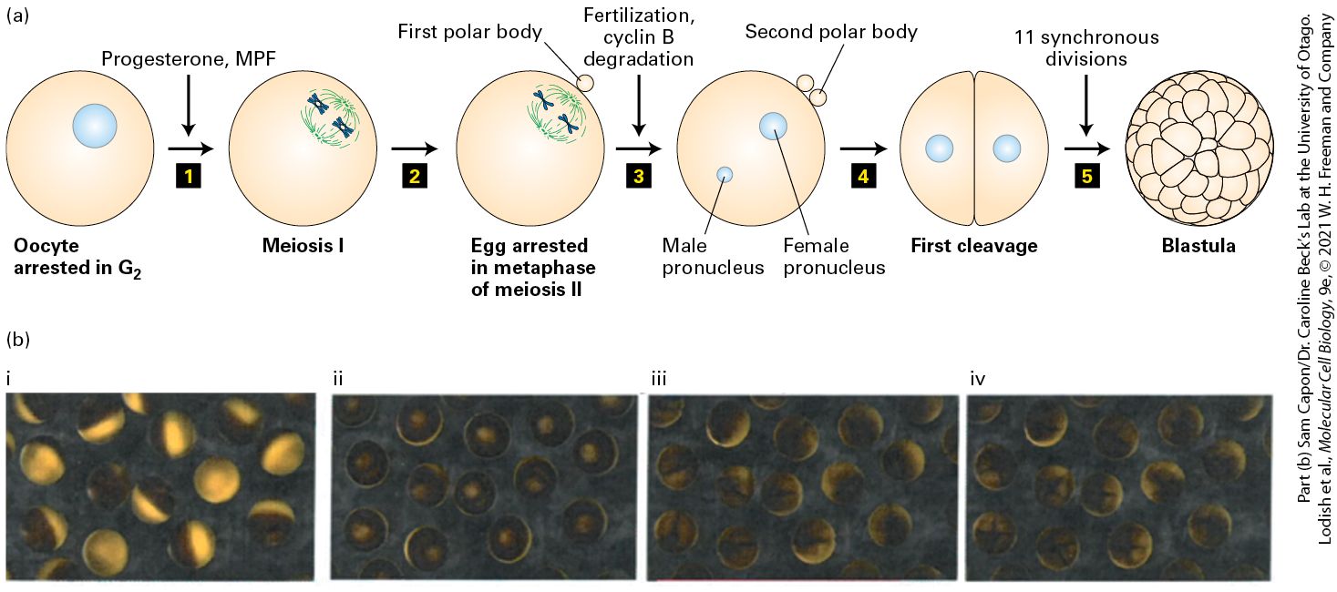
- G1 → S (DNA replication) → G2 → M (mitosis + cytokinesis). G0 = quiescent.
- Cyclin/Cdk
- CDKs are kinases; cyclins are regulatory subunits, periodically expressed. G1: cyclin D + CDK4/6; S: cyclin E/A + CDK2; M: cyclin B + CDK1.
- Cell-cycle checkpoints
- G1/S (Restriction): DNA damage → p53 → p21 → CDK inhibition. G2/M: damage / unreplicated DNA. M (SAC): unattached kinetochores hold APC/C.
- Mitosis stages
- Prophase → prometaphase → metaphase (alignment) → anaphase (cohesin cleaved by separase) → telophase → cytokinesis.
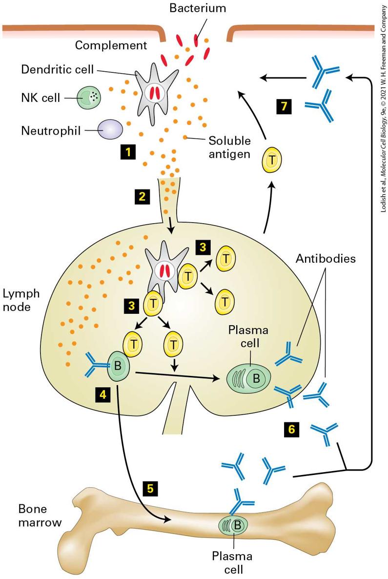
- Apoptosis
- Programmed cell death. Intrinsic (mitochondrial cyt c → apoptosome → caspase-9 → effector caspase-3/7); extrinsic (death receptor Fas/TNFR → caspase-8). Bcl-2 family balance.
- Necroptosis / pyroptosis
- Lytic forms of cell death — RIPK + MLKL; gasdermin pores (matters for Chivero's NEUR 1520 inflammasome content!).
- Autophagy
- Self-degradation via autophagosome → lysosome. mTORC1 inhibits, ATG genes execute.
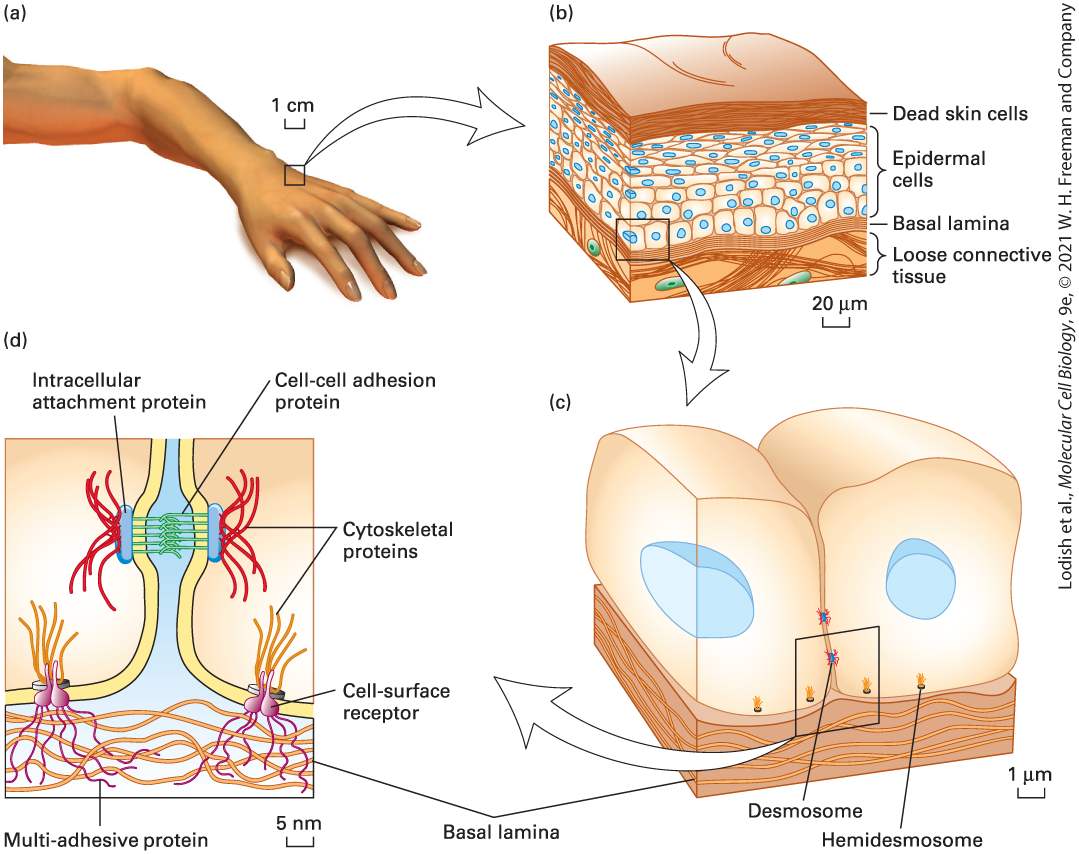
U13 · Cell-cell junctions — Johnson focus
📖 Lodish 9e · related chapters
- Tight junctions (zonula occludens)
- Apical seal; claudins + occludin form paracellular barrier. Define apical/basolateral polarity.
- Adherens junctions
- E-cadherin (Ca²⁺-dependent) homophilic binding. Cytoplasmic tail binds β-catenin → α-catenin → actin. Johnson's specialty.
- Desmosomes
- Spot-weld between cells; desmocollins/desmogleins → plakoglobin/desmoplakin → intermediate filaments (keratin).
- Gap junctions
- Connexin hexamer = connexon; two connexons span the gap → channel for small molecules (<1 kDa: ions, cAMP, IP₃). Johnson's specialty.
- Hemidesmosomes
- Cell-to-ECM (basal lamina) via integrin α6β4 → keratin IFs.
- Focal adhesions
- Integrins + actin; mechanosensing; signal via FAK + Src.
- Cadherin switching in EMT
- E-cadherin ↓, N-cadherin ↑ during epithelial-to-mesenchymal transition (development + cancer metastasis).
- Catenins as signals
- β-catenin doubles as a Wnt-pathway TF when not sequestered at adherens junctions.
U14 · Cancer + tissues + ECM
📖 Lodish 9e · related chapters
- Hanahan-Weinberg hallmarks
- Sustained proliferation, evasion of growth suppressors, resisting cell death, replicative immortality, angiogenesis, invasion + metastasis, deregulated metabolism, immune evasion + inflammation, genome instability.
- Oncogenes
- Gain-of-function mutations in proto-oncogenes (Ras, Myc, EGFR, BRAF). Dominant.
- Tumor suppressors
- Loss-of-function (p53, Rb, APC, PTEN, BRCA1/2). Recessive (Knudson 2-hit).
- Metastasis
- EMT → local invasion → intravasation → circulation → extravasation → colonization.
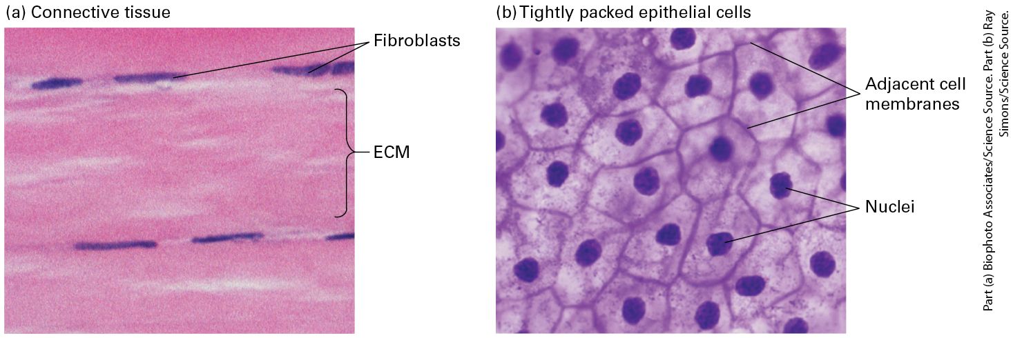
- ECM
- Collagen (most abundant), elastin, proteoglycans (heparan/chondroitin sulfate), fibronectin, laminin (basal lamina).
- Basal lamina
- Sheet of laminin + type IV collagen + perlecan. Separates epithelium from CT. Barrier disrupted in carcinoma invasion.
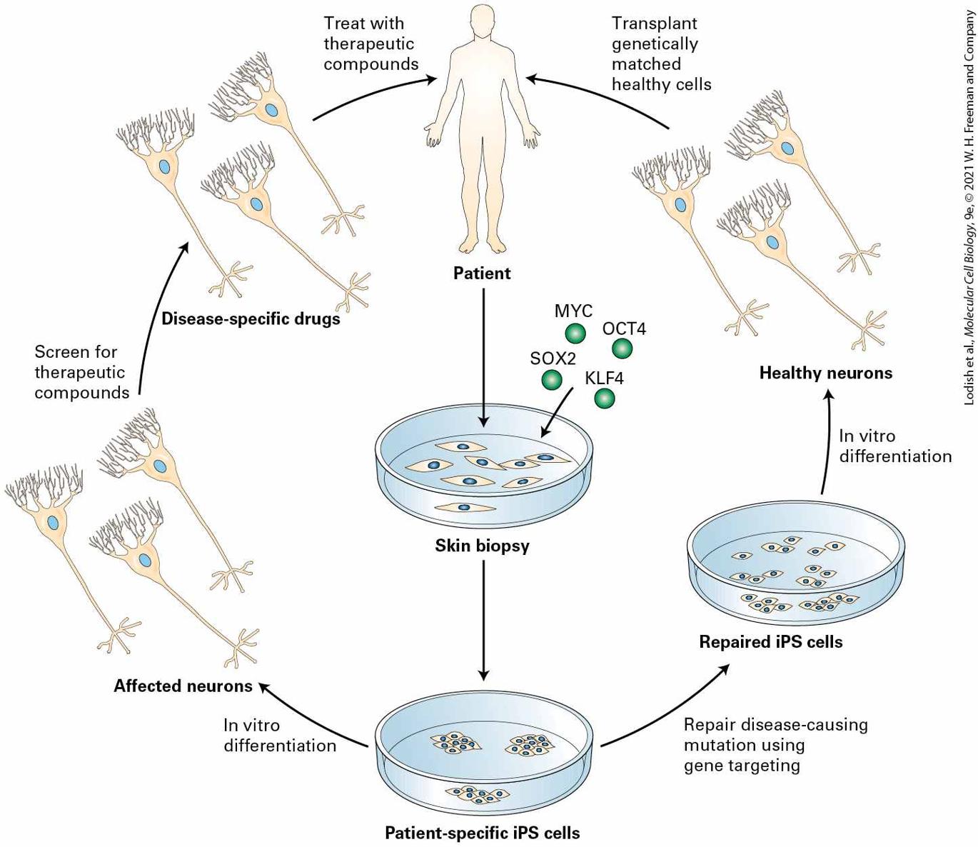
- Stem cells
- Self-renewal + multipotency. Niche-dependent. Adult (intestinal crypt, bone marrow, hair follicle).
U15 · Lab techniques (Northam's lab block)
📖 Lodish 9e · related chapters
- Tissue culture
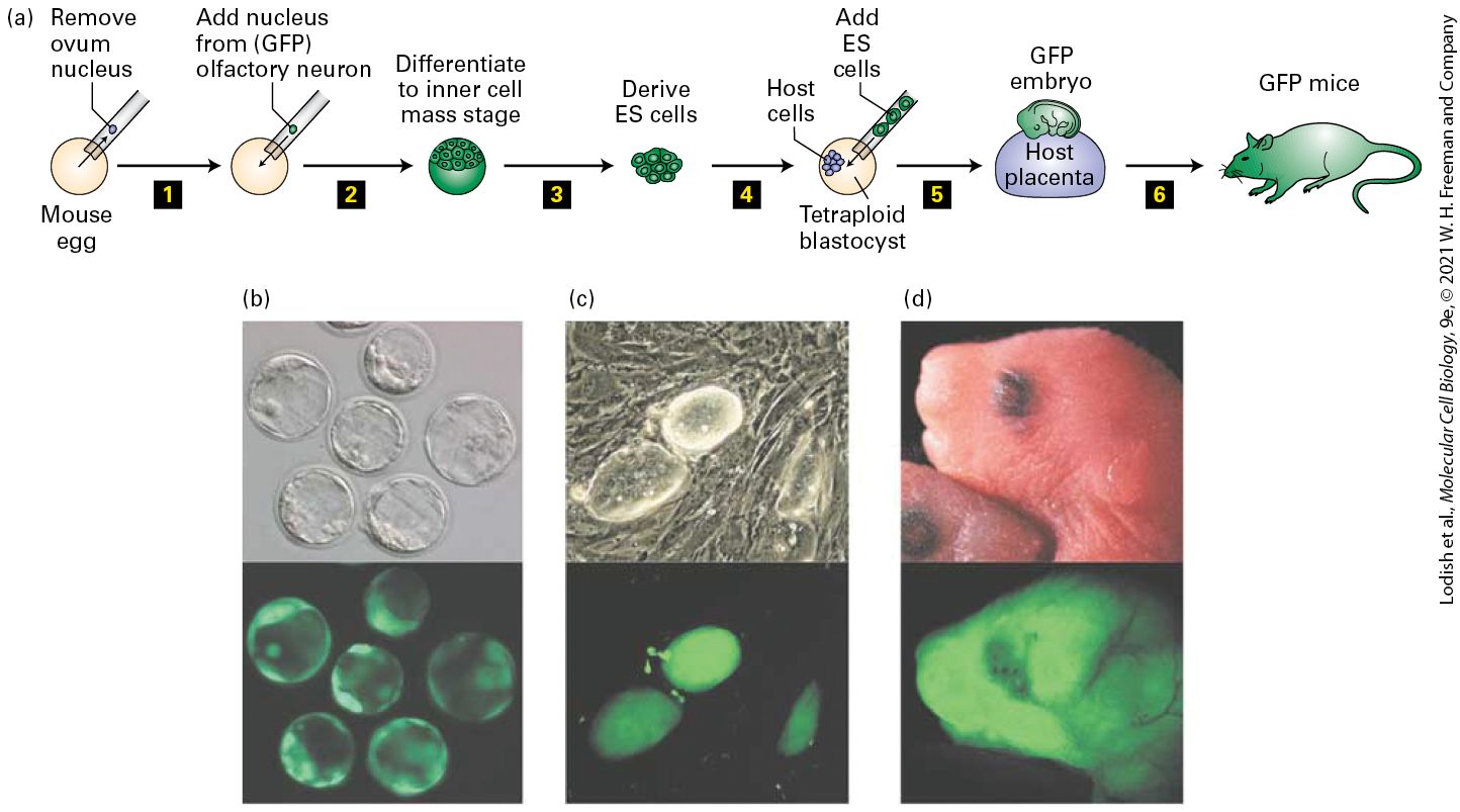
- Maintain mammalian cells in vitro: medium with FBS, sterile technique, passaging. Common lines: HeLa, HEK293, NIH-3T3, MCF-7.
- Trypsinization
- Protease detaches adherent cells for splitting/passaging.
- Transfection
- Introduce DNA/RNA: lipofection, electroporation, viral vectors (lentivirus).
- Immunofluorescence (IF)
- Fix → permeabilize → block → primary Ab → fluorescent secondary Ab → DAPI for nuclei → image. Localize proteins.
- Western blot
- SDS-PAGE → transfer → primary + secondary HRP Ab → ECL detection. Quantify protein.
- RT-qPCR
- RNA → cDNA (reverse transcriptase) → SYBR/TaqMan qPCR. Quantify mRNA. ΔΔCt analysis vs housekeeping gene.
- Flow cytometry / FACS
- Fluorescent markers + cytometer → analyze or sort cells by surface or DNA-content phenotypes.
- siRNA / shRNA / CRISPR
- Loss-of-function: siRNA transient, shRNA stable, CRISPR-Cas9 genomic edit.
- Live-cell imaging
- Fluorescent proteins (GFP/mCherry) + confocal/spinning disk; FRAP for diffusion; FRET for protein-protein interaction.
- High-throughput assays
- Multi-well plate readouts: viability (MTT, ATP), reporter, image-based phenotyping. Used in drug discovery.
Johnson + Northam exam tips
- Be prepared to diagram a junction (adherens, gap, tight, desmosome) labeled with proteins + linkages.
- Know EMT: cadherin switch, β-catenin re-localization, role in cancer.
- For lab: identify a microscopy technique from a sample image — bright-field vs phase vs IF vs confocal.
- Be ready to interpret RT-qPCR ΔΔCt data and a Western blot.
📚 Textbook companion · Lodish MCB 9e
Each unit above maps to chapters in the locally-OCR'd Lodish MCB 9e. Use the cards below as a quick visual jump into the embedded textbook reader — one figure per chapter, click to read the full chapter:
CH 01
Evolution: Molecules, Genes, Cells, and Organisms
CH 02
Chemical Foundations
CH 03
Protein Structure and Function
CH 04
Culturing and Visualizing Cells
CH 05
Fundamental Molecular Genetic Mechanisms
CH 06
Molecular Genetic Techniques
CH 07
Genes, Chromatin, and Chromosomes
CH 08
Transcriptional Control of Gene Expression
CH 09
Post-Transcriptional Gene Control
CH 10
Biomembrane Structure
CH 11
Transmembrane Transport of Ions and Small Molecules
CH 12
Cellular Energetics
CH 13
Moving Proteins into Membranes and Organelles
CH 14
Vesicular Traffic, Secretion, and Endocytosis
CH 15
Receptors, Hormones, and Cell Signaling
CH 16
Growth Factor and Cytokine Signaling Pathways That Control Gene Expression
CH 17
Cell Organization and Movement I: Microfilaments
CH 18
Cell Organization and Movement II: Microtubules and Intermediate Filaments
CH 19
The Eukaryotic Cell Cycle
CH 20
Integrating Cells into Tissues
CH 21
Responding to the Cellular Environment
CH 22
Stem Cells, Cell Asymmetry, and Regulated Cell Death
CH 23
Cells of the Nervous System
CH 24
Immunology
CH 25
Cancer