Ch 1Evolution: Molecules, Genes, Cells, and OrganismsRead full chapter →
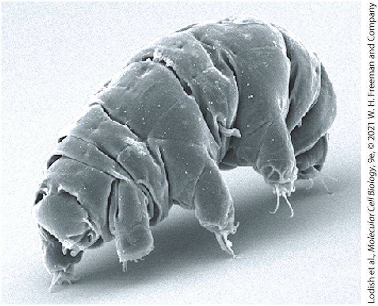
Chapter 1 Evolution: Molecules, Genes, Cells, and Organisms Tardigrades are tiny -long invertebrates that have evolved to survive exposures to extreme conditions that kill other life forms; they are the only metazoan organisms known to survive exposure to the deadly combination of low pressure and intense radiation in outer space. [Schokraie E., Warnken U., Hotz-Wagenblatt A., Grohme M. A., Hengherr S., et al. 2012. Comparative proteome analysis of Milnesium tardigradum in early embryonic state versus
Sections in this chapter
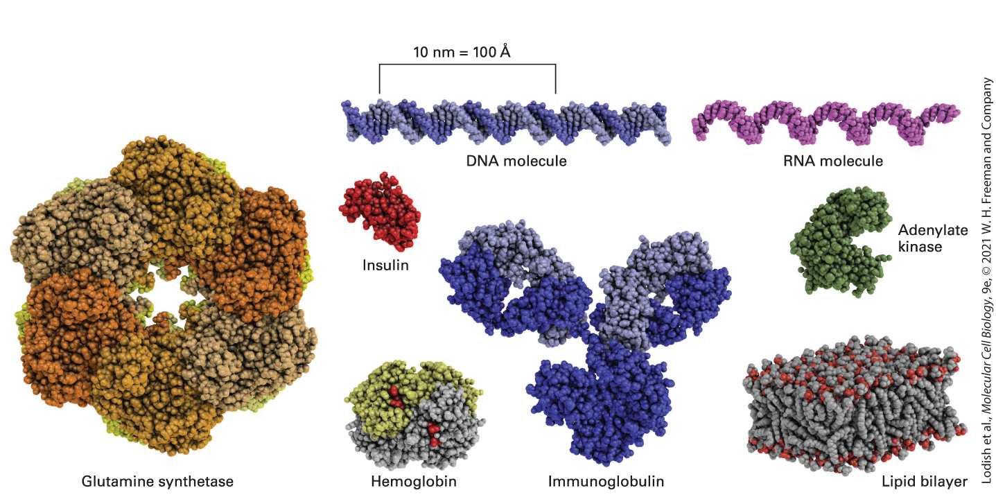
- 1.1 The Molecules of Life
- Proteins Give Cells Structure and Perform Most Cellular Tasks
- Nucleic Acids Carry Coded Information for Making Proteins at the Right Time and Place
- Phospholipids Are the Conserved Building Blocks of All Cellular Membranes
- Quality Control of All Cellular Macromolecules Is Essential for Life
- 1.2 Prokaryotic Cell Structure and Function
- Prokaryotes Comprise Two Kingdoms: Archaea and Eubacteria
- Many Bacteria Including Escherichia coli Are Widely Used in Biological Research
- 1.3 Eukaryotic Cell Structure and Function
- The Cytoskeleton Has Many Important Functions
- The Nucleus Contains the DNA Genome, Apparatuses for Synthesis of DNA and RNA, and a Fibrous Matrix
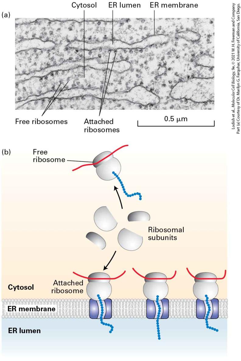
- The Endoplasmic Reticulum Is the Site of Synthesis of Most Membrane and Secreted Proteins as Well as Many Lipids
Ch 2Chemical FoundationsRead full chapter →
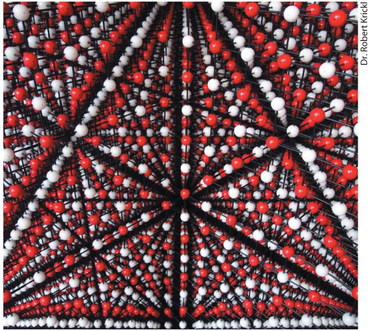
Chapter 2 Chemical Foundations Model of sodium chloride (NaCl) crystal. This photograph shows the largest (3.1 m tall) balland-stick physical model of a crystalline salt ever constructed, built from 40,110 balls by Robert Krickl.
Sections in this chapter
- The Electronic Structure of an Atom Determines the Number and Geometry of the Covalent Bonds It Can Make
- All Covalent Bonds Are Not Equal: Electrons May Be Shared Equally or Unequally in Covalent Bonds
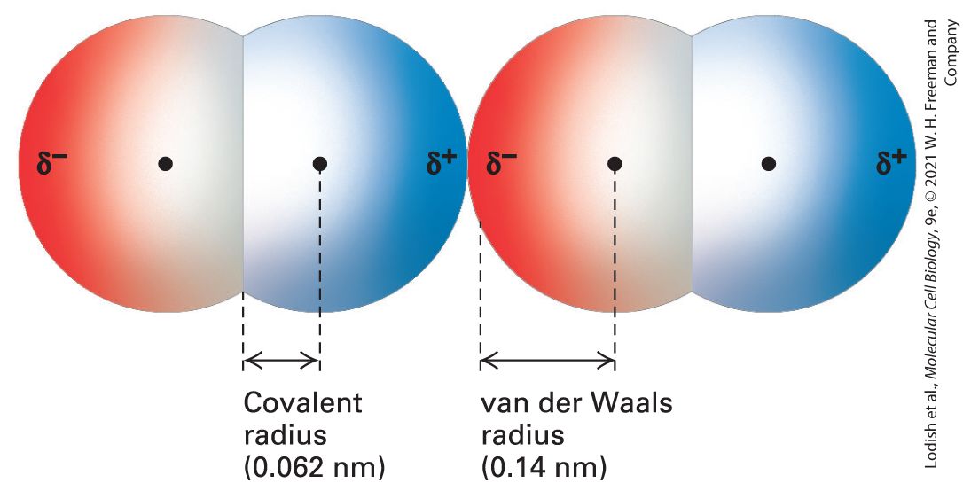
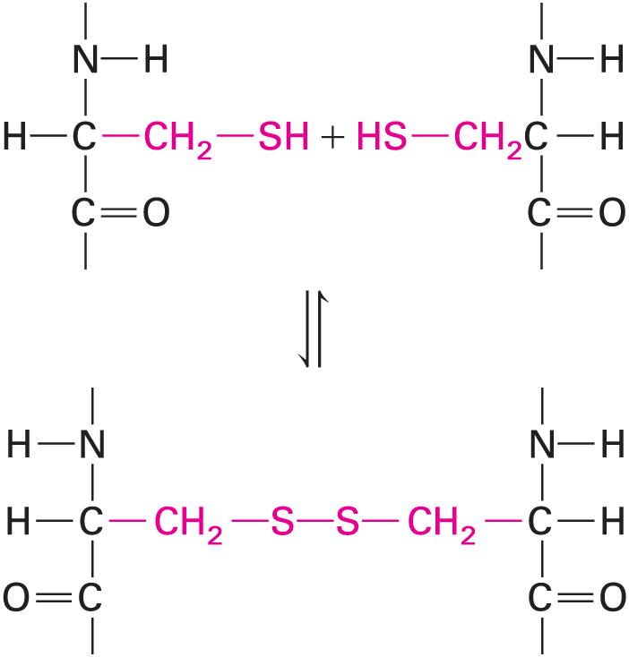
- Covalent Bonds Are Much Stronger and More Stable Than Noncovalent Interactions
- Ionic Bonds Are Noncovalent Interactions Formed by the Electrostatic Attractions Between Oppositely Charged Ions
- Hydrogen Bonds Are Noncovalent Interactions That Determine the Properties of Water and the Water Solubility of Uncharged Molecules
- Van der Waals Interactions Are Weak Attractive Interactions Caused by Transient Dipoles
- The Hydrophobic Effect Causes Nonpolar Molecules to Adhere to One Another
- Molecular Complementarity Due to Noncovalent Interactions Leads to a Lock-and-Key Fit Between Biomolecules
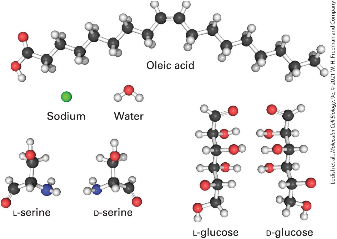
- 2.2 Chemical Building Blocks of Cells
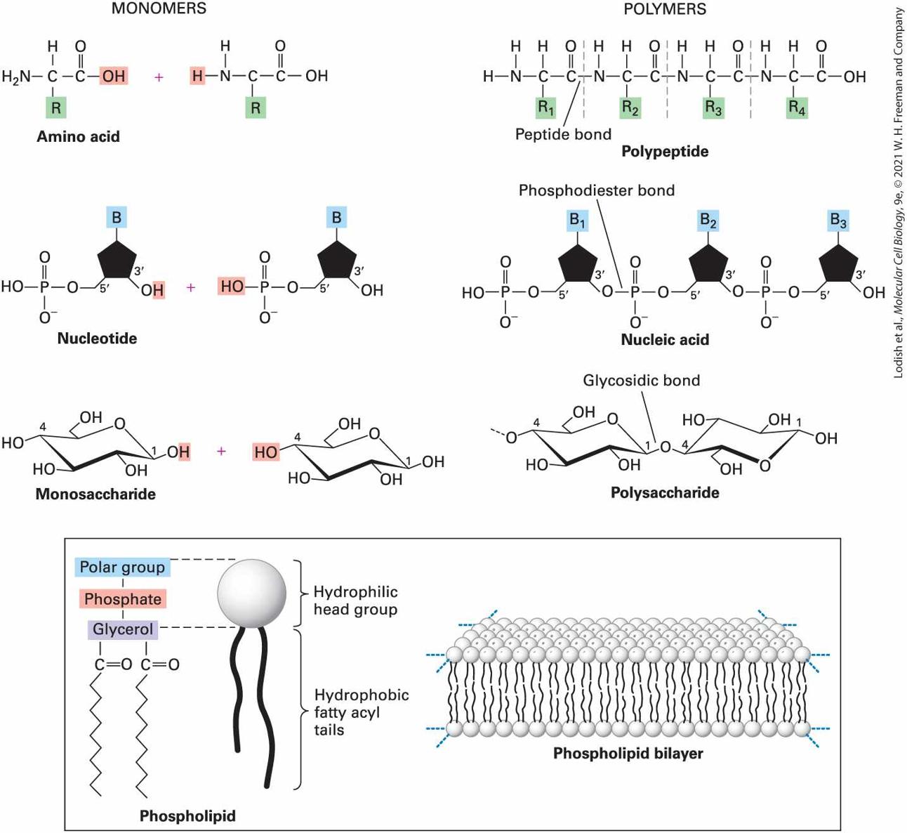
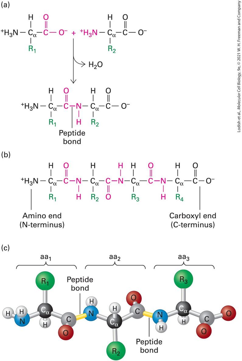
- Amino Acids Differing Only in Their Side Chains Compose Proteins
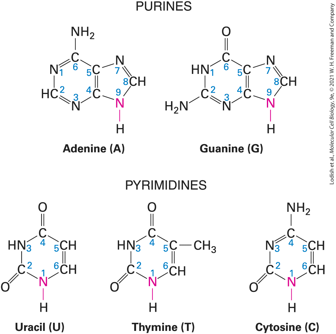
- Five Different Nucleotides Are Used to Build Nucleic Acids
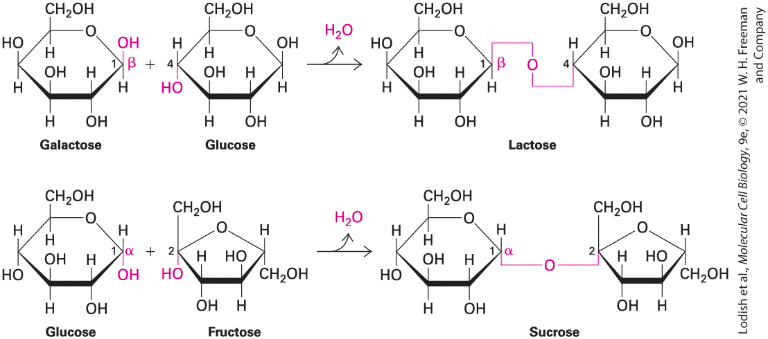
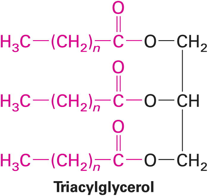
- Monosaccharides Covalently Assemble into Linear and Branched Polysaccharides
Ch 3Protein Structure and FunctionRead full chapter →
Chapter 3 Protein Structure and Function Computationally designed, hypothetical four-stranded fiber protein viewed down the fiber axis. Using advanced methods for protein design, the amino acid sequence of the
staphylococcal nuclease protein was modified so that the individual, folded protein chains (shown in different colors) would stack into long strands. The design predicts that four long strands would assemble into a four-stranded fiber held together by the binding of hydrophobic helices located in the center of the fiber. [Data from H. Shen et al., 2018, Science 362:705–709.]
Sections in this chapter
- 3.1 Hierarchical Structure of Proteins
- The Primary Structure of a Protein Is Its Linear Arrangement of Amino Acids
- Secondary Structures Are the Core Elements of Protein Architecture
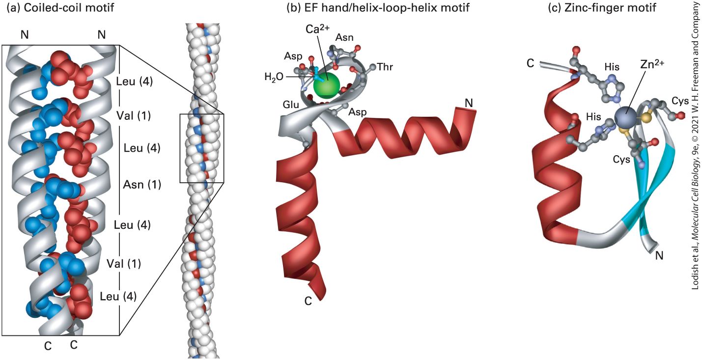
- Structural Motifs Are Regular Combinations of Secondary Structures
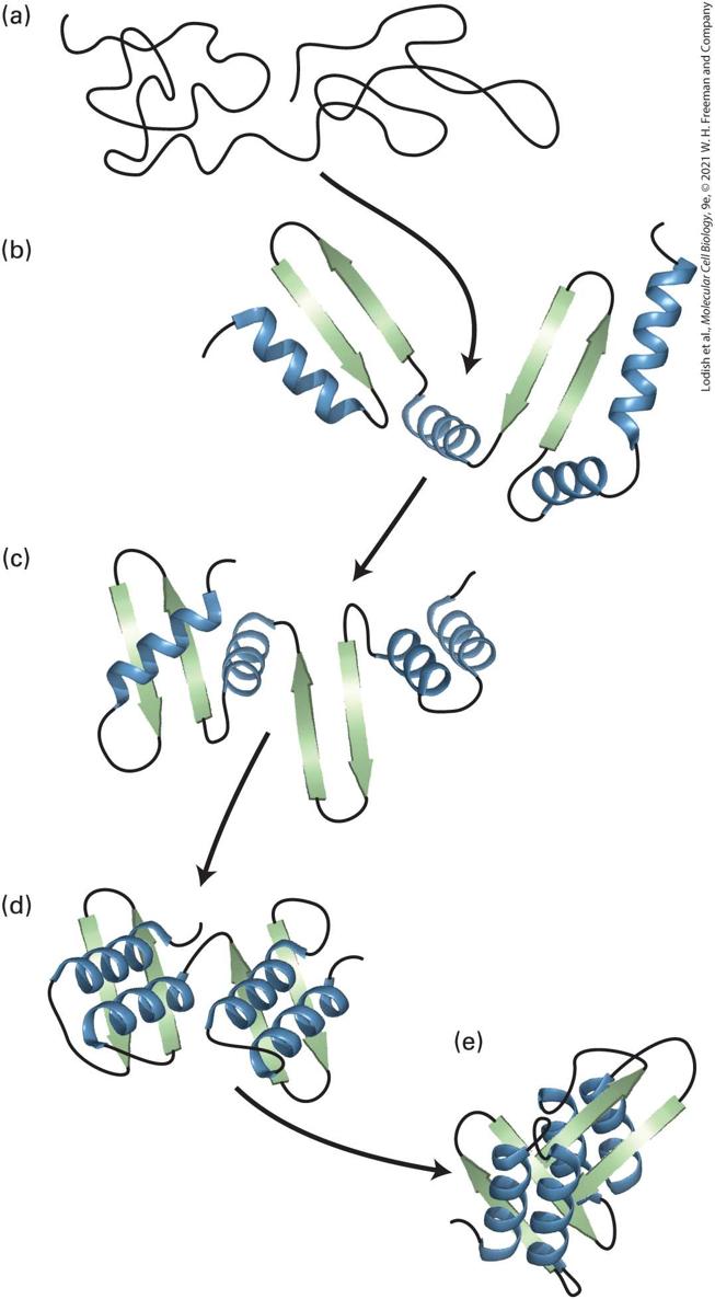
- Tertiary Structure Is the Overall Folding of a Polypeptide Chain
- Different Ways of Depicting the Conformation of Proteins Convey Different Types of Information
- Domains Are Modules of Tertiary Structure
- Comparing Protein Sequences and Structures Provides Insight into Protein Function and Evolution
- There Are Four Broad Structural Categories of Proteins
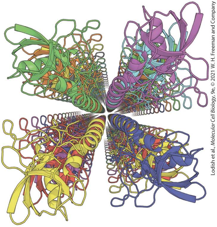
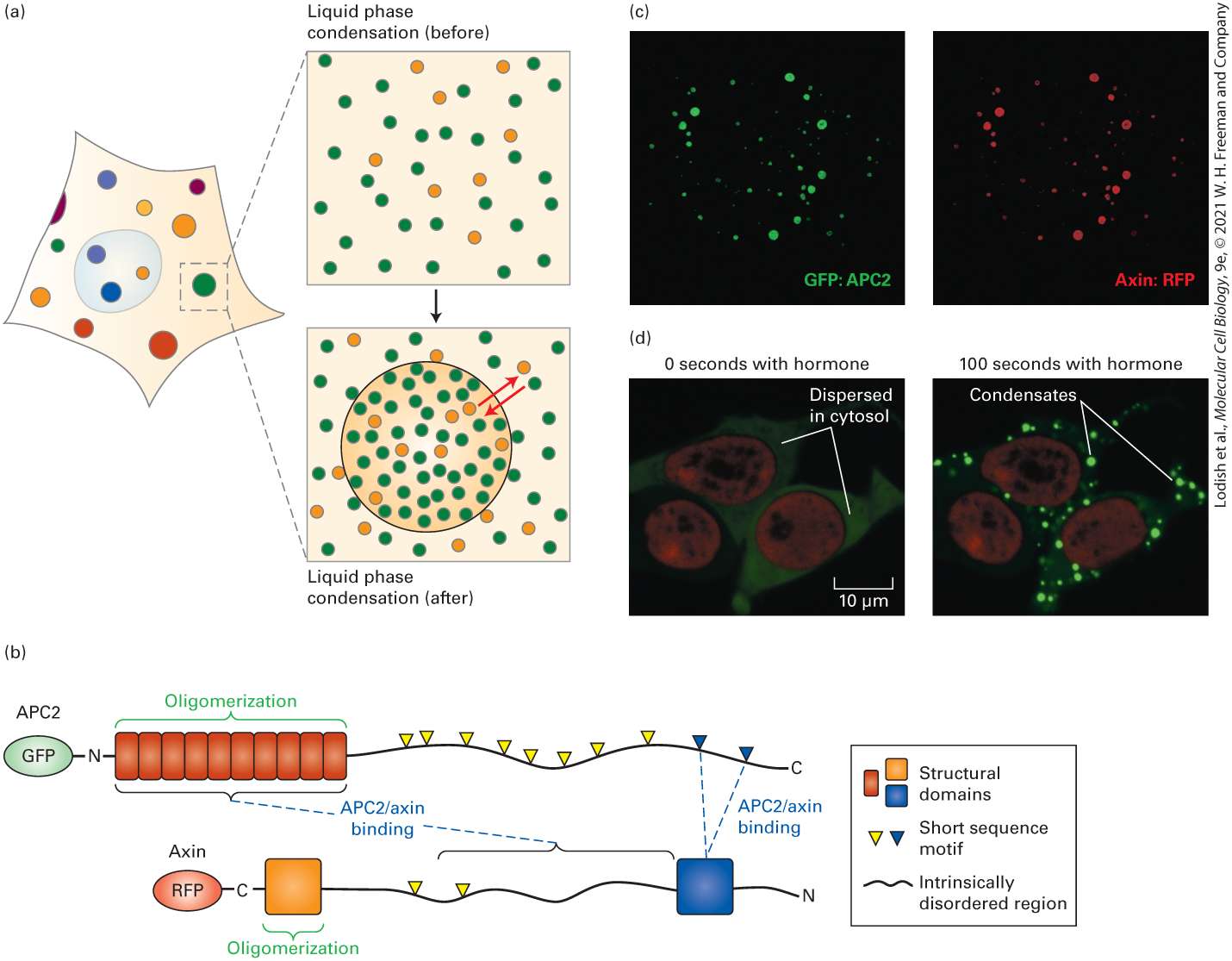
- Multiple Polypeptides Assemble into Quaternary Structures, Supramolecular Complexes, and Biomolecular Condensates
- Planar Peptide Bonds Limit the Shapes into Which Proteins Can Fold
- Protein Folding Is Promoted by Proline Isomerases
Ch 4Culturing and Visualizing CellsRead full chapter →
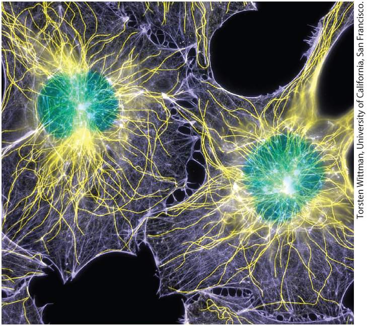
Chapter 4 Culturing and Visualizing Cells Fluorescence microscopy showing the locations of DNA (green), microtubules (yellow), and microfilaments (purple) in two cultured cells. The cells were chemically fixed and then rendered permeable to antibodies using a gentle detergent. Microtubules were stained with an antibody to tubulin; microfilaments were stained with a labeled toxin, phalloidin, that binds selectively to F-actin; and DNA was visualized with a DNA-binding dye.
Sections in this chapter
- Culture of Animal Cells Requires Nutrient-Rich Media and Special Solid Surfaces
- Primary Cell Cultures and Cell Strains Have a Finite Life Span
- Transformed Cells Can Grow Indefinitely in Culture
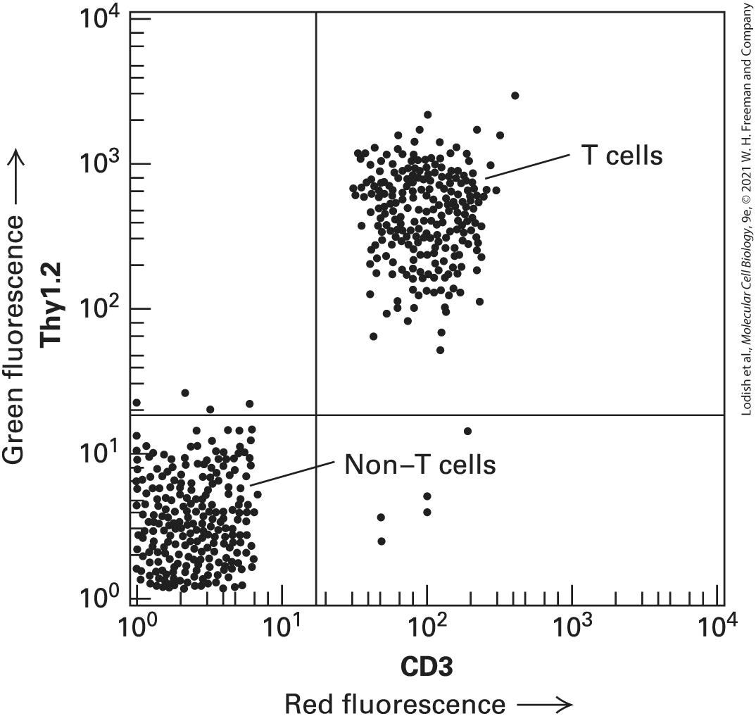
- Flow Cytometry Separates Different Cell Types
- Growth of Cells in Two-Dimensional and Three-Dimensional Culture Mimics the In Vivo Environment
- Stem Cells Can Differentiate in Culture to Make Organoids
- Hybridomas Produce Abundant Monoclonal Antibodies
- A Wide Variety of Cell Biological Processes Can Be Studied with Cultured Cells
- Drugs Are Commonly Used in Cell Biological Research
- 4.2 Light Microscopy: Exploring Cell Structure and Visualizing Proteins Within Cells
- The Resolution of the Conventional Light Microscope Is About 0.2 μm
- Phase-Contrast and Differential-Interference-Contrast Microscopy Visualize Unstained Live Cells
Ch 5Fundamental Molecular Genetic MechanismsRead full chapter →
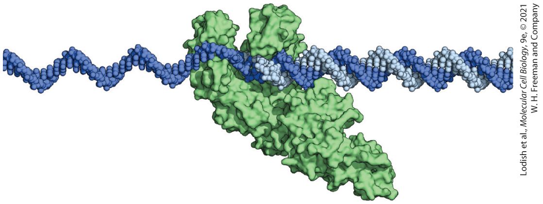
Chapter 5 Fundamental Molecular Genetic Mechanisms Model of DNA Polymerase I from Escherichia coli in the process of replicating DNA. The universal mechanism for copying information in DNA is replication by DNA polymerase, a remarkable copying and proofreading machine. The E. coli DNA polymerase I shown here can synthesize a complementary copy of the template strand at a rate of about 500 bases per second, making a mistake only once in every bases.
Sections in this chapter
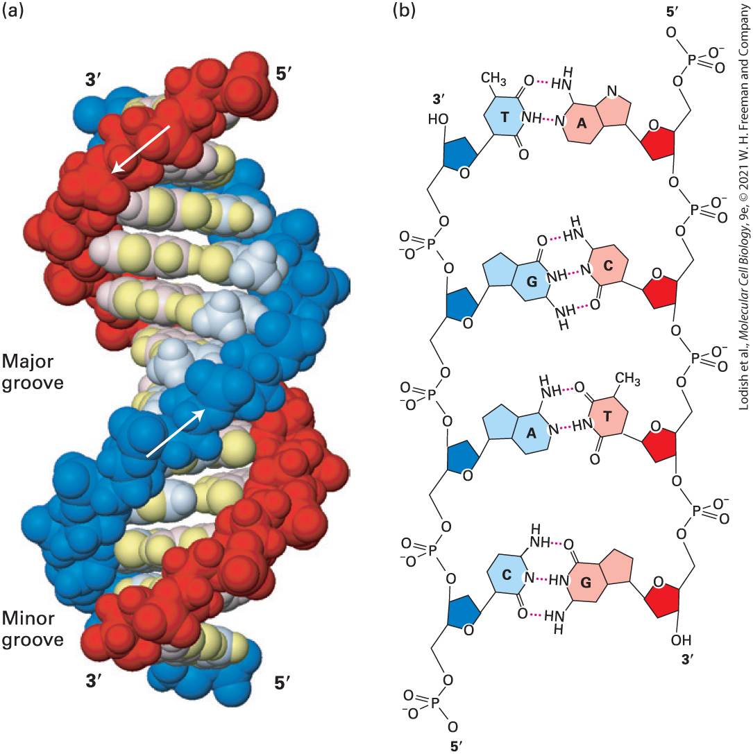
- Native DNA Is a Double Helix of Complementary Antiparallel Strands
- DNA Can Undergo Reversible Strand Separation
- DNA Molecules Can Acquire Torsional Stress
- 5.2 DNA Replication
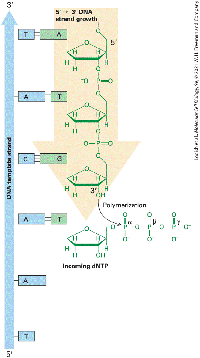
- DNA Polymerases Require a Template and a Primer to Replicate DNA
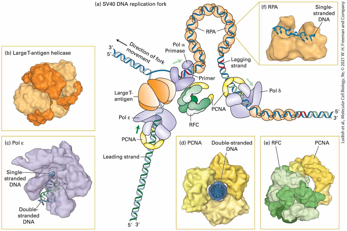
- Duplex DNA Is Unwound, and Daughter Strands Are Formed at the DNA Replication Fork
- A DNA Replication Fork Advances by Cooperation of Multiple Proteins
- DNA Replication Occurs Bidirectionally from Each Origin
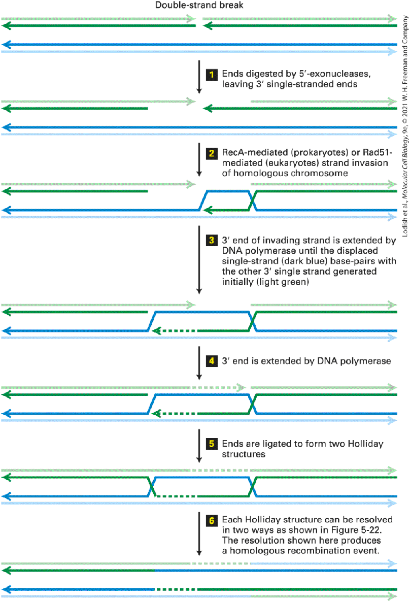
- 5.3 DNA Repair and Recombination
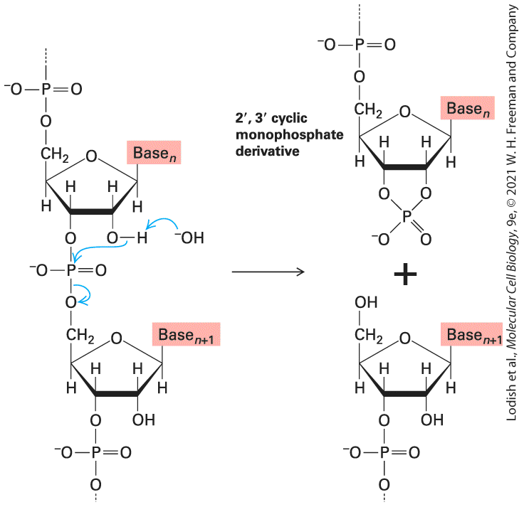
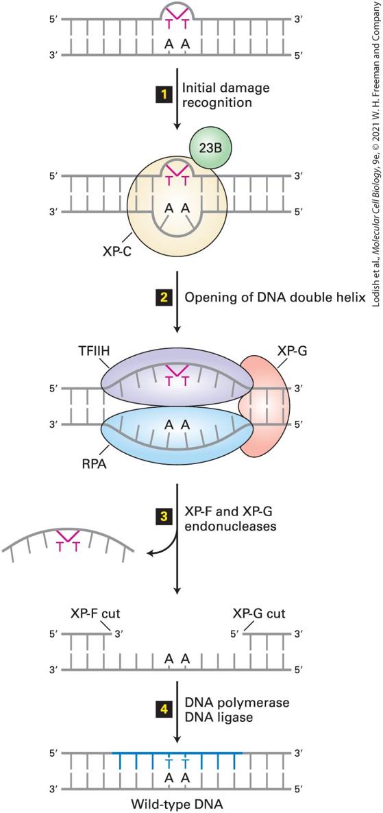
- Chemical and Radiation Damage to DNA Can Lead to Mutations
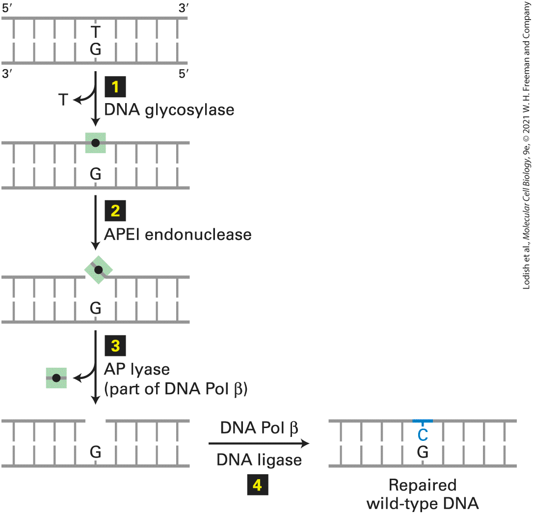
- Base Excision Repairs T-G Mismatches and Damaged Bases
- Mismatch Excision Repairs Other Mismatches and Small Insertions and Deletions
Ch 6Molecular Genetic TechniquesRead full chapter →
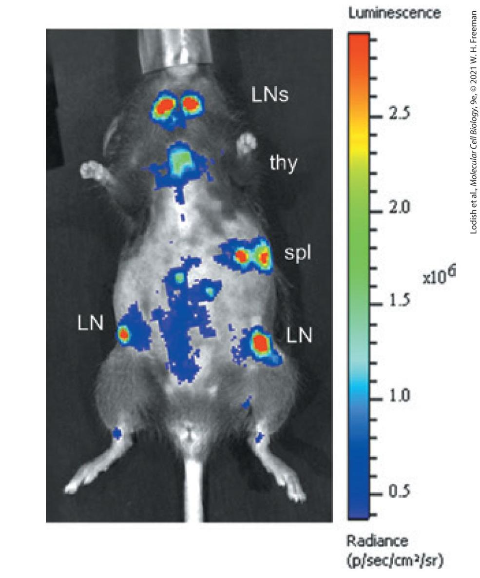
Genetic modifications make it possible to locate a specific cell type within the body of a living mouse. Scientists can label cells of the immune system by generating a transgenic mouse carrying the gene for insect luciferase expressed from the CD2 promoter, which is specifically expressed in T cells. After injection of the bioluminescent substrate for
luciferase, the light emitted through the skin is imaged for 30 seconds. T cells can be seen located in the lymph nodes (LNs), the thymus (thy), and the spleen (spl). [From J. W. Kleinovink et al., 2019. A dual-color bioluminescence reporter mouse for simultaneous in vivo imaging of T cell localization and function. Frontiers in Immunology 9:3097. https://www.ncbi.nlm.nih.gov/pmc/articles/PMC6333049]
Sections in this chapter
- 6.1 Using Genetic Analysis of Mutations to Identify and Study Genes
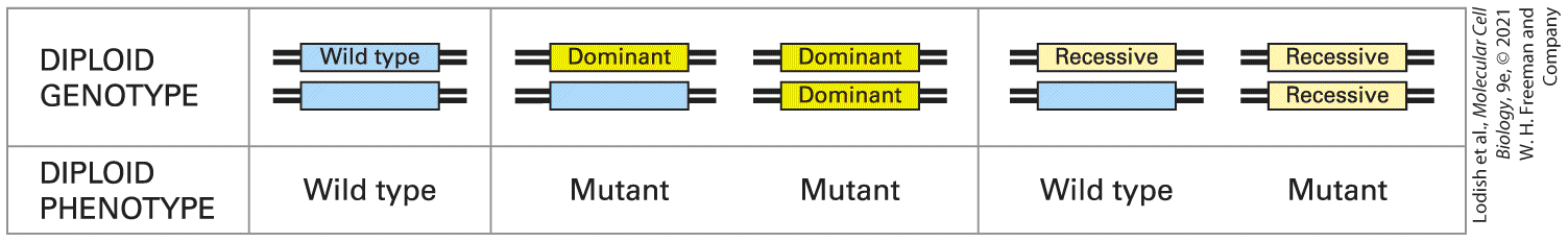
- Recessive and Dominant Mutant Alleles Generally Have Opposite Effects on Gene Function
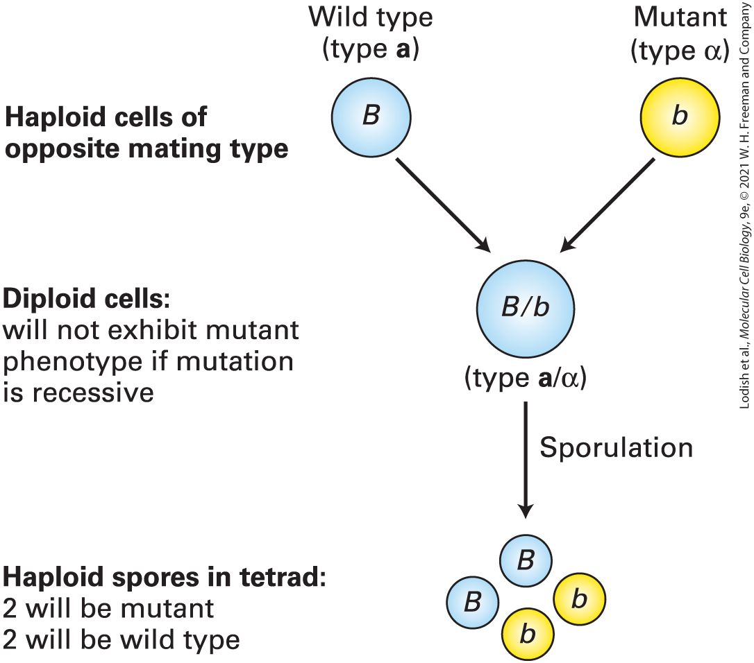
- Segregation of Mutations in Breeding Experiments Reveals Whether They Are Dominant or Recessive
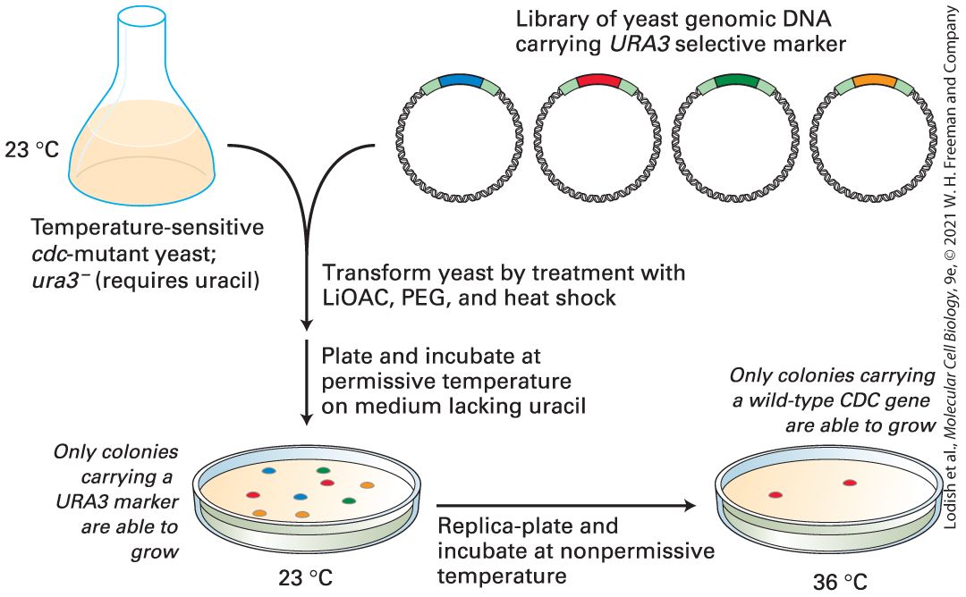
- Conditional Mutations Can Be Used to Study Essential Genes in Yeast
- Recessive Lethal Mutations in Diploids Can Be Identified by Inbreeding and Maintained in Heterozygotes
- Complementation Tests Determine Whether Different Recessive Mutations Are in the Same Gene
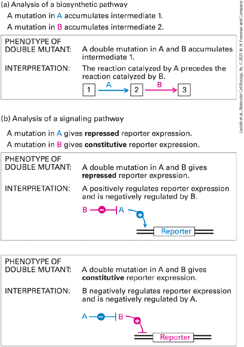
- Double Mutants Are Useful in Assessing the Order in Which Proteins Function
- Genetic Suppression and Synthetic Lethality Can Reveal Interacting or Redundant Proteins
- Global Analysis of Double Mutant Combinations Can Reveal Networks of Gene Functions
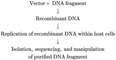
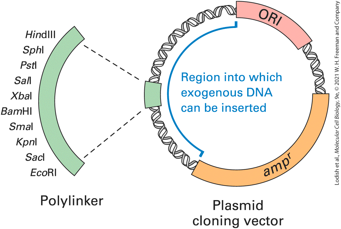
- 6.2 DNA Cloning and Characterization
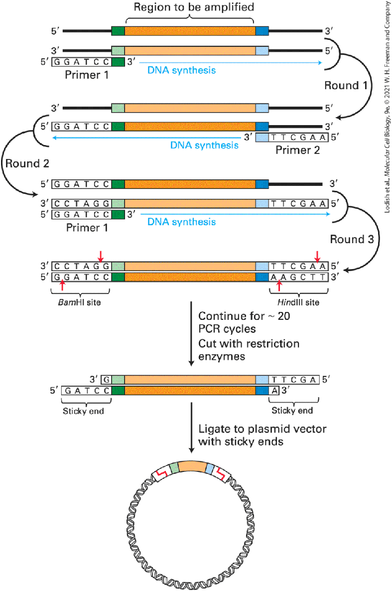
- Restriction Enzymes and DNA Ligases Allow Insertion of DNA Fragments into Cloning Vectors
- Isolated DNA Fragments Can Be Cloned into E. coli Plasmid Vectors
Ch 7Genes, Chromatin, and ChromosomesRead full chapter →
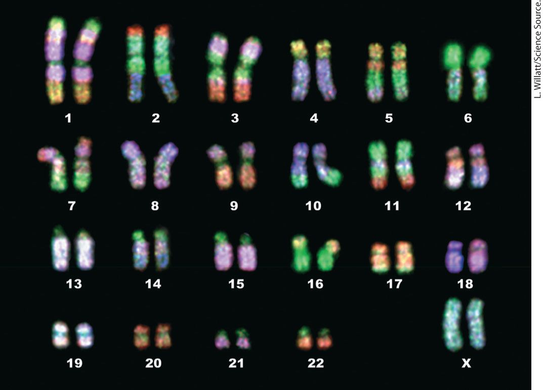
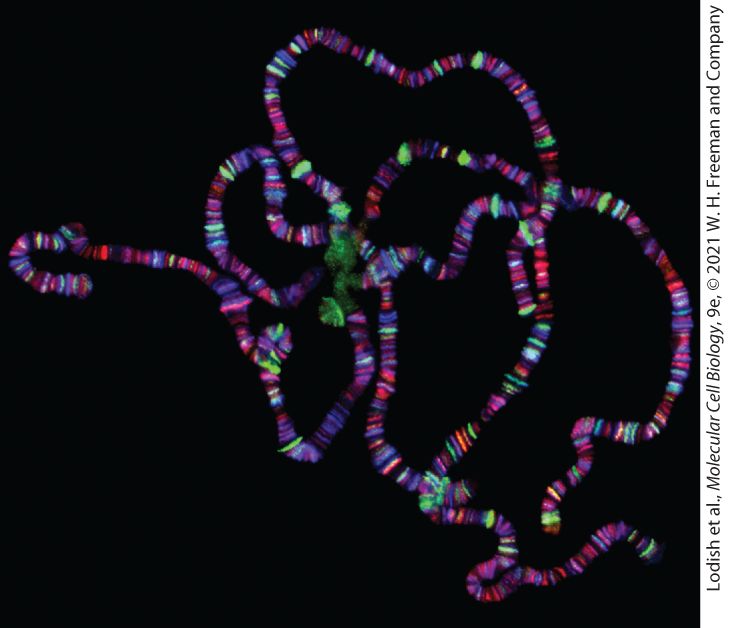
Chapter 7 Genes, Chromatin, and Chromosomes These brightly colored FISH-painted chromosomes are not only beautiful, but also useful in revealing chromosome anomalies and in comparing karyotypes of different species.
Sections in this chapter
- 7.1 Eukaryotic Gene Structure and Organization
- Most Genes of Multicellular Eukaryotes Contain Introns and Produce mRNAs Encoding Single Proteins
- Simple and Complex Transcription Units Are Found in Eukaryotic Genomes
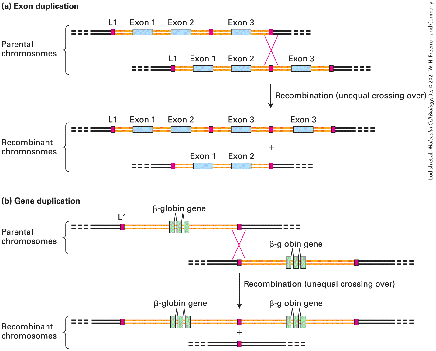
- Protein-Coding Genes May Be Solitary or Belong to a Gene Family
- Heavily Used Gene Products Are Encoded by Multiple Copies of Genes
- Nonprotein-Coding Genes Encode Functional RNAs
- Mrp Rna
- Genomes of Many Organisms Contain a Large Fraction of Noncoding DNA
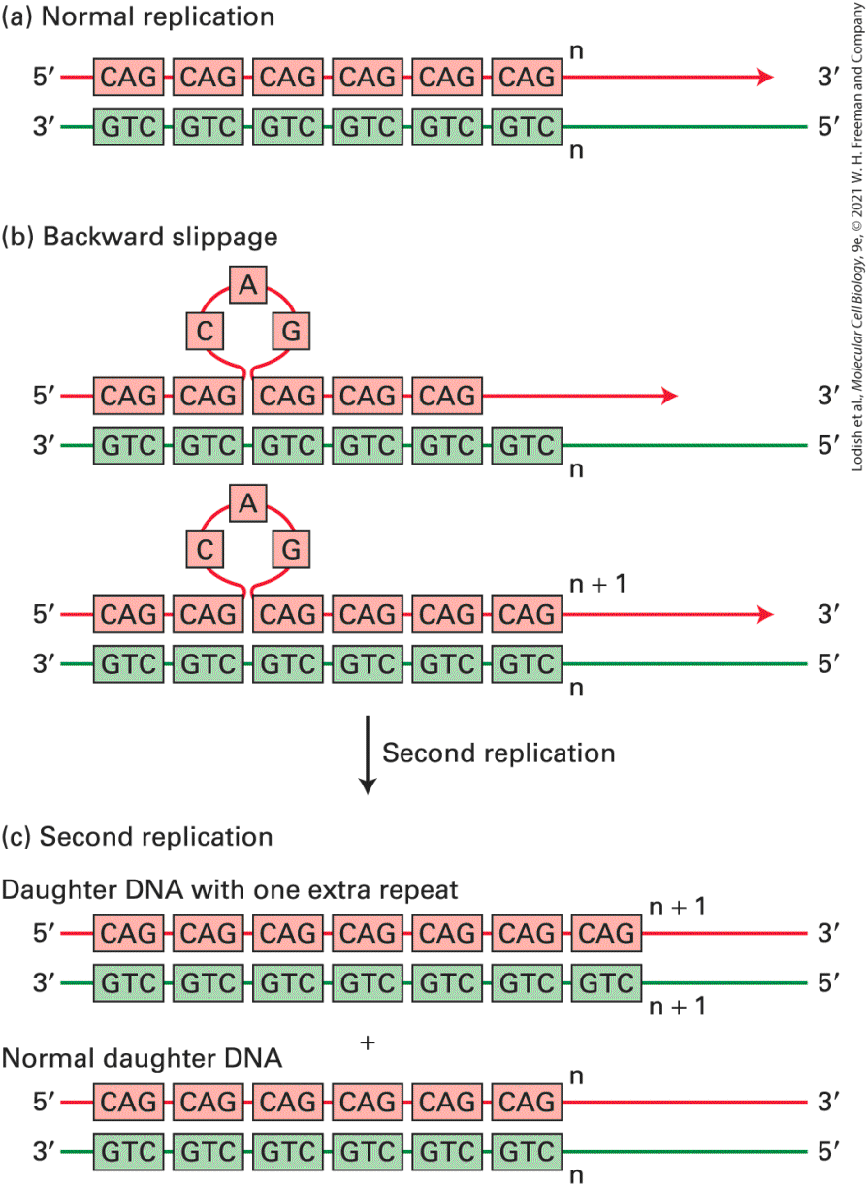
- Most Simple-Sequence DNAs Are Concentrated in Specific Chromosomal Locations
- DNA Fingerprinting Depends on Differences in Length of Simple-Sequence DNAs
- Unclassified Intergenic DNA Occupies a Significant Portion of the Genome
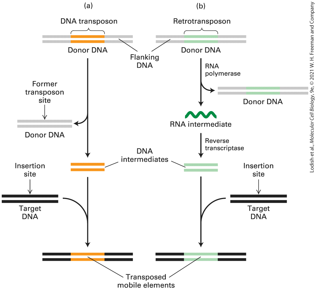
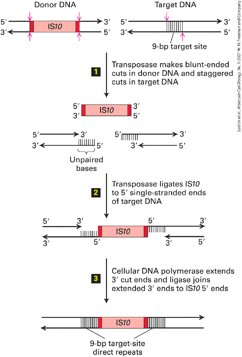
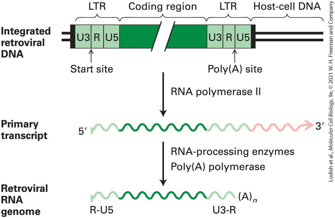
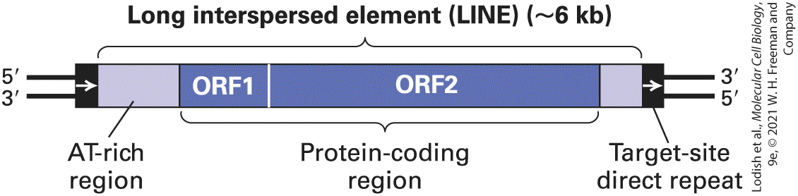
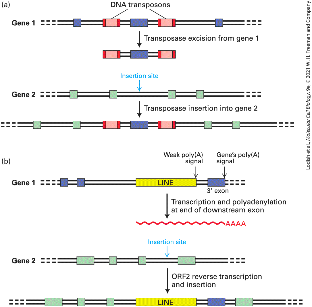
- 7.3 Transposable (Mobile) DNA Elements
Ch 8Transcriptional Control of Gene ExpressionRead full chapter →
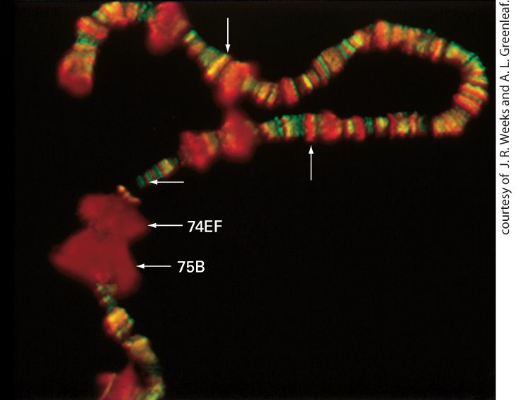
Chapter 8 Transcriptional Control of Gene Expression Drosophila polytene chromosomes stained with antibodies against a chromatin-remodeling ATPase called Kismet (blue), RNA polymerase II with low CTD phosphorylation (red), and RNA polymerase II with high CTD phosphorylation (green). [Reproduced with permission from The Company of Biologists, from S. Srinivasan et. al., 2005, “The Drosophila Trithorax Group Protein Kismet Facilitates an Early Step in
Sections in this chapter
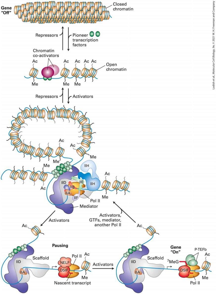
- 8.1 Overview of Eukaryotic Transcription
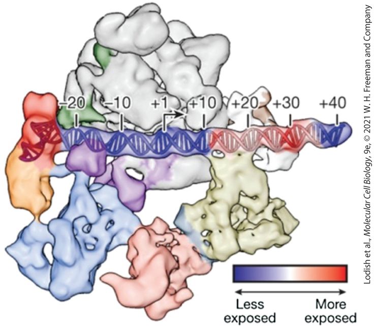
- Regulatory Elements in Eukaryotic DNA Are Found Both Close to and Many Kilobases Away from Transcription Start Sites
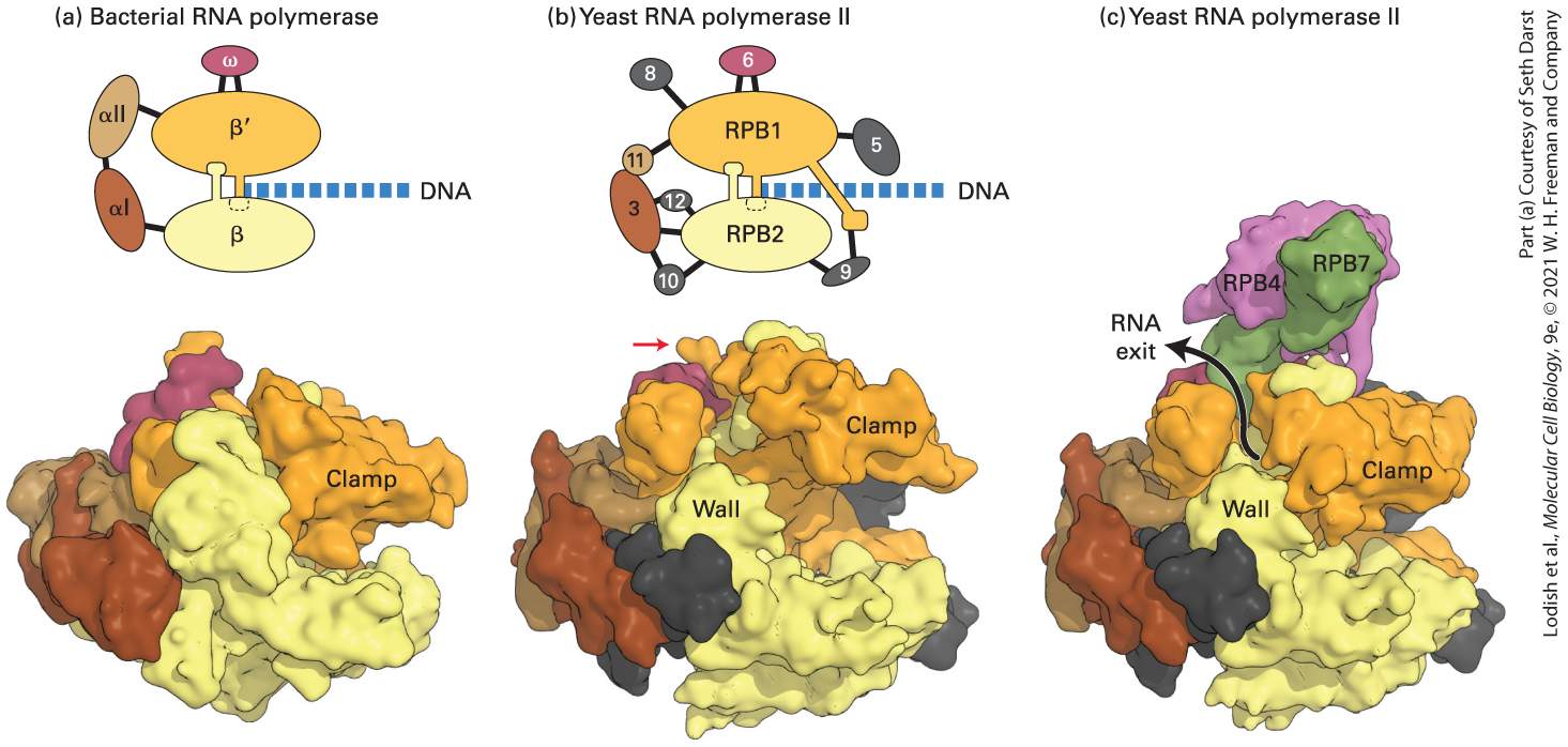
- Three Eukaryotic Nuclear RNA Polymerases Catalyze Formation of Different RNAs
- The Clamp Domain Enables RNA Polymerase II to Transcribe Long Stretches of DNA
- The Largest Subunit in RNA Polymerase II Has an Essential Carboxy-Terminal Repeat
- RNA Polymerase II Initiates Transcription at DNA Sequences Corresponding to the 5′ Cap of mRNAs
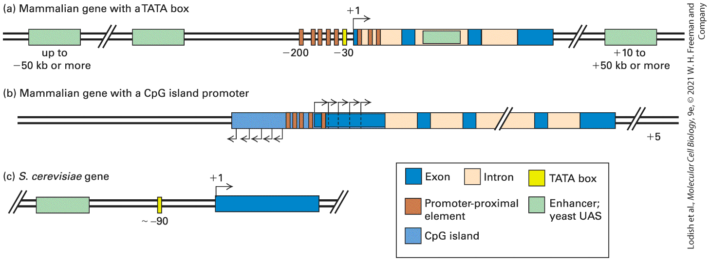
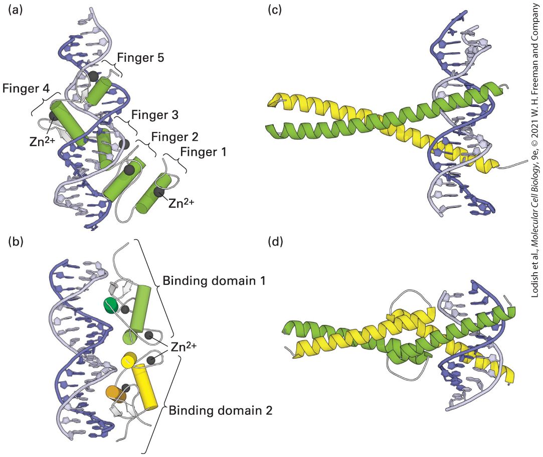
- The TATA Box, Initiators, and CpG Islands Function as Promoters in Eukaryotic DNA
- General Transcription Factors Position RNA Polymerase II at Transcription Start Sites and Assist in Initiation
- Elongation Factors Regulate the Initial Stages of Transcription in the Promoter-Proximal Region
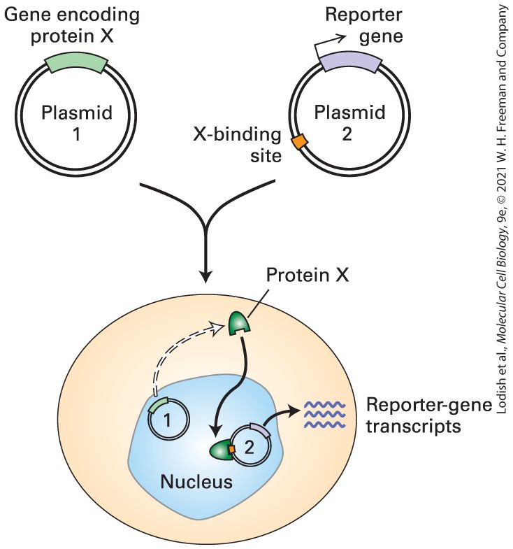
- Promoter-Proximal Elements Help Regulate Eukaryotic Genes
- Distant Enhancers Often Stimulate Transcription by RNA Polymerase II
- Most Eukaryotic Genes Are Regulated by Multiple Transcription-Control Elements
Ch 9Post-Transcriptional Gene ControlRead full chapter →
Chapter 9 Post-Transcriptional Gene Control Portion of a “lampbrush chromosome” from an oocyte of the newt Nophthalmus viridescens. The hnRNP protein associated with nascent RNA transcripts fluoresces red after staining with a monoclonal antibody.
Sections in this chapter
- 9.1 Processing of Eukaryotic Pre-mRNA
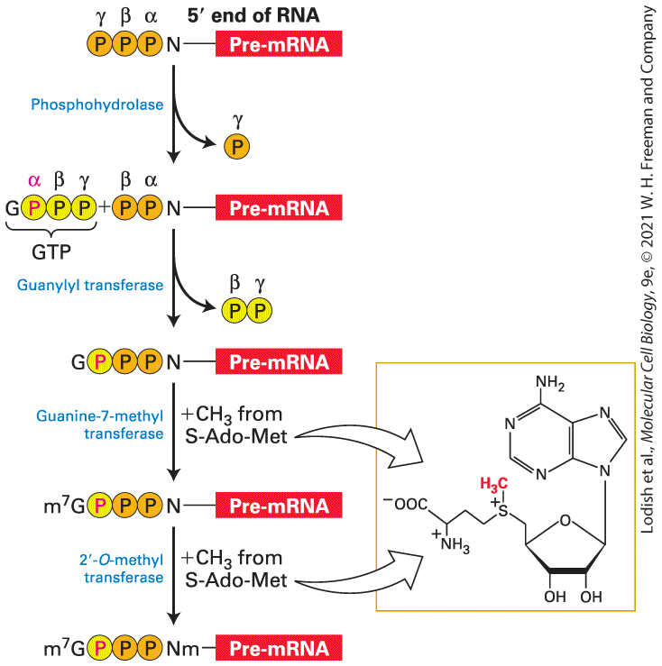
- The 5′ Cap Is Added to Nascent RNAs Shortly After Transcription Initiation
- Chain Elongation by RNA Polymerase II Is Coupled to the Presence of RNA Processing Factors
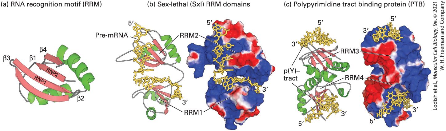
- A Diverse Set of Proteins with Conserved RNA-Binding Domains Associate with Pre-mRNAs
- Splicing Occurs at Short, Conserved Sequences in Pre-mRNAs via Two Transesterification Reactions
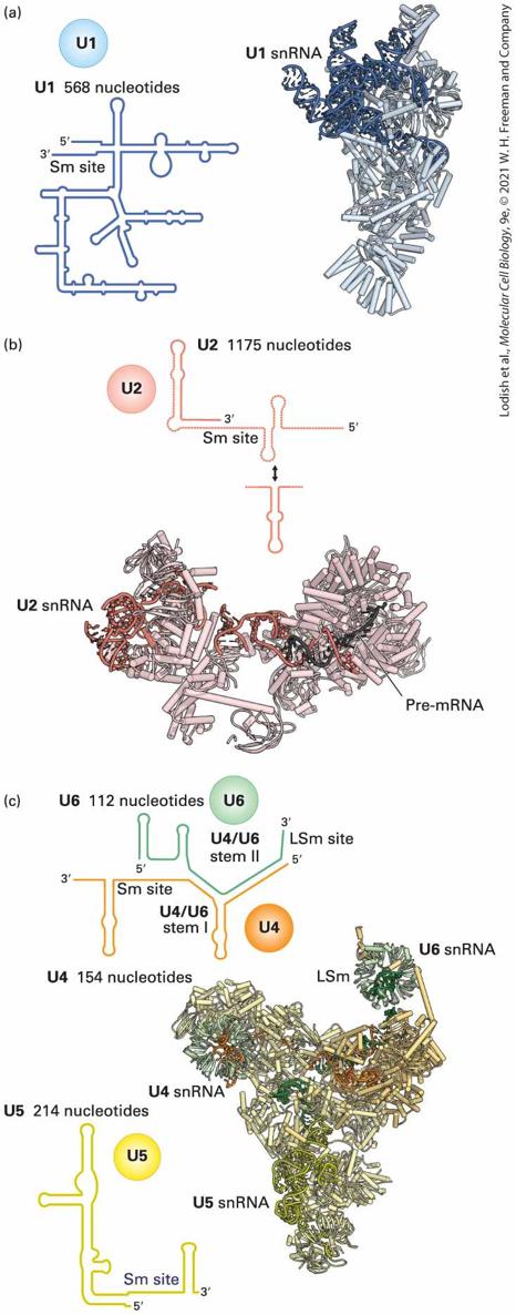
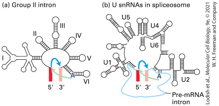
- During Splicing, snRNAs Base-Pair with Pre-mRNA to Select Splice Sites and Guide the Transesterification Reactions
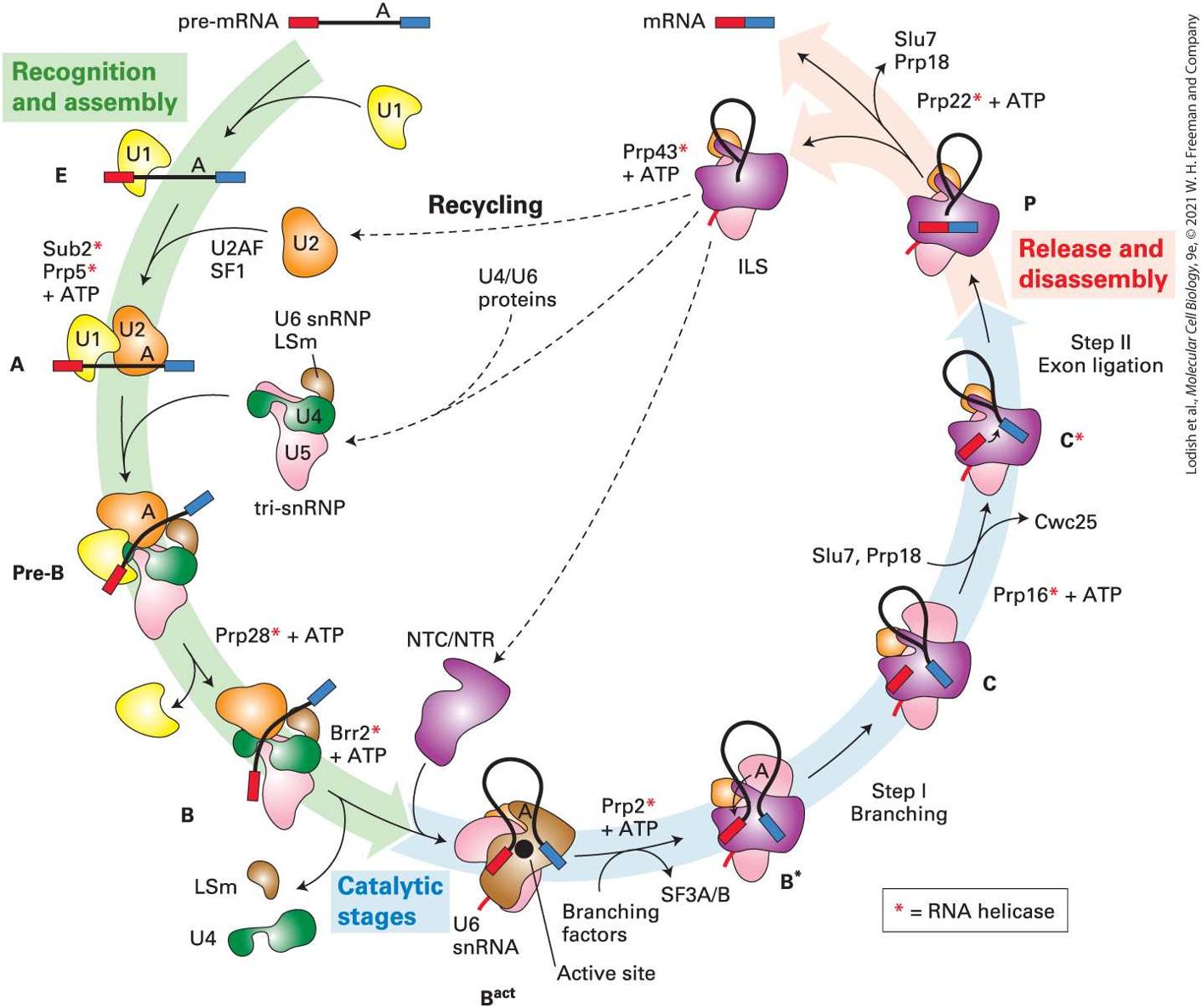
- Spliceosomes Catalyze Pre-mRNA Splicing
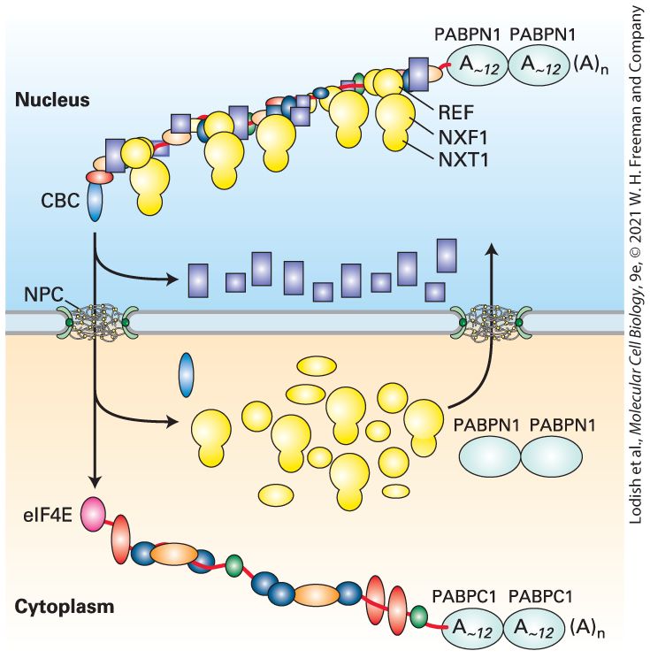
- 3′ Cleavage and Polyadenylation of Pre-mRNAs Are Tightly Coupled
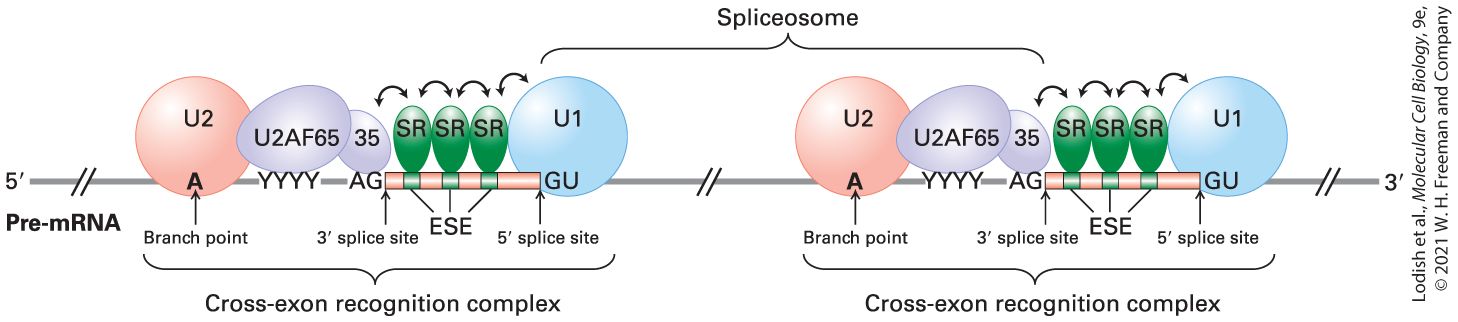
- Additional Nuclear Proteins Contribute to Splice-Site Selection in the Long Pre-mRNAs of Humans and Other Vertebrates
- Expression and Function of Related K+-Channel Protein Isoforms in Vertebrate Inner Ear Hair Cells
- Regulation of RNA Splicing Through Splicing Enhancers and Silencers Controls Drosophila Sexual Differentiation
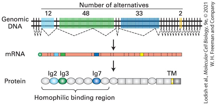
- Expression of Dscam Isoforms in Drosophila Retinal Neurons
Ch 10Biomembrane StructureRead full chapter →
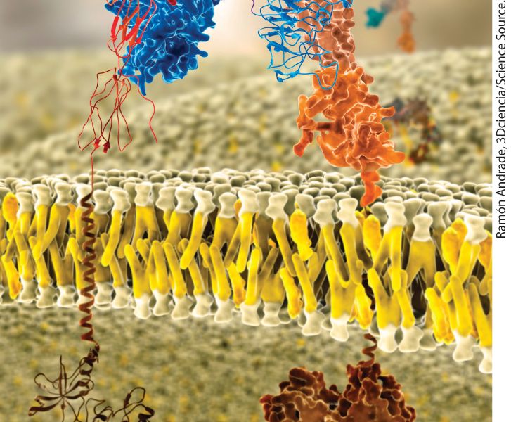
Chapter 10 Biomembrane Structure Molecular model of a lipid bilayer with embedded membrane proteins. Integral membrane proteins have distinct exoplasmic, cytosolic, and membrane-spanning domains. Shown here are portions of the insulin receptor, which regulates cell metabolism.
Sections in this chapter
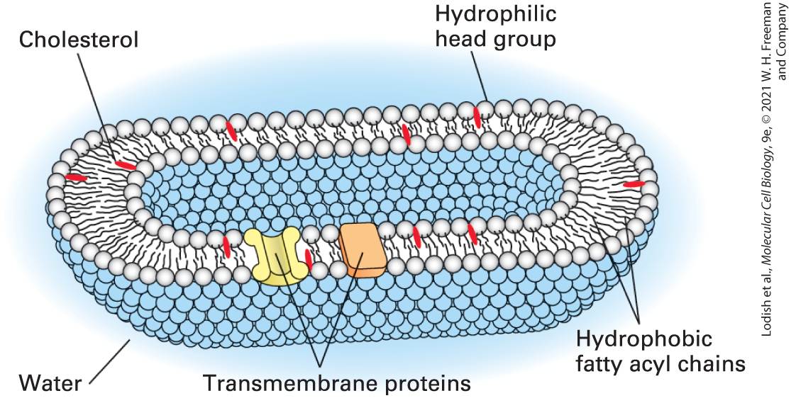
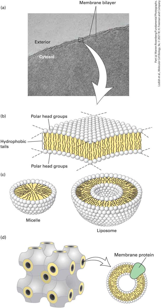
- Phospholipids Spontaneously Form Bilayers
- Phospholipid Bilayers Form a Sealed Compartment Surrounding an Internal Aqueous Space
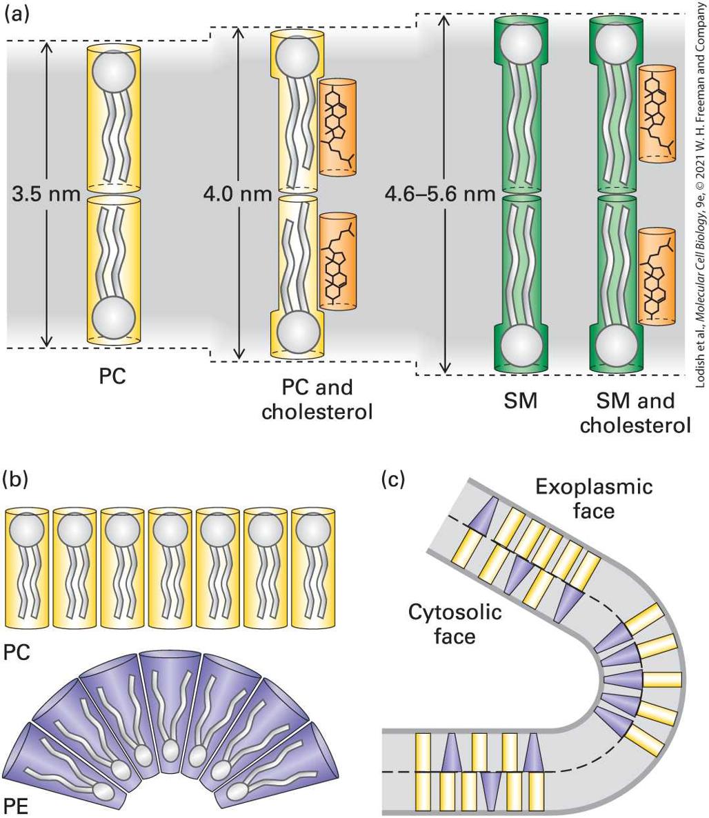
- Biomembranes Contain Three Principal Classes of Lipids
- Most Lipids and Many Proteins Are Laterally Mobile in Biomembranes
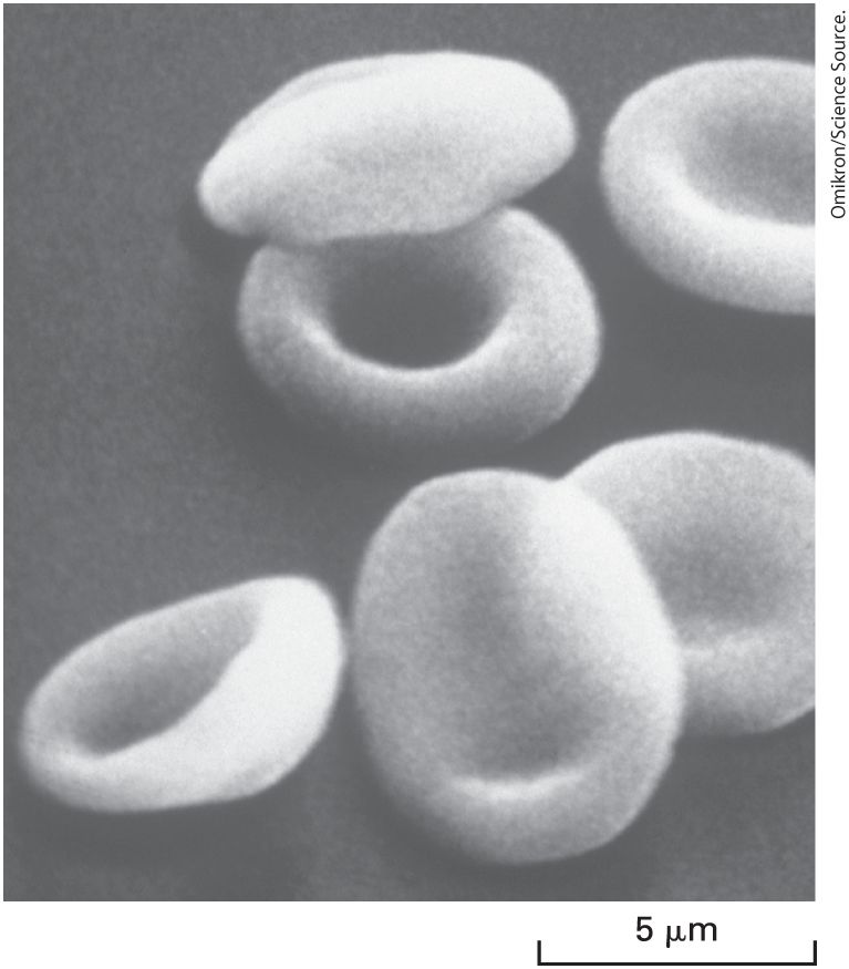
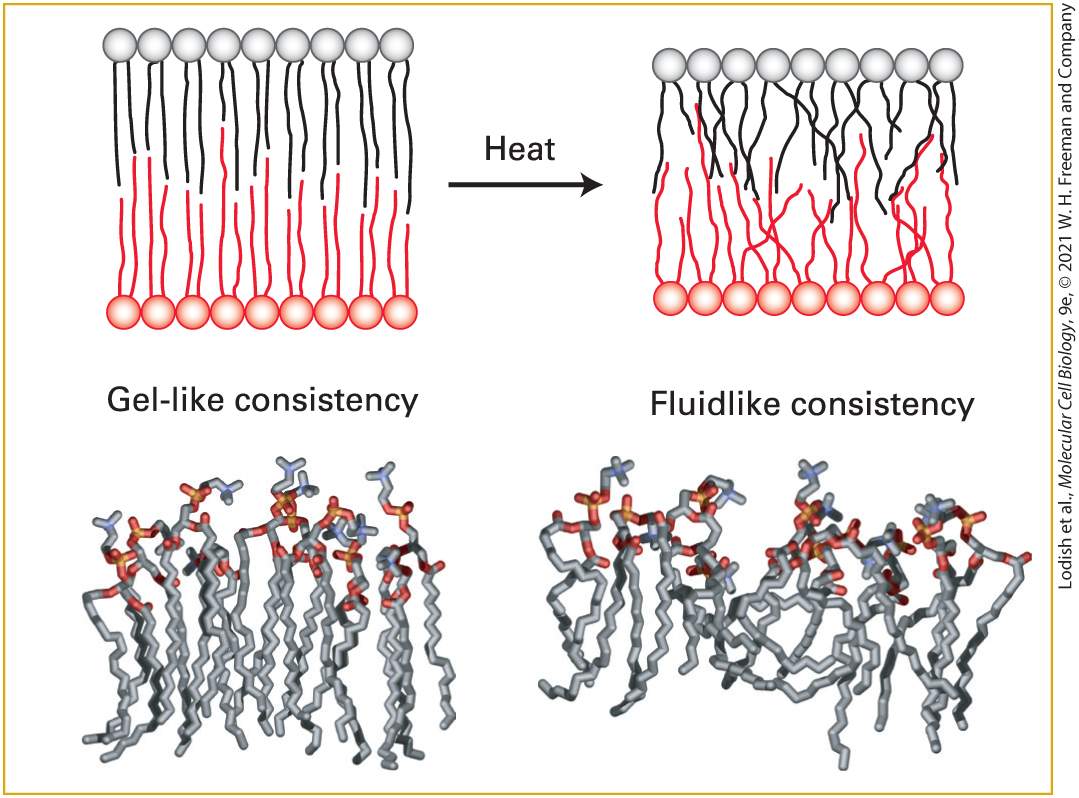
- Lipid Composition Influences the Physical Properties of Membranes
- Lipid Composition Is Different in the Exoplasmic and Cytosolic Leaflets
- Cholesterol and Sphingolipids Cluster with Specific Proteins in Membrane Microdomains
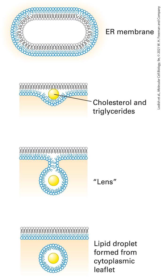
- Cells Store Excess Lipids in Lipid Droplets
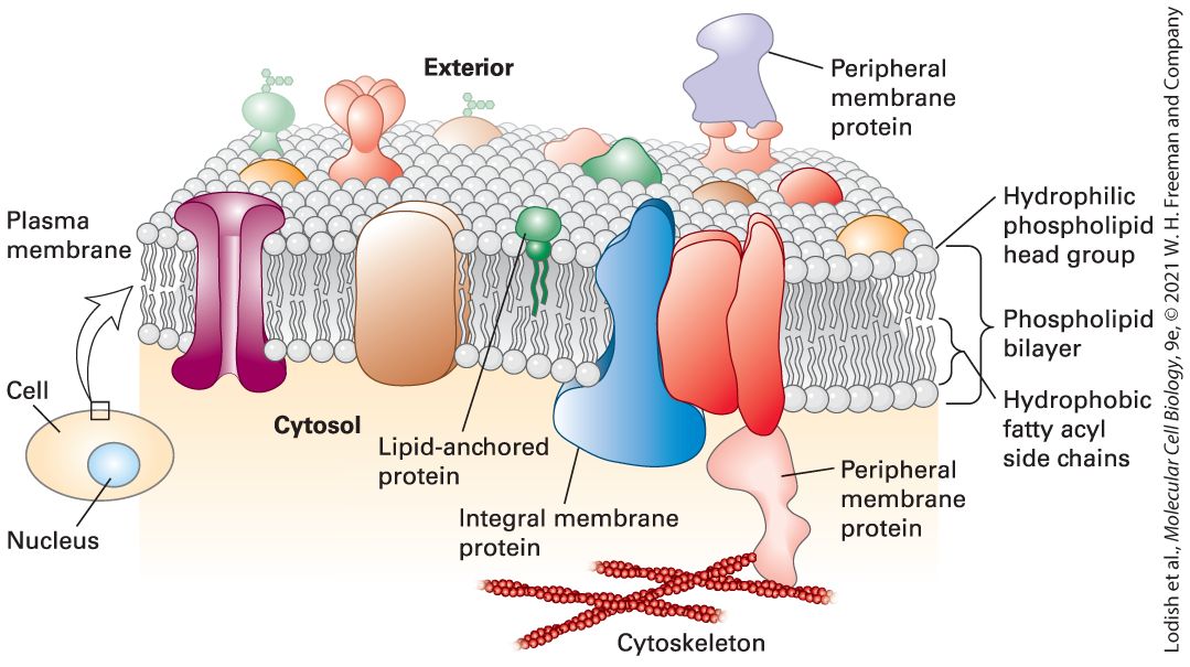
- 10.2 Membrane Proteins: Structure and Basic Functions
- Proteins Interact with Membranes in Three Different Ways
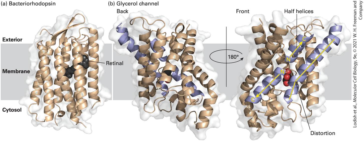
- Most Transmembrane Proteins Have Membrane-Spanning α Helices
- Multiple β Strands in Porins Form Membrane-Spanning “Barrels”
Ch 11Transmembrane Transport of Ions and Small MoleculesRead full chapter →
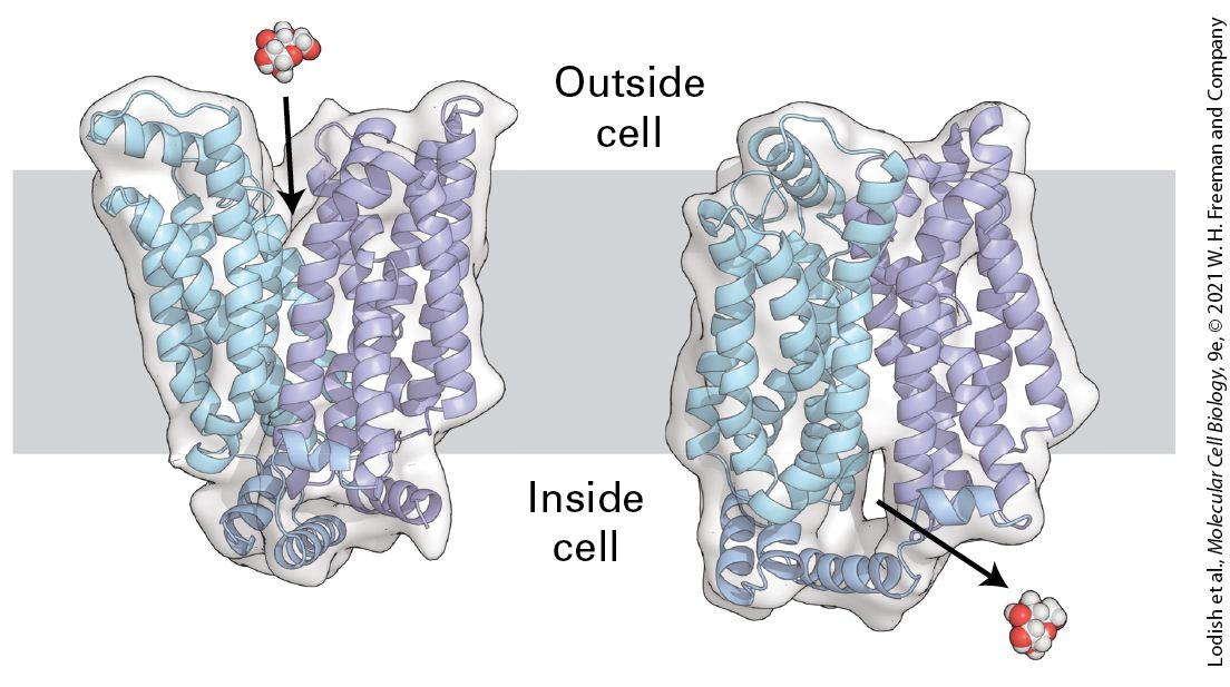
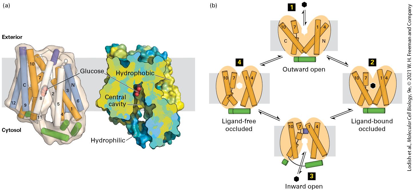
Chapter 11 Transmembrane Transport of Ions and Small Molecules Transmembrane, side view of the human glucose transporter GLUT1. Like other glucose transporters, GLUT1 transports glucose across the cell’s plasma membrane by alternating between an outward-facing and an inward-facing state. In the outward-facing state, a molecule of glucose binds to GLUT1. Glucose binding changes the conformation of the transporter such that it is now open to the inside of the cell, releasing glucose into the cytoplasm. [Data from D. Deng et al., 2014 Nature 510:121–125, PDB ID 4PYP.]
Sections in this chapter
- Only Gases and Small Uncharged Molecules Cross Membranes by Simple Diffusion
- Three Main Classes of Membrane Proteins Transport Molecules and Ions Across Cellular Membranes
- Uniport Transport Is Faster and More Specific than Simple Diffusion
- The Low Km of the GLUT1 Uniporter Enables It to Transport Glucose into Most Mammalian Cells
- The Human Genome Encodes a Family of Sugar-Transporting GLUT Proteins
- Transport Proteins Can Be Studied Using Artificial Membranes and Recombinant Cells
- Osmotic Pressure Causes Water to Move Across Membranes
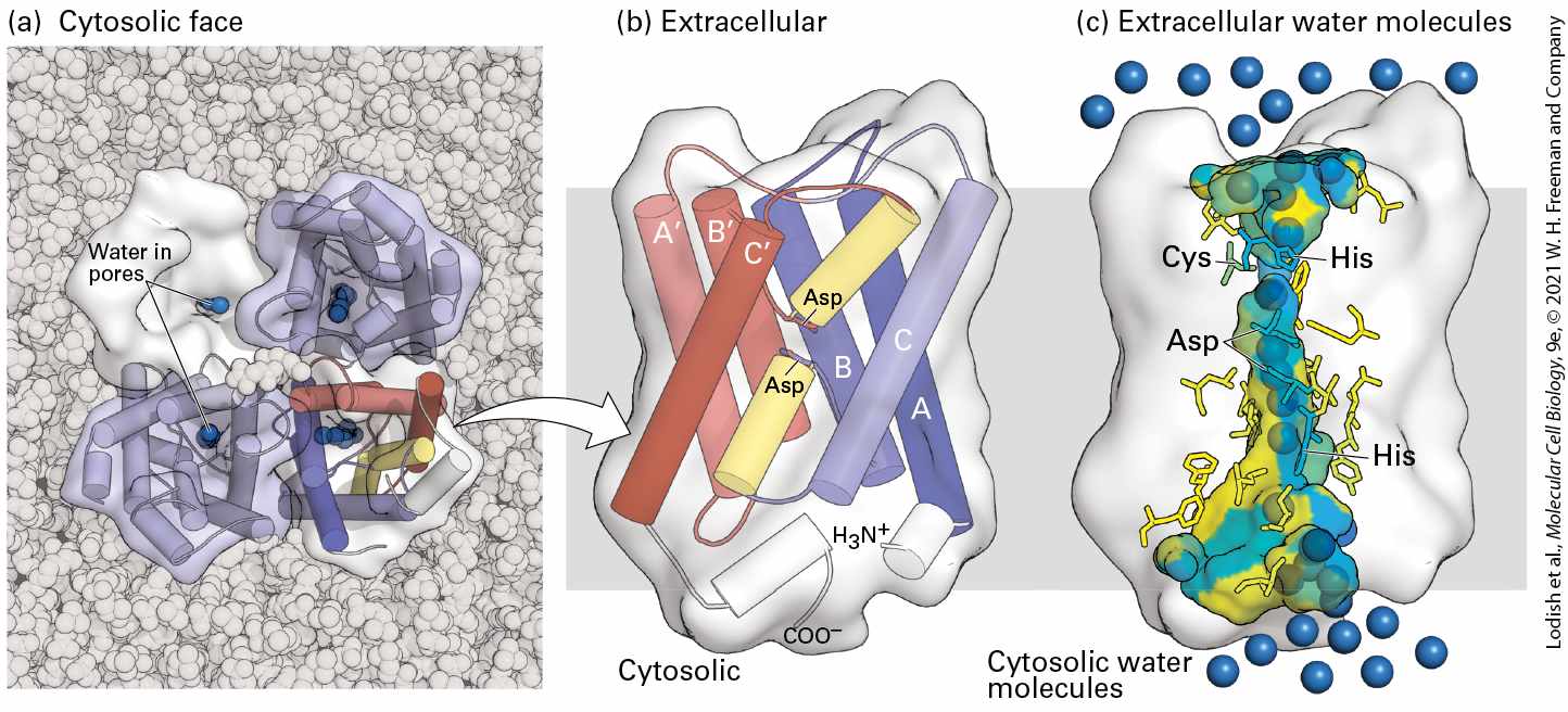
- Aquaporins Increase the Water Permeability of Cellular Membranes
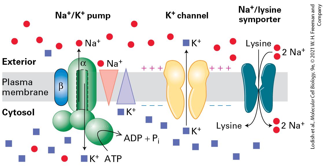
- There Are Four Main Classes of ATP-Powered Pumps
- ATP-Powered Ion Pumps Generate and Maintain Ionic Gradients Across Cellular Membranes
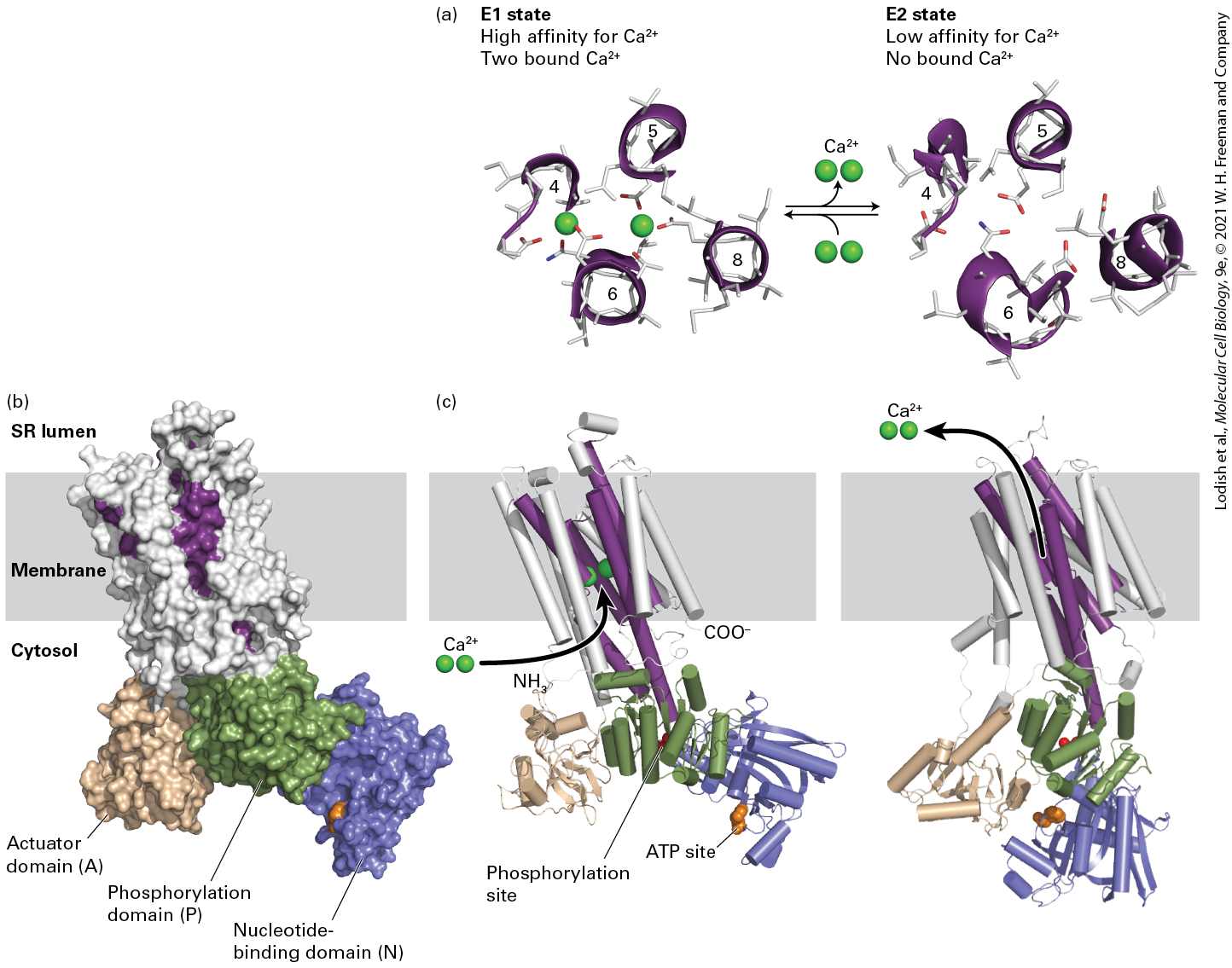
- Muscle Relaxation Depends on Ca2+ ATPases That Pump Ca2+ from the Cytosol into the Sarcoplasmic Reticulum
- The Mechanism of Action of the Ca2+ Pump Is Known in Detail
Ch 12Cellular EnergeticsRead full chapter →
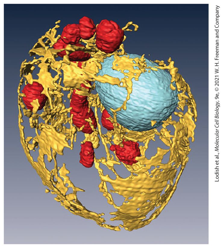
Chapter 12 Cellular Energetics Computer generated image of chromatophores in the photosynthetic purple bacterium Rhodobacter sphaeroides. Each chromatophore contains membrane proteins that use light to drive the synthesis of ATP. Energy is absorbed by pigments in two types of light-harvesting complexes (green and red) and transferred to reaction centers (light blue) where high-energy electrons are generated. Their energy is used by pumps (purple) to move protons into the chromatophores, generating a proton gradient. This powers ATP synthase (orange) to convert ADP + P to ATP. i
Sections in this chapter
- 12.1 Chemiosmosis, Electron Transport, the Proton-Motive Force, and ATP Synthesis
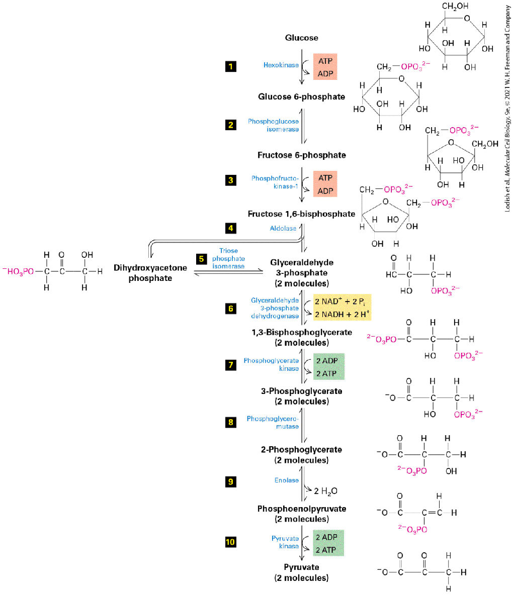
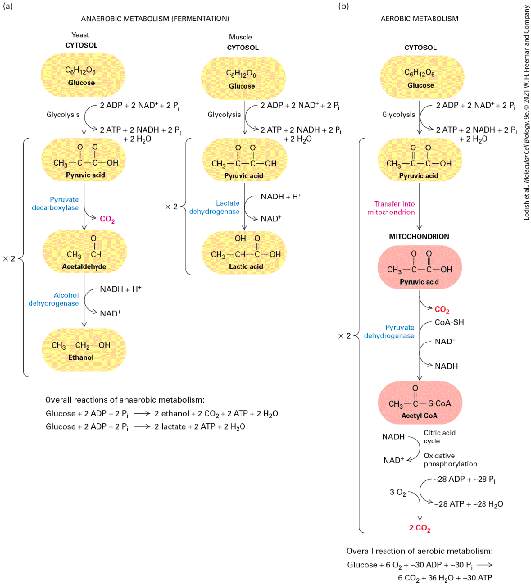
- 12.2 First Step of Harvesting Energy from Glucose: Glycolysis
- During Glycolysis (Stage I), Cytosolic Enzymes Convert Glucose to Pyruvate
- The Rate of Glycolysis Is Adjusted to Meet the Cell’s Need for ATP
- Glucose Is Fermented When Oxygen Is Scarce
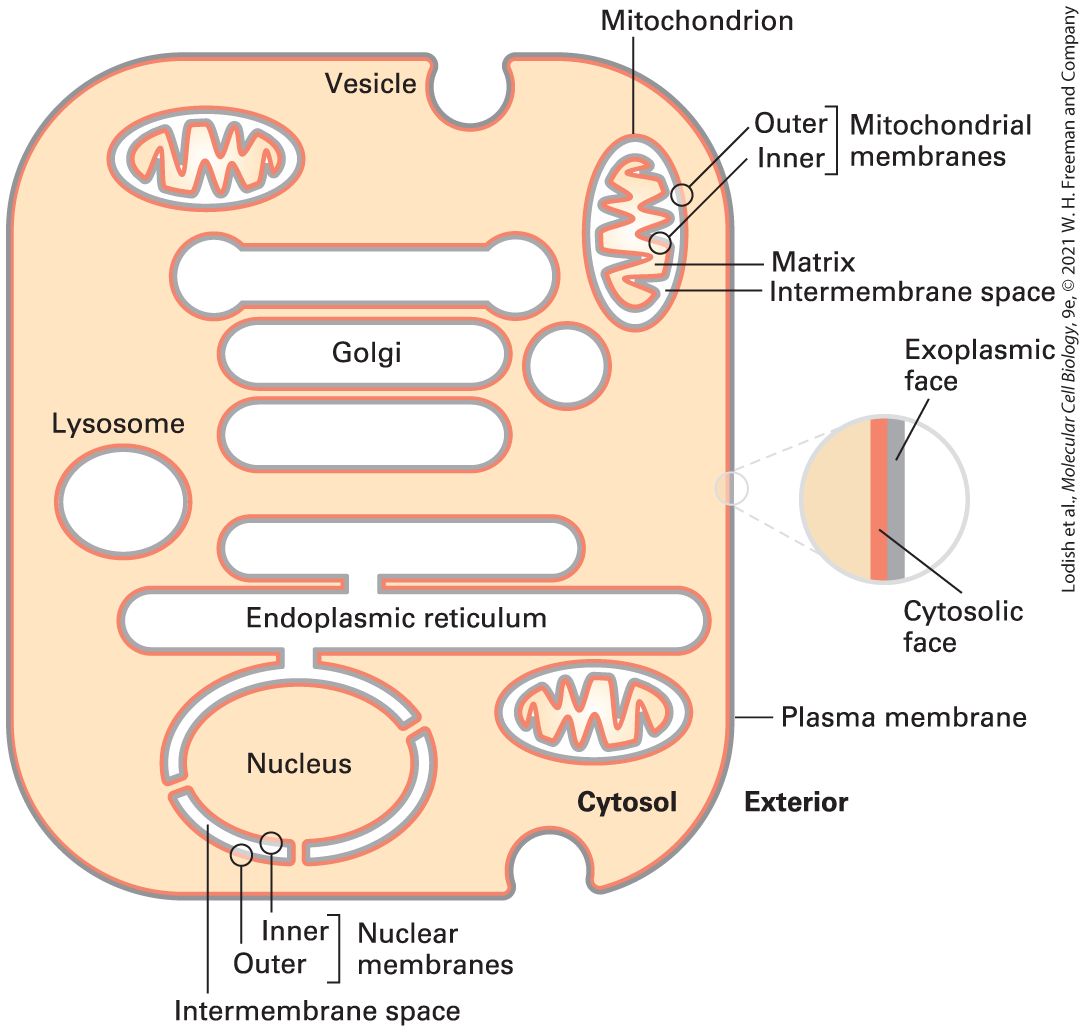
- Mitochondria Are Abundant, Multifunctional Organelles
- Mitochondria Have Two Structurally and Functionally Distinct Membranes
- Mitochondria Contain DNA and Evolved from a Single Endosymbiotic Event Involving Alphaproteobacterium
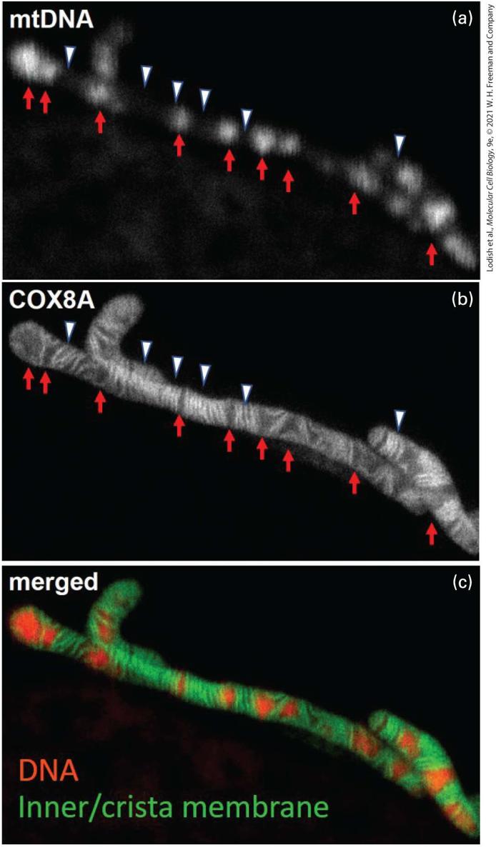
- The Size, Structure, and Coding Capacity of mtDNA Vary Considerably Among Organisms
- Mitochondrial DNA Is Located in the Matrix and Transferred During Mitosis to Daughter Cells by Cytoplasmic Inheritance
- Products of Mitochondrial Genes Are Not Exported
- Mutations in Mitochondrial DNA Cause Several Genetic Diseases in Humans
Ch 13Moving Proteins into Membranes and OrganellesRead full chapter →
A three-dimensional reconstruction of the internal membranes of a yeast cell using scanning electron microscopy. The cell wall has been removed and the organelles highlighted with false color to reveal the endoplasmic reticulum (yellow), mitochondria (red), and nucleus (blue). Cell diameter is . [From D. Wei et al., 2012, “High-Resolution Three-Dimensional Reconstruction of a Whole Yeast Cell Using Focused-Ion Beam Scanning Electron Microscopy,” Biotechniques 53(1):41–48.]
Sections in this chapter
- 13.1 Targeting Proteins to and Across the ER Membrane
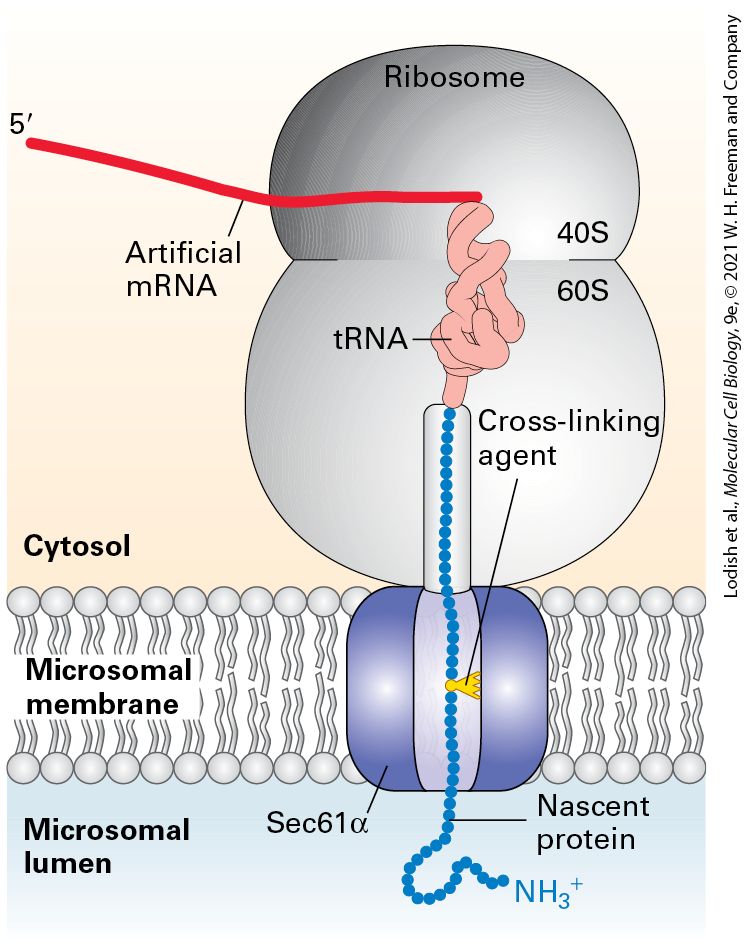
- Pulse-Chase Experiments with Purified ER Membranes Demonstrated That Secreted Proteins Cross the ER Membrane
- A Hydrophobic N-Terminal Signal Sequence Targets Nascent Secretory Proteins to the ER
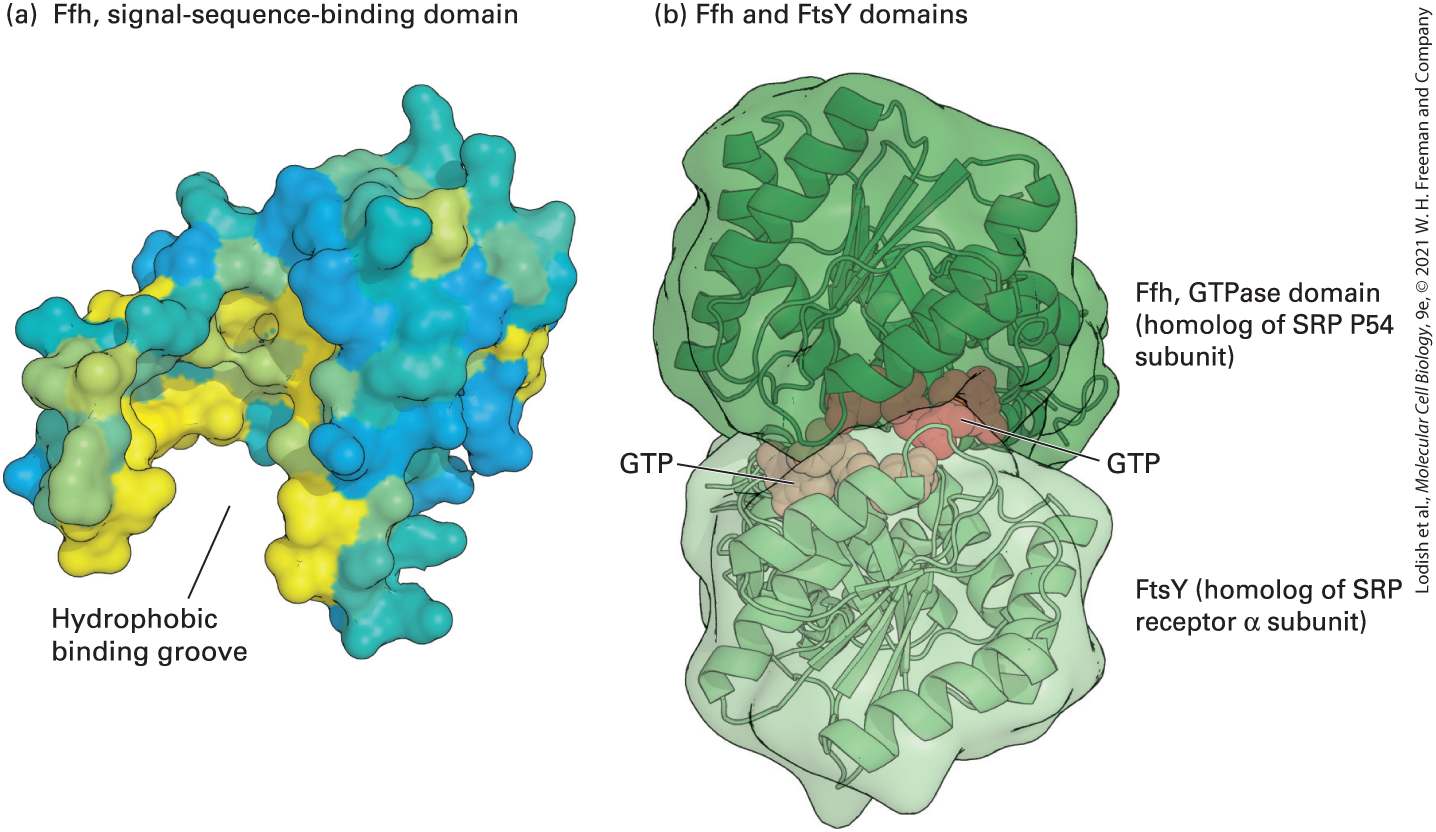
- Cotranslational Translocation Is Initiated by Two GTP-Hydrolyzing Proteins
- Passage of Growing Polypeptides Through the Translocon Is Driven by Translation
- ATP Hydrolysis Powers Post-Translational Translocation of Some Secretory Proteins in Yeast
- 13.2 Insertion of Membrane Proteins into the ER
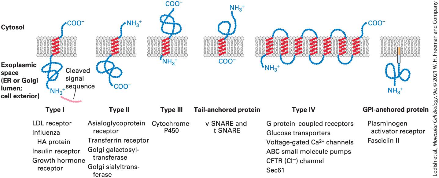
- Several Topological Classes of Integral Membrane Proteins Are Synthesized on the ER
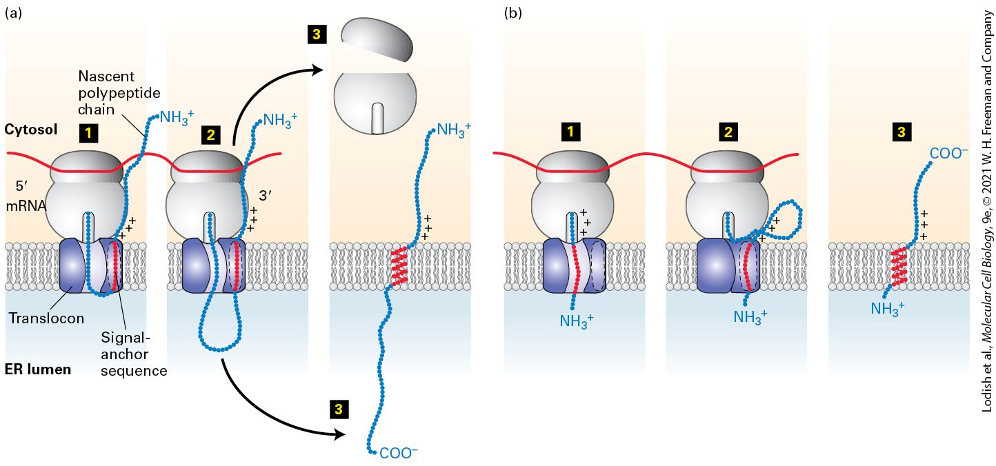
- Internal Stop-Transfer Anchor and Signal-Anchor Sequences Determine Topology of Single-Pass Proteins
- Type IV (Multipass) Proteins
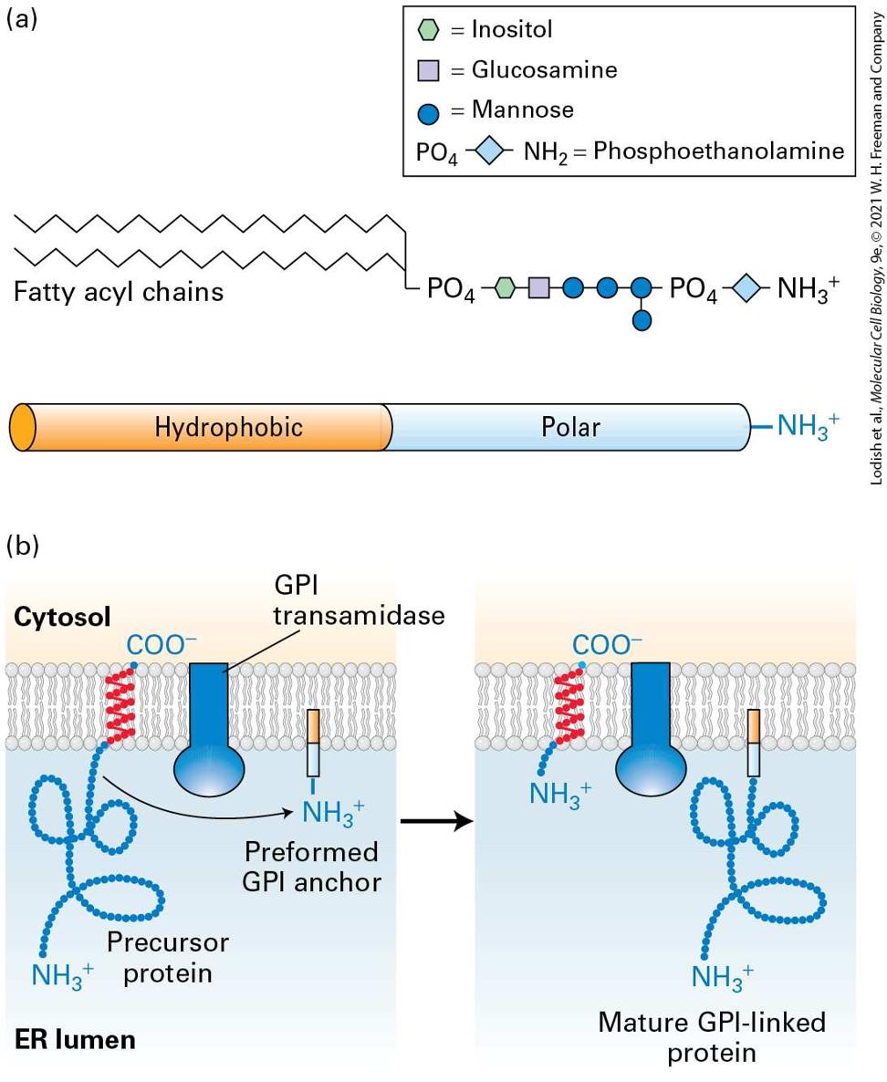
- A Phospholipid Anchor Tethers Some Cell-Surface Proteins to the Membrane
- The Topology of a Membrane Protein Can Often Be Deduced from Its Sequence
Ch 14Vesicular Traffic, Secretion, and EndocytosisRead full chapter →
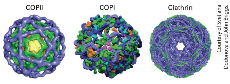
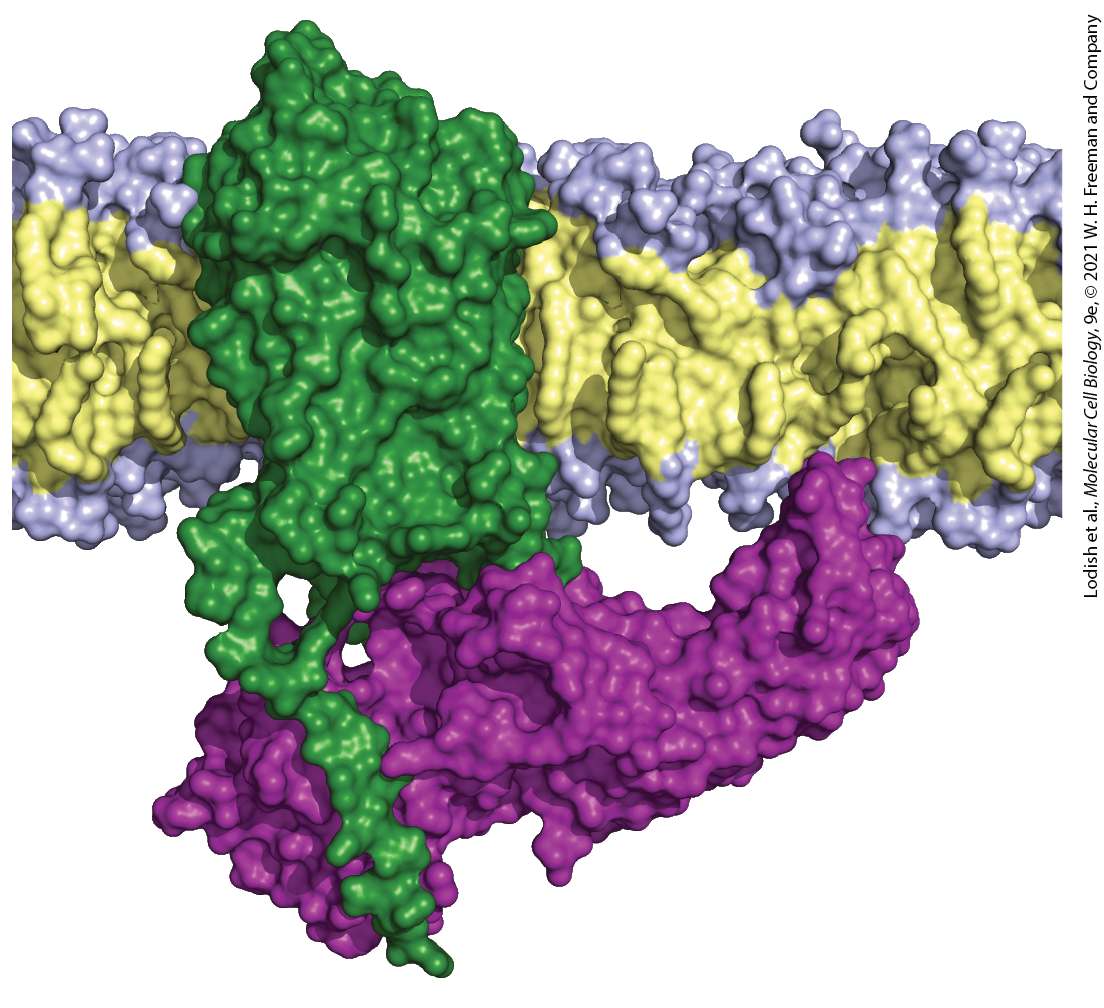
Chapter 14 Vesicular Traffic, Secretion, and Endocytosis Structural models of the coat protein complexes for three types of transport vesicles. The coat complexes are responsible for sculpting the curvature of the vesicle membrane and for selecting the types of cargo proteins incorporated into the vesicle. [Data from A. J. Noble and S. M. Stagg, 2015, Science 349(6244):142–143; https://doi.org/10.1126/science.aac6537.]
Sections in this chapter
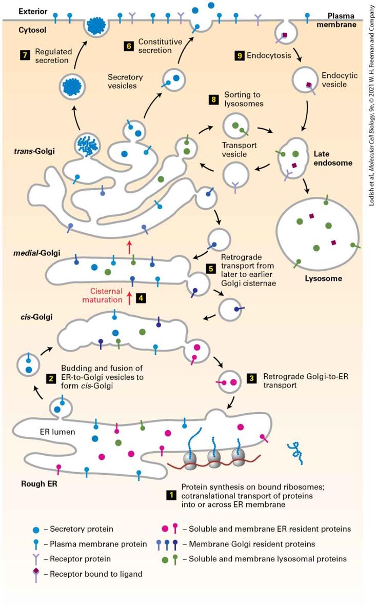
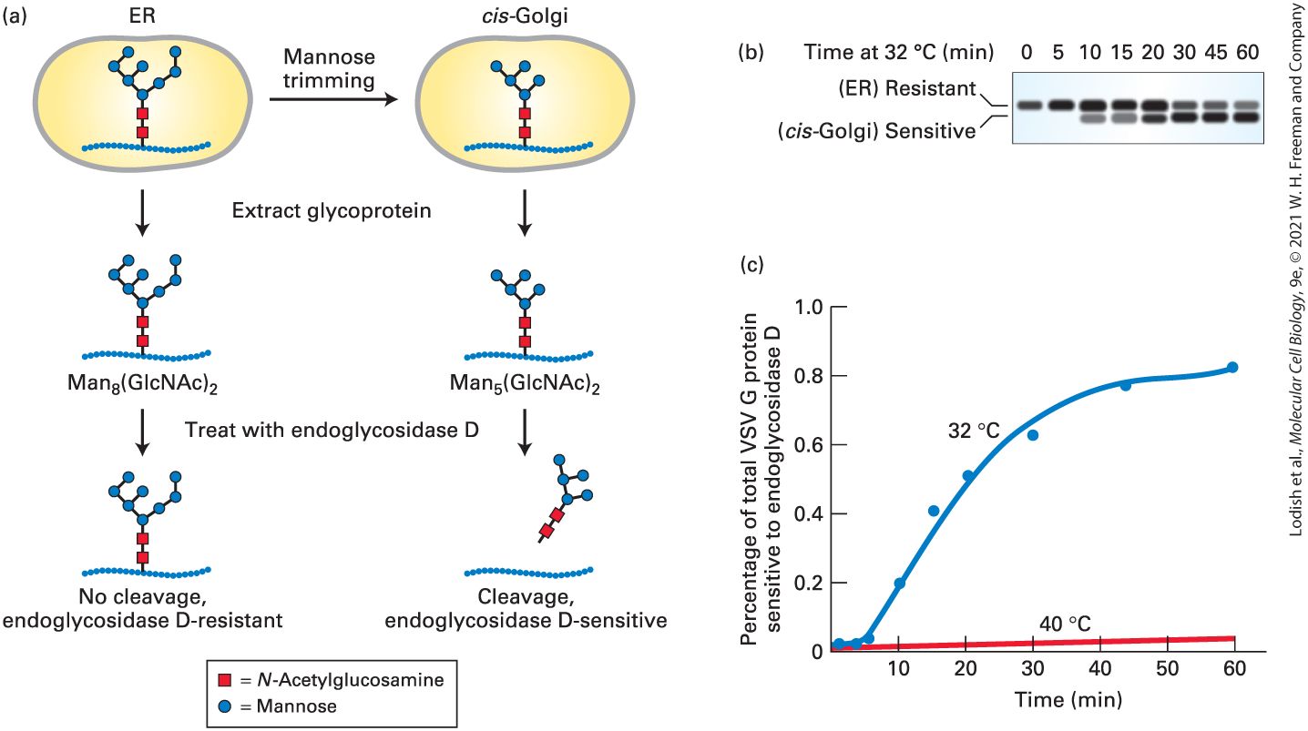
- Transport of a Protein Through the Secretory Pathway Can Be Assayed in Live Cells
- Yeast Mutants Define Major Stages and Components of Vesicular Transport
- Cell-Free Transport Assays Allow Dissection of Individual Steps in Vesicular Transport
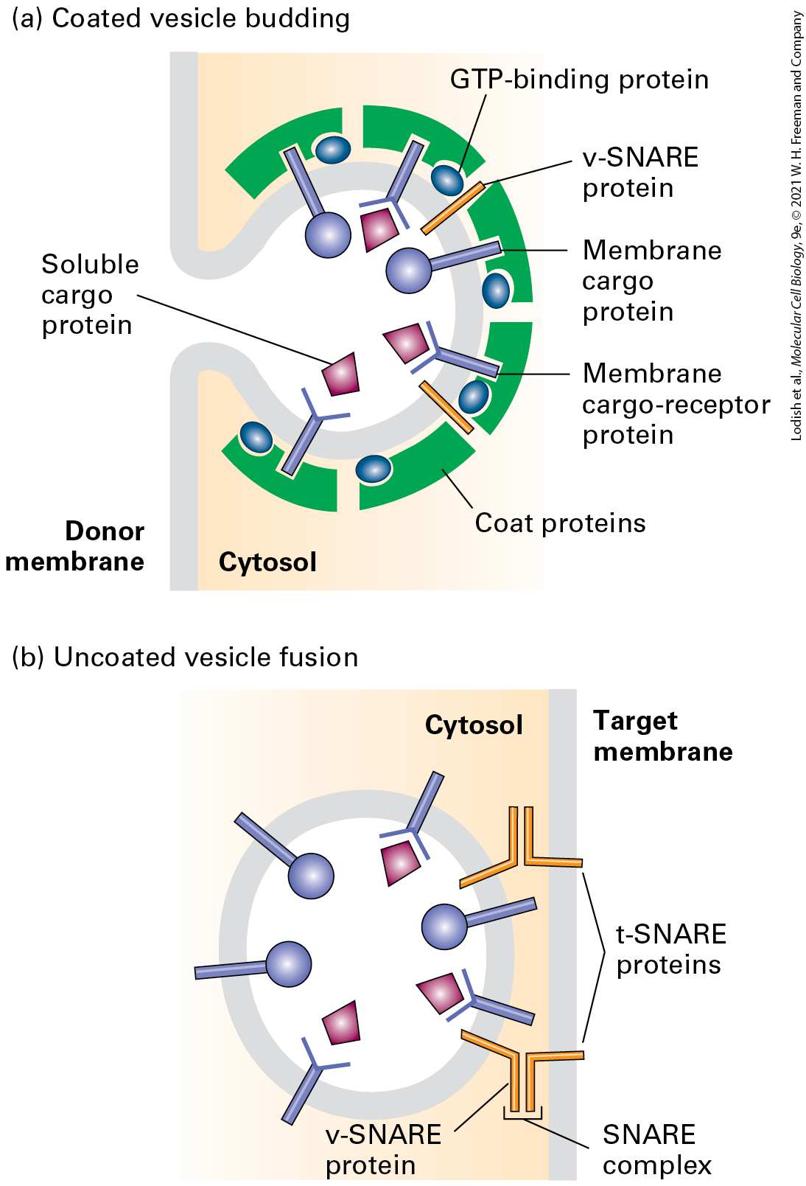
- Assembly of a Protein Coat Drives Vesicle Formation and Selection of Cargo Molecules
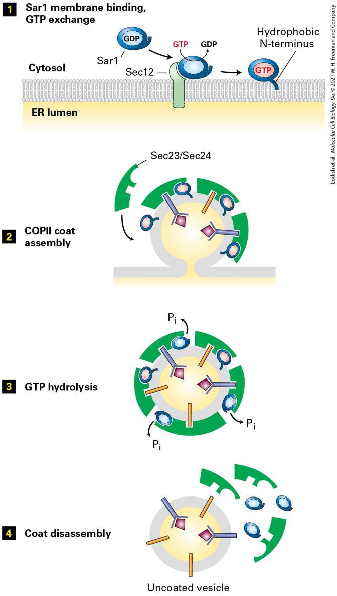
- A Conserved Set of GTPase Switch Proteins Controls the Assembly of Different Vesicle Coats
- Targeting Sequences on Cargo Proteins Make Specific Molecular Contacts with Coat Proteins
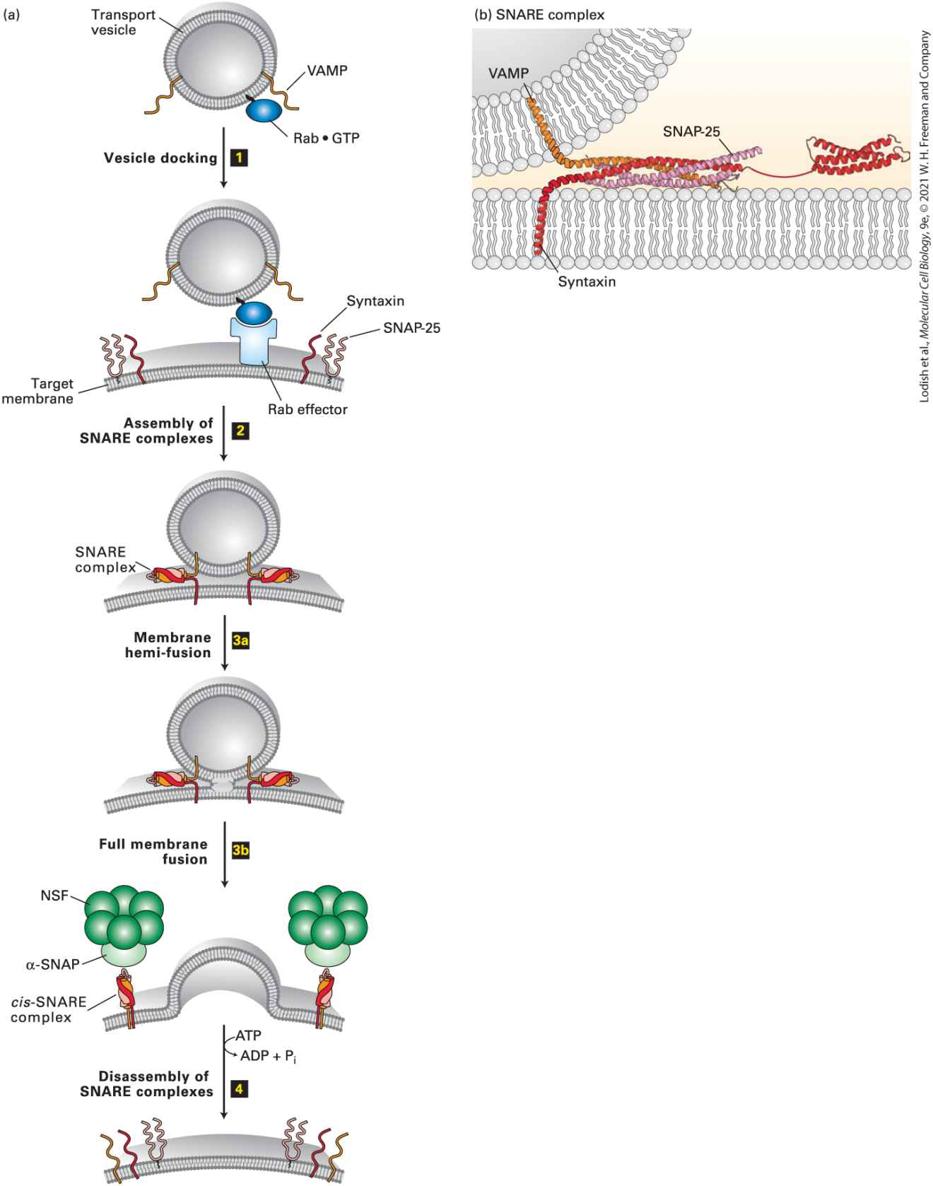
- Rab GTPases Control Docking of Vesicles on Target Membranes
- Paired Sets of SNARE Proteins Mediate Fusion of Vesicles with Target Membranes
- Dissociation of SNARE Complexes After Membrane Fusion Is Driven by ATP Hydrolysis
- 14.3 Early Stages of the Secretory Pathway
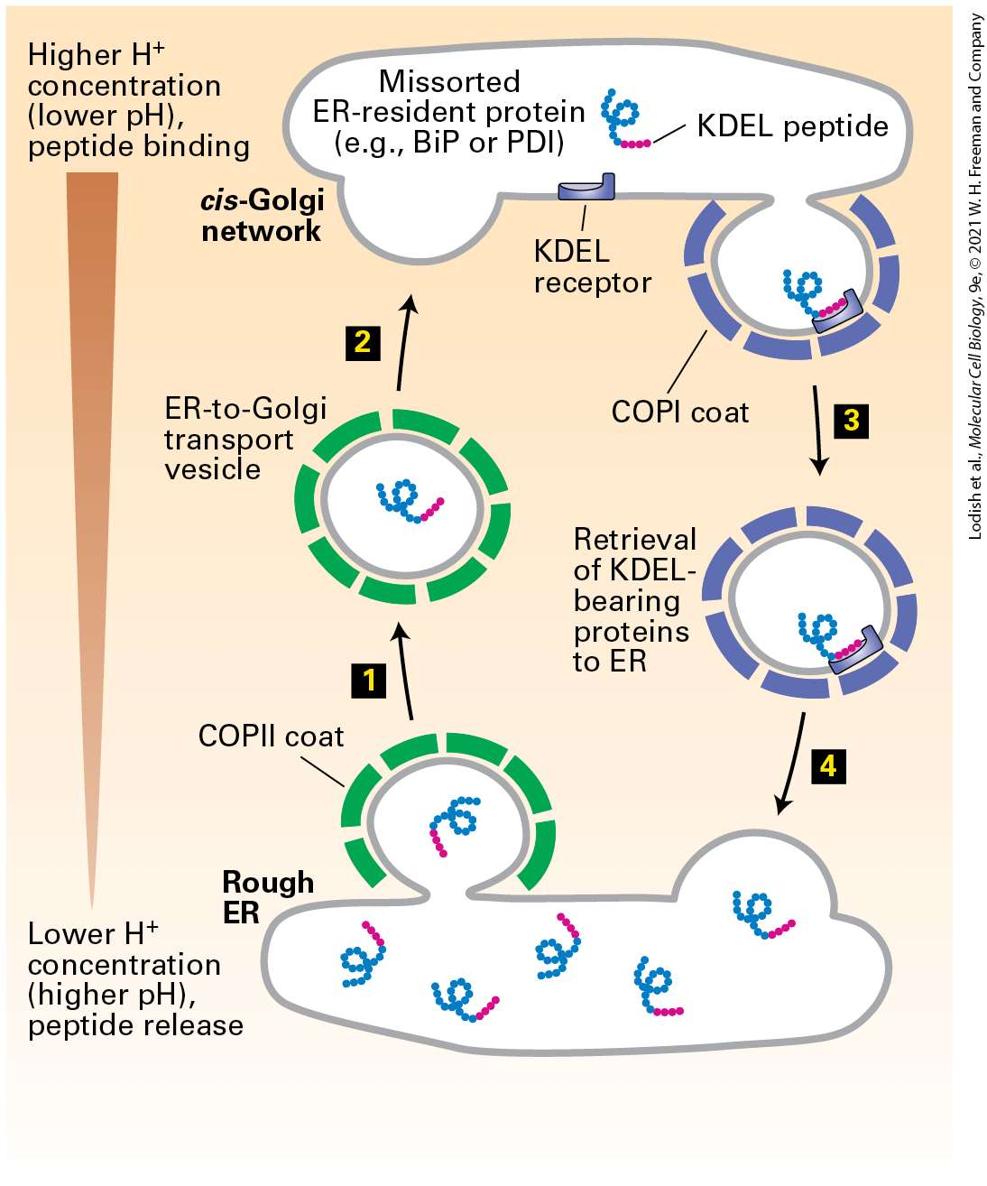
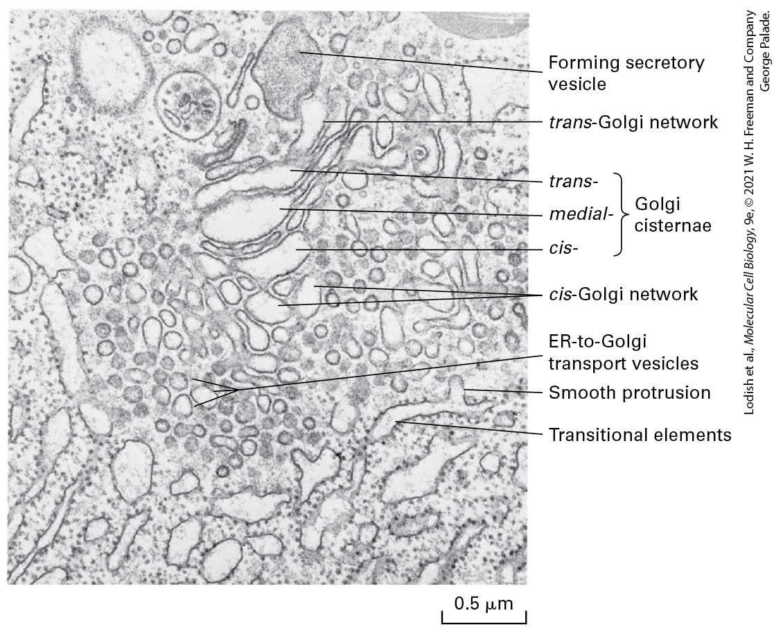
- COPII Vesicles Mediate Transport from the ER to the Golgi
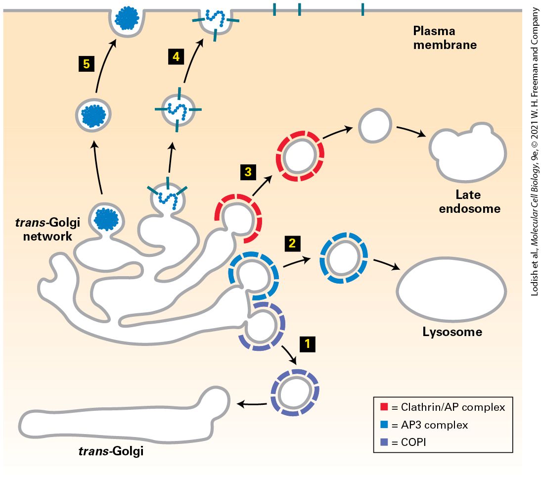
- COPI Vesicles Mediate Retrograde Transport Within the Golgi and from the Golgi to the ER
Ch 15Receptors, Hormones, and Cell SignalingRead full chapter →
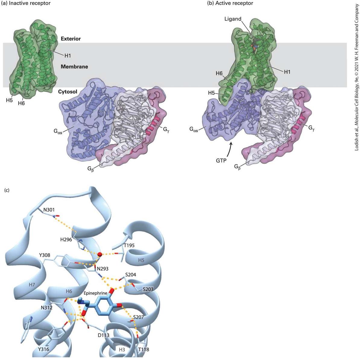
Chapter 15 Receptors, Hormones, and Cell Signaling Structure of a cell surface G protein–coupled receptor (green) bound to β-arrestin (purple). G protein–coupled receptors that are in the active state for a long period of time become phosphorylated, triggering binding of an arrestin and inhibition of further signaling by the receptor. [Data from Y. Kang et al., 2015, Nature 523:561–567, PDB ID 4zwj, and custom PDB.]
Sections in this chapter
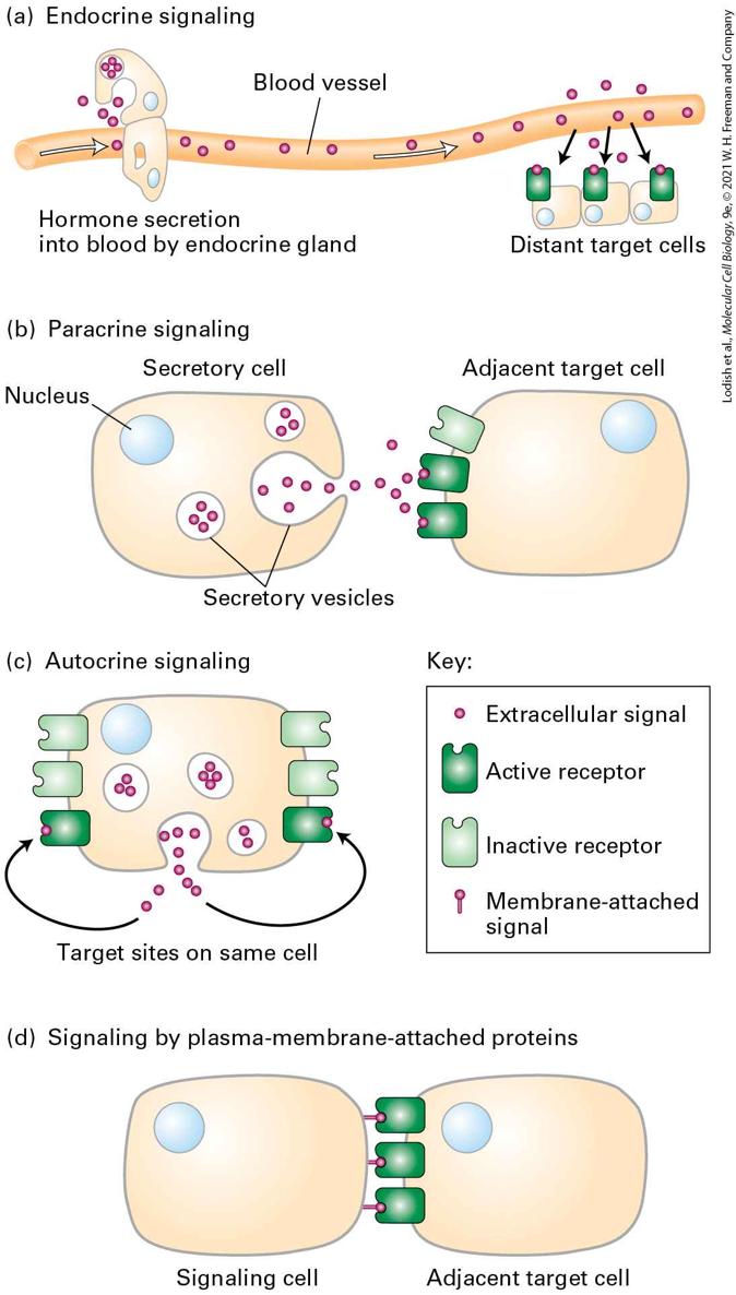
- Signaling Molecules Can Act Locally or at a Distance
- Signal Transduction Pathways Can Produce Rapid, Short-Term or Slow, Long-Term Changes in Cells, or Both
- Receptors Are Allosteric Proteins That Activate Signal Transduction Pathways
- Receptors Can Be in the Cytosol, Nucleus, or on the Cell Surface Membrane
- Most Receptors Bind Only a Single Type of Ligand or a Group of Closely Related Ligands
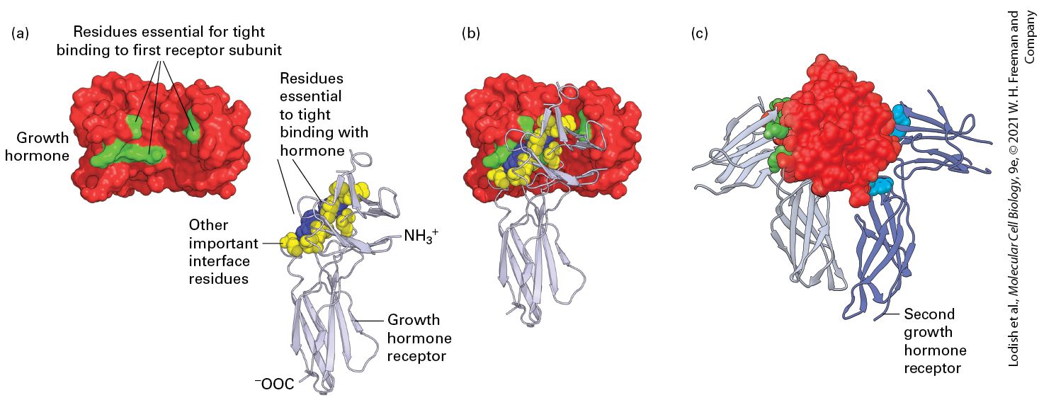
- Most Receptors Bind Their Ligands with High Affinity
- Second Messengers Are Used in Most Signal Transduction Pathways
- Protein Kinases and Phosphatases Participate in Signal Transduction Pathways by Covalently Modifying and Thus Activating or Inhibiting a Wide Variety of Proteins That Control Cellular States
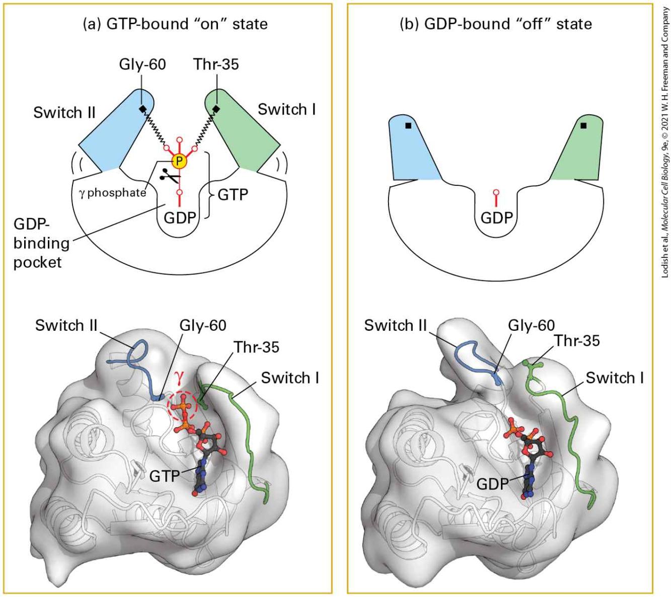
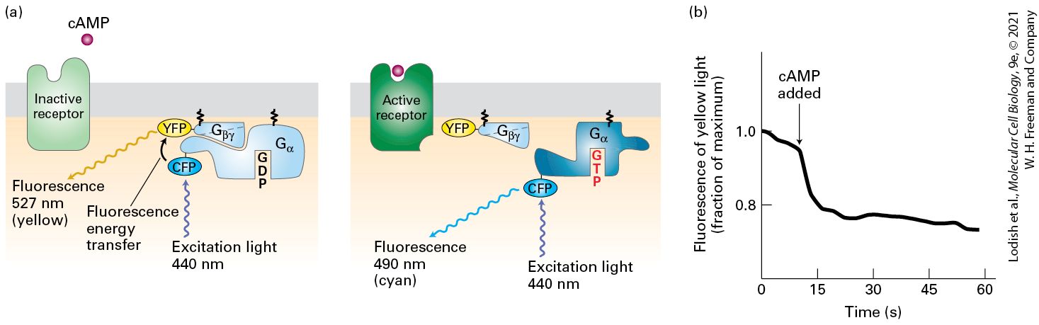
- GTP-Binding Proteins Are Frequently Used in Signal Transduction Pathways as On/Off Switches
- Signal Amplification and Feedback Repression Characterize Most Signal Transduction Pathways
- Binding Assays Are Used to Detect Receptors and Determine Their Affinity and Specificity for Ligands
- Near-Maximal Cellular Response to a Signaling Molecule Usually Does Not Require Activation of All Receptors
Ch 16Growth Factor and Cytokine Signaling Pathways That Control Gene ExpressionRead full chapter →
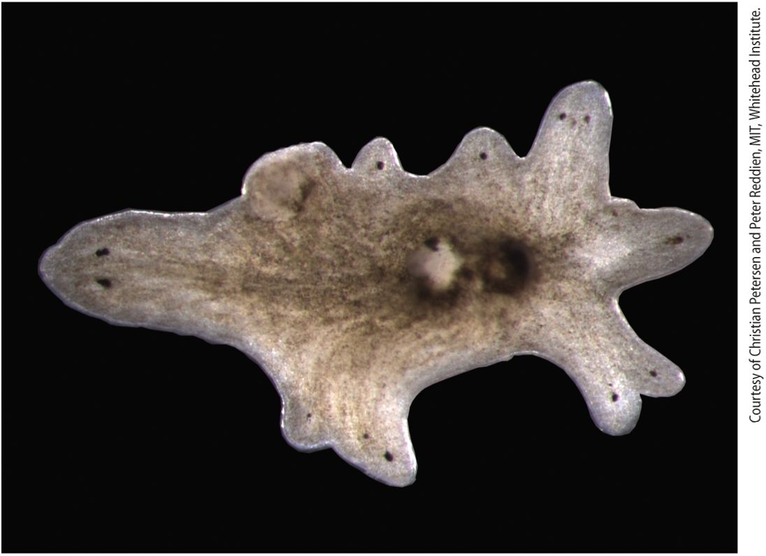
Chapter 16 Growth Factor and Cytokine Signaling Pathways That Control Gene Expression A planarian with multiple heads. Several hormones and their receptors regulate the regeneration of complex body parts in wounded planaria; the extracellular signaling protein Wnt promotes tail regeneration and inhibits head regeneration. In an experiment, the gene encoding beta-catenin-1, an essential protein in the Wnt signal transduction pathway, was inhibited by feeding animals an inhibitory double-stranded RNA. During the subsequent month, normal uninjured animals developed heads around their periphery in…
Sections in this chapter
- Binding of Ligand to the Extracellular Domain of an RTK Leads to Dimerization and Activation of Its Intrinsic Cytosolic Tyrosine Kinase
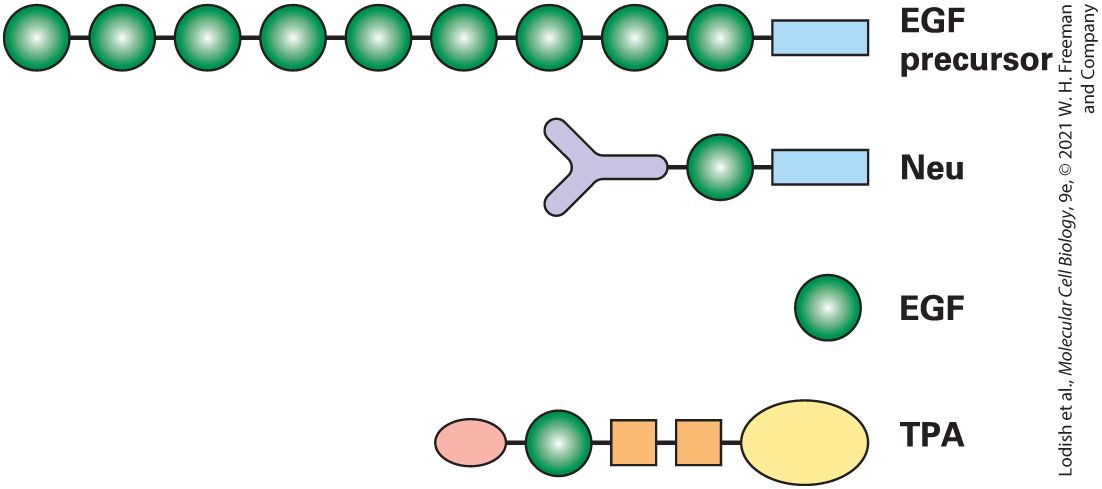
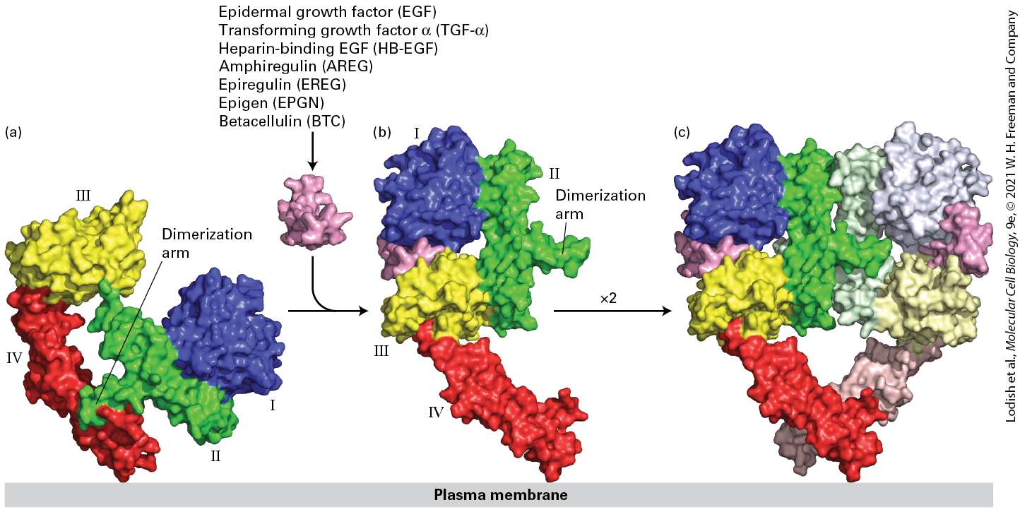
- Homo- and Hetero-Oligomers of Epidermal Growth Factor Receptors Bind Members of the Epidermal Growth Factor Family
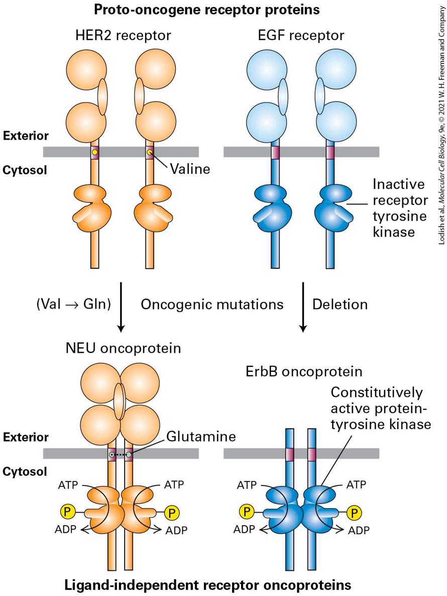
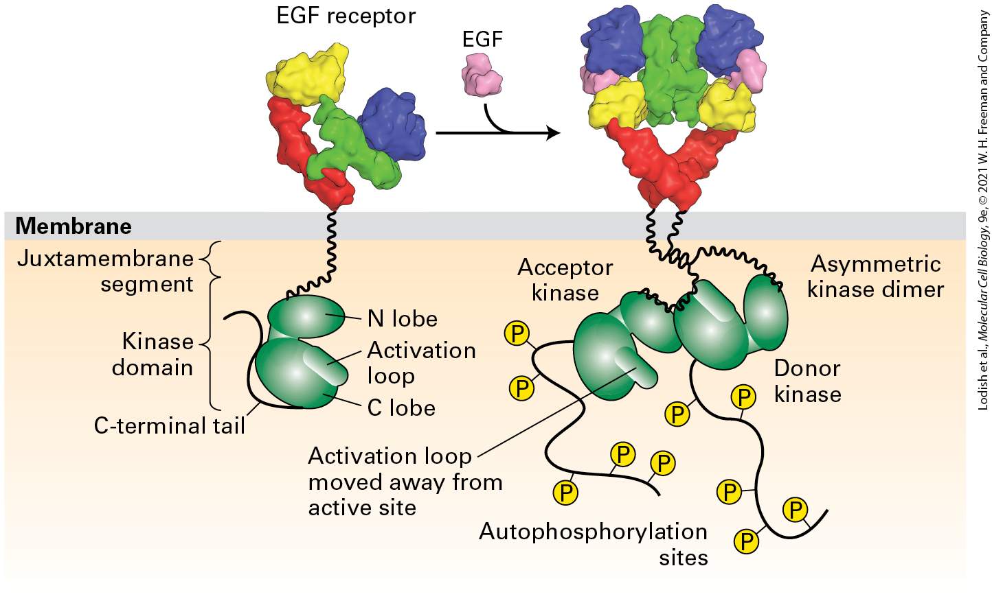
- EGF Receptor Homodimers
- EGF Receptor Heterodimers with HER2
- Ligand Binding to the EGF Receptor and Receptor Dimerization Results in the Formation of an Active Asymmetric Kinase Domain Dimer
- Signal Transduction After Activation of RTKs: Phosphotyrosine Residues on the Receptor Are Binding Surfaces for Multiple Proteins with SH2 Domains
- Receptor-Mediated Endocytosis and Lysosomal Degradation Squelch Signaling from RTKs
- 16.2 The Ras/MAP Kinase Signal Transduction Pathway
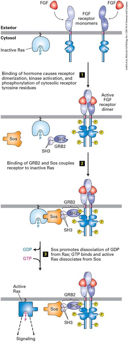
- Ras, a GTPase Switch Protein, Operates Downstream of Most RTKs and Cytokine Receptors
- Receptor Tyrosine Kinases Are Linked to Ras by Adapter Proteins
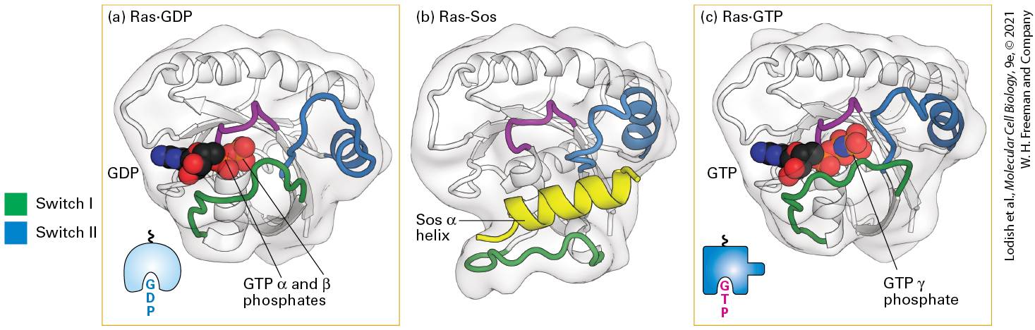
- Binding of Sos to Inactive Ras Causes a Conformational Change That Triggers an Exchange of GTP for GDP
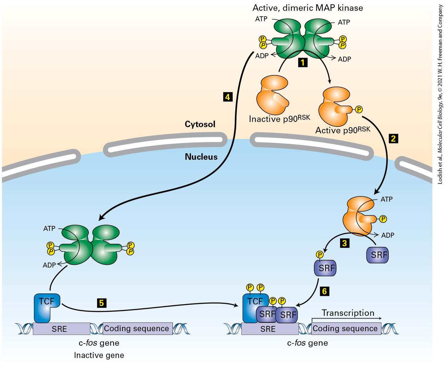
- Signals Pass from Activated Ras to a Cascade of Protein Kinases Ending with MAP Kinase
Ch 17Cell Organization and Movement I: MicrofilamentsRead full chapter →
Chapter 17 Cell Organization and Movement I: Microfilaments A section of mouse intestine stained for actin (red), the extracellular matrix protein laminin (green), and DNA (blue). Each blue dot of DNA indicates the presence of a cell. Actin in the microvilli on the apical end of the epithelial cells can be seen lining the surface facing the lumen (top). Actin can also be seen prominently in the smooth muscle that surrounds the intestine (bottom).
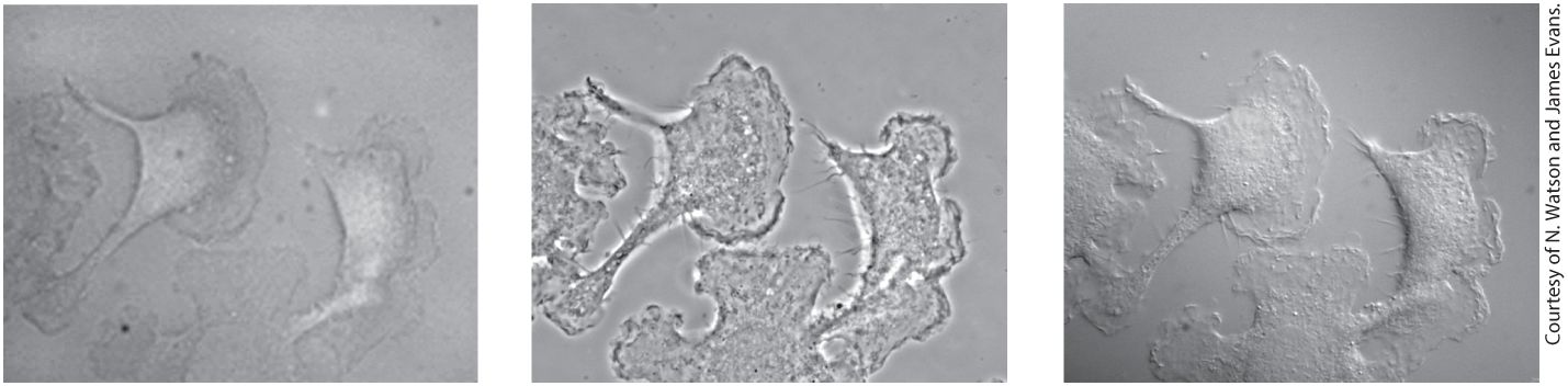
17.7 Cell Migration: Mechanism, Signaling, and Chemotaxis When we look through a microscope at the diversity of cells in nature, the variety of cell shapes and movements we see is astonishing. We may notice that some cells, such as vertebrate sperm, ciliates such as Tetrahymena, or flagellates such as Chlamydomonas, swim rapidly, propelled by cilia and flagella. Other cells, such as amoebae and human macrophages, move more sedately, propelled not by external appendages, but by coordinated movement of the cell itself. If we examine tissues under the microscope, we might notice that some cells c…
Sections in this chapter
- 17.1 Microfilaments and Actin Structures
- Actin Is Ancient, Abundant, and Highly Conserved
- G-Actin Monomers Assemble into Long, Helical F-Actin Polymers
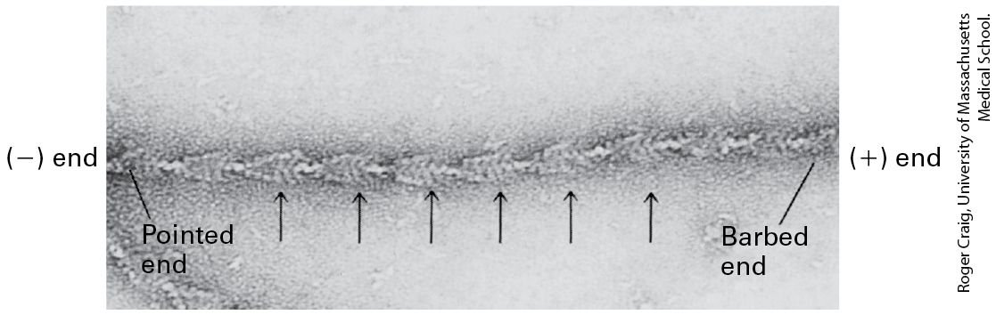
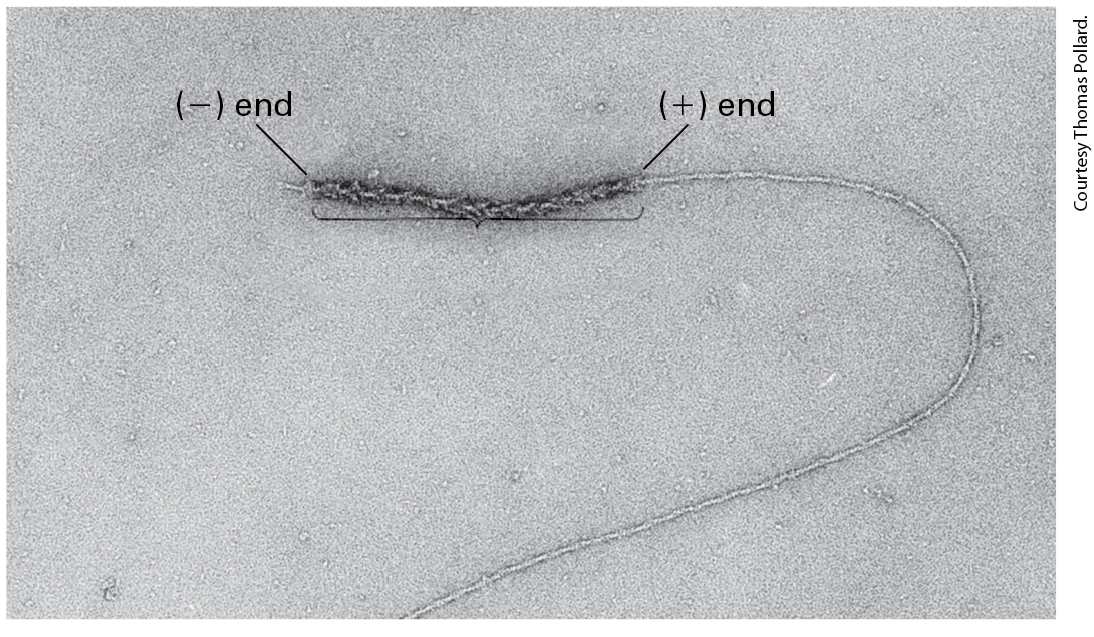
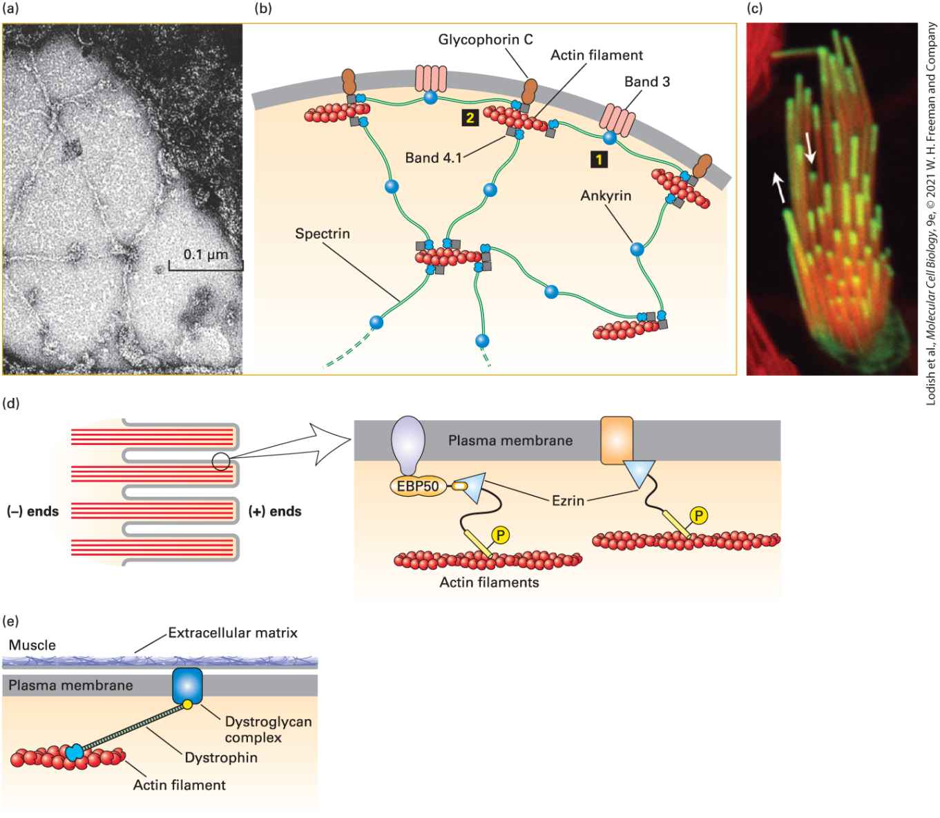
- F-Actin Has Structural and Functional Polarity
- 17.2 Dynamics of Actin Filaments
- Actin Polymerization In Vitro Proceeds in Three Steps
- Actin Filaments Grow Faster at (+) Ends than at (−) Ends
- Actin Filament Treadmilling Is Accelerated by Profilin and Cofilin
- Thymosin-β4 Provides a Reservoir of Actin for Polymerization
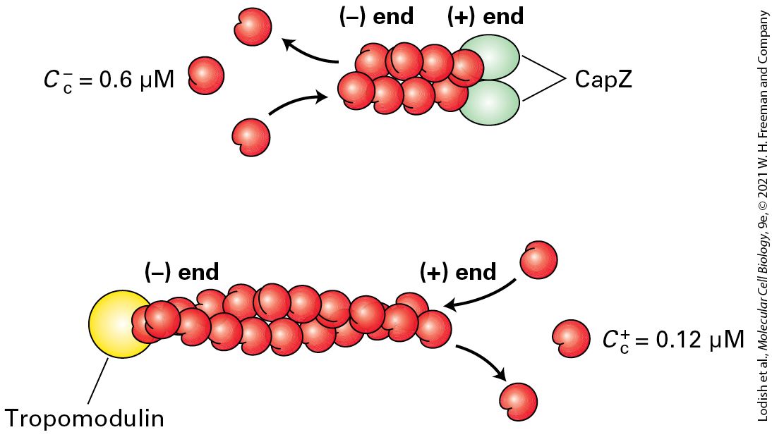
- Capping Proteins Block Assembly and Disassembly at Actin Filament Ends
- Formins Assemble Unbranched Filaments
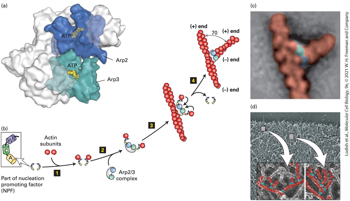
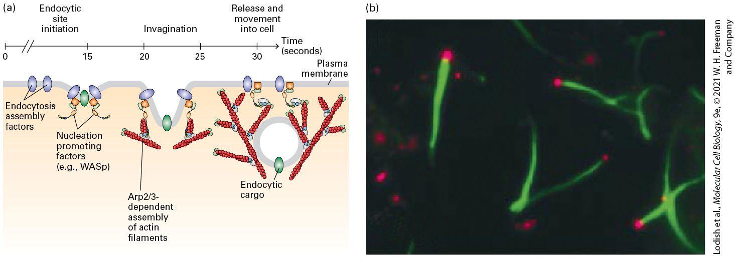
- The Arp2/3 Complex Nucleates Branched Filament Assembly
Ch 18Cell Organization and Movement II: Microtubules and Intermediate FilamentsRead full chapter →
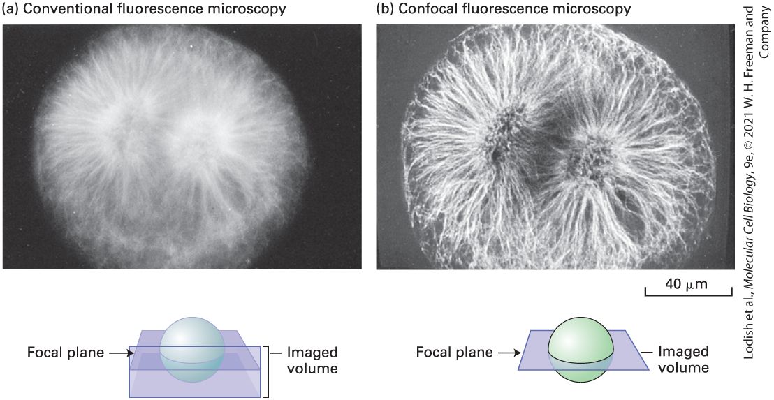
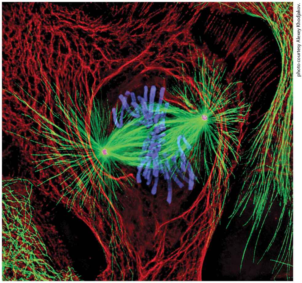
Chapter 18 Cell Organization and Movement II: Microtubules and Intermediate Filaments Newt lung cell in mitosis stained for centrosomes (magenta), microtubules (green), chromosomes (blue), and keratin intermediate filaments (red).
[Reprinted by permission of Nature Publishing Group, from A. Khodjakov, “Olympus/Nature competition: A 1, 2, 3 in Light Microscopy,” Nature, 2000, 408:423–424; permission conveyed through Copyright Clearance Center, Inc.]
Sections in this chapter
- 18.1 Microtubule Structure and Organization
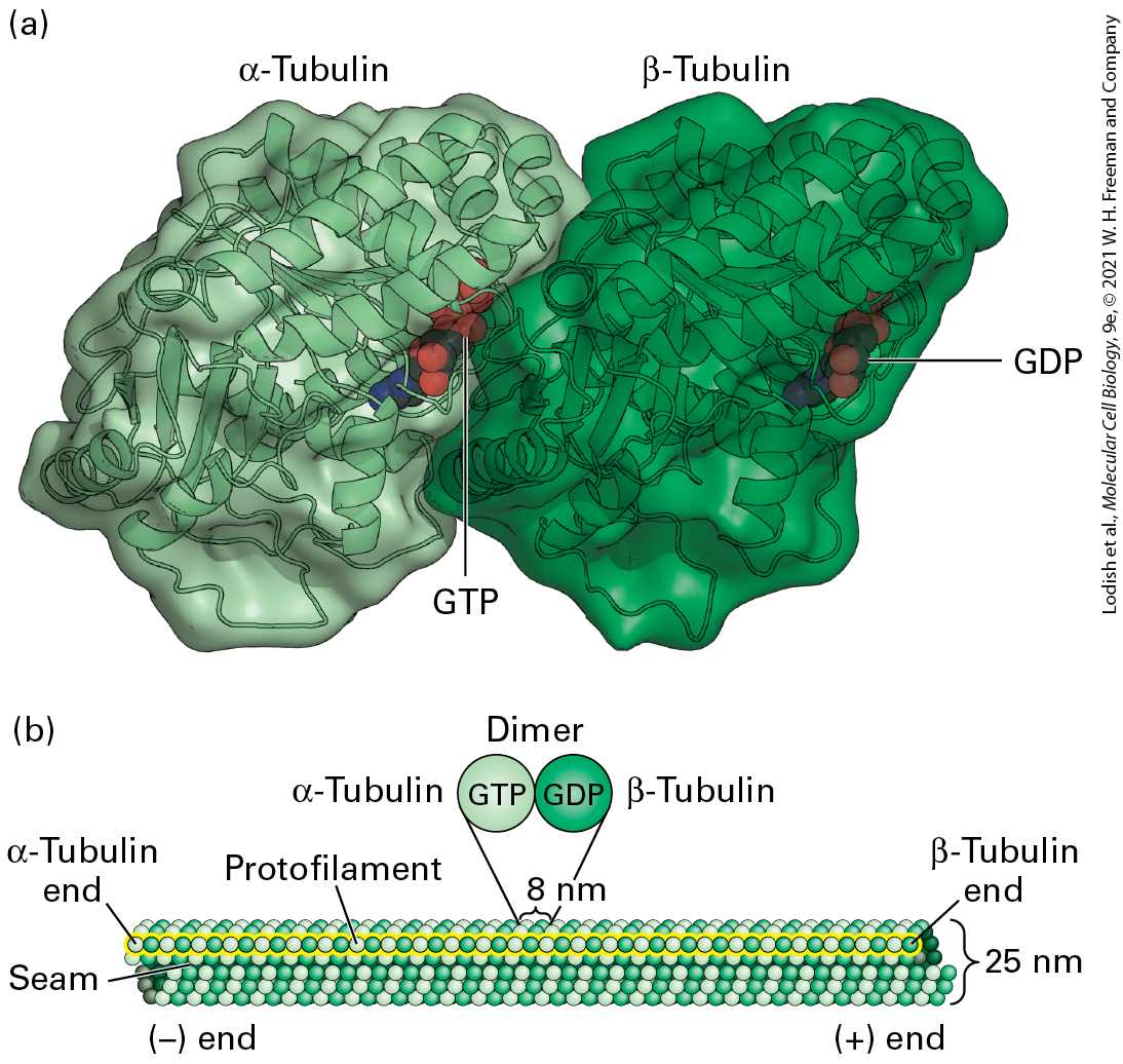
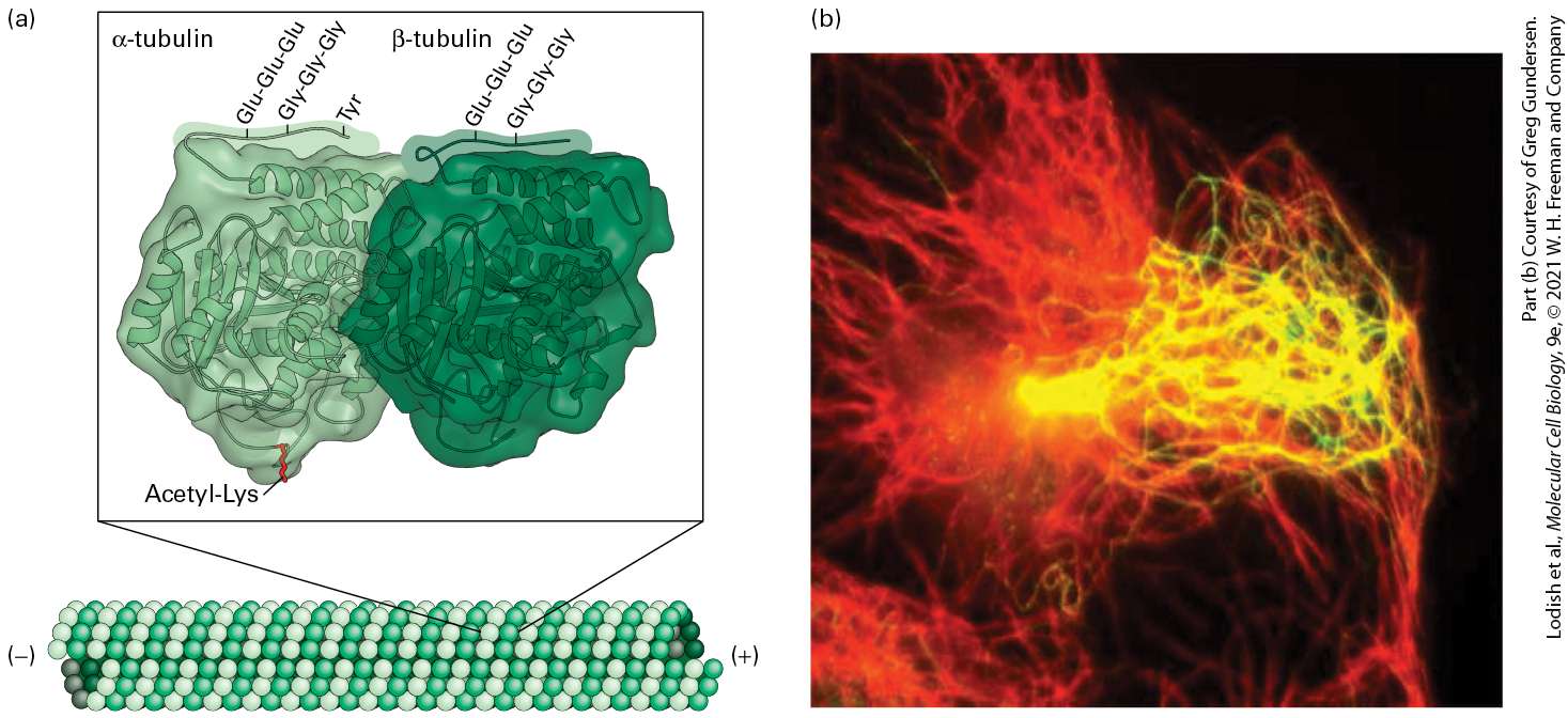
- Microtubule Walls Are Polarized Structures Built from αβ-Tubulin Dimers
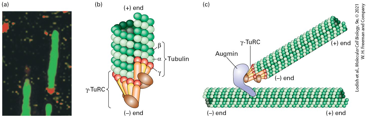
- Microtubules Are Assembled from MTOCs to Generate Diverse Configurations
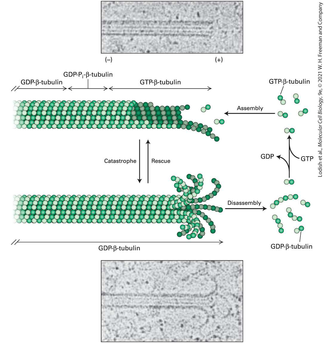
- Individual Microtubules Exhibit Dynamic Instability
- Localized Assembly and Search and Capture Help Organize Microtubules
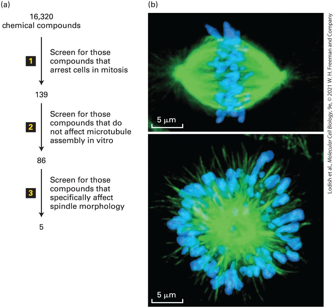
- Drugs Affecting Tubulin Polymerization Are Useful Experimentally and in Treatment of Diseases
- 18.3 Regulation of Microtubule Structure and Dynamics
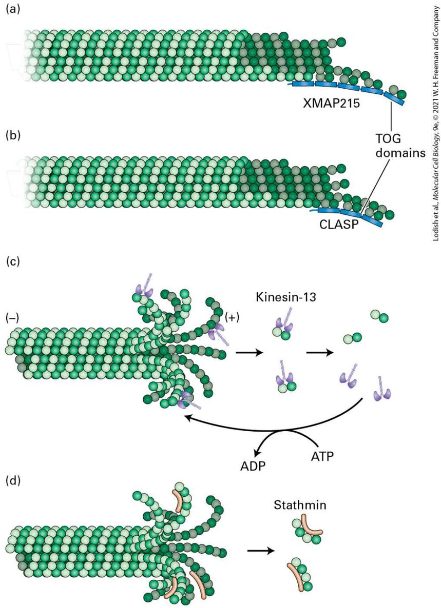
- Microtubules Are Stabilized by Side-Binding Proteins
- +TIPs Regulate the Properties and Functions of the Microtubule (+) End
- Severing Proteins Also Regulate Microtubule Dynamics
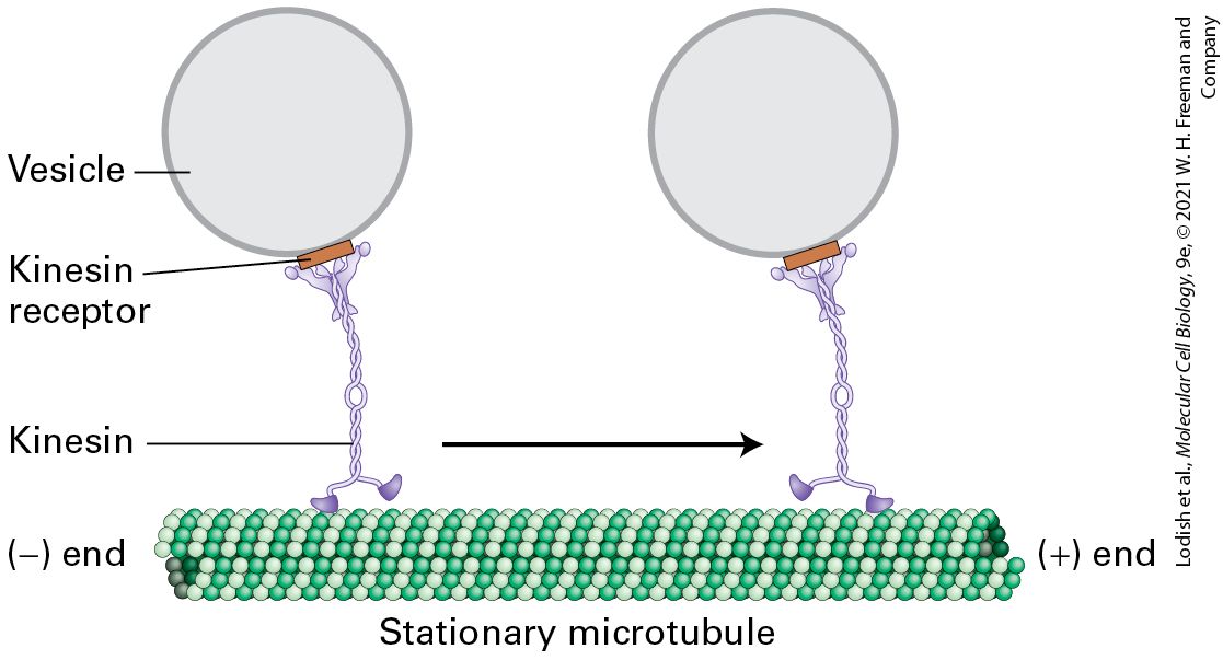
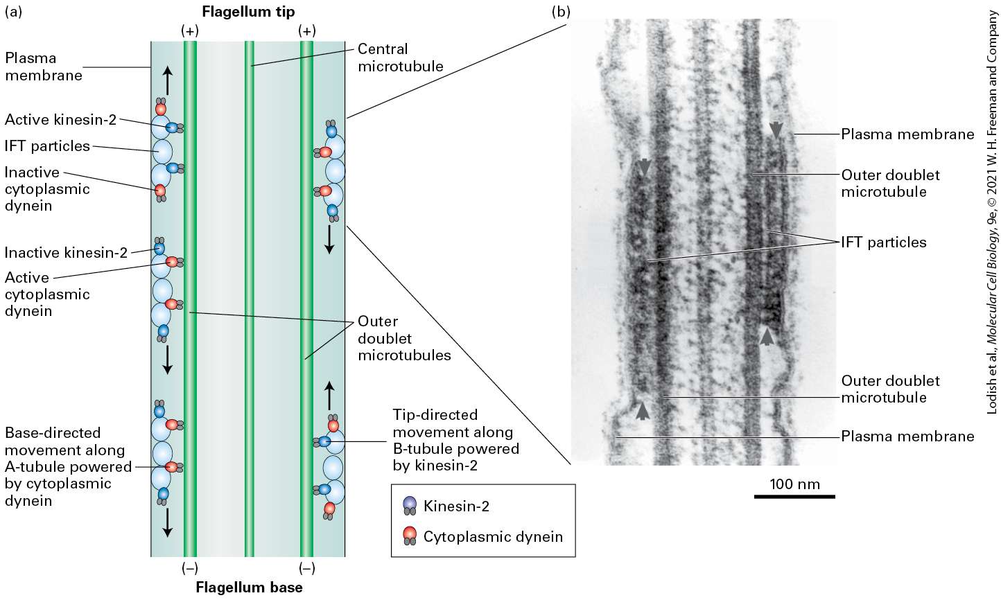
- Organelles in Axons Are Transported Along Microtubules in Both Directions
- Kinesin-1 Powers Anterograde Transport of Vesicles Down Axons Toward the (+) Ends of Microtubules
Ch 19The Eukaryotic Cell CycleRead full chapter →
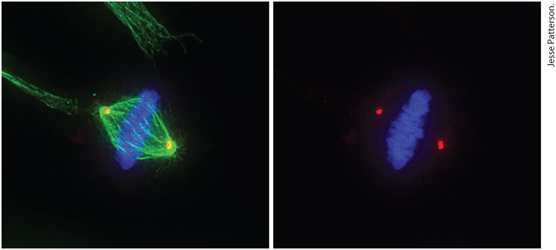
Chapter 19 The Eukaryotic Cell Cycle Micrograph on the left shows a human epithelial cell in metaphase. Following DNA replication, cells undergo mitosis to segregate their replicated chromosomes. The cell has aligned its chromosomes (blue) in the center of the mitotic spindle apparatus (green), prior to pulling them to opposite poles of the spindle, which will occur during anaphase. Thereafter, the cytoplasm of the cell is divided to produce two identical daughter cells. The micrograph on the right is the same cell without imaging the spindle apparatus in order to show the location of the cent…
Sections in this chapter
- 19.1 Overview of the Cell Cycle
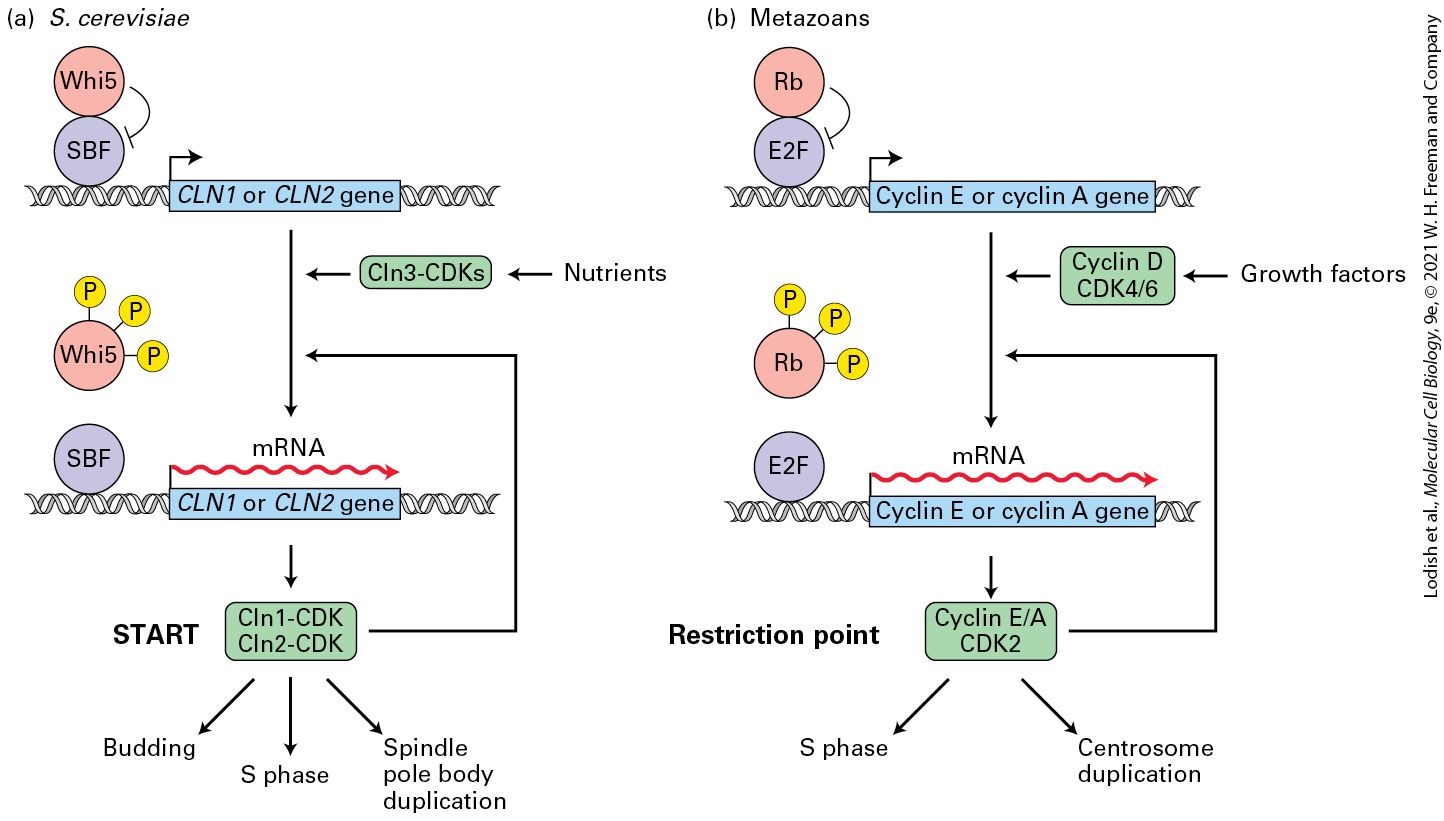
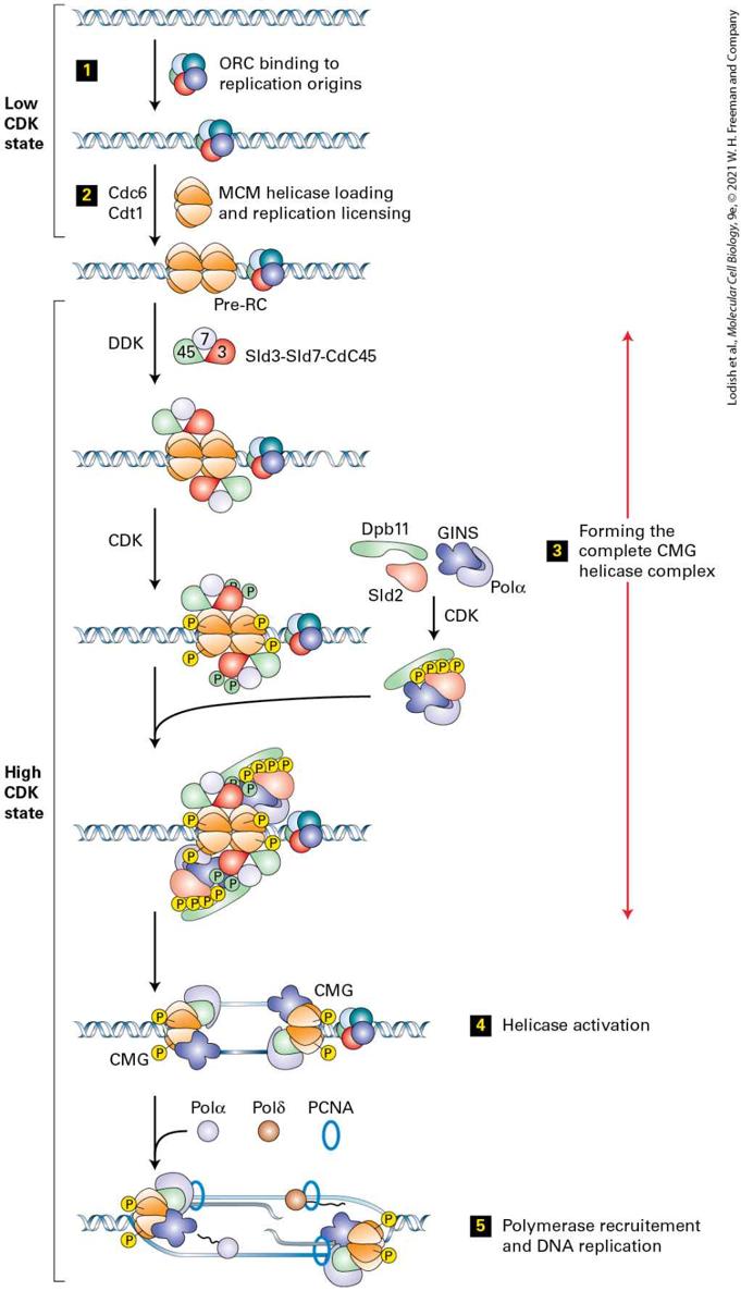
- G1 Controls Entry into S Phase
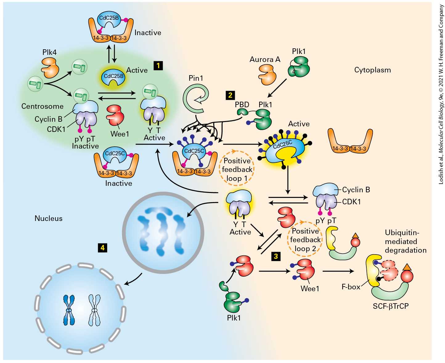
- G2 Phase Readies the Cell for Mitosis and Cell Division
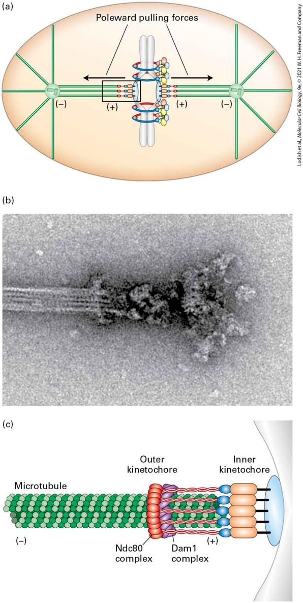
- Mitosis and Cytokinesis Occur During M-Phase
- Budding and Fission Yeasts Are Powerful Systems for Genetic Analysis of the Cell Cycle
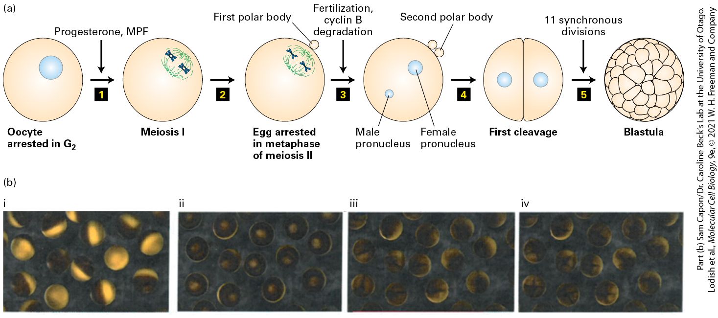
- Frog Oocytes and Early Embryos Facilitate Biochemical Characterization of the Cell Cycle Machinery
- The Study of Tissue Culture Cells Uncovers Cell Cycle Regulation in Mammals
- Researchers Use Multiple Tools to Study the Cell Cycle
- 19.3 Cell Cycle Progression and Control: Feedback Loops and Post-Translational Modification
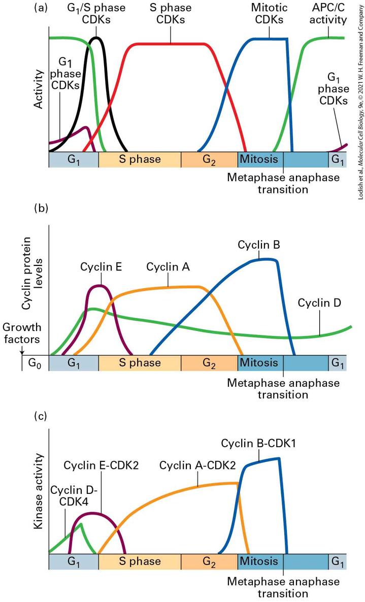
- Cyclin-Dependent Kinases Are Small Protein Kinases That Require a Regulatory Cyclin Subunit for Their Activity
- Cyclins Determine the Activity of CDKs
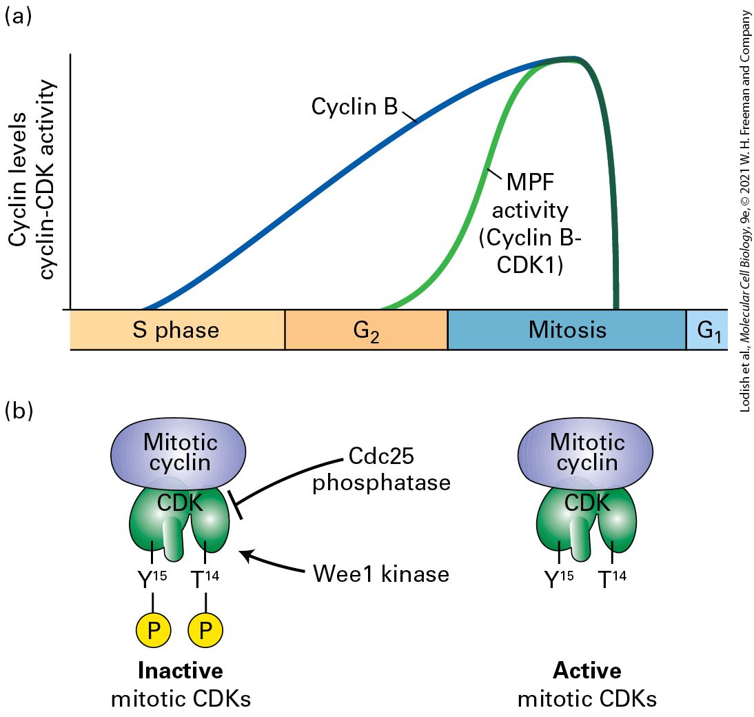
- CDKs Are Regulated by Activating and Inhibitory Phosphorylation
Ch 20Integrating Cells into TissuesRead full chapter →
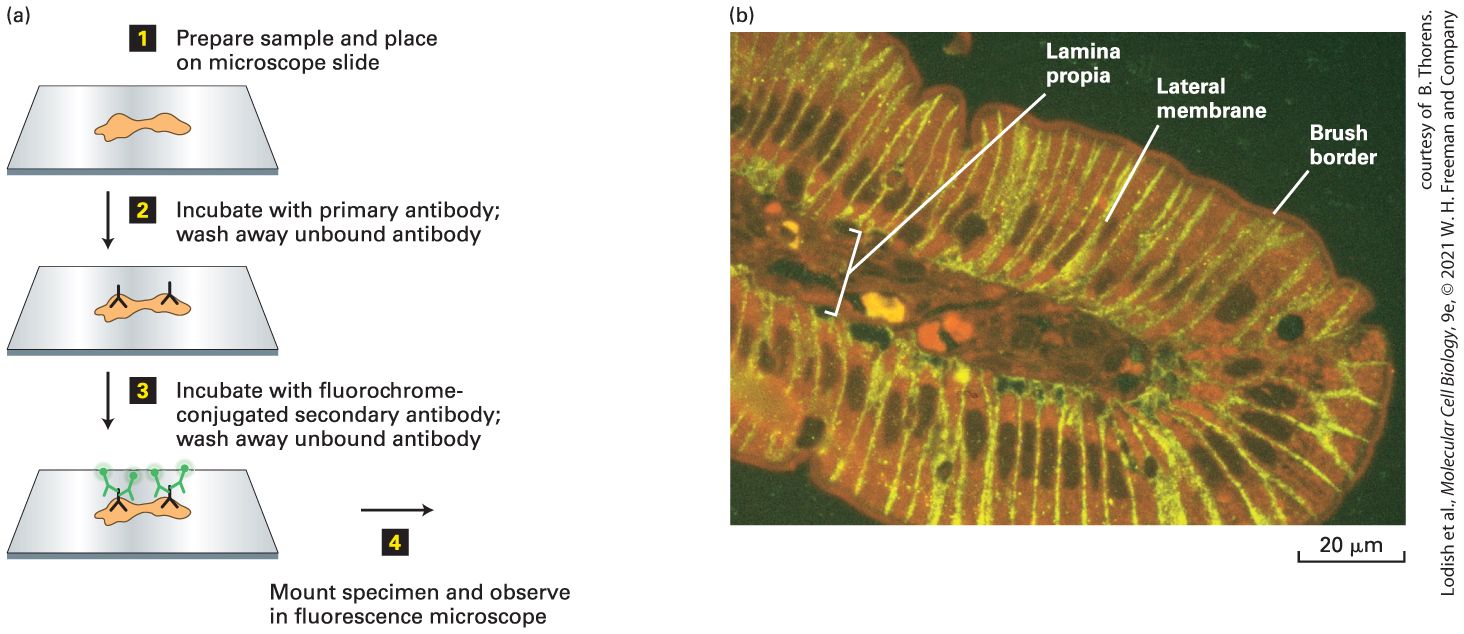
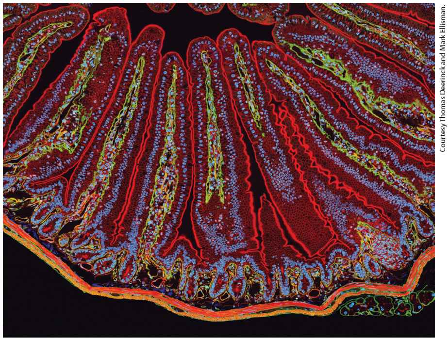
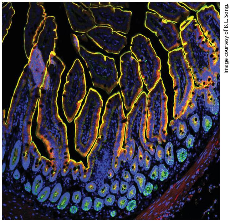
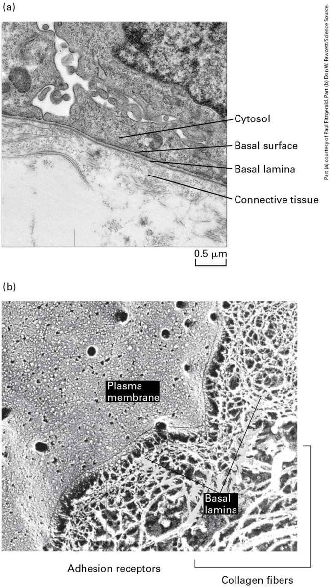
Chapter 20 Integrating Cells into Tissues Antibody staining of mouse small intestinal villi, fingerlike projections of cell layers (see Figures 20-13 through 20-15). NPC1L1, a protein located primarily on the plasma membrane involved with cholesterol metabolism, is green. Villin, which binds to actin bundles in microvilli (very small membrane projections on the apical surfaces of absorptive cells that
take up digested nutrients), is red. Nuclei (DNA) are blue. The image shows all colors merged together (colocalization of green and red appears yellow). The yellow lines show how the apical surfaces of the cells face the lumen of the small intestine (black) from which nutrients are absorbed.
Sections in this chapter
- Cell-Adhesion Molecules Bind to One Another and to Intracellular Proteins
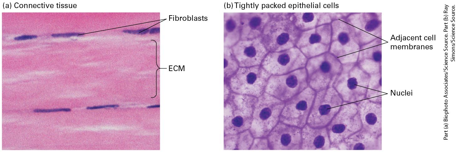
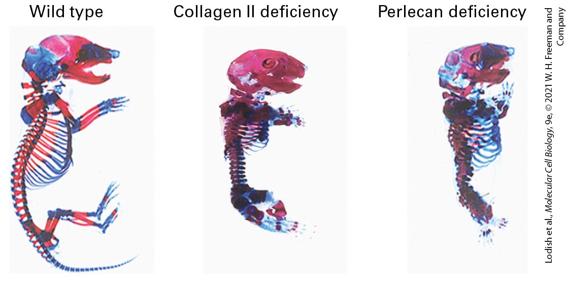
- The Extracellular Matrix Participates in Adhesion, Signaling, and Other Functions
- The Evolution of Multifaceted Adhesion Molecules Enabled the Evolution of Diverse Animal Tissues
- Cell-Adhesion Molecules Mediate Mechanotransduction
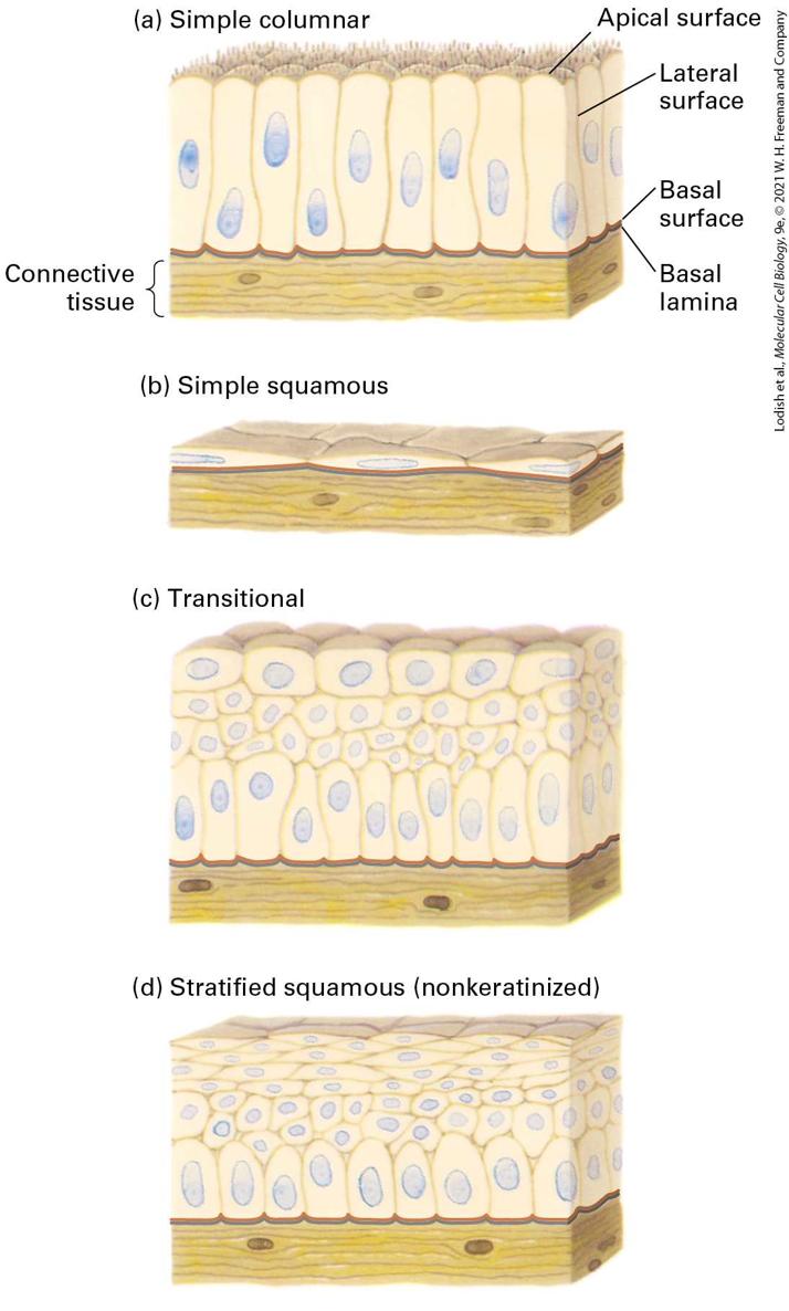
- Epithelial Cells Have Distinct Apical, Lateral, and Basal Surfaces
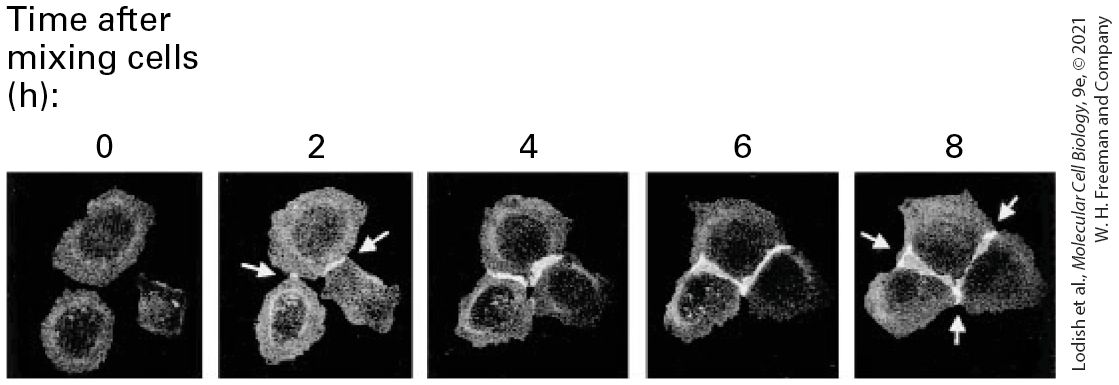
- Three Types of Junctions Mediate Many Cell-Cell and Cell-Matrix Interactions
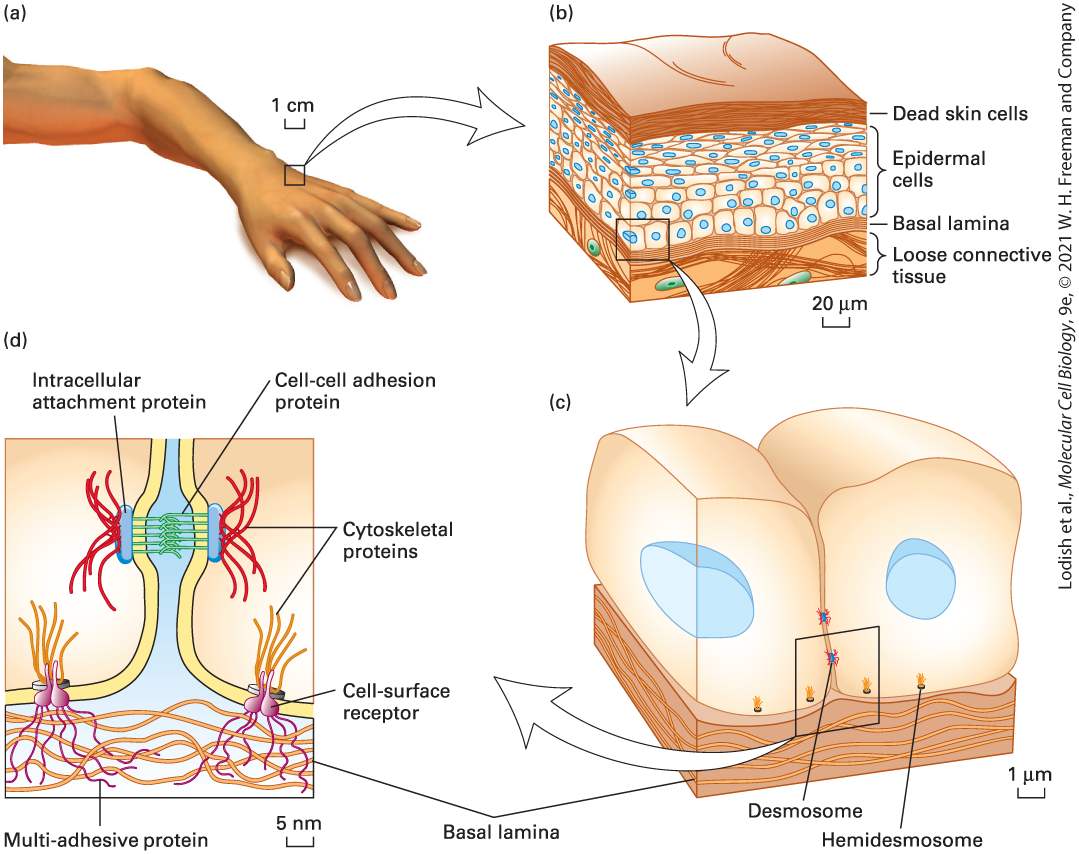
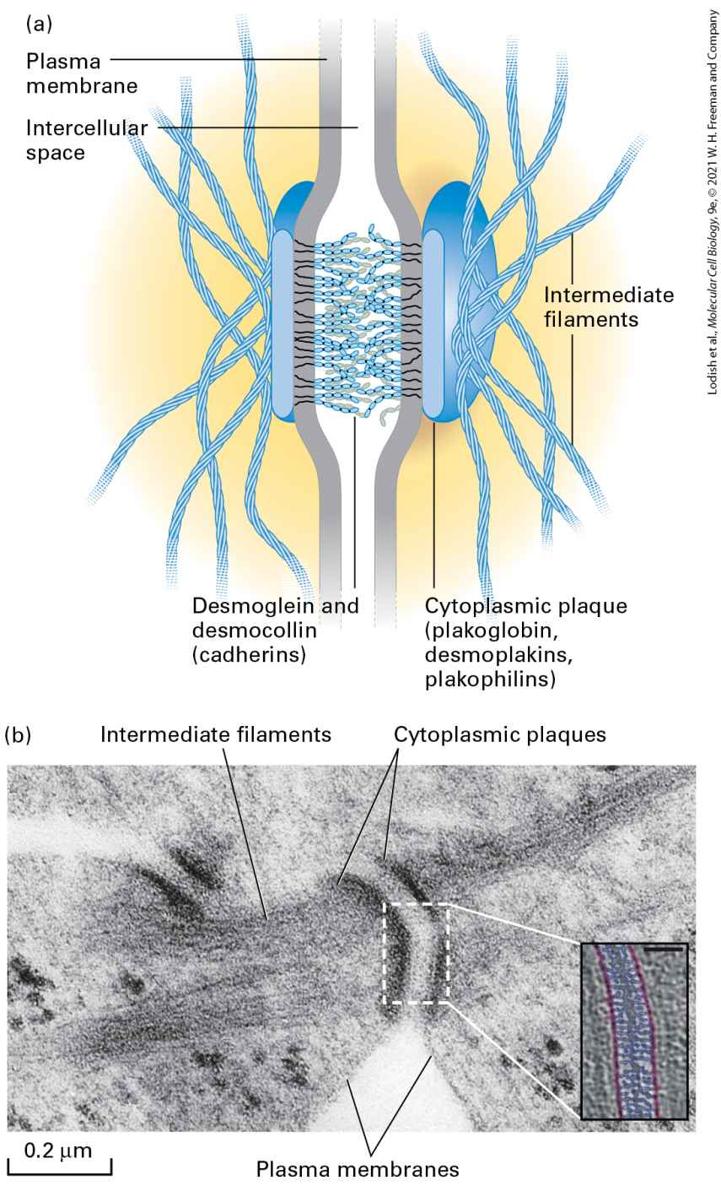
- Cadherins Mediate Cell-Cell Adhesions in Adherens Junctions and Desmosomes
- Integrins Mediate Cell-Matrix Adhesions, Including Those in Epithelial-Cell Hemidesmosomes
- Icam-1, Icam-2
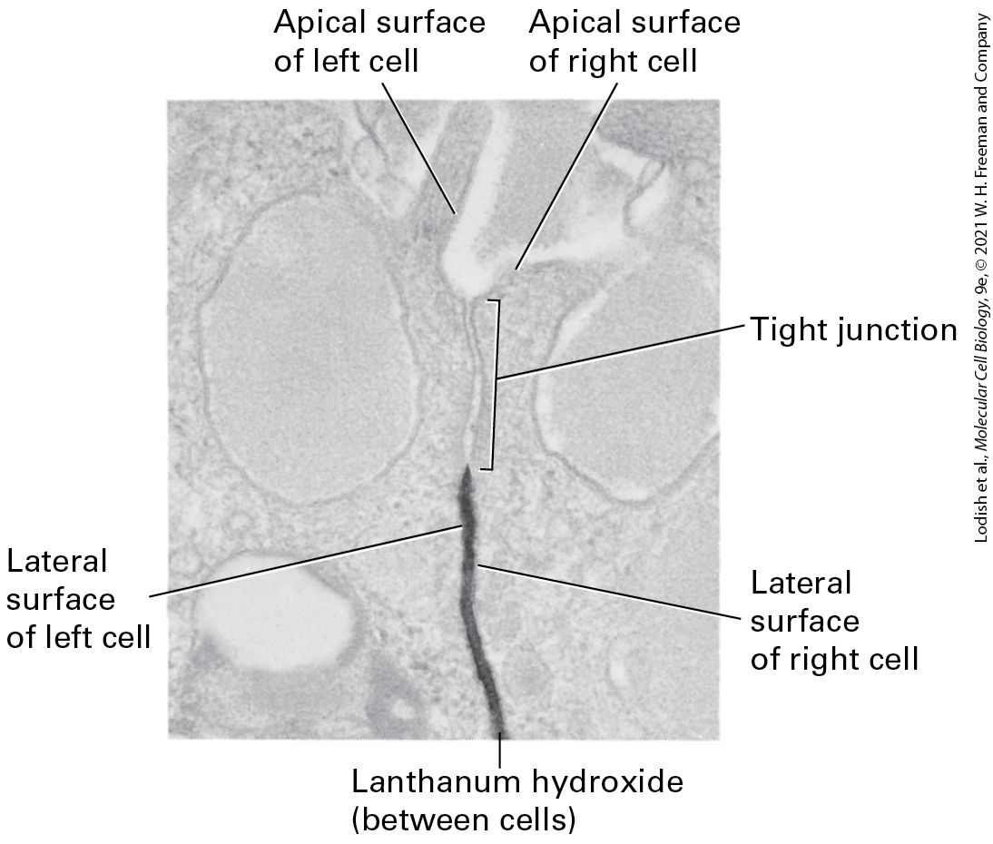
- Tight Junctions Seal Off Body Cavities and Restrict Diffusion of Membrane Components
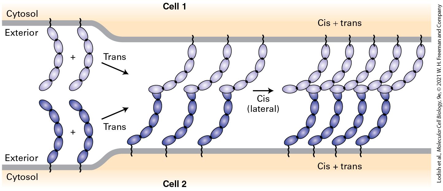
- Gap Junctions Composed of Connexins Allow Small Molecules to Pass Directly Between the Cytosols of Adjacent Cells
- Tunneling Nanotubes Can Mediate Metabolic Coupling and Transfer Organelles Between Animal Cells
Ch 21Responding to the Cellular EnvironmentRead full chapter →
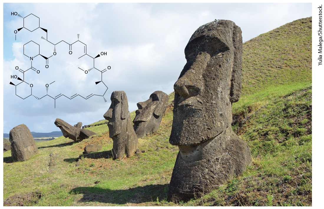
Chapter 21 Responding to the Cellular Environment Rapamycin (in black), a clinically important antifungal drug and inhibitor of the mTORC kinase complex, was isolated from the bacterium Streptomyces hygroscopicus found on Easter Island. It was named after the native name of the island, Rapa Nui.
Sections in this chapter
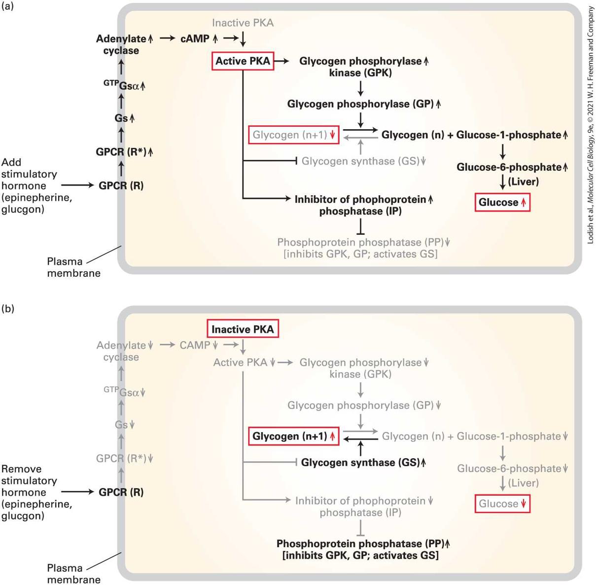
- Insulin and Glucagon Work Together to Maintain a Stable Blood Glucose Level
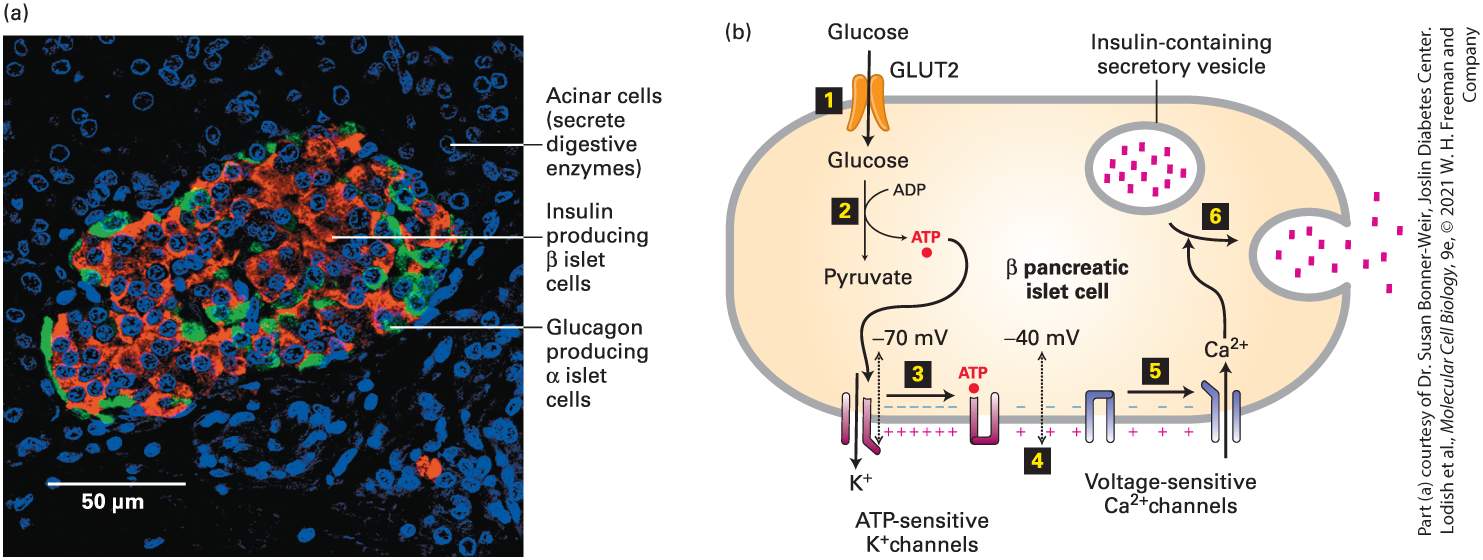
- A Rise in Blood Glucose Triggers Insulin Secretion from the β Islet Cells
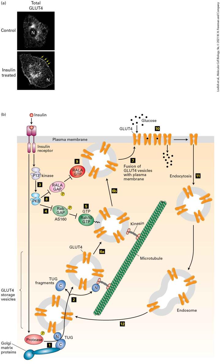
- In Fat and Muscle Cells, Insulin Triggers Fusion of Intracellular Vesicles Containing the GLUT4 Glucose Transporter with the Plasma Membrane, Thus Increasing the Rate of Glucose Uptake
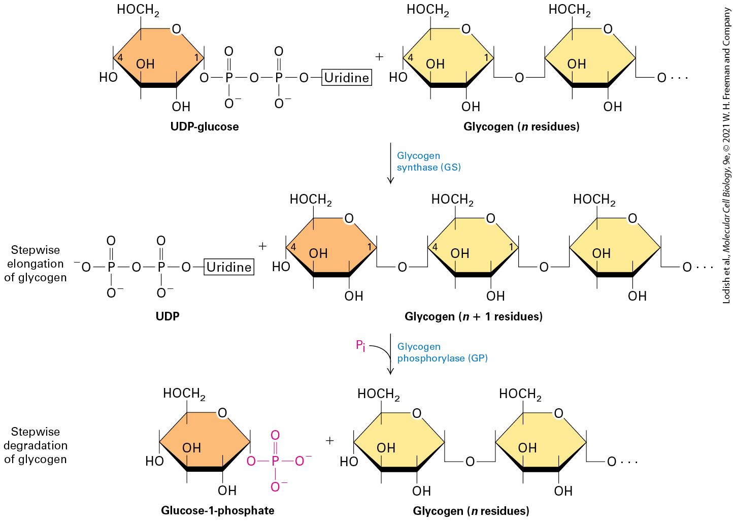
- In the Liver, Insulin Inhibits Glucose Synthesis, Accelerates the Rate of Glycolysis, and Enhances Storage of Glucose as Glycogen
- 21.2 Integrating Cell Growth Signals with Nutrient and Energy Levels
- The Active mTORC1 Complex Activates Many Anabolic Signal Transduction Pathways
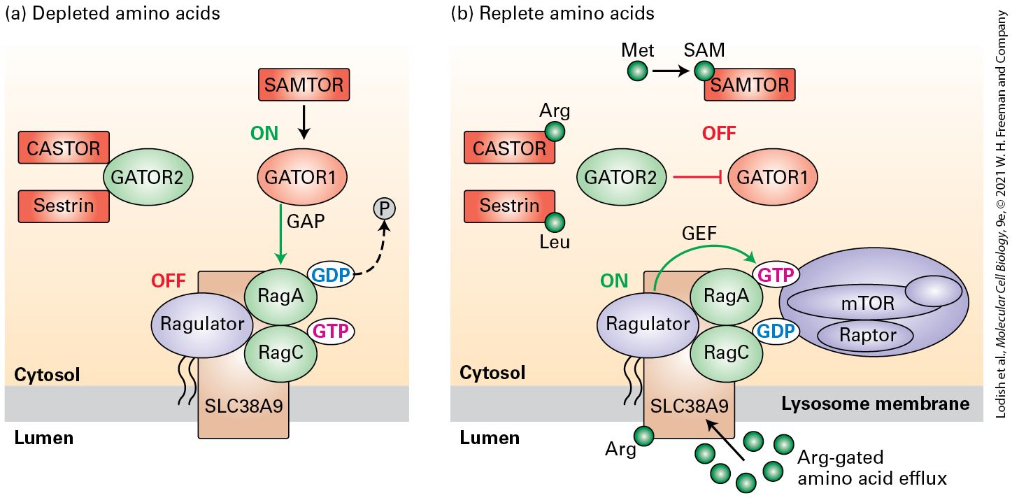
- mTORC1 Kinase Activation Requires Amino Acids, a High ATP:AMP Ratio, and Activation of Signal Transduction Pathways Downstream of Growth-Factor Receptors
- Fatty Acid and Cholesterol Biosynthesis as Well as Cholesterol Import Are Regulated at the Level of Gene Transcription
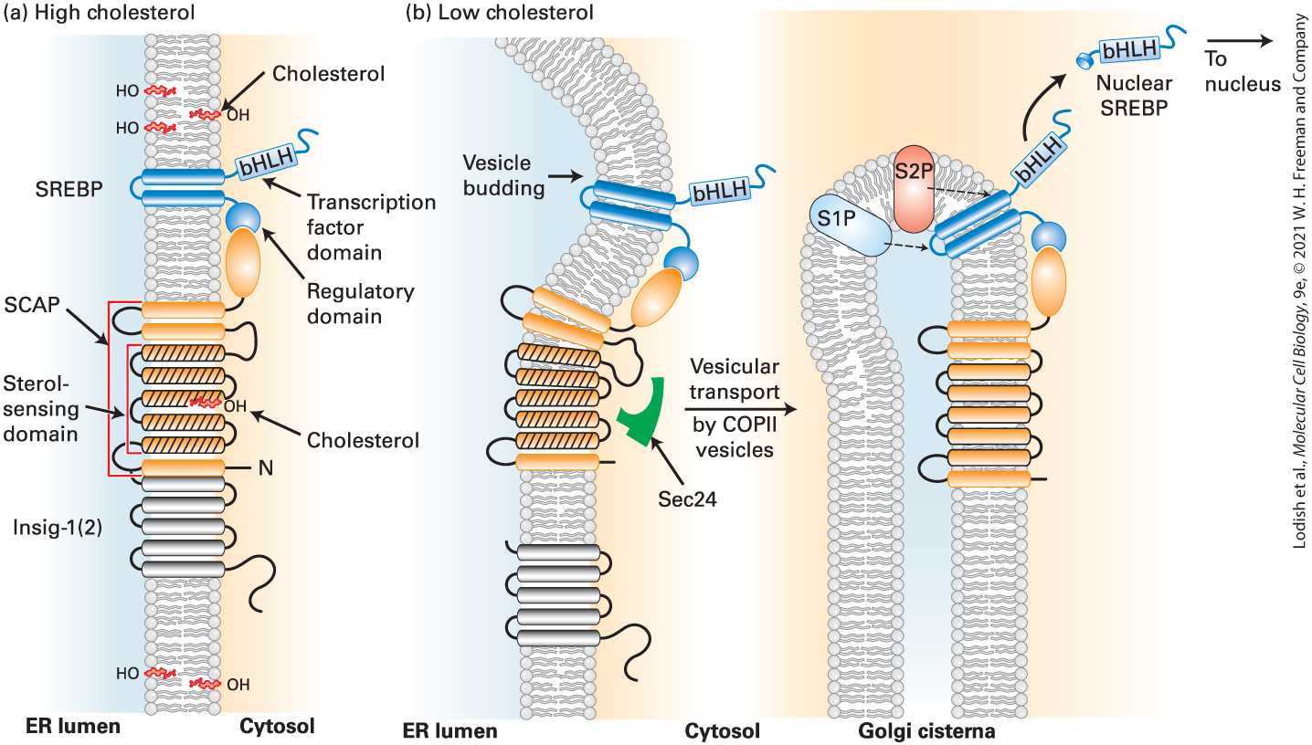
- The Endoplasmic Reticulum SCAP Protein Senses the Level of Cellular Cholesterol
- Regulated Intramembrane Proteolysis of SREBP in the Golgi Releases a bHLH Transcription Factor That Acts to Maintain Appropriate Phospholipid and Cholesterol Levels
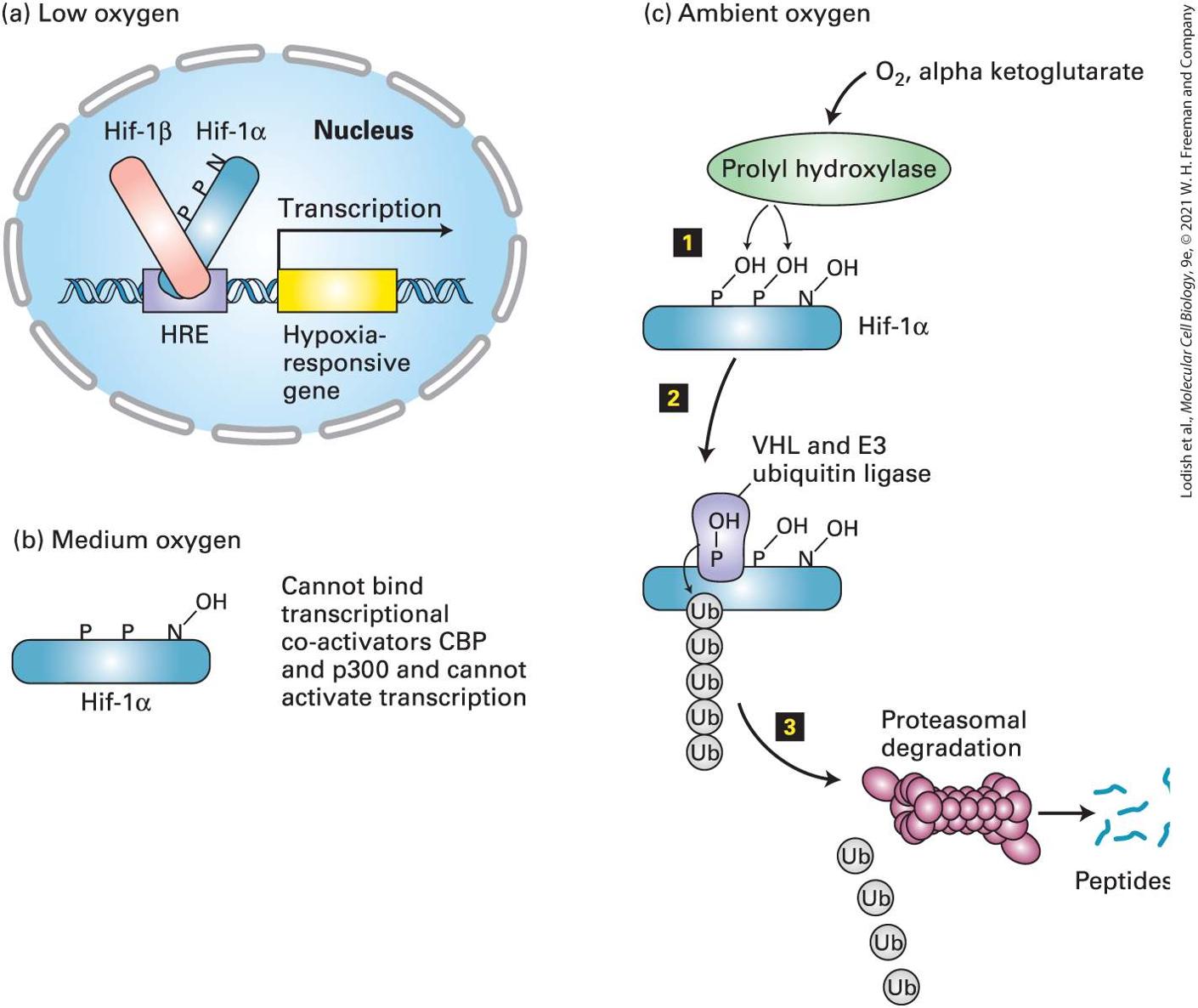
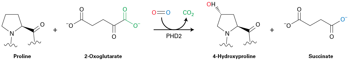
- Induction of the Erythropoietin Gene at Low Oxygen Levels
- Oxygen Sensing and Regulated Hif-1α Expression Is a Property of All Nucleated Mammalian Cells
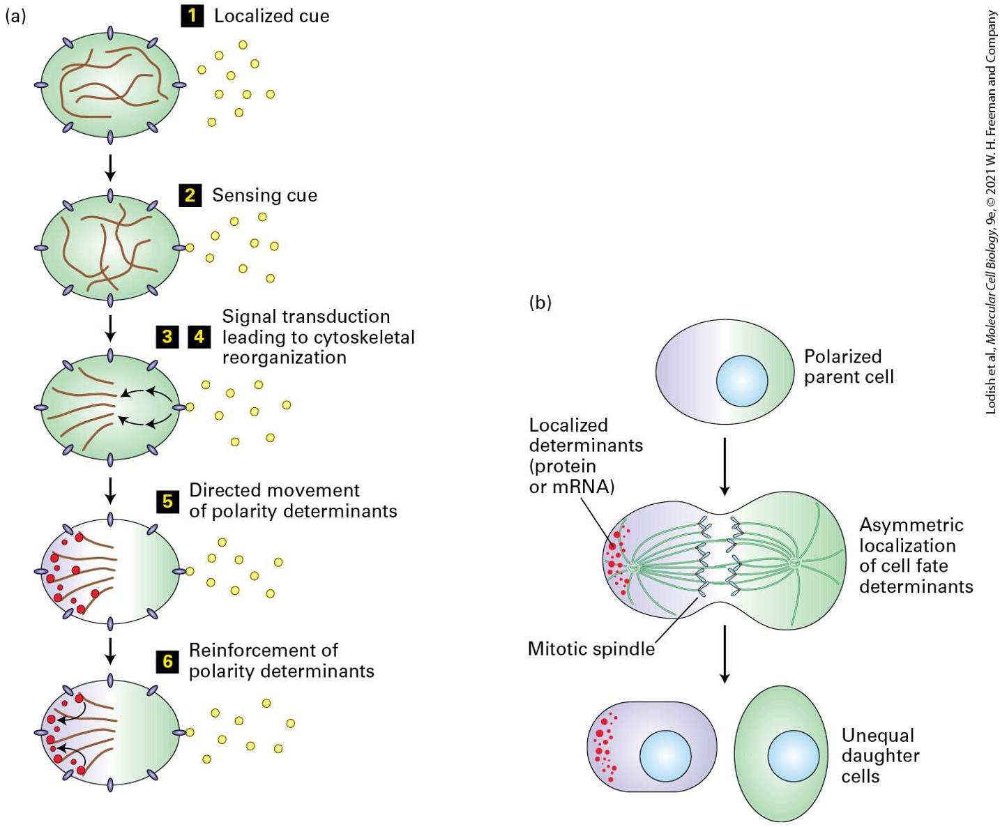
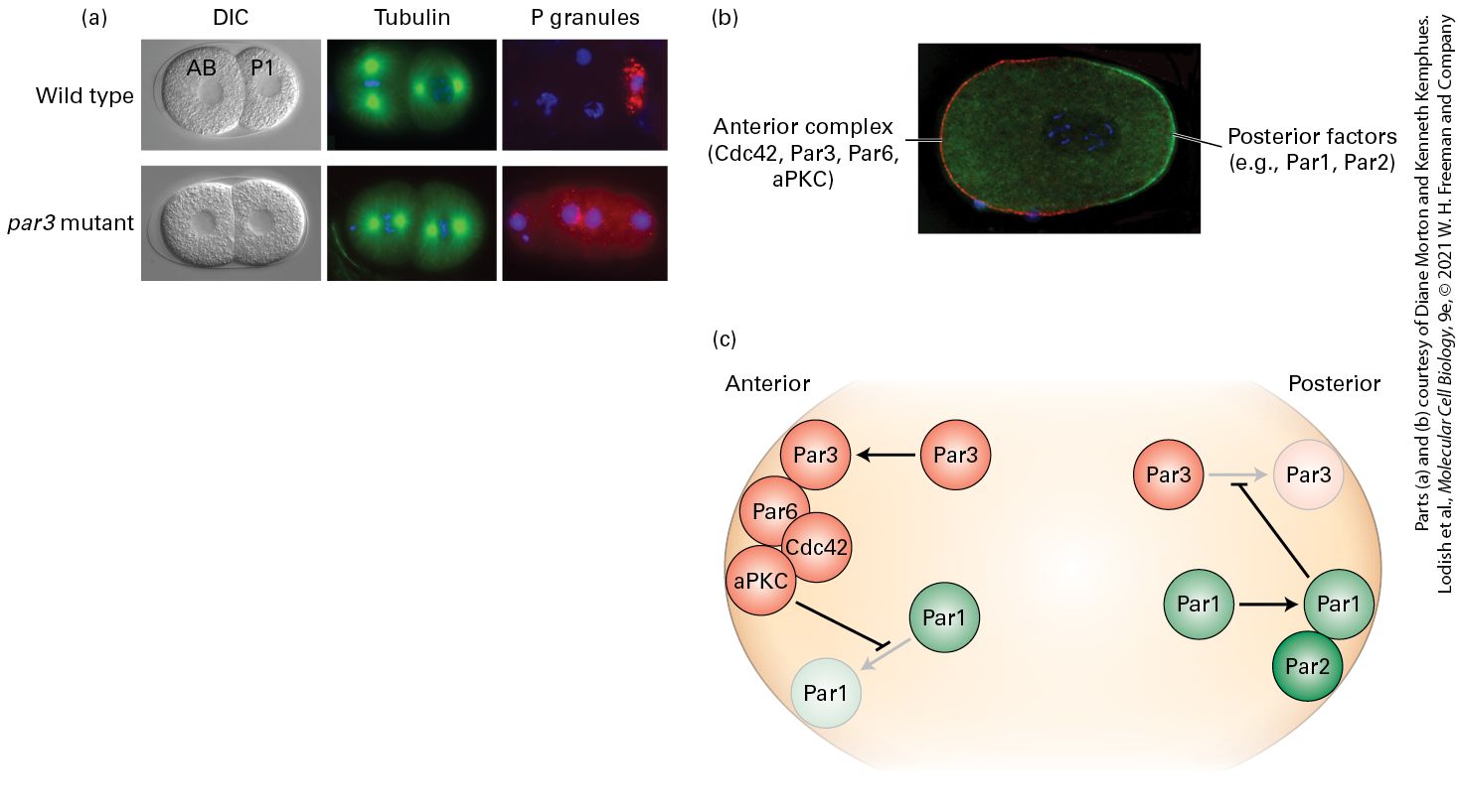
Ch 22Stem Cells, Cell Asymmetry, and Regulated Cell DeathRead full chapter →
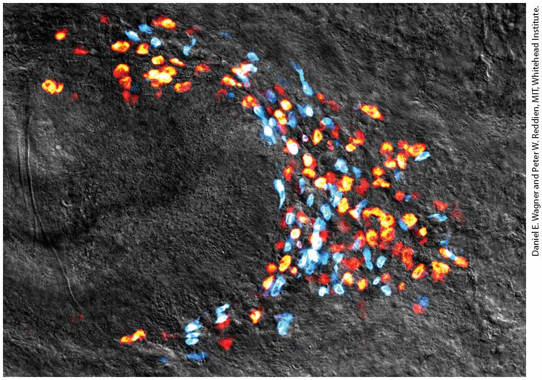
Chapter 22 Stem Cells, Cell Asymmetry, and Regulated Cell Death Pluripotent stem cells called neoblasts provide the cellular basis for regeneration in planarian flatworms. Shown is a colony of neoblasts (yellow, orange, and red), all derived from a single neoblast 14 days after regeneration of the tail was initiated by amputation; differentiating cells (blue), also derived from the single neoblast, are shown as well.
Sections in this chapter
- Fertilization Unifies the Genome
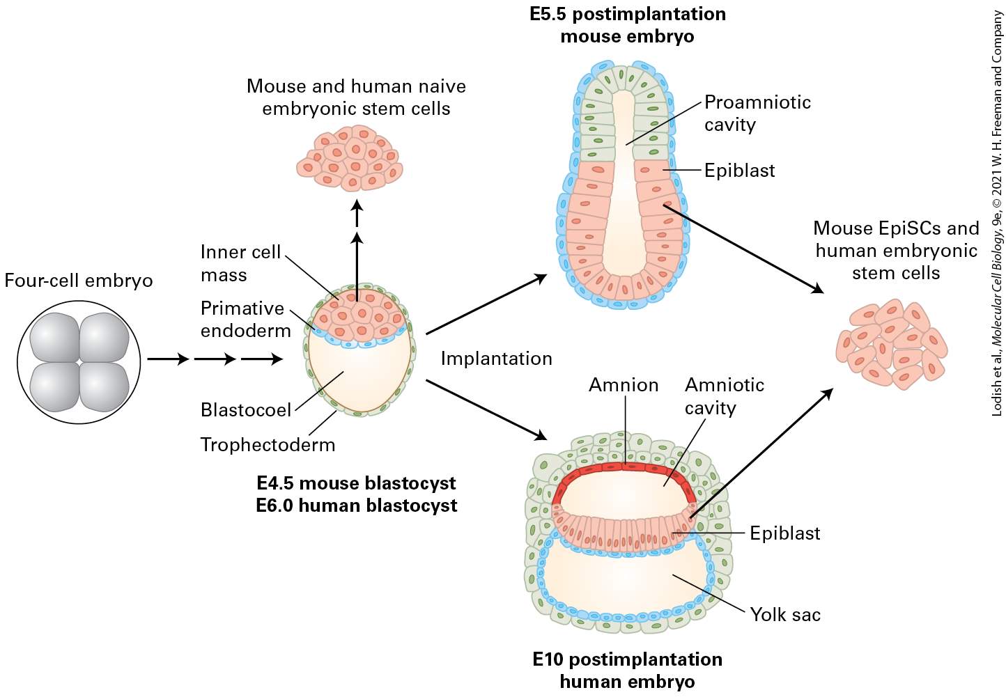
- Cleavage of the Mammalian Embryo Leads to the First Differentiation Events
- Pluripotent Cells of the Inner Cell Mass Are the Source of ES Cells
- Multiple Factors Control the Pluripotency of ES Cells
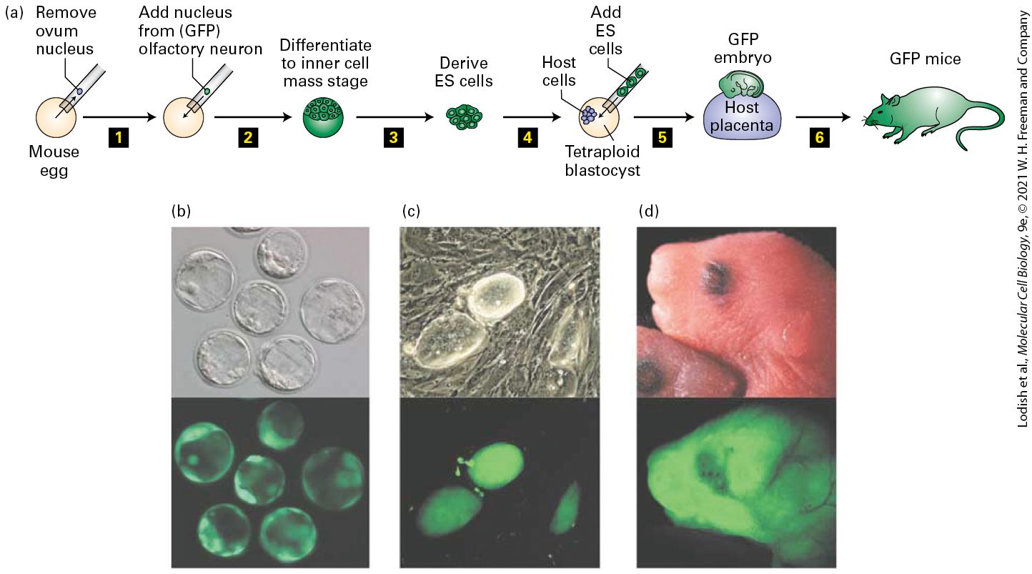
- Animal Cloning Shows That Epigenetic Changes During Differentiation Can Be Reversed
- Somatic Cells Can Generate iPS Cells
- Patient-Specific iPS Cells Can Be Used to Develop Potential Treatments for Many Diseases
- ES and iPS Cells Can Generate Functional Differentiated Human Cells
- Adult Planarians Contain Pluripotent Stem Cells
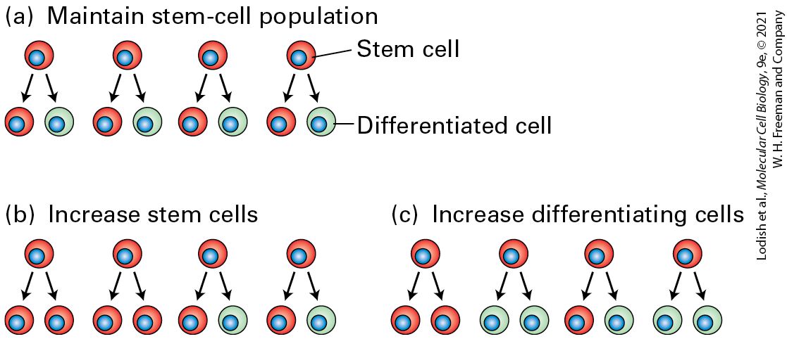
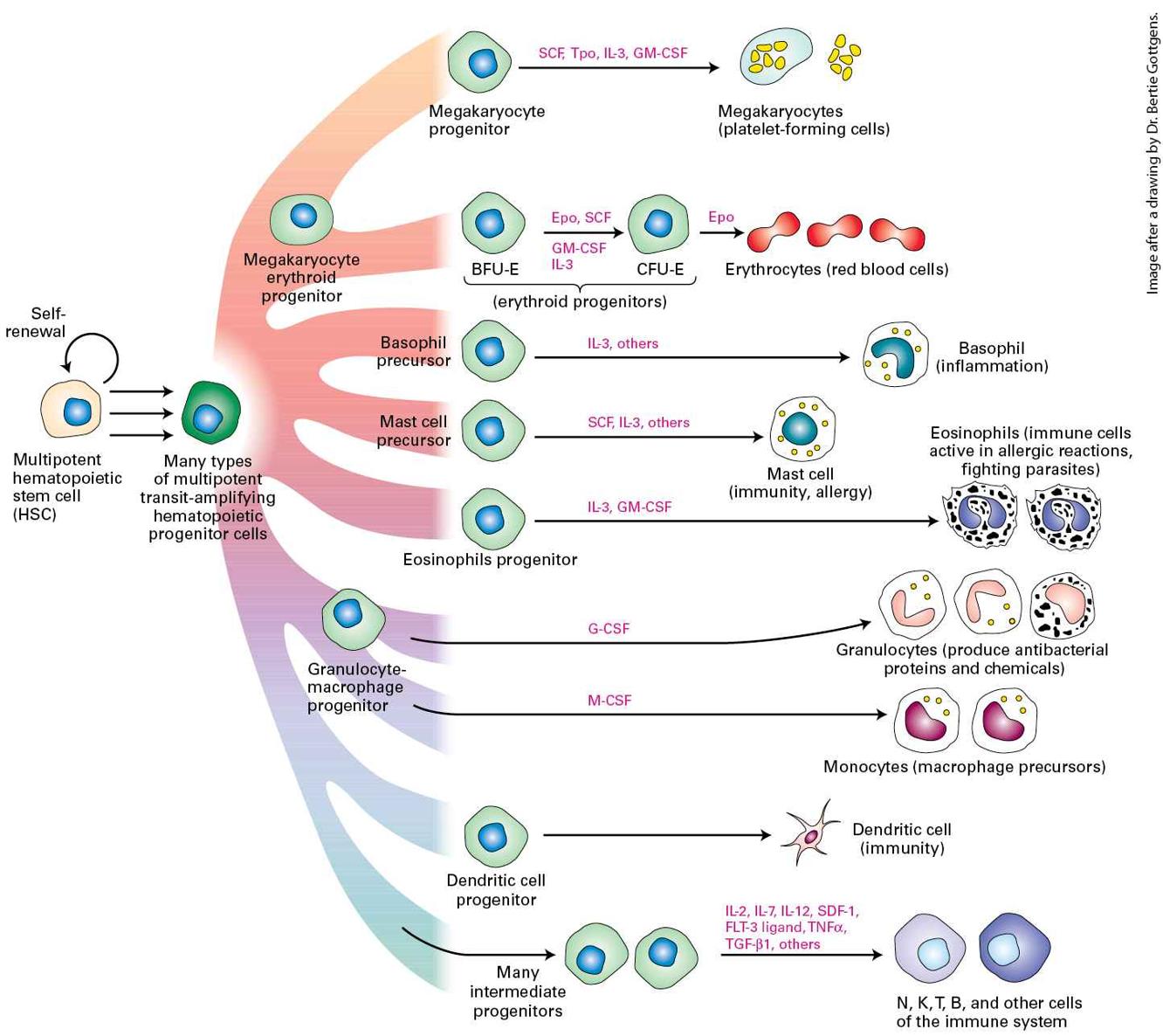
- Multipotent Somatic Stem Cells Give Rise to Both Stem Cells and Differentiating Cells
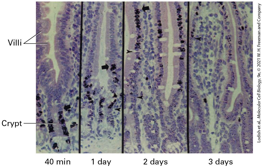
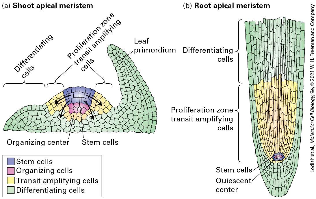
- Stem Cells for Different Tissues Occupy Sustaining Niches
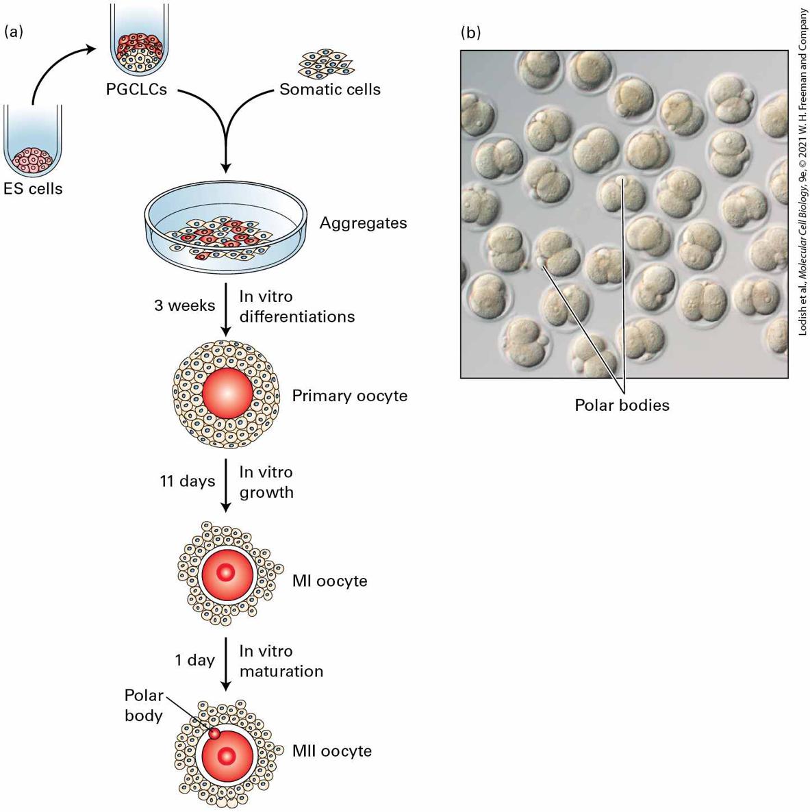
- Germ-Line Stem Cells in Many Organisms Produce Sperm or Oocytes
Ch 23Cells of the Nervous SystemRead full chapter →
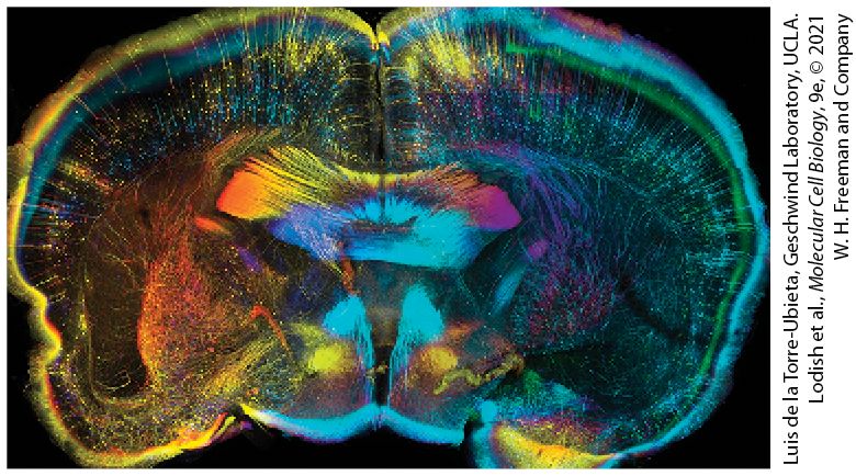
Chapter 23 Cells of the Nervous System Coronal slice of CLARITY-treated adult mouse brain expressing green fluorescent protein (GFP) in a subset of neurons (Thy1-GFP). CLARITY renders tissue optically transparent, permitting deep and complete imaging of tissues. Each section was stained with antibodies to GFP and color coded by depth to facilitate individual neuron visualization. This approach provides unprecedented opportunity to image intact brains at cellular resolution, paving the way to a comprehensive understanding of how the brain is wired.
Sections in this chapter
- 23.1 Neurons and Glia: Building Blocks of the Nervous System
- Information Flows Through Neurons from Dendrites to Axons
- Information Moves Along Axons as Pulses of Ion Flow Called Action Potentials
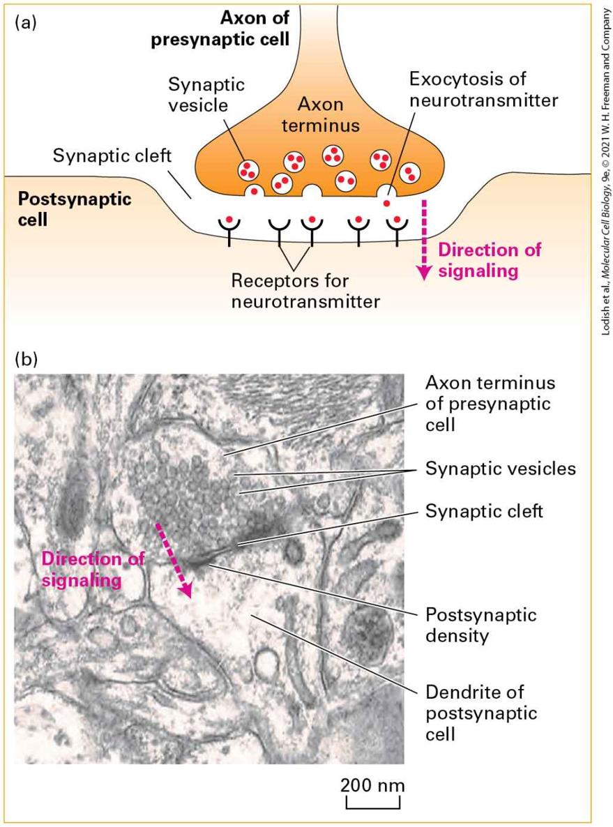
- Information Flows Between Neurons via Synapses
- The Nervous System Uses Signaling Circuits Composed of Multiple Types of Neurons
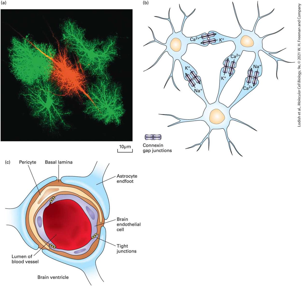
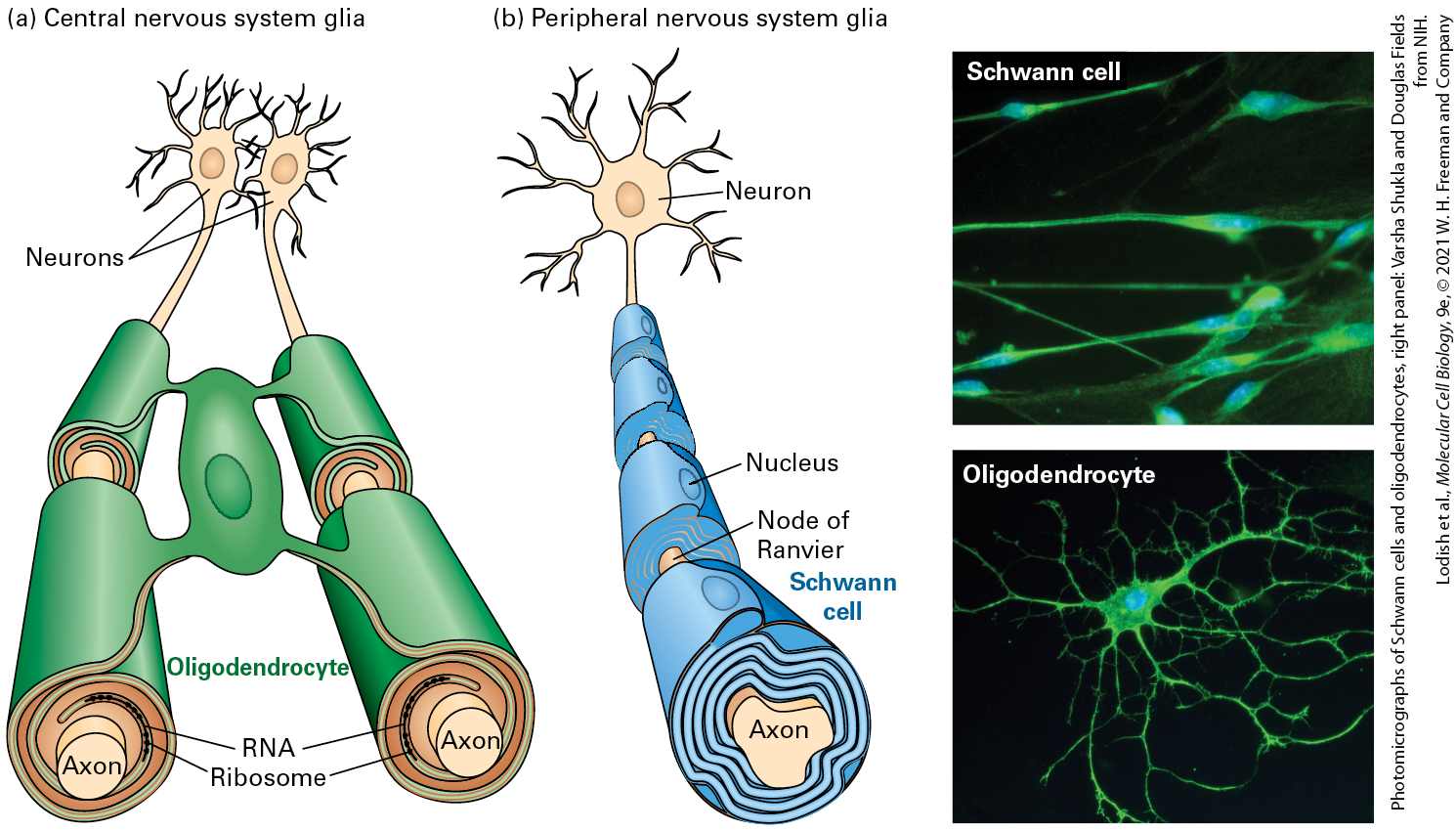
- Glial Cells Form Myelin Sheaths and Support Neurons
- Neural Stem Cells Form Nerve and Glial Cells in the Central Nervous System
- 23.2 Voltage-Gated Ion Channels and the Propagation of Action Potentials
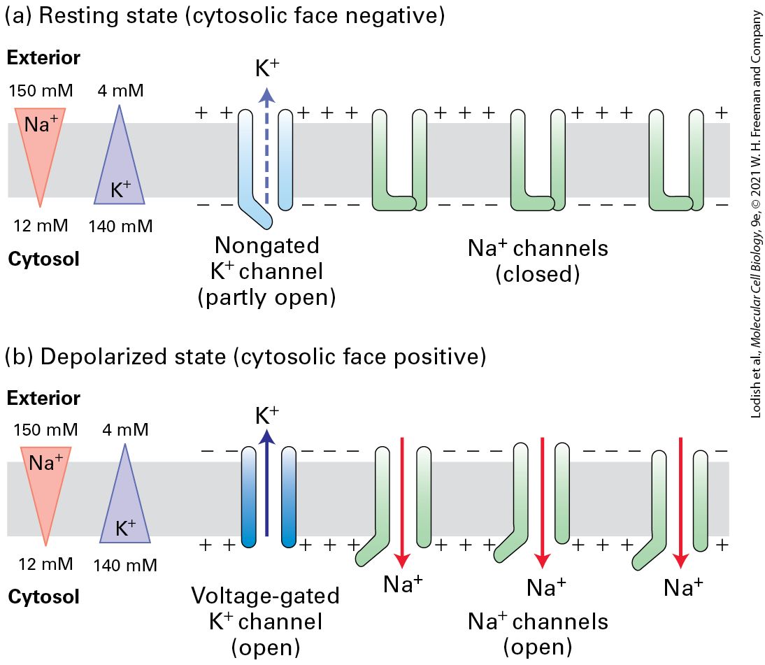
- The Magnitude of the Action Potential Is Close to ENa and Is Caused by Na+ Influx Through Open Na+ Channels
- Sequential Opening and Closing of Voltage-Gated Na+ and K+ Channels Generate Action Potentials
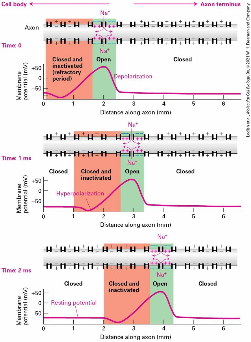
- Action Potentials Are Propagated Unidirectionally Without Diminution
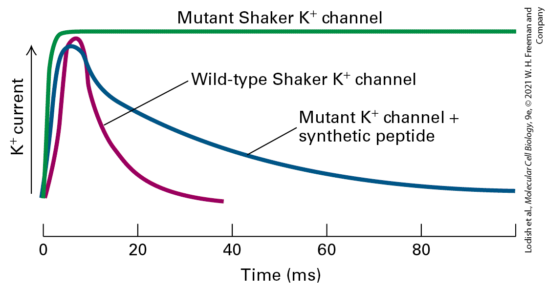
- All Voltage-Gated Ion Channels Have Similar Structures
Ch 24ImmunologyRead full chapter →
Sections in this chapter
- Pathogens Enter the Body Through Different Routes and Replicate at Different Sites
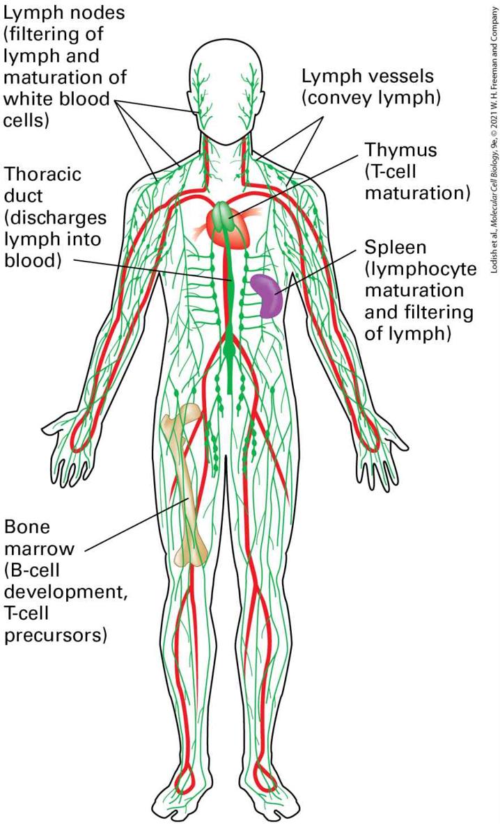
- Cells of the Innate and Adaptive Immune Systems Circulate Throughout the Body and Take Up Residence in Tissues and Lymph Nodes
- Mechanical and Chemical Boundaries Form a First Layer of Defense Against Pathogens
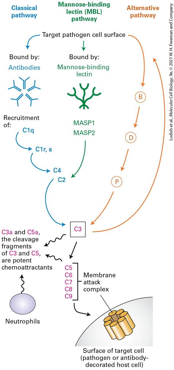
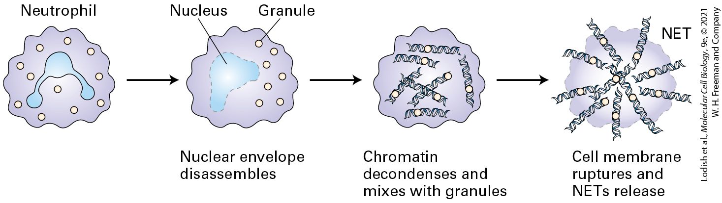
- Innate Immunity Provides a Second Line of Defense
- Inflammation Is a Complex Response to Injury That Encompasses Both Innate and Adaptive Immunity and Helps Destroy Pathogens
- Adaptive Immunity, the Third Line of Defense, Exhibits Specificity
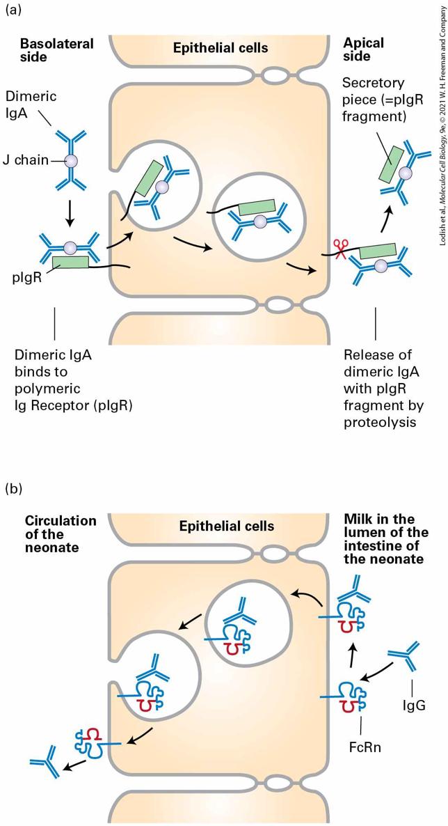
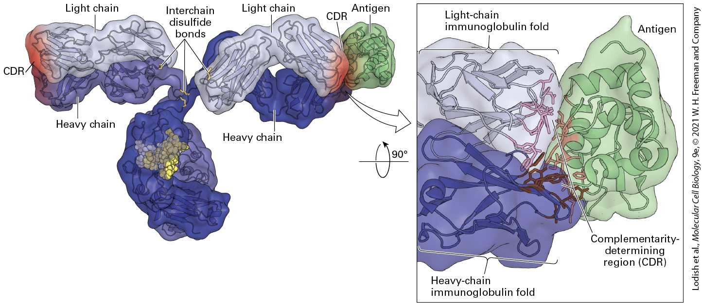
- Immunoglobulins Have a Conserved Structure Consisting of Heavy and Light Chains
- Multiple Immunoglobulin Isotypes Exist, Each with Different Functions
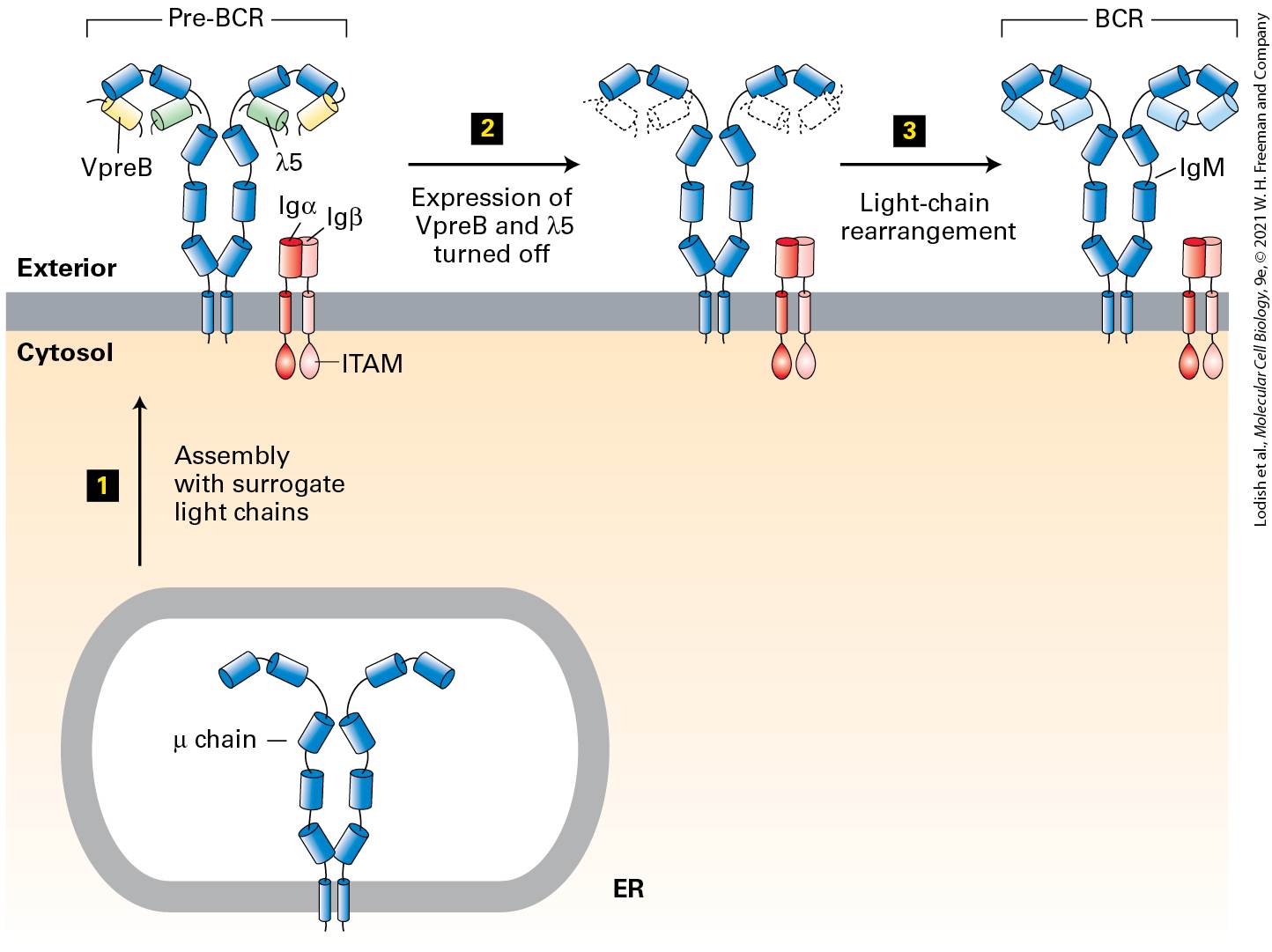
- Each Naive B Cell Produces a Unique Immunoglobulin
- Immunoglobulin Domains Have a Characteristic Fold Composed of Two β Sheets Stabilized by a Disulfide Bond
- An Immunoglobulin’s Constant Region Determines Its Functional Properties
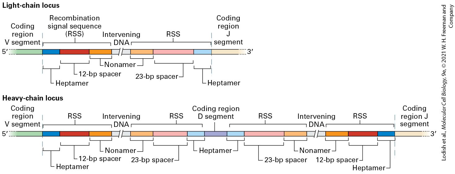
- 24.3 Generation of Antibody Diversity and B-Cell Development
Ch 25CancerRead full chapter →
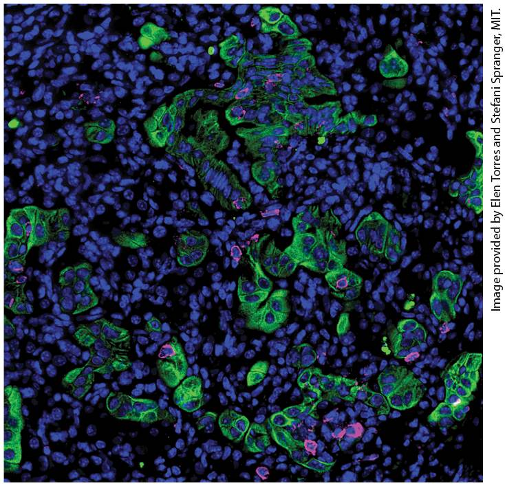
Chapter 25 Cancer Cells of the immune system often interact with cancer cells within the tumor microenvironment. This section of a lung adenocarcinoma shows all cell nuclei (blue), tumor cells (green) and T cells (magenta). Note the tendency of the T cells to be in contact with tumor cells.
Sections in this chapter
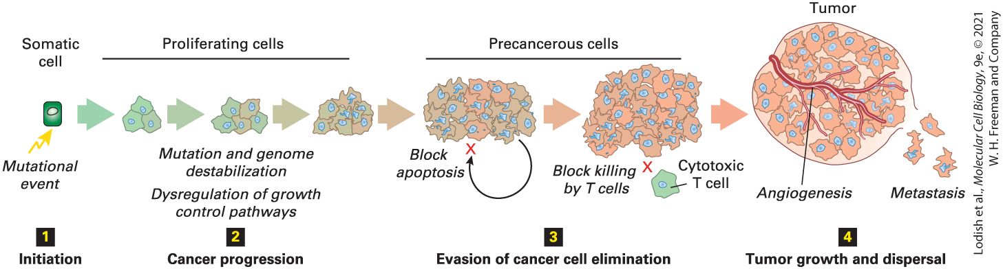
- 25.1 How Tumor Cells Differ from Normal Cells
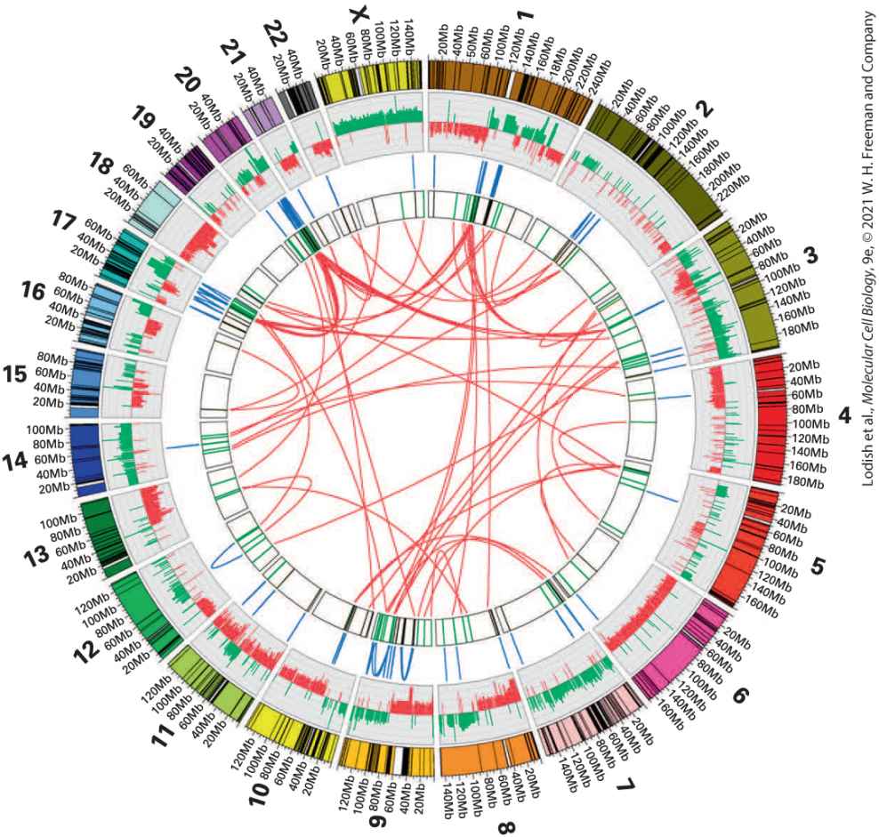
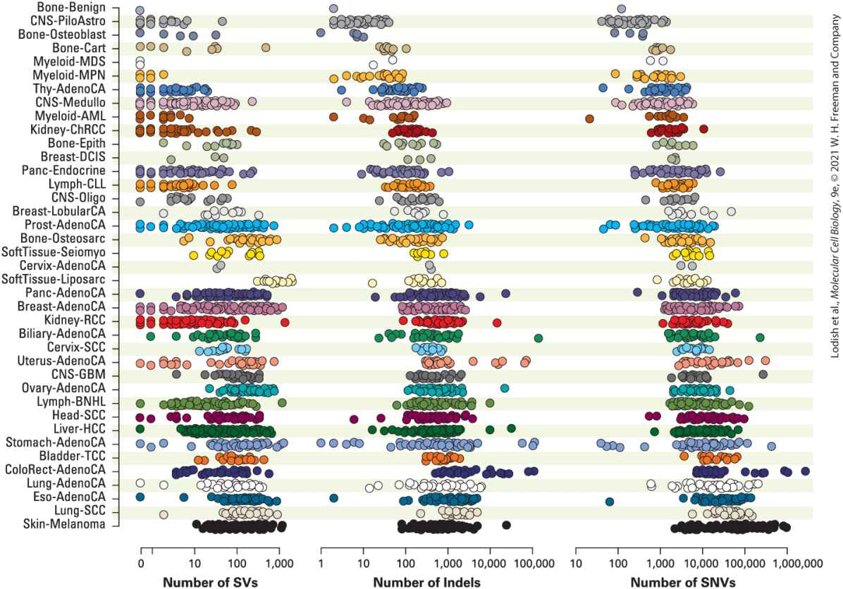
- The Genetic Makeup of Most Cancer Cells Is Dramatically Altered
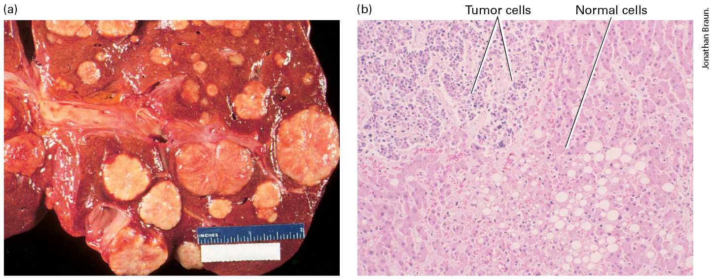
- Uncontrolled Proliferation Is a Universal Trait of Cancer
- Cellular Housekeeping Functions Are Fundamentally Altered in Cancer Cells
- Cancer Cells Exhibit Altered Cell-Cell Interactions to Form Heterogeneous Organs
- Tumor Growth Requires Formation of New Blood Vessels
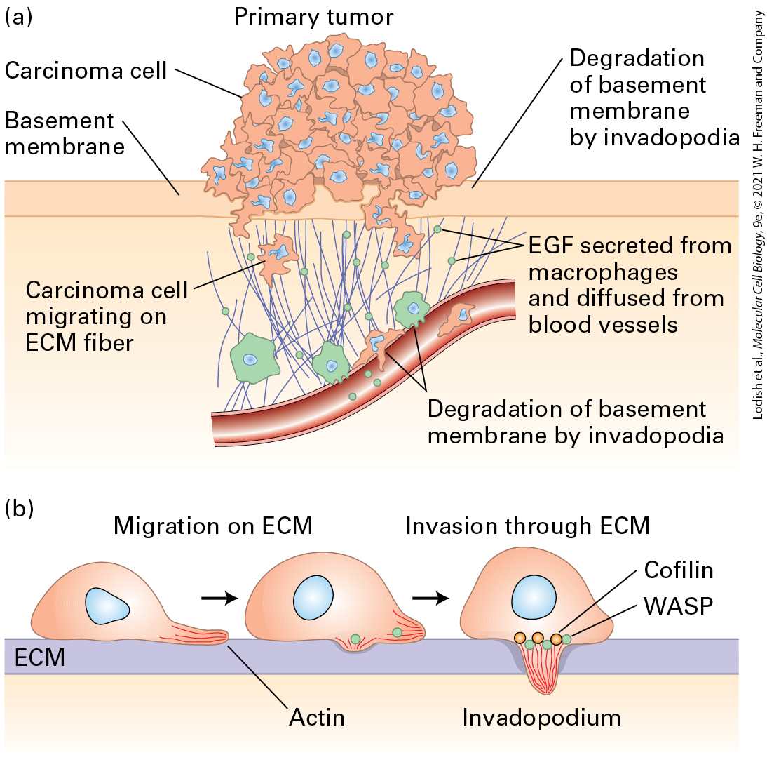
- Invasion and Metastasis Are Late Stages of Tumorigenesis
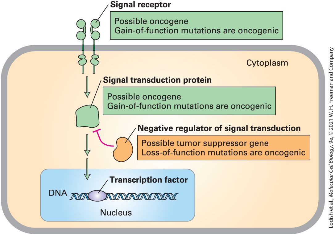
- 25.2 Genetic and Genomic Basis of Cancer
- Carcinogens Induce Cancer by Damaging DNA
- Some Carcinogens Have Been Linked to Specific Cancers
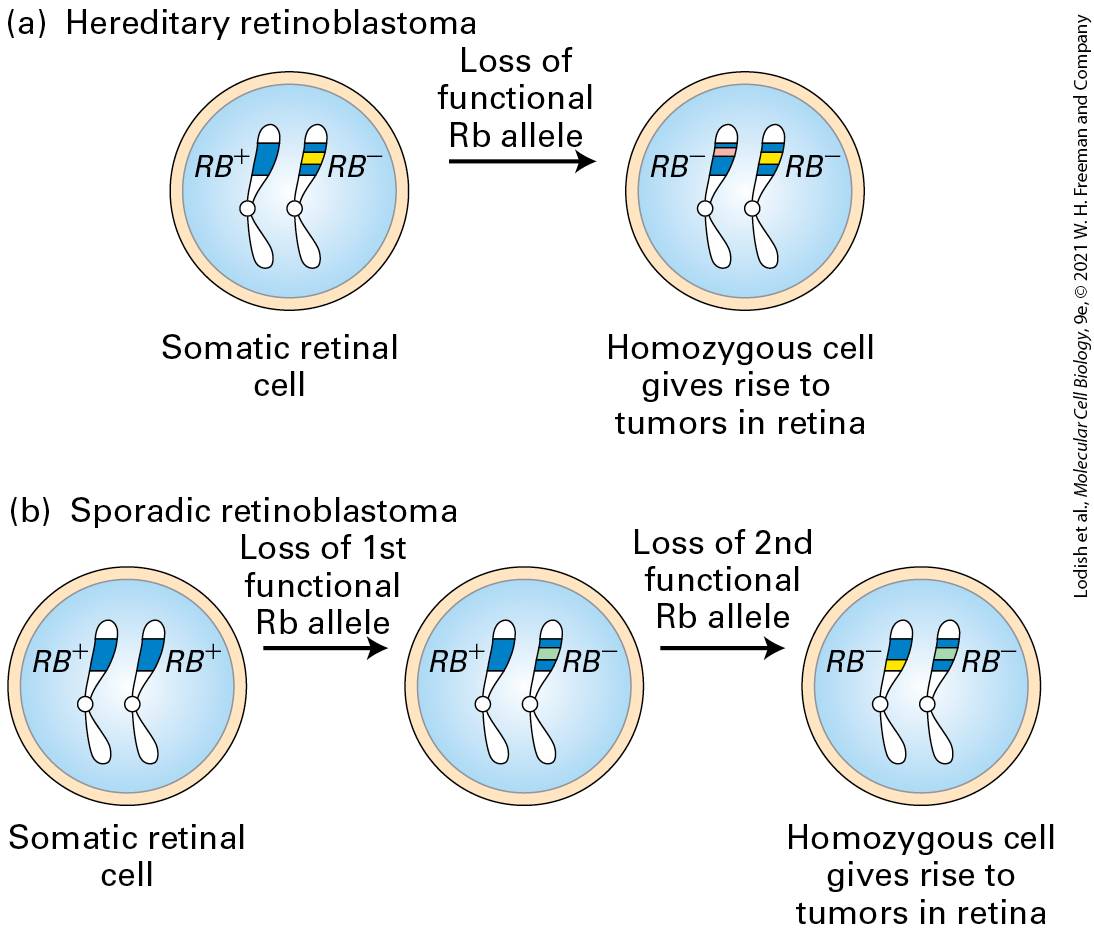
- Familial Syndromes That Cause Loss of DNA Repair Can Lead to Cancer
- Somatic Mutations in the DNA Damage Response Pathway Are Oncogenic