Chapter introduction
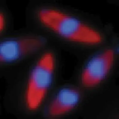
A nuclear option that protects phage from host defenses. Phage FKZ infects Pseudomonas aeruginosa cells and protects its genomic DNA from the host’s defenses by constructing a protective nucleus-like structure. mCherry (red) tags the host defense protein, a restriction endonuclease, and 4′,6-diamidino-2-phenylindole (DAPI;
Prior chapters of the textbook have described how cells express their genes, swim, form biofilms, and perform myriad other functions critical to their growth and survival. For simplicity, prior chapters have largely treated these activities as isolated processes. This closing chapter of Part 2 demonstrates how molecular processes can be integrated to provide higher-order cellular functions, and we use select areas of interest as examples. Molecular complexity will be demonstrated for chemotactic swimming, biofilm formation, and destruction of infecting viruses (phages) using defenses such as CRISPR. We also describe how phages launch counterdefenses to set off an evolutionary arms race with their hosts. We end the chapter describing how scientists in the emerging field of synthetic biology can engineer complex, integrated processes within cells with the goal of producing microbes of great benefit to humankind.
12.1 Chemotaxis: Movement with a DestinationUnit 5 · Regulation
We learned in Chapter 3 that many bacteria propel themselves by means of rotary flagella. But how do cells decide where to swim? Most flagellated cells have an elaborate sensory system for taxis, the ability to swim toward favorable environments (attractant signals, such as nutrients) and away from inferior environments (repellent signals, such as waste products). Taxis to specific chemicals is called chemotaxis. It is controlled by a sophisticated signal transduction cascade that begins with transmembrane chemoreceptor proteins that bind to attractant or repellent ligands, and it ends with a molecular switch that controls the direction of flagellar rotation. Chemotaxis is important to bacteria for a number of reasons. It provides a useful survival strategy in nature, keeping bacteria moving toward nutrients and, with chemoreceptors that sense repellents, away from trouble (for example, toxic compounds). For commensal bacteria or pathogens, chemotaxis can also be used to move the organism toward a cell surface to which it can attach. For example, the intestinal lining can exude chemical attractants that, like a beacon, will lead the bacteria in the intestine toward the cell surface where they can attach. In sum, chemotactic sensory perception plays a major role in structuring microbial communities, in affecting microbial activities, and in influencing various microbial interactions with their surroundings. In this section we connect the dots between the receptor and the flagellum to understand how the cell controls flagellar spin and, ultimately, its swim direction.
Moving with a Purpose
Chemotaxis requires a mechanism for the rotary flagella (singular, flagellum) to propel the cell toward attractants or away from repellents. Let’s first talk about how bacteria can change the direction of their swim. The flagellar rotor can rotate in the clockwise or counterclockwise direction (Fig. 12.1), and switching between the two directions is key to chemotactic swimming. The switch in rotation direction can be visualized directly in cells that are tethered to a microscopy slide by one of their flagella (Fig. 12.1). Because the flagellum is fixed in location, it is the cell itself that spins when the proton motive force drives flagellar rotation. Note that the spin of the cell would be opposite to the spin of the flagellum if the latter were free of its tether.
FIGURE 12.1 ■ Chemotaxis. A. Flagella are oriented in a bundle extending behind one pole. When the cell veers away from the attractant, the receptors send a signal that allows one or more flagella to switch rotation from counterclockwise (CCW) to clockwise (CW). This switched rotation disrupts the bundle of flagella, causing the cell to tumble briefly before it swims off in a new direction. B. The resulting pattern of movement is a “biased random walk” in which the tumbles reorient the cell randomly, but runs toward the attractant are generally longest, resulting in an overall migration toward the attractant.
In bacteria with multiple flagella, all motors coordinate their rotations. In the counterclockwise mode, all flagella sweep behind
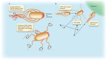
the cell, forming a rotating bundle that propels the organism forward in what is called smooth swimming, or a “run.” When flagellar rotors suddenly switch to a clockwise rotation, the bundle is disrupted and the bacterium “tumbles” in a random fashion. When the flagellar rotors switch back to counterclockwise rotation, swimming resumes, oriented in a new, random direction as a result of the tumble. The key to chemotaxis is a mechanism that suppresses the number of tumbles an organism makes when it moves from lower to higher concentration of an attractant chemical (Fig. 12.1). For instance, an organism moving toward an attractant may tumble only twice in 5 seconds. In contrast, an organism moving in the wrong direction (that is, toward a lower concentration of attractant) may tumble eight times in 5 seconds. If the organism suddenly finds itself going in the right direction, the sensory transduction system will suppress the number of tumbles, and the cell will continue moving in the right direction. The resulting pattern of movement generates a “biased random walk” in which the cell tends to migrate toward the attractant.
The frequency of tumbles versus runs is controlled by the activity of the switch complex located at the base of the flagellum. In Escherichia coli, the default rotation of the flagellar rotor is counterclockwise, which provides a run. Activation of the switch changes the rotation to clockwise, which provides a tumble. The more often the switch is activated, the higher the frequency of tumbles.
The frequency of switch activation is controlled by the upstream chemotaxis signal transduction system, which is sensitive to the attractants or repellents that are transported into the periplasm from the environment. Bacteria such as E. coli utilize transmembrane receptor proteins referred to as methyl-accepting chemotaxis proteins (MCPs, or chemoreceptors) to detect a wide array of attractants and repellents. These MCPs control the activity of a two-component signal transduction pathway (see Section 10.1) composed of the kinase protein CheA (pronounced “key-ay”) and the response regulator protein CheY. The cytoplasmic domain of each MCP binds to CheA (via an intermediary protein, CheW) and controls CheA activity. Notably, the MCPs are localized to one or both cell poles in highly structured arrays organized by the cytoplasmic CheA and CheW proteins (Fig. 12.2). This arrangement serves to heighten the sensitivity to chemoattractants or repellents by amplifying the signal of chemoreception. Binding to an attractant molecule allows a single chemoreceptor to control the function of up to 35 CheA proteins within the array, each of which can relay the signal to the flagella.
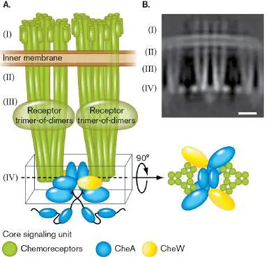
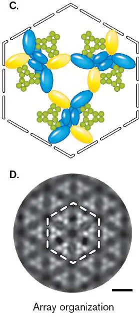
FIGURE 12.2 ■ Chemoreceptor array organization. A. MCPs are arrayed in trimer-of-dimer clusters organized by the cytoplasmic CheA and CheW proteins. B. Cryo-electron tomography reveals the MCP + CheA + CheW organization in a side view of the cell envelope. Roman numerals correspond to those in part A. C. A top-down schematic of the hexagonal structure of the MCP + CheA + CheW complex. D. The hexagonal structure is repeated throughout the array, as observed in this top-down view of the cryoelectron tomogram. Scale bars, 10 nm.
A. BURT ET AL. 2020. NAT COMMUN. 11 :743
A. BURT ET AL. 2020. NAT COMMUN. 11 :743
Let’s now examine how the MCPs work with the Che proteins to control flagellar spin. Recall from Section 10.1 that kinases like CheA can become “auto”-phosphorylated using ATP as the phosphate donor. When the MCP is not bound to a chemoattractant, CheA is free to autophosphorylate, forming CheA-P (Fig. 12.3, step 1). CheA-P then transfers its phosphate to CheY, and then CheY-P activates the flagellar switch, causing the cell to tumble. So long as the MCP continues to remain without chemoattractant, the CheA kinase can continue to acquire phosphates from ATP.
FIGURE 12.3 ■ Chemotaxis signaling pathway in Escherichia coli.
When a chemoattractant chemical, such as serine, binds to the periplasmic side of the MCP, the conformation of the cytoplasmic domain changes and inhibits the autophosphorylation activity of CheA (Fig. 12.3, step 2). CheA can no longer transfer a phosphate to CheY, and CheY-P is eventually dephosphorylated by CheZ, which is a slow but always active phosphatase. In its unphosphorylated state, CheY cannot activate the flagellar switch, which means that the flagellum rotates in the counterclockwise direction, and the cell performs a run.
Memory Helps Cells Move Up a Chemical Gradient
The chemotaxis signaling cascade just described makes sense when an organism first encounters a chemoattractant, but how does the cell know to keep moving into even higher concentrations? As it
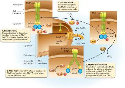
happens, chemotactic organisms possess a primitive form of memory. The conformational change in the MCP that inactivates CheA kinase activity also subjects the cytoplasmic side of the MCP to methylation (hence the name “methyl-accepting chemotaxis protein”) by CheR methylase (Fig. 12.3, step 3). Methylation of glutamate residues in the cytoplasmic struts of the MCP decreases the binding affinity of MCP to the attractant, effectively desensitizing the system. This means in order for MCP to maintain inhibition of CheA kinase and reward the cell with a continued run, an even higher concentration of the attractant must be present.
When the concentration of attractant is no longer high enough for MCP to bind, MCP can no longer block phosphorylation of CheA and CheY, and the organism will tumble.
When the cell moves away from attractant, the methylation switch is reset by another protein, CheB-P, which removes methyl groups so that the system is resensitized to attractant (Fig. 12.3, step 4). The system is elegantly fine-tuned in that CheA kinase is also the protein that phosphorylates CheB. So, as the cell moves away from attractant, CheA kinase phosphorylates CheY to produce tumble, and it phosphorylates CheB to reset the sensitization switch. Notably, while the basic chemotactic mechanism is evolutionarily conserved in bacteria, different genera use it differently. In the Gram-positive Bacillus subtilis, for example, ligand binding to an MCP stimulates CheA kinase, and CheY-P stimulates counterclockwise flagellar rotation and, thus, extended runs. This mechanism is the exact opposite of the one used in E. coli.
As we have seen, the chemotaxis machinery is quite complex, involving organized arrays of sensors, phosphorylation cascades, and a mechanism that confers memory of past conditions. One might ask, is all this really necessary, or wouldn’t a cell be just as well off with an unregulated flagellum that only generates runs? This question is perhaps best answered by the 1969 study of John Armstrong and Julius Adler at the University of Wisconsin–Madison. They generated two types of mutants in E. coli: one that was nonmotile, and one that was motile but not chemotactic. They used needle tips to inoculate these mutants as well as the motile and chemotactic wild-type strain onto a semisolid agar Petri plate (Fig. 12.4). The agar was soft enough to allow cells to swim within the medium during the incubation. These plates contained tryptone, a mixture of amino acid monomers and oligomers, which the strains could consume as their carbon and energy source. Importantly, these amino acids also served as the chemoattractants.
FIGURE 12.4 ■ Chemotaxis improves motility. Escherichia coli wild type and mutants inoculated at the same time in semisolid agar medium form colonies that expand to varying extents, according to their ability to both swim and perform chemotaxis toward tryptone.
J. ADLER. 2011. ANNU REV BIOCHEM. 80 :42–70
Over time, the wild-type strain expanded quickly from the point of inoculation, as witnessed by its large colony diameter. Cells in the colony consumed the local sources of tryptone, thus establishing a gradient of chemoattractant. This gradient continued to shift away from the colony center as the population expanded and consumed
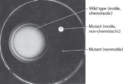
the tryptone. Perhaps not surprisingly, the nonmotile mutant formed a very compact colony relative to the wild type. In contrast to the nonmotile mutant, the motile but non-chemotactic mutant was able to expand beyond the point of inoculation. However, its colony diameter was significantly smaller than that of the wild type. The value of well-timed tumbles between runs is thus clearly evidenced by the vastly larger expansion of the chemotactic wild-type population relative to the non-chemotactic (and nonmotile) mutant(s).
Thought Question
12.1 The movie associated with Figure 12.1shows bacilli tethered to a glass slide by one of their flagella. Several bacteria rotate in opposite directions as their flagellar rotors switch from clockwise to counterclockwise rotation and back again (see Fig. 12.3). Which way will a bacillus rotate when an attractant is added? What would the rotating phenotypes be if you tethered the knockout mutants cheY, cheA, cheZ, and cheR to the slide and then added attractant?
Diverse Mechanisms of Taxis
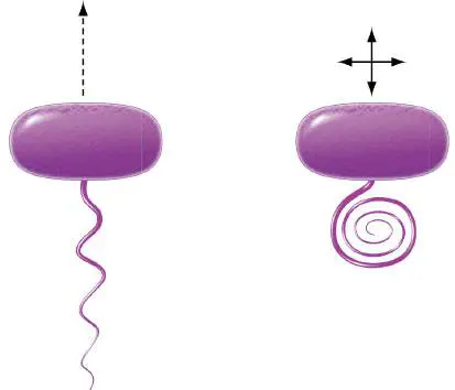
Some chemotactic bacteria do not form flagellar bundles, and they perform runs and tumbles in different ways. For example, the rod-shaped Rhodobacter sphaeroides, a purple phototrophic wetland bacterium, has a single flagellum positioned curiously at the middle of the rod rather than at one of the poles (Fig. 12.5). This flagellum provides a run when it spins at high speeds. If the spin slows down, the flagellar filament changes shape, forming a coil. This coil tumbles the cell as it spins. A return to higher spin speeds causes the flagellum to revert to a shape capable of producing a run. Hence, this organism adjusts the speed of flagellar rotation, rather than the direction, to provide the runs and tumbles of chemotaxis.
FIGURE 12.5 ■ Chemotaxis in Rhodobacter sphaeroides. High (left) versus low (right) speed of rotation by the single flagellum results in a run or a tumble, respectively.
Not all prokaryotes use chemical attractants or repellents to direct their movement. Some haloarchaea perform phototaxis, which is taxis toward light (see Section 19.5). Other microbes can respond to oxygen gradients (aerotaxis) either by directly binding oxygen or by sensing the rate of electron flow during aerobic respiration. An unusual form of taxis is magnetotaxis, the ability to sense and respond to magnetism. Found in pond water, magnetotactic bacteria orient themselves along Earth’s lines of magnetic field. Magnetotactic
bacteria can be collected from the environment by placing a magnet in a jar of pond water; bacteria orienting by the field lines collect nearby. The bacteria align their cell axis along the magnetic field using linear arrays of magnetosomes, microscopic membrane-enclosed crystals of the magnetic mineral magnetite, Fe 3 O 4 (see Fig. 2.45). The magnetosomes orient bacterial swimming toward the bottom of the pond. Magnetotactic bacteria are anaerobes or microaerophiles (requiring low oxygen), which prefer the lower part of the water column, where oxygen concentration is lowest. In the northern latitudes, where Earth’s magnetic field lines point downward, bacteria that are magnetotactic swim “downward” toward magnetic north.
Thought Question
12.2 How would a magnetotactic species have to behave if it were in the Southern Hemisphere instead of the Northern Hemisphere? In addition to swimming, some microbes can move on solid surfaces. This ability is especially important for biofilm development, as we will see in the next section. Flagellar rotation can propel cells on surfaces as well as in liquid. The surface mode is called swarming motility, which is also described in the next section, as well as in Section 18.4. Many bacteria such as Pseudomonas aeruginosa can use retractable type IV pili to pull themselves along surfaces in what is referred to as twitching motility (Fig. 12.6; also see Fig. 25.12).
FIGURE 12.6 ■ Type IV pili pull Pseudomonas aeruginosa cells during twitching motility. The white arrow shows the fixed position of the pilus tip during retraction. t = elapsed time in seconds. Scale bar, 2 μm.
J. M. SKERKER AND H. C. BERG. 2001. PROC NATL ACAD SCI USA. 98 (12):6901–4
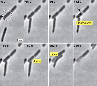
Myxococcus xanthus can perform a different type of motility altogether, called gliding motility, which has its own special apparatus for locomotion. M. xanthus combines twitching and gliding motility to hunt other bacteria such as E. coli (Fig. 12.7). M. xanthus releases degradative enzymes that cause its prey to lyse, so that it can feed on the released contents.
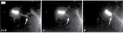
FIGURE 12.7 ■ Myxococcus hunts Escherichia coli by surface motility. Motile Myxococcus (bottom left at time zero) uses swarming and twitching motility to find E. coli prey. Release of enzymes causes the plasmolysis and lysis of the prey cell.
W. ZHANG ET AL. 2020. APPLIED AND ENVIRON MICROB. 86 (3):E02286–19
Each type of surface movement uses a different kind of machinery, but they can all be used for chemotaxis. P. aeruginosa cells twitch toward increasing gradients of phospholipids or their fatty acid components, whereas Vibrio parahaemolyticus swarms toward quorum-sensing autoinducers that they themselves make.
Chemotaxis involving surface motility can have complex signal transduction cascades, and it often includes Che proteins and MCP receptors, like the machinery used for chemotactic swimming.
Thought Question
12.3 What property of fatty acids makes them useful chemoattractants for surface motility, compared to sugars or amino acids that often serve as chemoattractants for swimming motility?
To Summarize
Chemotaxis is a behavior in which motile microbes swim toward favorable environments (chemoattractants) or away from unfavorable environments (chemorepellents).
The direction of flagellar motor rotation determines the type of movement. Counterclockwise rotation results in smooth swimming; clockwise rotation results in tumbling.
Chemoreceptors that bind attractants or repellents provide information that controls the direction of flagellar rotation. Random movement toward an attractant causes a drop in CheY-P levels, which enables counterclockwise rotation and smooth swimming.
Methyl-accepting chemotaxis proteins (MCPs) clustered at cell poles bind chemoattractants and initiate a series of events that lowers CheY-P levels. Reversible methylation or demethylation of MCPs desensitizes or sensitizes MCPs, respectively.
Magnetotaxis is a process of flagellar motility directed along magnetic field lines. Taxis involves magnetosomes, crystals of magnetite encased by intracellular membrane vesicles.
Surface motility can involve flagella, pili, or other specialized machines. Like swimming, it can also involve chemotaxis toward attractants.
Glossary
chemotaxis The ability of organisms to move toward or away from specific chemicals.
flagellum pl. flagella A filamentous structure for motility. In prokaryotes, a helical protein filament attached to a rotary motor; in eukaryotes, an undulating membrane-enclosed complex of microtubules and ATP-driven motor proteins.
flagellum pl. flagella A filamentous structure for motility. In prokaryotes, a helical protein filament attached to a rotary motor; in eukaryotes, an undulating membrane-enclosed complex of microtubules and ATP-driven motor proteins.
methyl-accepting chemotaxis protein (MCP)
Also called chemoreceptor. A cell-membrane signal transduction protein that becomes methylated during adaptation to a chemotactic signal.
MCP Also called chemoreceptor. A cell-membrane signal transduction protein that becomes methylated during adaptation to a chemotactic signal.
magnetotaxis The ability to direct motility along magnetic field lines. magnetosome An organelle that contains the mineral magnetite and thus enables microbes to sense a magnetic field.
swarming or swarming motility A behavior in which some microbial cells differentiate into large swarmer cells and swim together as a unit.
twitching motility A type of bacterial movement on solid surfaces in which a specific pilus extends and retracts.
gliding motility The movement of cells individually or as a collective over surfaces using specialized pili.
Fig. 2.45 FIGURE 2.45 ■ Magnetotactic cell visualized by cryo-electron tomography. A. A single cryo-EM scan lengthwise through Magnetospirillum magneticum. B. 3D model of M. magneticum based on multiple scans. C. Expanded view of the cell interior.
AAAS. ARASH KOMEILI ET AL. SCIENCE 311 :242–245, FIG. 1
NIH, THE JENSEN LABORATORY
NIH, THE JENSEN LABORATORY
Fig. 25.12
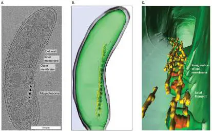
FIGURE 25.12 ■ Type IV pili. A. Model of pilus assembly and disassembly. In this example, PilA is the pilin protein, and PilC1 and Y1 form the attachment tip. Filament is approx. 6 nm in diameter. Assembly and disassembly require the hydrolysis of nucleoside triphosphate (NTP) and take place at the inner membrane, not in the periplasm. B. Photographic evidence of type IV pilus retraction in cells of Pseudomonas aeruginosa. Filament c attaches briefly at its distal tip (note straightening at 24 seconds) and then begins to retract.
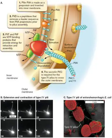
Fluorescent microscopy. t = time, in seconds. C. Type IV pili (green) are essential for enterohemorrhagic E. coli to attach to epithelial cells. SEM.
Source: Part A modified from Bardy et al. 2003. Microbiology 149: 295–
304.
SKERKER, J., ET AL. 2001. PNAS 98 : 6901.
J. XICOHTENCATL-CORTES ET AL. 2007. J CLIN INVEST. 117 :3519–29
12.2 Biofilm Formation by Pseudomonas aeruginosaUnit 5 · Regulation
Biofilms are now recognized as the predominant form of life for most microbes. The importance of biofilm formation has long been recognized in the medical and industrial fields (see Section 4.5). However, until the 1990s the concept of biofilm formation as a sophisticated process was not fully appreciated. Before we discuss this complexity, consider the alternative scenario, whereby a community on a surface arises by a founder “sticky” bacterium that is blind to whether it is still swimming as a planktonic cell or attached to a surface and merely follows the same program of growth and reproduction without a change in life strategy. What would happen to cells that try to keep swimming on a surface? Would the attached cells develop a community that is optimized for resource utilization, waste removal, predation avoidance, and antibiotic resistance? Would the cells be able to leave the surface once conditions were no longer optimal?
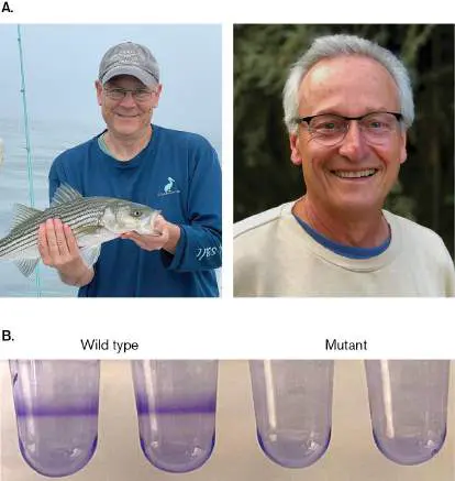
As we will see, microbes use physical and chemical cues to know exactly when they arrive at a surface, when there are enough of them nearby to collectively build the biofilm, and, once established, to determine how well life is going within the biofilm. Dissecting this process over the past few decades has involved many different approaches, including epifluorescence microscopy, biochemistry, and various “-omics” technologies. One of the key innovations effectively utilized by George O’Toole, now at the Geisel School of Medicine at Dartmouth, and Roberto Kolter at Harvard Medical School, was the application of the dye crystal violet to reveal biofilm communities that form as rings at the air-liquid interface in cultureware (Fig. 12.8). Specifically, when applied to communities that form in wells of plastic 96-well microtiter plates, this method allows for the high-throughput screening for genes involved in biofilm formation: mutants defective in this process are identified by their aberrant or missing crystal violet rings. Over the years this technique, used by multiple scientists, has identified a multitude of genes involved in biofilm development in a wide variety of microbes.
FIGURE 12.8 ■ Development of the high-throughput assay for genetic analysis of biofilms. A. George O’Toole (left) and Roberto Kolter (right) developed the crystal violet microtiter plate biofilm assay. B. Crystal violet staining of wild-type (left) and biofilm-deficient mutant (right) Pseudomonas spp. strains grown in microtiter wells.
GEORGE O’TOOLE
ROBERTO KOLTER
GEORGE O’TOOLE
In this section we explore several key molecular processes involved in the progression of biofilm formation, starting with attachment to the surface, and finishing with mechanisms of dispersal and return to the planktonic phase (as introduced in Section 4.5). For this discussion we focus on the opportunistic pathogen Pseudomonas aeruginosa, which can form biofilms in the lungs of cystic fibrosis patients and in catheter lines (see Section 23.4). Figure 12.9summarizes the stages of biofilm formation as they occur in P. aeruginosa. The cells attach to the surface to begin the process.
FIGURE 12.9 ■ Biofilm formation in Pseudomonas aeruginosa. Biofilm development involves activities that occur at different stages. The master regulator of the planktonic-to-sessile transition, bis-(3′–5′)-cyclic dimeric guanosine monophosphate (c-di-GMP), increases in concentration during development until signals to leave the biofilm are received. eDNA = extracellular DNA; EPS = extracellular polymeric substance.
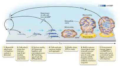
Once attached, the cells move along the surface and establish clusters of cells called microcolonies (Fig. 12.10). These microcolonies expand into mushroom-shaped macrocolonies that define the mature biofilm. Finally, when environmental changes occur, some cells dislodge themselves from the biofilm and reenter the planktonic phase. We will discuss these stages in turn, describing the actions taken by the cells as well as some of the molecular mechanisms that regulate and perform these steps. Critically, biofilm formation by P. aeruginosa remains an area of highly active research, as many questions about this complex life cycle remain unanswered.
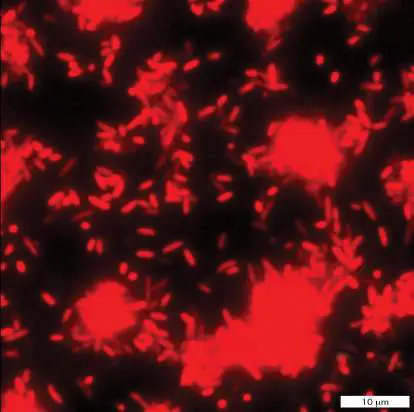
FIGURE 12.10 ■ Pseudomonas aeruginosa forms microcolonies via surface motility.
GEORGE O’TOOLE
c-di-GMP: Master Regulator of the Planktonic-Surface Transition
The key regulator of the surface-associated biofilm lifestyle is the second messenger molecule c-di-GMP [bis-(3′–5′)-cyclic dimeric guanosine monophosphate]. As described in Section 10.4, c-di-GMP is synthesized by diguanylate cyclases (DGCs) and is degraded by phosphodiesterases (PDEs). In planktonic cells, c-di-GMP concentrations are low (Fig. 12.9). Contact with the surface triggers c-di-GMP synthesis by DGCs, and maturation of the biofilm coincides with a progressive increase in cellular c-di-GMP concentration. Environmental signals can activate PDE enzymes that degrade c-di-GMP by subsets of cells within the biofilm, and the concomitant drop in c-di-GMP triggers processes that release these cells from the biofilm, including enzymes that degrade the extracellular matrix and release the protein tethers to that matrix. How c-di-GMP functions to regulate each of these transitions is still under active investigation. Many of the effectors (proteins regulated by binding c-di-GMP) have been discovered so far, but an unknown number of others are most certainly involved as well. What’s more, the Pseudomonas aeruginosa genome encodes about 40 DGC and PDE enzymes, many of which are still uncharacterized. These enzymes may function to transition the cells from one stage to the next or to maintain the cells in an active, viable state during prolonged biofilm existence.
To further contextualize this key regulator, c-di-GMP messaging is only part of the regulatory cascade that directs biofilm development. Additional secondary messengers such as cyclic AMP (cAMP) and guanosine tetraphosphate (ppGpp), as well as several interacting quorum-sensing systems (see Section 10.4), contribute to a sophisticated regulatory program that is exquisitely sensitive to the physical and chemical changes that occur during biofilm formation and dispersal. Altogether, this regulatory system controls expression of hundreds of genes, including those that promote virulence and confer tolerance to antibiotics.
Surface Attachment and Motility Initiate Biofilm Formation
Swimming cells contact the surface with their polar flagella, sometimes assisted by pili (Fig. 12.9, step 1). Attachment via the flagellum is reversible, and cells can readily detach and reenter the planktonic phase. Eventually the cells attach to the surface along their long axis, and their type IV pili attach to the surface, committing the cells to the surface (Fig. 12.9, step 2).
Contact with the surface triggers the onset of c-di-GMP synthesis, thereby initiating the progression toward mature biofilm formation. Cells can physically sense surface attachment through their flagella and by contact-mediated membrane disruption, both of which trigger c-di-GMP synthesis. In addition, the binding of type IV pili to a surface stimulates synthesis of more type IV pili that, upon binding, activate synthesis of c-di-GMP in a positive feedback loop. Biofilm development proceeds with the critical transition from swimming motility to surface motility (Fig. 12.9, step 3). For Pseudomonas, the flagellum, located at one pole, participates in both modes of motility. Recall that the flagellum is driven by a rotary motor, energized by the proton motive force to spin the dynamic rotor within the stator ring structure (see Section 3.6). The stators for swimming cells are composed of MotAB and MotCD protein complexes.
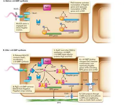
Once the cell attaches to that surface along its long axis, the MotCD stator assumes control of flagellar rotation to initiate swarming motility (Fig. 12.11A). This switch is thought to happen because the MotCD stator can provide the higher torque required for swarming motility. Swarming motility is assisted by the release of rhamnolipids, surfactants that decrease the surface tension at the liquid-solid interface. Note that in contrast to Vibrio species that undergo cell elongation and synthesis of multiple flagella along the long axis of the cell (see Section 18.4), Pseudomonas relies on the single polar flagellum to drive swarming motility.
FIGURE 12.11 ■ c-di-GMP signals the transition from a motile to a sessile state by stopping flagellar rotation and increasing adherence to the surface.
Pseudomonas also utilize their type IV pili for surface motility. These retractile pili mediate twitching motility, as described in Section 25.2, and can also assist during swarming motility by mediating cell-cell contact. During twitching motility, the type IV pili
can attach to surface-associated EPS (extracellular polymeric substance) molecules that are released by neighbor cells, using them as guides to follow and establish contact with their neighbors. During this movement along the surface, cells are actively growing and dividing, and some cells—particularly those with low c-di-GMP concentrations—detach from the surface to resume swimming motility (Fig. 12.9, step 4). Swimming cells can reestablish contact elsewhere along the surface; these combined forms of motility allow the Pseudomonas population to quickly colonize a surface.
Adhesion
Surface motility allows the cells to form the aggregates (microcolonies) that will ultimately develop into macrocolonies. Upon aggregation, cells need to stop moving (Fig. 12.9, step 5). To stop the flagellum from spinning, cells disengage and sequester the motor. This is accomplished by the protein FlgZ once it is activated by binding c-di-GMP (Fig. 12.11B ). The concentration of c-di-GMP increases through signals reinforcing that the cell is on a surface, and this eventually results in a FlgZ:c-di-GMP protein:ligand complex that functions to bind and sequester MotCD away from the flagellum.
Once released from the flagellum machinery, MotC can establish contacts with the DGC called SadC. When this happens, MotC stimulates SadC to produce even more c-di-GMP.
Aggregated cells reinforce the nonmotile, sessile state by producing molecular anchors. Cells release adhesive extracellular polymeric substance (EPS) molecules as well as adhesin proteins that extend from the cell envelope to bind the cell to the EPS and to neighboring cells. Release of these anchoring molecules is triggered by the accumulation of c-di-GMP described earlier.
The c-di-GMP–responsive regulator FleQ controls synthesis of the molecular anchors while also helping to regulate surface motility. FleQ monomers assemble to form a hexameric quaternary structure, most likely in the shape of a ring. This hexamer is able to bind to the regulatory sequences of several operons and control their expression, either positively or negatively depending on whether it is bound to c-di-GMP. In motile cells with low c-di-GMP concentration, the FleQ hexamer is not bound to c-di-GMP and consequently activates transcription of genes involved in flagellum synthesis while repressing transcription of genes involved in the synthesis of EPS and the adhesin protein (CdrA) (Fig. 12.11B ). When cells accumulate higher concentrations of c-di-GMP, this molecule can bind to the FleQ hexamer and change its quaternary structure. When bound to c-di-GMP, FleQ reverses its regulatory function, now repressing the flagellar genes and activating the genes for EPS and the CdrA adhesin. This regulatory step not only anchors the cell but also halts production of additional flagella, appendages that will no longer serve a purpose within a stationary biofilm enclosed by matrix.
Once synthesized, the CdrA adhesins extend beyond the outer membrane through pores built from monomers of CdrB, encoded in the same operon as CdrA. CdrA proteins adhere to both EPS and to other CdrA proteins that extend from neighboring cells to firmly anchor the microcolony aggregate to the surface.
Thought Question
12.4 Why do you think Pseudomonas aeruginosa regulates flagella at both the transcription and posttranslation levels during the transition from surface attachment to biofilm formation?
Biofilms Mature into Macrocolonies
Mushroom-shaped macrocolonies develop from microcolony precursors through release of molecules that form an extracellular matrix (Fig. 12.9, step 6). The extracellular matrix of the mature biofilm is a complex mix of EPS, protein, and, perhaps surprisingly, DNA. Extracellular DNA (eDNA) serves to promote adhesion to the surface, to spatially organize cells within the biofilm, and to provide overall stability of the structure. eDNA in the matrix originates within intact cells that release their cytoplasmic contents when triggered to lyse (Fig. 12.9, step 6). There are multiple ways that cells lyse, including the synthesis of the secondary metabolite pyocyanin, which produces the membrane-damaging oxidant hydrogen peroxide; cells may also undergo explosive lysis. Importantly, only a subset of cells need to lyse within the biofilm. Cells within the biofilm matrix secrete an array of proteins, many of which are virulence factors that help Pseudomonas colonize and persist in the host. These include exotoxin A, which inhibits translation in eukaryotic cells, a protease (LasA), and an elastase (LasB) that degrades elastin and collagen. Cells also release pyoverdine and pyochelin, secondary metabolites that help scavenge iron.
Rhamnolipid surfactants, which promote swarming during the motility phase of biofilm development, serve a different purpose in the development of macrocolonies. Rhamnolipids released by the growing microcolony coat the surrounding surface to establish “void” zones where cells can migrate through but not adhere (Figs. 12.9 and 12.12 ). These zones will form the channels between macrocolonies that are critical for nutrient acquisition and waste removal.
FIGURE 12.12 ■ Rhamnolipids create channels around macrocolonies. A. Green fluorescent protein (GFP)-labeled Pseudomonas aeruginosa (green) attempts to colonize the surface around established macrocolonies of P. aeruginosa (red) when fluid flow is interrupted. B. Rhamnolipids produced by the macrocolonies prevent adhesion in those areas, washing away the potential colonizers when flow resumes.
M. E. DAVEY ET AL. 2003. J OF BACTERIOLOGY 185 (3):1027–1036
M. E. DAVEY ET AL. 2003. J OF BACTERIOLOGY 185 (3):1027–1036
The transition from microcolony to macrocolony is controlled by a complex array of gene expression regulators. One important set of these regulators is the quorum-sensing (QS) systems. Recall from Section 10.4 that QS systems monitor cell density, which in Pseudomonas biofilms increases when the cells form microcolonies during surface motility.
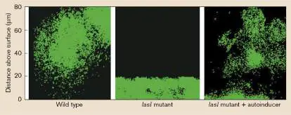
Quorum-sensing control of macrocolony development in Pseudomonas was discovered by a genetic study of the LasI autoinducer synthetase. Unlike wild-type cells, lasI mutants were unable to develop the mushroom-shaped biofilm structures, and instead formed a uniform monolayer (Fig. 12.13). When the autoinducer normally synthesized by LasI was supplied to the lasI mutant by addition to the medium, it could enter cells and activate the autoinducer-binding LasR transcriptional regulator and consequently restore macrocolony development.
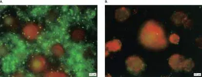
FIGURE 12.13 ■ Quorum sensing is required for macrocolony development. Side view of GFP-labeled Pseudomonas aeruginosa cells growing on a surface. Mutants lacking the lasI gene fail to develop the mushroom-shaped microcolony, unless supplied with the autoinducer product of LasI.
D. G. DAVIES ET AL. 1998. SCIENCE 280 (5361):295–298
Pseudomonas aeruginosa utilizes four different quorum-sensing systems, each of which synthesizes and responds to a unique autoinducer. Collectively, these QS systems regulate expression of hundreds of genes, as much as 6% of the genome. These QS systems cross-regulate each other in a complex hierarchy, with the LasI-LasR QS system as the master regulator. Fine-tuning of these QS systems is further provided by over a dozen additional regulators, including the sigma factor RpoS and the secondary metabolite ppGpp, that relay environmental cues to modify autoinducer production and response. Indeed, regulation of this QS cascade is quite complex and will not be detailed here.
Dispersion Returns Cells to the Planktonic State
Within the biofilm, cells are sensitive to changes in the external environment, such as drops in nutrient concentration, that signal when to leave the surface community and reenter the swimming
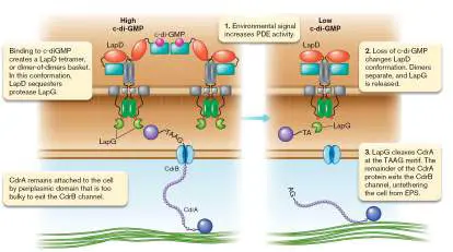
planktonic state in a process called dispersion. These signals initiate a regulatory cascade that activates c-di-GMP–degrading PDEs within the cell. The resulting drop in c-di-GMP concentration within these cells triggers a biofilm escape program involving flagellar synthesis and matrix dissolution (Fig. 12.9, step 7). Release from c-di-GMP restores FleQ’s ability to induce transcription of flagellar synthesis genes. Recall that cells within the biofilm are encased in an extracellular matrix composed of EPS, eDNA, and proteins. Cells triggered to disperse from the biofilm release extracellular enzymes that degrade this matrix and, through a mechanism of targeted proteolysis, also sever the connection between the CdrA adhesin and the EPS matrix.
In intact biofilms, CdrA is secured to the cell by a periplasmic domain too bulky to pass through the outer membrane channel formed by CdrB (Fig. 12.14). Cells sever the connection to the EPS by clipping off the bulky domain, allowing the remaining CdrA protein to exit the CdrB pore. Prior to dispersion, the protease responsible for this cleavage, LapG, is sequestered in an inactive state by LapD. Binding of c-di-GMP stabilizes the LapD proteins as a tetramer, or a dimer-of-dimers basket. This conformation binds and sequesters the periplasmic protease LapG. Degradation of c-di-GMP by phosphodiesterases (PDEs) changes the conformation of LapD to one that can no longer bind LapG. LapG, released into the periplasm, is now able to cleave the TAAG sequence within the periplasmic domain of CdrA, removing the bulky domain and freeing the cell from its tether. With the release from the matrix and the restoration of flagella, cells can escape the biofilm to enter the planktonic state once more.
FIGURE 12.14 ■ Dispersion signals sever the connection between cells and the EPS.
To Summarize
Biofilm formation in Pseudomonas aeruginosa is a developmental progression. After surface contact, cells move along the surface to aggregate into microcolonies that mature into macrocolonies.
c-di-GMP is a master regulator of the transition between the planktonic and the sessile state; it controls rotation of the flagellum and production of surface adhesins.
Quorum sensing regulates the transition from microcolonies to macrocolonies.
Macrocolony structure is established by an extracellular matrix composed of EPS, protein, and eDNA, and its immediate surroundings are protected by surface rhamnolipids that prevent colonization.
Dispersion occurs when cells within the biofilm receive signals from the environment to return to the planktonic
phase. Dissolution of the extracellular matrix and synthesis of new flagella facilitate the dispersion process.
Glossary
biofilm A community of microbes growing on a solid surface. planktonic cell An isolated cell, growing individually in a liquid without connections to other cells.
microcolony A small assembly of cells on a surface that initiate biofilm formation.
macrocolony A large cluster of cells embedded within an extracellular matrix. Macrocolonies constitute the mushroom-shaped structures of mature biofilms.
c-di-GMP Bis-(3′-5′)-cyclic dimeric guanosine monophosphate; a second messenger molecule that plays a key role in regulating biofilm formation.
diguanylate cyclase (DGC)
A class of enzymes that synthesize the second messenger molecule c-di-GMP.
phosphodiesterase (PDE)
A class of enzymes that degrade the second messenger molecule c-di-GMP.
type IV pili Retractable appendages used for surface attachment and twitching motility along the surface.
rhamnolipid A surfactant molecule composed of a rhamnose head group and a fatty acid tail.
extracellular polymeric substances (EPS)
See exopolysaccharides .
extracellular DNA (eDNA)
DNA located outside the cell, often released by cells to provide stability and structure to biofilms.
dispersion The process by which cells escape the biofilm structure and reenter the planktonic state.
12.3 CRISPR and Other Antiphage Defensesnot assigned
In many cases, the consequences of successful infection by a phage are dire for the
prokaryote host. The cell dies from lysis, and the released phage can go on to devastate
the remaining population. Given these consequences, prokaryotes have been under strong
evolutionary pressure to resist phage infection. Consistently, many of the evolved defenses
are located within “phage resistance” islands that get shared within the microbial
community via horizontal gene transfer (see Section 9.5).
This section describes the innovations prokaryotes employ to prevent phage infection
and/or spread throughout the population. We describe how one of these defenses, CRISPR,
has been exploited for gene editing in a number of organisms, including humans. This
section ends with a discussion of the phage’s response to these defenses, which leads to an
evolutionary arms race between predator and prey.
DNA Restriction and Modification
Since the 1950s, scientists have been aware of a system within bacteria that prevents
infection by destroying the phage’s DNA. This protection system, called “restriction and
modification,” or shortened to restriction-modification (RM), involves the enzymatic
cleavage (restriction) of alien DNA and the protective methylation (modification) of self
DNA (Fig. 12.15 ). Most bacteria and archaea produce DNA restriction endonucleases
(also known as restriction enzymes), enzymes that recognize specific short DNA sequences
(known as recognition sites) and cleave DNA at or near those sequences. This ability to
cleave DNA at specific sequences has provided molecular biologists with a tremendously
powerful tool for genetic engineering (see eAppendix 3).
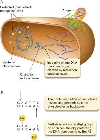
FIGURE 12.15 ■ Restriction of invading phage DNA. A. Phage DNA is injected
into a host, where restriction endonucleases can digest it at unprotected recognition
sites. Host DNA is protected because specific methylations of its own recognition sites
prevent the enzymes from cutting them. B. Recognition site for the EcoR I restriction-
modification system. Shown here are cleavage (top) and methyl modifications
(bottom). The DNA sequence shown is specifically recognized by the endonuclease and
methyltransferase enzymes. Me = methyl group (−CH 3).
Note: Restriction endonucleases are named according to the species from which the
enzyme was isolated. Thus, EcoR I is an enzyme from Escherichia coli. Previously written
as Eco R I, these enzymes no longer have the first three letters italicized.
There are four types of restriction endonucleases (Table 12.1 ), called types I through
IV. Type I and type III restriction endonucleases have their restriction and modification
activities combined in one multifunctional protein and cleave DNA some distance away from
the recognition site. Type II restriction endonucleases (which are used most often for
cloning) possess only endonuclease activity; a separate type II modification protein
methylates the same restriction site.
TABLE Main Types of Restriction-Modification Systems 12.1
System Restriction Number Recognition Cleavage Examples
and of site and
modification subunits characteristics modification
activities sites
Type I
Present in one Three 5–7 bp, Located 100 EcoK in E.
multifunctional different asymmetrical bp or more coli; StyLT
protein subunits from III in
recognition Salmonella
site enterica
Type II
Separate One or 4–6 bp, At or near EcoR I in E.
methylase and two palindromic recognition coli; Hind III
restriction identical site in
endonuclease subunits Haemophilus
enzymes per influenzae
activity
Type
III Present in one Two 5–7 bp, Located 24– Eco571 in E.
multifunctional different asymmetrical 26 bp from coli; BceS I
protein subunits recognition in Bacillus
site cereus
TABLE Main Types of Restriction-Modification Systems 12.1
Type IV
Separate Two Methylated At recognition McrBC in E.
methylase and different bases, up to 3 site or up to coli; Dpn I in
restriction subunits kb apart 30 bp away Streptococcus
endonuclease pneumoniae
enzymes
Importantly, because the recognition sites are quite short, by chance they are often also
found in multiple locations within the chromosome of the organism producing the restriction
endonuclease. So, how do bacteria avoid cleaving their chromosomes with their own
restriction endonucleases? They protect themselves with specific modification enzymes
called methyltransferases that use S -adenosylmethionine to attach methyl groups to the
restriction site sequences (Fig. 12.15B ). Methylation makes the sequence invisible to the
cognate (matched) restriction endonuclease (because the modified “A” no longer looks like
adenine to the restriction endonuclease). Only one strand of the sequence needs to be
methylated to protect the duplex from cleavage; thus, even newly replicated and
consequently hemimethylated (only one strand is methylated) DNA sequences are invisible
to the restriction endonuclease.
Type IV restriction endonucleases are a curious class of enzymes. Like type I–III
enzymes, type IV enzymes target specific DNA sequences. However, whereas type I–III
enzymes cleave only unmethylated DNA, type IV enzymes cleave only methylated DNA. As
one might imagine, this difference can create a dangerous situation for a cell expressing
both type I–III and type IV enzymes. The methyltransferases of the type I–III systems will
methylate the cell’s DNA, and if the type IV endonuclease targets the same DNA sequence
as any of the type I–III systems, it could “accidentally” cleave the cell’s chromosome and
possibly kill the cell.
Note: A variation on the restriction-modification theme tags host DNA by a different
means than methylation: replacing an oxygen atom with a sulfur atom in the phosphate
backbone of the DNA. The restriction endonucleases of these phosphorotioate (PT) systems
will only cut (phage) DNA lacking these sulfur atoms.
Thought Question
12.5 Escherichia coli has several DNA methyltransferases, such as Dam (see Chapter 7),
that methylate many bases in the genome. If a researcher wishes to transfer a plasmid
from E. coli into a new species, the presence of which type(s) of restriction-modification
systems in the new species might prompt a researcher to use a Dam-minus mutant of E.
coli?
CRISPR: Adaptive Immunity of Bacteria and Archaea
The restriction systems just described have been referred to as a primitive form of innate
immunity in bacteria and archaea. They are innate because they are already in surveillance
mode by the time foreign DNA enters the cell, and they cannot change the specific DNA
sequences they target during infection. Many bacteria and archaea also employ a second
system of defense, one that serves as a primitive form of adaptive immunity. This system,
first introduced in Chapter 6, is called CRISPR (c lustered r egularly i nterspaced s hort p
alindromic r epeats), and it works against incoming double-stranded DNA, such as that from
bacteriophages or plasmids. If a CRISPR-containing organism manages to survive a phage
attack, its CRISPR system can capture a piece of the invader’s genome and wield it as a
defense against future attack. There are at least six distinct types of CRISPR systems,
although they share overall similar function in adaptive immunity. Nearly 50% of bacteria
and 85% of archaea possess one or more of these CRISPR loci.
CRISPR anatomy. Emmanuelle Charpentier, currently at the Max Planck Unit for the
Science of Pathogens in Germany, and Jennifer Doudna at UC Berkeley (Fig. 12.16 )
played integral roles in deciphering the CRISPR mechanism. For their work, Charpentier and
Doudna received the 2020 Nobel Prize in Chemistry.
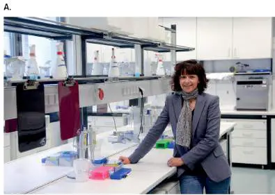
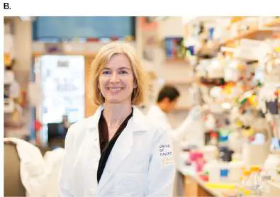
FIGURE 12.16 ■ Emmanuelle Charpentier (A) and Jennifer Doudna (B)
performed some of the seminal work on the CRISPR mechanism.
PETER STEFFEN/PICTURE-ALLIANCE/DPA/AP IMAGES
KEEGAN HOUSER, UC BERKELEY
A CRISPR locus on a bacterial or archaeal chromosome (Fig. 12.17 ) is composed of
short direct-repeat sequences (averaging 32 bp) separated by spacers of uniform length
(20–72 bp, depending on the species). Although the sequences of direct repeats are, by
definition, nearly identical, the sequences of the spacers vary. Note that these repeats and
spacers do not encode proteins. Near these sequence clusters lie CRISPR-associated gene
families (cas) that do encode proteins. A single species can have one or more of these cas
genes, as well as cas subtype genes (which in E. coli are called cse).
FIGURE 12.17 ■ The CRISPR-Cas adaptation and defense pathway. crRNA =
cis -repressed mRNA, or CRISPR RNA.
Source: Modified from Frank Hille et al. 2018. Cell 172 :1239–1259, fig. 1.
CRISPR function. Clues about the function of the variable spacer regions were
uncovered using bioinformatics, which revealed that some spacers bear sequence homology
to bacteriophage or plasmid genes. It turned out that cells harboring these spacers were
immune to the corresponding invaders, but related species lacking these spacers were
susceptible. Thus, CRISPR is perceived as a primitive microbial immune system.
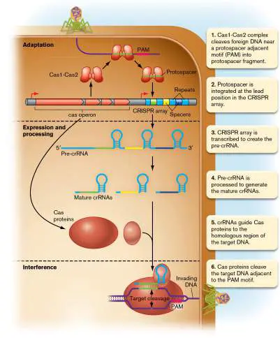
How does a CRISPR locus work? There are three stages of CRISPR-mediated immunity:
adaptation, expression and processing, and interference (Fig. 12.17 ). During adaptation,
bacteria with a CRISPR locus acquire new spacers by incorporating a piece of an invader’s
DNA. First, the Cas1 and Cas2 proteins recognize the foreign DNA and cleave it, generating
a protospacer fragment (Fig. 12.17 , step 1). In most CRISPR-Cas systems, part of the
recognition involves a short stretch of DNA called the protospacer adjacent motif (PAM)
located a few bases away from the cut site. For the Cas9 system of Streptococcus pyogenes
, the PAM sequence is 5′-NGG-3′, where N could be any of the four bases. The PAM motif is
not included in the protospacer, and this absence is important for distinguishing self from
foreign DNA, as we will see. Recombinase proteins of the Cas system integrate the
protospacer into the lead position in the CRISPR region (step 2). Once integrated, this
sequence is called a spacer. The new spacer in the CRISPR array helps the cell “remember”
that this sequence is foreign and, when encountered again, should be targeted for
destruction.
The expression and processing stage begins with the transcription of the CRISPR locus,
starting from the upstream leader sequence (Fig. 12.17 , step 3). The RNA transcript is
then cleaved and trimmed (processed) by some of the Cas products and host RNases into
small RNAs composed of a single spacer sequence (CRISPR RNA or crRNA, also called guide
RNA; step 4). In the final, interference, stage, crRNA associates with Cas proteins to form a
riboprotein complex that binds to a homologous sequence from an infecting phage or
plasmid and directs cleavage of the foreign DNA (steps 5 and 6). As a result, the infected
cell avoids destruction by the infecting phage.
Given that the crRNA directs the Cas system to regions of homology in DNA, what
prevents the disastrous event of Cas proteins cleaving the homologous spacer within the
CRISPR array on the chromosome? Most Cas systems identify the phage target by the PAM
sequence, which is found in the target DNA but is absent in the CRISPR array (Fig. 12.17
). The Cas systems of the interference stage only cleave DNA with homology to the crRNA if
the homologous region is also adjacent to a PAM. The CRISPR array is protected because it
lacks the PAM motif.
Novel CRISPR functions. A number of studies have now shown that CRISPR loci
contribute to cell function beyond providing immunity from phage infections. One novel
function of CRISPR loci may be to banish a lysogenic cell from a biofilm, so that phages
produced by the lysogen cannot kill the rest of the biofilm community. Figure 12.18
demonstrates this phenomenon with Pseudomonas aeruginosa.
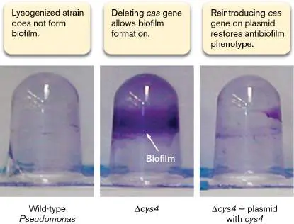
FIGURE 12.18 ■ The effect of CRISPR on Pseudomonas aeruginosa biofilm
formation. Shown are upside-down tubes in which P. aeruginosa lysogenized with
DMS3 phage were grown. Cells that form a biofilm stick to the side of the tube and are
not dislodged by washing. The biofilm is revealed after staining with crystal violet. cys4
is a Cas-encoding gene.
A. T. TUCKER ET AL. 2014. MBIO 5 :E01313–14
Figure 12.18A shows that a lysogenized (infected) cell of P. aeruginosa does not form
a biofilm. If this lysogen were a single cell among many biofilm-producing nonlysogens, it
would not associate with the biofilm. This self-exile saves the biofilm from infection, should
the prophage in the lysogen become active. The CRISPR locus is essential for this loss,
because disruption of the Cas-encoding gene cys4 restores a lysogen’s ability to form
biofilms (Fig. 12.18B ). Introducing a wild-type copy of cys4 (Fig. 12.18C ) into the
mutant cell once again prevents biofilm formation. Thus, a mechanism in which an infected
cell imposed self-exile would save the population.
Gene Editing with CRISPR
The CRISPR system has been modified for in vivo genetic engineering purposes, and one
CRISPR-Cas system has now been repurposed into a powerful molecular tool that can
genetically edit any gene in a eukaryotic cell. The technology has enabled precise gene
editing in species where this has never been possible. A striking example of this
technology’s potential was its use to disrupt latent HIV provirus in infected cells. As
described in Chapter 11, HIV DNA made by reverse transcriptase integrates into the host
genome. Current drug therapies can stop HIV replication and transmission but will not
expunge the provirus from infected cells. Scientists from the Lewis Katz School of Medicine
at Temple University used the CRISPR-Cas9 system to excise HIV provirus from the
genomes of living animals, offering hope that this system may provide a tool for curing HIV
infection in humans.
The system most often used is CRISPR-Cas9 from Streptococcus pyogenes. The key to
this tool is in programming a synthetic crRNA (now called a guide RNA, or gRNA) to direct
Cas9 endonuclease to a specific eukaryotic gene. The programmed gRNA is fused to what is
called the tracer RNA, a molecule that binds to and activates Cas9 (Fig. 12.19 ). How are
these genes introduced into eukaryotic cells? The gene that encodes Cas9 and the gene for
the programmed guide RNA are inserted into plasmids that are introduced into eukaryotic
cells via transformation, and the genes are transiently expressed in the nucleus.
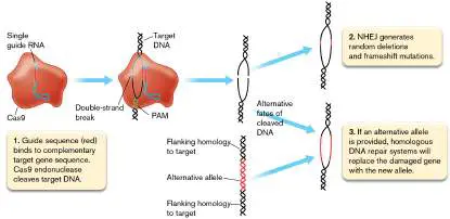
FIGURE 12.19 ■ CRISPR-Cas9 gene editing in eukaryotic cells. Targeted
endonuclease Cas9 from Streptococcus pyogenes can efficiently knock out eukaryotic
genes or replace them with modified forms. The eukaryotic cell’s own nonhomologous
end joining (NHEJ) enzymes achieve knockouts while homologous DNA repair systems
swap alleles.
The guide RNA binds to the Cas9 endonuclease and helps it recognize the target
eukaryotic sequence. The Cas9 endonuclease introduces a double-strand break in the
target DNA—a critical feature of this gene-editing tool. To survive, the eukaryotic cell will
try to re-join the blunt ends using nonhomologous end joining (NHEJ) repair (Fig. 12.19 ;
described in Section 9.2). However, the process often deletes a few bases, which may
cripple the gene. Once mutated, researchers can use the damaged gene to observe its
effect on cell physiology.
In 2021, a team of researchers from the United Kingdom and New Zealand used
CRISPR-Cas9 technology for gene editing in human patients suffering from transthyretin
amyloidosis. This disease, fatal if left untreated, is caused by the accumulation of amyloid
fibrils in the nerves and heart. These fibrils are composed of a misfolded transthyretin, a
protein whose normal function is to transport thyroxine and vitamin A. The patients in this
study were chosen because they all had the hereditary form of transthyretin amyloidosis, in
which a single copy of a dominant allele of the transthyretin gene was responsible for the
disease. The monogenic nature of this disease made it a good candidate for CRISPR gene
therapy, because only a single gene needed to be targeted.
The research team used a lipid nanoparticle to deliver the CRISPR components to the
cells of the liver. Within this nanoparticle were a gRNA complementary to the transthyretin
gene and the mRNA encoding the Cas9 protein, which, once released into the liver cell,
could be translated into the endonuclease. Because safety was a major concern, this
system was first tested in animal models. In the human patients, the approach was highly
successful, as over 93% of the examined liver cells showed the small mutations expected
from CRISPR editing. This success translated in a dramatic reduction of transthyretin protein
in the serum samples of these patients. Importantly, there were no major side effects
reported, and accidental editing of other genes was not observed. The success of this study
suggests that in the near future, editing of human genes could be an important therapy to
treat other diseases, especially those that have identified genetic bases.
CRISPR mutagenesis in bacteria and archaea. The application of CRISPR-Cas9
technology for genetic modification in bacteria and archaea has been more limited than
that in eukaryotes. One major reason is that many bacteria and archaea lack the machinery
for NHEJ repair, and without it the Cas9-mediated double-strand breaks have a high degree
of lethality. However, the NHEJ repair machinery can be transferred in from a related strain
to improve survival and facilitate CRISPR mutagenesis. For example, CRISPR mutagenesis
was improved for the archaeal methanogen Methanosarcina acetivorans when the NHEJ
repair machinery of the related species M. paludicola was introduced.
CRISPR-Cas9 technology can be customized for bacterial gene editing by replacing
double-strand-break formation with base substitution. Akihiko Kondo and colleagues at
Kobe University in Japan developed the Target-AID system, in which a catalytically
inactivated Cas9 is fused to a cytidine deaminase from the sea lamprey Petromyzon
marinus. The defective Cas9 (with the cytidine deaminase in tow) can still be guided by
gRNA to a specific target gene. The cytidine deaminase then converts cytidine residues to
thymidine. In this way, guide RNAs can be engineered such that the Target-AID system can
mutate cytosines at loci near PAM sites anywhere in the genome.
Manipulating microbial gene expression in the intestine. In addition to making
changes to DNA, the CRISPR-Cas9 system has been modified to serve as a regulator of
gene expression. Like Target-AID, CRISPR interference (CRISPRi) uses an inactive version of
Cas9 (dCas9) that no longer works as an endonuclease but can still be guided to target
DNA sequences. Once bound to its target, dCas9 blocks transcription. Timothy Lu’s
laboratory at the Massachusetts Institute of Technology used CRISPRi to control gene
expression in Bacteroides thetaiotaomicron growing in a mouse intestine. B.
thetaiotaomicron is an important bacterial member of mammalian gut microbiomes
(discussed in Sections 13.3 and 23.2).
The scientists designed an sgRNA (single guide RNA) to target a luciferase gene
that was part of the organism’s genome. The sgRNA and the gene for dCas9 were
integrated into the microbe’s genome (Fig. 12.20 ). The gene for dCas9 also included the
lacO operator, which means that dCas9 expression can be induced by the addition of IPTG (
i so p ropyl t hio g alactoside). Without IPTG, the circuit allows luciferase to be made, and
cells glow. Adding IPTG, however, induces dCas9 production. The sgRNA guides dCas9 to
the luciferase gene to turn it off.
FIGURE 12.20 ■ CRISPR interference gene circuit engineered in
Bacteroides thetaiotaomicron. When isopropyl thiogalactoside (IPTG) is added,
dCas9-sgRNA prevents transcription of the luciferase gene Nanoluc.
The investigators in this study demonstrated that the system worked when the
programmed organism resided in the mouse intestine and IPTG was added to drinking
water. The ability to precisely modulate gene expression in commensal organisms should
enable functional studies of the microbiome, noninvasive monitoring of in vivo
environments, and long-term targeted therapeutics.
Additional Mechanisms of Cell Defense from Phages
Besides restriction-modification and CRISPR, prokaryotes can employ an array of defenses
against phage predation. These defenses operate at different stages in the phage life cycle,
and they can even direct the cell to kill itself to prevent spread of the phage to other cells
in the population. We will first describe the mechanisms that protect the individual cell, and
then describe the mechanisms that infected cells use to sacrifice themselves for the
population.
Recall from Chapter 11 that phages target specific receptors on the cell surface,
including outer membrane proteins and the lipopolysaccharide (LPS) layer of the outer
membrane. Prokaryotes use various means to either hide or modify these receptors to
prevent phage adsorption (Fig. 12.21 ). Receptors can be masked through
posttranslational modifications such as glycosylations (see Section 8.4) or can be hidden
from contact by a masking protein, EPS layer, or capsule. Another method prokaryotes use
is to release decoys of themselves in the form of outer membrane vesicles: Phages can be
tricked into adsorbing and injecting their genetic information into vesicles that carry the
phage receptor.
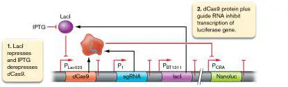
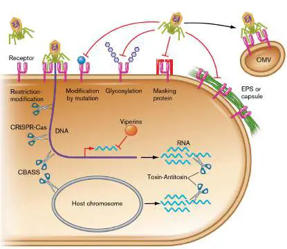
FIGURE 12.21 ■ Arsenal of antiphage defenses. Cells prevent phage adsorption
to the receptor by various means, including the release of outer membrane vesicles
(OMVs) as decoys. Some defenses, such as restriction-modification and CRISPR, cleave
viral DNA. Viperins block phage transcription. Toxin-antitoxin and CBASS destroy all
RNA or DNA in the cell, respectively.
Receptors can also be altered through mutation such that the amino acid motif
recognized by the phage is replaced. As you might imagine, mutations that affect the amino
acid sequence of a protein can often decrease the activity of that protein. This is an
important example of a fitness trade-off that typifies evolution: The bacteria lose fitness
because of the decreased functionality of the protein (for example, slower import through a
mutated transporter), but they benefit when under attack by phages that use that protein
to gain entry into and destroy the cell.
Despite their best efforts, prokaryotes cannot perfectly protect themselves from phage
adsorption, and they must rely on defenses within the cell to prevent the phage from
completing its lytic cycle. Most prokaryotes employ restriction-modification and CRISPR-Cas
systems, and some use additional measures to protect themselves from phages (Fig.
12.21 ). Several of these additional defenses target foreign DNA and share features of
restriction-modification or CRISPR-Cas.
Some prokaryotes survive phage infection by a different mechanism entirely: blocking
transcription of the phage genes. Viperins are enzymes that produce chain-terminating
nucleotides. These nucleotides cause premature transcription termination of phage RNA
polymerases but for reasons not yet understood have no effect on the RNA polymerase of
the host cell. Notably, eukaryote cells also employ viperin defenses to block viral
transcription.
Saving the population by “suicide.” Sometimes, phages bypass all defenses keeping
the cell alive; if left unchecked, they would produce progeny that would go on to infect
other cells in the population. Natural selection favors a host cell that can sacrifice itself if
doing so stops the spread of the phage to other, genetically related cells. One class of
antiphage sacrificial mechanisms, referred to as abortive infection, disrupts phage
replication as a consequence of cell death. As we will see, abortive infection often involves
indiscriminate destruction of RNA or DNA within the cell, a drastic measure in sharp contrast
to the sequence-specific forms of cleavage mediated by restriction enzymes or CRISPR-Cas.
One form of abortive infection involves toxin-antitoxin pairs, such as the MazE-MazF
system in E. coli described in Section 5.5. Activation of the MazF ribonuclease (toxin) leads
to the destruction of RNA, both host and viral (Fig. 12.21 ). This activity blocks the
translation essential for capsid protein synthesis but kills the host in the process.
A similar pathway kills the cell and phage by cleaving DNA rather than RNA (Fig. 12.21
). C yclic oligonucleotide- b ased a ntiphage s ignaling s ystem (CBASS) involves a
nonspecific DNA-cleaving nuclease that becomes activated when it binds a second
messenger called cyclic triadenylate (cA 3). cA 3 is synthesized when CBASS detects phage.
Notably, CBASS, as opposed to CRISPR-Cas and restriction-modification systems, recognizes
phage proteins rather than DNA.
Rather than destroy nucleic acids, some forms of population-level protection work by
preventing phage DNA from entering the capsid structures. Phage-inducible chromosomal
islands (PICIs) are mobile genetic elements that integrate into the host chromosome (Fig.
12.22 ). The PICI genes are maintained in an inactive state by a repressor protein until a
phage infects the cell (Fig. 12.22A ). One of the proteins made by the phage functions as
an antirepressor of the PICI repressor, which removes it from the PICI promoters and allows
expression of this gene cluster (Fig. 12.22B ).
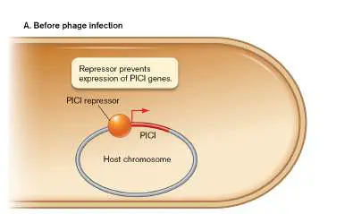
FIGURE 12.22 ■ PICI elements promote their own release by hijacking the
capsids of incoming phages.
Products of this PICI element function to hijack the infecting phage’s capsid for its own
transduction (see Section 9.3). The PICI element excises, replicates, and is packaged into
the phage’s capsid that has been specially modified by another PICI protein to have a
smaller volume, one that can no longer accommodate the larger phage DNA (Fig. 12.22B
). Host cells ultimately lyse when enough capsids are produced, but a large fraction of the
released capsids contain PICI DNA, which, when injected into a new host cell, will integrate
into the chromosome and remain dormant until the next phage infection. While acting
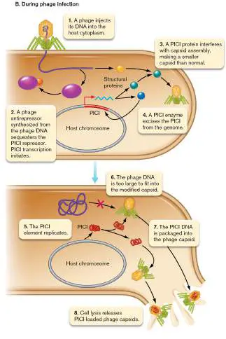
rather indirectly, the overall reduction in the burst size of infectious phage progeny by PICI
action is thus another means by which the population can benefit from defenses that
require death of the infected cell.
Phages Employ Counterdefenses
Prokaryotes and their phages are in an evolutionary arms race: As the outcome determines
the survival and reproduction of both host and phage, the stakes cannot be higher. The
innovation of a new host defense selects for phage that can overcome that defense, which
in turn promotes the innovation of yet another defense mechanism, and so forth. This
section closes with an in-depth look at an example series of host-phage innovations and
responses during evolution. Before that discussion, we will look at the broad suite of
options that phage may use to counteract host defense.
Defenses that prevent adsorption to the host can be overcome by spontaneous mutation
in the phage. Debbie Lindell (Fig. 12.23 ) and colleagues at the Technion–Israel Institute
of Technology have shown that in the presence of lytic phage, populations of the marine
cyanobacterium Prochlorococcus are rapidly taken over by mutants resistant to that phage.
These mutants have modified cell envelopes to which the phage can no longer bind.
However, in extended cultivation, phage mutants with modified tail proteins arose that
were able to overcome this means of resistance and resume infection of their host.
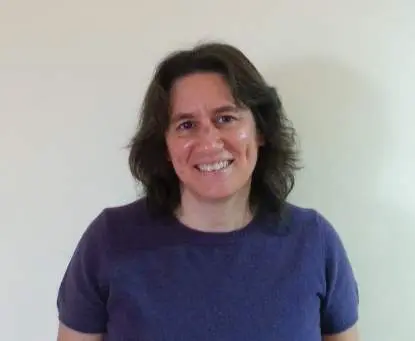
FIGURE 12.23 ■ Debbie Lindell and her research team have discovered
phage-resistant mutants of Prochlorococcus.
DEBBIE LINDELL
What is the nature of these mutations that restore adsorption to the host? Phage
lambda provides some examples. E. coli cells with mutations in the gene encoding the
outer membrane protein LamB develop immunity to phage lambda. However, mutations
affecting the tail fiber protein of lambda can restore lambda’s ability to adsorb and infect E.
coli by either now recognizing the mutant LamB receptor or bypassing LamB entirely and
redirecting binding to another outer membrane protein, OmpF.
Masked receptors can be unmasked by phage. For instance, cells protected by an EPS or
capsule layer can be infected by phages that deploy EPS-degrading enzymes on their tails.
Phages can avoid destruction from intracellular host defenses by several means. One
common method is to inactivate the key defense enzyme involved. Phage T7 produces a
protein Ocr, which binds the active site and inactivates type I restriction-modification
systems of its E. coli host. Likewise, there are many phages that produce anti-CRISPR
proteins that block the activity of the host Cas nucleases (see Fig. 6.24 ). Phage T4
expresses a protein that binds and inactivates the nuclease MazF involved in abortive
infection in E. coli.
Besides blocking the host nuclease, phages can utilize additional measures to protect
their DNA during infection. Because restriction-modification systems and CRISPR-Cas
systems target specific sequences, phage mutants with altered DNA sequences in these
recognition motifs can avoid detection and destruction by their hosts. Recently, several
phages were discovered with a novel mechanism of defense from CRISPR-Cas: enclosure in
a protective nucleus-like membrane structure. Another recently discovered phage
counterdefense repairs what the host defenses destroyed. Phage T4 overcomes the action
of the host Cas enzymes with enzymes of its own that recombine and repair its fragmented
genome. Finally, as discussed in eResearch Activity 12, phage DMS3 disarms its P.
aeruginosa host by disrupting the quorum-sensing system that regulates the synthesis of
the CRISPR-Cas defenses.
The ICP1 phage that infects Vibrio cholerae has acquired a remarkable way of
counteracting the PICI element in the host chromosome: destroy the PICI, and use its own
CRISPR-Cas to do so. ICP1 encodes a functional CRISPR-Cas system in its genome, with
spacers that are identical to a sequence within the PICI. Infection by ICP1 triggers the
activation of PICI, but the Cas system of ICP1 destroys the replicating copies of the PICI
before they interfere with phage encapsidation.
The phage-host evolutionary arms race carries on. The continual back-and-forth
nature of the evolutionary arms race between phages and their hosts is clearly illustrated
by the existence of a protein called IPI* in phage T4. IPI* is a small (76 amino acid) protein
that gets packaged into the capsid along with the genomic DNA. During infection it is small
enough to be coinjected with DNA through the tail sheath channel into the host cell. IPI*
functions to bind and inactivate a specific restriction endonuclease of T4’s host, E. coli.
Remarkably, this enzyme recognizes cytosines only if they have been modified into
glucosylated hydroxymethylcytosines. T4 phage lacking IPI* cannot infect E. coli containing
this enzyme, and a sustained arms race between predatory phage and prey bacterium
explains the presence of these unusual proteins. Below is one likely scenario for how this
arms race proceeded through the course of natural selection.
The ancestral form of T4 was susceptible to the standard restriction-modification
systems of its host that recognize cytosines (Fig. 12.24A ). Within the population of T4,
genetic variants were present with the ability to synthesize during infection the enzyme
dCMP hydroxymethyltransferase. This enzyme converts cytosines to
hydroxymethylcytosines (hm Cs) within the replicated DNA of the phage. We don’t know
exactly how the gp42 gene encoding this enzyme came to arrive within the T4 genome in
these variants, but for the sake of argument we’ll assume it was acquired via horizontal
gene transfer from another microbe’s genome (see Section 9.5).
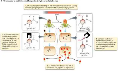
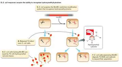
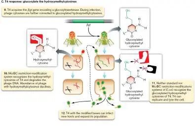
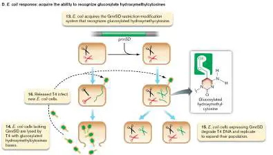
FIGURE 12.24 ■ Evolutionary arms race between phage T4 and its
Escherichia coli host. Panels A–E show the series of evolutionary responses of the
phage and host to the selective pressures of this predator-prey relationship. The color
of the phage DNA indicates the form of cytosine, and the color of the scissors indicates
the form of cytosine recognized by the endonuclease. RM = restriction-modification.
Importantly, hm Cs are not recognized by the standard host restriction endonucleases,
including the type IV restriction systems that recognize methylated cytosines. With the
selective pressure of host restriction endonucleases exerted on the T4 population, the
ancestral-type T4 phages were depleted while the T4 phages expressing gp42 expanded
their population through successive rounds of infection and lysis of the unprotected host (
Fig. 12.25 ).
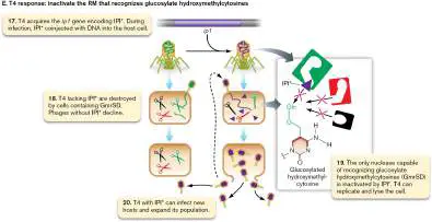
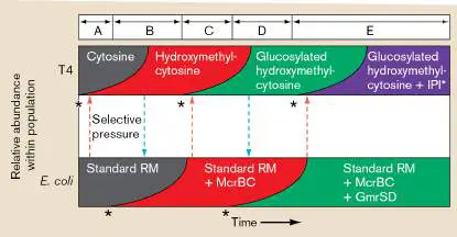
FIGURE 12.25 ■ Changes in cytosine structure or restriction-modification
defenses over time during the T4– Escherichia coli evolutionary arms race.
Note that selective pressure (dashed arrows) affects the fitness of two genotypes
already present in the population; under this pressure, the rare genotype expands
relative to the common one. Letters A–E refer to the panels of Figure 12.24: Each time
frame begins with the acquisition of the new gene that establishes the rare genotype
in either the phage or the host (asterisks).
Hydroxymethylcytosine bases are not recognized by normal systems, but rare variants
of E. coli acquired a novel restriction system, McrBC, that can recognize hm Cs and cleave
phage DNA containing those bases (Fig. 12.24B ). While cells lacking this system fell prey
to T4 phages with hm C bases, cells with McrBC could destroy the T4 DNA and continue
replication, expanding their population (Fig. 12.25 ).
As these McrBC cells grew to prominence, selective pressure was exerted on T4 to
further modify its cytosines. Rare variants of phage T4 were able to take base modification
one step further with the addition of sugar residues, forming glucosylated hm Cs (ghm Cs)
through the activity encoded by the gene βgt (Fig. 12.24C ). ghm C bases are not
recognized by McrBC, and thus phages with βgt could expand their population while phages
with only the hm C bases were destroyed (Fig. 12.25 ).
As the βgt phage grew to prominence, selective pressure was exerted on E. coli to
acquire restriction defenses that could recognize ghm C bases (Fig. 12.25 ). Cells within
the population that acquired a ghm C-targeting restriction enzyme, GmrSD, could eliminate
the phage and expand their population relative to the cells only able to target cytosines
and hm Cs (Figs. 12.24D and 12.25 ).
With selective pressure now acting on T4 to resist the new enzyme of the host, variants
expressing IPI* rose to prominence (Figs. 12.24E and 12.25 ). As mentioned earlier, IPI*
specifically blocks the activity of endonucleases that recognize ghm C bases.
The arms race between T4 and E. coli is far from over: Variants of GmrSD can avoid
blockage by IPI*, while new variants of IPI* can reinstate blockage of those GmrSD
variants. In addition to this interplay centered on restriction-modification, E. coli has
acquired different types of antiphage defense, such as the CRISPR-Cas system and the
MazE-MazF toxin-antitoxin system, and T4 has acquired counterdefenses to these
mechanisms, as described previously.
To Summarize
Restriction endonucleases protect prokaryotes from invasion by foreign DNA.
Restriction-modification enzymes methylate restriction target sites in the host DNA
to prevent self-digestion.
The CRISPR-Cas system is a small, interfering RNA system in many bacteria and
archaea that captures a piece of an invader’s DNA and uses it to fend off future
attacks, thus serving as a primitive form of adaptive immunity.
CRISPR can be applied to eukaryotes, bacteria, and archaea to change DNA
sequences or interfere with gene expression.
Prokaryote defenses can prevent phage adsorption, can complement restriction
endonucleases and CRISPR to further block phage replication, and can also sacrifice
the infected cell to save the population.
Phage utilize a broad suite of counterdefenses to overcome the defenses of
the host.
The evolutionary arms race between host cell and phage typifies the ongoing
series of innovations that prey and predator accumulate over evolutionary
timescales.
Glossary
restriction-modification (RM)
An antiphage defense system usually composed of a methyltransferase enzyme that
methylates DNA and a restriction endonuclease that cleaves unmethylated DNA.
restriction endonuclease or restriction enzyme
A bacterial enzyme that cleaves double-stranded DNA within a specific short sequence,
usually a palindrome.
methyltransferase
An enzyme that adds a methyl group to a base in DNA.
CRISPR
Clustered regularly interspaced short palindromic repeats. CRISPR consists of short
repeated DNA sequences in a bacterial or archaeal genome, derived from previous
bacteriophage or viral infection and conferring protection from future infection;
considered a prokaryotic “immune system.” In biotechnology, provides a means of
editing the human genome.
abortive infection
A class of antiphage defenses that kill the host cell before the phage can complete
replication.
phage-inducible chromosomal islands (PICIs)
Mobile genetic elements integrated within the host chromosome that hijack the capsids
produced by incoming phage for transfer to new host cells.
evolutionary arms race
The escalation of adaptations and counteradaptations that develop between predator
and prey or between competing species.
Fig. 6.24
FIGURE 6.24 ■ CRISPR defense of a bacterial cell and anti-CRISPR
counterdefense. A. A piece of phage DNA gets copied as a “spacer” into the host
genome. If the bacterium survives infection, later reinfection by the same kind of
phage causes transcription of the spacers into CRISPR RNA. A processed spacer
(crRNA) joins the Cas complex to recognize and cleave the phage DNA. B. Phage
may carry an anti-CRISPR gene (acr) encoding a protein Acr that blocks the host
bacterial CRISPR-Cas from binding phage DNA.
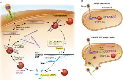
12.4 Synthetic Biology: Biology by Designnot assigned
What if we could, with all our knowledge of cells, genes, and gene circuits, make bacteria do things they wouldn’t ordinarily do, such as sensing explosives in mines and then telling us about it? Or detecting pathogens? Or keeping time? Or even, as Special Topic 7 highlights, storing information—much like a computer? These are all goals of synthetic biology. Synthetic biology is the design and construction of new biological parts, devices, and systems for a desired purpose by use of principles from electrical engineering.
How do we make these new parts or components? All living organisms already contain an instruction set encoded in DNA that determines what the creature looks like and what it does. Humans have been altering the genetic codes of plants and animals for millennia by selectively breeding individual plants and animals with desirable features. Today, scientists easily take pieces of genetic information from one organism and insert them directly into another, bypassing the need to breed. This is the basis of genetic engineering. Recent technological advances now enable scientists to synthesize and manipulate DNA in ways never before possible. By applying engineering principles to these genetic manipulations, researchers can take components of genes from several different organisms, link them together, and design customized organisms that do new things. For instance, Chang Li and colleagues (Harvard Medical School) engineered an Escherichia coli strain that prevented cancer in mice. The new strain contained the invasin gene from Yersinia pseudotuberculosis and the hemolysin gene from Listeria monocytogenes. The invasin gene enabled the modified E. coli to invade mouse cells. The hemolysin enabled delivery of an inhibitory small RNA molecule that decreased expression of a tumor-initiating gene. Amazingly, this novel E. coli prevented cancer in this mouse model.
Principles of Synthetic Biology
The key to synthetic biology is engineering. Engineering a life form from scratch is one of the most impressive feats that synthetic biology has accomplished to date (Special Topic 12). Which engineering principles are employed by synthetic-biology scientists? Many of the principles are borrowed from the field of electrical engineering and involve “logic gates.” An electronic logic gate (as in semiconductors) receives a tiny current as an input and produces voltage as an output. A genetic logic gate is a promoter and a gene. An input signal such as a regulatory protein affects the promoter, which drives the output signal (mRNA and protein). The output signal for one gate can be an input signal for another part of the logic circuit. These logic gates can be combined to produce biologically useful switches that control gene expression and cellular activity. Toggle, oscillator, and kill switches are examples that we will describe after introducing the types of genetic logic gates that compose them. As we will show, these switches can be used to make cells glow, keep time, or die if they are released accidentally into the environment. Many of the components used in building an electronic circuit have biological counterparts that are used for building complex genetic circuits. Figure 12.26shows three common logic gates used in synthetic biology. The first is a “buffer gate” that amplifies signals ( Fig. 12.26A). For a simple gene, the protein input activates a promoter; a message is then transcribed and a protein is made. With a “NOT gate,” a protein input represses a promoter, and the output protein is not made (Fig. 12.26B ). Finally, an “OR gate” involves several genes. For instance, two alternative gene output proteins can activate a third gene. So, in Figure 12.26C , the alternative input proteins I 1 and I 2 activate gene 1 or gene 2, respectively, to make product 1 or product 2 (Pr 1 or Pr 2). Either of those output signals can activate gene 3 to make product 3, Pr 3. Once you understand how these gates work, you can make any kind of circuit.
FIGURE 12.26 ■ Examples of logic gates used in electronics, and their synthetic-biology equivalents. A.
When inducer is added, the transcript for Pr 1 is made. Without inducer, Pr 1 is not made. B. When repressor is added, the transcript for Pr 1 is no longer made. C. When either I 1 or I 2 is added, the transcript for Pr 3 is made.
Toggle Switches
Electrical systems rely heavily on toggle switches to control whether a system is turned on or off. Similarly, synthetic-biology circuits depend on biological toggle switches. Figure 12.27illustrates a basic, genetically engineered toggle switch designed to control whether a gfp gene is turned on or off. The circuit involves two repressor genes (called NOT gates) whose products can repress each
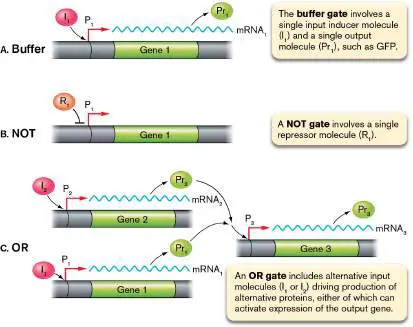
other’s transcription. The switch depends on whether one of two different inducer signals is present. Inducer 1 inactivates repressor 1, which means repressor 2 is produced. Repressor 2, in turn, stops transcription of the repressor 1 gene and the reporter gene. So, when inducer 1 is added, GFP is not made and the system is stably toggled off.
FIGURE 12.27 ■ Genetic toggle switch. Notice that repressor 1 and the reporter gfp are transcribed colinearly from promoter 2. Two outputs are possible, depending on which inducer is added. Adding inducer 1 stops fluorescence; adding inducer 2 triggers fluorescence. What happens if you add them both?
Alternatively, inducer 2 inactivates repressor 2, which means that the genes for repressor 1 and GFP are transcribed and the cell lights up. The system is stably toggled on because repressor 1 halts transcription of repressor 2. So, a genetic engineer can control the on/off switch of these cells by adding one or the other inducer. Of course, for this system to toggle, at least a small amount of both repressor proteins must be made at all times—just enough to bind to inducer molecules and have an effect on gene expression. Switches can also be engineered in bioreporter bacteria to record exposure to molecules such as antibiotics, as was shown in Figure 11.11.
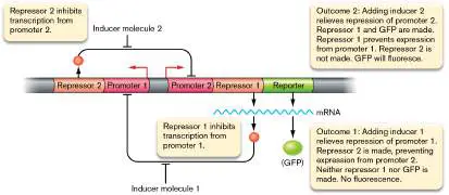
Thought Question
12.6 What would happen with the toggle switch shown in Figure 12.27if the genetic engineer could set repressor 1 and repressor 2 protein levels to be exactly equal? Imagine that this is done without either inducer present.
An Oscillator Switch
An oscillator is another important component of many electronic systems. In an electronic circuit, the oscillator produces a repetitive electronic signal viewed as a wave. In synthetic biology, scientists can also make an oscillating genetic circuit. Figure 12.28Ashows a basic genetic oscillator switch designed by Jeff Hasty’s laboratory at UC San Diego. The components come from some of the systems discussed in Chapter 10. One component is the gene for the AraC activator protein, which was linked by Hasty’s group to a hybrid operator region that included an ara activator sequence and a LacI repressor control sequence. The next component is the gene encoding the repressor LacI, which was also spliced to the hybrid ara activator–LacI repressor control sequences. The resulting circuit contains negative and positive feedback loops. The lac and ara operons are discussed in Section 10.1.
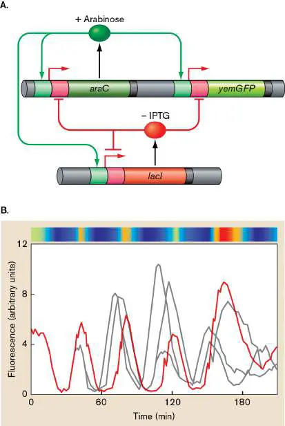
FIGURE 12.28 ■ A dual-feedback oscillator constructed in Escherichia coli. A. Network diagram. A hybrid promoter, P lac/ara (small pink and green boxes), drives the transcription of araC and lacI, forming positive and negative feedback loops. B. Single-cell fluorescence oscillations induced with 0.7% arabinose plus 2 mM IPTG (red line in the graph) or 1 mM IPTG (gray lines). The points represent experimental fluorescence values. The colored bar across the top represents the intensity of the fluorescence, from high (red) to low (blue).
Source: Modified from J. Stricker et al. 2008. Nature 456 :516–519.
SPECIAL TOPIC 12 Constructing the Smallest Genome for Cellular Life
What is the minimum number of genes needed to make a self-replicating life form, and what are the essential functions that these genes encode? One way to address this question is to find all of the nonessential genes of an existing organism— particularly an organism with a small genome—then remove them all and see whether the organism can still replicate. Because there may be hundreds of genes to delete, deleting them one at a time would be slow and laborious. A better way to perform this large-scale genome reduction is to build the chromosome from scratch, adding only the genes thought to be essential. This strategy was employed by Clyde Hutchison III, Hamilton Smith (Fig. ST 12.1 ), and colleagues at the J. Craig Venter Institute to build a viable, replicating microbe named JCVI-syn3.0, whose genome, built entirely from chemically synthesized DNA, encodes a mere 473 genes.
FIGURE ST 12.1 ■ Clyde Hutchison III (left) and Hamilton Smith at the J. Craig Venter Institute.
J. CRAIG VENTER INSTITUTE
Their approach began with strain JCVI-syn1.0, a synthetic reconstruction of the 1.08-Mb genome of Mycoplasma mycoides consisting of 901 genes (Fig. ST 12.2 ). Hypothesizing that not all genes in JCVI-syn1.0 were essential for growth, the scientists mutagenized the strain with a transposon and selected for viable transposon insertion mutants (see Section 9.4). The genes inactivated by the transposon insertions in these viable mutants were then identified and classified as “nonessential” for growth. Essential genes should not tolerate a transposon insertion, so they would be identified by their absence from the collection of viable mutants.
FIGURE ST 12.2 ■ Derivation of JCVI-syn3.0 from JCVI-syn1.0. Comparison of the syn1.0 (purple) and syn3.0 (green) genomes. Green fragments adjacent to the purple circle show the sections of the syn1.0 genome that were retained in the smaller syn3.0 genome.
Source: Modified from C. A. Hutchison III et al. 2016. Science 351
:aad6253, unnum. fig., panel B.
An initial genome built exclusively of the 240 identified essential genes did not grow, and thus some genes identified as nonessential were actually needed to establish a viable, replicating organism. What accounted for this need for “nonessential” genes? In some cases, the cell had two genes that could perform the same essential function; in other words, these genes had functional redundancy. Losing one gene through transposon insertion was nonlethal because the other
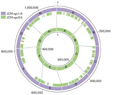
gene was not disrupted. However, attempts to build a genome that lacked both genes were not successful; some genes had to be added back.
Through trial and error, they arrived at version 3.0 of their synthetic genome: a viable, replicating life form with a genome of 531,490 bp and 473 genes (Fig. ST 12.2 ). Just how did they make their version 3.0 artificial chromosome and get it to start the essential processes of life, such as gene expression and chromosome replication? Short oligonucleotides (less than 100 bases) were synthesized chemically, and then subsequent steps were performed to stitch all of these together to make the 531,490-bp whole genome (Fig. ST 12.3 ).
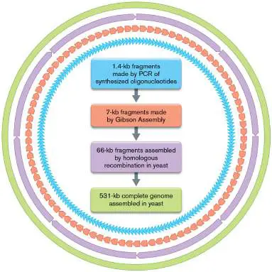
FIGURE ST 12.3 ■ Strategy for the synthesis of JCVI-syn3.0 from oligonucleotides.
Source: Modified from C. A. Hutchison III et al. 2016. Science 351
:aad6253, fig. 2.
The first stitching step used polymerase chain reaction (PCR) to assemble the oligonucleotides into 1.4-kb fragments. Five of these fragments were then stitched together using Gibson Assembly (described in eAppendix 3) to make 7-kb fragments. The final assemblies into eight 66-kb fragments and subsequently to one 531-kb circular chromosome were performed inside yeast cells. When transformed by these fragments, the yeast could assemble the fragments into large replicating clones via their homologous recombination machinery. Once the whole genome was assembled, it was transformed into a related Mycoplasma species, M. mycoides. The transplanted syn3.0 chromosome used the existing machinery of the host cell to replicate and express its genes, eventually displacing the host M. mycoides chromosome and gene products for its own.
The JCVI-syn3.0 synthetic organism forms clusters of spherical cells of varying diameter (Fig. ST 12.4A ). It is a heterotroph that requires extensive nutrient supplementation and grows about three times slower than the JCVI-syn1.0 strain. Its genome contains 438 protein-encoding genes and 35 untranslated RNA genes (rRNA, tRNA, and small RNAs), making it the smallest genome for an organism capable of growth in pure culture. The largest category of genes is for the expression of genomic information (Fig. ST 12.4B ). Surprisingly, 79 genes (17%) included in the minimum genome have no known function. It thus remains unclear which cell functions are absolutely essential for survival and replication. But at least now we have a clearer picture of what we don’t know.
FIGURE ST 12.4 ■ Characterization of JCVI-syn3.0. A. A cell cluster (SEM). B. Functional group assignment of the genome.
Source: Part B modified from C. A. Hutchison III et al. 2016. Science 351
:aad6253, fig. 6.
TOM DEERINCK AND MARK ELLISMAN OF THE NATIONAL CENTER FOR
IMAGING AND MICROSCOPY RESEARCH AT THE UNIVERSITY OF CALIFORNIA
AT SAN DIEGO
RESEARCH QUESTION
What types of techniques found in eAppendix 3 might be applied to test the function of the unknown genes?
Hutchison, C. A., III, R.-Y. Chuan, V. N. Noskov, N. Assad-Garcia, T.
J. Deerinck, et al. 2016. Design and synthesis of a minimal bacterial
genome. Science 351 :aad6253.
To see how this oscillator works, IPTG (the chemical that inactivates LacI) and L -arabinose (the sugar that activates AraC) are added at the same time. The increase in AraC production drives
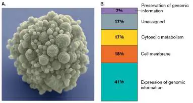
expression of GFP but also increases LacI repressor, which eventually represses araC. However, IPTG inactivates LacI protein, which enables renewed araC and lacI expression. As shown in Figure 12.28B , the differential activity of the two feedback loops drives oscillation back and forth between fluorescence (on) and no fluorescence (off). Changing the concentrations of arabinose and IPTG will modulate the oscillation frequency, making the circuit tunable.
System Noise
Before we discuss more complex circuits, we should talk about “noise.” Noise (variance from a mean) is a problem in any electrical circuit but also arises in biological circuits, because of fluctuations in gene expression among single cells in a population. The concept of noise in a biological or electrical system might be best explained by the following analogy: Imagine you are at a concert and your friend is trying to talk to you. You can’t understand what your friend is saying, because the sound of your friend’s voice is not rising above the music and all of the background noise. The band would have to go quiet in order for you to hear your friend.
The same is true for anything we want to measure. The less noise there is, the easier it is to measure what rises above it. In bacteria, translation inefficiency is a major contributor to noise, although many factors can influence the expression of a gene (Fig. 12.29). To be useful, synthetic biological circuits must operate with transcriptional and translational controls balanced in a way that minimizes noise. Choosing the right ribosome-binding sequence and promoter, as well as using a small RNA to control message stability or translation, can help to decrease noise.
FIGURE 12.29 ■ Some factors affecting “noise” in a biological circuit. RNAPs = RNA polymerases.
Engineered Riboswitches and Switchboards
In Chapter 10 we described how the cell uses riboswitches to sense cell metabolites and control the translation of mRNA molecules. Synthetic-biology scientists seized upon this concept and have learned to tailor small RNAs, called synthetic riboswitches, to control the translation of nearly any gene they want. Figure 12.30reveals that a basic riboswitch consists of two parts: a cis -repressed mRNA (crRNA; Fig. 12.30A) and a trans -activating RNA (taRNA; Fig. 12.30B ). (Note that, although they share the same abbreviation, these crRNAs are distinct from the crRNAs of CRISPRs from Section 12.3.) The crRNA sequence is linked to an output gene (hence “ cis ”) and, by forming a hairpin, hides a ribosome-binding site to prevent translation. The taRNA, once it is made, will promote translation of the output gene by base-pairing with the crRNA (Fig. 12.30C ). The base pairing releases the ribosome-binding site (RBS) for translation. Transcription and translation of the output gene can now be manipulated by whatever activates the promoters used to express the crRNA and taRNA genes. This process can decrease noise in a system.
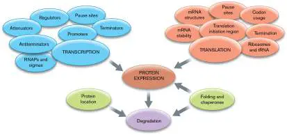
FIGURE 12.30 ■ The basic synthetic riboswitch. A. Cis - repressed mRNA (crRNA) folds to obscure a ribosome-binding site (RBS) needed to translate a downstream output gene. B, C. Trans -activating RNA (taRNA), driven by a different promoter (B) , can base-pair with crRNA (C) , thus opening up the hairpin to expose the RBS. Whatever activates the taRNA will activate translation of the output gene’s mRNA.
Members of James Collins’s laboratory (Harvard University)
designed a series of matched crRNA-taRNA riboswitches and linked them to various promoters and output genes. The design of these circuits, and their responses to various environmental signals, are shown in Figure 12.31. Notice that each riboswitch responded to only one signal.
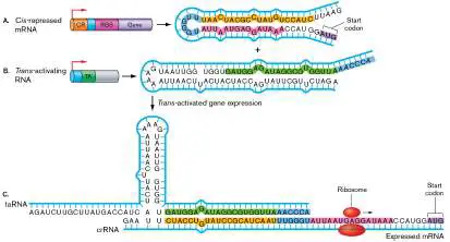
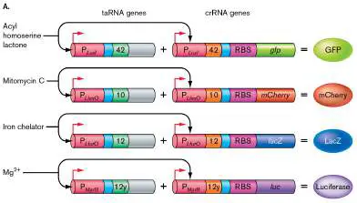
FIGURE 12.31 ■ Riboswitchboard. A. Four separate circuits were designed. Each circuit is controlled by a different promoter that senses a different environmental parameter: P LuxI (acyl homoserine lactone), P LlexO (mitomycin C), P LfurO (iron), or P MgrB (magnesium). Different taRNA-crRNA riboswitch pairs (labeled 42, 10, 12, and 12y) were used to control the translation of the output reporters for each circuit. B–E. These graphs show that each circuit responded to only a single environmental input signal. Inducers for GFP, mCherry, and LacZ (B, C, D) were added at time
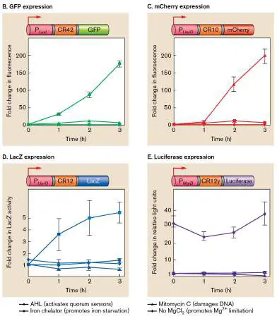
0. The inoculum for the luciferase circuit (E) was grown in low Mg 2+ and was active even at time 0.
The Collins group then linked these riboswitches to genes encoding carbon metabolism enzymes and placed them all in the same cell, essentially making a metabolic switchboard that could channel carbon flow through alternative metabolic pathways depending on which riboswitch was activated. Metabolic switchboards would have important industrial applications. For instance, the device could simultaneously sense a variety of metabolic states in a large-batch fermentation system and maximize the efficiency of an industrially beneficial pathway.
Kill Switches
Most of what we have described about synthetic biology has little chance of dangerous unintended consequences, but what about the long term? Biologists would like to engineer new strains that do new tasks, such as gobble up toxins in the environment. Remediating toxin contamination in nature, however, would require the release of genetically modified organisms outside the laboratory. This raises concerns about potential, unknown havoc that these organisms might cause.
To allay these concerns, scientists must engineer fail-safe mechanisms that kill the organism at a predetermined point. Hence the quest for effective “kill switches.” A genetically modified organism equipped with a kill switch can be made to commit “suicide” once the bacterium’s job is done. One proof-of-principle kill switch engineered by synthetic-biology techniques is shown in Figure 12.32.
FIGURE 12.32 ■ A synthetic-biology “kill switch,” or “suicide module.” A. Without tetracycline or arabinose, the TetR repressor protein binds the P LtetO operator and represses the ccdB gene. The gene is also equipped with a cis -repressed RNA (CR) that prevents translation. B. The addition of tetracycline removes TetR from the P LtetO operator, and L -arabinose enables AraC to activate the complementary trans -activating RNA gene (TA). The ccdB mRNA is translated by the ribosome, and CcdB causes DNA damage, which kills the cell.
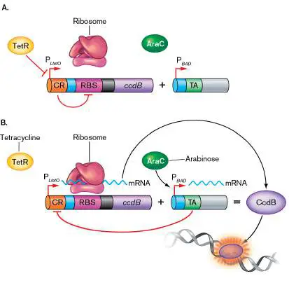
The gene for CcdB, a potent DNA-damaging toxin, was linked to the promoter P LtetO. This promoter is regulated by the TetR repressor protein. In the absence of tetracycline, TetR binds the promoter and prevents the transcription of ccdB. Cells with this switch in this off state are alive. Tetracycline controls the switch: when added, it binds and inactivates TetR, which then releases from the ccd promoter. As a result, the kill toxin gene ccdB is expressed and kills the cell.
Because transcription can be leaky, the engineers added a riboswitch mechanism (see Fig. 12.30) to prevent the organism from killing itself too early in this proof-of-concept experiment. They made sure that the translation of CcdB mRNA is blocked by a crRNA. A second component of the system, activated by AraC upon addition of arabinose, encodes the compensatory taRNA that can expose the RBS buried within the crRNA-CcdB message. So, both tetracycline and arabinose must be added for the cell to effectively produce CcdB and kill itself.
Although this system, as designed, is impractical for real-world use, its success shows that kill switches can be made. How could this kill switch be modified so that it could be used in the real world? What if repression of CcdB were tied to the presence of a toxic product found in the environment? Once the organism destroyed the toxin, the kill switch would be “thrown,” and the bacterium would kill itself.
BioBricks and Do-It-Yourself Synthetic Biology
The science of synthetic biology makes new logic circuits by linking promoters from one system to genes from another system. The art of synthetic biology is to combine these new genetic logic circuits in ways that produce new functional systems. But where do all the genetic “Lego-like” blocks come from? Laboratories around the world construct and deposit their “building materials” into a central registry at the Massachusetts Institute of Technology (MIT). Over the years, scientists from universities have assembled thousands of connectable pieces of DNA that they call BioBricks and have deposited them in the BioBricks Foundation registry at MIT. BioBricks range from those that kill cells to one that makes cells smell like bananas. MIT also hosts a weekend-long synthetic-biology showdown called the International Genetically Engineered Machine (iGEM) competition. The winner’s trophy of the iGEM competition is a large, aluminum Lego (Fig. 12.33).

FIGURE 12.33 ■ Winners of the 2021 iGEM competition Grand Prize trophy. Left: The team from the Polytechnic University of Valencia won Grand Prize at the 2018 competition for their design of Printeria, a combination of software and hardware that genetically engineers bacteria. Right: The BioBricks trophy.
THE IGEM FOUNDATION AND JUSTIN KNIGHT
THE IGEM FOUNDATION AND JUSTIN KNIGHT
Despite its potential for good, synthetic biology raises the concern that anyone, theoretically, can do it. In fact, the lure of constructing a new organism, combined with the ever-lowering cost of equipment needed to carry out these experiments, has spawned a community of do-it-yourself (DIY) genetic engineers, including high school students and so-called garage scientists. Most of these DIY efforts are well intentioned and could yield useful products as a result of outside-the-box thinking, but we should be mindful, again, of unintended consequences.

To Summarize
Synthetic biology applies engineering principles to design and construct new biological parts for a desired purpose. Synthetic-biology circuits use genetic logic gates , such as buffer gates, NOT gates, and OR gates.
A toggle switch , built from two NOT gates, can turn a gene on or off by sensing two different chemical signals. An oscillator switch will repeatedly turn on and off in response to a signal.
System noise is the fluctuation of gene expression among single cells of a population. System noise can dull the clarity of a response circuit. Riboswitches engineered into a genetic circuit can minimize noise.
Genetic kill switches can eliminate a genetically modified organism when its job is done.
BioBricks are connectable pieces of DNA that can be used in various combinations for synthetic biology.
Glossary
synthetic biology The genetic construction of novel organisms with useful functions.
noise Variance from a mean in an assay. A system in which individual results vary greatly from a mean that was derived from many results is a noisy system.
Figure 11.11
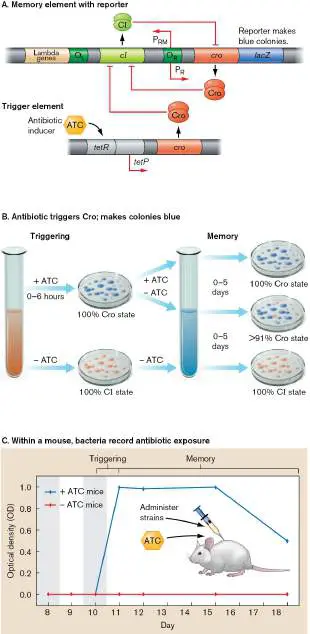
FIGURE 11.11 ■ Antibiotic reporter bacterium uses a CI /Cro switch. A. The CI /Cro switch was engineered to record exposure to an antibiotic (ATC). The Cro “memory” is triggered by an inducer that turns off CI expression over many cell generations. B. The ATC signal turns colonies blue. Blue colonies persist for 5 days. C. The bacterial detector works when administered to mice. Seven days after addition and removal of ATC, the engineered bacteria still make blue colonies (Cro state).
Source: Jonathan Kotula et al. 2014. PNAS 111 :4838.
eResearch Activity 12
Can Phage Jam a Host’s Communications to Block Antiphage Defense?
Phages employ a vast array of counterdefenses in order to protect themselves from host antiphage systems such as CRISPR-Cas and restriction-modification (see Section 12.3 in the printed book). What if phages were able to prevent such defenses from even being made in the first place by their hosts? The Pseudomonas aeruginosa – infecting phage DMS3 appears to do just that by producing a protein capable of interfering with the quorum-sensing system that activates expression of CRISPR-Cas. What’s more, the same phage protein blocks extension of the type IV pilus, which is the receptor for this phage. With the receptor missing, the injected DMS3 phage prevents superinfection from other phages and keeps the host cytoplasm to itself.
Megha Shah (Fig. ERA 12.1 ) and colleagues at the University of Toronto were interested in the unknown functions of genes in the DMS3 genome. They began their study by cloning those genes individually on plasmids and screening for phenotypes when transformed into the P. aeruginosa host. Their attention was drawn immediately to a phage protein called Aqs1. Overexpression of this protein gave the P. aeruginosa cells two new phenotypes: an overproduction of pyocyanin and decreased twitching motility on a surface (Fig. ERA 12.2 ). The researchers knew from prior studies that pyocyanin synthesis was under the control of the complex quorum-sensing cascade of P. aeruginosa (see Section 12.2 in the printed book). Perhaps the phage protein was interfering with this system.
FIGURE ERA 12.1 ■ Megha Shah of the University of Toronto.
MEGHA SHAH

FIGURE ERA 12.2 ■ Production of pyocyanin (top) or twitching motility (bottom) by Pseudomonas aeruginosa containing an empty vector (EV; left) or a plasmid that expresses the Aqs1 protein of phage DMS3. Aqs1 expression both increases pyocyanin (blue-green) production and prevents surface migration (purple) on semisolid agar via twitching motility.
M. SHAH. 2021. MOL CELL 81 (3):5571–83.
To test their hypothesis, they first asked whether Aqs1 could physically bind to any of the known quorum-sensing regulatory proteins of P. aeruginosa. They tested for binding through a clever genetic approach involving the reassembly of a protein cut in half through genetic manipulation. The bacterial adenylate cyclase two-hybrid (BACTH) technique is very similar to the yeast two-hybrid
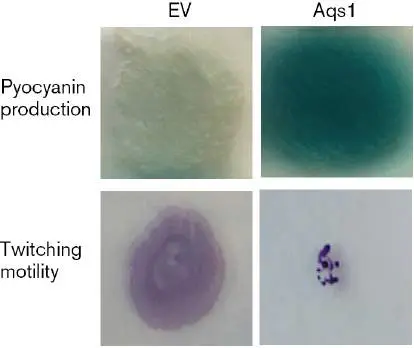
system (see eAppendix 3) and functions in the following way: The two domains of the adenylate cyclase protein are separated, and each domain is individually expressed by an open reading frame (ORF) in which is fused the gene of another protein, one of these proteins called bait and the other called prey. If the bait and prey proteins bind each other tightly, the domains of adenylate cyclase they are fused to can assemble into a functional enzyme. The activity of this reconstituted enzyme can then be quantified to determine the degree to which the two proteins bind.
The BACTH assay used in this study set the Aqs1 protein as bait and the known quorum-sensing proteins as prey. The negative control was simply the empty vector (EV), which is the plasmid expressing the domain of adenylate cyclase but without the prey protein fused to it (Fig. ERA 12.3 ). Some expression was observed for the empty vector, but expression was far more pronounced when the prey protein was LasR. None of the other quorum-sensing proteins had this large increase, so the researchers concluded that Aqs1 binds specifically to LasR.
FIGURE ERA 12.3 ■ Two-hybrid assays for contact between Aqs1 and proteins of the quorum-sensing system of Pseudomonas aeruginosa. Miller units are arbitrary units for beta-galactosidase activity, whose expression
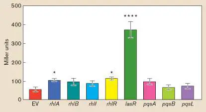
is controlled by cyclic AMP (cAMP). Cyclic AMP is synthesized if the two domains of adenylate cyclase are brought into proximity by the prey and bait fusion proteins. Of the genes tested in the assay, only lasR was able to produce a protein that could bind Aqs1, as indicated by a marked increase in Miller units relative to the empty vector (EV) control that lacks an inserted gene. Number of asterisks indicates degree of significance in difference relative to EV.
Now that they had evidence that Aqs1 bound LasR, the next step was to see if this binding had any influence on LasR’s ability to control gene expression as a quorum-sensing regulatory protein. First they purified Aqs1 and LasR proteins, converting the latter into its active form by adding the autoinducer molecule to which it binds. Next they purified DNA containing the operator region upstream of the lasB gene promoter to which activated LasR binds.
With these reagents prepared, the researchers performed electrophoretic mobility shift assays (EMSAs) to look for evidence that Aqs1 can block LasR binding to the lasB operator. By itself, the lasB DNA was highly mobile during electrophoresis and migrated to the bottom of the gel (Fig. ERA 12.4 ). In the presence of LasR, the lasB DNA was far less mobile, due to LasR binding, and it remained near the top of the gel. This treatment confirmed that their assay can detect LasR binding to lasB under normal conditions. When Aqs1 was added to lasB DNA and LasR, the high mobility was restored for lasB, indicating that Aqs1 was able to prevent LasR from binding its DNA target. How does Aqs1 interfere with LasR binding to lasB DNA? Aqs1 by itself did not alter the mobility of the lasB DNA, so it was unlikely to restrict LasR-lasB binding by blocking a site on the DNA. Rather, it was more likely that Aqs1 binds to the LasR protein and prevents it from binding to the lasB DNA.
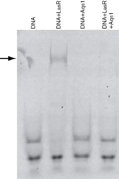
FIGURE ERA 12.4 ■ EMSA of lasB operator DNA in the presence or absence of proteins LasR and Aqs1. DNA migrated over time from top to bottom before the gel was stained. Arrow indicates the hindered mobility of lasB DNA that resulted when LasR was present and able to bind (lane 2). Aqs1 did not hinder lasB mobility, suggesting it does not bind the DNA (lane 3). However, Aqs1 was able to prevent the mobility shift caused by LasR (lane 4).
M. SHAH. 2021. MOL CELL 81 (3):5571–83.
After characterizing this function of Aqs1, the researchers applied BACTH and other assays to discover that Aqs1 also binds and inhibits the PilB protein, the ATPase that assembles type IV pili on the surface. Blocking this function prevents superinfection by phage that would otherwise use this receptor to adsorb to the P. aeruginosa host. It is remarkable how efficient DMS3 is with its defenses, in that it is able to block both quorum sensing and type IV pilus construction with a single 69-amino-acid-long protein.
Further Exploration
Given the importance of both quorum sensing and twitching motility to biofilm formation in Pseudomonas aeruginosa (see Section 12.2 in the printed book), how might surface populations of P. aeruginosa appear if DMS3 infection occurred at one of the stages of biofilm development?
Shah, M., V. L. Taylor, D. Bona, Y. Tsao, S. Y. Stanley, et al. 2021. A phage-encoded anti-activator inhibits quorum sensing in Pseudomonas aeruginosa. Molecular Cell 81 :571–583.
CHAPTER REVIEW
Review Questions
1. Describe how Escherichia coli senses a chemical gradient and changes its behavior in response.
2. How does methylation of MCP proteins affect chemotactic signal transduction?
3. How does magnetotaxis differ from chemotaxis?
4. What are the key stages of biofilm formation in Pseudomonas aeruginosa?
5. How does c-di-GMP function as the master regulator of the transition from planktonic to sessile forms of life? 6. Swarming motility and twitching motility both facilitate movement on a surface. What is different about the machinery that drives them?
7. What are the components of the extracellular matrix of a biofilm?
8. What is the value of rhamnolipid release during macrocolony formation?
9. What role does quorum sensing play in biofilm development?
10. Explain how a decrease in c-di-GMP triggers the detachment of P. aeruginosa cells from EPS.
11. Discuss how bacteria use restriction endonucleases to protect themselves against invading bacteriophages. 12. Describe how CRISPR-Cas systems acquire foreign DNA and use it to protect a cell from infection.
13. What are some of the other means besides restriction endonucleases and CRISPR-Cas by which cells protect themselves from phages?
14. What are some of the ways in which phages counteract the defenses of the host?
15. What is an evolutionary arms race, and how does this apply to phage-host interactions?
16. Describe how BioBricks and logic gates are used in synthetic biology.
Thought Questions
1. How might signal transduction through the Che proteins work for situations when the MCP chemoreceptor binds a repellent rather than an attractant?
2. In nature, bacteria often form mixed-species biofilms, rather than single-species ones such as the biofilm of Pseudomonas aeruginosa described in this chapter.
Consider the various stages of biofilm formation. Can you think of potential interactions between species where biofilm formation would be enhanced? Inhibited? Can invaders cheat the established biofilm?
3. Host defenses and phage counterdefenses have been treated in this chapter as interactions between a single species of host infected by a single genotype of phage. How might the effectiveness of each defense or counterdefense change if different hosts and/or infectious phages are present in the environment?
Key Terms
abortive infection (476)
biofilm (463)
c-di-GMP (464)
chemotaxis (458)
CRISPR (471)
diguanylate cyclase (DGC) (464) dispersion (468)
extracellular DNA (eDNA) (467) extracellular polymeric substance (EPS) (466) evolutionary arms race (477) flagellum (458)
gliding motility (462)
macrocolony (464)
magnetosome (462)
magnetotaxis (462)
methyl-accepting chemotaxis protein (MCP) (459) methyltransferase (470)
microcolony (464)
noise (486)
phage-inducible chromosomal islands (PICIs) (476) phosphodiesterase (PDE) (464) planktonic cell (463)
restriction endonuclease (469) restriction-modification (RM) (469) rhamnolipid (465)
swarming motility (462)
synthetic biology (481)
twitching motility (462)
type IV pili (465)
Recommended Reading
Ahator, Stephen D., and LianHui Zhang. 2019. Small is mighty —chemical communication systems in Pseudomonas aeruginosa. Annual Review of Microbiology 73 :559–578.
Berens, Christian, Florian Groher, and Beatrix Suess. 2015. RNA aptamers as genetic control devices: The potential of riboswitches as synthetic elements for regulating gene expression. Biotechnology Journal 10 :246–257.
Burt, Alister, C. Keith Cassidy, Peter Ames, Maria Bacon-Verloop, Megghane Baulard, et al. 2020. Complete structure of the chemosensory array core signalling unit in an E. coli minicell strain. Nature Communications 11 :743.
Cherny, Kathryn E., and Karin Sauer. 2020. Untethering and degradation of the polysaccharide matrix are essential steps in the dispersion response of Pseudomonas aeruginosa biofilms. Journal of Bacteriology 202 :e00575-19.
Collins, Alan J., T. Jarrod Smith, Holger Sondermann, and George A. O’Toole. 2020. From input to output: The Lap/c-di-GMP biofilm regulatory circuit. Annual Review of Microbiology 74:607–631.
Davey, Mary E., Nicky C. Caiazza, and George A. O’Toole. 2003. Rhamnolipid surfactant production affects biofilm architecture in Pseudomonas aeruginosa PAO1. Journal of Bacteriology 185 : 1027–1036.
Davies, David G., Matthew R. Parsek, James P. Pearson, Barbara H. Iglewski, J. W. Costerton, et al. 1998. The involvement of cell-to-cell signals in the development of a bacterial biofilm. Science 280 :295–298.
Gillmore, Julian D., Ed Gane, Jorg Taubel, Justin Kao, Mariana Fontana, et al. 2021. CRISPR-Cas9 in vivo gene editing for transthyretin amyloidosis. New England Journal of Medicine 385 :493–502.
Hampton, Hannah, G., Bridget N. J. Watson, and Peter C. Fineran. 2020. The arms race between bacteria and their phage foes. Nature 577 :327–336.
Ishino, Yoshizumi, Mart Krupovic, and Patrick Forterre. 2018. History of CRISPR-Cas from encounter with a mysterious repeated sequence to genome editing technology. Journal of Bacteriology 200 :e00580-17.
Laventie, Benoît-Joseph, Matteo Sangermani, Fabienne Estermann, Pablo Manfredi, Rémi Planes, et al. 2019. A surface-induced asymmetric program promotes tissue colonization by Pseudomonas aeruginosa. Cell Host & Microbe 25 :140–152.
Locke, James C., Jonathan W. Young, Michelle Fontes, Maria J. Hernandez Jimenez, and Michael B. Elowitz. 2011.
Stochastic pulse regulation in bacterial stress response. Science 334 :366–369.
Mimee, Mark, Alex C. Tucker, Christopher A. Voigt, and Timothy K. Lu. 2015. Programming a human commensal bacterium, Bacteroides thetaiotaomicron, to sense and respond to stimuli in the murine gut microbiota. Cell Systems 1 :62–71. Parkinson, John S., Gerald L. Hazelbauer, and Joseph J. Falke. 2015. Signaling and sensory adaptation in Escherichia coli chemoreceptors: 2015 update. Trends in Microbiology 23:257–266.
Reichhardt, Courtney, Cynthis Wong, Daniel Passos da Silva, Daniel J. Wozniak, and Matthew R. Parsek. 2018. CdrA interactions within the Pseudomonas aeruginosa biofilm matrix safeguard it from proteolysis and promote cellular packing. mBio 9 :e01376-18.
Rifat, Dalin, Nathan T. Wright, Kristen M. Varney, David J. Weber, and Lindsay W. Black. 2008. Restriction endonuclease inhibitor IPI* of bacteriophage T4: A novel structure for a dedicated target. Journal of Molecular Biology 375 :720–734.
Rostøl, Jakob T., and Luciano Marraffini. 2019. (Ph)ighting phages: How bacteria resist their parasites. Cell Host & Microbe 25 :184–194.
Rovner, Alex J., Adrian D. Haimovich, Spencer R. Katz, Zhe Li, Michael W. Grome, et al. 2015. Recoded organisms engineered to depend on synthetic amino acids. Nature 518:89–93.
Samson, Julie E., Alfonso H. Madagán, Mourad Sabri, and Sylvain Moineau. 2013. Revenge of the phages: Defeating bacterial defences. Nature Reviews. Microbiology 11 :675–687. Schwartz, Daniel A., and Debbie Lindell. 2017. Genetic hurdles limit the arms race between Prochlorococcus and the T7-like podoviruses infecting them. ISME Journal 11 :1836–1851. Seed, Kimberley D., David W. Lazinski, Stephen D.
Calderwood, and Andrew Camilli. 2013. A bacteriophage encodes its own CRISPR/Cas adaptive response to evade host innate immunity. Nature 494 :489–491.
Glossary
chemotaxis The ability of organisms to move toward or away from specific chemicals.
flagellum pl. flagella A filamentous structure for motility. In prokaryotes, a helical protein filament attached to a rotary motor; in eukaryotes, an undulating membrane-enclosed complex of microtubules and ATP-driven motor proteins.
methyl-accepting chemotaxis protein (MCP)
Also called chemoreceptor. A cell-membrane signal transduction protein that becomes methylated during adaptation to a chemotactic signal.
microcolony A small assembly of cells on a surface that initiate biofilm formation.
magnetotaxis The ability to direct motility along magnetic field lines. magnetosome An organelle that contains the mineral magnetite and thus enables microbes to sense a magnetic field.
swarming or swarming motility A behavior in which some microbial cells differentiate into large swarmer cells and swim together as a unit.
twitching motility A type of bacterial movement on solid surfaces in which a specific pilus extends and retracts.
gliding motility The movement of cells individually or as a collective over surfaces using specialized pili.
biofilm A community of microbes growing on a solid surface. planktonic cell An isolated cell, growing individually in a liquid without connections to other cells.
macrocolony A large cluster of cells embedded within an extracellular matrix. Macrocolonies constitute the mushroom-shaped structures of mature biofilms.
c-di-GMP Bis-(3′-5′)-cyclic dimeric guanosine monophosphate; a second messenger molecule that plays a key role in regulating biofilm formation.
diguanylate cyclase (DGC)
A class of enzymes that synthesize the second messenger molecule c-di-GMP.
phosphodiesterase (PDE)
A class of enzymes that degrade the second messenger molecule c-di-GMP.
type IV pili Retractable appendages used for surface attachment and twitching motility along the surface.
rhamnolipid A surfactant molecule composed of a rhamnose head group and a fatty acid tail.
extracellular polymeric substances (EPS)
See exopolysaccharides .
extracellular DNA (eDNA)
DNA located outside the cell, often released by cells to provide stability and structure to biofilms.
dispersion The process by which cells escape the biofilm structure and reenter the planktonic state.
restriction-modification (RM)
An antiphage defense system usually composed of a methyltransferase enzyme that methylates DNA and a restriction endonuclease that cleaves unmethylated DNA. restriction endonuclease or restriction enzyme A bacterial enzyme that cleaves double-stranded DNA within a specific short sequence, usually a palindrome.
methyltransferase An enzyme that adds a methyl group to a base in DNA. CRISPR Clustered regularly interspaced short palindromic repeats. CRISPR consists of short repeated DNA sequences in a bacterial or archaeal genome, derived from previous bacteriophage or viral infection and conferring protection from future infection; considered a prokaryotic “immune system.” In biotechnology, provides a means of editing the human genome.
abortive infection A class of antiphage defenses that kill the host cell before the phage can complete replication.
phage-inducible chromosomal islands (PICIs)
Mobile genetic elements integrated within the host chromosome that hijack the capsids produced by incoming phage for transfer to new host cells.
evolutionary arms race The escalation of adaptations and counteradaptations that develop between predator and prey or between competing species.
synthetic biology The genetic construction of novel organisms with useful functions.
noise Variance from a mean in an assay. A system in which individual results vary greatly from a mean that was derived from many results is a noisy system.