Ch 1: The Microbial WorldRead full chapter →
Sections in this chapter
- 1 The Microbial World
- Microbiology in Motion
- I Exploring the Microbial World
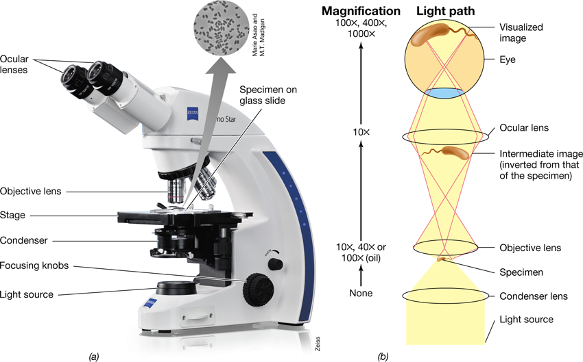
- 1.1 Microorganisms, Tiny Titans of the Earth
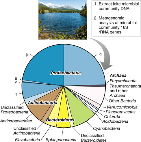
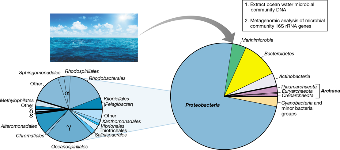
- Figure 1.1 Microbial communities.
- Figure 1.2 Microbial applications.
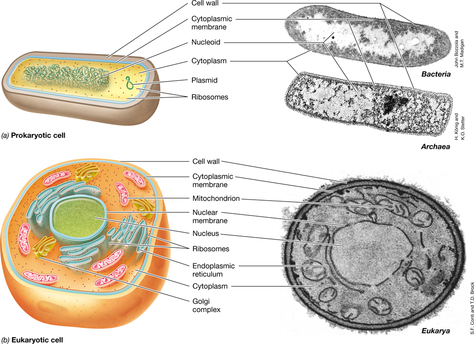
- Figure 1.3 Microbial cells.
- Check Your Understanding
- 1.2 Structure and Activities of Microbial Cells
- Elements of Microbial Structure
- Figure 1.4 Microbial cell structure.
- Mastering Microbiology
Ch 2: Microbial Cell Structure and FunctionRead full chapter →
Sections in this chapter
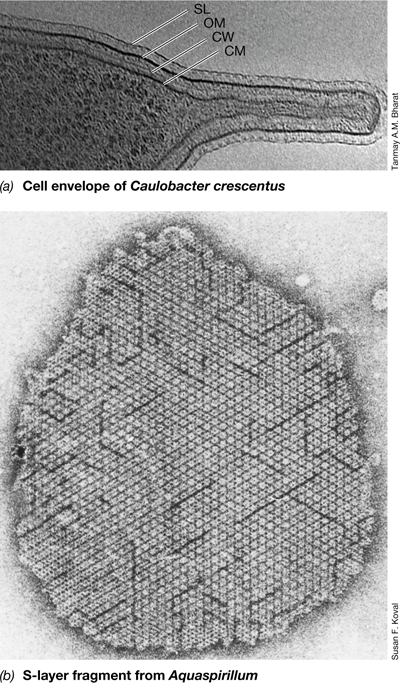
- 2 Microbial Cell Structure and Function
- Exploring the Microbial Cell
- I The Cell Envelope
- 2.1 The Cytoplasmic Membrane
- Bacterial Cytoplasmic Membranes
- Figure 2.1 Phospholipid bilayer membrane.
- Mastering Microbiology
- Figure 2.2 Structure of the cytoplasmic membrane.
- Archaeal Cytoplasmic Membranes
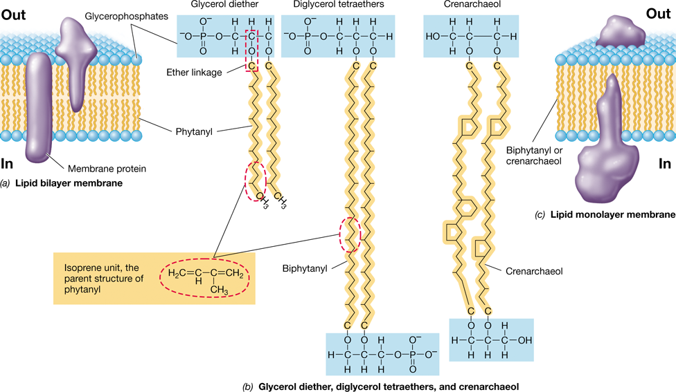
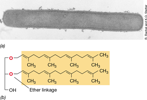
- Figure 2.3 Major lipids of ***Archaea*** and the architecture of archaeal membranes.
- Cytoplasmic Membrane Function
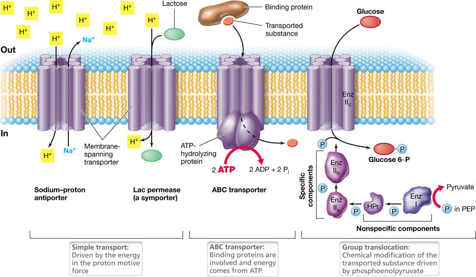
- Figure 2.4 The major functions of the cytoplasmic membrane.
Ch 3: Microbial MetabolismRead full chapter →
Sections in this chapter
- 3 Microbial Metabolism
- Life Begins with Metabolism
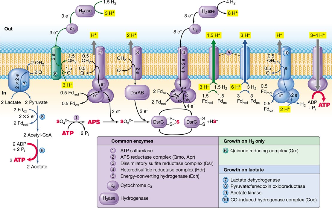
- Figure 3.1 Metabolic coupling with respect to energy conservation and electron flow.
- I Fundamentals of Metabolism
- 3.1 Defining the Requirements for Life
- Free Energy
- Reducing Power
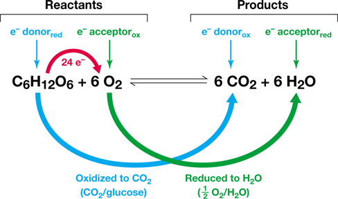
- Figure 3.2 Example of an oxidation–reduction reaction.
- Metabolic Classes of Microorganisms
- Figure 3.3 Classification of metabolic types based on energy sources.
- Check Your Understanding
- 3.2 Electron Transfer Reactions

Ch 4: Microbial Growth and Its ControlRead full chapter →
Sections in this chapter
- 4 Microbial Growth and Its Control
- Growing Their Own Way
- I Culturing Microbes and Measuring Their Growth
- 4.1 Feeding the Microbe: Cell Nutrition
- Chemical Makeup of a Cell
- Figure 4.1 Elemental and macromolecular composition of a bacterial cell.
- Carbon, Nitrogen, and Other Macronutrients
- Micronutrients: Trace Metals and Growth Factors
- Table 4.1 Micronutrients needed by microorganismsa
- Check Your Understanding
- 4.2 Growth Media and Laboratory Culture
- Classes of Culture Media
Ch 5: Viruses and Their MultiplicationRead full chapter →
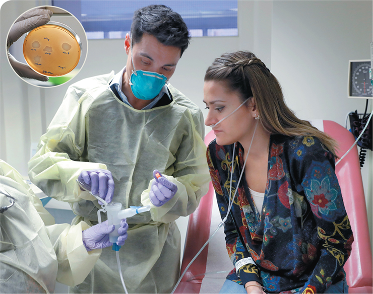
Acquiring an antibiotic-resistant infection or “superbug” is one of medicine’s biggest nightmares. What can medical practitioners do to treat the patient? Besides drugs, viruses known as bacteriophages have been recruited to specifically target and kill bacteria.
Despite microbiologists’ tinkering with using bacteriophages as antimicrobials for decades, their actual application in medicine has been minimal. However, the emergence of antibiotic resistance has led to renewed focus on using these tiny microbes as therapeutic agents. The photo above shows Ella Balasa (right side of photo), a microbiologist who has cystic fibrosis. Cystic fibrosis is a genetic disease that results in a buildup of thick mucus in the lungs. This mucus allows bacteria to flourish in the lungs, which results in infections and subsequent lung damage that can be fatal. Ella had b…
Sections in this chapter
- When Antibiotics Fail, Bacteriophage Therapy to the Rescue
- I The Nature of Viruses
- 5.1 What Is a Virus?
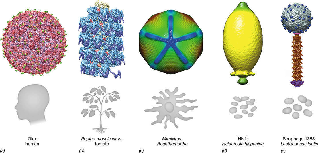
- Figure 5.1 Virion morphology and their hosts.
- Viral Components and Activities
- Figure 5.2 Comparison of naked and enveloped virus particles.
- Viral Diversity and Hosts
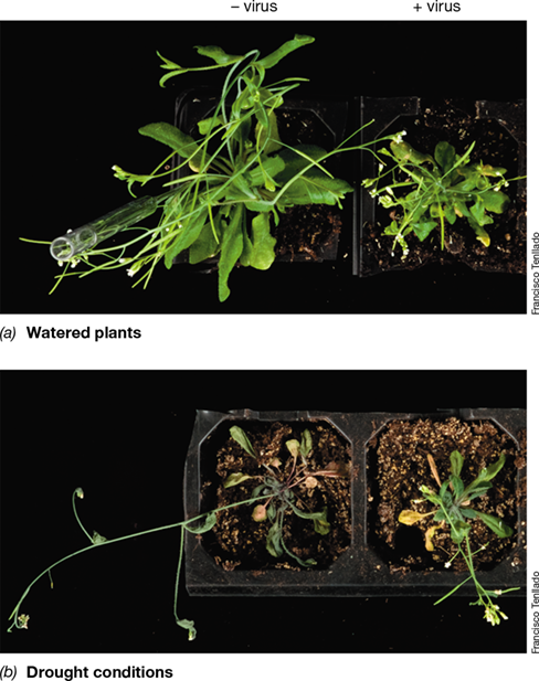
- Figure 5.3 Virus-infected ***Arabidopsis thaliana*** plants and response to drought.
- Check Your Understanding
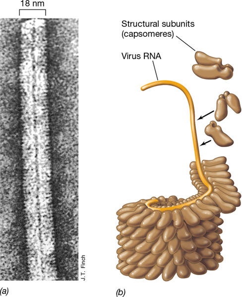
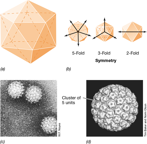
- 5.2 Structure of the Virion
- Figure 5.4 The giant and asymmetrical ***Pandoravirus***.
- Virion Structure
Ch 6: Molecular Information Flow and Protein ProcessingRead full chapter →
Sections in this chapter
- 6 Molecular Information Flow and Protein Processing
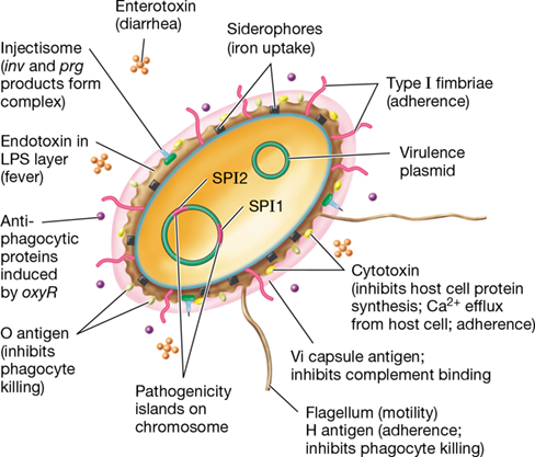
- Injectisomes: *Salmonella*’s Mode of Attack
- I Molecular Biology and Genetic Elements
- 6.1 DNA and Genetic Information Flow
- Figure 6.1 Genetic information flow and the components of the nucleic acids.
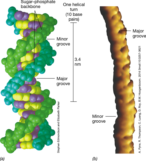
- Properties of the Double Helix
- Figure 6.2 DNA structure.
- Figure 6.3 Arrangement of the DNA double helix.
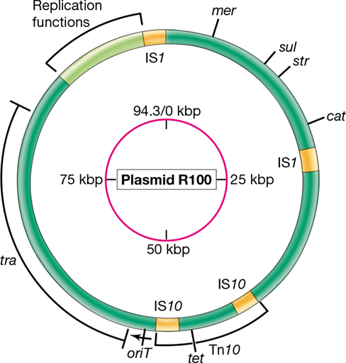
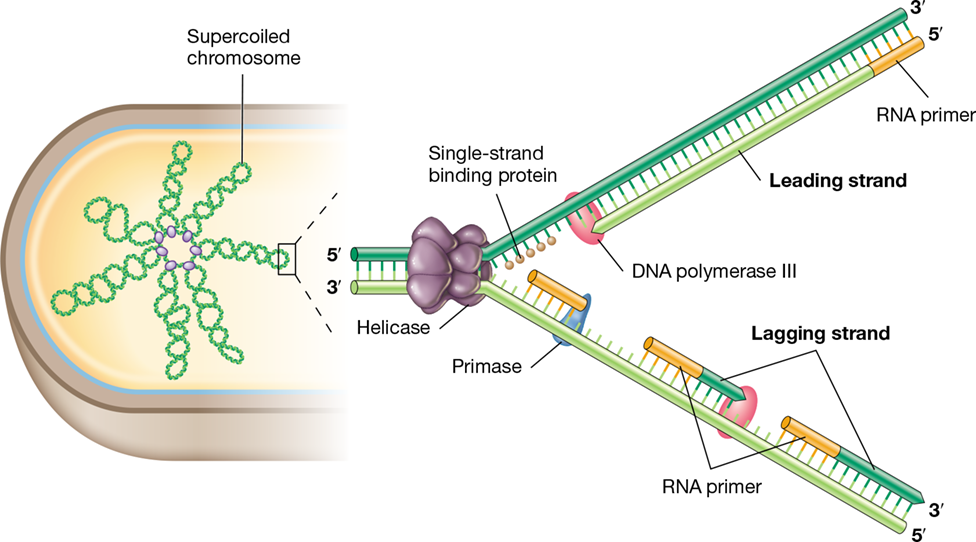
- Size, Shape, and Supercoiling of DNA
- Figure 6.4 Supercoiled DNA and DNA gyrase.
- Genes and the Steps in Biological Information Flow
- Figure 6.5 Synthesis of the three types of informational macromolecules in the processes of replication (DNA→DNA), transcription (DNA→RNA), and translation (RNA→protein).
Ch 7: Microbial Regulatory SystemsRead full chapter →
Sections in this chapter
- 7 Microbial Regulatory Systems
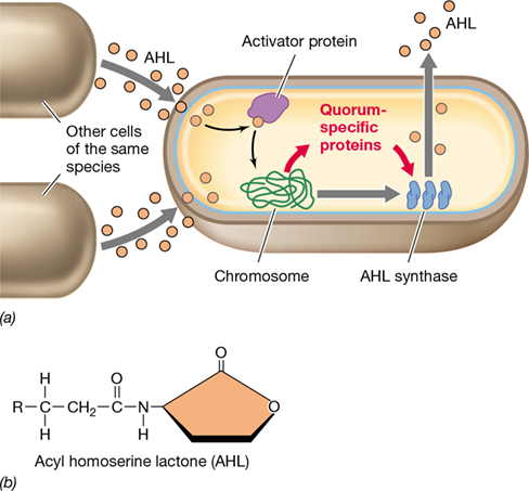
- As Bacterial Cells Chatter, Viruses Eavesdrop
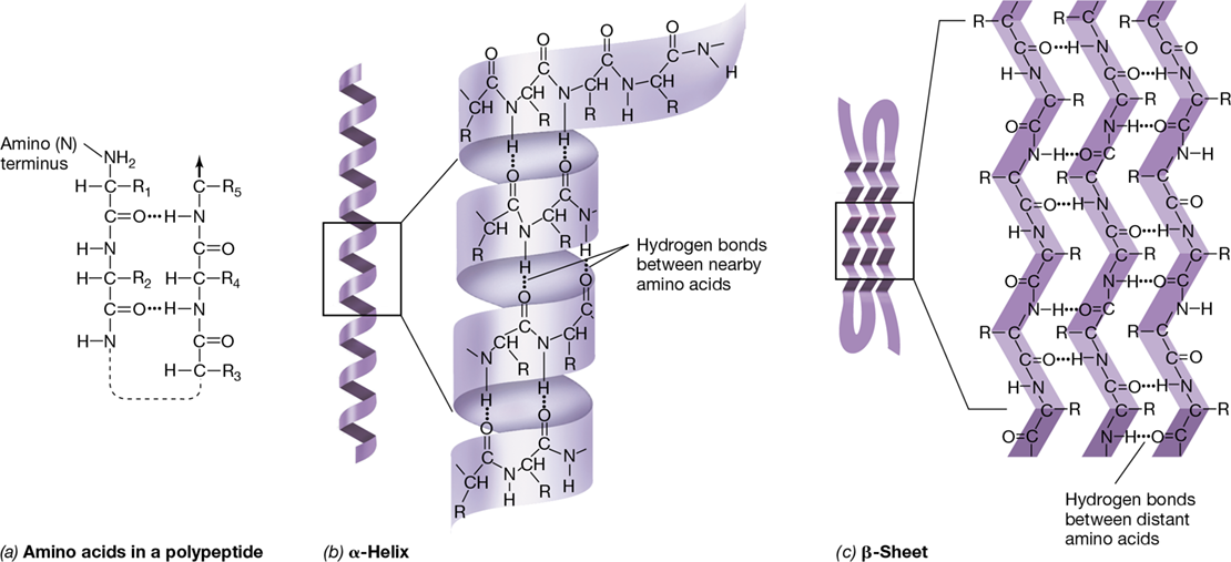
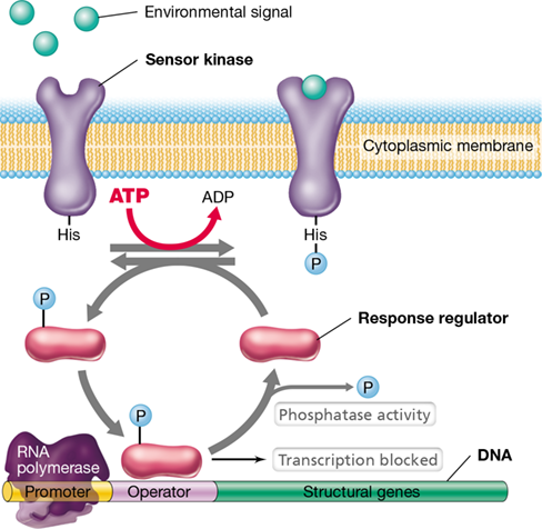
- Figure 7.1 Gene expression and regulation of protein activity.
- I: DNA-Binding Proteins and Transcriptional Regulation
- I DNA-Binding Proteins and Transcriptional Regulation
- 7.1 DNA-Binding Proteins
- Interaction of Proteins with Nucleic Acids
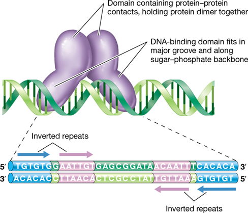
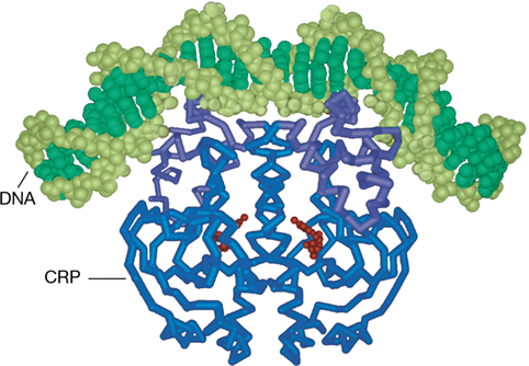
- Figure 7.2 DNA-binding proteins.
- Structure of DNA-Binding Proteins
- Figure 7.3 The helix-turn-helix structure of some DNA-binding proteins.
- Check Your Understanding
- 7.2 Transcription Factors and Effectors
Ch 8: Molecular Aspects of Microbial GrowthRead full chapter →
Sections in this chapter
- 8 Molecular Aspects of Microbial Growth
- Membrane Vesicles: Nano Vehicles Transporting Important Cargo
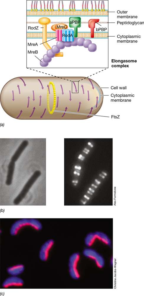
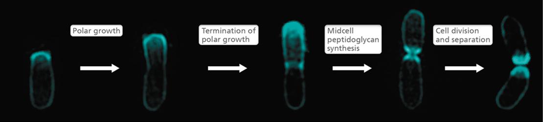
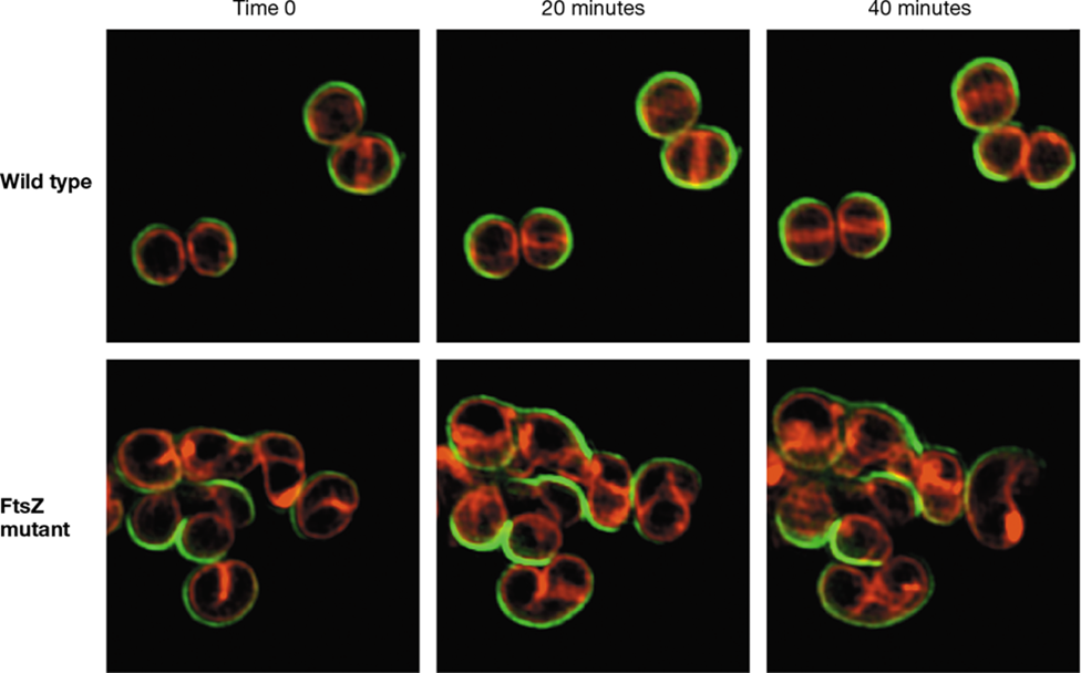
- I Bacterial Cell Division
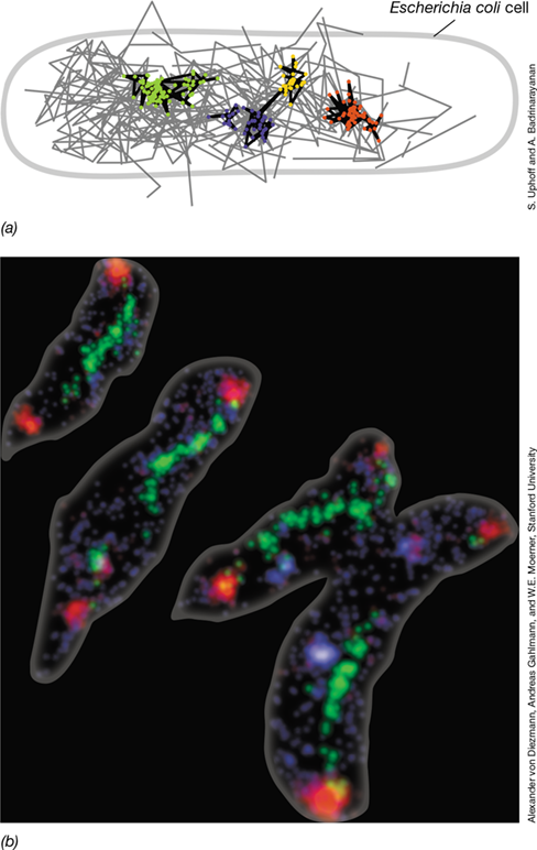
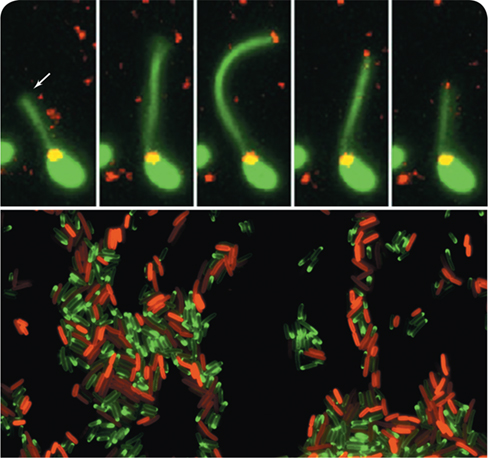
- 8.1 Visualizing Molecular Growth
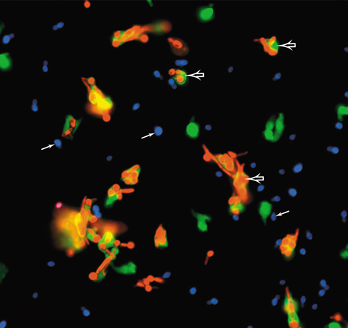
- Fluorescent Tagging
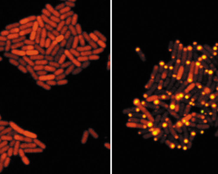
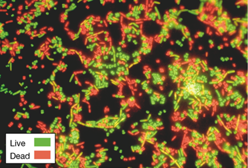
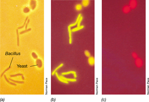
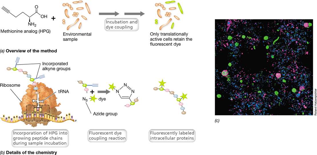
- Figure 8.1 Fluorescence micrographs illustrating molecular growth characteristics.
- Super-Resolution Techniques
- Figure 8.2 Super-resolution imaging of molecular growth characteristics.
- Check Your Understanding
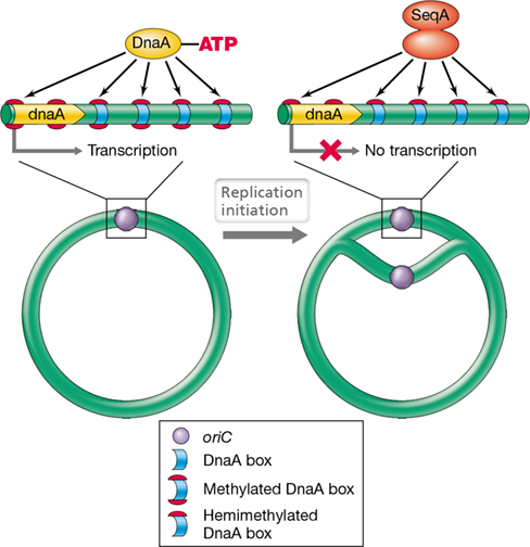
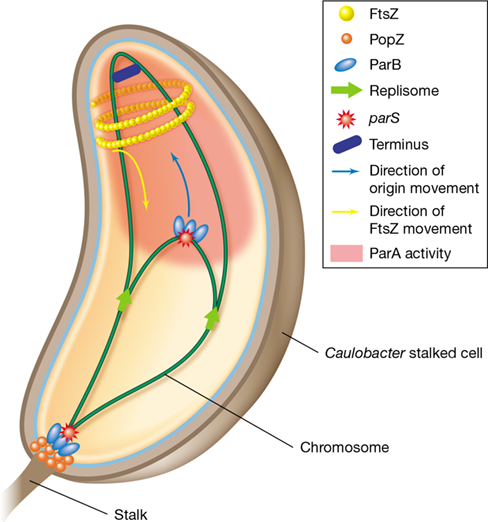
- 8.2 Chromosome Replication and Segregation
- Figure 8.3 Overview of the bacterial cell cycle.
- Regulation of Chromosome Replication Initiation
Ch 9: Genetics of Bacteria and ArchaeaRead full chapter →
Sections in this chapter
- 9 Genetics of Bacteria and Archaea
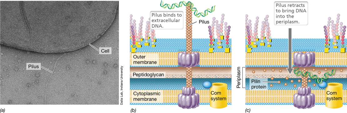
- Live Cell Imaging Captures Bacterial Promiscuity
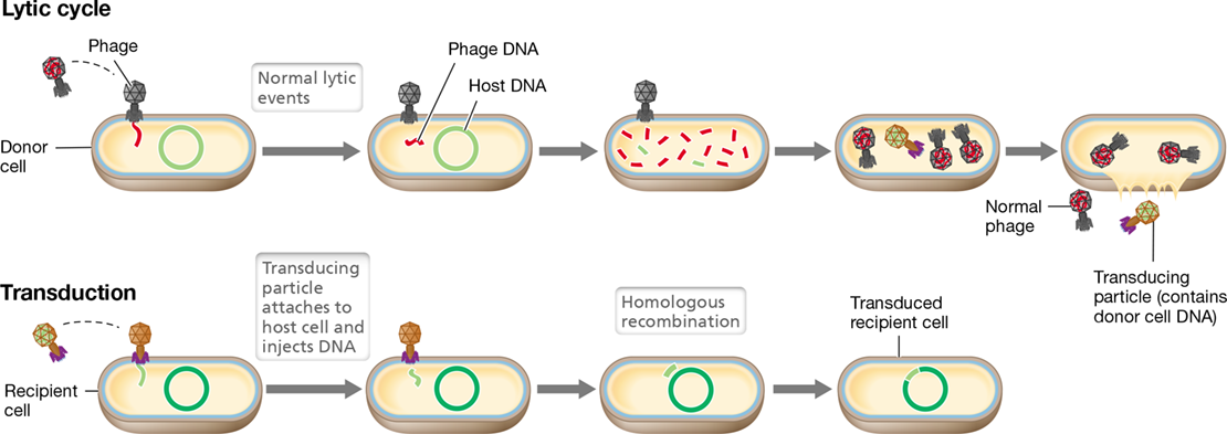
- Figure 9.1 Overview of bacterial and archaeal genetics.
- I Mutation
- 9.1 Mutations and Mutants
- Figure 9.2 Wild-type versus mutant phenotype.
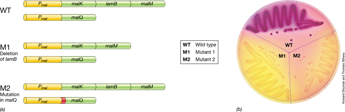
- Isolation of Mutants: Screening versus Selection
- Figure 9.3 Selectable and nonselectable mutations.
- Mastering Microbiology
- Isolation of Nutritional Auxotrophs
- Figure 9.4 Screening for nutritional auxotrophs.
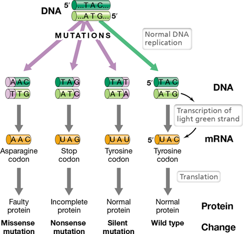
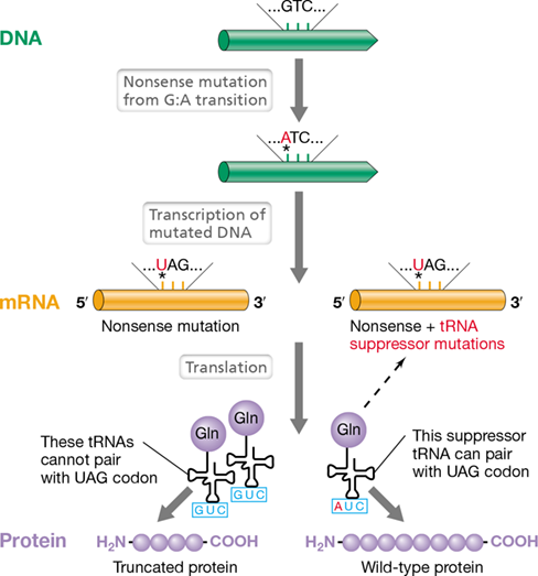
- 9.2 Molecular Basis of Mutation

Ch 10: Microbial Genomics and Other OmicsRead full chapter →
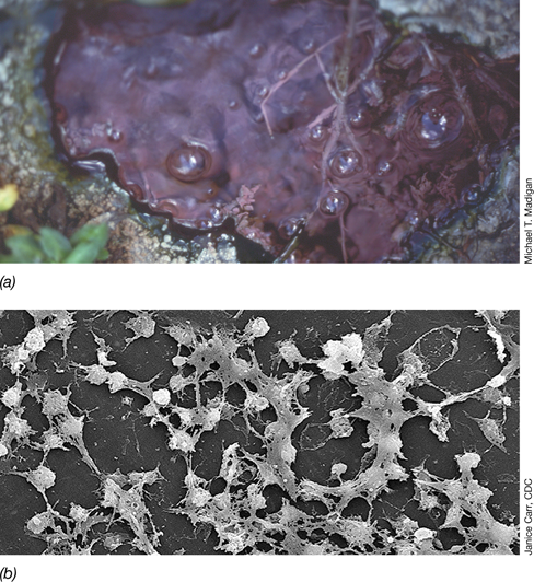
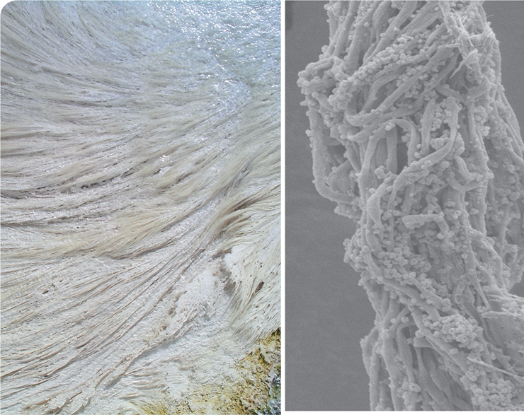
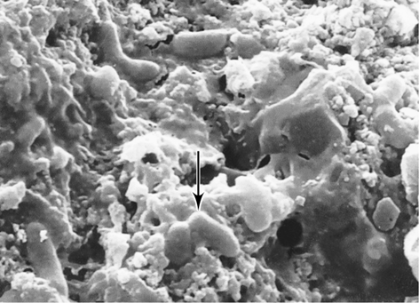
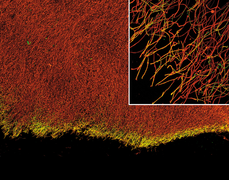
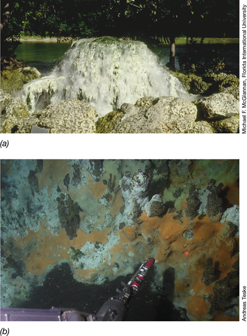
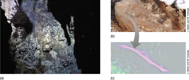
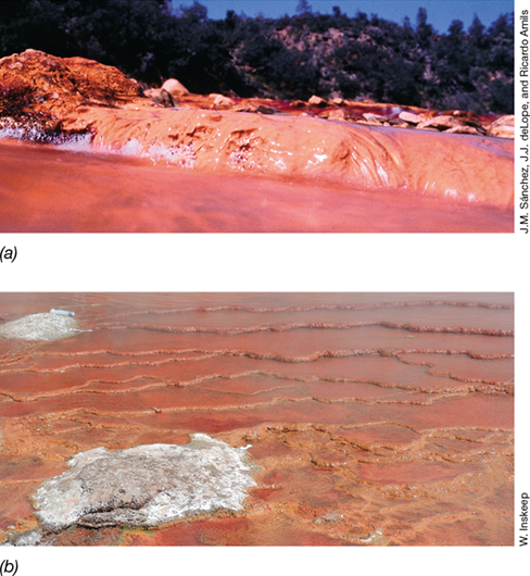
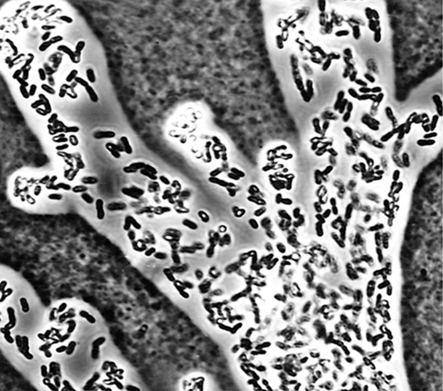
Mammoth Hot Springs (MHS) in Yellowstone National Park (USA) displays many geochemical traits similar to those on Mars. Because of this, geomicrobiologists study these hot, oxygen-limited and sulfur-rich springs for microbial fossils whose fingerprints of life (biomarkers) could be used to detect life on other planets. The photo on the left shows filamentous microbial mats from MHS that resemble fettuccine pasta as a result of mineral encrustation. Tens of centimeters long, these streamers contain extremophilic microbes that entomb themselves in 5 millimeters of travertine (calcium carbonate) …
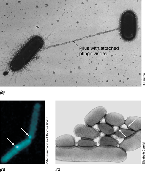
Using a combination of geochemical measurements, microscopy, and metagenomic, metatranscriptomic, and metaproteomic approaches, scientists have unraveled many of the mysteries surrounding the MHS streamers. These mats are dominated by the thermophilic, chemolithotrophic bacterium Sulfurihydrogenibium yellowstonense, which uses CO2 and reduced sulfur as carbon and energy sources, respectively. Multi-omic data sets indicate that these remarkable cells produce pili as well as extracellular polymeric substances to latch onto one another and form streamers in the fast-moving spring water. Once th…
Sections in this chapter
- Omics Tools Unravel Mysteries of “Fettuccine” Rocks
- I Genomics
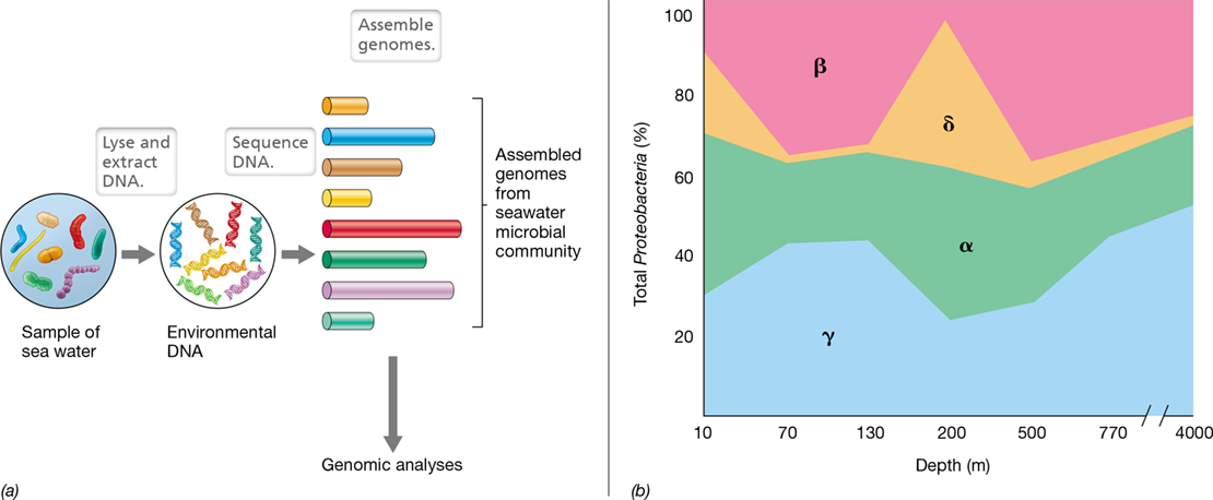
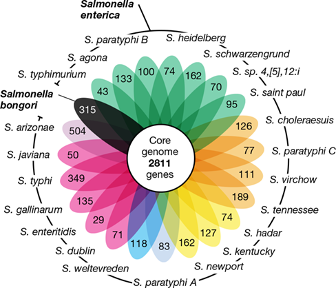
- Figure 10.1 Utility of microbial genome sequences.
- 10.1 Introduction to Genomics
- Genomics: Then and Now
- Table 10.1 Genomes of select species of *Bacteria* and *Archaea*a
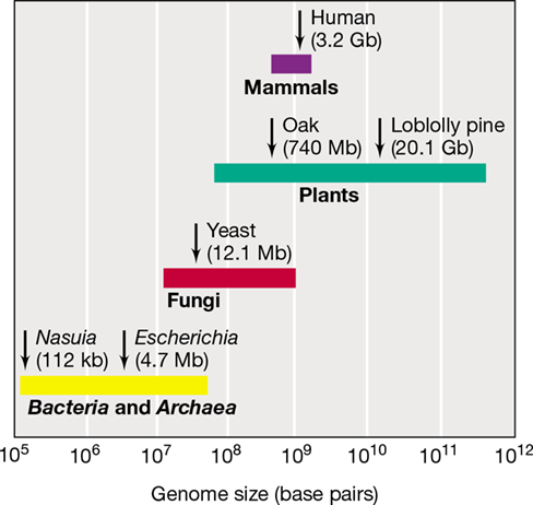
- Figure 10.2 Genome sizes of microbial cells and higher organisms.
- What Can Genomes Tell Us?
- Figure 10.3 Diverse examples of what genomes can tell us.
- Check Your Understanding
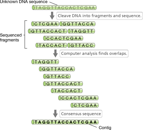
- 10.2 Sequencing and Annotating Genomes
- DNA Sequencing
Ch 11: Viral Genomics and DiversityRead full chapter →
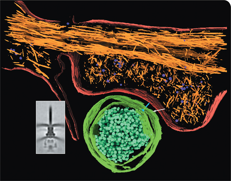
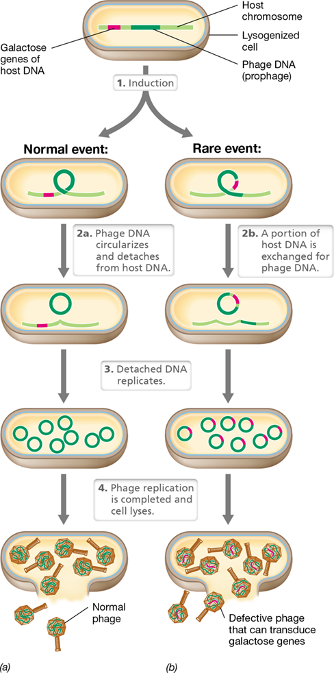
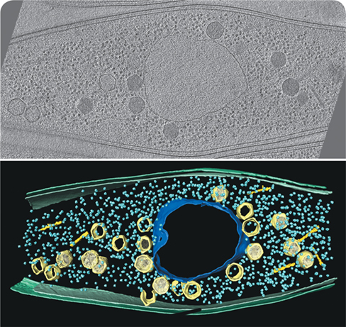
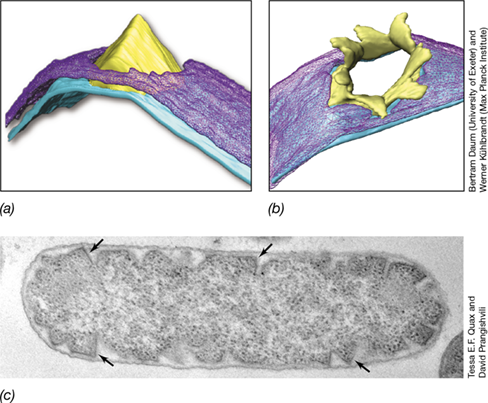
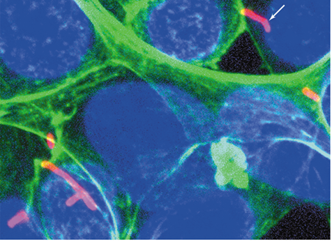
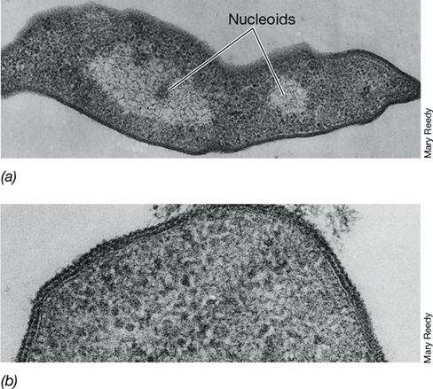
A membrane-bound nucleus is a defining feature of eukaryotic cells that differentiates them from their prokaryotic counterparts. Remarkably, molecular biologists have discovered a group of large Pseudomonas bacteriophages whose genomes encode structures resembling the eukaryotic nucleus and mitotic spindle. The obvious question is why, and to understand these remarkable events, scientists needed to understand the roles of both eukaryotic-like structures.
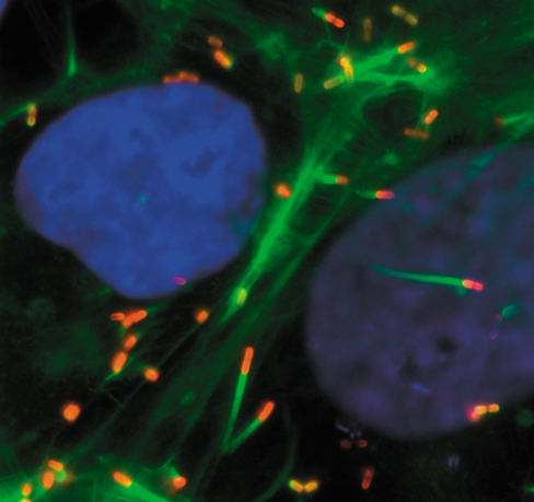
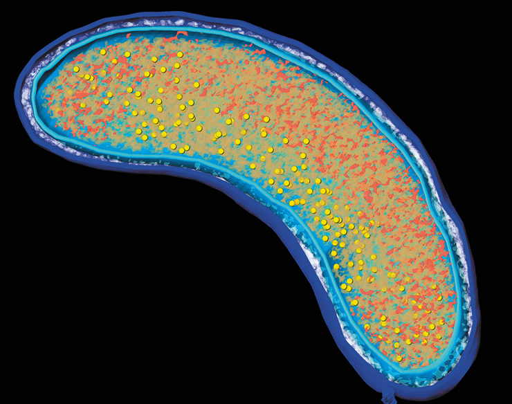
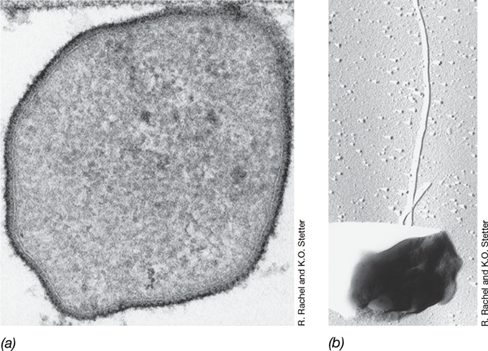
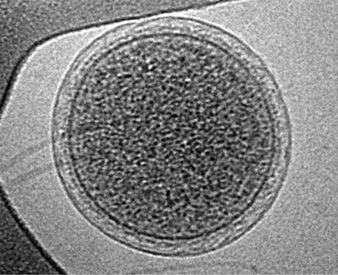
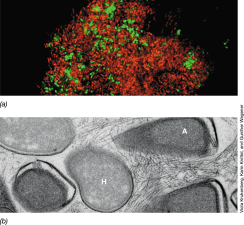
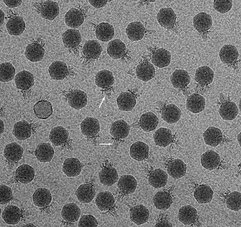
Using fluorescent labeling and cryo-electron microscopy, it was determined that the phage-encoded nucleus contains enzymes specific for replicating and transcribing phage DNA. Once empty phage capsids are assembled at the host membrane, they move rapidly toward the phage nucleus to be filled with phage DNA. The top photo shows a cryo-tomogram slice of a Pseudomonas chlororaphis cell infected with the phage 201ϕ2-1 (phage virions are about 0.1 μm in diameter), and the bottom image highlights the following structures in color: nucleus-like structure (blue), capsids (light yellow), phage tails …
Sections in this chapter
- Bacteriophages Mimicking Eukaryotes—Discovery of a Phage-Encoded Nucleus and Spindle
- I Viral Genomes and Classification
- 11.1 Size and Structure of Viral Genomes
- Figure 11.1 Comparative genomics.
- The Baltimore Scheme: DNA Viruses
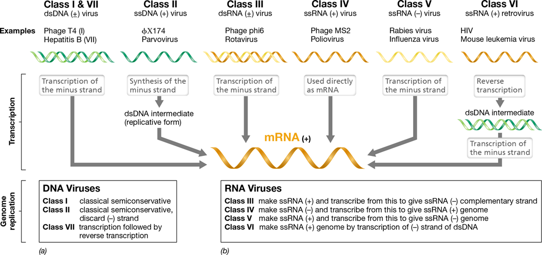
- Figure 11.2 The Baltimore classification of viral genomes.
- The Baltimore Scheme: RNA Viruses
- Hosts for Viruses of Each Baltimore Class
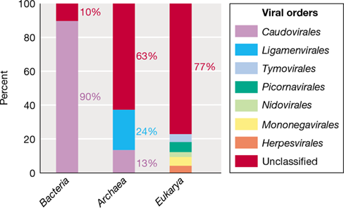
- Figure 11.3 Viral hosts and viral diversity.
- Viral Protein Synthesis
- Check Your Understanding
- 11.2 Viral Taxonomy and Phylogeny
Ch 12: Biotechnology and Synthetic BiologyRead full chapter →
Sections in this chapter
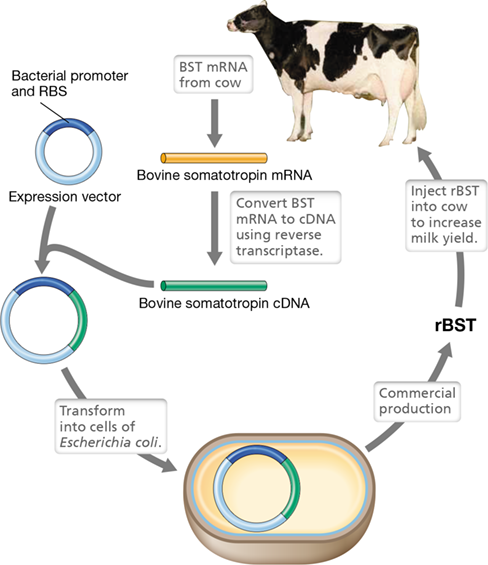
- 12 Biotechnology and Synthetic Biology
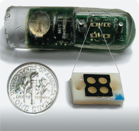
- An Ingestible Biosensor: Using Bacteria to Monitor Gastrointestinal Health
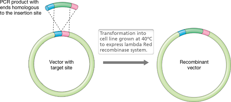
- Figure 12.1 Genetic engineering and biobrick assembly.
- I Tools of the Genetic Engineer
- 12.1 Manipulating DNA: PCR and Nucleic Acid Hybridization
- Figure 12.2 The polymerase chain reaction (PCR).
- Mastering Microbiology
- PCR and Polymerases
- PCR Applications and RT-PCR
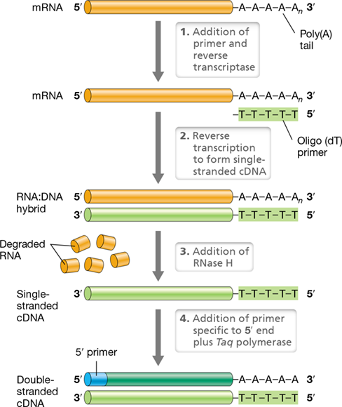
- Figure 12.3 Reverse transcription PCR.
- Gel Electrophoresis and Nucleic Acid Hybridization
- Figure 12.4 Agarose gel electrophoresis of DNA.

Ch 13: Microbial Evolution and Genome DynamicsRead full chapter →
Sections in this chapter
- 13 Microbial Evolution and Genome Dynamics
- Exploring Viral Genesis
- I: Early Earth and the Origin and Diversification of Life
- I Early Earth and the Origin and Diversification of Life
- 13.1 Formation and Early History of Earth
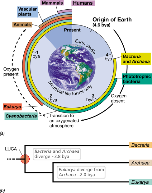
- Origin of Earth
- Figure 13.1 Major landmarks in biological evolution, Earth’s changing atmospheric chemistry, and microbial metabolic diversification.
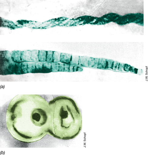
- Figure 13.2 Ancient microbial life.
- Origin of Cellular Life
- Mastering Microbiology
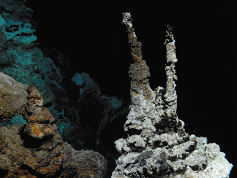
- Figure 13.3 Submarine mounds and their possible link to the origin of life.
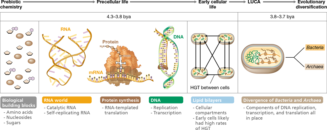
- Figure 13.4 Events hypothesized to precede the origin of cellular life.
Ch 14: Metabolic Diversity of MicroorganismsRead full chapter →
Sections in this chapter
- 14 Metabolic Diversity of Microorganisms
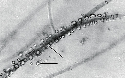
- Ferreting Out the Peculiar Life of Iron Bacteria
- I Introduction to Metabolic Diversity
- 14.1 Foundational Principles of Metabolic Diversity: Energy and Redox
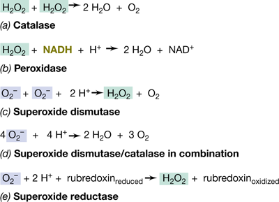
- Figure 14.1 Diversity of respiration reactions.
- Conservation of Energy
- Chemotrophic Metabolism
- Table 14.1 Energy yields from the oxidation of various inorganic electron donorsa
- Reducing Power and Redox Balance
- Figure 14.2 The reaction scheme for flavin-based electron bifurcation.
- Assimilative and Dissimilative Processes
- Check Your Understanding

Ch 15: Ecological Diversity of BacteriaRead full chapter →
Sections in this chapter
- 15 Ecological Diversity of Bacteria
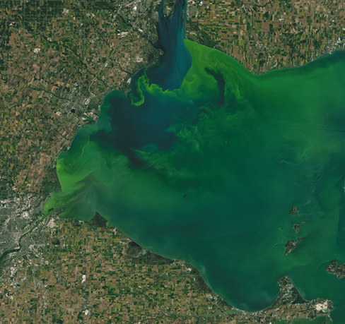
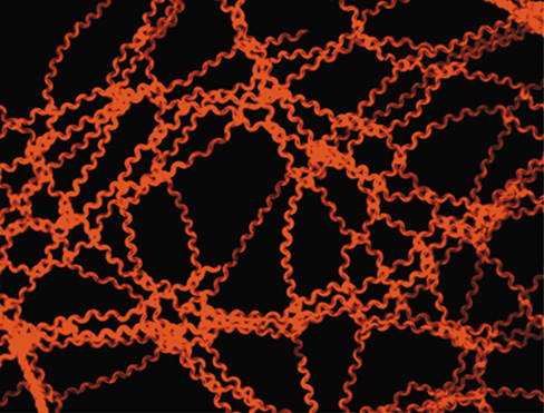
- Cyanobacterial Diversity and Environmental Change
- I Ecological Diversity Among Microorganisms
- 15.1 Making Sense of Microbial Diversity
- Figure 15.1 Major functional traits mapped across major phyla of *Bacteria* and *Archaea*.
- Check Your Understanding
- II Ecological Diversity of Phototrophic *Bacteria*
- 15.2 Overview of Phototrophic *Bacteria*
- Phylogeny and Classification of *Cyanobacteria*
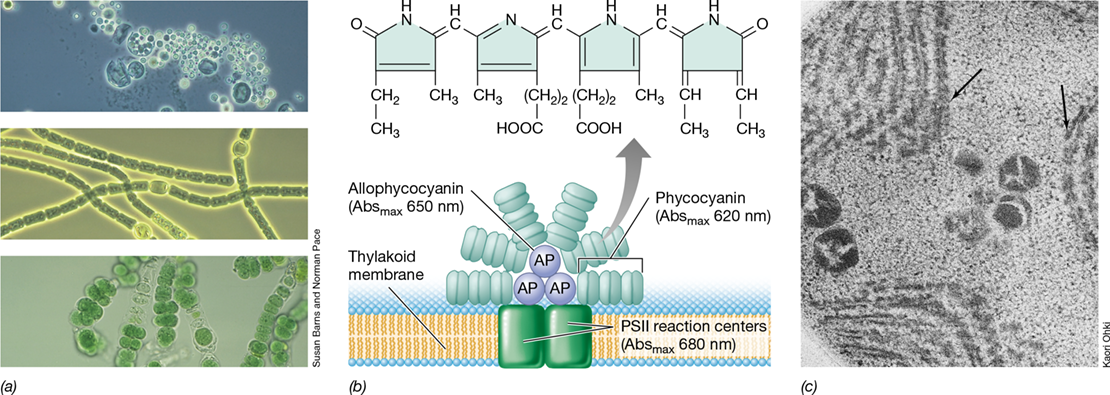
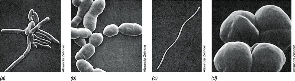
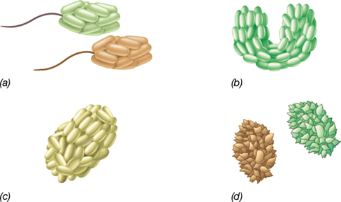
- Figure 15.2 The five major morphological types of *Cyanobacteria*.
- Figure 15.3 Taxonomically informative traits mapped onto the phylogeny of *Cyanobacteria*.
- Physiology and Photosynthetic Membranes
Ch 16: Phylogenetic Diversity of BacteriaRead full chapter →
Sections in this chapter
- 16 Phylogenetic Diversity of Bacteria
- Bacterial Diversity and Human Health
- Figure 16.1 Some major phyla of *Bacteria* based on 16S ribosomal RNA gene sequence comparisons.
- I Proteobacteria
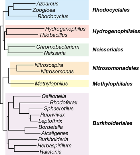
- Figure 16.2 Phylogenetic tree and metabolic links of some key genera of *Proteobacteria*.
- 16.1 Alphaproteobacteria
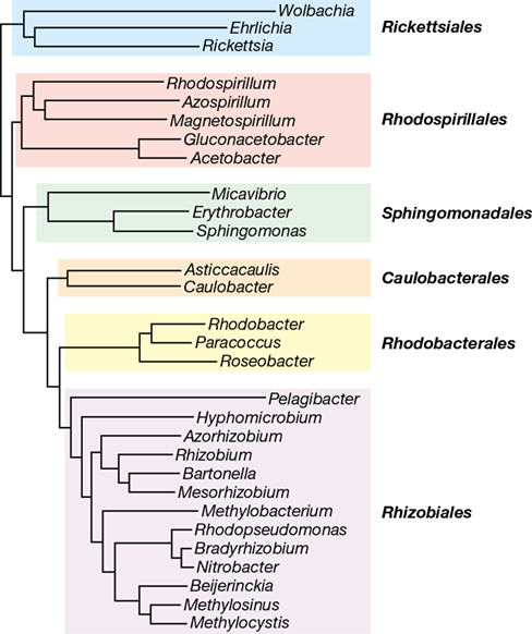
- Figure 16.3 Major orders of *Proteobacteria* in the class *Alphaproteobacteria*.
- Table 16.1 Notable genera of *Alphaproteobacteria*
- Key Genera: *Bartonella, Methylobacterium, Pelagibacter, Rhizobium, Agrobacterium*
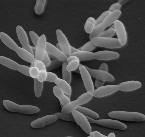
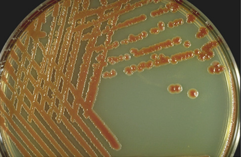
- Figure 16.4 Colonies of *Rhizobium mongolense*.
- Figure 16.5 Rickettsias growing within host cells.
- Figure 16.6 *Wolbachia*.
Ch 17: Diversity of ArchaeaRead full chapter →
Sections in this chapter
- 17 Diversity of Archaea
- Methanogens and Global Climate Change
- Figure 17.1 Schematic representation of the phylogeny of the major taxonomic orders within the domain *Archaea*.
- I *Euryarchaeota*
- Key Genera: *Halobacterium, Haloferax, Natronobacterium*
- Figure 17.2 Hypersaline habitats for halophilic *Archaea*.
- Hypersaline Environments: Chemistry and Productivity
- Taxonomy and Physiology of Extremely Halophilic *Archaea*
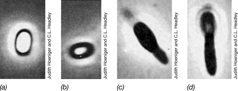
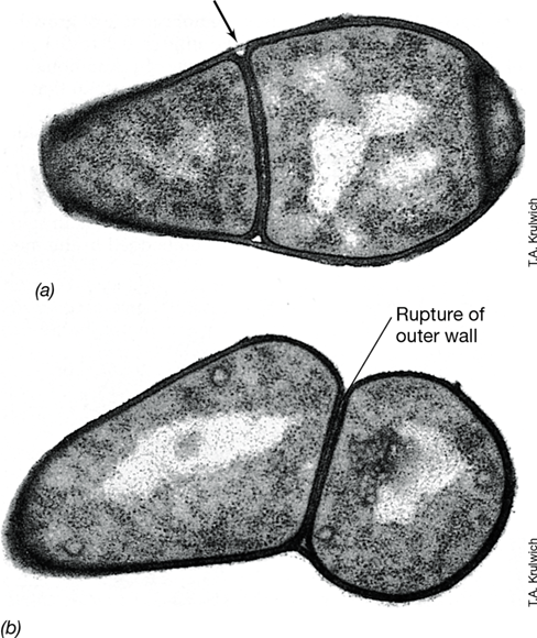
- Figure 17.3 Electron micrographs of thin sections of the extreme halophile *Halobacterium salinarum*.
- Water Balance in Extreme Halophiles
- Table 17.1 Concentration of ions in cells of *Halobacterium salinarum*a
- Mastering Microbiology

Ch 18: Diversity of Microbial EukaryaRead full chapter →
Sections in this chapter
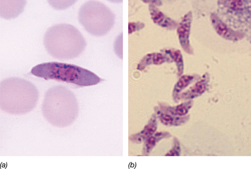
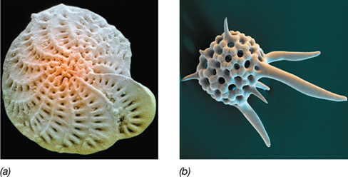
- 18 Diversity of Microbial Eukarya
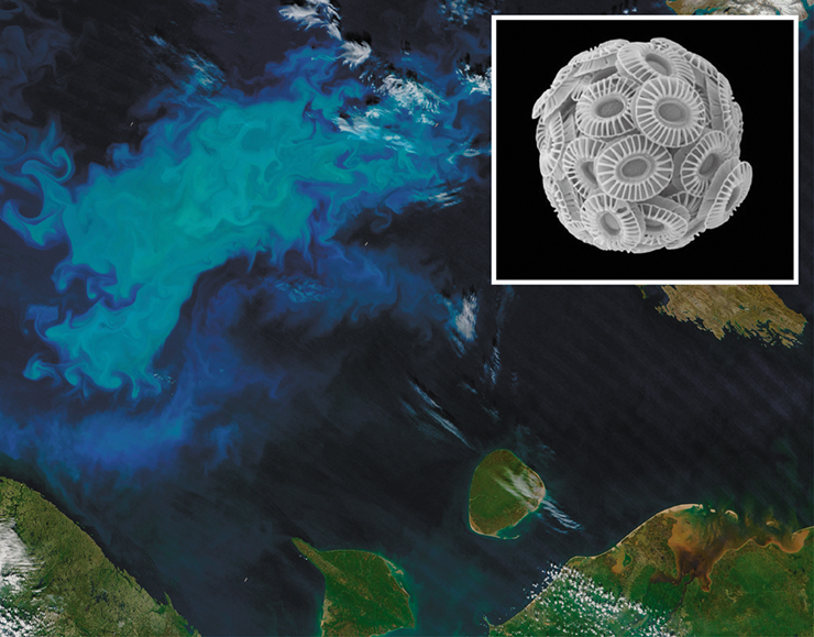
- Coccolithophores, Engineers of Global Climate
- I Organelles and Phylogeny of Microbial *Eukarya*
- 18.1 Endosymbioses and the Eukaryotic Cell
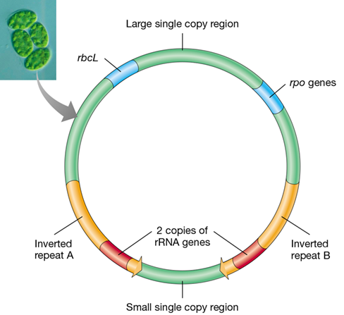
- Figure 18.1 Organellar DNA.
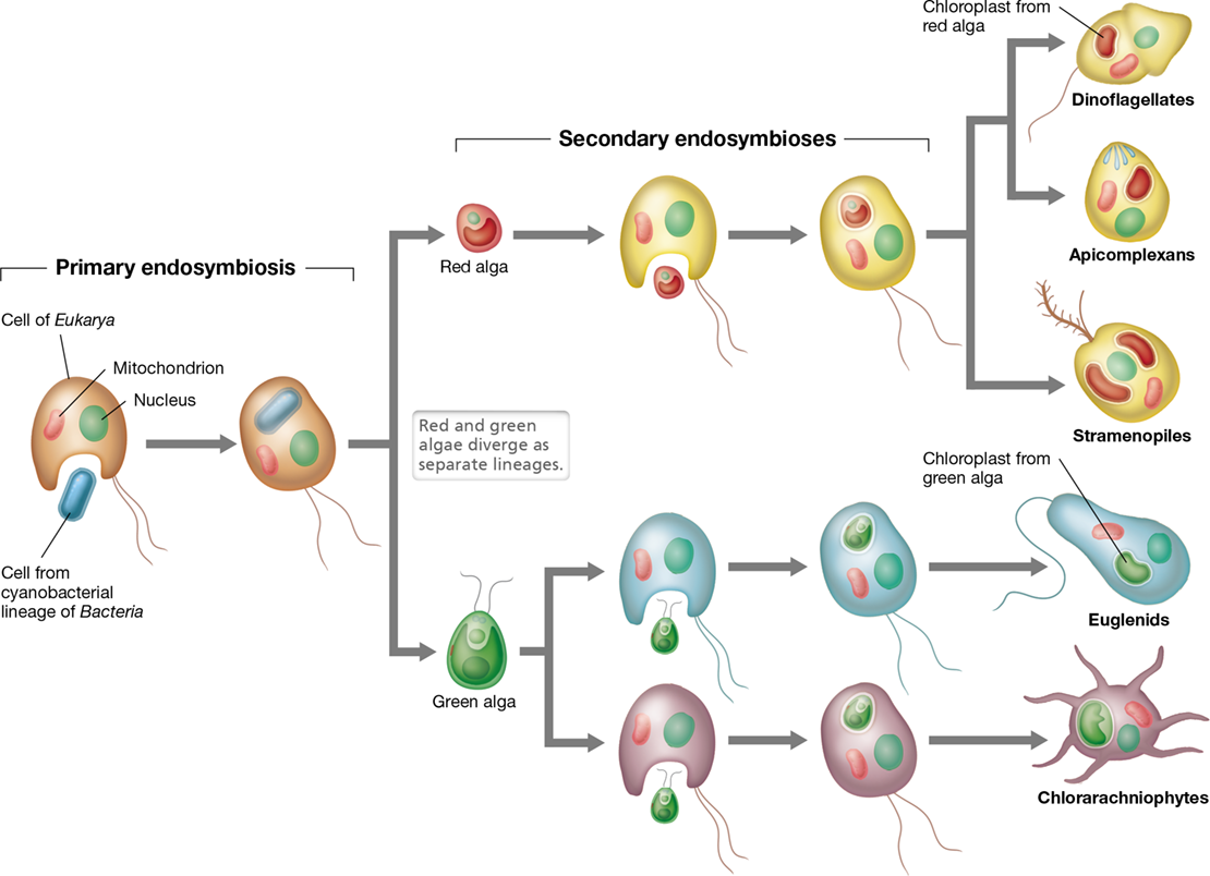
- Primary Endosymbioses
- Figure 18.2 Endosymbioses.
- Secondary Endosymbioses
- Check Your Understanding
- 18.2 Phylogenetic Lineages of *Eukarya*
- Eukaryotic Evolution: The Big Picture
- Figure 18.3 Phylogenetic tree of *Eukarya*.
Ch 19: Taking the Measure of Microbial SystemsRead full chapter →
Sections in this chapter
- 19 Taking the Measure of Microbial Systems
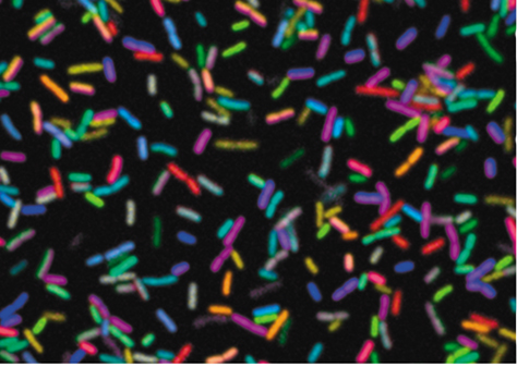
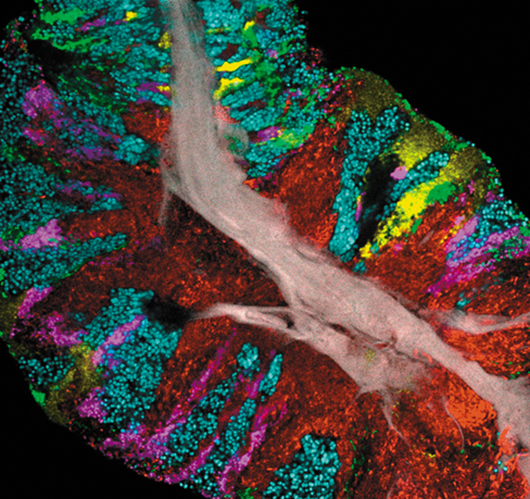
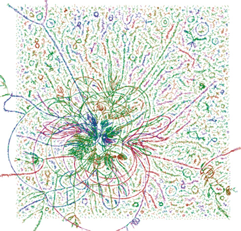
- Touring Microbial Biogeography Using Combinatorial Imaging
- I: Culture-Dependent Analyses of Microbial Communities
- I Culture-Dependent Analyses of Microbial Communities
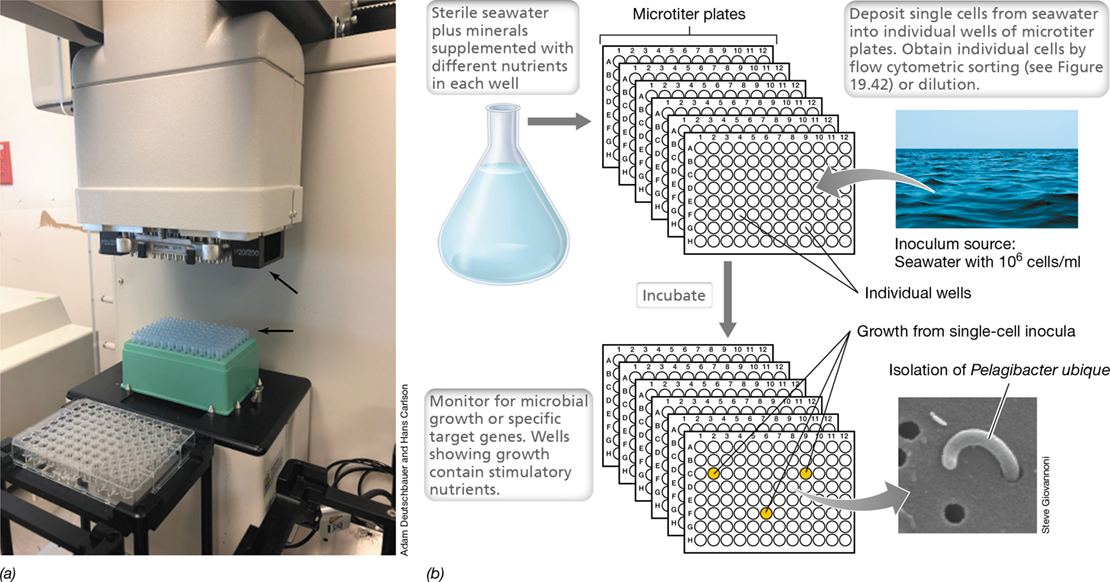
- 19.1 Enrichment Culture Microbiology
- Table 19.1 Some enrichment culture methods for phototrophic bacteria (main C source, CO2)
- Table 19.2 Some enrichment culture methods for nonphototrophic bacteriaa
- Inocula
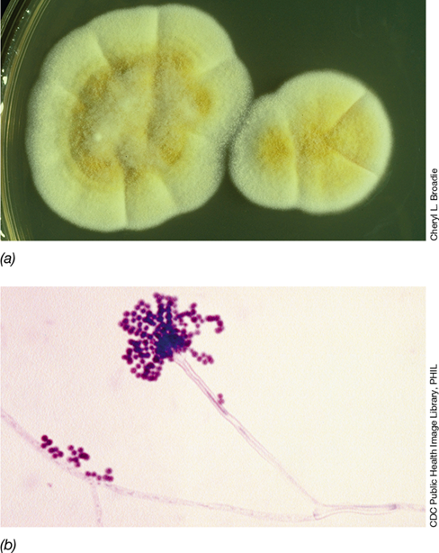
- Figure 19.1 The isolation of *Azotobacter*.
- Enrichment Culture Outcomes
- Mastering Microbiology
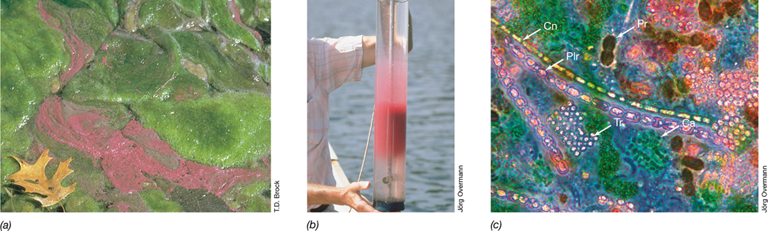
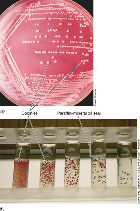
- The Winogradsky Column
Ch 20: Microbial EcosystemsRead full chapter →
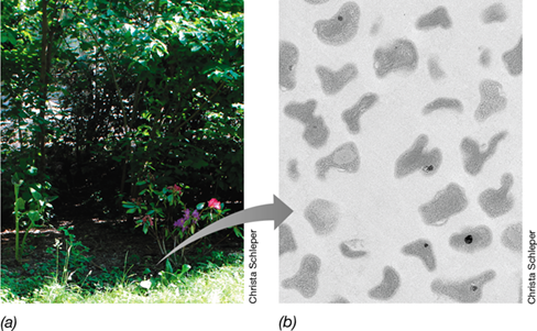
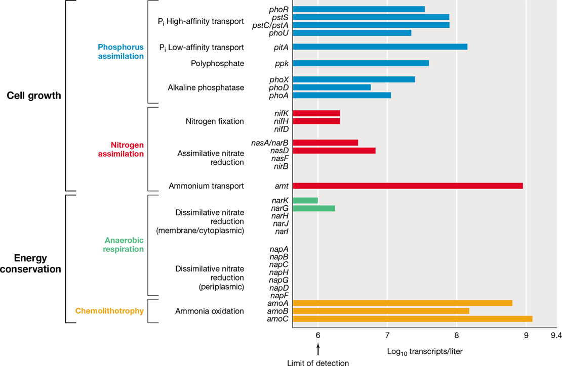
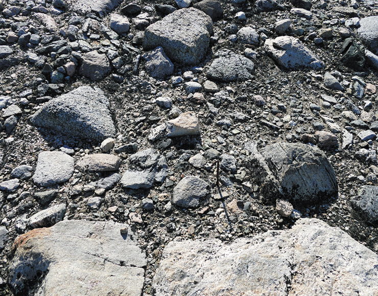
Antarctica is one of the harshest environments on Earth, experiencing extremely cold temperatures, high levels of ultraviolet radiation, and limited availability of carbon, nitrogen, and water. Even so, rocky desert Antarctic soils (photo) support microbial communities whose diversity rivals that of temperate soils. Cyanobacteria fix enough carbon dioxide (CO2) to support heterotrophic communities in many soils of Antarctica. However, the virtual absence of cyanobacteria in some other Antarctic soils raises the question of what energy source (other than light) sustains these microbial communit…
Researchers combined metagenomics with biochemical measurements to probe this mystery. The first hint was the discovery of Calvin cycle genes for CO2 fixation in Actinobacteria that composed about half of the total soil microbiota (the Calvin cycle is a major autotrophic pathway). Since these Actinobacteria are not phototrophic, CO2 fixation—which requires ATP—had to be supported by energy sources other than sunlight. The answer again emerged from metagenomics by identifying genes in the Actinobacteria encoding aerobic respiration of molecular hydrogen (H2) and carbon monoxide (CO) and t…
Sections in this chapter
- Living on Fumes
- I Microbial Ecology
- 20.1 General Ecological Concepts
- Ecosystems and Habitats
- Species Diversity in Microbial Habitats
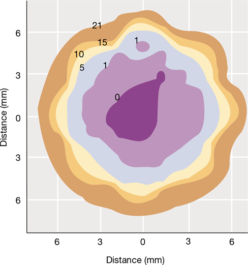
- Figure 20.1 Microbial species diversity: Richness versus abundance.
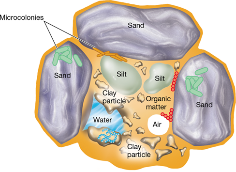
- Table 20.1 Resources and conditions that govern microbial growth in nature
- Check Your Understanding
- 20.2 Ecosystem Service: Biogeochemistry and Nutrient Cycles
- Figure 20.2 Populations, guilds, and communities.
- Energy Inputs to the Ecosystem
- Biogeochemical Cycling

Ch 21: Nutrient CyclesRead full chapter →
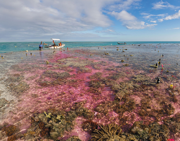
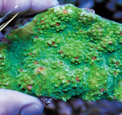
Carbon dioxide (CO2) released from the burning of fossil fuels is contributing to global warming and ocean acidification. Although all ecosystems will ultimately be impacted by this, coral reef ecosystems are particularly sensitive. About a quarter of the oceans’ coral reefs have already been lost to thermal stress and pollution, and ongoing acidification is impairing their formation of calcium carbonate skeletons. Although occupying less than 1% of the ocean floor, coral reefs support about one-quarter of all marine species that provide food for millions of people. Thus it is essential that w…
In an experimental study of adding CO2 to ocean waters bathing a natural coral reef ecosystem, it was clear that even a minor reduction in pH greatly reduces the rate of biogenic calcification. This study, performed at a section of the Great Barrier Reef (Australia), used tidal flow from an upper to a lower lagoon to adjust the pH of water bathing the lower section of reef. A 4000-gallon floating tank of seawater was first acidified by bubbling with CO2 and then pumped from the edge of the upper lagoon during tidal change, reducing the pH in the lower lagoon from ambient pH (8.13) to a treatme…
Sections in this chapter
- An Uncertain Future for Coral Reef Ecosystems
- I Carbon, Nitrogen, and Sulfur Cycles
- 21.1 The Carbon Cycle
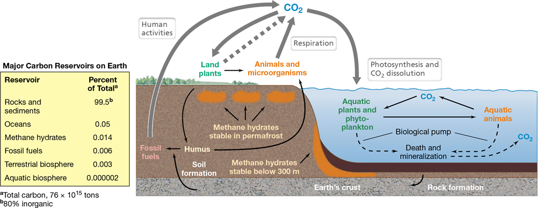
- Figure 21.1 The carbon cycle.
- Carbon Reservoirs
- Photosynthesis and Decomposition
- Figure 21.2 Redox cycle for carbon.
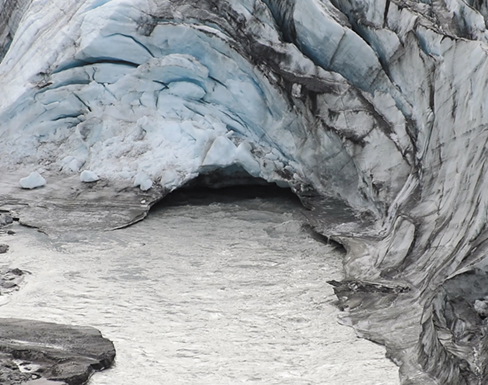
- Methane Hydrates
- Figure 21.3 Burning methane hydrate.
- Mastering Microbiology
- Figure 21.4 Seasonal flares of methane bubbling from methane hydrates.
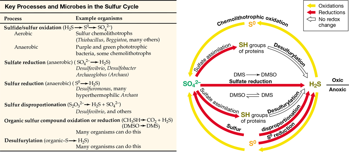
- Figure 21.5 Sulfur-oxidizing microbial mats overlying a deep-sea methane seep.


Ch 22: Microbiology of the Built EnvironmentRead full chapter →
Sections in this chapter
- 22 Microbiology of the Built Environment
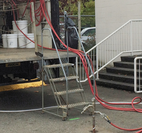
- Sending Microbes to Clean Up after Polluters
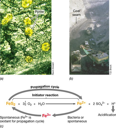
- I Mineral Recovery and Acid Mine Drainage
- 22.1 Mining with Microorganisms
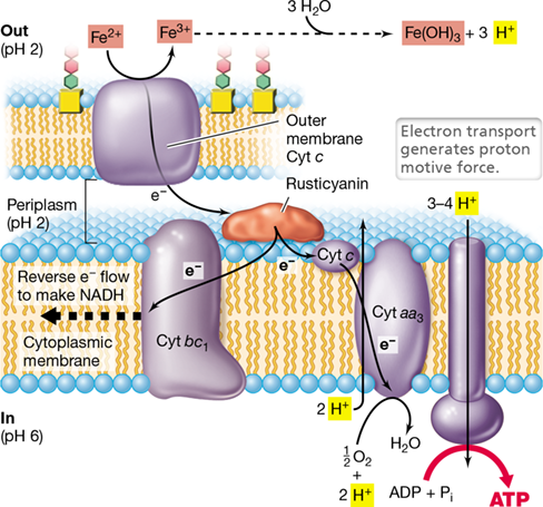
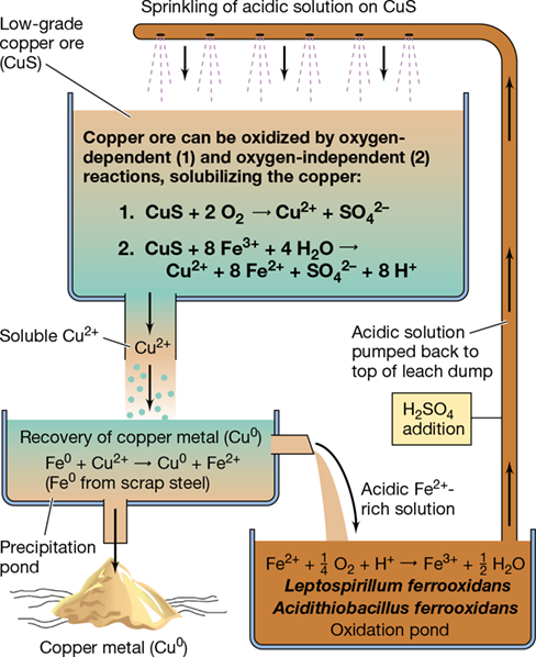
- Figure 22.1 The leaching of low-grade copper ores using iron-oxidizing bacteria.
- The Leaching Process
- Figure 22.2 Arrangement of a leaching pile and reactions in the microbial leaching of copper sulfide minerals to yield metallic copper.
- Mastering Microbiology
- Metal Recovery
- Other Microbial Leaching Processes: Uranium and Gold
- Figure 22.3 Gold bioleaching.
- Check Your Understanding
Ch 23: Microbial Symbioses with Microbes, Plants, and AnimalsRead full chapter →
Sections in this chapter
- 23 Microbial Symbioses with Microbes, Plants, and Animals
- Coral Fluorescence Provides the Guiding Light for Their Symbiotic Algae
- I Symbioses Between Microorganisms
- 23.1 Lichens
- Figure 23.1 Lichens.
- Figure 23.2 Lichen structure.
- Lichen Components
- Molecular Studies of Lichens
- Check Your Understanding
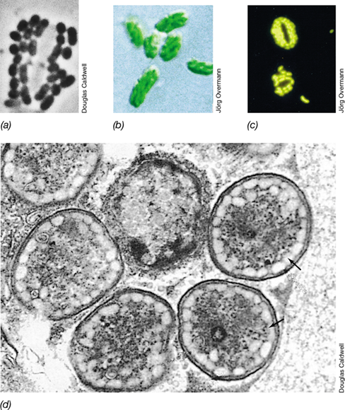
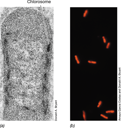
- 23.2 “Chlorochromatium aggregatum”
- Nature of the Consortium
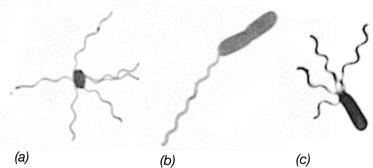
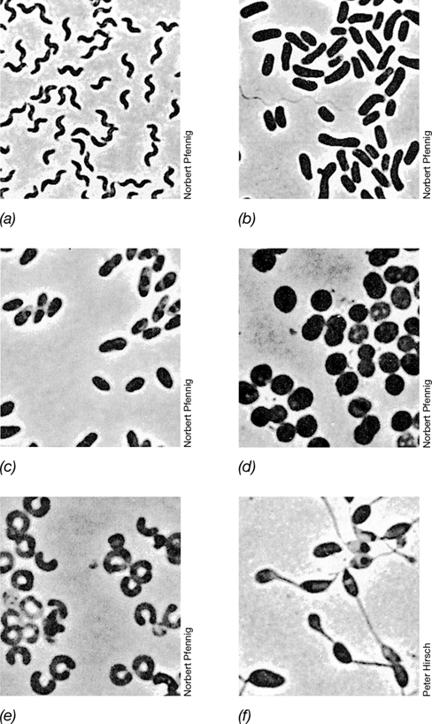
- Figure 23.3 Drawings of some motile phototrophic green bacterial consortia found in freshwater lakes.
Ch 24: Microbial Symbioses with HumansRead full chapter →
Sections in this chapter
- 24 Microbial Symbioses with Humans
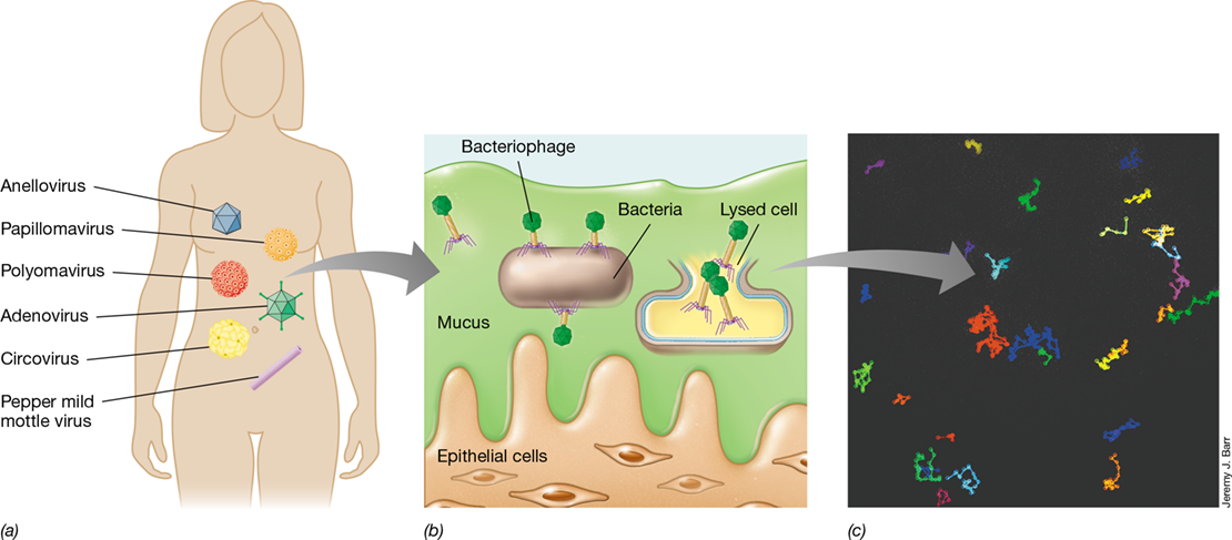
- One of the Most Abundant Viruses on Earth Discovered First in the Human Viral Microbiome
- I: Structure and Function of the Healthy Adult Gastrointestinal and Oral Microbiomes
- I Structure and Function of the Healthy Adult Gastrointestinal and Oral Microbiomes
- 24.1 Overview of the Human Microbiome
- Figure 24.1 Microbial habitats of the human body.
- Table 24.1 Major human microbiome research programs
- Methodologies for Probing the Human Microbiome
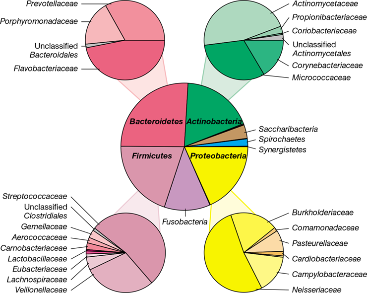
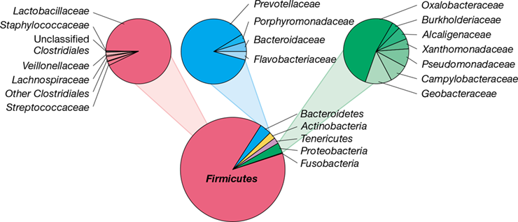
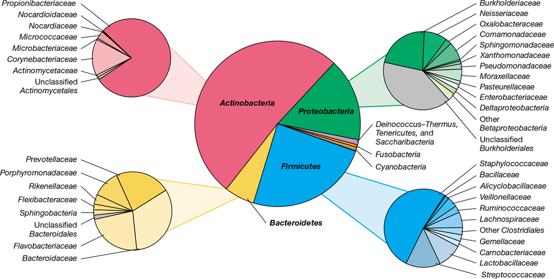
- A Snapshot of the Human Microbiome
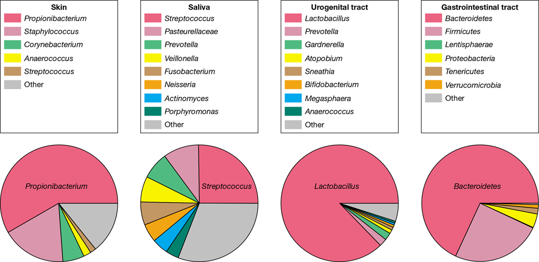
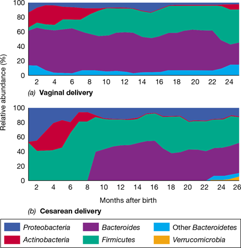
- Figure 24.2 Overview of major microbial populations in the body sites sampled by human microbiome projects.
- Check Your Understanding
- 24.2 Gastrointestinal Microbiota
Ch 25: Microbial Infection and PathogenesisRead full chapter →
Sections in this chapter
- 25 Microbial Infection and Pathogenesis
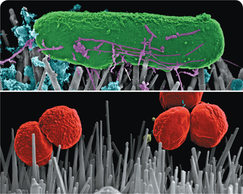
- Killing Pathogens on Contact
- I Human–Pathogen Interactions
- 25.1 Microbial Adherence
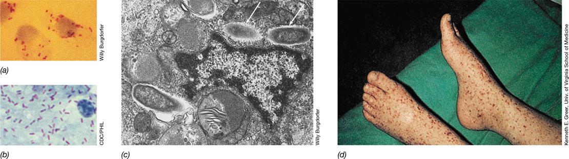
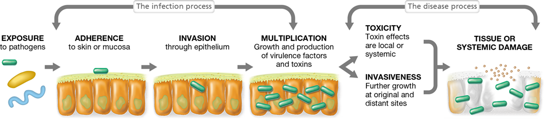
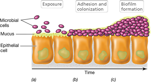
- Figure 25.1 Microbial pathogenesis.
- Adherence Molecules
- Mastering Microbiology
- Figure 25.2 Bacterial adherence.
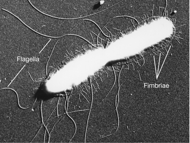
- Adherence Structures: Capsules, Fimbriae, Pili, and Flagella
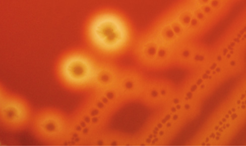
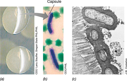
- Figure 25.3 The bacterial capsule as a facilitator of pathogen attachment.
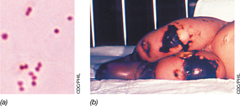
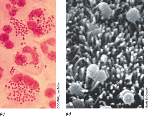
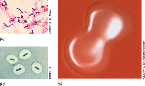
- Figure 25.4 Capsules and colonies of *Streptococcus pneumoniae*.
- Figure 25.5 Fimbriae.
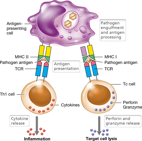
Ch 26: Innate Immunity: Broadly Specific Host DefensesRead full chapter →
Sections in this chapter
- 26 Innate Immunity: Broadly Specific Host Defenses
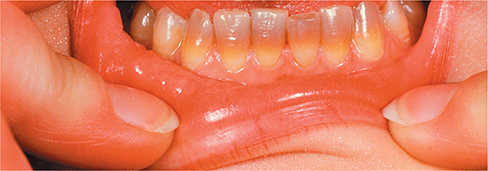
- Periodontal Disease and Alzheimer’s: Evidence for Causation?
- I Fundamentals of Host Defense
- 26.1 Basic Properties of the Immune System
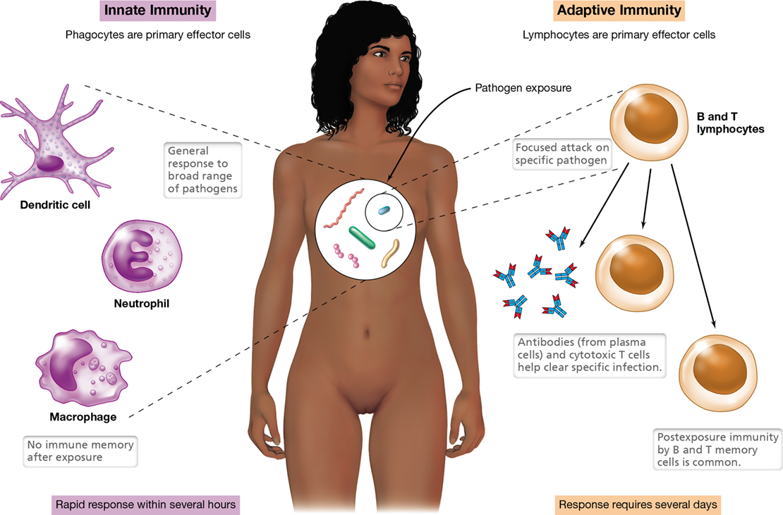
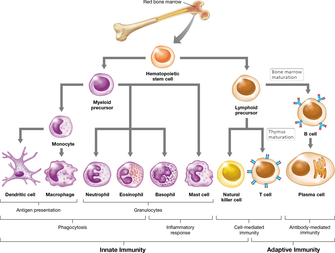
- Figure 26.1 Overview of the two-pronged immune response.
- Mastering Microbiology
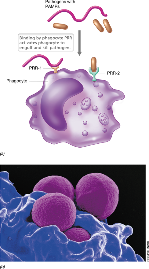
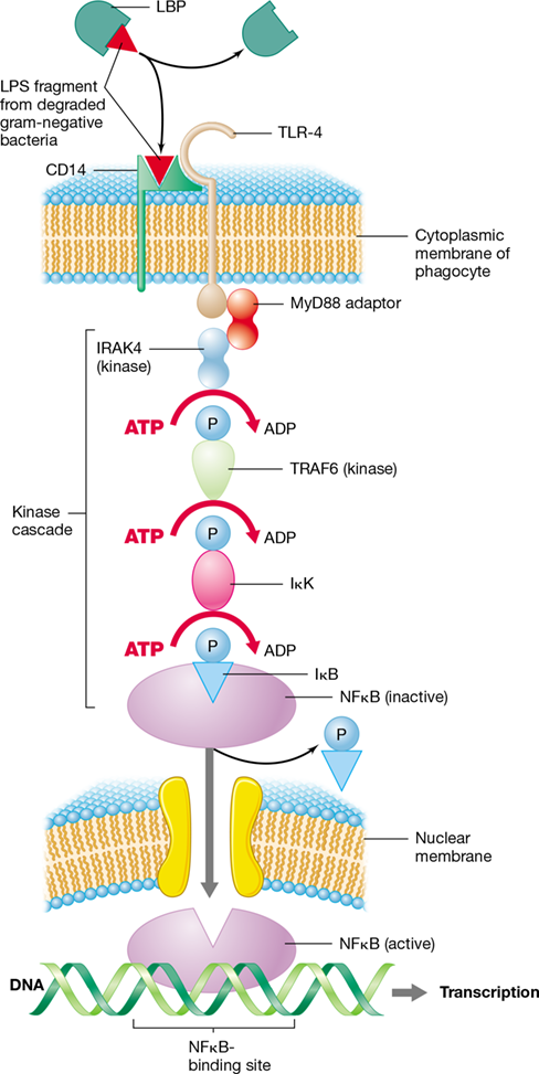
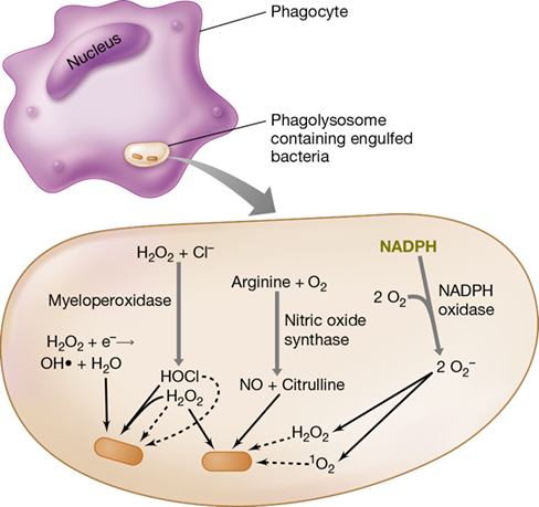
- Principles of Innate Immunity
- Principles of Adaptive Immunity
- Check Your Understanding
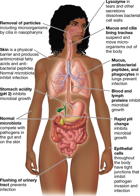
- 26.2 Barriers to Pathogen Invasion
- Natural Host Resistance
- Figure 26.2 Barriers to infection in the human body.

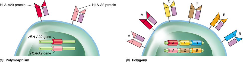
Ch 27: Adaptive Immunity: Highly Specific Host DefensesRead full chapter →
Sections in this chapter
- 27 Adaptive Immunity: Highly Specific Host Defenses
- Controlling HIV through “Public” T Cell Receptors on CD4 T Cells
- I Principles of Adaptive Immunity
- 27.1 Specificity, Memory, Selection Processes, and Tolerance
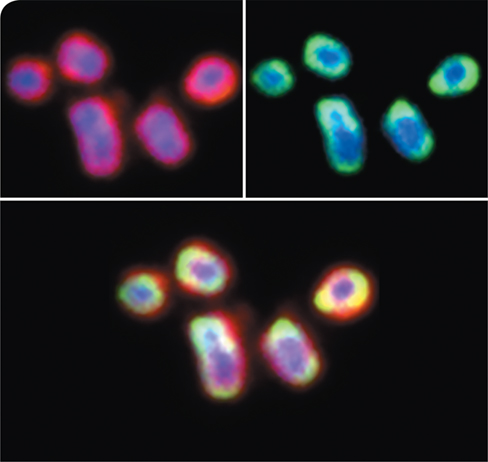
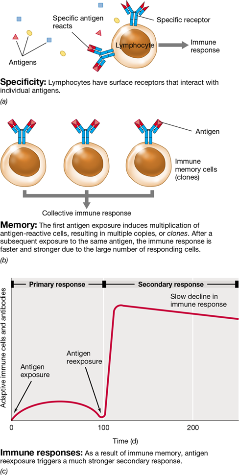
- Immune Specificity and Memory
- Figure 27.1 Specificity and memory in the adaptive immune response.
- T Cell Selection and Tolerance
- Figure 27.2 T cell selection and clonal deletion.
- Mastering Microbiology
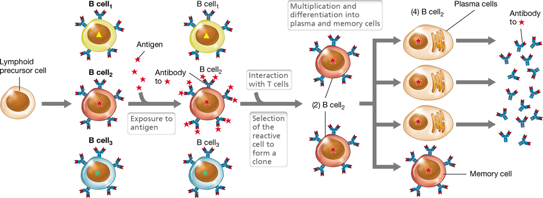
- B Cell Selection and Tolerance
- Figure 27.3 B cell clonal selection and expansion.
- Check Your Understanding

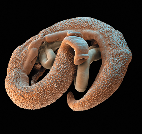
Ch 28: Immune Disorders and Antimicrobial TherapyRead full chapter →
Sections in this chapter
- 28 Immune Disorders and Antimicrobial Therapy
- Preventing Autoimmunity with . . . Parasitic Worms?
- Mastering Microbiology
- I: Disorders and Deficiencies of the Immune System
- I Disorders and Deficiencies of the Immune System
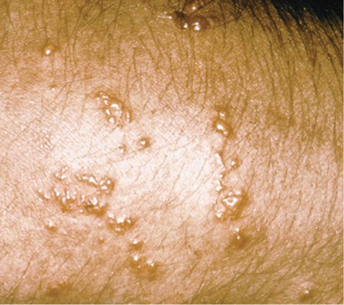
- 28.1 Allergy, Hypersensitivity, and Autoimmunity
- Table 28.1 Hypersensitivitya
- Immediate Hypersensitivity
- Figure 28.1 Immediate hypersensitivity.
- Figure 28.2 Hives due to immediate hypersensitivity.
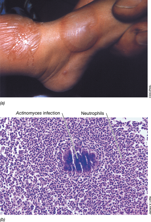
- Delayed-Type Hypersensitivity
- Figure 28.3 Delayed-type hypersensitivity.


Ch 29: Diagnosing Infectious DiseasesRead full chapter →
Sections in this chapter
- 29 Diagnosing Infectious Diseases
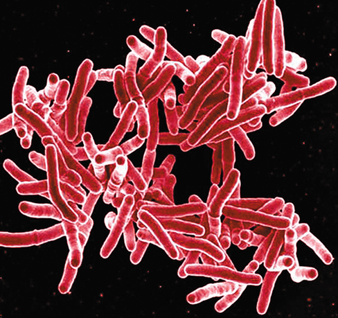
- Shedding New Light on Diagnosing Tuberculosis
- I Microbiology and the Healthcare Environment
- 29.1 The Clinical Microbiology Laboratory
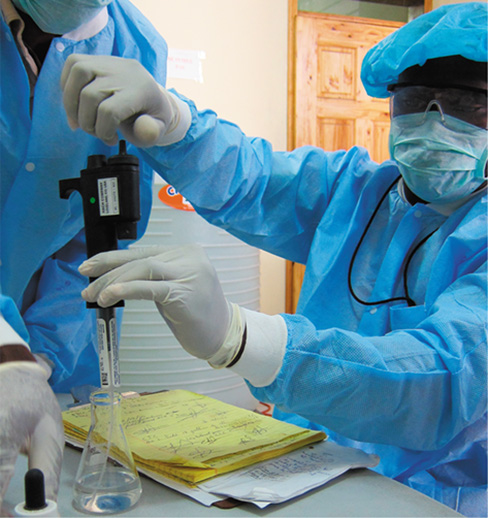
- Laboratory Safety
- Figure 29.1 Standard apparel for clinical laboratory safety.
- Mastering Microbiology
- Table 29.1 Microbiology laboratory safety standards
- Biological Containment and Biosafety Levels
- Figure 29.2 Conducting research in a BSL-4 (biosafety level 4) laboratory.
- Check Your Understanding
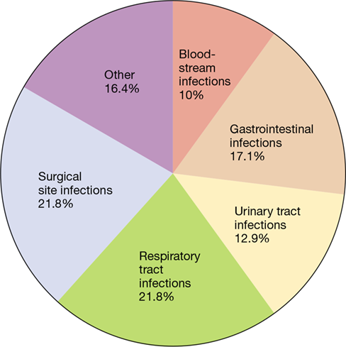
- 29.2 Healthcare-Associated Infections
Ch 30: Epidemiology and Public HealthRead full chapter →
Sections in this chapter
- 30 Epidemiology and Public Health
- A New Urgent Threat Is Emerging in Public Health Microbiology
- I Principles of Epidemiology
- 30.1 The Language of Epidemiology
- Mastering Microbiology
- Disease Incidence and Prevalence
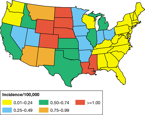
- Figure 30.1 The concepts of disease incidence and disease prevalence.
- The Scope of Disease
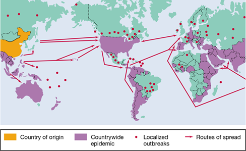
- Figure 30.2 Endemic, epidemic, and pandemic disease.
- Stages of Disease
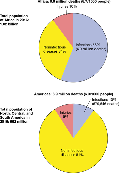
- Mortality, Morbidity, and DALY
- Table 30.1 Worldwide deaths due to infectious diseasesa