Chapter introduction
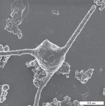
Microbes grow everywhere, from the deepest ocean to the human body. Our own bodies contain as many microbes as human cells, including 100 trillion bacteria in the digestive tract. Throughout history, humans have had a hidden partnership with microbes that ranges from food production to mineral mining. Microscopes first revealed the tiny organisms at work in our bodies and in our environment. In the twentieth century, microbial genetics led to recombinant DNA and sequenced genomes. Today, we discover the microbial foundations of our global ecology and climate. What are these microbes? From childhood, we hear that we are surrounded by microscopic organisms, or “germs,” which we cannot see. In 2019, a novel virus emerged and caused a disease, COVID-19, that upended human society all around the globe. The disease-causing microorganism, a coronavirus known as SARS-CoV-2, changed the course of history.
Earlier, in the 1980s, a very different microbe—the thermophilic bacterium Thermus aquaticus —provided the first heat-stable DNA polymerase of the polymerase chain reaction (PCR). Today PCR makes our most accurate test for infection with the coronavirus, providing our best defense against it. Overall, PCR has changed human history by reading genomes and transforming forensic detection.
In Chapter 1 we introduce the concept of a microbe and survey the history of microbial discovery. We explain how to show which pathogen causes a disease. Finally, we address the exciting century of molecular microbiology, in which microbial genetics, genomics, and evolution have transformed the practice of medicine and our understanding of the natural world.
1.1 From Germ to Genome: What Is a Microbe?not assigned
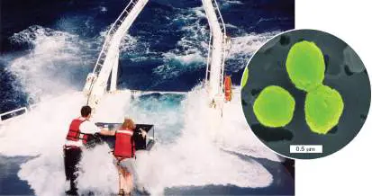
Life began early in the history of planet Earth in the form of microscopic organisms, or “microbes.” Over the eons, those microbes evolved to shape our atmosphere, our geology, and the energy cycles of all ecosystems. For the first 2 billion years, all life was microbial. Then, as now, a vast realm of that microbial diversity resided in Earth’s oceans. Oceans constitute the largest biome on Earth’s surface, a community where the biomass is dominated by microbes ( Fig. 1.1). The waves pounding the research vessel in the photograph contain trillions of photosynthetic Prochlorococcus bacteria, tiny cells that make much of the oxygen gas we breathe. FIGURE 1.1 ■ Marine microbiologists sample bacteria such as the phototroph Prochlorococcus. Inset: Colorized scanning electron micrograph of Prochlorococcus.
ERIK ZINSER (AUTHOR)
ANNE THOMPSON, CHISHOLM LAB, MIT
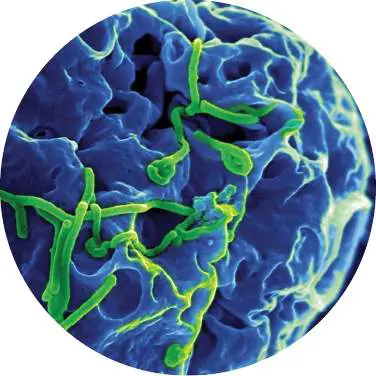
Yet, before we devised microscopes in the seventeenth century, we humans were unaware of the unseen living organisms that surround us, that float in our air and water, and that inhabit our own bodies. Microbes fix nitrogen for plants, and they make vitamins, such as vitamin B 12. In the ocean, microbes produce biomass for the food web that feeds the fish we eat, and microbes consume toxic wastes such as the oil from the Deepwater Horizon spill in the Gulf of Mexico in 2010. At the same time, pathogens (agents of disease) such as SARS-CoV-2 take our lives—and researchers risk their lives to study them. Working with deadly pathogens requires containment at biosafety level 3 or 4 (BSL-3 or BSL-4). The highest containment level, BSL-4 requires sealed suits and respiratory equipment (Fig. 1.2). BSL-4 is required, for example, to study Ebola virus infection of cells in tissue culture. Despite all our advances in medicine and public health, humans continue to die of microbial diseases.
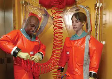
FIGURE 1.2 ■ Researching deadly pathogens. Biosafety level 4 containment is required to study the most dangerous pathogens. Inset: Ebola virus (green) budding from monkey cells in tissue culture; requires BSL-4 containment.
H. S. PHOTOS/ALAMY STOCK PHOTO
SCIENCE SOURCE/SCIENCE SOURCE
Today, microbes provide new tools that affect human society. For example, the use of heat-stable bacterial DNA polymerase (a DNA-replicating enzyme) in a technique called the polymerase chain reaction (PCR ; see eAppendix 3) enables us to detect minute amounts of DNA in traces of blood or fossilized bone. Microbial technologies led us from the discovery of the double helix to the sequence of the human genome, the total genetic information that defines our species.
A Microbe Is a Microscopic Organism
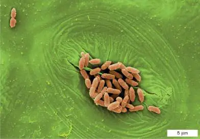
A microbe is commonly defined as a living organism that requires a microscope to be seen. All around us, microbes exhibit diverse forms and lifestyles (Fig. 1.3). Rod-shaped bacteria such as Escherichia coli are among the 100 trillion inhabitants of our intestines, where they help digest our food. Alternatively, E. coli may colonize the plants we eat (Fig. 1.3A). Microbial eukaryotes (cells with nuclei) such as the voracious Stentor engulf aquatic prey (Fig. 1.3B ). Archaea are a life form distinct from both bacteria and eukaryotes. Some archaea grow in extreme environments, such as concentrated salt (Fig. 1.3C ). And all kinds of life host viruses. For example, herpes simplex virus infects human cells (Fig. 1.3D ). Microbial cells range in size from 5 millimeters (mm) down to less than 0.2 micrometer (μm), and viruses may be tenfold smaller (Table 1.1). A. Bacteria: E. coli B. Eukaryote: Stentor
C. Archaea: Halophiles
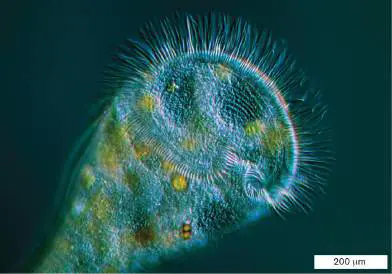
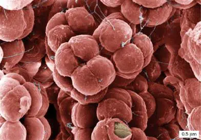
D. Virus: Herpes simplex virus FIGURE 1.3 ■ Representative kinds of microbes. A. Bacterium: E. coli on leaf stomate. B. Eukaryote: Stentor, a predatory protist. C. Archaeon: Halococcus. D. Virus: Herpes simplex virus (with envelope removed). Colorized.
SCIMAT/SCIENCE SOURCE
BLICKWINKEL, ALAMY STOCK PHOTO
EYE OF SCIENCE/SCIENCE SOURCE
LOUISE HUGHES/SCIENCE SOURCE
TABLE 1.1 Sizes of Some Microbes Microbe Description Approximate size
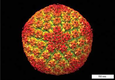
TABLE 1.1 Sizes of Some Microbes Varicella-zoster Virus that causes 100 nanometers virus 1 chickenpox and (nm) = 10 −7 shingles meter (m)
Prochlorococcus Photosynthetic 500 nm = 5 × 10 −7 marine bacterium m Escherichia coli Bacterium growing 2 micrometers (μm)
within human = 2 × 10 −6 m intestine Spirogyra Aquatic alga that 5 cm = 0.05 m forms long (chain of cells)
filaments of cells Pelomyxa Ameba (a protist) 5 millimeters (mm)
that consumes = 5 × 10 −3 m bacteria in soil or water Some microbes consist of a single cell, the smallest unit of life, a membrane-enclosed compartment of water solution containing molecules that carry out metabolism. Each microbe contains a genome used to reproduce its own kind. Microbial cells acquire food, gain energy to build themselves, and respond to environmental change. Microbes evolve at rapid rates—often fast enough to see evolution occur in the laboratory (discussed in Chapter 17). Much of Earth’s microbial inventory remains a mystery. Barely 0.1% of the microbes in our biosphere can be cultured in the laboratory; even the digestive tract of a newborn infant contains species of bacteria unknown to science.
The more diverse kinds of microbes we discover, the more contradictions we find with our definitions of a microbe. For example: Super-size microbial cells. Most single-celled organisms require a microscope to render them visible, and thus they fit the definition of a microbe. Nevertheless, some bacteria grow to sizes large enough to see with the unaided eye. The marine sulfur bacterium Thiomargarita namibiensis, called the “sulfur pearl of Namibia,” grows to a thousand times larger than Prochlorococcus (Fig. 1.4A). The related bacterium Thiomargarita magnifica grows a thread-like cell nearly a centimeter long. Even more surprising, a single cell of the “killer alga” Caulerpa taxifolia grows feather-like fronds from stalks up to several meters long (Fig. 1.4B ). This alga forms multiple nuclei and extends without cell division. Caulerpa now covers many acres beneath the coastal waters of California.
Microbial populations and communities. Many microbes form complex multicellular assemblages such as biofilms and fruiting bodies. Within a microbial population, or a community of multiple species, microbial cells may be differentiated into distinct types that complement each other’s functions, as in multicellular organisms. Yet, multicellular animals such as mites and tardigrades require a microscope for us to see but are not considered microbes.
Viruses. A virus is a noncellular particle containing genetic material that takes over the metabolism of a cell to generate more virus particles (see Chapter 6). Some viruses consist of only a short chromosome packed in protein. Other kinds of viruses, such as pandoraviruses that infect amebas, have the size and complexity of a cell. Although viruses are not fully functional cells, some viral genomes may have evolved from cells.
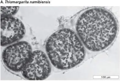
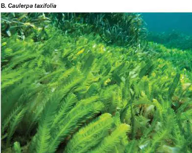
FIGURE 1.4 ■ A giant microbial cell. A. Thiomargarita namibiensis, a marine sulfur bacterium (light microscopy). B. Caulerpa taxifolia, an invasive alga that consists of a single multinucleated cell.
H. N. SCHULZ ET AL., 1999. SCIENCE 284 :493–495
O.DIGOIT/ALAMY STOCK PHOTO
Note: Each section of text contains Thought Questions that may
have various answers. Possible responses are provided in the ebook and at the back of the printed book.
Thought Questions
1.1 The minimum size of most microbial cells is about 0.2 μm. How can even smaller cells be discovered? What factors may determine the minimum size of a cell?
1.2 If viruses are not functional cells, are they “alive”?
In practice, our definition of a microbe derives from tradition as well as genetic considerations. In this book we consider microbes to include prokaryotes (cells lacking a nucleus, including bacteria and archaea), as well as certain classes of eukaryotes (cells with a nucleus), such as algae, fungi, and protozoa (discussed in Chapter 20 ). The bacteria, archaea, and eukaryotes—known as the three “domains”—evolved from a common ancestral cell (see Section 1.5 and Chapter 21). Viruses and even smaller infectious particles are discussed in Chapters 6 and 11.
Note: The formal names of the three domains are Bacteria,
Archaea, and Eukarya. Members of these domains are called bacteria (singular, bacterium), archaea (singular, archaeon), and eukaryotes (singular, eukaryote), respectively. The microbiology literature includes alternative spellings for some of these terms, such as “archaean” and “eucaryote.”
Microbial Genomes Are Sequenced
Our modern concept of a microbe has deepened through the use of two major research tools: advanced microscopy (see Chapter 2) and the sequencing of genomes (see Chapter 7). A genome is the total genetic information contained in an organism’s chromosomal DNA. The genes in a microbe’s genome and the sequence of DNA tell us a lot about how that microbe grows and associates with other species. For example, if a microbe’s genome includes genes for nitrogenase, a nitrogen-fixing enzyme, that microbe probably can fix nitrogen from the atmosphere into proteins—its own proteins and those of associated plants. And by comparing DNA sequences of different microbes, we can figure out how closely related they are and how they evolved.
The first method of DNA sequencing that was fast enough to sequence large genomes was developed by Fred Sanger (1918–2013) at the University of Cambridge (Fig. 1.5). This achievement—which jump-started the study of molecular biology—earned Sanger the 1980 Nobel Prize in Chemistry, together with Walter Gilbert and Paul Berg. Sanger and his colleagues used the new method to sequence DNA containing tens of thousands of base pairs, such as the DNA of the human mitochondrion (plural, mitochondria).
FIGURE 1.5 ■ Fred Sanger devised the method of DNA sequence analysis used to sequence the first genomes.
BETTMANN/GETTY IMAGES
But most genomes of cells contain millions or even billions of base pairs (complementary bases paired on two strands of DNA helix). In 1995, scientists completed the first genome sequence of a cellular microbe, the bacterium Haemophilus influenzae (Fig. 1.6A). H. influenzae causes several diseases including meningitis in children, a disease now prevented by the Hib vaccine. The H. influenzae genome has nearly 2 million base pairs, which specify about 1,700 genes. The sequence was determined by a large team of scientists led by Craig Venter, Hamilton Smith, and Claire Fraser (Fig. 1.6B ) at The Institute for Genomic Research (TIGR). The TIGR team devised a computational strategy for assembling large amounts of sequence data, which Fraser used to sequence many microbial genomes. This strategy was later used to sequence the human genome.
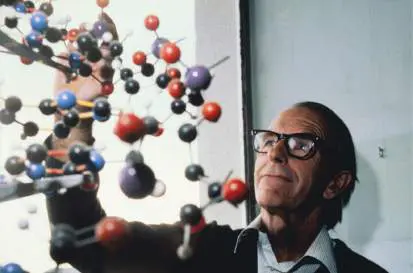
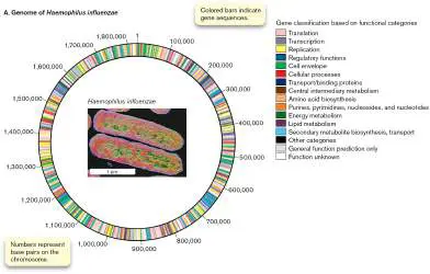
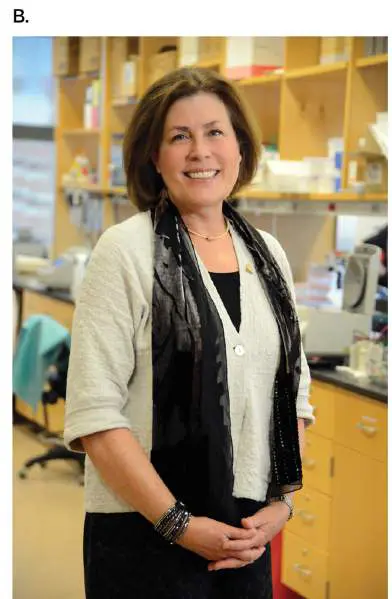
FIGURE 1.6 ■ Bacterial genomes were sequenced. A. The genome of Haemophilus influenzae, a bacterium that causes ear infections and meningitis, was the first DNA sequence completed for a cellular organism. Inset: Colorized electron micrograph of H. influenzae. B. Claire Fraser, past president of The Institute for Genomic Research (TIGR), sequenced numerous microbial genomes.
CNRI/SCIENCE SOURCE
INSTITUTE OF GENOME SCIENCE, UNIVERSITY OF MARYLAND SCHOOL OF MEDICINE
Today we sequence new bacterial genomes daily. In addition to sequencing individual genomes, computational strategies are used to sequence thousands of genomes of microbes sampled from a natural environment, such as the ocean or soil, or the intestinal tract of a cow. The collection of sequences taken directly from the environment is called a metagenome. Now, metagenomes are sequenced for microbial communities of medical interest, such as that of the human colon. Human gut microbes contain 100 times more genes in their metagenome (DNA of all microbes in a community) than the human genome contains—and many of these microbial genes contribute to our health!
Comparing genomes has revealed a set of core genes shared by all organisms. These core genes add further evidence that all living beings on Earth, including humans, share a common ancestry. Genomes are discussed further in Chapter 7, and the evolution of genomes and metagenomes is discussed in Chapters 17 and 21.
To Summarize
A microbe is a living organism that requires a microscope to be seen. Some organisms exist in both microscopic and macroscopic forms.
Microbes are found all around us, as part of human bodies and throughout our environment.
Major categories of microbes include bacteria, archaea, microbial eukaryotes, and viruses. Viruses are noncellular and require replication within a host cell.
Microbes may grow in communities such as a biofilm. A community with a shared habitat may include more than one species of organism, both microscopic and macroscopic. Microbial capabilities are defined by their genome sequences.
Glossary
pathogen A bacterial, viral, or fungal agent of disease.
polymerase chain reaction (PCR)
A method to amplify DNA in vitro using many cycles of DNA denaturation, primer annealing, and DNA polymerization with a heat-stable polymerase.
microbe An organism or virus too small to be seen with the unaided human eye.
virus A noncellular particle containing a genome that can replicate only inside a cell.
prokaryote An organism whose cell or cells lack a nucleus. Both bacteria and archaea are prokaryotes.
eukaryote An organism whose cells contain a nucleus. All eukaryotes are members of the domain Eukarya.
Bacteria One of the three domains of life, consisting of organisms with a last common ancestor not shared with members of Archaea or Eukarya. Organisms are prokaryotic (lacking nuclei, unlike eukaryotes) and possess primarily ester-linked phospholipid membranes (like eukaryotes, unlike archaea).
Archaea One of the three domains of life, consisting of organisms with a last common ancestor not shared with members of Bacteria or Eukarya. Organisms are prokaryotic (lacking nuclei, unlike eukaryotes) and possess ether-linked phospholipid membranes (unlike bacteria).
Eukarya One of the three domains of life, consisting of organisms with a last common ancestor not shared with members of Archaea or Bacteria. Cells possess nuclei, unlike cells of bacteria and archaea.
genome The complete genetic content of an organism. The sequence of all the nucleotides in a haploid set of chromosomes.
metagenome The sum of genomes of all members of a community of organisms.
1.2 Microbes Shape Human Historynot assigned
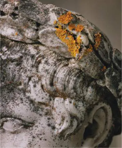
Today our knowledge of microbes is enormous, and it keeps growing. We know that microbes produce half the oxygen in our atmosphere, and that overall, microbial ecology is fundamental to our biosphere, mediating much of the human-caused change in global climate. Yet throughout most of human history, we were unaware of how microbes shaped our culture. Yeasts and bacteria made foods such as bread and cheese (Fig. 1.7A), as well as alcoholic beverages (discussed in Chapter 16). “Rock-eating” bacteria, known as “lithotrophs,” leached copper and other metals from ores exposed by mining, enabling ancient human miners to obtain these metals. The lithotrophic oxidation of minerals for energy generates strong acids, which accelerate breakdown of the ore. Today, about 20% of the world’s copper, as well as some uranium and zinc, is produced by bacterial leaching. Unfortunately, microbial acidification also consumes the stone of ancient monuments (Fig. 1.7B )—a process intensified by airborne acidic pollution.
A B
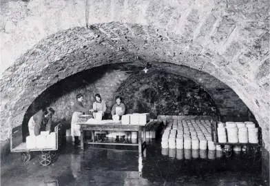
FIGURE 1.7 ■ Production and destruction by microbes. A. Roquefort cheeses ripening in France. B. Statue at the cathedral of Cologne, Germany, decaying from the action of lithotrophic microbes. The process is accelerated by acid rain.
BETTMANN/GETTY IMAGES
JOHNER IMAGES/GETTY IMAGES
How did people find out about microbes? Table 1.2lists the discoveries throughout history that have brought us to our current level of knowledge. Microscopists in the seventeenth and eighteenth centuries formulated key concepts about microbes and their existence, including their means of reproduction and death. In the nineteenth century, the “golden age” of microbiology, scientists established the fundamental principles of disease pathology and microbial ecology that are still in use today. This period laid the foundation for modern biology, in which genetics and molecular biology provide powerful tools for scientists to manipulate microorganisms for medicine, research, and industry.
TABLE Microbes and Human History 1.2
Date Microbial discovery Discoverer(s)
Microbes impact human culture without detection 10,000 Food and drink are produced Egyptians, BCE by microbial fermentation. Chinese, and others 1500 BCE Tuberculosis, polio, leprosy, Egyptians and smallpox are evident in mummies and tomb art.
50 BCE Copper is recovered from mine Roman metal water acidified by sulfur-workers under oxidizing bacteria. Julius Caesar 1362 CE Plague transmission is Ibn al-Khatib observed. (Granada)
TABLE Microbes and Human History 1.2
1546 CE Syphilis and other diseases Girolamo are seen to be contagious. Fracastoro (Padua)
Early microscopy and the origin of microbes 1676 Microbes are observed under a Antonie van microscope. Leeuwenhoek (Netherlands)
1688 Spontaneous generation is Francesco Redi disproved for maggots. (Italy)
1717 Smallpox is prevented by Turkish women inoculation of pox material, taught Lady a form of immunization. Mary Montagu, who brought the practice to England 1765 Microbe growth in organic Lazzaro material is prevented by Spallanzani boiling in a sealed flask. (Padua)
1798 Cowpox vaccination prevents Edward Jenner smallpox. (England)
1835 Fungus causes disease in Agostino Bassi silkworms (first pathogen to de Lodi (Italy) be demonstrated in animals).
TABLE Microbes and Human History 1.2
1847 Chlorine as antiseptic wash for Ignaz doctors’ hands decreases Semmelweis pathogens. (Hungary)
1881 Bacterial spores survive John Tyndall boiling but are killed by (Ireland)
cyclic boiling and cooling.
“Golden age” of microbiology: principles and methods established 1855 Sanitation shows statistical Florence correlation with mortality Nightingale (Crimean War). (England)
1857 Microbial fermentation Louis Pasteur produces lactic acid or (France)
alcohol.
1864 Microbes fail to appear Louis Pasteur spontaneously, even in the (France)
presence of oxygen.
1866 Microbes are defined as a Ernst Haeckel class distinct from animals (Germany) and plants.
1867 Antisepsis during surgery Joseph Lister prevents patient death. (England)
TABLE Microbes and Human History 1.2
1881 First artificial vaccine is Louis Pasteur developed (against (France)
anthrax).
1882 First pure culture of colonies, Robert Koch Mycobacterium tuberculosis (Germany)
, is grown on solid medium.
1877– Koch’s postulates are based Robert Koch 1884 on anthrax and tuberculosis. (Germany) 1884 Gram stain is devised to Hans Christian distinguish bacteria from Gram human cells. (Denmark)
1886 Intestinal bacteria include Theodor Escherichia coli, the future Escherich model organism. (Austria)
1889 Bacteria oxidize iron and Sergei sulfur and fix CO 2 Winogradsky (lithotrophy). (Russia)
1889 Bacteria isolated from root Martinus nodules are proposed to fix Beijerinck nitrogen. (Netherlands)
1892, The concept of a virus is Dmitri Ivanovsky 1899 proposed to explain tobacco (Russia) and mosaic disease. Martinus
TABLE Microbes and Human History 1.2
Beijerinck (Netherlands)
Cell biology, biochemistry, and genetics 1908 Antibiotic chemicals are Paul Ehrlich synthesized and identified (Germany)
(chemotherapy).
1911 Viruses are found to be a Peyton Rous cause of cancer in chickens. (USA)
1917 Bacteriophages are recognized Frederick Twort as viruses that infect (England) and bacteria. Félix d’Herelle (France)
1924 The ultracentrifuge is invented Theodor and used to measure the Svedberg size of proteins. (Sweden)
1928 Streptococcus pneumoniae Frederick Griffith bacteria are transformed by (England) material from dead cells.
1929 Penicillin, the first widely Alexander successful antibiotic, is Fleming isolated from a fungus in (Scotland), 1941. Howard Florey (Australia), and Ernst
TABLE Microbes and Human History 1.2
Chain (England)
1933 First African-American earns a Ruth E. Moore PhD in microbiology, on the (USA)
bacteriology of tuberculosis.
1933– The transmission electron Ernst Ruska and 1945 microscope is invented and Max Knoll used to observe cells. (Germany)
1937 The tricarboxylic acid cycle is Hans Krebs discovered. (Germany)
1938 The microbial “kingdom” is Herbert subdivided into prokaryotes Copeland (Monera) and eukaryotes. (USA)
1938 Bacillus thuringiensis spray is Insecticide produced as the first manufacturers bacterial insecticide. (France)
1941 One gene encodes one George Beadle enzyme in Neurospora. and Edward Tatum (USA)
1941 Poliovirus is produced in John Enders, human tissue culture. Thomas Weller, and Frederick Robbins (USA)
TABLE Microbes and Human History 1.2
1944 DNA is the genetic material Oswald Avery, that transforms S. Colin MacLeod, pneumoniae. and Maclyn McCarty (USA)
1945 The bacteriophage replication Salvador Luria mechanism is elucidated. (Italy) and Max Delbrück (Germany), working in the USA 1946 Bacteria transfer DNA by Edward Tatum conjugation. and Joshua Lederberg (USA)
1946– X-ray diffraction crystal Dorothy 1956 structures are obtained for Hodgkin, John the first complex biological Bernal, and co-molecules: penicillin and workers vitamin B 12. (England)
1950 Anaerobic culture technique is Robert Hungate devised to study anaerobes (USA)
of the bovine rumen.
1950 The E. coli K-12 genome Esther Lederberg carries a latent (USA) and bacteriophage lambda.
TABLE Microbes and Human History 1.2
André Lwoff (France)
1951 Transposable elements in DNA Barbara are discovered in maize and McClintock later shown in bacteria. (USA)
1952 DNA is injected into a cell by a Martha Chase bacteriophage. and Alfred Hershey (USA)
Molecular biology and recombinant DNA 1953 Overall structure of DNA is Rosalind Franklin identified by X-ray and Maurice diffraction analysis as a Wilkins double helix. (England)
1953 Double-helical DNA consists of James Watson antiparallel chains (USA) and connected by the hydrogen Francis Crick bonding of AT and GC base (England)
pairs.
1959 Expression of the messenger Arthur Pardee RNA for the E. coli lac (England); operon is regulated by a François Jacob repressor protein. and Jacques Monod (France)
TABLE Microbes and Human History 1.2
1960 Radioimmunoassay for Rosalyn Yalow detection of biomolecules is and Solomon developed. Bernson (USA)
1961 The chemiosmotic theory, Peter Mitchell which states that and Jennifer biochemical energy is stored Moyle in a transmembrane proton (England) gradient, is proposed and tested.
1966 The genetic code by which Marshall DNA information specifies Nirenberg, Har protein sequences is Gobind deciphered. Khorana, and others (USA)
1967 Bacteria can grow at Thomas Brock temperatures above 80°C in (USA)
hot springs at Yellowstone National Park.
1968 Serial endosymbiosis is Lynn Margulis proposed to explain the (USA)
evolution of mitochondria and chloroplasts.
1969 Retroviruses contain reverse Howard Temin, transcriptase, which copies David RNA to make DNA. Baltimore, and Renato
TABLE Microbes and Human History 1.2
Dulbecco (USA)
1972 Inner and outer membranes of Mary Osborn Gram-negative bacteria ((USA)
Salmonella) are separated by ultracentrifugation.
1973 A recombinant DNA molecule Stanley Cohen, is made in vitro (in a test Annie Chang, tube). Robert Helling, and Herbert Boyer (USA)
1974 A rotary motor drives the Howard Berg, bacterial flagellum. Michael Silverman, and Melvin Simon (USA)
1975 mRNA-rRNA base pairing Joan Steitz and initiates protein synthesis in Karen Jakes E. coli. (USA); Lynn Dalgarno and John Shine (Australia)
1975 The dangers of recombinant Paul Berg, DNA are assessed at the Maxine Singer, Asilomar Conference. and others (USA)
TABLE Microbes and Human History 1.2
1975 Monoclonal antibodies are George Köhler produced indefinitely in (Germany) and tissue culture by Cesar Milstein hybridomas, antibody- (UK)
producing cells fused to cancer cells.
1977, A DNA sequencing method is Fred Sanger, 1980 invented and used to Walter Gilbert, sequence the first genome and Allan of a virus. Maxam (England and USA)
1977 Archaea are identified as a Carl Woese third domain of life, the (USA)
others being eukaryotes and bacteria.
1978 The first protein catalog, Fred Neidhardt, based on 2D gels, is Peter O’Farrell, compiled for E. coli. and colleagues (USA)
1978 Biofilms are a major form of William existence of microbes. Costerton and others (Canada)
1979 Smallpox is declared World Health eliminated—a global Organization
TABLE Microbes and Human History 1.2
triumph of immunology and public health.
Genomics, structural biology, and molecular ecology 1981 Invention of the polymerase Kary Mullis (USA) chain reaction (PCR) makes available large quantities of DNA.
1981– Self-splicing and self-Thomas Cech, 1986 replicating RNA is Sidney Altman, discovered in the protist Jennifer Tetrahymena. Doudna, and Jack Szostak (USA)
1982 Archaea are discovered with Karl Stetter optimal growth above (Germany)
100°C.
1982 Viable but noncultured Rita Colwell and bacteria contribute to Norman Pace ecology and pathology. (USA)
1982 Prions, infectious agents Stanley Prusiner consisting solely of protein, (USA)
are characterized.
1983 Human immunodeficiency Françoise Barré- virus (HIV) is discovered as Sinoussi and
TABLE Microbes and Human History 1.2
the cause of AIDS. Luc Montagnier (France); Robert Gallo (USA)
1983 Genes are introduced into Eugene Nester, plants by use of Mary-Dell Agrobacterium tumefaciens Chilton, and plasmid vectors. colleagues (USA)
1984 Acid-resistant Helicobacter Barry Marshall pylori grow in the stomach, and J. Robin where they cause gastritis. Warren (Australia)
1987 Geobacter bacteria that can Derek Lovley and generate electricity are colleagues discovered. (USA)
1988 Prochlorococcus is identified Sallie Chisholm as Earth’s most abundant and colleagues marine phototroph. (USA)
1995 First genome is sequenced for Craig Venter, a cellular organism, Hamilton Haemophilus influenzae. Smith, Claire Fraser, and others (USA)
TABLE Microbes and Human History 1.2
2006 First metagenomes are Jillian Banfield, sequenced, from Iron Craig Venter, Mountain acid mine and others drainage and from the (USA)
Sargasso Sea.
2006 Gardasil vaccine prevents Patented by genital human Georgetown papillomavirus (HPV), the University and most common sexually other transmitted infection. institutions (USA and Australia)
2012 CRISPR-Cas9 bacterial self-Jennifer Doudna defense mechanism is used (USA) and for programmable gene Emmanuelle editing. Charpentier (France)
2013 A lentiviral vector, a Michael Kalos, genetically modified form of Stephan HIV, cures a person of Grupp, Carl cancer. June, and colleagues (USA)
1988– Escherichia coli long-term Richard Lenski, 2022 evolution experiment Zachary reaches 50,000 generations Blount, and and continues.
TABLE Microbes and Human History 1.2
colleagues (USA)
2019 A coronavirus (SARS-CoV-2) is Li Wenliang found to be the cause of the (China)
COVID-19 pandemic.
2020 First mRNA vaccines are Pfizer-BioNTech, approved for human use, to Moderna, and prevent SARS-CoV-2 National infection. Institutes of Health (USA and Germany)
Microbial Disease Devastates Human Populations
Microbial infectious diseases such as the bubonic plague and tuberculosis have profound effects on human history (Fig. 1.8). The Black Death (bubonic plague) wiped out a third of Europe’s population in the fourteenth century. Bubonic plague is caused by Yersinia pestis, a bacterium spread by fleas of rats and humans. Ironically, the plague-induced population decline enabled the social transformation that led to the Renaissance, a period of unprecedented cultural advancement. In the nineteenth century, the bacterium Mycobacterium tuberculosis stalked overcrowded cities, and tuberculosis was so common that the pallid appearance of tubercular patients became a symbol of tragic youth in European arts, such as Puccini’s opera La Bohème. Today, strains of tuberculosis that resist all known antibiotics stalk human communities throughout the world. A leading infectious cause of death, M. tuberculosis infects one-third of the world’s population.
FIGURE 1.8 ■ Infectious disease pandemics throughout human history.
In the twentieth century, global pandemics were caused by viruses (see Chapter 11). Influenza virus in 1918 caused more deaths than World War I, so swiftly that coffins were stacked in city streets. The AIDS pandemic spread more slowly, over decades, but the deaths of so many young people profoundly shaped culture and economies in the 1980s and ’90s. The AIDS Memorial Quilt consists of 48,000 panels, each stitched in memory of an individual who died of AIDS ( Fig. 1.9). The quilt can be viewed interactively online at aidsmemorial.org.
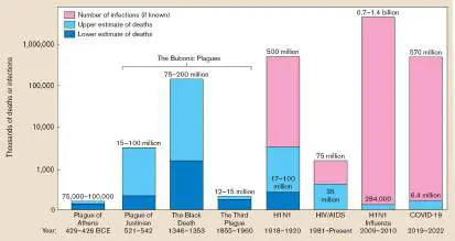
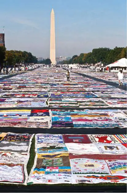
FIGURE 1.9 ■ The AIDS Memorial Quilt. Panels representing individuals who died of AIDS were displayed before the Washington Monument in 1992. A traveling exhibition continues, and the entire collection of panels is presented online.
RICHARD ELLIS/ALAMY STOCK PHOTO
The coronavirus SARS-CoV-2, which caused the COVID-19 pandemic, infected more than 500 million people and caused more than 6 million deaths between late 2019 and mid-2022. It caused social and economic devastation for the United States and many countries around the globe. This book presents SARS-CoV-2 in several contexts: devising an mRNA vaccine (Chapter 1); the coronavirus replication cycle (Chapter 6); the production of the Pfizer-BioNTech mRNA vaccine (Chapter 16); and COVID-19 epidemiology (Chapter 28).
Medical Statistics and Health Disparities
Historians traditionally emphasize the role of warfare in shaping human destiny and the brilliance of leaders or the advantage of new technology in determining which civilizations rise or fall. Yet throughout history, more soldiers have died of microbial infections than of wounds in battle. Assessment of the effects of disease on large populations required medical statistics—a important invention for public health.
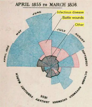
The significance of disease in warfare was first recognized by the British nurse and statistician Florence Nightingale (1820–1910; Fig. 1.10A). Better known as the founder of professional nursing, Nightingale also founded the science of medical statistics. She used methods invented by French statisticians to demonstrate the high mortality rate due to disease among British soldiers during the Crimean War. To show the deaths of soldiers due to various causes, she devised the “polar area chart” (Fig. 1.10B ). In this chart, blue wedges represent deaths due to infectious disease, red wedges represent deaths due to wounds, and black wedges represent all other causes of death. Infectious disease accounts for more than half of all mortality.
A B
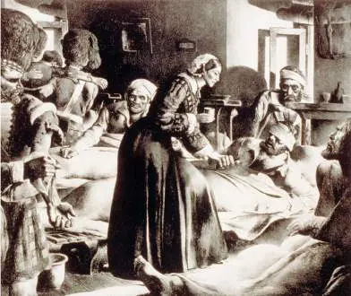
FIGURE 1.10 ■ Florence Nightingale, founder of medical statistics. A. Florence Nightingale was the first to use medical statistics to demonstrate the significance of mortality due to disease. B. Nightingale’s polar area chart of mortality data during the Crimean War.
EVERETT HISTORICAL/SHUTTERSTOCK
SMITH COLLECTION/GADO/GETTY IMAGES
Before Nightingale, no one understood the effect of disease on armies or on other crowded populations, such as in cities.
Nightingale’s statistics convinced the British government to improve army living conditions and to upgrade the standards of army hospitals. In modern epidemiology, statistical analysis continues to be a crucial tool in determining the causes of disease. For example, statistical analysis of COVID-19 case reports revealed the exponential spread of infection and helped public health directors predict the need for containment measures and hospital preparation.
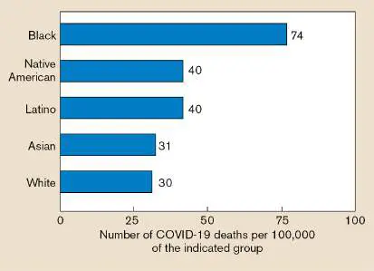
Medical statistics also reveal the important reality of health disparities. Health disparities are defined as differences in the incidence, prevalence, mortality, and social burden of diseases that exist among specific populations. Such disparities commonly affect Black people, Latino people, indigenous peoples, and LGBTQ people. For example, health disparities drastically affected mortality from COVID-19 (Fig. 1.11). Similar disparities appear in many other infectious diseases such as chlamydia and hepatitis B. The causes of health disparities involve institutional racism, disparities in access to quality health care, and lower socioeconomic status. Health disparities are discussed in detail in Chapter 28.
FIGURE 1.11 ■ COVID-19 death rates for different ethnic groups in 2020. Death rates for Americans with COVID-19 infection show disparities that are based on ethnicity.
Source: The COVID Tracking Project, https://covidtracking.com/.
Microscopes Reveal the Microbial World
The seventeenth century was a time of growing inquiry and excitement about the “natural magic” of science and patterns of our world, such as the laws of gravitation and motion formulated by Isaac Newton (1642–1727). Robert Boyle (1627–1691) performed the first controlled experiments on the chemical conversion of matter. Physicians attempted new treatments for disease involving the application of “stone and minerals” (that is, chemicals)—what today we would call “chemotherapy.” Minds were open to consider the astounding possibility that our surroundings, indeed our very bodies, were inhabited by tiny living beings.
Robert Hooke observes the microscopic world. The first microscopist to publish a systematic study of the world as seen under a microscope was Robert Hooke (1635–1703). As curator of experiments for the Royal Society of London, Hooke built a compound microscope—a magnifying instrument containing two or more lenses that multiply their magnification in series. With his microscope, Hooke observed biological materials such as nematode “vinegar eels,” mites, and mold filaments. Hooke published drawings of these microbes in Micrographia (1665), the first publication of objects observed under a microscope (Fig. 1.12).
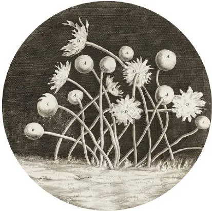
FIGURE 1.12 ■ Robert Hooke’s Micrographia. Mold sporangia, drawn by Hooke in 1665 from his observations of objects using a compound microscope.
LIBRARY OF CONGRESS
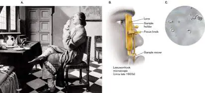
Hooke was the first to observe distinct units of living material, which he called “cells.” Hooke first named the units cells because the shape of hollow cell walls in a slice of cork reminded him of the shape of monks’ cells in a monastery. But his crude lenses achieved at best 30-fold power (30×), and he never observed single-celled bacteria. Antonie van Leeuwenhoek observes bacteria with a single lens. Hooke’s Micrographia inspired other microscopists, including Antonie van Leeuwenhoek (1632–1723), who became the first individual to observe single-celled microbes (Fig. 1.13A). As a young man, Leeuwenhoek lived in the Dutch city of Delft, where he worked as a cloth draper, a profession that introduced him to magnifying glasses. The magnifying glasses were used to inspect the quality of the cloth, enabling the worker to count the number of threads. Later in life, Leeuwenhoek took up the hobby of grinding ever-stronger lenses to see into the world of the unseen.
Leeuwenhoek ground lenses stronger than Hooke’s, which he used to build single-lens magnifiers, complete with sample holder and focus adjustment (Fig. 1.13B ). First he observed insects, including lice and fleas; then the relatively large single cells of protists and algae; then, ultimately, bacteria. One day he applied his microscope to observe matter extracted from between his teeth. He wrote, “To my great surprise [I] perceived that the aforesaid matter contained very many small living Animals, which moved themselves very extravagantly.”
Over the rest of his life, Leeuwenhoek recorded page after page on the movement of microbes, reporting their size and shape so accurately that in many cases we can determine the species he observed (Fig. 1.13C ). He performed experiments, comparing, for example, the appearance of “small animals” from his teeth before and after drinking hot coffee. The disappearance of microbes from his teeth after drinking a hot beverage suggested that heat killed microbes—a profoundly important principle for the study and control of microbes ever since.
FIGURE 1.13 ■ Antonie van Leeuwenhoek. A. A portrait of Leeuwenhoek, the first person to observe individual microbes. B. “Microscope” (magnifying glass) used by Leeuwenhoek. C.
Micrograph obtained using a replica of Leeuwenhoek’s microscope.
BETTMANN/GETTY IMAGES
BRIAN J. FORD
Leeuwenhoek is believed to have died, ironically, of a disease contracted from sheep whose bacteria he had observed. Historians have often wondered why it took two centuries for Leeuwenhoek and his successors to determine the link between microbes and disease. Although observers such as Agostino Bassi de Lodi (1773–1856) had noted cases of microbes associated with pathology (see Table 1.2), the very ubiquity of microbes—most of them actually harmless—may have obscured their more deadly roles. In addition, it was hard to distinguish between microbes and the single-celled components of the human body, such as blood cells and sperm. It was not until the nineteenth century that human tissues could be distinguished from
microbial cells by the application of differential chemical stains such as the Gram stain (discussed in Chapter 2).
Thought Question
1.3 Why do you think it took so long for humans to connect microbes with infectious disease? What innovations helped make the connection?
Spontaneous Generation: Do Microbes Have Parents?
The observation of microscopic organisms led priests and philosophers to wonder where these tiny beings came from. In the eighteenth century, scientists and church leaders intensely debated the question of spontaneous generation. Spontaneous generation is the concept that living creatures such as maggots could arise spontaneously, without parental organisms. Chemists of the day tended to support spontaneous generation, as it appeared similar to the way chemicals changed during reaction. Christian church leaders, however, supported the biblical view that all organisms have “parents” going back to the first week of creation.
The Italian priest Francesco Redi (1626–1697) showed that maggots in decaying meat were the offspring of flies. Meat kept in a sealed container, excluding flies, did not produce maggots. Thus, Redi’s experiment argued against spontaneous generation for macroscopic organisms. The meat still putrefied, however, producing microbes that seemed to arise “without parents.”
To disprove spontaneous generation of microbes, another Italian priest, Lazzaro Spallanzani (1729–1799), showed that a sealed flask of meat broth sterilized by boiling failed to grow microbes. Spallanzani also noticed that microbes often appeared in pairs. Were these two parental microbes coupling to produce offspring or did one microbe become two? Through long and tenacious observation, Spallanzani watched a single microbe grow in size until it split in two. Thus he demonstrated cell fission, the process by which cells arise by the splitting of preexisting cells.
Even Spallanzani’s experiments, however, did not put the matter to rest. Proponents of spontaneous generation argued that the microbes in the priest’s flask lacked access to oxygen and therefore could not grow. The pursuit of this question was left to future microbiologists, including the French microbiologist Louis Pasteur (1822–1895; Fig. 1.14A). In addressing spontaneous generation and related questions, Pasteur and his contemporaries laid the foundations for modern microbiology.
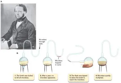
FIGURE 1.14 ■ Louis Pasteur, founder of medical microbiology and immunology. A. Pasteur’s contributions to the science of microbiology and immunology earned him lasting fame. B. Swan-necked flask. Pasteur showed that, after boiling, the contents in such a flask remain free of microbial growth, despite access to air.
THE PRINT COLLECTOR/ALAMY STOCK PHOTO
Louis Pasteur reveals the biochemical basis of microbial growth. As a child in rural France, Pasteur’s main interest was art, especially portraiture. He drew portraits of friends and family and aspired to be a professional artist. But his family convinced him to take up a more secure profession in science. So he studied chemistry —and applied an artist’s intensity of observation.
Pasteur wrote his doctoral thesis on the structure of organic crystals. Crystals exhibit aspects of visual beauty that piqued his interest. From his earlier correspondence on art, we know that Pasteur was fascinated by mirror symmetry. In crystals, he discovered the fundamental chemical property of chirality, the fact that some organic molecules exist in two forms that differ only by mirror symmetry; in other words, the two structures are mirror images of one another, like the right and left hands. Pasteur found that when microbes were cultured on a nutrient substance containing both mirror forms, only one mirror form was consumed. He concluded that the metabolic preference for one mirror form was a fundamental property of life. Subsequent research has confirmed that most molecules of organisms, such as DNA and proteins, are found in only one of their mirror forms.
As a chemist, Pasteur was asked to help with a problem encountered by French manufacturers of wine and beer. The alcohol in beverages comes from fermentation, a process by which microbes gain energy by converting sugars into alcohol. In the time of Pasteur, however, the conversion of grapes or grain to alcohol was believed to be a spontaneous chemical process. No one could explain why some fermentation mixtures produced vinegar (acetic acid) instead of alcohol. Pasteur discovered that fermentation is actually caused by living yeast, a single-celled fungus. In the absence of oxygen, yeast produces alcohol as a terminal waste product. But when the yeast culture is contaminated with bacteria, the bacteria outgrow the yeast and produce acetic acid instead of alcohol. (Fermentative metabolism is discussed in Chapter 13.)
Pasteur’s work on fermentation led him to test a key claim made by proponents of spontaneous generation. The proponents claimed that Spallanzani’s failure to find spontaneous appearance of microbes was due to lack of oxygen. From his studies of yeast fermentation, Pasteur knew that some kinds of microbes do not require oxygen for growth. So he devised an unsealed flask with a long, bent “swan neck” that admitted air but prevented the passage of dust that carried microbes (Fig. 1.14B ). When beef broth in the flask was boiled, the sterile broth remained clear, showing no growth of microbes. The famous swan-necked flasks remained free of microbial growth for many years. But when a flask was tilted so that the broth reached the dust, microbes grew immediately. Thus, Pasteur disproved the idea that lack of oxygen was the reason for the failure of spontaneous generation in Spallanzani’s flasks.
Even Pasteur’s work did not prove that microbial growth requires preexisting microbes. The Irish scientist John Tyndall (1820–1893) attempted an experiment similar to Pasteur’s but sometimes found the opposite result. Tyndall found that some kinds of broth, such as hay infusion, gave rise to microbes no matter how long they were sterilized by boiling. The microbes appear because hay infusion is contaminated with a heat-resistant form of bacteria called “endospores” (or “spores”). The spore form can be eliminated only by repeated cycles of boiling and resting, in which the spores germinate to the growing, vegetative form that is killed at 100°C.
It was later discovered that endospores could be killed by boiling under pressure, as in a pressure cooker, which generates higher temperatures than can be obtained at atmospheric pressure. The steam pressure device called the autoclave became the standard way to sterilize materials for the controlled study of microbes. (Microbial control and antisepsis are discussed further in Chapter 5.)
How Did Life Originate?
Spontaneous generation was discredited as a continual source of microbes. Yet at some point in the past, the first living organisms must have originated from nonliving materials. How did the first microbes arise?
The earliest fossil evidence of cells in the geological record appears in rock that formed as long ago as 4 billion years (discussed in Chapter 17). Although the nature of the earliest reported fossils remains controversial, it is generally accepted that “microfossils” from over 2 billion years ago were formed by living cells. Moreover, the living cells that formed microfossils looked remarkably similar to bacterial cells of today, forming chains of simple rods or spheres ( Fig. 1.15).
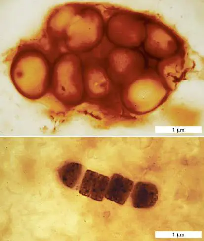
FIGURE 1.15 ■ Microfossils of ancient cyanobacteria. These fossils, from the Bitter Springs Formation, Australia, are about 850 million years old.
J. WILLIAM SCHOPF
The exact composition of the first environment for life is controversial. The components of the first living cells may have formed from spontaneous reactions sparked by ultraviolet absorption or electrical discharge. American chemists Stanley Miller (1930–2007) and Harold C. Urey (1893–1981) argued that the environment of early Earth contained mainly reduced compounds—compounds that have a strong tendency to donate electrons, such as ferrous iron, methane, and ammonia. In 1953, Miller attempted to simulate the highly reduced conditions of early Earth to test whether ultraviolet absorption or electrical discharge could cause reactions producing the fundamental components of life (Fig. 1.16A). He boiled a solution of water containing hydrogen gas, methane, and ammonia and applied an electrical discharge (comparable to a lightning strike). The electrical discharge excites electrons in the molecules and causes them to react. Astonishingly, the reaction produced a number of amino acids, including glycine, alanine, and aspartic acid. A similar experiment in 1961 by Spanish-American researcher Joan Oró (1923– 2004; Fig. 1.16B ) combined hydrogen cyanide and ammonia under electrical discharge to obtain adenine, a fundamental component of DNA and of the energy carrier adenosine triphosphate (ATP). A B
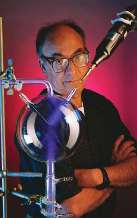
FIGURE 1.16 ■ Simulating early Earth’s chemistry. A. Stanley Miller with the apparatus of his early-Earth simulation experiment. B. Biochemist Joan Oró demonstrated the formation
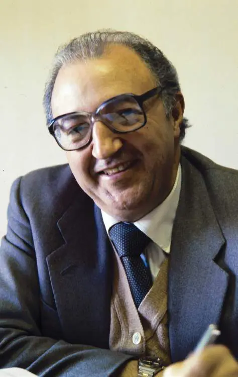
of adenine and other biochemicals from reaction conditions found in comets.
JIM SUGAR/GETTY IMAGES
ALBUM/ALAMY STOCK PHOTO
More recent evidence has modified this view, but it is agreed that the strong electron acceptor oxygen gas (O 2) was absent until the evolution of the first oxygen-producing photosynthetic microbes. Today, all our cells are composed of highly reduced molecules that are readily oxidized (lose electrons to O 2). This seemingly hazardous composition may reflect our cellular origin in the chemically reduced environment of early Earth. The experimental basis for the origin of life and evolution is discussed in detail in Chapter 17.
To Summarize
Microbes affected human civilization for centuries before humans guessed at their existence through their contributions to our global biosphere, food and drink production, and infectious diseases.
Disease pandemics such as the Black Death in medieval Europe and COVID-19 today shape human history.
Florence Nightingale statistically quantified the impact of infectious disease on human populations. Medical statistics reveal health disparities.
Robert Hooke and Antonie van Leeuwenhoek were the first to record observations of microbes through simple microscopes.
Spontaneous generation is the theory that microbes arise spontaneously, without parental organisms. Lazzaro Spallanzani showed that microbes arise from preexisting microbes and demonstrated that heat sterilization can prevent microbial growth.
Louis Pasteur discovered the microbial basis of fermentation. He also showed that providing oxygen does not enable spontaneous generation.
John Tyndall showed that repeated cycles of heat were necessary to eliminate spores formed by certain kinds of bacteria.
Earth’s first living organisms arose from nonliving materials. Evidence from microfossils and chemical simulations supports the origin of microbial life within the first 100 million years of Earth’s existence.
Glossary
spontaneous generation The theory, much debated in the eighteenth century, that under current Earth conditions life can arise spontaneously from nonliving matter.
fermentation Also called fermentative metabolism. 1. The production of ATP via substrate-level phosphorylation, using organic compounds as both electron donors and electron acceptors. 2. Industrial fermentation is the production of microbial products that are made by microbes grown in fermentation vessels; it may include respiratory metabolism to maximize microbial growth.
autoclave A device that uses pressurized steam to sterilize materials by raising the temperature above the boiling point of water at standard pressure.
1.3 Medical Microbiologynot assigned
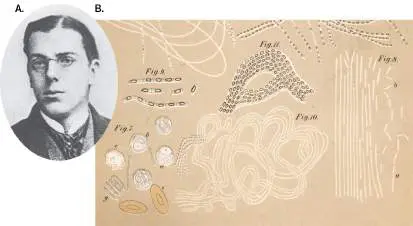
Over the centuries, thoughtful observers such as Ibn al-Khatib and Girolamo Fracastoro (see Table 1.2) noted the transmission of diseases that we now know are caused by microbes. Ultimately, researchers developed the germ theory of disease, the theory that many diseases are caused by a specific pathogen. The first scientific basis for determining that a specific microbe causes a disease was devised by the German physician Robert Koch (1843–1910; Fig. 1.17A). As a college student, Koch conducted biochemical experiments on his own digestive system. Koch’s curiosity about the natural world led him to develop principles and methods crucial to microbial investigation using microscopy (Fig. 1.17B ) and pure culture.
FIGURE 1.17 ■ Robert Koch, founder of the scientific method of microbiology. A. Koch as a university student. B. Koch’s sketch of anthrax bacilli in mouse blood.
MUSEUM IN THE ROBERT KOCH INSTITUTE, BERLIN
SCIENCE HISTORY IMAGES/ALAMY STOCK PHOTO
Growth of Microbes in Pure Culture
Unlike Pasteur, who was a university professor, Koch took up a medical practice in a small Polish-German town. To make space in his home for a laboratory to study anthrax and other deadly diseases, his wife curtained off part of his patient examination room.
Anthrax interested Koch because its epidemics in sheep and cattle caused economic hardship among local farmers. Today, anthrax is no longer a major problem for agriculture, because its transmission is prevented by effective environmental controls and vaccination. It has, however, gained notoriety as a bioterror agent because anthrax bacteria can survive for long periods in the dormant desiccated form of an endospore. In 2001, anthrax spores sent through the mail contaminated post offices, as well as an office building of the U.S. Senate, causing several deaths.
To investigate whether anthrax was a transmissible disease, Koch used blood from an anthrax-infected cow carcass to inoculate a rabbit. When the rabbit died, he used its blood to inoculate a second rabbit, which then died in turn. The blood of the unfortunate animal had turned black with long, rod-shaped bacilli. Upon introduction of these bacilli into healthy animals, the animals became ill with anthrax. Thus, Koch demonstrated an important principle of epidemiology: the chain of infection, or transmission of a disease. In retrospect, his choice of anthrax was fortunate, because anthrax microbes generate disease very quickly, multiply in the blood to high numbers, and remain infective outside the body for long periods. Koch and his colleagues then applied their experimental logic and culture methods to a more challenging disease: tuberculosis. In Koch’s day, tuberculosis caused one-seventh of all reported deaths in Europe; today, tuberculosis bacteria continue to infect millions of people worldwide. Koch’s approach to anthrax, however, was less applicable to tuberculosis, a disease that develops slowly after many years of dormancy. Furthermore, the causative bacterium, Mycobacterium tuberculosis, is small and difficult to distinguish from human tissue or from different bacteria of similar appearance associated with the human body. How could Koch prove that a particular bacterium caused a particular disease?
What was needed was to isolate a pure culture of microorganisms, a culture grown from a single “parental” cell. Previous researchers had achieved pure cultures by a laborious process of serially diluting suspended bacteria until a culture tube contained only a single cell. Alternatively, inoculating a solid surface such as a sliced potato could produce isolated colonies —distinct populations of bacteria, each grown from a single cell. For M. tuberculosis, Koch inoculated serum, which then formed a solid gel after heating. Later he refined the solid-substrate technique by adding gelatin to a defined liquid medium, which could then be chilled to form a solid medium in a glass dish. For his successful determination that M. tuberculosis causes tuberculosis, Koch was awarded the 1905 Nobel Prize in Physiology or Medicine.
A covered version of the solid-substrate technique is the Petri dish (or Petri plate ), which was invented by a colleague, Julius Richard Petri (1852–1921). The Petri dish is a round dish with vertical walls covered by an inverted dish of slightly larger diameter. Today the Petri dish, generally made of disposable plastic, remains an indispensable part of the microbiological laboratory.
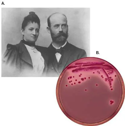
Another improvement in solid-substrate culture was the replacement of gelatin with materials that remain solid at higher temperatures, such as the gelling agent agar (a polymer of the sugar galactose). The use of agar was recommended by Angelina Hesse (1850–1934), a microscopist and illustrator, to her husband, Walther Hesse (1846–1911), a young medical colleague of Koch (Fig. 1.18). Agar comes from red algae (seaweed), which is used by East Indian birds to build nests; it is the main ingredient in the delicacy “bird’s nest soup.” Dutch colonists used agar to make jellies and preserves, and a Dutch colonist from Java introduced it to Angelina Hesse. The Hesses used agar to develop the first effective growth medium for tuberculosis bacteria. Pure culture and growth conditions are discussed further in Chapters 4 and 5.
FIGURE 1.18 ■ Angelina and Walther Hesse. A. Portrait of the Hesses, who first used agar to make solid-substrate media for bacterial growth. B. Colonies from a streaked agar plate.
NATIONAL LIBRARY OF MEDICINE
DR. MICHAEL GABRIDGE/VISUALS UNLIMITED, INC.
Some kinds of microbes cannot be grown in pure culture; that is, without other organisms. For example, intracellular pathogens such as Chlamydia trachomatis, the cause of trachoma and of infections of the reproductive tract, must be cultivated in human host tissue culture. And all viruses can be cultured only within their host cells
(see Chapter 6). The discovery of viruses is explored at the end of this section.
Koch’s Postulates
Koch used his studies of anthrax to formulate his famous set of criteria for establishing a causative link between an infectious agent and a disease. These four criteria are known as Koch’s postulates ( Fig. 1.19):
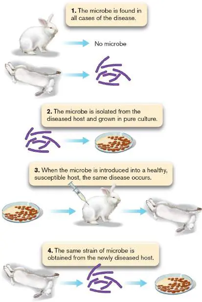
FIGURE 1.19 ■ Koch’s postulates defining the causative agent of a disease.
1. The microbe is found in all cases of the disease. For anthrax, Koch found large rod-shaped bacteria in the blood of sick rabbits.
2. The microbe is isolated from the diseased host and grown in pure culture. Koch isolated bacteria from blood samples and cultured them in a rabbit’s eyeball (later, this was done in culture dishes).
3. When the microbe is introduced into a healthy, susceptible host (or animal model), the host shows the same disease. The cultured bacteria were introduced into healthy rabbits, which became sick and died of anthrax.
4. The same strain of microbe is obtained from the newly diseased host. Blood from the newly infected rabbits showed rod-shaped bacteria that were cultured in the same way as the original bacteria.
Koch’s postulates continue to be used to determine whether a given strain of microbe causes a disease. An example is Lyme disease (Lyme borreliosis), a tick-borne infection first described in New England and shown in 1981 to be caused by the bacterium Borrelia burgdorferi. Nevertheless, the postulates remain only a guide; individual diseases and pathogens may confound one or more of the criteria. For example, in the case of AIDS, the concentration of HIV is so low that initially no virus could be detected in patients with active symptoms. It took the invention of the polymerase chain reaction (PCR), a method of producing any number of copies of DNA or RNA sequences, to detect the presence of HIV. Current research on microbial disease is presented in Chapter 26.
Another difficulty with many human diseases is the absence of an animal host that exhibits the same disease. For COVID-19, many animals can be infected by various coronaviruses but not the same SARS-CoV-2 that infects humans; and if they are infected, they may not show the same range of symptoms. In some cases, diseases that are curable or self-limiting may be tested on human volunteers in clinical trials. But for diseases without a cure, experimental inoculation of humans is banned by law.
In rare cases, researchers have voluntarily exposed themselves to a proposed pathogen. For example, Australian researcher Barry Marshall ingested Helicobacter pylori to convince skeptical colleagues that this organism could colonize the extremely acidic stomach. H. pylori turned out to be the causative agent of gastritis and stomach ulcers, conditions that had long been thought to be caused by stress rather than infection. For the discovery of H. pylori and its role in gastritis, Marshall and colleague J. Robin Warren won the 2005 Nobel Prize in Physiology or Medicine.
Thought Questions
1.4 How could you use Koch’s postulates (Fig. 1.19) to demonstrate the causative agent of influenza? What problems not encountered with anthrax would you need to overcome?
1.5 The original formulation of Koch’s first postulate stated, “The microbe is found in all cases of disease but is absent from healthy individuals.” Why do you think subsequent medical researchers modified this postulate?
Immunization Prevents Disease
Identifying the cause of a disease is, of course, only the first step in developing an effective therapy and preventing further transmission. Early microbiologists achieved some remarkable insights on how to control pathogens (see Table 1.2).
The first clue of how to protect an individual from a deadly disease came from the dreaded smallpox. In the eighteenth century, smallpox virus (also called variola virus) infected a large fraction of the European population, killing or disfiguring many people. The existence of the virus was then unknown, but in countries of Asia and Africa the incidence of smallpox was decreased by the deliberate inoculation of children with material from smallpox pustules. Inoculated children usually developed a mild case of the disease and were protected from smallpox thereafter.
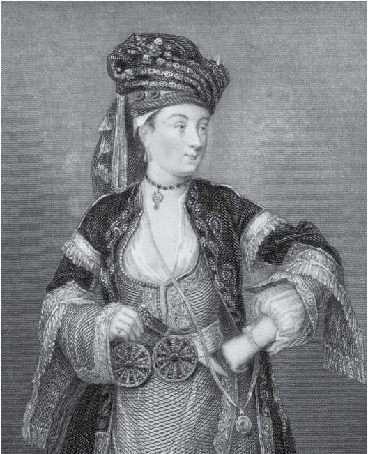
The practice of smallpox inoculation (or variolation) was introduced from Turkey to Europe in 1717 by Lady Mary Montagu, a smallpox survivor (Fig. 1.20A). While traveling in Turkey, Lady Montagu learned that many elderly women there had perfected the art of variolation: “The old woman comes with a nut-shell full of the matter of the best sort of small-pox, and asks what vein you please to have opened.” During a period outside the host, the virus becomes “attenuated”; that is, loses some of its molecular structure required for infection. The attenuated virus stimulates the immune system with much lower mortality than does the fully virulent virus. Lady Montagu arranged for the procedure on her own son and then brought the practice back to England. A similar practice of smallpox inoculation was introduced to the American colonies by an enslaved person, Onesimus, from the Coromantee people of Africa. Onesimus convinced his enslaver, Reverend Dr. Cotton Mather, to promote smallpox inoculation as a defense against an epidemic that was devastating Boston.
Preventive inoculation with smallpox was dangerous, however, because some infected individuals still contracted serious disease and were contagious. Thus, doctors continued to seek a better method of prevention. In England, milkmaids claimed that they were protected from smallpox after they contracted cowpox (caused by vaccinia virus), a related but much milder disease. English physician Edward Jenner (1749–1823) confirmed this claim by deliberately infecting patients with matter from cowpox lesions (Fig. 1.20B ). The practice of cowpox inoculation was called vaccination, after the vaccinia virus, which was derived from the Latin word vacca, meaning “cow.” At the time, the practice was highly controversial, as people feared they would somehow turn into cows (Fig. 1.20C ). Today, unfortunately, modern immunizations still raise irrational concerns. Failure to accept immunization leads to outbreaks of preventable disease, such as the measles outbreak in 2019, with more than 1,200 cases across the United States.
A.
B.
C.
FIGURE 1.20 ■ Smallpox vaccination. A. Lady Mary Wortley Montagu, shown in Turkish dress. The artist avoided showing
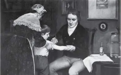
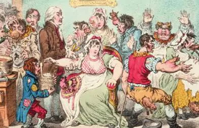
Montagu’s facial disfigurement from smallpox. B. Dr. Edward Jenner, depicted vaccinating 8-year-old James Phipps with cowpox matter from the hand of milkmaid Sarah Nelmes, who had caught the disease from a cow. C. Eighteenth-century newspaper cartoon depicting public reaction to cowpox vaccination.
HULTON ARCHIVE/GETTY IMAGES
POPPERFOTO/GETTY IMAGES
LIBRARY OF CONGRESS
Pasteur was aware of vaccination as he studied the course of various diseases in experimental animals. In the spring of 1879, he was studying fowl cholera, a transmissible disease of chickens with a high death rate. He had isolated and cultured the bacteria that had killed the chickens, but he left his work during the summer for a long vacation. No refrigeration was available to preserve cultures, and when he returned to work, the aged bacteria failed to cause disease in his chickens. Pasteur then obtained fresh bacteria from an outbreak of disease elsewhere, as well as some new chickens. All of the new chickens, exposed only to the fresh bacteria, contracted the disease. But the fresh bacteria failed to make the original chickens sick (those that had been exposed to the aged bacteria). Grasping the clue from his mistake, Pasteur had the insight to recognize that an attenuated strain of microbe, altered somehow to eliminate its potency to cause disease, could still confer immunity to the virulent disease-causing form.
Pasteur was the first to recognize the significance of attenuation and extend the principle to other pathogens. We now know that the molecular components of pathogens generate immunity, the resistance to a specific disease, by stimulating the immune system, an organism’s exceedingly complex cellular mechanisms of defense (see Chapters 23 and 24). Understanding the immune system awaited the techniques of molecular biology a century later, but nineteenth-century physicians developed several effective examples of immunization, the stimulation of an immune response by deliberate inoculation with an attenuated pathogen.
The way to attenuate a strain depends on the pathogen. Heat treatment or aging for various periods often turns out to be the most effective approach. The original success of prophylactic smallpox inoculation was due to natural attenuation of the virus during the time between acquisition of smallpox matter from a diseased individual and inoculation of the healthy patient. A far more elaborate treatment was required to combat the most famous disease for which Pasteur devised a vaccine: rabies.
The rabid dog loomed large in folklore, and rabies was dreaded for its particularly horrible and inevitable course of death. Pasteur’s vaccine for rabies required a highly complex series of heat treatments and repeated inoculations. Its success led to his instant fame (Fig. 1.21). Grateful survivors of rabies founded the Pasteur Institute, one of the world’s greatest medical research institutions, whose scientists in the twentieth century discovered HIV.
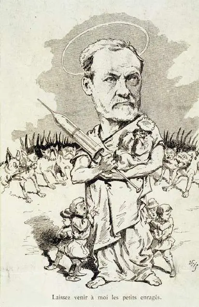
FIGURE 1.21 ■ Pasteur cures rabies. This cartoon in a French newspaper depicts Louis Pasteur protecting children from rabid dogs.
INSTITUT PASTEUR/MUSÉE PASTEUR
Today, vaccines are constructed by molecular cloning of parts of pathogens, such as the capsid protein vaccine for human papillomavirus (Gardasil). Even more effective are the mRNA vaccines against SARS-CoV-2, which were developed in just a few months at the height of the COVID-19 pandemic. Special Topic 1 presents how one of the first mRNA vaccines was devised for coronavirus by Kizzmekia Corbett and colleagues.
SPECIAL TOPIC 1 An mRNA Vaccine for COVID-19
In 2020, thousands of Americans were dying of COVID-19. The country—and the world at large—was desperate for a vaccine. At the National Institutes of Health (NIH), a radical new kind of vaccine was invented: an artificial messenger RNA (mRNA) that teaches host cells to stimulate an immune response (Fig. ST 1.1A ). In principle, an RNA vaccine would be safer and faster to produce than the attenuated virus vaccines described in Section 1.3. But an mRNA vaccine poses many challenges of molecular biology—and never before had one been approved by the U.S. Food and Drug Administration (FDA). Two pharmaceutical companies, Pfizer (in partnership with BioNTech) and Moderna, each achieved a vaccine with much support from publicly funded research. An NIH research team partnering with Moderna included more than fifty collaborators, led by viral immunologist Kizzmekia Corbett (Fig. ST 1.1B ).
A B
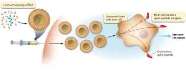
FIGURE ST 1.1 ■ An mRNA vaccine for SARS-CoV-2. A. The mRNA vaccine causes host cells to make viral spike proteins and process antigens to stimulate an immune
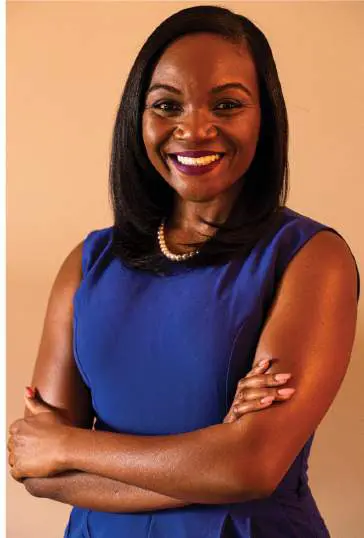
response. B. Viral immunologist Kizzmekia Corbett led the NIH-Moderna research team.
TIM NWACHUKWU/GETTY IMAGES
To make an mRNA vaccine, we must identify a viral protein that is antigenic (able to stimulate an immune response). A cloned gene encoding this viral protein is expressed in bacteria to make mRNA. The mRNA is then isolated from the bacteria and encapsulated in lipids, forming a liposome. The liposomes are injected into a patient, where they fuse with host immune cells. The host cells then express viral proteins from the mRNA. The viral proteins are processed to antigenic peptides, which the cell presents on its cell surface. These antigens then stimulate the immune system (discussed in Chapter 24).
Like all experimental work involving humans, the basic biology had to be worked out first in model animals (see Koch’s postulates in Section 1.3). The viral antigen chosen was the famous spike protein of the virus envelope, which binds the receptor on the host cell. A key achievement of Corbett’s team was to stabilize the spike antigen by mutating the mRNA sequence that encodes it. Corbett had previously made such a mutant spike variant of the related coronavirus MERS-CoV. She now constructed a similar mRNA mutant for the SARS-CoV-2 spike protein.
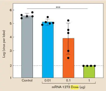
After solving numerous other technical problems, the mutant mRNA, called mRNA-1273, was tested in mice for its function as a vaccine. The researchers inoculated mice with various doses of the mRNA-1273 and then, after 7 weeks, infected them with SARS-CoV-2. The virus titer (concentration) was measured in the mouse lungs (Fig. ST 1.2A ). Those mice that had received the higher doses of mRNA-1273 showed lower virus titer—a sign of protection from infection.
The mice showed protection, but had the vaccine generated immune memory? Corbett’s team then measured the antibody titer in mice that had been immunized with mRNA-1273 (Fig. ST 1.2B ). The higher the dose that was received, the higher the concentration of SARS-CoV-2–specific antibodies that was found in the mouse serum. These experiments (with others) enabled the NIH-Moderna team to obtain approval for the clinical human trial that led to emergency approval of a SARS-CoV-2 vaccine for humans.
A. Higher vaccine doses cause decrease in virus B. Higher vaccine doses generate more antibodies
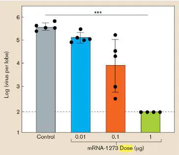
FIGURE ST 1.2 ■ SARS-CoV-2 mRNA vaccine protects mice. A. Virus-infected mice with higher doses of mRNA-1273 vaccine show lower virus titers in lung. B. Mice with increasing dose of mRNA-1273 produce higher titer of IgG immunoglobin (antibodies).
RESEARCH QUESTION
What other questions about the mRNA-1273 vaccine do you think the researchers needed to test before conducting a human clinical trial?
Corbett, Kizzmekia S., Darin K. Edwards, Sarah R. Leist, Olubukola
M. Abiona, Seyhan Boyoglu-Barnum, et al. 2020. SARS-CoV-2 mRNA
vaccine design enabled by prototype pathogen preparedness. Nature 586:
567–571.
Antiseptics and Antibiotics
Before the work of Koch and Pasteur, many patients died of infections transmitted unwittingly by their own doctors. In 1847, Hungarian physician Ignaz Semmelweis (1818–1865) noticed that for women in childbirth, the death rate due to puerperal fever was much higher in his own hospital than in a birthing center run by midwives. He guessed that the doctors in his hospital were transmitting pathogens from cadavers that they had dissected. So he ordered the doctors to wash their hands in chlorine, an antiseptic agent (a chemical that kills microbes). The mortality rate fell, but this revelation displeased other doctors, who refused to accept Semmelweis’s findings.
In 1865, the British surgeon Joseph Lister (1827–1912) noted that half of his amputee patients died of sepsis. Lister knew from Pasteur that microbial contamination might be the cause. So he began experiments to develop the use of antiseptic agents, most successfully carbolic acid (phenol), to treat wounds and surgical instruments. After initial resistance, Lister’s work, with the support of Pasteur and Koch, drew widespread recognition. In the twentieth century, surgeons developed fully aseptic environments for surgery; that is, environments completely free of microbes.
The problem with most antiseptic chemicals that killed microbes was that if taken internally, they would also kill the patients. Researchers sought a “magic bullet,” an antibiotic molecule that would kill only microbes, leaving their host unharmed.
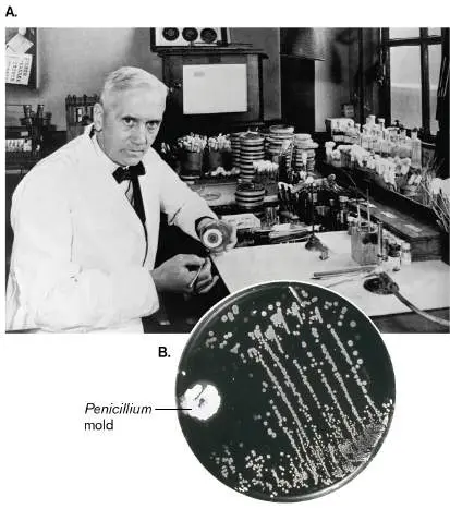
An important step in the search for antibiotics was the realization that microbes themselves produce antibiotic compounds. This conclusion followed from the famous accidental discovery of penicillin by the Scottish medical researcher Alexander Fleming (1881–1955; Fig. 1.22A). In 1929, Fleming was culturing Staphylococcus, which infects wounds. He found that one of his plates of Staphylococcus was contaminated with a mold, Penicillium notatum, which he noticed was surrounded by a clear region free of Staphylococcus colonies (Fig. 1.22B ). Following up on this observation, Fleming showed that the mold produced a substance that killed bacteria. Today we know this substance as penicillin.
FIGURE 1.22 ■ Alexander Fleming, discoverer of penicillin. A. Fleming in his laboratory. B. Fleming’s original plate of bacteria with Penicillium mold inhibiting the growth of bacterial colonies.
DAVIES/GETTY IMAGES
MEDISCAN/VISUALS UNLIMITED
In 1941, biochemists Howard Florey (1898–1968) and Ernst Chain (1906–1979) purified the penicillin molecule, which we now know inhibits formation of the bacterial cell wall. Penicillin saved the lives of many Allied troops during World War II, the first war in which an antibiotic became available to soldiers.
The second half of the twentieth century saw the discovery of many new and powerful antibiotics. Most of the new antibiotics, however, were made by little-known bacteria and fungi from endangered ecosystems—a circumstance that focused attention on wilderness preservation. Furthermore, the widespread and often indiscriminate use of antibiotics selects for pathogens to evolve resistance to antibiotics. As a result, antibiotics have lost their effectiveness against certain strains of major pathogens. For example, multidrug-resistant Mycobacterium tuberculosis and methicillin-resistant Staphylococcus aureus (MRSA) are now serious threats to public health. To combat evolving drug resistance, we continually need to research and develop new antibiotics and devise practices that limit spread of resistance. Microbial biosynthesis of antibiotics is discussed in Chapter 15, and the medical use of antibiotics is discussed in Chapter 27.
Thought Questions
1.6 Why do you think some pathogens generate immunity readily, whereas others evade the immune system?
1.7 How do you think microbes protect themselves from the antibiotics they produce?
The Discovery of Viruses
Viruses are much smaller than the host cells they infect; most are too small to be seen by a light microscope. So how were they discovered? In 1892, the Russian botanist Dmitri Ivanovsky (1864–1920) studied tobacco mosaic disease, a condition in which the leaves become mottled and the crop yield is decreased or destroyed altogether. Ivanovsky knew that the disease was transmissible, but he was surprised to find that the agent of transmission could pass through a porcelain filter having a pore size (0.1 μm) that blocked known microbes. Later, the Dutch plant microbiologist Martinus Beijerinck (1851–1931) conducted similar filtration experiments. Beijerinck concluded that because the agent of disease passed through a filter that retained bacteria, it could not be a bacterial cell. Beijerinck used the word “virus,” although neither he nor Ivanovsky understood that the cause of tobacco mosaic disease was an infectious noncellular particle (Fig. 1.23).
The “filterable agent” of disease was ultimately purified by the American scientist Wendell Stanley (1904–1971), who processed 4,000 kilograms (kg) of infected tobacco leaves. Stanley obtained a sample of infective virus particles pure enough to crystallize in a 3D array comparable to crystals composed of inert chemicals. The crystal was analyzed by X-ray crystallography (discussed in Chapter 2) to reveal the molecular structure of tobacco mosaic virus (Fig. 1.23A) —a feat that earned Stanley the 1946 Nobel Prize in Chemistry. The fact that an object capable of biological reproduction could be stable enough to be crystallized amazed scientists, ultimately leading to a new, more mechanical view of living organisms. Today, we consider viruses “subcellular organisms.”
The individual particle of tobacco mosaic virus consists of a helical tube of protein subunits containing its genetic material coiled within ( Fig. 1.23B ). Stanley thought the virus was a catalytic protein, but colleagues later determined that it contained RNA as its genetic material. The structure of the coiled RNA was solved through X-ray crystallography by the British scientist Rosalind Franklin (1920–1958). Other viruses that have RNA genomes include influenza virus and HIV (the virus that causes AIDS); viruses with DNA genomes include human papillomavirus (HPV) and herpesviruses. We now know that all kinds of animals, plants, and microbial cells can be infected by viruses—and carry endogenous viruses that may benefit their hosts. Viral function and disease are discussed in Chapters 6, 11, and 26.
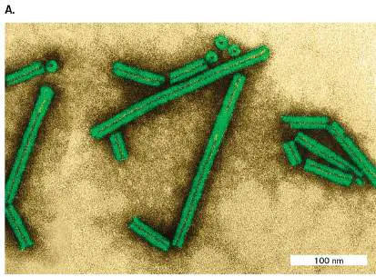
FIGURE 1.23 ■ Tobacco mosaic virus (TMV). A. Particles of tobacco mosaic virus (colorized transmission electron micrograph). B. In TMV, a capsid of proteins surrounds an RNA chromosome.
DENNIS KUNKEL MICROSCOPY/SCIENCE SOURCE
To Summarize
Robert Koch devised techniques of pure culture to study a single species of microbe in isolation. A key technique is culture on solid medium using agar, as developed by Angelina and Walther Hesse, in a double-dish container devised by Julius Petri.
Koch’s postulates provide a set of criteria to establish a causative link between an infectious agent and a disease.
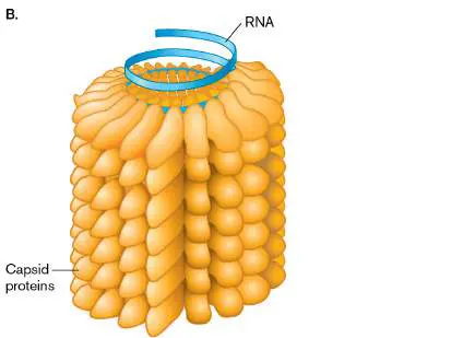
Edward Jenner established the practice of vaccination, or inoculation with cowpox to prevent smallpox. Jenner’s discovery was based on earlier observations by Lady Mary Montagu and others that a mild case of smallpox could prevent future cases.
Louis Pasteur developed the first vaccines that were based on attenuated strains, such as the rabies vaccine.
Ignaz Semmelweis and Joseph Lister showed that antiseptics could prevent the transmission of pathogens from doctor to patient.
Alexander Fleming discovered that the Penicillium mold generates a substance that kills bacteria.
Howard Florey and Ernst Chain purified the substance penicillin, the first commercial antibiotic to save human lives. Dmitri Ivanovsky and Martinus Beijerinck discovered viruses as filterable agents of infection that are not cells. Wendell Stanley first crystallized virus particles.
Glossary
germ theory of disease The theory that many diseases are caused by microbes.
chain of infection The serial passage of a pathogenic organism from an infected individual to an uninfected individual, thus transmitting disease. pure culture A culture containing only a single strain or species of microorganism. A large number of microorganisms that all descended from a single individual cell.
colony A visible cluster of microbes on a plate, all derived from a single founding microbe. Usually consists of a clone , except for infrequent mutations.
Petri dish or Petri plate A round dish with vertical walls covered by an inverted dish of slightly larger diameter. The smaller dish can be filled with a substrate for growing microbes.
agar A polymer of galactose that is used as a gelling agent. Koch’s postulates Four criteria, developed by Robert Koch, that should be met for a microbe to be designated the causative agent of an infectious disease.
vaccination Exposure of an individual to a weakened version of a microbe or a microbial antigen to provoke immunity and prevent development of disease upon reexposure.
immunity A body’s resistance to a specific disease.
immune system An organism’s cellular defense system against pathogens. immunization The stimulation of an immune response by deliberate inoculation with a weakened pathogen, in hopes of providing immunity to disease caused by the pathogen.
antiseptic Describing a chemical that kills microbes. Also, the chemical itself.
antibiotic A molecule that can kill or inhibit the growth of selected microorganisms.
aseptic Free of microbes.
Fig. 1.19: FIGURE 1.19 ■ Koch’s postulates defining the causative agent of a disease.
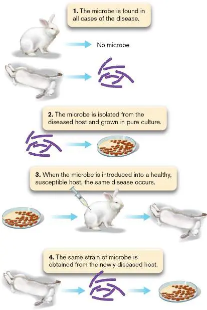
1.4 Environment and Ecologynot assigned
Koch’s growth of microbes in pure culture founded the systematic study of microbial physiology and biochemistry. But how does pure culture relate to “natural” environments, such as a forest or a human intestine, where countless kinds of microbes interact with each other and with multicellular hosts? The opposite of pure culture is the Winogradsky column (Fig. 1.24). The Russian microbiologist Sergei Winogradsky (1856–1953) devised this model wetland ecosystem containing regions of enrichment for microbes of diverse metabolism.
FIGURE 1.24 ■ Winogradsky column. A wetland model ecosystem designed by Sergei Winogradsky.
PHOTO BY TIMOTHY A. WILKERSON
A typical Winogradsky column consists of a glass tube containing mud (a source of wetland bacteria) mixed with shredded newsprint (an organic carbon source) and calcium salts of sulfate and carbonate (an inorganic carbon source for autotrophs). After
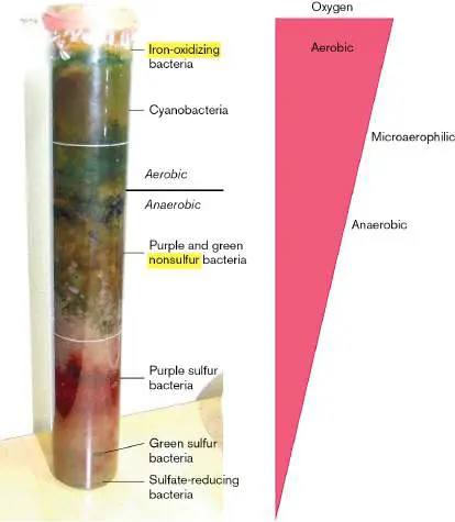
exposure to light for several weeks, several zones of color develop, full of mineral-metabolizing bacteria. At the top, cyanobacteria conduct photosynthesis, using light energy to split water and produce molecular oxygen. Below, purple sulfur bacteria use photosynthesis to split hydrogen sulfide, producing sulfur. At the bottom, with O 2 exhausted, bacteria reduce (donate electrons to) alternative electron acceptors such as sulfate. Sulfate-reducing bacteria produce hydrogen sulfide and precipitate iron.
In hindsight, the invention of pure culture eclipsed the equally important study of microbial ecology (discussed in Chapters 21 and 22). Microbes cycle the many minerals essential for all life, including all atmospheric nitrogen gas and much of the oxygen. Yet, less than 0.1% of all microbial species can be cultured in the laboratory. In natural environments, uncultured microbes make up the majority of Earth’s entire biosphere. Only the outer skin of Earth supports complex multicellular life. The depths of Earth’s crust, to at least 3 kilometers (km) down, as well as the atmosphere 15 km out into the stratosphere, remain the domain of microbes. So, to a first approximation, Earth’s ecology is microbial ecology.
Environmental Microbes Support Ecosystems
The first microbiologists to culture microbes in the laboratory selected the kinds of nutrients that feed humans, such as beef broth or potatoes. Winogradsky, however, suspected that other kinds of microbes living in soil or wetlands consume more exotic fare. Soil samples were known to oxidize hydrogen gas, and this activity was eliminated by treatment with heat or acid, suggesting microbial origin. Ammonia in sewage was oxidized by donating electrons to oxygen, forming nitrate. Nitrate formation was eliminated by antibacterial treatment. These findings suggested the existence of microbes that “eat” hydrogen gas or ammonia instead of beef or potatoes, but no one could isolate these microbes in culture. Winogradsky waded through marshes to discover microbes with metabolisms quite alien from human digestion. For example, he discovered that species of the bacterium Beggiatoa oxidize hydrogen sulfide (H 2 S) to sulfuric acid (H 2 SO 4). Beggiatoa fixes carbon dioxide into biomass without consuming any organic food. Organisms that feed solely on inorganic minerals are known as chemolithotrophs, or lithotrophs (discussed further in Chapters 4 and 14).
The lithotrophs studied by Winogradsky could not be grown on Koch’s plate media containing agar or gelatin. The bacteria that Winogradsky isolated can grow only on inorganic minerals; in fact, some species are actually poisoned by organic food. For example, nitrifiers convert ammonia to nitrate, forming a crucial part of the nitrogen cycle in natural ecosystems. Winogradsky cultured nitrifiers on a totally inorganic solution containing ammonia and silica gel, which supported no other kind of organism. This experiment was an early example of enrichment culture, the use of selective growth media that support certain classes of microbial metabolism while excluding others.
Later microbial ecologists showed that bacteria perform unique roles in geochemical cycling, the global interconversion of inorganic and organic forms of nitrogen, sulfur, phosphorus, and other minerals. Without these essential conversions (nutrient cycles), no plants or animals could live. Bacteria and archaea fix nitrogen (N 2) by reducing it to ammonia (NH 3), the form of nitrogen assimilated by plants. This process is called nitrogen fixation (Fig. 1.25). Other bacterial species oxidize ammonium ions (NH +) in several
4
stages back to nitrogen gas. These microbial cycles are heavily affected by human technology leading to climate change (discussed further in Chapters 21 and 22).
FIGURE 1.25 ■ The global nitrogen cycle. All life depends on these oxidative and reductive conversions of nitrogen—most of which are performed only by microbes.
Thought Question
1.8 Why don’t all living organisms fix their own nitrogen? Consider the structure of a dinitrogen molecule, N≡N.
Today, microbes with unusual properties, such as the ability to digest toxic wastes or withstand extreme temperatures, have
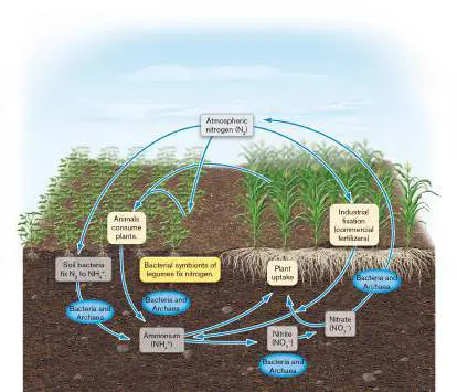
valuable applications in industry and bioremediation. For this reason, microbial ecology is a priority for funding by the National Science Foundation (NSF). From 1998 to 2004, the NSF was directed by marine microbial ecologist Rita Colwell (Colwell’s research is described in Chapter 21). At NSF, Colwell founded the Biocomplexity Initiative to study complex interactions between microbes and other life in the environment. Such research includes the discovery of extremophiles, microbes from environments with extreme heat, salinity, or acidity. For example, hydrothermal vents in the ocean floor spew superheated water and minerals that feed hyperthermophiles (Fig. 1.26A). A record-setting hyperthermophile is the archaeon Geogemma barossii (Fig. 1.26B ), isolated by microbiologist Kazem Kashefi from the Mothra vent field named for the fictional monster. G. barossii reduces rust (iron oxide; Fe 2 O 3) to the magnetic mineral magnetite (Fe 3 O 4) while growing in an autoclave at 121°C (250°F), a temperature high enough to kill all other known organisms. A simple magnet can be used to show that Geogemma converts nonmagnetic Fe 2 O 3 to the magnetic Fe 3 O 4 (Fig. 1.26C ).
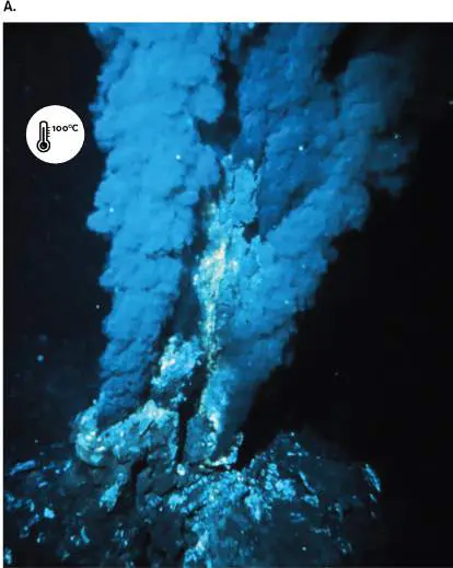
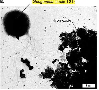
FIGURE 1.26 ■ An extreme thermophile reduces iron oxide to magnetite. A. Hydrothermal vent at ocean floor. B. Geogemma barossii is a round archaeon with a tuft of flagella (transmission electron micrograph). C. Kazem Kashefi, now at Michigan State University, pulls a live culture of “strain 121” ( Geogemma) out of an autoclave generally used to kill all living organisms at 121°C (250°F).
OAR/NURP/NOAA
K. KASHEFI AND D. R. LOVLEY. 2003. SCIENCE 301 :934
KAZEM KASHEFI/MICHIGAN STATE UNIVERSITY
Microbial Endosymbiosis with Plants and Animals
The pure-culture model of microbiology, a powerful tool of discovery, nonetheless disregarded the fact that outside the laboratory, all
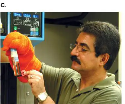
microbes live in the presence of other kinds of life. Many live in endosymbiosis with multicellular organisms. Endosymbiosis is the partnership of a host organism with its associated endosymbionts, microbes that grow within a host body or within a host cell. The first person to describe endosymbiosis was Martinus Beijerinck, who observed nitrogen-fixing bacteria called rhizobia (singular, rhizobium) within the cells of plants. Rhizobia induce the roots of legumes such as soybean plants to form special nodules to fix nitrogen into biomass, which is shared with the plant cells. Microbial endosymbiosis occurs everywhere. Endosymbiotic microbes make essential nutritional contributions to host animals. Invertebrates such as hydras and corals harbor endosymbiotic phototrophs that provide products of photosynthesis in return for protection and nutrients. Other endosymbionts produce antibiotics or toxins that thwart predators. Among vertebrates, ruminant animals such as cattle, as well as insects such as termites, require digestive bacteria such as Bacteroides to break down cellulose and other plant polymers. Even humans obtain about 15% of our nutrition from bacteria growing within the colon (Fig. 1.27). FIGURE 1.27 ■ Intestinal microbiota form a biofilm. Blowup: Bacterial biofilm on the surface of a residual food particle.
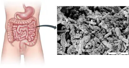
AMERICAN SOCIETY FOR MICROBIOLOGY
MACFARLANE, S., ET AL. 2005. J. APPL. ENVIRON. MICROBIOL. 71 :7483
Some intestinal bacteria, such as Escherichia coli, grow as biofilms, organized multispecies communities adhering to a surface —in this case the surface of colonic epithelial cells. Biofilms play major roles in all ecosystems and within parts of the human body (discussed in Chapters 4, 13, and 21). The biofilm shown magnified in Figure 1.27is attached to the surface of a digested food particle.
Today we know that all multicellular organisms possess a microbiota, or microbiome , the collection of all microbes associated with an organism or habitat. Remarkably, our concept of “multicellular organism” has changed to include its microbiome as a functional part of the organism. Physicians consider the human microbiome to be a part of the body, as essential as a limb or an organ. Bacteria that normally inhabit the human intestine and skin protect our bodies from infection by pathogens. Gut bacteria regulate the development of our immune system and even send signals to the brain (discussed in Chapter 13). In 2016, the U.S. government announced the National Microbiome Initiative to advance understanding of how microbiomes contribute to our health and the environment. Microbiomes are discussed further in Chapters 21 and 23.
Note: The term “micro biota ” refers to the ecological community
of microbes living within or upon an organism, such as the human body. “Micro biome ” refers to the community of microbes associated with an organism or with a different defined habitat, such as soil or plants; the term emphasizes the microbes’ collective DNA sequences. Despite these subtle distinctions, we use the terms interchangeably in this book. Another term, “metagenome,” refers specifically to the collective DNA sequences found in all the microbes of a microbiome.
Thought Question
1.9 Could endosymbiosis occur today; that is, could a small microbe be engulfed by a larger one and evolve into an endosymbiont, and then into an organelle? Explain.
Microbial Life on Other Planets
The abundance of life on Earth—and the evidence of life’s appearance early in Earth’s history—leads us to ask whether microbial life has emerged on other planets. Molecules that spontaneously formed in Stanley Miller’s and Joan Oró’s experiments are also found in meteorites and comets. This observation led Oró to propose that the first chemicals of life could have come from outer space, perhaps carried by comets. Furthermore, at the time that life arose on Earth, Earth’s geochemistry resembled that of other planets, such as Mars. In 2012, to seek evidence for Martian life, NASA landed the Mars Science Laboratory, or Curiosity rover, near the base of a mountain on the planet Mars (Fig. 1.28). The car-sized rover has a laser to drill into rock, X-ray and fluorescence analyzers, and camera microscopes. As of this writing, Curiosity continues its mission, testing the Martian soil for water, organic compounds, and other potential evidence of microbial life. The question became more interesting in 2018 when the orbital radar detector MARSIS found evidence of water buried beneath Mars’s southern ice cap—similar to ice-buried lakes that support microbial life on Earth.
FIGURE 1.28 ■ Mars Curiosity rover. The rover explores Mars for evidence of microbial life.
NASA/JPL-CALTECH/MSSS
To Summarize
Sergei Winogradsky developed the first system of enrichment culture, called the Winogradsky column, to grow microbes from natural environments.
Chemolithotrophs (or lithotrophs) metabolize inorganic minerals, such as ammonia, instead of the organic nutrients used by the microbes isolated by Koch.
Geochemical cycling depends on bacteria and archaea that cycle nitrogen, phosphorus, and other minerals throughout
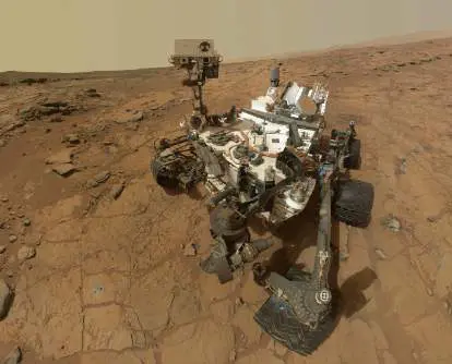
the biosphere.
Endosymbionts are microbes that live within host organisms and may provide essential functions for their hosts, such as nitrogen fixation, digestion of food molecules, or protection from predation.
Martinus Beijerinck was the first to demonstrate that nitrogen-fixing rhizobia grow as endosymbionts within leguminous plants.
The microbiome , or microbial community, of a multicellular host is now considered a functional part of the host organism. NASA’s Curiosity rover now explores the planet Mars for signs of microbial life outside Earth.
Glossary
Winogradsky column A glass tube containing a stratified environment that causes specific microbes to grow at particular levels; a type of enrichment culture for the growth of microbes from wetland environments.
photosynthesis The metabolic ability to absorb and convert solar energy into chemical energy for biosynthesis. Autotrophic photosynthesis, or photoautotrophy, includes CO 2 fixation.
enrichment culture The use of selective growth media to allow only certain microbes to grow.
geochemical cycling The global interconversion of various inorganic and organic forms of elements.
nitrogen fixation The ability of some prokaryotes to reduce inorganic diatomic nitrogen gas (N) to two ammonium ions (2NH +).
2 4
extremophile An organism that grows only in an extreme environment; that is, an environment including one or more conditions that are “extreme” relative to the conditions for human life.
endosymbiosis An intimate association between different species in which one partner population grows within the body of another organism. endosymbiont An organism that lives as a symbiont inside another organism. biofilm A community of microbes growing on a solid surface.
microbiota or microbiome The total community of microbes associated with an organism (such as the human body) or with a defined habitat (such as soil or plants).
1.5 The Microbial Family Treenot assigned
The bewildering diversity of microbial life forms presented nineteenth-century microbiologists with a seemingly impossible task of classification. So little was known about life under the lens that natural scientists despaired of ever learning how to distinguish microbial species. The famous classifier of species, Swedish botanist Carl von Linné (Carolus Linnaeus, 1707–1778), called the microbial world “chaos.” Linnaeus would have been astonished to see the intricate family tree of life revealed by Carl Woese in the twentieth century, consisting of three domains: Bacteria, Archaea, and Eukarya (Fig. 1.29A). Of these domains, Archaea was unknown in Linnaeus’s time, and the idea that archaea gave rise to eukaryotes— including humans—would have been impossible to imagine.
A B
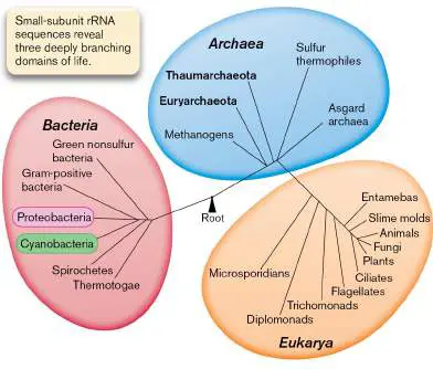
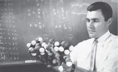
FIGURE 1.29 ■ Carl Woese and the three domains of life. A. Three domains form a monophyletic tree that is based on small-subunit ribosomal RNA (rRNA) sequences. The length of each branch approximates the time of divergence from the last common ancestor. B. Carl Woese first used rRNA sequence to determine the phylogeny of living organisms.
AP PHOTO
Microbes Are a Challenge to Classify
Before the twentieth century, taxonomists faced two challenges as they attempted to classify microbes. First, the resolution of the light microscope revealed little more than the outward shape of microbial cells, and vastly different kinds of microbes looked more or less alike (discussed in Chapter 2). This challenge was overcome as advances in biochemistry and microscopy made it possible to distinguish microbes by metabolism and cell structure, and ultimately by DNA sequence.
Second, microbes do not readily fit the classic definition of a species; that is, a group of organisms that interbreed. Unlike multicellular eukaryotes, microbes generally reproduce asexually. When they do exchange genes, they may do so with related strains or with distantly related species (discussed in Chapter 9). Nevertheless, microbiologists have devised working definitions of microbial species that enable us to usefully describe populations (discussed in Chapter 17). The most useful classification defines relatedness on the basis of similarity of DNA sequence: the more sequence two organisms share, the shorter the time since their populations diverged from a common ancestor. But early taxonomists had no DNA sequence information.
Note: The names of microbial species are frequently changed to
reflect new understanding of genetic relationships. For example, the causative agent of bubonic plague was formerly called Bacterium pestis (1896), Bacillus pestis (1900), and Pasteurella pestis (1923), but it is now called Yersinia pestis (1944). The older names, however, still appear in the literature—a point to remember when carrying out research. Names of bacteria and archaea are compiled in the List of Prokaryotic Names with Standing in Nomenclature (LPSN).
Taxonomists first tried to classify microbes as either animals or plants, which since ancient times had been considered the two “kingdoms” or major categories of life. For example, algae and fungi were included with plants. But German naturalist Ernst Haeckel (1834–1919) recognized that microbes differed from both plants and animals in fundamental aspects of their lifestyle, cell structure, and biochemistry. Haeckel proposed that microscopic organisms constitute a third kind of life—neither animal nor plant—which he called Monera. In the twentieth century, biochemical studies revealed profound distinctions even within the Monera. In particular, microbes such as protists and algae contain a nucleus enclosed by a nuclear membrane, whereas bacteria do not. Herbert Copeland (1902–1968) proposed a system of classification that divided Monera into two groups: the Eukaryotes (protists, or protozoa and algae) and the Prokaryotes (bacteria). Copeland’s four-kingdom classification (plants, animals, protists, and prokaryotic bacteria) was later modified by Robert Whittaker (1920–1980) to include fungi as a fifth kingdom of eukaryotic microbes. But all these systems faced a challenge from the new science of genome sequences.
Archaea Differ from Bacteria and Eukaryotes
In 1977, Carl Woese (1928–2012), at the University of Illinois, was wondering how to measure the time since divergence of the most distantly related life forms. He recalled his earlier postdoctoral work on the coding of information in RNA molecules (Fig. 1.29B ). Woese and a colleague, George Fox, used the sequence of the gene for 16S ribosomal RNA (16S rRNA) as a “molecular clock,” a gene whose sequence differences can be used to measure the time since the divergence of two species (discussed in Chapter 17). In constructing their tree of life, they included certain prokaryotes that live in seemingly hostile environments, such as the boiling sulfur springs of Yellowstone, or that conduct unusual kinds of metabolism, such as production of methane (methanogenesis). The divergence of rRNA genes showed that some of these prokaryotes were as distant from bacteria as they were from eukaryotes. These organisms represent a distinct domain of life: Archaea.
The archaea resemble the bacteria in their relatively simple cell structure and in their lack of a nucleus; thus, both archaea and bacteria are prokaryotes. But the gene expression machinery of archaea is more similar to that of eukaryotes. Archaea are found in a wide range of environments, and certain species, such as the autoclave-cultured archaeon Geogemma (Fig. 1.26B), grow in environments more extreme than any that support bacteria. Other kinds of archaea grow alongside bacteria in common soil or water—or even within the human gut or skin (discussed in Chapter 19). Woese’s discovery replaced the classification scheme of five kingdoms with three equally distinct domains: Bacteria, Archaea, and Eukarya (Fig. 1.29A). But the genomes of undiscovered life had further surprises in store. In the twenty-first century, Jillian Banfield and co-workers at UC Berkeley pioneered the sequencing of metagenomes—the DNA sequences of entire microbial communities ( Fig. 1.30). In 2004, Banfield’s research team sequenced the first environmental metagenome, which was obtained from a sample of extremely acidic mine drainage. Later environmental metagenomes revealed ultrasmall bacteria and archaea that can pass through a filter with 0.2-μm pores. Previously, the existence of such small cells was thought impossible.
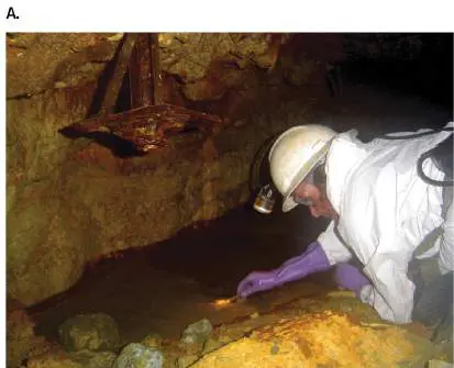
FIGURE 1.30 ■ Jillian Banfield sequences metagenomes. A. Banfield obtains samples from extremely acidic drainage at Richmond Mine, California. B. Ultrasmall bacterium obtained from a groundwater sample whose metagenomes revealed previously unknown clades of bacteria.
Source: Birgit Luef et al. 2015. Nat. Commun. 6 :6372.
MITCH JONES/JILLIAN BANFIELD
B. LUEF ET AL. 2015. NAT. COMMUN. 6 :6372
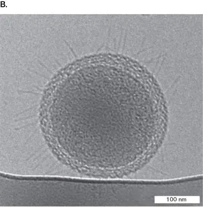
Eukaryotes Evolved through Endosymbiosis
During the early twentieth century, microscopists wondered about the nature of subcellular structures such as mitochondria and chloroplasts, which have the appearance of cells within cells. Lynn Margulis (1938–2011) at the University of Massachusetts was fascinated by organelles amid the diversity of microbial eukaryotes ( Fig. 1.31). Margulis tried to explain why eukaryotic cells contain mitochondria and chloroplasts, membranous organelles that possess their own chromosomes. She proposed that eukaryotes evolved by merging with bacteria to form composite cells by intracellular endosymbiosis, in which one cell internalizes another that grows within it. The endosymbiosis may ultimately generate a single organism whose formerly independent members are now incapable of independent existence.
FIGURE 1.31 ■ Lynn Margulis and the serial endosymbiosis theory. Inset: Lynn Margulis proposed that organelles evolve through endosymbiosis.
NANCY R. SCHIFF/GETTY IMAGES
Margulis proposed that early in the history of life, respiring bacteria similar to Escherichia coli were engulfed by pre-eukaryotic cells, where they evolved into mitochondria, the eukaryote’s respiratory organelles. Similarly, she proposed that a phototroph related to cyanobacteria was taken up by a eukaryote, giving rise to the chloroplasts of phototrophic algae and plants. In Margulis’s
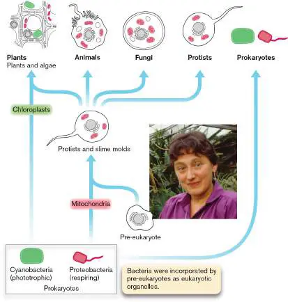
model, the nature of the original pre-eukaryote that took up precursors to mitochondria was unclear. Today, genomic evidence suggests that the original pre-eukaryote may have been an archaeon (discussed in Chapter 19).
The endosymbiosis theory was controversial because it implied a polyphyletic, or multiple, ancestry of living species, inconsistent with the long-held assumption that species evolve only by divergence from a common ancestor (monophyletic ancestry). Ultimately, DNA sequence analysis produced compelling evidence of the bacterial origin of mitochondria and chloroplasts. Both of these classes of organelles contain circular molecules of DNA, whose sequences show unmistakable homology (similarity) to those of bacteria. Genome sequences now reveal many cases of horizontal gene transfer among all three domains of life.
Thought Question
1.10 What arguments support the classification of Archaea as a third domain of life? What arguments support the classification of archaea and bacteria together, as prokaryotes, distinct from eukaryotes?
To Summarize
Classifying microbes was a challenge historically because of the difficulties in observing distinguishing characteristics of different categories.
Ernst Haeckel recognized that microbes constitute a form of life distinct from animals and plants.
Herbert Copeland and Robert Whittaker classified prokaryotes as a form of microbial life distinct from eukaryotic microbes such as protists.
Carl Woese discovered a domain of prokaryotes, Archaea, whose genome sequences diverge equally from those of bacteria and those of eukaryotes. Archaea grow in a wide range of environments; some species grow under conditions that exclude bacteria and eukaryotes.
Jillian Banfield pioneered the sequencing of metagenomes that reveal previously unknown forms of microbial life.
Lynn Margulis proposed that eukaryotic organelles such as mitochondria and chloroplasts evolved by endosymbiosis from prokaryotic cells engulfed by pre-eukaryotes.
Glossary
polyphyletic Having multiple evolutionary origins.
monophyletic Having a single evolutionary origin; that is, diverging from a common ancestor.
Fig. 1.26B: B. Geogemma barossii is a round archaeon with a tuft of flagella (transmission electron micrograph).
K. KASHEFI AND D. R. LOVLEY. 2003. SCIENCE 301 :934
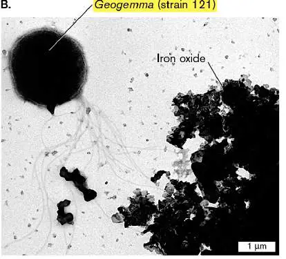
1.6 Cell Biology and the DNA Revolutionnot assigned
During the twentieth century, amid world wars and societal transformations, the field of microbiology exploded with new knowledge (see Table 1.2). More than 99% of what we know about microbes today was discovered after 1900 by scientists too numerous to cite in this book. New tools of microscopy and genomic analysis offered unprecedented applications for human medicine and industry (discussed in Chapters 7–12). Electron microscopy and biochemistry revealed the fundamental structure and function of cell membranes and proteins. For example, an electron micrograph of a stained section of Chlorobium bacteria reveals unique light-harvesting organelles called “chlorosomes” (Fig. 1.32). The chlorosomes conduct photosynthesis at extremely low light levels, such as that of long-wavelength energy radiated from thermal vents at the ocean floor.
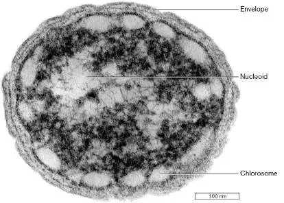
FIGURE 1.32 ■ Electron micrograph of Chlorobium species, a photosynthetic bacterium. The thin section reveals the nucleoid (containing DNA), the light-harvesting chlorosomes, and envelope membranes.
NIELS-ULRIK FRIGAARD ET AL. 2002 J. BACTERIOL. 184 :3368
Cell Membranes and Macromolecules
In 1900, the study of cell structure was still limited by the resolution of the light microscope and by the absence of tools that could take apart cells to isolate their components. Both of these limitations were overcome by the invention of powerful instruments. Just as society was being transformed by machines ranging from jet airplanes to vacuum cleaners, the study of microbiology was also being transformed by machines. Two instruments had exceptional impact: The electron microscope revealed the internal structure of cells (see Chapter 2), and the ultracentrifuge enabled isolation of subcellular parts (see Chapter 3).
The electron microscope. In the 1920s, at the Technical University in Berlin, student Ernst Ruska (1906–1988) was invited to develop an instrument for focusing rays of electrons. Ruska recalled, from his childhood, that his father’s microscope could magnify fascinating specimens of plants and animals, but that its resolution was limited by the wavelength of light. He was eager to devise lenses that could focus beams of electrons, with wavelengths far smaller than that of light, to reveal living details never seen before. Ultimately, Ruska built lenses to focus electrons using specially designed electromagnets. Magnetic lenses were used to complete the first electron microscope in 1933 (Fig. 1.33). Early transmission electron microscopes achieved about tenfold greater magnification than the light microscope, revealing details such as the ridged shell of a diatom. Further development steadily increased magnification, to as high as a millionfold. Today’s electron microscopes can visualize phospholipid membranes and protein complexes such as individual antibodies. Exciting kinds of microscopy are presented in Chapter 2.
FIGURE 1.33 ■ An early transmission electron microscope.
HAGLEY MUSEUM AND ARCHIVE/SCIENCE SOURCE
Subcellular structures, however, raised many questions about cell function that visualization alone could not answer. Biochemists showed that cell function involves numerous chemical transformations mediated by enzymes. A milestone in the study of
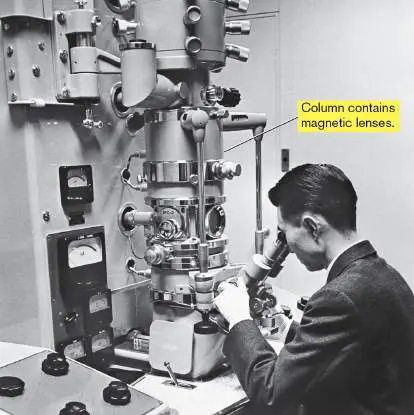
metabolism was the elucidation by German biochemist Hans Krebs (1900–1981) of the tricarboxylic acid cycle (TCA cycle, or Krebs cycle), by which the products of sugar digestion are converted to carbon dioxide. The TCA cycle provides energy for many bacteria and for the mitochondria of eukaryotes. But even Krebs understood little of how metabolism is organized within a cell; he and his contemporaries considered the cell a “bag of enzymes.” The full understanding of cell structure required experiments on isolated parts of cells.
The ultracentrifuge. Centrifugation can separate whole cells from the fluid in which they are suspended. The first centrifuges spun samples in a rotor with centrifugal force of a few thousand times that of gravity. In the nineteenth century, biochemists proposed that even greater centrifugal forces could separate components of lysed cells, even macromolecules such as proteins. The Swedish chemist Theodor Svedberg (1884–1971), at the University of Uppsala, built such a machine: the ultracentrifuge. By the twentieth century, ultracentrifuges had achieved rotation rates so high that they required a vacuum to avoid burning up like a space reentry vehicle. Ultracentrifuges isolated protein complexes such as ribosomes and DNA molecules such as plasmids (small circular pieces of DNA). Experiments combining electron microscopy and ultracentrifugation revealed how membranes govern energy transduction within bacteria and within organelles such as mitochondria and chloroplasts. In the 1960s, English biochemists Peter Mitchell (1920–1992) and Jennifer Moyle (1921–2016)
proposed and tested a revolutionary idea called the chemiosmotic theory. The chemiosmotic theory states that the reduction-oxidation (redox) reactions of the electron transport system store energy in the form of a gradient of protons (hydrogen ions) across a membrane, such as the bacterial cell membrane or the inner membrane of the mitochondrion. The energy stored in the proton gradient, in turn, drives the synthesis of ATP (discussed in Chapter 14).
Microbial Genetics Leads the DNA Revolution
As the form and function of living cells emerged in the early twentieth century, a largely separate line of research revealed patterns of heredity of cell traits. In eukaryotes, the Mendelian rules of inheritance were rediscovered and connected to the behavior of subcellular structures called chromosomes. Frederick Griffith (1879– 1941) showed in 1928 that an unknown substance from dead bacteria could carry genetic information into living cells, transforming harmless bacteria into a strain capable of killing mice— a process called transformation. Some kind of “genetic material” must be inherited to direct the expression of inherited traits. Biochemists thought the inherited material might be protein, because of the tremendous variety of amino acid sequences. Then, in 1944, Oswald Avery (1877–1955) and colleagues showed that the genetic material for transformation is deoxyribonucleic acid, or DNA. An obscure acidic polymer, DNA had been previously thought too uniform in structure to carry information; its precise structure was unknown. As World War II raged among nations, scientists embarked on an epic struggle: the quest for the structure of DNA.
The double helix. The tool of choice to discover the structure of molecules was X-ray crystallography, a method developed by British physicists in the early 1900s. The field of X-ray analysis included an unusual number of women, including Dorothy Hodgkin (1910–1994), who later won a Nobel Prize for the structures of penicillin and vitamin B 12 (discussed in Chapter 2). In 1953, crystallographer Rosalind Franklin joined a laboratory at King’s College London to study the structure of DNA (Fig. 1.34A). As a woman and as a Jew who supported relief work in Palestine, Franklin felt socially isolated at the male-dominated Protestant university; her work was disparagingly called “witchcraft.” Nevertheless, her exceptional X-ray micrographs (Fig. 1.34B ) revealed for the first time that the common form of DNA was a double helix.
Without Franklin’s knowledge, her colleague Maurice Wilkins (1916–2004) showed her data to a competitor, James Watson at the University of Cambridge. The pattern led Watson and Francis Crick (1916–2004) to propose that the four bases of the DNA “alphabet” were paired in the interior of Franklin’s double helix (Fig. 1.34C ). They published their model in the journal Nature, while denying that they had used Franklin’s data. The discovery of the double helix earned Watson, Crick, and Wilkins the 1962 Nobel Prize in Physiology or Medicine. Franklin died of ovarian cancer before the prize was awarded. Before her death, however, she had turned her efforts to the structure of ribonucleic acid (RNA). She determined the helical form of the RNA chromosome within tobacco mosaic virus, the first viral RNA to be characterized.
FIGURE 1.34 ■ The DNA double helix. A. Rosalind Franklin discovered that DNA forms a double helix. B. X-ray diffraction pattern of DNA, obtained by Franklin. C. James Watson (left) and Francis Crick discovered the complementary pairing between bases of DNA and the antiparallel form of the double helix.
SCIENCE SOURCE
OMIKRON/SCIENCE SOURCE
A. BARRINGTON BROWN/SCIENCE SOURCE
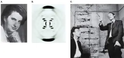
Modern X-ray crystallography (discussed in Chapter 2) reveals with atomic precision the structure of DNA, including its complementary base pairs (Fig. 1.35A). The complementary pairing of DNA bases led to the development of techniques for DNA sequencing, the reading of a sequence of DNA base pairs. Figure 1.35B shows a portion of the DNA sequence from bacterial DNA isolated by an undergraduate student. (The sequencing process is described in Chapter 7.) In the data, each color represents a fluorescent signal from one of the four bases: adenine (A), guanine (G), cytosine (C), or thymine (T). Each peak represents a DNA fragment terminating in that particular base. The order of fragment lengths yields the sequence of bases in one strand. Reading the DNA sequence enabled microbiologists to determine the beginning and endpoint of microbial genes, and ultimately entire genomes, as discussed in Section 1.1.
FIGURE 1.35 ■ DNA. A. The structure of DNA, based on modern X-ray crystallography. B. A DNA sequence fluorogram obtained from bacterial genomic DNA. Each colored trace represents the fluorescence of one of the four bases terminating a fragment of DNA. Units represent number of DNA bases.
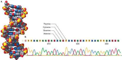
Reading the genomes enabled microbiologists to see the history of microbial evolution, reaching back to a time even before the advent of DNA—to a pre-DNA world when the cell’s chromosomes were actually composed of ribonucleic acid (RNA). This hypothetical world without DNA is called the RNA world. How did life function in the RNA world? We hypothesize that cells used RNA for all the functions of DNA and protein, including information storage and replication, and for biochemical catalysis. RNA molecules capable of catalysis, called ribozymes, were discovered in 1982 by Thomas Cech at the University of Colorado and Sidney Altman at Harvard University, who jointly earned the Nobel Prize in Chemistry in 1989 ( Fig. 1.36). That same year, Jennifer Doudna and Jack Szostak at Harvard showed how an RNA molecule from the protozoan Tetrahymena could catalyze its own replication. These achievements support the theory that early organisms were composed primarily of RNA.
FIGURE 1.36 ■ Discovery of catalytic RNA. A. Tom Cech holds a flask containing protists that make catalytic RNA, the kind of molecule that in early cells may have served both genetic
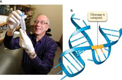
and catalytic functions. B. Diagram of a catalytic RNA, where horizontal bars represent bases. The RNA catalyzes cleavage of itself.
JEREMY PAPASSO/DIGITAL FIRST MEDIA/BOULDER DAILY CAMERA VIA GETTY
IMAGES
How do DNA and RNA sequences convey information in the cell? To read the DNA language required deciphering the genetic code— how triplets of DNA “letters” specify the amino acid units of proteins. This story is discussed in Chapter 8.
The DNA revolution began with bacteria. What amazed the world about DNA was that such a simple substance, composed of only four types of subunits, is the genetic material that determines all the different organisms on Earth. The promise of this insight was first fulfilled in bacteria and bacteriophages, whose small genomes and short generation times made key experiments possible (see Chapters 6–9). Bacterial tools were later extended to animals and plants; for example: Bacteria readily recombine DNA from unrelated organisms. The mechanisms of bacterial recombination led to construction of artificially recombinant DNA, or “gene cloning.” Recombinant DNA ultimately enabled us to transfer genes between the genomes of virtually all types of organisms.
Bacterial DNA polymerases are used for polymerase chain reaction (PCR) amplification of DNA. A hot spring in Yellowstone National Park yielded the bacterium Thermus aquaticus, whose DNA polymerase could survive many rounds of cycling to near-boiling temperature. The Taq polymerase formed the basis of a multibillion-dollar industry of PCR amplification of DNA, with applications ranging from genome sequencing to forensic identification.
Gene regulation discovered in bacteria provided models for animals and plants. The first key discoveries of gene expression were made in bacteria and bacteriophages.
Regulatory DNA-binding proteins were discovered in bacteria and then subsequently found in all classes of living organisms. CRISPR-Cas9 is a molecular mechanism of bacterial defense against bacteriophages. This mechanism was developed as a means of editing human genomes for gene therapy. The use of CRISPR-Cas9 for human genome editing earned the 2020 Nobel Prize in Chemistry for Emmanuelle Charpentier (currently at the Max Planck Unit for the Science of Pathogens) and Jennifer Doudna, at UC Berkeley (Fig. 1.37). CRISPR-Cas9 is described in Chapter 12.
FIGURE 1.37 ■ Nobel Prize for CRISPR-Cas9. Emmanuelle Charpentier and Jennifer Doudna won the 2020 Nobel Prize in Chemistry for CRISPR-Cas9 discovery and application for human genome editing.
REUTERS/ALAMY STOCK PHOTO
In the 1970s, when the DNA revolution began, its implications drew public concern. The use of recombinant DNA to make hybrid organisms—organisms combining DNA from more than one species
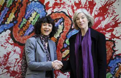
—seemed “unnatural.” We now know that in natural environments, genes frequently move between species. Furthermore, recombinant DNA technology raised the specter of placing deadly genes that produce toxins such as botulin into innocuous human-associated bacteria such as E. coli.
The unknown consequences of recombinant DNA so concerned molecular biologists that in 1975 they held a conference to assess the dangers of and restrict recombinant DNA experimentation. The conference, led by Paul Berg and Maxine Singer at Asilomar (Pacific Grove, California), was possibly the first time in history that a group of scientists organized and agreed to regulate and restrict their own field.
On the positive side, the emerging world of molecular biology excited the imagination of young scientists and entrepreneurs. The pioneering biotechnology company Genentech was founded in 1976 by Robert Swanson and bacterial geneticist Herbert Boyer, from UC San Francisco. Growing numbers of students entered the field of molecular biology, seeking to invent medical cures—or even to clone dinosaurs, as in Michael Crichton’s novel and film Jurassic Park (1993). While the idea of cloning a dinosaur remains science fiction, the tools of microbial genetics opened a window into the past by letting us read the DNA of long-dead organisms preserved in museums. In 1980, the first patent was upheld for a living organism —a bacterium genetically modified to catabolize oil components from petroleum spills. Since then, it has become accepted to patent modified mice for cancer studies.
Thought Question
1.11 State an argument in favor of patenting a microbial isolate or a gene sequence. What argument can be made against patenting microbes or genes?
Microbial Discoveries Transform Medicine and Industry
Twentieth-century microbiology transformed the practice of medicine and generated entire new industries of biotechnology and bioremediation. After the discovery of penicillin, Americans poured millions of dollars of private and public funds into medical research. In 1938, the March of Dimes was founded to discover a polio vaccine; today, polio has been nearly eliminated. Research on microbes and other aspects of biology has grown with support from U.S. government agencies, such as the National Institutes of Health and the National Science Foundation, as well as from governments of other countries, particularly the European nations, Japan, and China. Further support comes from private foundations, such as the Pasteur Institute, the Wellcome Trust, and the Howard Hughes Medical Institute. Medical research generates astonishing advances, such as the use of human immunodeficiency virus (HIV, the cause of AIDS) to devise gene therapy agents that cure cancer (discussed in Chapter 11).
Research in microbiology includes fields as diverse as medicine and space science (Table 1.3). These fields all recruit microbiologists (Fig. 1.38). Industrial and applied biology (see Chapter 16) use bacteria to clone and produce therapeutic proteins, such as insulin for diabetics. Recombinant viruses make safer vaccines. At the frontiers of science, we use microbes for “synthetic biology,” the construction of novel organisms with useful functions (see Chapter 12). For example, synthetic biology may design bacteria with an on/off switch to report the presence of arsenic in environmental samples.
FIGURE 1.38 ■ Microbiologists at work. Students at Kenyon College conduct research on bacterial gene expression.
JOAN SLONCZEWSKI, KENYON COLLEGE
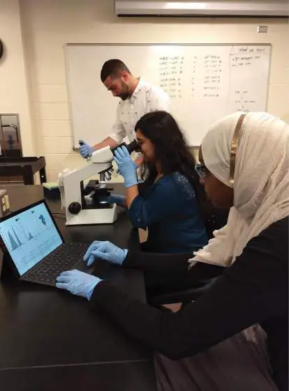
On a global level, the management of our planet’s biosphere, with the challenges of pollution and global warming, increasingly depends on our understanding of microbial populations. For example, as Earth heats up, frozen soil at the polar regions activates microbial decomposition, releasing carbon dioxide and methane gases that accelerate global warming. Microbiologists play critical roles in the field of climate science (see Chapter 22).
Fields of Research in
TABLE 1.3
Microbiology
Field Subject of study Experimental Fundamental questions about microbiology microbial form and function, genetics, and ecology Medical microbiology The mechanism, diagnosis, and treatment of microbial disease Epidemiology Distribution and causes of disease in humans, animals, and plants Immunology The immune system and other host defenses against infectious disease Food microbiology Fermented foods and food preservation Industrial microbiology Production of drugs, cloned gene products, and biofuels
Fields of Research in
TABLE 1.3
Microbiology
Environmental Microbial diversity and microbial microbiology processes in natural and artificial environments Bioremediation The use of microbial metabolism to remediate human wastes and industrial pollutants Forensic microbiology Analysis of microbial strains as evidence in criminal investigations Climate science The study of weather patterns and temperature changes over time, including processes mediated by microbial populations
To Summarize
Cell structure was revealed by new tools such as the electron microscope and the ultracentrifuge.
Genetics of bacteria, bacteriophages, and fungi in the early twentieth century revealed fundamental insights about gene transmission that apply to all organisms.
Structure and function of the genetic material, DNA , emerged from a series of experiments in the twentieth century.
Molecular microbiology generated key advances , such as the cloning of the first recombinant molecules and the invention of DNA sequencing technology.
Genome sequence determination and bioinformatic analysis became the tools that shape the study of biology in the twenty-first century.
Microbial discoveries transformed medicine and industry. Biotechnology produces new kinds of pharmaceuticals and industrial products. Synthetic biology engineers new kinds of organisms with useful functions.
Glossary
electron microscope A microscope that obtains high resolution and magnification by using magnetic lenses to focus electron beams on samples. ultracentrifuge A machine that subjects samples to high centrifugal forces and can be used to separate subcellular components.
chemiosmotic theory A theory stating that the products of oxidative metabolism store their energy in an electrochemical gradient that can drive cellular processes such as ATP synthesis.
transformation The internalization of free DNA from the environment into bacterial cells.
DNA sequencing A technique to determine the order of bases in a DNA sample. RNA world A model of early life in which RNA performed all the informational and catalytic roles of today’s DNA and proteins. eResearch Activity 1
Do Bacteria Make Neurotransmitters for the Human Brain?
Our intestines support a vast community of diverse bacteria that help digest our food. Some products of this bacterial digestion actually modulate the function of our nervous system—and possibly directly affect our brain. For example, bacteria that catabolize glutamic acid (an amino acid) release the waste product 4-aminobutanoate (GABA). In our nervous system, GABA is a major neurotransmitter. Could gut bacteria be producing our neurotransmitters?
Philip Strandwitz, a young scientist at the Massachusetts Institute of Technology (MIT), set out to test the possibility that bacteria produce neurotransmitters that influence our brain. He focused on Bacteroides bacteria, which are known to catabolize many kinds of molecules in our gut. In a remarkable experiment, Strandwitz’s research team compared “functional connectivity” (a brain signal associated with depression) with the level of Bacteroides bacteria excreted in human feces (Fig. ERA 1.1 ). The human subjects had depressive disorder, which is associated with the brain region tested. Strandwitz asked whether functional connectivity as a depression signal might depend on Bacteroides bacteria. In fact, he found an inverse correlation between the Bacteroides and the depressive signal in the brain: The more Bacteroides were found in the gut microbiome, the lower the brain’s depression signal.

FIGURE ERA 1.1 ■ Bacteroides bacteria show an inverse correlation with a brain signal of depression. A. Philip Strandwitz conducts experiments to test the role of gut bacterial neurotransmitters in human brain function. B. The depression signal of “functional connectivity” in the brain’s prefrontal cortex is significantly decreased in human patients whose digestive tract contains more Bacteroides.
PHILIP STRANDWITZ
P. STRANDWITZ ET AL. 2019. NAT MICROBIOL. 4 :396–403
Strandwitz pursued a possible connection between mental depression and gut microbial metabolism. He knew that some Bacteroides strains convert glutamic acid (glutamate) to GABA, a neurotransmitter that may decrease anxiety and depression. Strandwitz wondered whether the gut microbiome includes GABA producers and consumers. To identify GABA consumers, bacteria from the human gut were plated on rich medium containing yeast extract. After up to a week, smaller colonies were then plated in the presence of Bacteroides (Fig. ERA 1.2A ). One novel strain grew
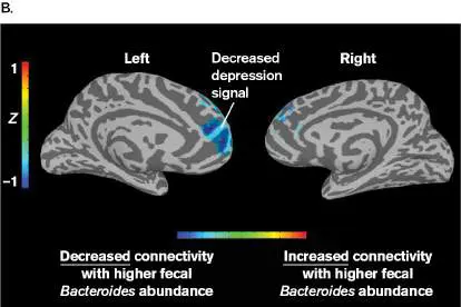
microcolonies in the presence of Bacteroides. This strain, designated KLE1738, was found to require GABA for growth and, remarkably, could not grow on glucose or any amino acids as a carbon source.
The GABA-dependent strain KLE1738 was then used to identify additional gut species that produce GABA. When gut bacteria were plated on a lawn of KLE1738, those colonies that produce GABA were each surrounded by a halo of KLE1738 microcolonies. These new GABA producers were then screened for GABA production. Some species produced GABA in response to acid stress (pH 4–5), which would occur from stomach passage, whereas others produced GABA at pH 6–7, which is typical of the intestinal tract (Fig. ERA 1.2B ). Together, the GABA producers and consumers might maintain a steady-state GABA level in the gut. While no causation is proven, these observations have led Strandwitz and his team to pursue possible mechanisms by which gut GABA could affect neural connections in the brain.
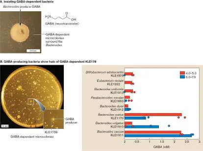
FIGURE ERA 1.2 ■ Isolating gut bacteria that produce GABA. A. New GABA-producing bacteria are identified by their surrounding halo of GABA-dependent bacteria that grow up from the background lawn. B. Many isolates increase GABA production at lower pH (pH 4–5), but some of the new strains make GABA at neutral pH.
P. STRANDWITZ ET AL. 2019. NAT MICROBIOL. 4 :396–403
P. STRANDWITZ ET AL. 2019. NAT MICROBIOL. 4 :396–403
Further Exploration
What experiments in mice do you think might show more directly the mechanism of how gut bacterial GABA production modulates behavior? How might the role of steady-state GABA levels be tested? What human organ systems might mediate the connection between gut bacteria GABA and neural connections?
Strandwitz, Philip, Ki Hyun Kim, Darya Terekhova, Joanne K. Liu, Anukriti Sharma, et al. 2019. GABA-modulating bacteria of the human gut microbiota. Nature Microbiology 4 :396–403.
CHAPTER REVIEW
Review Questions
1. Explain the apparent contradictions in defining microbiology as the study of microscopic organisms or as the study of single-celled organisms.
2. What is the genome of an organism? How do genomes of viruses differ from those of cellular microbes?
3. Under what conditions might microbial life have originated? What evidence supports current views of microbial origin?
4. List the ways in which microbes have affected human life throughout history.
5. Summarize the key experiments and insights that shaped the controversy over spontaneous generation. What questions were raised, and how were they answered? 6. Explain how microbes are cultured in liquid and on solid media. Compare and contrast the culture methods of Koch and Winogradsky. How did their different approaches to microbial culture address different questions in microbiology?
7. Explain how a series of observations of disease transmission led to the development of immunization to prevent disease.
8. Summarize key historical developments in our view of microbial taxonomy. What attributes of microbes have made them challenging to classify?
9. Explain how various discoveries in “natural” bacterial genetics were used to develop recombinant DNA technology.
Thought Questions
1. How do Earth’s microbes contribute to human health? Include examples of environmental microbes outside the human body and of microbes associated with the human body.
2. When space scientists seek evidence for life on Mars, why do you think they expect to find microbes rather than creatures like the “alien monsters” often depicted in science fiction?
3. Why do you think so many environmental microbes cannot be cultured in laboratory broth or agar media? 4. Outline the different contributions to medical microbiology and immunology of Louis Pasteur, Robert Koch, and Florence Nightingale. What methods and assumptions did they have in common, and how did they differ?
5. Outline the different contributions to environmental microbiology of Sergei Winogradsky and Martinus Beijerinck. Why did it take longer for the significance of environmental microbiology to be recognized, as compared with pure-culture microbiology?
6. What kinds of evidence support the common ancestry of life from cells with RNA chromosomes? Could cells with RNA chromosomes exist today? Why or why not?
Key Terms
agar (16)
antibiotic (21)
antiseptic (20)
Archaea (5)
aseptic (21)
autoclave (14)
Bacteria (5)
biofilm (26)
chain of infection (16) chemiosmotic theory (32) colony (16)
DNA sequencing (32)
electron microscope (31) endosymbiont (25)
endosymbiosis (25)
enrichment culture (24) Eukarya (5)
eukaryote (5)
extremophile (25)
fermentation (14)
genome (5)
geochemical cycling (24) germ theory of disease (16) immune system (19)
immunity (19)
immunization (19)
Koch’s postulates (17) metagenome (5)
microbe (3)
microbiota (microbiome) (26) monophyletic (30)
nitrogen fixation (24) pathogen (2)
Petri dish (16)
photosynthesis (23)
polymerase chain reaction (PCR) (3) polyphyletic (30)
prokaryote (5)
pure culture (16)
RNA world (33)
spontaneous generation (13) transformation (32)
ultracentrifuge (31)
vaccination (19)
virus (4)
Winogradsky column (23)
Recommended Reading
Albers, Sonja-Verena, Patrick Forterre, David Prangishvili, and Christa Schleper. 2013. The legacy of Carl Woese and Wolfram Zillig: From phylogeny to landmark discoveries. Nature Reviews. Microbiology 11 :713–719.
Anguela, Xavier M., and Katherine A. High. 2019. Entering the modern era of gene therapy. Annual Review of Medicine 70:273–288.
Asch, David A., Nazmul Islam, Natalie E. Sheils, Yong Chen, Jalpa A. Doshi, et al. 2021. Patient and hospital factors associated with differences in mortality rates among Black and White US Medicare beneficiaries hospitalized with COVID-19 infection. JAMA Network Open 4 :e2112842.
Blount, Zachary D., Christina Z. Borland, and Richard E. Lenski. 2008. Historical contingency and the evolution of a key innovation in an experimental population of Escherichia coli. Proceedings of the National Academy of Sciences USA 105:7899–7906.
Brock, Thomas D. 1999. Robert Koch: A Life in Medicine and Bacteriology. ASM Press, Washington, DC.
Brokowski, Carolyn, and Mazhar Adli. 2019. CRISPR ethics: Moral considerations for applications of a powerful tool. Journal of Molecular Biology 431 :88–101.
Dinc, Gulten, and Yesim I. Ulman. 2007. The introduction of variolation “A La Turca” to the West by Lady Mary Montagu and Turkey’s contribution to this. Vaccine 25 :4261–4265.
Dubos, Rene. 1998. Pasteur and Modern Science. Translated by Thomas Brock. ASM Press, Washington, DC.
Fleishmann, Robert D., Mark D. Adams, Owen White, Rebecca A. Clayton, Ewen F. Kirkness, et al. 1995. Whole-genome random sequencing and assembly of Haemophilus influenzae Rd. Science 269 :496–512.
Gann, Alexander, and Jan Witkowski. 2010. The lost correspondence of Francis Crick. Nature 467 :519–524.
Hansen, Bert. 2021. Pasteur’s lifelong engagement with the fine arts: Uncovering a scientist’s passion and personality. Annals of Science 78 :334-386.
Hesse, Wolfgang. 1992. Walther and Angelina Hesse—early contributors to bacteriology. ASM News 58 : 425–428.
Jinek, Martin, Krzystof Chylinski, Ines Fonfara, Michael Hauer, Jennifer A. Doudna, and Emmanuelle Charpentier. 2012. A programmable dual-RNA-guided DNA endonuclease in adaptive bacterial immunity. Science 337 :816–821.
Levin, Petra A. 2022. A bacterium that is not a microbe. Science 376 :1379–1380.
Luef, Birgit, Kyle R. Frischkorn, Kelly C. Wrighton, Hoi-Ying N. Holman, Giovanni Birarda, et al. 2015. Diverse uncultivated ultra-small bacterial cells in groundwater. Nature Communications 6 :6372.
Maddox, Brenda. 2002. The Dark Lady of DNA. HarperCollins, New York.
Margulis, Lynn. 1968. Evolutionary criteria in Thallophytes: A radical alternative. Science 161 :1020–1022.
Polack, Fernando P., Stephen J. Thomas, Nicholas Kitchin, Judith Absalon, Alejandra Gurtman, et al. 2020. Safety and efficacy of the BNT162b2 mRNA Covid-19 vaccine. New England Journal of Medicine 383 :2603–2615.
Sherman, Irwin W. 2006. The Power of Plagues. ASM Press, Washington, DC.
Thomas, Gavin. 2005. Microbes in the air: John Tyndall and the spontaneous generation debate. Microbiology Today (November 5): 164–167.
Thompson, Anne W., and Kathleen Kouba. 2019. Differential activity of coexisting Prochlorococcus ecotypes. Frontiers in Marine Science 6 :701.
Glossary
pathogen A bacterial, viral, or fungal agent of disease.
polymerase chain reaction (PCR)
A method to amplify DNA in vitro using many cycles of DNA denaturation, primer annealing, and DNA polymerization with a heat-stable polymerase.
microbe An organism or virus too small to be seen with the unaided human eye.
virus A noncellular particle containing a genome that can replicate only inside a cell.
prokaryote An organism whose cell or cells lack a nucleus. Both bacteria and archaea are prokaryotes.
eukaryote An organism whose cells contain a nucleus. All eukaryotes are members of the domain Eukarya.
Bacteria One of the three domains of life, consisting of organisms with a last common ancestor not shared with members of Archaea or Eukarya. Organisms are prokaryotic (lacking nuclei, unlike eukaryotes) and possess primarily ester-linked phospholipid membranes (like eukaryotes, unlike archaea).
Archaea One of the three domains of life, consisting of organisms with a last common ancestor not shared with members of Bacteria or Eukarya. Organisms are prokaryotic (lacking nuclei, unlike eukaryotes) and possess ether-linked phospholipid membranes (unlike bacteria).
Eukarya One of the three domains of life, consisting of organisms with a last common ancestor not shared with members of Archaea or Bacteria. Cells possess nuclei, unlike cells of bacteria and archaea.
genome The complete genetic content of an organism. The sequence of all the nucleotides in a haploid set of chromosomes.
metagenome The sum of genomes of all members of a community of organisms.
spontaneous generation The theory, much debated in the eighteenth century, that under current Earth conditions life can arise spontaneously from nonliving matter.
fermentation Also called fermentative metabolism. 1. The production of ATP via substrate-level phosphorylation, using organic compounds as both electron donors and electron acceptors. 2. Industrial fermentation is the production of microbial products that are made by microbes grown in fermentation vessels; it may include respiratory metabolism to maximize microbial growth. autoclave A device that uses pressurized steam to sterilize materials by raising the temperature above the boiling point of water at standard pressure.
germ theory of disease The theory that many diseases are caused by microbes. chain of infection The serial passage of a pathogenic organism from an infected individual to an uninfected individual, thus transmitting disease. pure culture A culture containing only a single strain or species of microorganism. A large number of microorganisms that all descended from a single individual cell.
colony A visible cluster of microbes on a plate, all derived from a single founding microbe. Usually consists of a clone , except for infrequent mutations.
Petri dish or Petri plate A round dish with vertical walls covered by an inverted dish of slightly larger diameter. The smaller dish can be filled with a substrate for growing microbes.
Koch’s postulates Four criteria, developed by Robert Koch, that should be met for a microbe to be designated the causative agent of an infectious disease.
vaccination Exposure of an individual to a weakened version of a microbe or a microbial antigen to provoke immunity and prevent development of disease upon reexposure.
immunity A body’s resistance to a specific disease.
immune system An organism’s cellular defense system against pathogens. immunization The stimulation of an immune response by deliberate inoculation with a weakened pathogen, in hopes of providing immunity to disease caused by the pathogen.
antiseptic Describing a chemical that kills microbes. Also, the chemical itself.
antibiotic A molecule that can kill or inhibit the growth of selected microorganisms.
Winogradsky column A glass tube containing a stratified environment that causes specific microbes to grow at particular levels; a type of enrichment culture for the growth of microbes from wetland environments.
photosynthesis The metabolic ability to absorb and convert solar energy into chemical energy for biosynthesis. Autotrophic photosynthesis, or photoautotrophy, includes CO 2 fixation.
enrichment culture The use of selective growth media to allow only certain microbes to grow.
geochemical cycling The global interconversion of various inorganic and organic forms of elements.
nitrogen fixation The ability of some prokaryotes to reduce inorganic diatomic nitrogen gas (N) to two ammonium ions (2NH +).
2 4
extremophile An organism that grows only in an extreme environment; that is, an environment including one or more conditions that are “extreme” relative to the conditions for human life.
endosymbiosis An intimate association between different species in which one partner population grows within the body of another organism. endosymbiont An organism that lives as a symbiont inside another organism. biofilm A community of microbes growing on a solid surface.
microbiota or microbiome The total community of microbes associated with an organism (such as the human body) or with a defined habitat (such as soil or plants).
polyphyletic Having multiple evolutionary origins.
monophyletic Having a single evolutionary origin; that is, diverging from a common ancestor.
electron microscope A microscope that obtains high resolution and magnification by using magnetic lenses to focus electron beams on samples. ultracentrifuge A machine that subjects samples to high centrifugal forces and can be used to separate subcellular components.
chemiosmotic theory A theory stating that the products of oxidative metabolism store their energy in an electrochemical gradient that can drive cellular processes such as ATP synthesis.
transformation The internalization of free DNA from the environment into bacterial cells.
DNA sequencing A technique to determine the order of bases in a DNA sample. RNA world A model of early life in which RNA performed all the informational and catalytic roles of today’s DNA and proteins. aseptic Free of microbes.
agar A polymer of galactose that is used as a gelling agent.