Chapter introduction
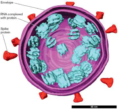
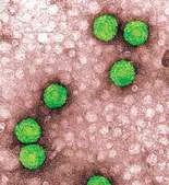
The virion of coronavirus SARS-Co This cryo-electron tomogram is colorized V-2 causes COVID-19 disease.
to represent viral structures. The envelope (purple) is a
A virus infects a host cell and uses the cell’s machinery to form progeny virions. In ecosystems, viruses cycle nutrients, control host populations, and promote host diversity. Viruses may kill their host cells or they may copy themselves into their host genome. In humans, endogenous viral DNA evolved into many portions of our genome. Deadly viruses can be engineered as vectors for lifesaving Most of the viruses we tend to hear about are those that cause epidemics, such as seasonal influenza outbreaks and the COVID-19 pandemic. But the majority of human-associated viruses go unnoticed, and some actually enhance our health. For example, our gut contains numerous bacteriophages that control our populations In research, viruses provide tools and model systems for our discovery of the fundamental principles of molecular biology. For example, the CRISPR antiviral defense in bacteria gives us tools to bacteriophages provides a background for the molecular biology we will encounter in Part 2 of this book (Chapters 7–12). And remarkably, we now engineer human viruses to deliver gene therapy and kill cancers (discussed in Chapter 11).
In Chapter 6 we introduce the major themes of virus structure and function and the ways that viruses manipulate host cells for their own reproduction. The molecular biology of viral infection and replication is explored further in Chapter 11. Viral disease pathology and epidemiology are discussed in Chapters 25–27.
6.1 Viruses in EcosystemsUnit 3 · Structure
The global pandemic known as COVID-19 (coronavirus disease of 2019) began with a pathogen that emerged in Wuhan, China, in December 2019. The pathogen was designated SARS-CoV-2 (severe acute respiratory syndrome coronavirus 2), a virus closely related to known coronaviruses infecting animals and humans. Evidence points to wild-caught animals in a meat market as the most probable source. In the United States, the new coronavirus eventually spread from humans to animals such as deer.
Within months of its discovery, coronavirus SARS-CoV-2 reached humans in every country in the world, including the United States ( Fig. 6.1). A nasal swab test was developed using quantitative PCR (presented in Section 6.5), and millions of tests were administered. Testing was essential, but it was not sufficient to slow transmission of what became one of the most contagious diseases we have known. To slow transmission of the virus, citizens were told to stay 6 feet apart—by, for example, staying within the boundaries of circles painted in a park. Businesses were ordered closed, air travel was restricted, and large public gatherings such as weddings were banned. Nevertheless, by January 2022 the world had suffered 500 million known cases of COVID-19 and 6 million deaths (Fig. 6.1C ). The actual death toll—and the human cost due to economic disruption—is thought to be far larger. The disease process is described in Chapter 26; here in Chapter 6, we present the cell biology of coronavirus infection (see Section 6.5).
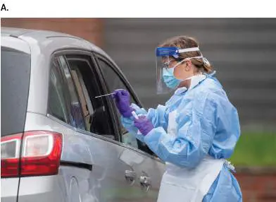

FIGURE 6.1 ■ The COVID-19 pandemic of 2020–2022. A. A nurse administers a drive-by nasopharyngeal swab test for SARS-CoV-2 coronavirus, the cause of the COVID-19 pandemic. B. Circles painted in a city park to maintain distance between family groups and thus decrease virus transmission. C. Global map of the COVID-19 pandemic, as of January 15, 2022. Disk diameters represent the cumulative number of confirmed cases in state regions. Source: Part C copyright 2022 Johns Hopkins University Center for Systems Science and Engineering, all rights reserved.
PRESS ASSOCIATION VIA AP IMAGES
VANESSA CARVALHO/SHUTTERSTOCK
What are viruses—and how can they cause such destruction, changing the course of human history? Beyond humans, all living organisms are subjected to viruses that play important roles in ecosystems by limiting populations and promoting diversity. The details of virus function are extraordinarily diverse, and this book can cover only a few examples. Table 6.1lists the viruses we cover in detail in Chapter 6, Chapter 11, and eAppendix 4.
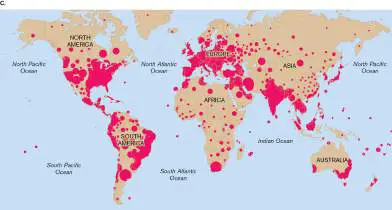
TABLE Virus Infection and Life Cycles
6.1 Covered in Detail
Chapter Host and section or Virus disease appendix Bacteriophage lambda Escherichia coli Section 11.1 lysis or lysogeny Bacteriophage M13 Escherichia coli eAppendix 4 slow release or persistent infection Bacteriophage T4 Escherichia coli eAppendix 4 lysis Caulimovirus (cauliflower Cruciferous plants Section 6.5 mosaic virus; CaMV) (cabbage family)
Hepatitis C virus (HCV) Humans: liver eAppendix 4 infection and failure Herpes simplex virus Humans: oral and Section 11.5 (HSV-1 and HSV-2) genital herpes infections Human immunodeficiency Humans: Section 11.3 virus (HIV), a lentivirus acquired immunodeficie ncy syndrome (AIDS)
Human papillomavirus Humans: warts or Section 6.5 (HPV) cancer (various strains)
Influenza A virus (IAV) Humans: related Section 11.2 variants infect birds and swine Poliovirus (enterovirus Humans: eAppendix 4 C), a picornavirus poliomyelitis, enteric and brain infection with paralysis SARS-CoV-2 Humans: severe Section 6.5 acute respiratory syndrome (SARS) in 2019 (COVID-19)
A virus is defined as a noncellular particle that infects a host cell and directs it to produce progeny particles. For example, a virus infects the tiny marine cyanobacterium Prochlorococcus (Fig. 6.2). As Prochlorococcus fixes CO 2 by photosynthesis and its population grows, viral infections spread and break down the cells, releasing their organic molecules, which other forms of life can use. Matthew Sullivan’s research shows how marine viruses play a global role in cycling carbon, nitrogen, and sulfur (discussed in Chapter 22). FIGURE 6.2 ■ Bacteriophage (phage; bacterial virus) infection. A. Prochlorococcus bacteria infected by bacteriophage P-SSP7 (cryo-EM section, colorized). B. Matthew Sullivan at Ohio State University isolates marine bacteriophages and dissects their replication cycles. C. The bacteriophage that infects Prochlorococcus. D. To infect a bacterium, the phage inserts its DNA into a host cell. The phage DNA directs the cell to make progeny phages. Source: Parts A and C modified from K. Murata et al. 2017. Sci. Rep. 7 :44176.
K. MURATA ET AL. 2017. SCI. REP. 7 :44176, FIG. 1B
K. MURATA ET AL. 2017. SCI. REP. 7 :44176, FIG. 5A
THE OHIO STATE UNIVERSITY
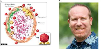
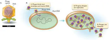
The virus particle, also called a virion, generally consists of a viral genome (DNA or RNA) contained within a protein capsid (Fig. 6.2C ). The capsid may or may not contain appendages such as a spike and tail fibers. It must possess a mechanism to get its genome into the host cell, either by insertion (Fig. 6.2D ) or by entry of the entire virus (most viruses of eukaryotes).
In the oceans, viruses were once thought to be inconsequential because researchers used host strains to try to isolate viruses and found none. But the host organisms used by most researchers were not common in marine waters. In the late 1980s, researchers started concentrating seawater and looking at it— microscopy revealed some 10 million virus-like particles per milliliter of water! In fact, viruses are the dominant consumer of marine microbes. These include viruses of bacteria, called bacteriophages or phages. For example, the virus infecting the bacterium Prochlorococcus in Figure 6.2is a bacteriophage, known as phage P-SSP7.
Viruses Infect Specific Hosts
Different types of viruses infect different kinds of host cells. For example, bacteriophage T2 infects a specific strain of the bacterium Escherichia coli. Within the host, the phage genome directs production and assembly of progeny virions (Fig. 6.3A). Virions are released when the host cell lyses. As cells lyse, their disappearance can be observed as a plaque, a clear spot within a lawn of bacterial cells (Fig. 6.3B ). Each plaque arises from a single virion, or phage particle, that lyses a host cell and spreads progeny to infect adjacent cells. The plaque count represents the number of individual infective virions from the phage suspension that was spread on the plate.
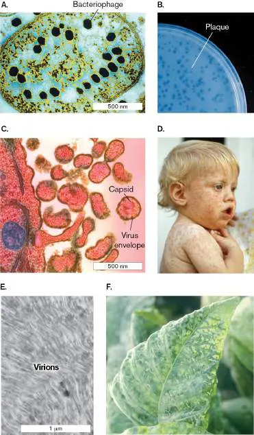
FIGURE 6.3 ■ Virus infections and disease. A. Bacteriophage T2 particles pack in an array within an Escherichia coli cell (TEM). B. Bacteriophage infection forms plaques of lysed cells on a lawn of bacteria. C. Measles virions bud out of human cells in tissue culture (TEM). D. Child infected with measles shows a rash of red spots. E. Tobacco leaf section is packed with tobacco mosaic virus (TMV) particles. F. Tomato leaf infected by TMV shows mottled appearance.
LEE D. SIMON/STAMMERS/SCIENCE SOURCE
DR. EDWARD CHAN/VISUALS UNLIMITED, INC.
NIBSC/SCIENCE SOURCE
LOWELL GEORGIA/SCIENCE SOURCE
B. ZECHMANN ET AL. 2011. J. VIS. EXP. 56:E2950
NORM THOMAS/SCIENCE SOURCE
A virus that infects humans is the measles virus (Fig. 6.3C ). The measles virus has an envelope that is derived from the host cell plasma membrane as the virus exits the host cell. When the virus infects a new host cell, the envelope fuses with the host cell plasma membrane, releasing the viral contents into the cytoplasm of the cell. After replicating within the infected cell, newly formed measles virions become enveloped by host cell membrane as they bud out of the host cell. The spreading virus causes an immune reaction with a rash of red spots on the skin of infected patients (Fig. 6.3D ). Measles illness generally resolves without treatment, but infections can be fatal (one in 500 cases).
Plants are infected by viruses such as tobacco mosaic virus (TMV). Within the plant cell, virions accumulate to high numbers (Fig. 6.3E ) and travel through interconnections to neighboring cells. Infection by tobacco mosaic virus results in mottled leaves and stunted growth (Fig. 6.3F ). Plant viruses cause major economic losses in agriculture worldwide.
Are viruses “alive”? Historically, viruses were defined as nonliving particles, because at first the virion was the only form to be visualized, and isolated virions behaved like inert chemicals. The Russian botanist Dmitri Ivanovsky (1864–1920) and the Dutch microbiologist Martinus Beijerinck (1851–1931) first proposed the existence of viruses as infectious agents that passed through a filter too small for cells to pass. Certain kinds of these infectious agents were actually crystallized from solution. Wendell Stanley (1904–1971) earned the 1946 Nobel Prize in Chemistry for the first crystallization of a virus, tobacco mosaic virus (TMV). He later crystallized poliovirus. The crystallization of viruses reinforced their definition as nonliving.
But what happens after a virus infects a cell? The viral components interact with the cell’s parts in a dynamic way, fully part of the cell’s living processes. The replication cycles of bacteriophages were first studied by English bacteriologist Frederick William Twort (1877–1950) and by French microbiologist Félix d’Herelle (1873– 1949). To actually reveal what went on in the host cell required electron microscopy (discussed in Chapter 2).
Integrated Viral Genomes
Some viruses do more than replicate within a cell: They can integrate their own genomes into the host genome. In effect, such viruses become a part of the host organism. A virus that integrates its genome into the DNA of a bacterial genome is called a prophage. Viruses transfer enormous quantities of genes among microbial populations, such as the genes for toxin production acquired by human-associated bacteria. Within a human cell, an integrated viral genome is called a provirus. A permanently integrated provirus transmitted via the germ line is called an endogenous virus. Remarkably, our human genome includes endogenous viral genomes that express essential human genes.
How were integrated viral genomes discovered? In the 1950s, genetic analysis of bacteriophage lambda in E. coli showed evidence of phage gene expression from within host genomes.
Bacteriophage lambda is discussed at length in Chapter 11 . Early in the twentieth century, American cancer researcher Peyton Rous (1879–1970) discovered that some avian viruses cause tumors. The tumor generation results from integration of a viral genome. For this discovery, Rous won the 1966 Nobel Prize in Physiology or Medicine.
But the biggest surprise emerged from sequencing the genomes of host organisms. Many host cell traits are expressed by integrated viral genomes that actually confer benefits on their host. For example, Nostoc cyanobacteria possess a prophage that encodes nitrogenase, an enzyme for nitrogen fixation. Many human pathogens, such as Staphylococcus aureus, contain prophages that encode toxins that enhance the pathogen’s virulence. At the same time, human genomes show many genes from viral genomes that integrated into host DNA during the course of our primate evolution. Over time, these viral sequences mutated and got “stuck” in the host. As much as half of the human genome may have originated from viruses or from virus-like elements of DNA called transposons (discussed in Chapter 9). For example, some integrated viral genomes express placental proteins that are essential for early development of human embryos.
Dynamic Nature of Viruses
Emerging evidence supports a dynamic view of viral existence and leads us to revisit the question: What is a virus? Some viruses may interconvert among very different forms (Fig. 6.4): FIGURE 6.4 ■ Virus as a subcellular organism. The virion, or virus particle, consists of a nucleic acid genome contained by a protein capsid. A virion may infect a host cell and form a virocell that synthesizes progeny virions. Alternatively, infection may lead to integration of the viral genome within the host cell genome. Integration may last indefinitely or else may lead to production of virions.
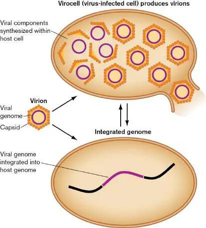
Virion, or virus particle. The virion is an inert particle consisting of nucleic acid enclosed by a protein capsid. Some viruses package enzymes and possess a lipid envelope. A virion does not carry out any metabolism or energy conversion.
Intracellular replication complex. Within a host cell, the viral gene products direct the cell’s enzymes to assemble progeny virions (discussed in Section 6.5). Virus assembly requires recruitment of host ribosomes, as well as intricate collaboration between host and viral proteins. This virus-directed cell may be called a virocell.
Viral genome integrated within host genome (provirus).
Some types of viral genomes may integrate within a host chromosome as a provirus and replicate as part of the host. Integration may be permanent; alternatively, the viral genome may be reactivated to start assembling virions.
The inert nature of the virion (particle), which lacks metabolism, argues that viruses are nonliving. However, we know that cellular life forms such as bacteria can convert into inert forms, such as endospores, that remain viable for thousands of years. The virion assembly process argues that viruses are living organisms. Assembly includes metabolism and production of progeny, processes we see for obligate intracellular bacteria such as chlamydias. Furthermore, the genomes of large viruses such as chloroviruses show evidence of reductive evolution (evolutionary loss of genes) from a cell. On the other hand, genome integration argues for a view of viruses as host cell components. Many bacteria regulate phage-encoded genes along with their own genes (discussed in Chapter 10 ). For example, Vibrio cholerae, the cause of cholera, uses bacterial genes to regulate expression of the cholera toxin, which is encoded by an integrated prophage. For the V. cholerae bacterium, the viral prophage is a functional part of the cell. Viruses that enter and leave microbial genomes can transfer valuable genes among different species, conferring traits such as the ability to metabolize new food sources. For example, bacteriophages can transfer genes encoding catabolic enzymes for complex carbon sources with soil or gut microbiomes. In the ocean, viruses transfer genes for photosynthesis and catabolism that enhance virocell production of viruses. How and where did viruses originate? Did they form within host cells or did they arise as independent entities? Giant viral genomes encode numerous metabolic enzymes, including aminoacyl-tRNA synthetases (enzymes that attach an amino acid to the transfer RNA for translation). The size and complexity of giant viruses argue that these viruses evolved by reductive evolution of an intracellular parasitic bacterium. Similar arguments are made for large viruses such as the smallpox viruses and herpesviruses. On the other hand, small viruses with small RNA genomes, such as influenza virus and human immunodeficiency virus (HIV), look more like something that arose from parts of a cell. For example, the key retroviral enzyme reverse transcriptase (which copies RNA to DNA) shows homology to telomerase, a host cell enzyme that maintains chromosome ends.
Note: Some scientists consider viruses “not alive.” Other scientists
consider viruses a noncellular life form that requires a host cell for replication.
Viruses Limit Host Populations and Increase Diversity
In ecosystems, acute viruses (which rapidly kill their hosts) act as predators or parasites to limit host population density. They also recycle nutrients from their host bodies. An increase in host population density increases the rate of transmission of viral pathogens. As the host population declines, viruses are less likely to find a new host before they lose infectivity, while many of the remaining hosts have undergone selection for resistance. Thus, viruses can limit host density without causing extinction of the host. The sum of viral populations in an ecosystem is called the virome. In a host community, the overall effect of the virome is to increase host diversity. Each virus species has a limited host range and requires a critical population density to sustain the chain of infection. In marine phytoplankton, a virus limits its particular host species to a population density far lower than what would be sustainable by the available resources without competition. The resources then support other species resistant to the given virus (but susceptible to other ones). Thus, overall, marine viruses prevent the dominance of any one species and foster the evolution of many distinct host species. The continual threat of marine viral epidemics explains the great diversity of phytoplankton ranging from silica-shelled diatoms to toxin-producing dinoflagellates.
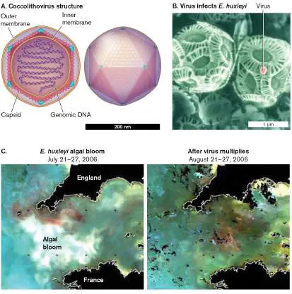
Viruses can dissipate marine algal blooms large enough to see from outer space. Figure 6.5shows an example of a marine virus infecting cells of the alga Emiliana huxleyi. These algal cells, called coccolithophores, are known for their calcite plates, or “coccoliths,” which coat each cell. The virus particle (Fig. 6.5A) consists of a polyhedral protein capsid with outer and inner membranes, containing a coiled DNA genome. The virus attaches one of its corner proteins to the surface of a coccolith and inserts its DNA. When coccolithophores overgrow, they can generate a bloom that covers the ocean for thousands of square kilometers (Fig. 6.5C ). In the figure, the pale clouds in the water are the reflected light from billions of coccoliths. Yet within a few days, this gigantic bloom is wiped out by viruses.
FIGURE 6.5 ■ Virus infection controls marine algal bloom. A. Coccolithovirus structure. This virus infects the coccolithophore, a eukaryotic alga, Emiliana huxleyi. B. E. huxleyi infected by Coccolithovirus. C. Left: Bloom of E. huxleyi off Plymouth, England, detected by the MODIS imaging sensor aboard a NASA satellite while it was remote-sensing light reflectance from chlorophyll. Right: Image after virus infection. Source: Part A modified from https://viralzone.expasy.org/589.
NATIONAL SCIENCE FOUNDATION
A. HIGHFIELD ET AL. 2014. VIROLOGY 466–67 :138
Viruses Cycle Nutrients and Share Host Genes
Throughout the ocean, viruses play a decisive role in controlling algal blooms. By lysing the algae as they grow, viruses return algal carbon and minerals to the surface water before the algae starve to death and their bodies sink. When biomass sinks to the ocean depths, its minerals become unavailable for phototrophs and their consumers. Thus, viruses are major players in the cycling and sequestering of atmospheric CO 2.
In marine ecosystems such as coral reefs, viruses play several important roles, as shown by the field research of Rebecca Vega Thurber from Oregon State University (Fig.
6.6 ). Most critically, herpes-related viruses infect coral and lead to coral bleaching. Coral bleaching occurs when corals expel their symbiotic algae, turn white, and ultimately die from lack of carbon fixation. Coral bleaching now affects coral reefs all over the planet, a process exacerbated by global warming. The coral-associated virome includes different viruses infecting coral tissues, coral endosymbionts, and coral-associated bacteria.
FIGURE 6.6 ■ Viruses cycle nutrients in coral reef ecosystems. A. Bacteriophages and eukaryotic viruses convert marine organisms into dissolved organic molecules and particles. The organic particles settle to the benthos (marine sediment), where they provide carbon and nitrogen for corals and other
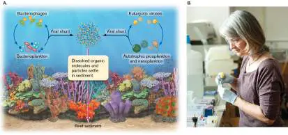
marine life. B. Rebecca Vega Thurber studies the virome of coral reefs.
Source: Part A modified from R. V. Thurber et al. 2017. Nat. Rev. Microbiol. 15
:205.
OREGON STATE UNIVERSITY
The most widespread role of marine viruses is the cycling of food molecules (Fig. 6.6A). Viral infection and lysis convert the bodies of phytoplankton (photosynthetic microbes) and consumers into detritus consisting of small organic particles and soluble molecules. The virus-generated detritus gets taken up by consumers (heterotrophs), and minerals are absorbed by phototrophs. This viral generation of detritus is called the viral shunt. The viral shunt returns some organic matter to microbial consumers in the upper region of the ocean (discussed in Chapter 21). Some carbon is thus diverted from the ocean sink and returns to the CO 2 cycle. The viral shunt also plays a major part in marine cycling of phosphorus, nitrogen, and iron.
Thought Question
6.1 Suppose a certain virus depletes the population of an algal bloom. What will happen if some of the algae are genetically resistant?
Persistent viruses (viruses that don’t immediately kill the host) may benefit the host population overall. If a virus can evade the host defenses, it may replicate, and thus persist, in the hosts. Humans harbor many obscure herpesviruses and cold-type viruses that stimulate normal development of the immune system. In natural mammal populations, viruses that have been shed from persistently infected mammals can infect and kill uninfected competitors of the same species. The loss of competitors means more food and territory for the infected host. Thus, a persistent virus can act as a bioweapon that kills off competitor populations susceptible to the virus. An example is the koala retrovirus, KoRV, which has become endogenous (persistent) in many koala populations of Australia. When KoRV is transmitted to koalas that lack the retrovirus, it kills most of those newly infected—except for the few in which the virus persists. In these infected animals, the immune system controls the virus but cannot eradicate it.
Integrated viral genomes can evolve complex symbiotic interactions with multiple hosts, combining mutualism and parasitism. An example is the relationship of polydnaviruses, wasps, and caterpillars (Fig. 6.7). Parasitoid wasps lay their eggs inside a living insect caterpillar, where the larvae must hatch and feed without stimulating the caterpillar’s defense. The wasp genome contains an integrated genome (or provirus) of a polydnavirus. The polydnavirus forms its own genomes and virions only within the wasp ovary. When the wasp deposits her eggs inside the host caterpillar, she also deposits her mutualistic virions. The virions express proteins that prevent the caterpillar cells from undergoing encapsulation, a process that would otherwise wall off the wasp eggs and kill them.
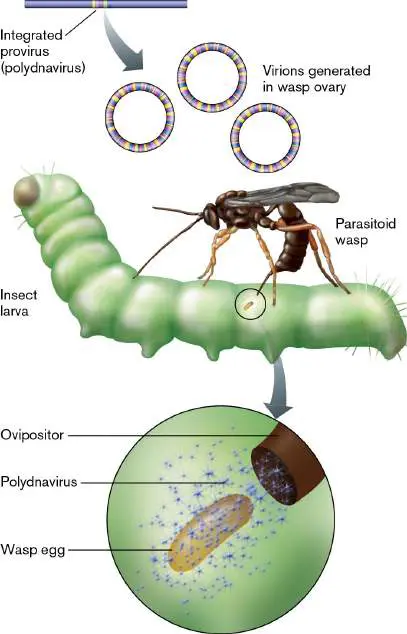
FIGURE 6.7 ■ The relationship of polydnaviruses, wasps, and caterpillars. Parasitoid wasps lay their eggs inside a living insect caterpillar. When a female wasp deposits her eggs inside the caterpillar, she also deposits her symbiogenic polydnavirus virions. The virions express wasp genes in the caterpillar, where they prevent the encapsulation process that would otherwise wall off the wasp egg and kill it.
Viral Disease
When viral infection causes harm to an animal or plant, the host has a disease. The range of host species infected by a given virus is known as its host range. Some viruses can infect only a single host species; for example, human immunodeficiency virus (HIV) infects only humans. Close relatives of humans, such as the chimpanzee, are not infected by HIV, although they are susceptible to a highly related virus, simian immunodeficiency virus (SIV). By contrast, West Nile virus, transmitted by mosquitoes, infects many species of birds and mammals. West Nile virus has a much broader host range than HIV and SIV have.
For humans, viruses cause many forms of illness. The impact of viral diseases on human history and culture would be hard to overstate. More people died of influenza in the global epidemic of 1918 than in the battles of World War I. Poliovirus is famous for causing the outbreaks of poliomyelitis that swept the United States during the first half of the twentieth century. President Franklin Roosevelt, himself a victim of the disease, established a national foundation called the March of Dimes to develop a vaccine. The spectacular success of the March of Dimes set the pattern for future public support of research on cancer and AIDS. In 2020, the COVID-19 pandemic devastated social life and economies around the globe, and vaccines were developed in record time (see Section 16.6). Chronic viral infections, however, are more common than acute disease and are an everyday part of our lives. The most frequent infections of college students are due to respiratory pathogens such as rhinovirus (the common cold) and Epstein-Barr virus (infectious mononucleosis), as well as sexually transmitted viruses such as herpes simplex virus (HSV) and papillomavirus (genital warts). Some viruses, such as rhinoviruses, are eliminated rapidly by our immune system, whereas others, such as herpesviruses, establish a lifelong latent infection. Viruses also affect human industry; for example, bacteriophages (literally, “bacteria-eaters”) infect cultures of Lactococcus during the production of yogurt and cheese. Plant pathogens such as cauliflower mosaic virus and rice dwarf virus cause substantial losses in agriculture.
In contrast to our vast arsenal of antibiotics (effective against bacteria), the number of antiviral drugs remains small. Because the machinery of viral growth is largely that of the host cell, viruses present relatively few targets that can be attacked by antiviral drugs without harming the host. An exception is HIV, the cause of AIDS. Molecular studies of HIV have yielded several major classes of antiviral drugs, such as AZT and protease inhibitors. The molecular biology behind anti-HIV drugs is discussed in Chapters 11 and 27.
Thought Question
6.2 Search the Internet for specific viruses: Can you find viruses that have a narrow host range and others that have a broad host range?
To Summarize
A virus particle, or virion, has a nucleic acid genome enclosed by a protein capsid. Within a host cell, the viral genome can generate an intracellular replication complex, forming a virocell that produces virions. Alternatively, the genome sequence can become integrated within host cell DNA. All classes of organisms are infected by viruses. Usually the hosts are limited to a particular host range of closely related strains or species.
Are viruses living or nonliving? A virion consists of an inert particle lacking metabolism. But some viral genomes resemble those of cells, and viral replication directs a metabolic program within the host cell.
Acute viral infection limits host population density. Virus-associated mortality may increase the genetic diversity of host species.
Persistent viruses remain in hosts , where they may evolve traits that confer positive benefits in a virus-host mutualism.
Marine viruses infect most phytoplankton, releasing their minerals in the upper water, where they are available for other phototrophs. Viral activity substantially affects the global carbon balance.
Viruses transfer genes between host genomes. Viral gene transfer is a major source of cellular genome evolution.
Glossary
virus A noncellular particle containing a genome that can replicate only inside a cell.
virion A virus particle.
capsid The protein shell that surrounds a virion’s nucleic acid. Within an enveloped virus, such as HIV, the capsid may be called a core particle.
bacteriophage Also called phage. A virus that infects bacteria.
phage See bacteriophage .
plaque A cell-free zone on a lawn of bacterial cells caused by viral lysis. virion A virus particle.
prophage A phage genome integrated into a host genome.
provirus A viral genome that is integrated into the host cell genome. provirus A viral genome that is integrated into the host cell genome. endogenous virus A virus whose genome is encoded within the germ line of a host animal; may be considered a functional part of the host. virocell A cell infected by a virus that reprograms it to maximize production of virus particles.
virome The genomes of all the viruses that inhabit a particular organism or environment.
viral shunt The release by viral lysis of cell contents as organic material available for microbial consumers in the upper region of the ocean.
host range The species that can be infected by a given pathogen.
6.2 Virus StructureUnit 3 · Structure
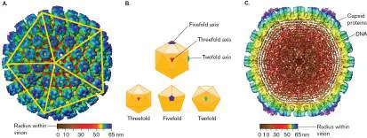
A virion possesses a genome of either DNA or RNA that is contained by proteins composing the capsid. The shape of the virion depends on the species of virus. The virion may be symmetrical or asymmetrical or combine aspects of both. For example, herpesvirus has a DNA genome spooled within an icosahedral (20-sided) capsid (Fig. 6.8). The papillomavirus virion consists solely of a capsid containing a genome, whereas the tailed bacteriophages have an elaborate delivery device to transfer the viral genome into the host cell. FIGURE 6.8 ■ Herpesvirus: icosahedral capsid symmetry. A. Icosahedral capsid of herpes simplex virus 1 (HSV-1), with envelope removed. Imaging of the capsid structure is based on computational analysis of cryo-transmission electron microscopy (cryo-TEM). Images of 146 virus particles were combined digitally to obtain this model of the capsid at 2-nm resolution. B.
Icosahedral symmetry includes fivefold, threefold, and twofold axes of rotation. C. The icosahedral capsid contains spooled DNA. Source: Parts A and C modified from C. Z. Hong Zhou et al. 1999. J. Virol. 73
:3210.
Many kinds of viruses possess a membrane envelope that is derived from membrane of the previous host cell. For example, an
envelope encloses the capsid of a herpesvirus. The protein capsid of an enveloped virus is called a core particle. Some viruses such as coronavirus contain no icosahedral capsid but have an RNA genome wound around nucleocapsid proteins, which overall is enclosed by an envelope membrane. The membrane typically incorporates viral proteins that mediate host infection (discussed shortly).
The structure of a virion keeps its genome intact, and it enables infection of the appropriate host cell. First, the stable capsid protects the viral genome from degradation and enables the genome to be transmitted outside the host. Second, in order for the viral genome to reproduce, the virion must either insert its genome into the host cell or disassemble within the host. In the process, the original particle loses its stable structure and its own identity as such. But if viral reproduction succeeds, then it yields numerous progeny virions. Understanding virus structure is crucial for devising vaccines and drug therapies. For example, the Gardasil vaccine for human papillomavirus, HPV (recommended for all children before adolescence), is composed of capsid proteins from nine different HPV strains. The molecular basis of viral structure and drugs is presented in Chapter 11.
Thought Question
6.3 What will happen if a virus particle remains intact within a host cell and fails to release its genome?
Symmetrical Virions
The capsid of a symmetrical virus may be one of two types: icosahedral or filamentous (helical). Some capsids have an intermediate form, such as the conical form of an HIV core particle. The advantage of geometrical symmetry is that it provides a way to form a package out of repeating protein units generated by a small number of genes and encoded by a short chromosomal sequence. The smaller the viral genome, the more genome copies can be synthesized from the host cell’s limited supply of nucleotides. Nevertheless, some symmetrical viruses, such as herpesvirus and Mimivirus, have much larger genomes. Large genomes offer a greater range of functions for viral components.
Icosahedral viruses. Many viruses such as herpesvirus package their genome in an icosahedral capsid (Fig. 6.8A) . An icosahedral capsid takes the form of a polyhedron with 20 identical triangular faces. In the capsid, each triangle can be composed of three identical but asymmetrical protein units. Each triangular face of the capsid is determined by the same genes encoding the same protein subunits. The actual form of viral subunits can vary greatly, generating very different complex shapes for different virus species. But no matter the pattern of subunits in the triangular unit, the structure overall shows the rotational symmetry of an icosahedron (Fig. 6.8B ): threefold symmetry around the axis through two opposed triangular faces, fivefold symmetry around an axis through opposite points, and twofold symmetry around an axis through opposite edges.
Thought Question
6.4 For a viral capsid, what is the advantage of an icosahedron (20-sided solid), as shown in Figure 6.8, instead of some other polyhedron, such as a cube or a tetrahedron?
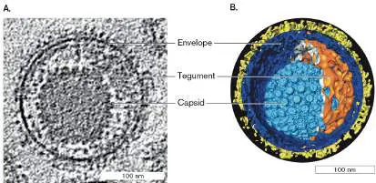
Virus particles can be observed by standard transmission electron microscopy (TEM), but the details of capsid structure (as in Fig. 6.8) require digital reconstruction of cryo-electron microscopy (cryo-EM) images (discussed in Section 2.6). Recall from Chapter 2 that in cryo-EM, the viral samples for TEM are flash-frozen, allowing high-voltage electron beams to image a cell without stain. The images show the internal contents of individual virions. By digitally combining and processing cryo-transmission electron microscopy (cryo-TEM) images from a number of capsids, we build a 3D reconstruction for the entire virus particle. Within the icosahedral capsid, the herpesvirus genome is spooled tightly (Fig. 6.8C ). During virion synthesis, the viral DNA is packaged under high pressure. A molecular motor powered by ATP drives viral DNA into the viral capsid (discussed in Chapter 11). Viruses that possess an envelope include membrane from the host cell in which the virion formed; the membrane contains embedded proteins specified by the viral genome. The envelope and capsid contents of herpesvirus are shown in Figure 6.9. A coronavirus such as SARS-CoV-2 has a different structure (see the chapter-opening image) in which the RNA genome is packaged in bead-like nucleoprotein complexes. The nucleoprotein-complexed RNA is then spooled within an envelope containing embedded spike proteins. The famous coronavirus spike protein (protein S) enables it to infect human cells very efficiently, giving rise to the COVID-19 pandemic. FIGURE 6.9 ■ Envelope and tegument surround the herpesvirus capsid. A. Section showing envelope and tegument proteins surrounding the capsid (cryo-EM). B. Cutaway reconstruction of the herpes virion (cryo-EM tomography).
NATIONAL INSTITUTE OF ARTHRITIS AND MUSCULOSKELETAL AND SKIN DISEASES
ALASDAIR STEVEN, NIH
The space between the capsid and the envelope contains “tegument” proteins, such as enzymes that interact with the host cell to depress its defense responses. Tegument proteins are expressed during infection of a host cell and then get packaged in the virion. Both viral and host proteins may be packaged as tegument. The mature envelope bristles with glycoprotein spike proteins that attach it to the capsid. The spike proteins enable the virus to attach to and infect the next host cell. Further details of herpesvirus structure and function are presented in Section 11.5.
Note: Distinguish the viral envelope (phospholipid membrane
derived from a host cell membrane) from the bacterial cell envelope (protective layers outside the bacterial cell membrane). The bacterial envelope is discussed in Chapter 3.
Filamentous viruses. A second major category of virus structure is that of filamentous viruses. Unlike an icosahedral capsid, which usually has a fixed size, a helical capsid can vary in length to accommodate different lengths of nucleic acid.
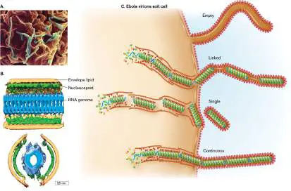
A well-known filamentous virus is Ebola virus (Fig. 6.10), which causes a fatal disease of humans and related primates. Ebola virus caused a major epidemic in western Africa in 2014, and it sparked another outbreak in 2018 (discussed in Chapter 28). The virus has become known for the iconic micrographs of its long, twisted filaments. Cryo-EM tomography reveals the helical form of the RNA genome, packaged in nucleocapsid proteins (Fig. 6.10B ). Tandem arrays of multiple encapsidated Ebola genomes are contained loosely within the viral envelope, which can therefore twist into the flexible forms seen in Figure 6.10A. Timothy Booth’s lab at the National Microbiology Laboratory, Winnipeg, Canada, showed how the process of enveloping nascent Ebola virions generates filaments that may contain a single genome, multiple linked genomes, or even an empty filament (Fig. 6.10C ).
FIGURE 6.10 ■ Filamentous viruses. A. Ebola virions budding out of a cell (SEM). B. Cryo-EM tomogram showing 3D structure of the Ebola virion (top: side view; bottom: end view). C. Ebola virions budding out as tandem arrays of multiple RNA genomes packaged in the nucleocapsid, held together within a flexible lipid envelope.
AMI IMAGES/SCIENCE SOURCE
TIM BOOTH/PHAC/ASPC
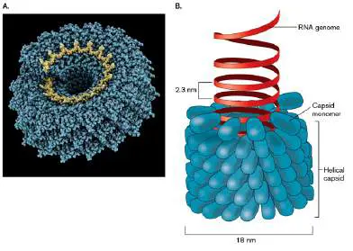
At the molecular level, filamentous viruses show helical symmetry. The pattern of capsid monomers forms a helical tube around the genome, which usually winds helically within the tube. In a helical capsid, the genome is a single-stranded RNA (as in Ebola virus or tobacco mosaic virus) or DNA (as in bacteriophage M13, presented in eAppendix 4). Figure 6.11shows how the RNA strand of tobacco mosaic virus (TMV) winds in a spiral within a tube of capsid monomers laid down in a spiral array. Unlike Ebola, TMV has no lipid
envelope—just the RNA genome wound within a tube of protein. Such a tube can be imagined as a planar array of subunits that coils around such that each row connects to the row above, generating a spiral. The length of the helical capsid may extend up to 50 times its width, generating a flexible filament.
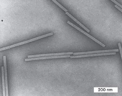
FIGURE 6.11 ■ Tobacco mosaic virus: helical symmetry. A. The helical filament of tobacco mosaic virus (TMV) contains a single-stranded RNA genome coiled inside (X-ray model). Inset: TMV virions (TEM). B. Components of the TMV virion.
DR. JOHN FINCH/SCIENCE SOURCE
LAGUNA DESIGN/SCIENCE SOURCE
Filamentous phages cause problems for human medicine and industry. One infects the Gram-positive species Propionibacterium freudenreichii, a key fermenting agent for Swiss cheese. Another filamentous phage, CTXphi (CTXφ), integrates its sequence into the genome of Vibrio cholerae, where it carries the deadly toxin genes required for cholera. On the other hand, filamentous phages such as M13 have been used for biotechnology, to clone products as proteins fused to the subunits of the helical capsid. Another application, in nanotechnology, is the use of a filamentous phage to nucleate the growth of crystalline “nanowires” for electronic devices.
Asymmetrical Virions
Many viruses have a capsid, or a core particle, that is asymmetrical. An important group of viruses with asymmetrical capsids is the poxviruses, such as vaccinia (the source of smallpox vaccine, derived from cowpox virus). In 2022, the monkeypox virus (now called mpox) emerged as a global pathogen spread by intimate contact.
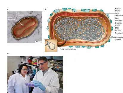
Monkeypox transmission can be prevented by smallpox vaccines. The virion of a poxvirus consists of an oval-shaped particle nearly as large as a cell (200–300 nm). Vaccinia has a relatively large double-stranded DNA genome of 190 kilobases (kb), encoding 250 genes. The DNA is stabilized by covalent connection of its two strands in a loop at each end (Fig. 6.12Aand B ). The core envelope encloses the nucleocapsid-coated DNA, as well as a large number of accessory proteins. The accessory proteins are needed early in viral infection, such as initiation proteins for the transcription of viral genes and RNA-processing enzymes that modify viral messenger RNA (mRNA) molecules. The proteins may be found either inside the capsid or in the tegument between the core envelope and the outer membrane, which is coated with surface tubules of protein. Large asymmetrical viruses contain so many enzymes that they appear to have evolved from degenerate cells.
FIGURE 6.12 ■ Vaccinia poxvirus. A. Vaccinia virion observed in aqueous medium (colorized TEM). B. A pox virion includes an outer membrane and a core envelope membrane containing envelope proteins enclosing the double-stranded DNA genome and accessory proteins. The DNA is stabilized by a hairpin loop at each end. C. Allison Titong and Paulo Verardi examine vaccinia virus in tissue culture to develop advanced vaccines and cancer treatments.
J. HEUSER. 2005. J. CELL BIOL. 169 (2). REPUBLISHED WITH PERMISSION OF
ROCKEFELLER UNIVERSITY PRESS
PETER MORENUS/UCONN PHOTO
Vaccinia virus offers opportunities for new applications, such as conversion to vaccines for other viruses, and engineering to combat tumors. For example, students in Paulo Verardi’s laboratory at the University of Connecticut (Fig. 6.12C ) engineered an on/off switch for vaccinia that will make the virus safer for immunocompromised patients. The switch involves a recombinant tetracycline repressor
gene, whose product confers resistance to tetracycline. Repressor controls and engineering are discussed in Chapters 10 and 12. Another class of asymmetrical viruses is influenza (molecular details are presented in Chapter 11). Influenza viruses are RNA viruses in which a set of unique RNA segments is coated with nucleocapsid proteins. The influenza virus packages the different RNA segments into separate helical packages of different sizes, contained together within a membrane envelope. Separate chromosome packaging enables influenza virus to make “mistakes” and pack different numbers of RNA segments into different virions. The process enables rapid evolution of new strains (see Chapter 11).
Tailed Viruses
Many bacteriophages supplement the icosahedral capsid or head coat with an elaborate delivery device. For example, bacteriophage T4 ( Fig. 6.13) has an icosahedral “head” containing the pressure-packed DNA, attached to a helical “neck” that channels the nucleic acid into the host cell. The tail baseplate has six jointed tail fibers that stabilize the structure on the host cell surface. After phage infection, the production of virions within a cell requires a factorylike assembly line of phage tail parts (described for phage T4 in eAppendix 4).
FIGURE 6.13 ■ Bacteriophage T4 capsid. A. Phage T4 particle with protein capsid containing packaged double-stranded DNA genome. The capsid has a sheath with tail fibers that facilitate attachment to the surface of the host cell. After
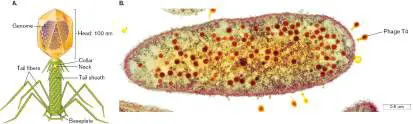
attachment, the sheath contracts and the core penetrates the cell surface, injecting the phage genome. B. E. coli infected by phage T4 (colorized brown; TEM).
M. MAEDER/DEPT. OF MICROBIOLOGY, BIOZENTRUM/SCIENCE SOURCE
Viroids
Are there infectious agents even simpler than small viruses? For some infectious agents, the nucleic acid genome is itself the entire infectious particle; there is no protective capsid. Such agents are called viroids, which are infectious agents of plants. A viroid encodes no genes, but it hijacks the plant cell’s RNA polymerase to replicate itself. Viroids infect many kinds of fruits and vegetables, entering the plant cell through a damaged cell wall. For example, citrus viroids cause economic losses in the citrus industry.
The potato spindle tuber viroid (Fig. 6.14) consists of a circular, single-stranded molecule of RNA that doubles back on itself to form base pairs interrupted by short, unpaired loops. This unusual circularized form avoids breakdown by host RNase enzymes. The RNA folds up into a globular structure that interacts with host cell proteins. Host RNA polymerase replicates the RNA. The host RNA polymerase normally requires a DNA template, but the replication process is modified during viroid infection. During infection, the RNA polymerase thus replicates progeny copies of the viroid, which encodes no products other than itself. Viroids can cause as much host destruction as “true viruses,” and some authors, particularly in plant pathology, classify them as “viruses without capsids.”
FIGURE 6.14 ■ Viroids: infective RNA. The potato spindle tuber viroid consists of a circular single-stranded RNA (ssRNA) that hybridizes internally.
Some viroids have catalytic ability, comparable to that of enzymes made of protein. An RNA molecule capable of catalyzing a reaction is called a ribozyme (discussed in Chapter 9). Viral ribozymes may be able to cleave themselves or other specific RNA molecules. Their ability to cleave very specific RNA sequences has applications in medical research, such as cleaving human mRNA involved in cancer.
Prions
Can an infectious agent propagate without a genome of its own? A remarkable class of infectious agents is believed to consist solely of protein. These agents, known as prions, are thought to be aberrant proteins arising from the host cell. Prions gained notoriety when they were implicated in brain infections such as Creutzfeldt-Jakob disease, a variant form of which is known as “mad cow” disease because it may be transmitted through defective proteins in beef from diseased cattle. Other diseases believed to be caused by prion transmission include scrapie, a disease of sheep, and kuru, a degenerative brain disease that was found in a tribe of people who customarily consumed the brains of deceased relatives.
In prion-associated diseases, the infective agent is unaffected by treatments that destroy RNA or DNA, such as nucleases or UV irradiation. A prion is an aberrant form of a normal cell protein that assumes an abnormal conformation or tertiary structure (Fig. 6.15A ). The prion form of the protein acts by binding to normally folded proteins of the same class and altering their conformation to that of the prion. The multiplying prion then alters the conformation of other normal subunits, forming harmful aggregates in the cell and ultimately leading to cell death. In the brain, prion-induced cell death leads to tissue deterioration and dementia (Fig. 6.15B ). Prion diseases are unique because they can be transmitted by an infective protein instead of by DNA or RNA, and they propagate the conformational change of existing molecules without synthesizing entirely new infective molecules.
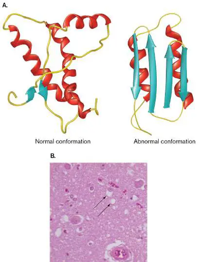
FIGURE 6.15 ■ Prion disease. A. The normal conformation of a protein, compared to the abnormal (prion) conformation. The abnormal form “recruits” normally folded proteins and changes their conformation into the abnormal form. (PDB code: 1AG2) B. Section of a human brain showing “spongiform” holes (arrows)
typical of Creutzfeldt-Jakob disease.
RALPH C. EAGLE/SCIENCE SOURCE
A prion disease can be initiated by infection with an aberrant protein. More rarely, the cascade of protein misfolding can start with the spontaneous misfolding of an endogenous host protein. The chance of spontaneous misfolding is greatly increased in individuals who inherit certain alleles encoding the protein; thus, Creutzfeldt-Jakob disease can be inherited genetically from one person with one mutant allele.
To Summarize
A viral capsid is composed of repeated protein subunits —a structure that maximizes the structural capacity while minimizing the number of genes needed for construction. The capsid, or core particle, packages the viral genome and delivers it into the host cell.
Icosahedral capsids have regular, 20-sided symmetry. Filamentous (helical) capsids have uniform width , generating a flexible filamentous virion.
Enveloped virions are enclosed within phospholipid membrane derived from the host cell. The core particle consists of a genome (RNA or DNA) packaged by nucleocapsid proteins. The core may be surrounded by tegument or accessory proteins. The envelope includes virus-specific spike proteins.
Viroids that infect plants consist of an RNA hairpin with no capsid.
Prions are infectious proteins that induce a cell’s native proteins to fold incorrectly and impair cell function.
Glossary
capsid The protein shell that surrounds a virion’s nucleic acid. Within an enveloped virus, such as HIV, the capsid may be called a core particle.
envelope A structure external to the cell membrane, such as the cell wall or outer membrane of a bacterium. For a virus, the envelope is a membrane enclosing the capsid or core particle.
core particle A viral capsid that encloses its nucleic acid genome and is surrounded by an envelope.
spike protein A viral glycoprotein that connects the membrane to the capsid or the matrix and may be involved in viral binding to host cell receptors.
spike protein A viral glycoprotein that connects the membrane to the capsid or the matrix and may be involved in viral binding to host cell receptors.
filamentous virus A viral structure type consisting of a helical capsid surrounding a single-stranded nucleic acid.
viroid An infectious naked nucleic acid.
prion An infectious agent that causes propagation of misfolded host proteins; usually consists of a defective version of the host protein.
6.3 Viral Genomes and ClassificationUnit 2 · Genomes
Viral genomes are structurally more diverse than those of cells. A given type of virus may have a genome that consists of either RNA or DNA, single-or double-stranded, linear or circular. The form of the genome has key consequences for the mode of infection and for the course of a viral disease. Viral genomes are used as the basis of virus classification.
Viral Genomes: Small or Large
Small viruses commonly have a small genome, encoding fewer than ten genes. For example, cauliflower mosaic virus (diameter 50 nm) has a genome encoding only seven genes (Fig. 6.16A), which actually overlap each other in sequence. This overlap in sequence is made possible by the use of different reading frames—start positions to define the first base of the codon for translation to an amino acid sequence (discussed in Chapter 8).
FIGURE 6.16 ■ Simple viral genomes. A. Cauliflower mosaic virus has a circular genome of double-stranded DNA, whose strands are interrupted by nicks. The genome encodes seven overlapping genes. B. Avian leukosis virus is a single-stranded RNA retrovirus resembling eukaryotic mRNA. Three genes (gag, pol, and env) encode polypeptides that are eventually cleaved to form a total of nine functional products. LTR = long terminal repeat.
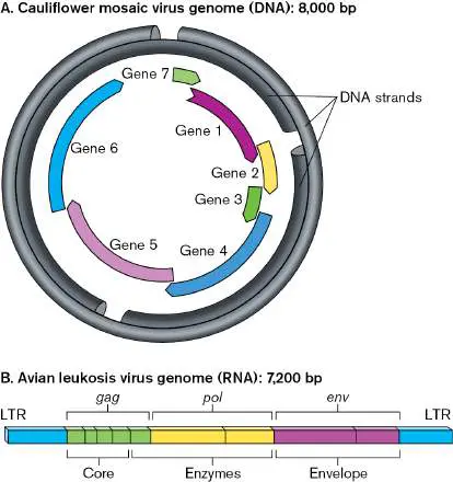
The smallest of viral genomes generally consist of RNA. RNA viruses include some of today’s most important human pathogens, such as influenza virus, hepatitis C virus, and human immunodeficiency virus (HIV). The RNA genome of avian leukosis virus (Fig. 6.16B ) has protein-encoding genes grouped by functional categories of core capsid, replication enzymes, and envelope proteins (proteins embedded in the envelope phospholipid bilayer).
At the opposite end of the scale, the “giant viruses” have genomes of double-stranded DNA comprising 300–2,500 genes. A giant virus called Mimivirus (diameter 300 nm), which infects amebas and may cause human pneumonia, is itself as large as some bacteria and has a bacterium-sized genome of 1.2 million base pairs, encoding more than 1,000 genes (Fig. 6.17). This genome is twice the size of genomes of some bacteria such as Mycoplasma genitalium.
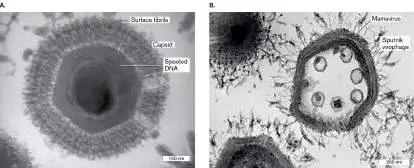
FIGURE 6.17 ■ Genome of Mimivirus. The genome of this giant virus specifies numerous enzymes with cell functions. Discovered in 2003 by French virologist Didier Raoult and colleagues, Mimivirus has a genome that encodes numerous cell functions, including DNA repair and protein folding by chaperones. The virus gains entry to its ameba host by phagocytosis, because its large particle size makes the virus particle resemble a bacterium that the ameba could engulf for food. Once taken up, the mimivirus capsid opens to release viral enzymes and DNA. Within the ameba’s cytoplasm, the viral enzymes generate a replication complex that produces progeny mimiviruses. Mimiviruses are so large that they can actually become infected themselves by smaller viruses, such as the Sputnik virophage, or “virus-eater” (Fig. 6.18).
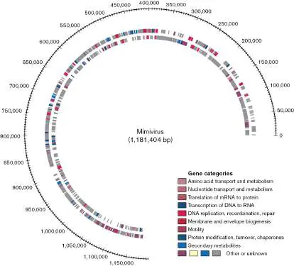
FIGURE 6.18 ■ Giant virus infecting an ameba. A. Mimivirus is larger than some bacteria (TEM). B. Sputnik virophage infects Mamavirus, a relative of Mimivirus (TEM).
DR. RAOULT/SCIENCE SOURCE
DIDIER RAOULT/CNRS
Giant viruses are intriguing because their genomes specify so many enzymes with housekeeping cell functions, such as nutrient transport, mRNA translation, and even cell motility. Such large, cell-like genomes suggest the likelihood that a virus evolved from a parasitic cell. A surprising source of giant viruses is the frozen polar environments of the Arctic and Antarctic regions. Antarctic lakes reveal giant viruses related to Mimivirus and the Sputnik virophage. Frozen ancient viruses have been successfully revived and inoculated into an ameba host, where they generated progeny virions. The revival of ancient viruses leads to concern that as tundra melts during inevitable climate change, the melting permafrost will release human pathogens from long-dead hosts, such as smallpox victims buried hundreds of years ago.
Thought Question
6.5 Giant viruses show evidence of integrating many genes from cellular hosts, such as genes that specify transfer RNA (tRNA). What kind of fitness advantage might favor acquisition of host genes?
The International Committee on Taxonomy of Viruses
How do we classify the bewildering diversity of viruses? Today we classify organisms by the relatedness of their gene sequences. The definition of a virus species, however, is problematic, given the small size and high mutability of viral genomes and the ability of different viruses to recombine their genome segments within an infected host cell. Furthermore, not all viruses are monophyletic; that is, descended from a common ancestor. In fact, different classes of viruses appear to have evolved from different sources; for example, from parasitic cells or from host cell components such as DNA replication enzymes. Viruses are classified by genome composition, virion structure, and host range.
For purposes of study and communication, a working classification system has been devised by the International Committee on Taxonomy of Viruses (ICTV). The ICTV classification system is based on several criteria: Genome composition. The nucleic acid of the viral genome can vary remarkably with respect to physical structure: It may consist of DNA or RNA, it may be single-or double-stranded, it may be linear or circular, and it may be whole or segmented (that is, divided into separate “chromosomes”). Genomes are classified by the Baltimore method (discussed shortly).
Capsid symmetry. The protein capsid, or core particle, may be helical or icosahedral, with various levels of symmetry. Envelope. The presence of a host-derived envelope, and the envelope structure, if present, are characteristic of related viruses.
Size of the virus particle. Related viruses generally share a similar size range; for example, enteroviruses such as poliovirus are only 30 nm across (about the size of a ribosome), whereas poxviruses are 200–400 nm across, as large as a small bacterium.
Host range. Closely related viruses usually infect the same or related hosts. However, viruses with extremely different hosts can show surprising similarities in genetics and structure. For example, both rabies virus and potato yellow dwarf virus are enveloped, bullet-shaped viruses of the rhabdovirus family.
Note: In nomenclature, families of viruses are designated by
Latin names with the suffix “-viridae”; for example, Papillomaviridae. Nevertheless, the common forms of such family names are also used; for example, “papillomaviruses.” Within a family, a virus species is simply capitalized, as in “Papillomavirus.”
The Baltimore Virus Classification
Given the wide variety of viral structures, how do we determine their relatedness? In general, viruses of the same genome class (such as double-stranded DNA) show greater evidence of shared ancestry with each other than with viruses of a different class of genome (such as RNA). In 1971, David Baltimore proposed that the primary distinctions among classes of viruses should be the genome composition (RNA or DNA) and the route used to express messenger RNA (mRNA). The form of the genome is critical for mechanisms such as virus involvement in carcinogenesis. Baltimore, together with Renato Dulbecco (1914–2012) and Howard Temin (1943– 1994), was awarded the 1975 Nobel Prize in Physiology or Medicine for discovering how tumor viruses cause cancer.
All cells and viruses need to make messenger RNA to produce their fundamental protein components. The production of mRNA from the viral genome is central to a virus’s ability to propagate its kind. Cellular genomes always make mRNA by copying double-stranded DNA. For viruses, however, different kinds of genomes require fundamentally different mechanisms to produce mRNA. The different means of mRNA production generate distinct groups of viruses with shared ancestry.
So far, the genome composition and mechanisms of replication and mRNA expression define seven fundamental groups of virus species (Fig. 6.19).
FIGURE 6.19 ■ Baltimore classification of viral genomes. A. Seven categories of viral genome composition and replication mechanism. B. David Baltimore studies viral sequence DNA at the California Institute of Technology. dsDNA = double-stranded DNA.
SLADE PAUL/CONTRIBUTOR/GETTY IMAGES
Group I. Double-stranded DNA viruses such as the herpesviruses and poxviruses make their own DNA polymerase or use that of the host for genome replication. Their genes can be transcribed directly by a standard RNA polymerase, in the same way that a cellular chromosome would be transcribed. The RNA polymerase used can be that of the host cell or it can be encoded by the viral genome.
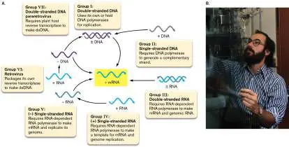
Group II. Single-stranded DNA viruses such as canine parvovirus require the host DNA polymerase to generate the complementary DNA strand. The double-stranded DNA can then be transcribed by host RNA polymerase.
Group III. Double-stranded RNA viruses require a viral RNA-dependent RNA polymerase to generate mRNA by transcribing directly from the RNA genome. Because the RNA polymerase is required immediately upon infection, such viruses package a viral RNA polymerase with their genome before exiting the host cell. A major class of double-stranded RNA viruses is the reoviruses, including rotavirus, a cause of diarrhea in children. Another reovirus has been engineered to destroy human tumors without infecting normal cells. This “oncolytic” reovirus, called Reolysin, is now in clinical trials for cancer therapy.
Group IV. (+) sense single-stranded RNA viruses such as the coronavirus SARS-CoV-2 consist of a positive-sense (+) strand (the coding strand) that can serve directly as mRNA to be translated to viral proteins. Replication of the RNA genome, however, requires synthesis of the complementary (−) strand by a viral RNA-dependent RNA polymerase. The (−) strand then serves as a template for (+) strand synthesis.
Group V. (− ) sense single-stranded RNA viruses such as influenza virus have genomes that consist of template, or “negative-sense,” RNA. Thus, they need to package a viral RNA-dependent RNA polymerase for transcribing (−) RNA to (+) mRNA. The (−) strand RNA viral genomes are often segmented; that is, they consist of multiple separate linear chromosomes—a key factor in the evolution of killer strains of influenza (see Section 11.2).
Group VI. Retroviruses , or RNA reverse-transcribing viruses, such as HIV and feline leukemia virus, have genomes that consist of (+) strand RNA. Instead of RNA polymerase, retroviruses package a reverse transcriptase, which transcribes the RNA into a double-stranded DNA (for details, see Section 11.3). The double-stranded DNA is then integrated into the host genome, where it directs the expression of the viral genes. Group VII. Pararetroviruses , or DNA reverse-transcribing viruses , have a replication cycle that requires reverse transcriptase. For example, the pararetrovirus hepatitis B virus (a hepadnavirus) first copies its double-stranded DNA genomes into RNA, and then reverse-transcribes the RNA to progeny DNA using a reverse transcriptase packaged in the original virion. In contrast, plant pararetroviruses, such as cauliflower mosaic virus (CaMV; a caulimovirus), generate an RNA intermediate that replicates using a reverse transcriptase made by the host cell. Many plant genomes include a gene for reverse transcriptase. Cauliflower mosaic virus is of enormous agricultural significance for its use as a vector to construct pesticide-resistant food crops. Examples of these seven fundamental groups are presented in Table 6.2.
Groups of Viruses—
TABLE 6.2 Baltimore
Classification
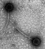
Taxonomic group with Virus example examples Group I. Double-stranded DNA viruses Bacteriophage lambda infects Escherichia coli.
Phage lambda Chloroviruses infect algae, controlling algal blooms.
BIOPHOTO
ASSOCIATES/SCIENCE SOURCEHerpesviruses cause chickenpox, genital infections, and birth defects.
Poxviruses cause smallpox and monkeypox (mpox).
Papillomavirus strains cause warts and tumors.


Group II. Single-stranded DNA viruses Anelloviruses are found in human blood plasma; they cause no known harm.
Bacteriophage M13 infects E. coli.
Geminiviruses infect tomatoes Geminivirus and other plants. CINDY L. MCKENZIE/USDA Parvoviruses cause disease in cats, dogs, and other animals. Group III. Double-stranded RNA viruses Birnaviruses infect fish.
Cystoviruses infect bacteria. Reoviruses such as rotavirus cause severe diarrhea in Rotavirus children. Other reoviruses are in
JAMES GATHANY/CDC
clinical trials to fight tumors (oncolysis).
Group IV. (+) sense single-stranded RNA viruses
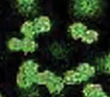
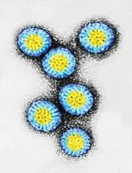

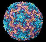

Rhinovirus Coronaviruses such as SARS-SCOTT CAMAZINE/VISUALS CoV-2 cause severe respiratory UNLIMITED, INC. disease.
Flaviviruses cause hepatitis C, Zika fever (Zika virus), West Nile disease, yellow fever, and dengue fever.
Poliovirus infects human intestinal epithelium and nerves.
Tobacco mosaic virus infects plants.
Group V. (–) sense single-stranded RNA viruses Filoviruses such as Ebola virus cause severe hemorrhagic disease.
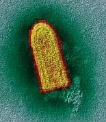
Rabies virus Orthomyxoviruses cause EYE OF SCIENCE/SCIENCE influenza. SOURCE Paramyxoviruses cause measles and mumps.
Rhabdovirus causes rabies.
Group VI. Retroviruses (RNA reverse-transcribing

viruses)
Feline leukemia virus (FeLV), Rous sarcoma virus (RSV) , and avian leukosis virus (ALV) cause cancer.
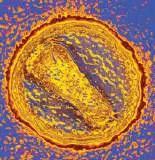
Lentiviruses include human immunodeficiency virus (HIV) , the cause of AIDS.
Engineered “lentivectors” are Human immuno-used for gene therapy. deficiency virus
JAMES CAVALLINI/SCIENCE
SOURCE
Group VII. Pararetroviruses (DNA reverse-transcribing viruses)
Caulimoviruses (such as cauliflower mosaic virus, or CaMV) infect many kinds of Caulimovirus vegetables. CaMV provides the SCIENCE PHOTO best vector tools for plant LIBRARY/SCIENCE SOURCE biotechnology. Hepadnaviruses such as hepatitis B virus infect the human liver.
Molecular Evolution of Viruses

The phylogeny, or genetic relatedness, of viruses can be determined within families. For example, the herpesvirus family includes double-stranded DNA viruses that cause several human and animal diseases, such as chickenpox, oral and genital herpes infection, and respiratory and genital infections in horses. Herpesvirus genomes consist of double-stranded DNA, 120–220 kilobases (kb) encoding about 70–200 genes; an example is that of varicella-zoster virus, the causative agent of chickenpox (Fig. 6.20A). The genome includes two “unique” segments of genes, one long and one short (U L and U S), joined by two inverted repeats (IRs). Other herpesvirus genomes share similar structure, though they differ in gene order and IR position.
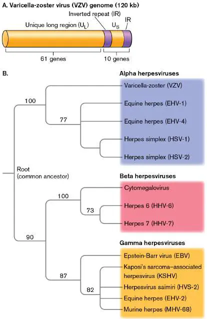
FIGURE 6.20 ■ Phylogeny of herpesvirus genomes. A. Genome structure of human varicella-zoster virus (VZV), the causative agent of chickenpox. B. Phylogeny of human and animal herpesviruses, based on whole-genome sequence analysis comparing clusters of orthologous groups of genes. Numbers indicate approximate percentage of sequence shared by divergent genomes from a common ancestor.
The large size of herpesvirus DNA genomes allows effective measurement of genetic divergence on the basis of sequence comparison. Measurement of sequence relatedness and divergence enables us to propose phylogenetic trees (discussed in Chapter 17). The relatedness of different herpesviruses that evolved from a common ancestor can be measured using orthologous genes, or orthologs. Orthologs are genes of common ancestry in two genomes that share the same function (a topic discussed in Chapter 9). For cellular organisms, we often use the ribosomal RNA gene sequences to measure relatedness. Viruses have no ribosomal RNA, but closely related viruses share other orthologous genes. In pairs of orthologs, the amount of difference in sequence correlates approximately with the time following divergence from a common ancestor (a topic discussed in Chapter 17).
A tree of genomic divergence (or phylogeny) was devised for herpesviruses. The comparison of all gene pairs places herpes strains into three related classes designated alpha, beta, and gamma (Fig. 6.20B ). The alpha class includes human varicella-zoster virus and the oral and genital herpesviruses (HSV-1 and HSV-2), as well as two equine herpesviruses. The beta class includes cytomegalovirus, a common cause of congenital infections (present at birth), as well as two lesser-known viruses. The gamma class includes Epstein-Barr virus, which causes infectious mononucleosis and is the proposed cause of multiple sclerosis. Other gamma class viruses include Kaposi’s sarcoma–associated herpesvirus and several viruses of nonhuman animals.
Gene comparison generates a tree for closely related viruses such as herpesvirus. But how can we assess phylogeny of more distantly related viruses that share no genes—and even have genomes of different kinds of nucleic acids? Unlike cells, viruses do not possess genes universal for all species, such as a gene encoding 16S or 18S ribosomal RNA. For viruses, no gene is universally shared, and thus no single phylogenetic tree of life can be made for viruses. Furthermore, viral genomes are highly mosaic; that is, they evolved from multiple sources. Mosaic genomes result from recombination or reassortment of chromosomes from different viruses coinfecting a host. Reassortment of influenza virus genomes generates dangerous strains, as discussed in Section 11.2.
Thought Question
6.6 When two different viruses infect a cell, how might viruses with different kinds of genomes (RNA versus DNA) combine and share genetic content in their progeny?
To Summarize
Viruses contain infective genomes of RNA or DNA. A viral genome may be single-or double-stranded, linear or circular.
Giant viruses have 300–2,500 genes and may have evolved from intracellular parasitic cells.
Smaller viral genomes comprise fewer than ten genes. Such viruses might have evolved from cell parts.
Classification of viruses is based on a variety of criteria, including genome composition, virion structure, and host range.
The Baltimore virus classification emphasizes the form of the genome (DNA or RNA, single-or double-stranded) and the route to generate messenger RNA.
Phylogeny of closely related viruses can be calculated by comparing all related pairs of genes from the viral genomes.
Glossary
segmented genome A viral genome that consists of more than one nucleic acid molecule.
RNA-dependent RNA polymerase An enzyme that produces an RNA complementary to a template RNA strand.
RNA reverse-transcribing virus See retrovirus .
reverse transcriptase (RT)
An enzyme that produces a double-stranded DNA molecule from a single-stranded RNA template.
pararetrovirus Also called DNA reverse-transcribing virus. A virus with a double-stranded DNA genome that generates an RNA intermediate and thus requires reverse transcriptase to generate progeny DNA genomes.
ortholog or orthologous gene A gene present in more than one species that derived from a common ancestral gene and encodes the same function. ortholog or orthologous gene A gene present in more than one species that derived from a common ancestral gene and encodes the same function. retrovirus Also called RNA reverse-transcribing virus. A single-stranded RNA virus that uses reverse transcriptase to generate a double-stranded DNA.
6.4 Bacteriophages: The Gut ViromeUnit 3 · Structure
The best-known bacteriophages are those of the mammalian digestive tract. Gut bacteriophages, or “coliphages,” are part of a microbial community that modulates human digestion, the immune system, and mental health. Historically, coliphages have provided some of the most fundamental insights in molecular biology. In 1952, Alfred Hershey and Martha Chase showed that the transmission of DNA by a bacteriophage to a host cell led to the production of progeny bacteriophages, thus confirming that DNA is the hereditary material. And two years before, Esther Lederberg at the University of Wisconsin discovered bacteriophage lambda hiding its genome within that of Escherichia coli K-12. Phage lambda became the foremost model for molecular biology of gene mobility in the twentieth century (described in Section 11.1). Many fundamental concepts of the genetic unit and the basis of gene transcription came from experiments with bacteriophages, as discussed in Chapters 7–9.
Bacteriophages Infect a Host Cell
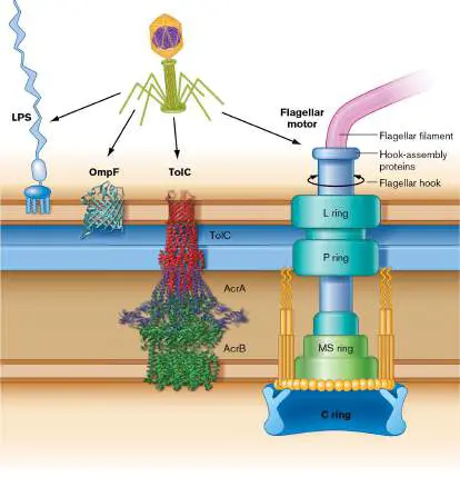
Here we outline the phage replication cycles within host enteric bacteria. To commence an infection cycle, bacteriophages need to contact and attach to the surface of an appropriate host cell. Contact and attachment are mediated by cell-surface receptors, proteins or other macromolecules on the host cell surface that are specific to the host species and that bind to a specific viral component. A cell-surface receptor for a virus is actually a molecule with an important function for the host cell, but the virus has evolved to take advantage of it. For example, a given type of phage that infects Salmonella enterica can use a specific outer membrane protein such as OmpF, which is a porin, or TolC, which is part of a drug efflux complex (Fig. 6.21). Alternatively, a phage might bind to LPS (lipopolysaccharide; see Chapter 3). The phage-receptor binding is usually highly specific; a bacterium can evolve resistance through single-amino-acid mutations in its protein. The lambda phage receptor protein (maltose porin) of E. coli is described in Chapter 11.
FIGURE 6.21 ■ Phages bind Salmonella receptors. Various kinds of host molecules can serve as a phage receptor, such as LPS components, membrane proteins and complexes (OmpF and TolC), and flagellar proteins.
Most bacteriophages deliver only their genome into a cell through the cell envelope, thus avoiding the need for the capsid to penetrate the molecular barrier of the cell wall. For example, the phage T4 virion has a sheath that contracts, bringing the head near the cell surface to inject its DNA (Fig. 6.22A). The pressure of the spooled DNA—as high as 50 atmospheres (atm)—is released, expelling the DNA into the cell. After the genome has been inserted, the phage
capsid remains outside, attached to the cell surface. The empty capsid is termed a “ghost” because of its pale appearance in an electron micrograph.
FIGURE 6.22 ■ Bacteriophage reproduction: lysis and lysogeny. A. A lytic phage particle attaches to the cell surface by its tail fibers and then contracts to inject its DNA. The empty capsid remains outside as a “ghost.” Right: Cryo-EM model of phage T7 injecting DNA. B. Lysis (left) occurs when the phage genome
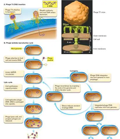
reproduces progeny phage particles, as many as possible, and then lyses the cell to release them. In phage lambda, lysogeny (right) can occur when the phage genome integrates itself into that of the host. The phage genome is replicated along with that of the host cell. The phage DNA, however, can direct its own excision by expressing a site-specific DNA recombinase. This excised phage chromosome then initiates a lytic cycle.
B. HU ET AL. 2013. SCIENCE 339 :576
The lytic cycle. A lytic cycle of replication generates a large number of progeny phages and then lyses the defunct cell. The lytic replication cycle requires these steps: Host recognition and attachment. A phage particle must contact a receptor molecule and adhere to a host cell.
Genome entry. The phage genome must enter the host cell and gain access to the cell’s machinery for gene expression and genome replication.
Assembly of phages. Phage components must be expressed and assembled. Components usually “self-assemble”; that is, the joining of their parts is favored thermodynamically.
Exit and transmission. Progeny phages must exit the host cell, and then reach new host cells to infect.
In a lytic cycle, when a phage particle delivers its genome into a cell, it immediately reproduces as many progeny phage particles as possible ( Fig. 6.22B ). The process of reproduction involves replicating the phage genome, as well as expressing phage mRNA to make enzymes and capsid proteins. Some phages, such as T4, digest the host DNA to increase the efficiency of phage production. Phage particles assemble, and the host cell lyses, releasing progeny phages.
The phage adsorbs to its host receptor and inserts its double-stranded DNA into the host cytoplasm. The phage genes are then expressed by the host cell RNA polymerase and ribosomes. “Early genes” are expressed first in the lytic cycle. Other phage-expressed proteins then work together with the cellular enzymes and ribosomes to replicate the phage genome and produce phage capsid proteins. The capsid proteins self-assemble into capsids and package the phage genomes—a process that takes place in defined stages, like a factory assembly line. At last, a “late gene” from the phage genome expresses an enzyme that lyses the host cell wall, releasing the mature virions. Lysis is also referred to as a burst, and the number of virus particles released is called the burst size.
Lysogeny. A temperate phage , such as phage lambda, can infect and lyse cells like a virulent phage, but it also has an alternative pathway: to integrate its genome within the host chromosome (Fig. 6.22B ). The phage is said to “lysogenize” the host, in a cycle called lysogeny. Phage lambda has a linear genome of double-stranded DNA, which circularizes upon entry into the cell. The circularized genome then recombines into that of the host by site-specific recombination of DNA. In site-specific recombination, a recombinase enzyme aligns the phage genome with the host DNA and exchanges the DNA backbone linkages with those of the host genome. (This process of DNA backbone recombination is explained in Chapter 9.) The DNA recombination event thus integrates the phage genome into that of the host. Now integrated, the phage genome is a prophage. The presence of the prophage prevents further infection (superinfection) by other virions of the same type.
In lysogeny, the prophage DNA is replicated along with that of the host cell as the host reproduces (Fig. 6.22B ). The host gains the benefit of resistance to superinfection. Implicit in the term “lysogeny,” however, is the ability of such a strain to generate a lytic burst of phage. For a lysogen to enter lysis, the prophage directs its own excision from the host genome by an intramolecular process of site-specific recombination. The two ends of the phage genome exchange their DNA linkages so as to come apart from the host DNA, which now closes its circle with the prophage removed. As the phage DNA exits the host genome, it circularizes and initiates a lytic cycle, destroying the host cell and releasing phage particles.
How does a lysogen “decide” to reactivate and begin a lytic cycle? The decision between lysogeny and lysis is determined by proteins that bind DNA and repress the transcription of genes for virus replication (discussed in Chapter 11). Exit from lysogeny into lysis can occur at random times or it can be triggered by environmental stress such as UV light, which damages the cell’s DNA. The regulatory switch of lysogeny responds to environmental cues indicating the likelihood that the host cell will survive and continue to propagate the phage genome. If a cell’s growth is strong, it is more likely that the phage DNA will remain inactive, whereas events that threaten host survival will trigger a lytic burst. Similarly, in animal viral infections such as herpes, an environmental stress triggers reactivation of a virus that was dormant within cells (a latent infection). Reactivation of a latent herpes infection results in painful outbreaks of skin lesions.
During the exit from lysogeny, the virus can acquire host genes and pass them on to other host cells. The process of transferring host genes is known as transduction. Sometimes a transducing bacteriophage picks up a bit of host genome and transfers it to a new host cell. In another kind of transduction, the entire phage genome is replaced by host DNA packaged in the phage capsid, resulting in a virus particle that transfers only host DNA. Host DNA transferred by viruses can become permanently incorporated into the infected host genome (Table 6.3). These former viral genes evolve into host genes that express products with new functions useful to the host.
Integrated Viral Genomes That
TABLE 6.3
Provide Host Traits
Bacterial Human Human protein or product endogenous regulator from retrovirus provided by HERV Prophage (host prophage (HERV)
infected) gene Phage C1 (Botulinum HERV-W Syncytin-1 (retroviral Clostridium toxin (Env protein; botulinum) c1) placental fusion)
Beta phage (Diphtheria HERV-FRD Syncytin-2 (retroviral Corynebacterium toxin (Env protein; diphtheriae) tox) placental fusion)
Lambda (E. coli Cell HERV INSL4 (insulin-like O157:H7) envelop protein 4; e placental protein (development)
bor)
Phage 933 (E. coli Shiga HERV-E MID1 (midline O157:H7) toxin (development; stx) prevents Opitz syndrome)
Epsilon 34 (LPS HERV-E Apolipoprotein C1 Salmonella synthesi (liver function)
enterica) s enzyme (rfb)
Epsilon 34 (Type III HERV-E Endothelin type B Salmonella secreted receptor (placenta enterica) toxin (function)
sopE)
TSST-1 (Toxic HERV-L Beta-1, 3-Staphylococcus shock galactosyltransfera aureus) syndrom se (colon and e toxin (mammary gland speA) function)
CTSφ (Vibrio Cholera LINE-1 ATRN (soluble cholerae) toxin (attractin; ctxAB) modulates inflammation)
The mechanisms of phage-mediated transduction are discussed in Chapter 9. In natural environments, phage transduction mediates much of the recombination of bacterial genomes. In the laboratory, the ability of phages to transfer genes provided some of the first vectors for recombinant DNA technology.
The slow-release cycle. A phage may persist in a living host by using a slow-release replication cycle. A slow-release replication cycle differs from lysis and lysogeny in that phage particles reproduce without destroying the host cell (Fig. 6.23). Slow release is performed by filamentous phages such as phage M13 (presented in eAppendix 4). In slow-release replication, the single-stranded circular DNA of M13 serves as a template to synthesize a double-stranded intermediate. The double-stranded intermediate slowly generates single-stranded progeny genomes, which are packaged by supercoiling and coated with capsid proteins. The phage particles then extrude through the cell envelope without lysing the cell. The host cell continues to reproduce, though more slowly than uninfected cells do, because many of its resources are diverted to virus production. FIGURE 6.23 ■ Slow-release cycle of a persistent phage. In the slow-release replication cycle, a filamentous phage produces
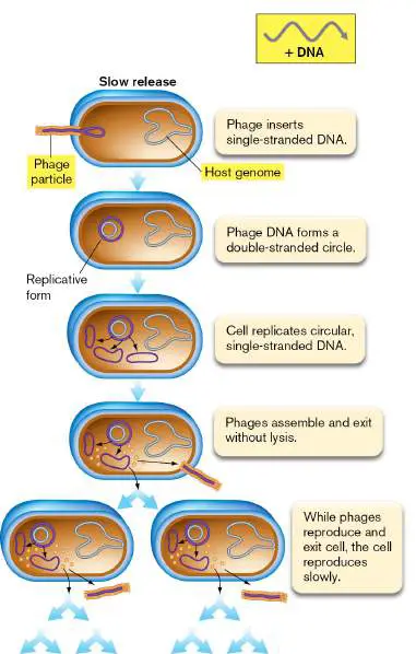
phage particles without lysing the cell. The host continues to reproduce itself, but more slowly than uninfected cells do, because many of its resources are being used to make phages.
Thought Question
6.7 What are the relative advantages and disadvantages (to a virus) of the slow-release strategy, compared with the strategy of a temperate phage, which alternates between lysis and lysogeny?
Integrated Viruses as Host Cell Parts
Many prophages and endogenous viruses function as part of their host cell. Table 6.3gives examples of prophages that express virulence genes for pathogenic bacteria—a benefit to the pathogen, enabling it to better colonize the host animal. For example, phage C1 in Clostridium botulinum expresses the peptide botulinum toxin. This toxin causes the paralysis associated with botulism (and is also the basis of the Botox cosmetic treatment). In Corynebacterium diphtheriae, the cause of diphtheria, the diphtheria toxin is expressed by beta phage. Other virulence factors (proteins that enhance disease) expressed by prophages include envelope proteins that help defend the bacterium from the immune system. Analogous to bacterial prophages, many human genes and gene control elements have evolved from human viruses, particularly human endogenous retroviruses (HERVs). Retroviruses are a category of virus that includes HIV, the cause of AIDS (discussed in Section 11.3). HERVs are reverse-transcribing RNA viruses whose DNA copies became permanently fixed in our chromosomes. For example, placental proteins called “syncytin” mediate cell fusion during an early stage of placental development, allowing the fused syncytium to implant in the uterine lining and access the maternal blood supply. Two genes for syncytin evolved from HERV genes encoding retroviral envelope proteins. The details of the remarkable story of endogenous retroviruses—and the related story of retroviral gene therapy—are discussed in Chapter 11.
Bacterial Defenses
In natural environments, viruses commonly outnumber cellular microbes by tenfold or more. So, how have host cells evolved to defend themselves? Several remarkable resistance mechanisms have evolved. Their molecular basis is explained in greater detail in Chapters 9 and 12. Genetic resistance. All bacteria acquire random mutations in their genomes as they reproduce (see Chapters 7 and 9). When attacked by bacteriophages, bacterial populations undergo natural selection; mutants that happen to be harder to infect will survive. Bacteria resist phage infection by expressing a gene that encodes an altered host receptor protein, which fails to bind the viral coat protein. Alternatively, a different cell protein evolves to block phage binding to the receptor. An evolutionary “arms race” ensues, in which phages may evolve enzymes that cleave the host defense molecules.
Restriction endonucleases. Bacteria modify their DNA by adding methyl groups to bases within certain sequences. The bacteria then express restriction endonucleases, enzymes that cleave DNA lacking the methylated patterns—which includes potential viral DNA (see Chapter 9). However, phage genomes composed of RNA or of modified DNA (such as phage T4, discussed in Chapter 11) escape cleavage by these enzymes. CRISPR: a bacterial immune system. Amazingly, bacteria possess an adaptive defense against viruses that is analogous to an immune system. (Adaptive immunity of humans is presented in Chapter 24.) The bacterial adaptive defense involves short DNA sequences homologous to DNA of phages that could infect the cell. The sequences are called c lustered r egularly i nterspaced s hort p alindromic r epeats (CRISPR). These series of short sequences were first characterized in the 1990s by Francisco Mojica at the University of Alicante, Spain, who proposed that they might represent an adaptive defense against phages. Since then, numerous other research groups figured out components of the mechanism, most famously Jennifer Doudna and Emmanuelle Charpentier, who won the 2020 Nobel Prize in Chemistry for using CRISPR mechanisms to edit eukaryotic genomes. CRISPR is an example of fundamental science, supported by the National Science Foundation, which later turned out to have astonishing applications for medicine and industry.
In bacterial genomes, where do the CRISPR short repeats come from? When a phage attacks a bacterium, if bacterial enzymes succeed in destroying the phage DNA, they may copy a tiny piece of it as a CRISPR segment, inserted as a spacer at the head of a long line of about 30 CRISPR sequences (Fig. 6.24A). Now the adapted host cell “remembers” infection by the specific phage—along with many other previous phages from previous infections that had inserted other spacers. The next time the adapted host cell is attacked by the same phage, all of its genomic CRISPR sequences are expressed as RNA. The CRISPR RNA is cleaved into small sequences (crRNA) containing one spacer from the original bacterial CRISPR DNA. The crRNA binds to the Cascade protein complex (or Cas complex), which now detects phage DNA homologous to its virus-derived crRNA. The Cas-crRNA complex proceeds to cleave the phage DNA, preventing phage replication.
FIGURE 6.24 ■ CRISPR defense of a bacterial cell and anti-CRISPR counterdefense. A. A piece of phage DNA gets copied as a “spacer” into the host genome. If the bacterium survives infection, later reinfection by the same kind of phage causes transcription of the spacers into CRISPR RNA. A processed spacer (crRNA) joins the Cas complex to recognize and cleave the phage DNA. B. Phage may carry
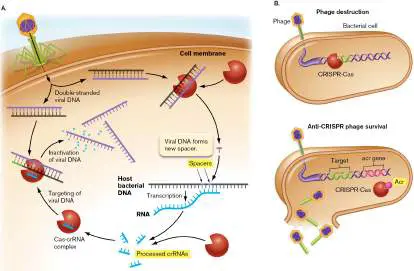
an anti-CRISPR gene (acr) encoding a protein Acr that blocks the host bacterial CRISPR-Cas from binding phage DNA.
Diverse bacteria show many variations on the CRISPR theme. For example, in 2019, Jonathan Strecker and co-workers discovered a CRISPR-associated transposase (DNA transfer enzyme) within the cyanobacterium Scytonema hofmanni. This CRISPR transposase could be used for editing large pieces of DNA in human genomes.
In response to CRISPR, how do phages adapt? Not surprisingly, phages have evolved several means of defense. One mechanism involves the “anti-CRISPR” phage protein family Acr. Acr proteins inhibit the CRISPR-Cas system of specific host bacteria. Different Acr proteins act by various mechanisms, one of which is to bind the bacterial host’s Cas complex ( Fig. 6.24B ). An Acr protein binding to a Cas subunit can prevent the binding of Cas to phage DNA, in some cases by acting as a DNA mimic for the Cas DNA-binding site. Thus, phage infection may proceed despite the presence of homologous CRISPR sequences in the host genome.
CRISPR mechanisms of bacterial defense and applications for gene technology are discussed in Section 12.3.
Thought Question
6.8 How else, besides Acr proteins, might a phage evolve resistance to the CRISPR host defense (outlined in Fig. 6.24)?
Bacteriophages Within the Gut Community
Phages are an important interactive part of the gut virome, the community of viruses within a human’s or other animal’s intestinal tract. We summarize some of the major phage-host interactions within the human intestinal tract in Figure 6.25. Note that this simplified diagram omits the viruses that infect human body cells; we focus here on the phages that infect gut bacteria. Surprisingly, phages are so important to the human host that the gut takes them up into the blood and tissues. The phages are taken up by transcytosis, an important cellular process for selective uptake of macromolecules. Transcytosis may enable phages to interact with development of the immune system. Another possibility is that phages might actually defend host tissues from wayward bacteria that escape the gut lumen.
FIGURE 6.25 ■ The gut bacteriophage community. Bacteriophages enter the intestine, where they infect intestinal bacteria. Most intestinal bacteria carry prophages. Phages also modulate the immune system.
The lumen of the human intestines contains a remarkably dense microbial community. Bacterial and archaeal cells are estimated at 10 11 – 10 13 cells per gram, whereas phage particles may be 10 9 per gram. The phage particle count is likely underestimated, as it is based on detection by electron microscopy and plate counts of viability (discussed in Section 6.4). Phages commonly infect all the major taxa of the gut lumen, including Bacteroidetes, Firmicutes, and Proteobacteria. In addition, more than half the genomes sequenced from gut bacteria show lysogeny,
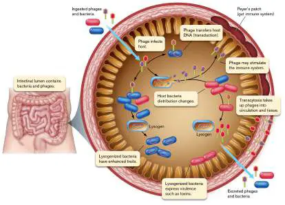
commonly with several different types of prophages in a given genome. Prophages may protect the gut bacteria from superinfection; that is, infection by other phages in the gut that cause lysis.
The gut community receives a continual influx of new bacteria and phages, while shedding present members (Fig. 6.25). When a new type of phage enters, what happens? If the phage encounters a susceptible host bacterium, it may undergo a lytic cycle, lysogeny, or slow release, depending on the phage’s genetic program. Cell lysis may have the effect of depopulating a dominant population of the bacterial community. Bacterial depopulation by phages might lead to intestinal “dysbiosis,” deterioration of health due to loss of health-enhancing bacteria. Other bacterial species, less healthy for the human host, might increase in population. But now, a different phage species (shown in red in Fig. 6.25 ) may depopulate the newly risen species, restoring equilibrium in the community. Still other phages may transfer useful genes from newcomer bacteria, by a process called transduction (see Chapter 9). Such genes may encode enzymes to metabolize new kinds of food molecules.
Some prophages of pathogenic bacteria express virulence factors, such as the Shiga toxin of Shigella and of E. coli O157:H7 (Table 6.3). At the same time, other effects of phages may be positive: Phages may limit the bacterial numbers to levels that the human immune system can tolerate. Lysogenized bacteria may use quorum sensing to detect host cell populations and “decide” whether to start a lytic cycle.
Phage particles may modulate the immune system by suppressing T-cell activation and tumor formation. (T cells are discussed in Chapter 24.)
Phages may attack biofilms. Biofilms of pathogens such as Pseudomonas aeruginosa may be eroded by phage infection.
Our bodies take up gut phages by transcytosis. The uptake of phages throughout our tissues implies that phages have more direct positive benefits, such as priming the immune system.
The positive potential of bacteriophages has led researchers to investigate the engineering of phages for phage therapy. An idea dating back to the early twentieth century, phage therapy was eclipsed by the rise of antibiotics. Today, as we face growing antibiotic resistance in pathogens, we are devising therapeutic uses of bacteriophages ( eResearch Activity 6; also see Chapters 5 and 11).
To Summarize
Host cell-surface receptors mediate the attachment of bacteriophages to a cell and confer host specificity.
Lytic cycle. A bacteriophage injects its DNA into a host cell, where it uses host gene expression machinery to produce progeny virions.
Lysogeny. Some bacteriophages can insert their genome into that of the host cell, which then replicates the phage genome along with its own. A lysogenic bacterium can initiate a lytic cycle. Gene transfer. Genes are transferred by phage processes of transduction and lysogeny.
Slow release. Some bacteriophages use the host machinery to make progeny that bud from the cell slowly, slowing growth of the host without lysis.
Bacterial host defense. Bacteria have evolved several forms of defense against bacteriophage infection, such as altered receptor proteins, restriction endonucleases, and CRISPR integration of phage DNA sequences.
The gut bacteriophage community includes phages that infect, lyse, or lysogenize bacterial hosts. Gut phages affect bacterial community structure, transfer genes among bacteria, and modulate the human gut immune system. Some phages are taken up by host tissues via transcytosis.
Glossary
cell-surface receptor A transmembrane protein that senses a specific extracellular signal and may be the docking site for a specific virus.
lysis Also called burst. The rupture of the cell by a break in the cell wall and membrane.
burst size The number of virus particles released from a lysed host cell. temperate phage A phage capable of lysogeny.
lysogeny A viral life cycle in which the viral genome integrates into and replicates with the host genome but retains the ability to initiate host cell lysis.
site-specific recombination Recombination between DNA molecules that do not share long regions of homology but do contain short regions of homology specifically recognized by the recombination enzyme.
transduction The transfer of host genes between bacterial cells via a phage head coat.
CRISPR Clustered regularly interspaced short palindromic repeats. CRISPR consists of short repeated DNA sequences in a bacterial or archaeal genome, derived from previous bacteriophage or viral infection and conferring protection from future infection; considered a prokaryotic “immune system.” In biotechnology, provides a means of editing the human genome.
transcytosis The movement of a cell or substance from one side of a polarized cell to the other side, using an intracellular route.
6.5 Coronavirus SARS-CoV-2 and Other Viral PathogensUnit 3 · Structure
Viruses of animals and plants solve problems similar to those faced by bacteriophages: host attachment, genome entry and gene expression, virion assembly, and virion release. The more complex structure of eukaryotic cells, however, requires viral replication cycles that are more complex. (Eukaryotic cell structure is reviewed in eAppendix 2). Viral reproduction may involve intracellular compartments such as the nucleus or secretory system and may depend on tissue and organ development in multicellular organisms. Human virus replication reveals potential targets for antiviral drugs, such as the Pfizer drug Paxlovid (nirmatrelvir plus ritonavir), which inhibits a coronavirus protease—the first oral antiviral agent approved by the FDA for COVID-19.
We start by introducing the diverse ways that different viruses attach to and enter the animal cell (Fig. 6.26). We then outline the replication cycles of coronavirus SARS-CoV-2, human papillomavirus, and caulimovirus (a virus of plants). Later, in Chapter 11, we present in molecular detail the replication cycles of bacteriophage lambda, influenza virus, lentivirus HIV, and herpes simplex virus.
FIGURE 6.26 ■ Receptor binding and genome uncoating. A. Coronavirus SARS-CoV-2 attaches to angiotensin-converting enzyme 2 (ACE2), a glycoprotein required by the host cell to bind a lymphocyte integrin, a cell-surface matrix protein required for cell-cell adhesion. After binding a specific receptor on the host cell membrane, the coronavirus enters the cell by endocytosis. Within the endosome, the viral genome is uncoated.
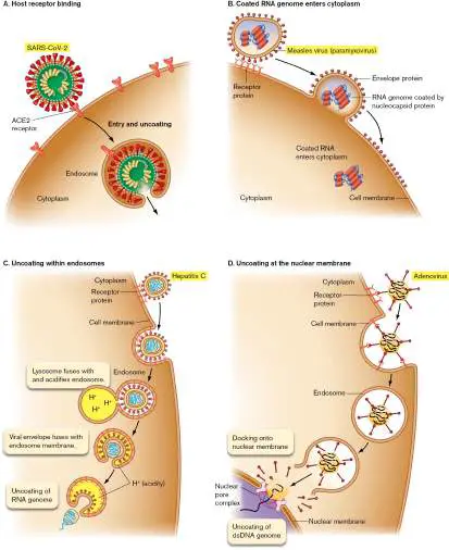
B. Measles virus: The coated RNA genome enters the cytoplasm. C. Hepatitis C: The genome is uncoated within an endosome activated by lysosome fusion and acidification. D. Adenovirus: The genome is uncoated at the nuclear pore complex.
Animal Viruses Bind Host Receptors and Enter the Cell
Like bacteriophages, animal viruses evolve surface proteins that bind specific receptors on their host cell. An example of a human virus-receptor interaction is that of SARS-CoV-2 (see Table 6.2, group IV), which causes COVID-19. The coronavirus envelope spike protein binds ACE2, a human glycoprotein (protein with sugar chains) that has important functions in tissues throughout the host, including regulation of blood pressure and blood vessel function in the heart and lungs. The coronavirus spike protein evolved a domain of amino acid residues that binds ACE2 and triggers endocytosis of the virus.
Host receptors play a key role in determining the host range, the group of host species permitting infection. Within a host, receptor molecules can also determine the viral tropism, or ability to infect a particular tissue type within a host. Some viruses, such as Ebola virus, exhibit broad tropism, infecting many kinds of host tissues, whereas others, such as papillomavirus, show tropism for only one type (in the case of papillomavirus, the epithelial tissues). Tropism may depend on the virus’s ability to interact with the cytoplasm or it may require the presence of an appropriate host cell receptor protein that can bind the viral surface attachment protein. For example, cats and ferrets happen to possess ACE2 receptor proteins with high amino acid similarity to that of humans. Thus, both animals are susceptible to SARS-CoV-2, and ferrets are sufficiently susceptible to use as a model system for study of airborne virus transmission (Special Topic 6).
Thought Question
6.9 How might humans undergo natural selection for resistance to coronavirus SARS-CoV-2 infection? Is such evolution likely? Why or why not?
Genome entry and uncoating. Most animal viruses, unlike bacteriophages, enter the host cell as an intact core particle; that is, the genome within a capsid. The contents of the virion may then interact with the cell in several different ways. For SARS-CoV-2, the entire virion is taken up by endocytosis (Fig. 6.26A). In endocytosis, the cell membrane forms a vesicle around the virion and engulfs it, forming an endocytic vesicle, or endosome (reviewed in eAppendix 2). Within the endosome, the internalized capsid then undergoes uncoating, a process in which the capsid comes apart, releasing the viral genome into the cytoplasm.
By contrast, the measles virus, a paramyxovirus, enters the cell by binding host receptor proteins, causing the viral envelope to fuse with the host cell membrane (Fig. 6.26B ). The measles RNA genome coated by nucleocapsid proteins is then released directly into the cytoplasm. For other types of viruses, such as the flavivirus hepatitis C, the virion undergoes endocytosis, then the endocytic vesicle fuses with a lysosome, whose acidity activates entry of the capsid into the cytoplasm (Fig. 6.26C ). The capsid then comes apart, and the viral genome is uncoated.
Yet other kinds of viruses, such as adenovirus, enter the cell by endocytosis but require transport to the nucleus (Fig. 6.26D ). After endocytosis and lysosome fusion, the adenoviral genome loses some of its capsid proteins (partial uncoating). A capsid protein then disrupts the endocytic membrane, allowing the remaining capsid to exit. The capsid then docks at a nuclear pore complex and injects its DNA genome into the nucleus. The adenoviral DNA is replicated by its own adenoviral DNA polymerase (carried in the virion), but it uses host nuclear histones for DNA packing, as well as host transcription factors available in the nucleus.
How does a virus replicate within an animal cell? The details of a replication cycle vary considerably, depending on the given virus. An important factor is the form of the viral genome (see Baltimore classification, Fig. 6.19). A DNA genome can use some or all of the host replication enzymes. An RNA genome, however, must encode either an RNA-dependent RNA polymerase to generate an RNA template or, in the case of retroviruses, a reverse transcriptase to generate a DNA template.
Coronavirus: (+) strand RNA Virus
The coronavirus has a structure typical of enveloped viruses: a core particle of RNA packaged in proteins (nucleoprotein complexes linked by RNA) with a surrounding envelope and spike proteins ( Fig. 6.27). The appearance of the “crown” of spike proteins in micrographs inspired the name “coronavirus.”
FIGURE 6.27 ■ SARS-CoV-2 virion structure and genome. ORFs (open reading frames) and genes are shown staggered where translation begins in a different reading frame (triplet start position) from that of the adjacent upstream gene. The (+) strand RNA genome has a eukaryotic cap (7-methyl-Gppp) attached at the 5′ end (indicated as orange ball). Within the host cell, all genome copies and mRNA molecules have a cap attached by the host cell’s capping enzyme. 3′ UTR = 3′ untranslated region; AAA = 5′ poly-A tail.
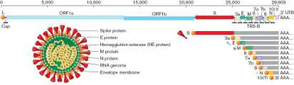
The SARS-CoV-2 genome (Fig. 6.27) consists of plus-strand RNA. Its 5´ end obtains a cap (7-methylguanosine) from the previous host cell, making it look like normal capped eukaryotic mRNA (described in Chapter 8). Most parts of the virion are encoded by genes at the 3′ end of RNA, such as the spike protein (S), envelope proteins (E, M), and nucleocapsid protein (N). Genome replication will require early expression of an RNA-dependent RNA polymerase (replicase), encoded by a portion of the open reading frame ORF1ab. “Open reading frame” means a base sequence defined by start and stop codons, capable of translation by a ribosome. (Gene expression is discussed in detail in Chapter 8.) Coronavirus replication. Infection starts when the coronaviral spike protein (S) binds the ACE2 receptor of a host cell (Fig. 6.28, step 1). Receptor-spike binding triggers endocytosis of the virion (step 2). Once the infecting virion (virus particle) uncoats in the cytoplasm, its envelope merges with the endosome membrane in a manner that releases the nucleocapsid-coated RNA.
FIGURE 6.28 ■ SARS-CoV-2 replication cycle. The virion binds the ACE2 receptor. It then undergoes endocytosis and uncoats in the host cytoplasm. The released RNA genome is translated by host ribosomes to synthesize the RNA-dependent RNA polymerase (replicase). The replicase synthesizes (−) strand RNA, which serves as a template for transcription to mRNAs. Endoplasmic reticulum (ER) membrane becomes transformed into viral factories (double-membrane vesicles) that organize virion assembly and transport to the Golgi complex. From the Golgi, virions move to the cell membrane and bud out. The (+) strand viral genome can start translation of its genes as soon as it enters a host cytoplasm (Fig. 6.28, step 3). The open reading frame (ORF) 1ab is immediately translated by host
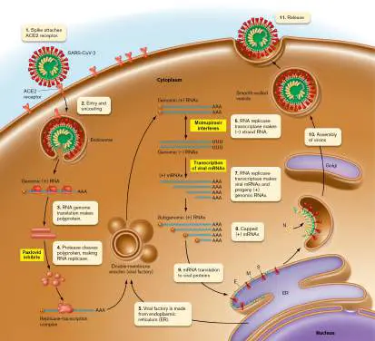
ribosomes, forming a polyprotein; that is, a peptide that needs to be cleaved to form one or more functional proteins. Polyproteins must be cleaved by a viral protease (step 4). The coronaviral protease is the target of the inhibitor nirmatrelvir, a component of the Pfizer antiviral medication Paxlovid.
The ORF1ab polyprotein is cleaved to form two subunits of the RNA-dependent RNA polymerase (replicase). Like all RNA viruses, coronavirus requires a special polymerase to make progeny genomes from an RNA template. Most RNA-template polymerases have high rates of mutation compared to DNA polymerases. This is one reason why RNA viruses rapidly mutate and generate variant strains so quickly. For example, in 2021 during the COVID-19 pandemic, the Delta variant of SARS-CoV-2 spread rapidly because this mutant virus generated 1000-fold higher virus concentration in humans than that of the original strain. The subsequent Omicron variant had a large number of mutations and spread even faster.
SPECIAL TOPIC 6 Found in Ferrets: Air Currents Carry SARS-CoV-2
When the COVID-19 pandemic began, everyone wanted to know how to avoid transmission of the deadly pathogen. Was the virus transmitted only by coughed droplets? Or could it hang around in the air and travel on air currents? The stakes were high, because the easier the spread, the more severe distancing measures were required, with enormous social and economic disruptions. The project was funded by the U.S. National Institutes of Health, the Dutch Research Council, and the European Commission.
To test this question, Sander Herfst’s lab group at Erasmus University, Netherlands, established a transmission experiment using ferrets (Fig. ST 6.1 ). Ferrets are an important model system for influenza transmission because the cells lining their nasal passages have molecular receptors for influenza virus that resemble those found in human nasal passages (discussed in Chapter 11). It turns out that ferrets also show ready transmission of SARS-CoV-2, the human-adapted coronavirus.
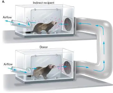
FIGURE ST 6.1 ■ Virus transmission between ferrets. A. A donor ferret is inoculated with virus, then housed in the bottom cage. The next day, an indirect recipient ferret is housed in the top cage. The cages are connected through a duct system. Air flows into the bottom cage and out from the top cage. The entire setup is contained within a biosafety level 3+ laboratory. B. A researcher studying respiratory viruses holds a ferret to be used as a model for human transmission.
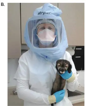
N. NAVRATIL. 2016. LAB ANIM. 45:440–441
In Herfst’s lab, Jasmin Kutter and co-workers devised an apparatus to allow detection of indirect transmission; that is, introduction of virus from an infected ferret into the air, which then carries virus to a recipient ferret who never directly contacts the first one (Fig. ST 6.1A ). The experimental design consisted of pairs of donor and recipient ferrets. Each donor ferret was inoculated intranasally with virus. The donor ferret was housed in a cage that had an air duct connection to the recipient ferret cage, directly above. The air duct was constructed with four 90-degree turns; this made it unlikely that a ferret cough or sneeze could directly propel virus from one animal into the other cage.
Three different viruses were tested in this device: SARS-CoV-2, the cause of COVID-19; SARS-CoV, the cause of an earlier human coronavirus outbreak in 2003; and influenza A strain H1N1. For each ferret pair, both donor and recipient were sampled for virus from nose and throat every two days. In the case of SARS-CoV-2, two out of the four ferret pairs showed virus present in the recipient, in days following positive infection of the donor. A representative example of the data is shown in Figure ST 6.2 .
FIGURE ST 6.2 ■ Virus detected in nasal swabs from donor and recipient ferrets. A representative donor-recipient pair is shown. RT-qPCR amplification of SARS-CoV-2 viral RNA is plotted in real-time PCR doublings up to 40 (the cutoff number of doubling cycles) minus the number of doublings observed (Ct) (see Fig. 6.30).
In the experiment shown, the levels of SARS-CoV-2 virus were measured by reverse-transcriptase quantitative polymerase chain reaction (RT-qPCR), a technique described later in Figure 6.30.
In their experiments, Kutter and Herfst found that all three respiratory viruses are capable of indirect airborne transmission between two host individuals. This finding had a profound impact upon health guidance from the Centers for Disease Control and Prevention (CDC). The CDC ultimately had to recognize that in an enclosed space, anyone can receive or
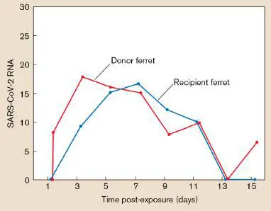
transmit airborne viruses from anyone else in the room, regardless of how far apart and careful the individuals are.
RESEARCH QUESTION
How would you follow up this experiment to test the distance the virus can travel or to test the effectiveness of shields and head coverings as barriers? What limitations would you need to overcome?
Kutter, Jasmin S., Dennis de Meulder, Theo M. Bestebroer, Pascal Lexmond, Ard Mulders, et al. 2021. SARS-CoV and SARS-CoV-2 are transmitted through the air between ferrets over more than one meter distance. Nature Communications 12 :1–8.
For further steps of virion construction, a viral factory is formed out of the host endoplasmic reticulum (Fig. 6.28, step 5). The SARS-CoV-2 replicase now uses the (+) RNA template to synthesize (−) strand complementary RNA (step 6). The (−) strand RNA then serves as a template to synthesize new (+) strand RNA genomes for progeny virions, as well as mRNA molecules encoding all the viral proteins (step 7). All new RNA molecules are capped at the 5´ end by a host enzyme (step 8). Viral packaging proteins are synthesized (step 9), and all the parts ultimately must be assembled together to make new virions (step 10). The Golgi complex secretes completed virions to the cell surface, and exocytosis then releases the progeny virions outside the cell (step 11).
For most of the processes described above, virus components are synthesized and assembled within double-membrane vesicles derived from the endoplasmic reticulum (ER), also called viral factories. The viral factories envelop core particles (genomes bound by nucleocapsid proteins) and transport them to the Golgi complex (reviewed in eAppendix 2). A cryo-EM view of viral factories is shown in Figure 6.29. The virtual stacked sections of cryo-EM show the contours of viral-factory vesicles. Early in virus development, the spike proteins assemble upon the inner surface of ER-derived membranes. The vesicle membrane invaginates to enclose RNA-protein complexes. (Uncoated RNA connects all the complexes but is not visualized.) Finally, a progeny virion is complete, ready for export by the Golgi.
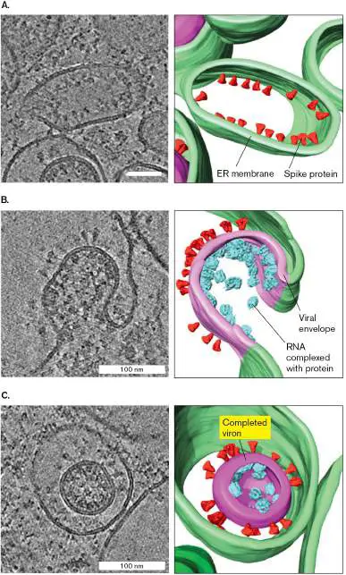
FIGURE 6.29 ■ Viral factories (replication complexes) of coronavirus SARS-CoV-2. Membrane contours of viral factories derived from coronavirus endoplasmic reticulum (cryo-EM tomography). A. Spike proteins assemble upon ER-derived membranes. B. Vesicle membrane invaginates to enclose RNA-protein complexes (uncoated RNA is not shown). C. Virion is complete, ready for export.
Source: Steffen Klein et al. 2020. Nat. Commun. 11 :5885.
S. KLEIN ET AL. 2020. NAT COMMUN. 11:5885
Once the progeny virions are released, they are carried by vascular networks throughout the body. But how do they get transmitted to a new host organism? An experiment testing coronavirus transmission in ferrets is described in Special Topic 6. qPCR amplification of viral RNA. The viral replication process generates tremendous amounts of viral RNA. This RNA can be detected from nasal secretions. The detection of viral RNA is the basis of the famous COVID-19 PCR test that was used for many months to diagnose cases (Fig. 6.30).
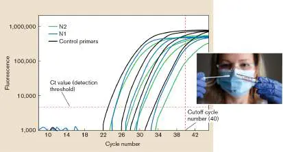
FIGURE 6.30 ■ RT-qPCR amplification of coronavirus RNA. The sample RNA molecules are copied to DNA by reverse transcriptase (RT) followed by quantitative PCR (qPCR)
amplification of the DNA. Primer sets N1 and N2 are designed to amplify regions of the gene encoding N (nucleocapsid) protein. Each amplification cycle leads to release of a hybridized probe and activation of fluorescence. Inset: Nasal swab sample for COVID-19 PCR test. Ct value = cycle number.
VYACHESLAV LOPATIN/ALAMY STOCK PHOTO
The COVID-19 PCR test is based on amplification of a portion of a SARS-CoV-2 gene, most commonly the N gene encoding nucleocapsid protein. First, the RNA must be reverse-transcribed to DNA using the retroviral enzyme reverse transcriptase (described in Section 11.3). The region of DNA sequence between two primers then undergoes exponential amplification by polymerase chain reaction (PCR; described in eAppendix 3). In quantitative PCR (qPCR; also called real-time PCR), the reaction yields a burst of fluorescence at each cycle. The greater the starting amount of target RNA, the earlier the cycle number starts to yield rise of fluorescence. Cycle number (Ct value) is compared with a control to determine relative amounts of RNA. If the cycle number reaches a critical threshold, the test reports detection of coronavirus RNA.
Human Papillomavirus: Double-Stranded DNA Virus
An example of a double-stranded DNA virus is human papillomavirus (HPV; see Table 6.2, group I). Strains of HPV cause cervical cancer, throat cancer, and warts (Fig. 6.31). The discovery that human papillomaviruses cause cancer earned Harald zur Hauzen the 2008 Nobel Prize in Physiology or Medicine (shared with Françoise Barré- Sinoussi and Luc Montagnier for their discovery of human immunodeficiency virus, presented in Chapter 11).
FIGURE 6.31 ■ Human papillomavirus. A. Certain strains of human papillomavirus (HPV) cause warts on the genitals or anus. B. HPV virion model (cryo-EM). C. HPV genome.
KEN GREER/VISUALS UNLIMITED
HPV is the most common sexually transmitted infection in the United States and one of the most common worldwide. Certain strains infect the skin, whereas others infect the mucous membranes through genital or anal contact (sexual transmission). Both cancer and warts are preventable by the Gardasil vaccine for children when administered by age twelve.
The HPV virion consists of a DNA within a capsid, along with matrix proteins, without envelope. The DNA genome of a papillomavirus is surprisingly simple, with only about eight genes ( Fig. 6.31C ). Yet these few genes manage both an active reproduction cycle and a dormant cycle within the host cell. Some of the genes are encoded in overlapping sequence, a common feature of small viruses. Note, however, that other DNA viruses such as the herpesviruses (see Section 11.5) can have much larger genomes, encoding hundreds of proteins including enzymes more typical of living cells.
The HPV gene products interact with host proteins to alter the growth of the host tissues, forming warts or tumors, depending on the HPV strain. For example, proteins E6 and E7 inactivate specific host tumor suppressor proteins, thus starting the conversion of host cell to tumor. HPV initially infects basal epithelial cells, requiring
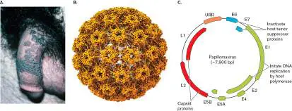
access through a cut or abrasion in the skin or mucous membrane. Instead of envelope spike proteins (as in coronavirus, earlier), the HPV capsid proteins (L1, L2) bind host receptors (Fig. 6.32). Several host receptors are involved, such as heparan sulfate proteoglycan (HSPG). HSPG is an important host cell surface protein attached to the glycan (sugar chain) heparan sulfate, which is used medically as an anticoagulant.
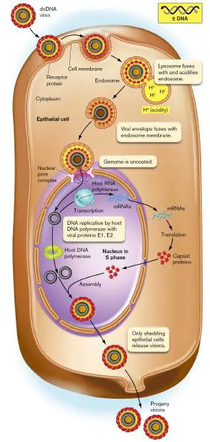
FIGURE 6.32 ■ Papillomavirus life cycle. HPV, a double-stranded DNA virus, enters the cytoplasm, where the protein coat disintegrates. The viral DNA enters the nucleus for replication and transcription by host polymerases.
HPV virions enter the host cell by endocytosis, followed by lysosome fusion and acidification (Fig. 6.32). The acidification triggers virion uncoating and releases the viral genome of circular double-stranded DNA. The uncoated viral DNA passes through the nuclear pore complex to enter the host cell nucleus, where it is called an episome. The nucleus must be in S phase (DNA synthesis) in order to support HPV replication.
Unlike RNA viruses, the HPV episome consists of DNA that can get replicated by the host DNA polymerase. But initiation is modified by the two virus-encoded proteins E1 and E2. E1 is a viral DNA helicase (see Chapter 7), whereas E2 helps E1 bind the viral origin of replication. Together, these proteins cause the cell to prioritize viral replication over its own DNA synthesis.
The viral DNA gets transcribed by the host RNA polymerase. Viral mRNA molecules are exported to the cytoplasm for translation of capsid proteins, which return to the nucleus for assembly of virions. Ultimately the progeny virions must exit the cell, but the HPV exit process is not well understood.
The process of HPV replication is complicated by the developmental progression of basal cells into keratinocytes (mature epithelial cells), and ultimately cells to be shed or sloughed off from the surface (Fig. 6.33). Viral replication is largely inhibited until the basal cells start to differentiate into keratinocytes. Host cell differentiation induces the viral DNA to replicate and undergo transcription by host polymerases. The mRNA transcripts then exit the nuclear pores, as do host mRNAs, for translation in the cytoplasm. The translated capsid proteins, however, return to the nucleus for assembly of the virion. Nuclear virion assembly is typical of DNA viruses (with the exception of poxviruses, which replicate entirely in the cytoplasm).
FIGURE 6.33 ■ Human papillomavirus causes cancer. HPV enters the cervical epithelium, where it infects basal epithelial cells. The DNA uncoats and forms an episome (circularized). As cells differentiate, new virions are synthesized and released by shedding cells. Some HPV genomes may integrate into host DNA. Integrated HPV genomes express proteins that transform host cells into cancer cells.
How do HPV virions disseminate? As the keratinocytes containing HPV complete differentiation, the cells start to come apart and are shed from the epithelial surface (Fig. 6.33). Cells release HPV virions during this shedding process. Virion-releasing cells show partly accelerated growth, leading to formation of warts.
Basal cells containing viral episomes generally do not cause cancer. However, the integrated HPV genome up-regulates expression of oncogenes (cancer-causing genes) to form oncoproteins E6 and E7. E6 inactivates the host tumor suppressor protein p53, whereas E7 inactivates retinoblastoma protein pRb, a checkpoint marker for cells to enter S phase. These oncoproteins can
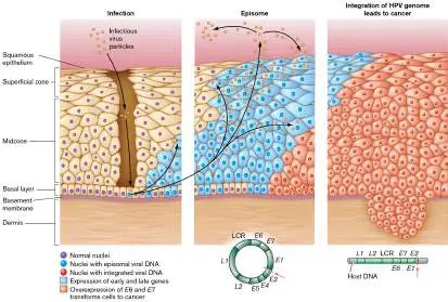
thus cause inappropriate start of host chromosome replication, leading to uncontrolled cell proliferation. This process can transform host cells into cancer cells, which invade the dermis and grow out of control. In some cases, the HPV genome integrates into the genome of cancer-transformed host cells.
Certain HPV strains are more likely to cause cancer, whereas others more likely cause warts (inappropriate tissue growth that is not cancerous). Both warts and cancer provide HPV with more host cells in S phase to produce virions. The most common strains that cause warts or cancer are both preventable by the Gardasil vaccine.
Thought Question
6.10 From the standpoint of a virus, what are the advantages and disadvantages of replication by the host polymerase compared with replication by a polymerase encoded by the virus’s own genome?
Oncogenic Viruses
As many as 20% of human cancers are caused by oncogenic viruses, such as Epstein-Barr virus (which causes lymphomas) and hepatitis C virus (which causes liver cancer). (Hepatitis C replication is presented in eAppendix 4.) When these viruses infect a cell, instead of destroying it the virus may transform the cell to divide and grow out of control. For a virus, the advantage of cancer transformation is that it expands the population of infected cells that produce virus particles or that replicate a viral genome hidden within a host chromosome.
How do oncogenic viruses cause cancer? Several different mechanisms enable different types of viruses to transform normal host cells so that they proliferate abnormally and form tumors. Oncogenes. A retrovirus such as feline leukemia virus (FeLV) may carry an oncogene, which can transform the host cells. Usually an oncogene encodes an abnormal form of an important host protein that controls cell proliferation. The normal host form is called a “proto-oncogene.”
Genome integration. Certain viruses can integrate their genome into a host chromosome. The integrated viral genome expresses proteins that stimulate host cell division and may ultimately lead to growth of tumors.
Cell cycle control. Oncogenic viruses such as papillomaviruses express viral proteins that interact with host cell cycle controls and can stimulate uncontrolled growth.
Viral capacities for gene transfer and host genome control may be manipulated artificially and used for gene therapy. In fact, some of the most dangerous viruses, such as HIV, are being engineered to make nonvirulent gene delivery devices (discussed in Section 11.4). HIV-derived vectors form the basis of CAR-T therapy for leukemia and other cancers, first approved by the U.S. Food and Drug Administration in 2017.
While some kinds of human viruses cause chronic infections and even cancer, others appear in our bodies indefinitely while causing no apparent disease. For example, anelloviruses commonly appear in our blood plasma, without known disease. Children frequently shed enteroviruses (viruses of the intestinal tract, related to poliovirus) with no known effects. Some evidence supports the idea that low-level chronic viral infections might actually enhance the function of our immune system.
Plant Viruses
All kinds of plants are subject to viral infection. Plant viruses pose enormous challenges to agriculture, especially where the concentrated growth of a single strain of food crop (monoculture) provides ideal conditions for a virus to spread.
Plant virus entry to host cells. In contrast to animal viruses and bacteriophages, plant viruses infect cells by mechanisms that do not involve specific membrane receptors. The reason may be that plant cell membranes are covered by thick cell walls impenetrable to virion uptake or genome insertion. Thus, the entry of plant viruses usually requires mechanical transmission —nonspecific access through physical damage to tissues, such as abrasions of the leaf surface caused by a feeding insect. Mechanical transmission of plant viruses is limited by the cell wall. Most plant viruses gain entry to cells by one of three routes: Contact with damaged tissues. Viruses such as tobacco mosaic virus appear to require nonspecific entry into broken cells.
Transmission by an animal vector. Insects and nematodes transmit many kinds of plant viruses. For example, the geminiviruses are inoculated into cells by plant-eating insects such as aphids, beetles, and grasshoppers.
Transmission through seed. Some plant viruses enter the seed and infect the next generation.
An economically important plant virus is the potyvirus called plum pox virus, a major pathogen of plums, peaches, and other stone fruits. Plum pox virus, a group IV (+) strand RNA virus (see Table 6.2), is transmitted by aphids (Fig. 6.34). After infection, the spread of the virus generates streaked leaves and flowers, as well as ring-shaped pockmarks on the surfaces of the fruit and of the stone within.
FIGURE 6.34 ■ Plum pox is caused by potyvirus. A. Potyvirus, a filamentous (+) strand RNA virus, approximately 800 nm in length (TEM). B. Potyvirus is transmitted by aphids, which suck the plant sap and release the virus into the damaged tissues. C. Streaking of flowers caused by potyvirus infection. D. Ring-shaped pockmarks appear on the infected fruit.
CENTRE FOR BIOIMAGING, ROTHAMSTED RESEARCH/SCIENCE SOURCE
SCOTT BAUER, AGRICULTURAL RESEARCH SERVICE, USDA
C. AVILEZ/ALAMY STOCK PHOTO
NATURE AND SCIENCE/ALAMY STOCK PHOTO
Plant virus transmission through plasmodesmata. Within a plant, the thick cell walls prevent a lytic burst or budding out of virions. Instead, plant virions spread to uninfected cells by traveling through plasmodesmata (singular, plasmodesma ). Plasmodesmata are membrane channels that connect adjacent plant cells (Fig. 6.35). The outer part of the channel connects the cell membranes of the two cells; the inner part connects the endoplasmic reticulum of one cell to that of the other. Passage through the plasmodesmata requires action by movement proteins whose expression is directed by the viral genome. In some cases, the movement proteins transmit the entire plant virion; in other cases, only the viral genome is small enough to pass through. The infection strategies of plant viral genomes may have features in common with those of viroids, which lack capsids altogether.
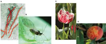
FIGURE 6.35 ■ Plant cells connected by plasmodesmata. Plasmodesmata offer a route for plant viruses to reach uninfected cells.
Cauliflower mosaic virus replication cycle. A plant virus important for agriculture is cauliflower mosaic virus (CaMV), a caulimovirus. Caulimovirus infects a wide range of cruciferous vegetables such as broccoli, cabbage, and cauliflower. The virus is a Baltimore group VII pararetrovirus, possessing a DNA genome that requires transcription to RNA in the cytoplasm, followed by reverse transcription to form DNA genomes for progeny virions. CaMV is an important tool for biotechnology because it has a highly efficient promoter for gene transcription, enabling high-level expression of
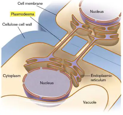
cloned genes. Vectors derived from caulimoviruses are used to construct transgenic plants.
CaMV is transmitted by secretions from an insect whose bite damages plant tissues, providing access to the cytoplasm (Fig. 6.36). While no plant-specific receptor is required, recent research has identified virus receptors in the mouthparts of the aphid. The CaMV genome moves from the cytoplasm to the cell nucleus through a nuclear pore. Within the nucleus, two promoters on its DNA genome direct transcription to RNA. The two RNA transcripts exit the nucleus for translation by host ribosomes to make viral proteins. A host reverse transcriptase, present in plant cells, copies the RNA into DNA viral genomes. After virions are assembled in the cytoplasm, virus-encoded proteins, called movement proteins, help transfer the virions through plasmodesmata into an adjacent cell.
FIGURE 6.36 ■ Caulimovirus life cycle. The cauliflower mosaic virus (CaMV; inset), a DNA pararetrovirus, uses host RNA polymerase to copy its DNA into RNA and uses host reverse transcriptase (RT) to make DNA copies.
A CaMV promoter sequence is commonly used in gene transfer vectors for plant biotechnology because transcription of the gene (such as one that confers pesticide resistance on the host) linked to the viral promoter is very efficient. In the field, 10% of cruciferous
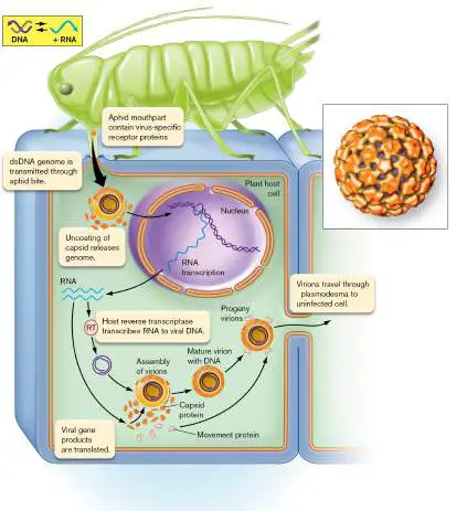
vegetables are typically infected with cauliflower mosaic virus. Some critics of gene technology fear that the prevalence of the CaMV promoter in transgenic crops may lead to the evolution of new pararetroviruses.
Animal and Plant Host Defenses
How do animals and plants defend themselves from virus infection? Because viruses are ubiquitous, a wide range of defense mechanisms have evolved. Defenses important for humans are part of our immune system, presented in Chapters 23 and 24.
Genetic resistance. As we saw for bacteria, animal and plant hosts continually experience mutations, some of which lead to strains that resist viral infection by halting adsorption or some other key step of the virus’s replication cycle. When a virus becomes widespread, natural selection favors resistant strains. But commercial livestock and crops are typically a monoculture in which no resistant variants are available. Thus, when an outbreak arises, an entire crop may be destroyed. To save a crop, it may be interbred with a wild strain that possesses genes conferring resistance. An example of genetic resistance in humans is resistance to HIV/AIDS. The basis of this resistance is a defective allele encoding a T-lymphocyte cell-surface protein that acts as a coreceptor, which is required for binding of virus HIV-1. The role of coreceptors in HIV infection and resistance is discussed further in Section 11.3. Immune system. The immune systems of humans and other animals possess extensive cell machinery to thwart viral infection. A component of our innate immunity is the class of proteins called interferons, which recognize general signs of viral infections, such as the presence of double-stranded RNA (see Chapter 23). For adaptive immunity, viral proteins expressed in the cell membrane of an infected cell are recognized by specific antibodies that stimulate immune cells to destroy the infected cell and halt its viral production. The antibodies recognize a specific virus strain, such as a strain of influenza virus to which the individual has been exposed previously. For example, during the 2009 flu epidemic, many individuals over the age of 50 had some protection arising from exposure to a similar strain in an earlier epidemic (see Chapter 24). RNA interference. RNA interference, or RNAi, is a mechanism by which mRNA molecules expressed by a viral genome are recognized by a host protein-RNA complex that shuts down further expression. RNA interference was first discovered in plants, where the system is most extensive, but it is now known to be widespread among all eukaryotes and archaea (discussed in Chapter 9). The mechanisms of RNA interference are now being engineered for use in gene therapy to halt gene expression in cancer and in inherited diseases.
Emergence of Viral Pathogens
Where does a “new virus” come from? Most human pathogens come from other humans or from related animals referred to as vectors. Certain human-infecting viruses persist in the wild, such as rabies virus and West Nile virus. Their persistence requires broad host ranges: rabies infects many different mammals, and West Nile virus infects birds as well as humans and horses. Understanding the epidemiology of rabies or of West Nile encephalitis requires understanding the behavior and seasonal migration patterns of wild organisms. While the COVID-19 pandemic was caused by a coronavirus previously unknown to science, previous coronavirus outbreaks were traced genetically to viruses found in bats and in camels. As of this writing, the origin of SARS-CoV-2 remains unknown.
Other emerging viruses arise as variants of endemic milder pathogens. Viruses long associated with a host, such as the common-cold viruses (rhinoviruses), tend to have evolved a moderate disease state that provides ample opportunities for host transmission. A virus that “jumps” from an animal host, however, may cause a more acute syndrome with higher mortality. The best-known cases are the exceptionally virulent emerging strains of influenza, which generally result from intracellular recombination of human strains with strains from pigs or ducks (discussed in Chapter 11). For example, in 2013 the avian influenza strain H7N9 emerged from poultry in China, where it killed several people before it was contained. Changes in the distribution patterns of insect vectors and animal hosts can generate new epidemics of a pathogen in regions where the virus could not spread before. Such changes in distribution can be brought about by many factors, including global climate change (see Chapter 28).
To Summarize
Host cell-surface receptors mediate animal virus attachment to a cell and confer host specificity and tropism. Animal DNA viruses either inject their genome or enter the host cell by endocytosis. The viral genome requires uncoating for gene expression.
RNA viruses use an RNA-dependent RNA polymerase to transcribe their messenger RNA.
Retroviruses use a reverse transcriptase to copy their genomic sequence into DNA for insertion in the host chromosome.
Oncogenic viruses transform the host cell to become cancerous. Mechanisms of oncogenesis by different types of viruses include insertion of an oncogene into the host genome, integration of the entire viral genome, and expression of viral proteins that interfere with host cell cycle regulation.
Plant viruses enter host cells by transmission through a wounded cell surface or by an animal vector, and they travel to adjacent cells through plasmodesmata.
Pararetroviruses contain DNA genomes but generate an RNA intermediate that requires reverse transcription to DNA for progeny virions.
Emerging viral pathogens commonly arise by mutation from pathogens of related animals.
Glossary
tropism The ability of a virus to infect a particular tissue type. endocytosis The invagination of the cell membrane to form a vesicle that contains extracellular material.
uncoating The release of a viral genome from its capsid, following entry of the virion into a host cell.
polyprotein A long peptide translated from one open reading frame but later cleaved into separate proteins with different functions. protease An enzyme that cleaves protein.
viral factory Also called replication complex. An intracellular membrane compartment with viral proteins that synthesize progeny virions. oncogenic virus A virus that causes cancer.
transform To cause bacteria to take up exogenous DNA. In eukaryotes, to convert cultured cells into cancer cells.
mechanical transmission A nonspecific means of transfer of a pathogen to a host, such as the transmission of a virus through a wound or of a bacterial pathogen from the body surface of a vector (such as a fly). plasmodesma pl. plasmodesmata A membrane channel in plants that connects adjacent plant cells.
Fig. 6.19 FIGURE 6.19 ■ Baltimore classification of viral genomes. A. Seven categories of viral genome composition and replication mechanism. B. David Baltimore studies viral sequence DNA at the California Institute of Technology. dsDNA = double-stranded DNA.
SLADE PAUL/CONTRIBUTOR/GETTY IMAGES
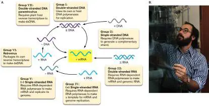
6.6 Culturing VirusesUnit 3 · Structure
To learn how microbes grow, we culture them in the laboratory. So how do we culture a virus? A complication of virus culture is the need to grow the virus within a host cell. Therefore, any virus culture system must be a double culture of host cells plus viruses. Culturing viruses of multicellular animals and plants involves additional complications, because viruses show tropism for particular tissues or organs. Viruses may replicate in tissue culture, but the tissue culture does not show all the properties of an organ within a living organism. Therefore, a virus propagated in tissue culture will evolve to lose some of the virulence factors needed to infect an animal.
Batch Culture
Batch culture, or culture in an enclosed vessel of liquid medium, enables growth of a large population of viruses for study.
Bacteriophages can be inoculated into a growing culture of bacteria, usually in a culture tube or a flask. The culture fluid is then sampled over time and assayed for phage particles. The growth pattern usually takes the form of a step curve (Fig. 6.37).
FIGURE 6.37 ■ One-step growth curve for a bacteriophage. After initial infection of a liquid culture of host cells, the titer of virus drops to near zero as all virions attach to the host. During the eclipse period, progeny phages are being assembled within the cell. As cells lyse (the rise period), virions are released until they reach the final plateau.
To observe one cycle of phage reproduction, phages are added to host cells at a high multiplicity of infection (MOI ; ratio of phage to cells) such that every host cell is infected. The phage particles immediately adsorb to surface receptors of host cells and deliver their DNA. As a result, intact virions are virtually undetectable in the growth medium. This short period after infection is called the eclipse period. For some species, it is possible to distinguish between the eclipse period and a latent period, the time during which the inserted phage genome directs production of progeny virions. The virions accumulate within the cell but have not yet emerged in the medium. In animal viruses, the latent period is less distinct because
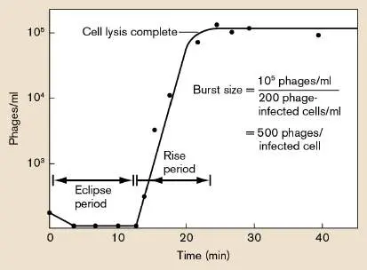
large numbers of virions usually generate progeny through budding out of the host cell (Fig. 6.38).
FIGURE 6.38 ■ One-step growth curve for an animal virus. The titer of extracellular virus drops to near zero during the latent period, as all virions adsorb to the host. Then progeny virions begin to emerge by budding out from the infected cell. The growth curve may take hours to level off; the “burst” event is not defined as clearly as for phages.
Note: The “latent period” of a lytic virus is the period between
initial phage-host contact and the first appearance of progeny phage. This period must be distinguished from the “latent infection ” of a virus that maintains its genome within a host cell without reproducing virions.
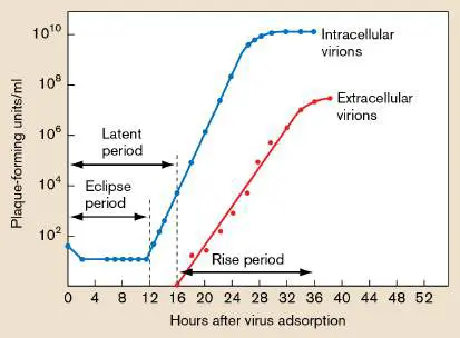
As cells begin to lyse and liberate progeny viruses, the culture enters the rise period, during which virus particles appear in the growth medium. The rise period ends when all the progeny viruses have been liberated from their host cells. If the number of viruses that go on to inoculate additional host cells is small, then the virus concentration at the end point, divided by the original concentration of inoculated phage, approximates the burst size; that is, the number of viruses produced per infected host cell. To estimate the burst size, we can divide the concentration of progeny virions by the concentration of infected cells.
The burst size, together with the cell density prior to lysis, determines the concentration of the resultant suspension of virus particles, called a lysate. In the case of bacteriophages, a lysate of phage particles can be extremely stable, remaining infective at room temperature for many years. Eukaryotic viruses, however, tend to be less stable and need to be maintained in culture or deep freeze.
Thought Question
6.11 Why does bacteriophage reproduction give a step curve, whereas cellular reproduction generates an exponential growth curve? (Compare Fig. 6.37with Fig. 4.22.) Could you design an experiment in which viruses generate an exponential growth curve? Under what conditions does the growth of cellular microbes give rise to a step curve?
Tissue Culture of Animal Viruses
In the case of animal and plant viruses, the multicellular nature of the host is an important factor in the pathology and transmission of the pathogen (discussed in Chapters 25 and 26). Animal viruses can be cultured within whole animals by serial inoculation, where virus is transferred from an infected animal to an uninfected one. Culture within animals ensures that the virus strain maintains its original virulence (ability to cause disease). But the process is expensive and laborious, involving the large-scale use of animals.
A historic event in 1949 was the first successful growth of a virus in tissue culture. Poliovirus, the causative agent of the devastating childhood disease poliomyelitis, was grown in human cell tissue culture (Fig. 6.39) by John F. Enders, Thomas J. Weller, and Frederick Robbins at Children’s Hospital in Boston. As heralded that year in Scientific American: “It means the end of the ‘monkey era’ in poliomyelitis research.... Tissue-culture methods have provided virologists with a simple in vitro method for testing a multitude of chemical and antibiotic agents.” Since then, tissue culture has remained the most effective way to study the molecular biology of animal and plant viruses and to develop vaccines and antiviral agents.
FIGURE 6.39 ■ Poliovirus replication in human tissue culture. Before infection (0 hours), the cultured cells grow in a smooth layer. At 8 hours, infected cells have detached from the culture dish. By 24 hours, cells have lysed or in some cases clumped with other cells.
J. FLINT ET AL. PRINCIPLES OF VIROLOGY, FOURTH ED. 2015. BUNDLE
Some viruses can be propagated in a tissue culture of cells growing confluently on a surface. The cells must be immortalized—
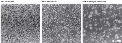
that is, genetically altered—to continue cell division indefinitely. The fluid bathing the tissue layer is sampled for virus concentration. As in the case of bacteriophage batch culture, we can define an eclipse period, a latent period before appearance of the first progeny virions in the culture fluid, and a rise period. In tissue culture, the time course of animal virus replication is usually much longer (hours or days) than that of bacteriophages (typically less than an hour under optimal conditions). The burst size of animal viruses, however, is typically several orders of magnitude larger than that of phages. The reason for the larger burst size is that the volume of a host cell is much larger than that of a bacterial host, thus providing a larger supply of materials to build virions.
Thought Question
6.12 What kinds of questions about viruses can be addressed in tissue culture, and what questions require infection of an animal model?
Plaque Isolation and Assay of Bacteriophages
For the investigation of cellular microbes, an important tool is the culturing of individual colonies on a solid substrate that prevents dispersal throughout the medium, as described in Chapters 1 and 4. Plate culture of colonies enables us to isolate a population of microbes descended from a common progenitor. But viruses cannot be isolated as “colonies.” The reason is that although viruses can be obtained at incredibly high concentrations, they disperse in suspension. Even on a solid medium, viruses never form a solid visible mass comparable to the mass of cells that constitutes a cellular colony.
In viral plate culture, viruses from a single progenitor lyse their surrounding host cells, forming a clear area called a plaque . Each plaque arises from a single infected bacterium that bursts, its phage particles diffusing to infect neighboring cells (Fig. 6.40). FIGURE 6.40 ■ Plating a phage suspension to count isolated plaques. A suspension of bacteria in rich broth culture is inoculated with a low proportion of phage particles (multiplicity of infection is approximately 0.1). The culture is then spread on a
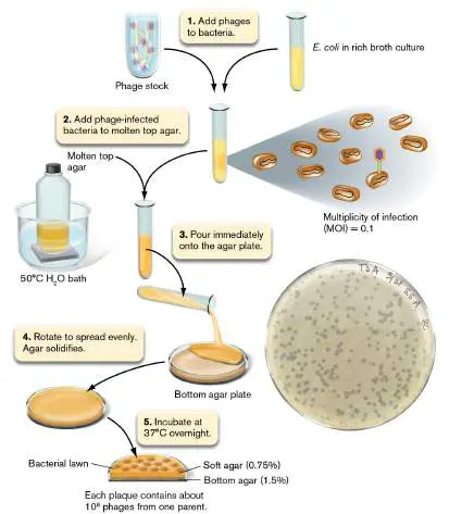
plate of rich agar where bacteria multiply. Each plaque arises from a single infected bacterium that bursts, its phage particles diffusing to infect neighboring cells.
JOAN SLONCZEWSKI
To perform a plaque assay of bacteriophages, a diluted suspension of phages is mixed with bacterial cells in soft agar, and the mixture is then poured over a nutrient agar plate (Fig. 6.40). Where no bacteriophages are present, the bacteria grow homogeneously as a “lawn,” an opaque sheet over the surface (confluent growth). Where bacteriophages are included, each one infects a cell, replicates, and spreads progeny phages to adjacent cells, killing them as well. The loss of cells results in a round, clear area seemingly cut out of the bacterial lawn. Plaques can be counted and used to calculate the concentration of phage particles, or plaque-forming units (PFUs ), in a given suspension of liquid culture (Fig. 6.40, inset). The liquid culture can be analyzed by serial dilution in the same way one would analyze a suspension of bacteria.
Plaque Isolation and Assay of Animal Viruses
For animal viruses, the plaque assay has to be modified because it requires infection of cells in tissue culture. Tissue culture usually involves growth of cells in a monolayer on the surface of a dish containing fluid medium, which would quickly disperse any viruses released by lysed cells. To solve this problem, in 1952 Renato Dulbecco, at the California Institute of Technology, modified the tissue culture procedure for plaque assays (Fig. 6.41A). In Dulbecco’s method, the tissue culture with liquid medium is first inoculated with virus. After sufficient time to allow for viral attachment to cells, the fluid is removed and replaced by a gel medium. The gel retards the dispersal of viruses from infected cells, and as the host cells die, plaques can be observed. Figure 6.41B shows a plate culture of human coronavirus infection of colon carcinoma cells.
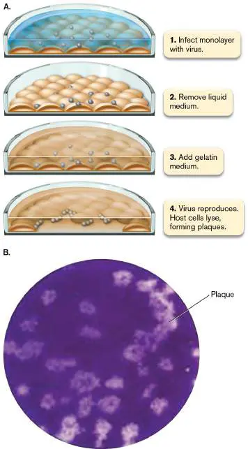
FIGURE 6.41 ■ Plate culture of animal viruses. A. Modified plaque assay for animal viruses. The gelled medium retards the dispersal of progeny virions from infected cells, restricting new infections to neighboring cells. The result is a visible clearing of cells (a plaque) in the monolayer. B. Plaque assay in which human coronavirus suspension was plated on a monolayer of colon carcinoma cells in tissue culture.
© P. HERZOG ET AL. 2008. VIROL. J. 5 :138
Animal viruses that do not kill their host cells require a different kind of assay that is based on identification of a “focus” (plural, foci), a group of cells infected by the virus. A focus may be identified using a fluorescent antibody—a method called fluorescent-focus assay. Another type of focus assay can be used to isolate oncogenic viruses, which transform their host cells into cancer cells. The cancer cells lose contact inhibition; they grow up in a pile instead of remaining in the normal monolayer. These piles of transformed cells, or transformed foci, can easily be visualized and counted. This procedure is known as the transformed-focus assay.
To Summarize
Culturing viruses requires growth in host cells.
Batch culture of viruses generates a step curve.
Plate culture involves replication of phages on a bacterial lawn or animal viruses in tissue culture.
Plaque assay is a method of culturing viruses in which single phages or virions each generate an isolated clearing of host cells. The plaques can be counted to enumerate the infectious virions in a suspension, called plaque-forming units.
Glossary
batch culture The growth of bacteria in a closed system without additional input of nutrients.
multiplicity of infection (MOI)
The ratio of infecting virions to host cells.
eclipse period The time in the viral life cycle after viral genome injection into a host cell but before complete virions are formed.
latent period The time in the viral life cycle when progeny virions have formed but are still within the host cell.
rise period The period of time in the viral life cycle when cells lyse and viral progeny are liberated.
burst size The number of virus particles released from a lysed host cell. lysate The contents of broken cells; may include virus particles. virulence A measure of the severity of a disease caused by a pathogenic agent.
plaque-forming unit (PFU)
A measure of the concentration of phage particles in liquid culture.
Fig. 4.22 FIGURE 4.22 ■ Bacterial growth curves. A. and B. Theoretical growth curves of a bacterial suspension measured by optical density (OD) at a wavelength of 600 nm. Linear-scale (A) and logarithmic-scale (B) plots of OD 600. Note that the log plot is a straight line. C. Phases of bacterial growth in a typical batch culture.
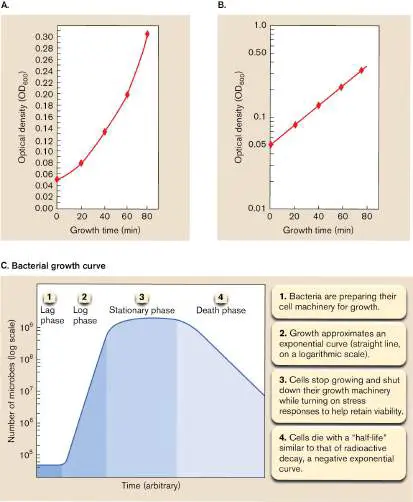
eResearch Activity 6
Muddy and ZoeJ: Can Phages Discovered by Undergraduates Save a Patient’s Life?
Bacteriophages are a common subject of undergraduate laboratory projects, as they replicate fast and pose little risk to the beginner. Who could imagine that phages discovered by beginner undergraduates could destroy pathogens that nearly killed a patient? That was an outcome of a multi-university undergraduate phage discovery program called Science Education Alliance–Phage Hunters Advancing Genomics and Evolutionary Science (SEA-PHAGES) (Fig. ERA 6.1 ). In the SEA-PHAGES program, funded by the Howard Hughes Medical Institute (HHMI), undergraduates isolate novel phages from environmental soil samples by plating them on the host bacterium Mycobacterium smegmatis.
FIGURE ERA 6.1 ■ Undergraduates in the SEA-PHAGES program. A. Students discover phages that infect a harmless
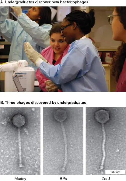
species, Mycobacterium smegmatis. B. Three of the 10,000 mycophages were shown to infect deadly Mycobacterium abscessus (TEM).
ERIC BRADLEY/HOWARD HUGHES MEDICAL INSTITUTE
COURTESY GRAHAM HATFULL/THE HATFULL LABORATORY
M. smegmatis is a harmless member of the human genital microbiome (microbial community). But the bacterium has close relatives that cause devastating disease, such as M. tuberculosis and M. abscessus. Could a phage that infects M. smegmatis also provide a new weapon against a pathogenic relative? Graham Hatfull, the SEA-PHAGES program director at the University of Pittsburgh, hoped to find out. Hatfull maintains a collection of 15,000 new phage isolates in storage, and his program sequences the genomes of many of them.
One day Hatfull heard from a physician in London, Helen Spencer, whose teenage patient Isabelle Carnell-Holdaway was dying of M. abscessus infection. The patient was immunocompromised after a double-lung transplant for cystic fibrosis, and no antibiotics fought off her infection. The patient’s mother wondered if a phage could fight the pathogen—and Spencer wanted to try this last-ditch effort.
So Hatfull got to work screening the SEA-PHAGES collection. His research team tested phages for lysis of bacteria by spotting a tenfold dilution series of phage concentrations on a lawn of their original M. smegmatis host (Fig. ERA 6.2 ). Similar concentrations were then plated on a strain of M. abscessus that had infected Spencer’s patient. Most did not lyse the new host—but three phages, originally named by their undergraduate discoverers, did cause some clearing. Phage Muddy showed the strongest lysis at all concentrations. Phage ZoeJ showed weak lysis; and knowing the phage genome, Hatfull’s team knocked out a repressor of lysogeny to eliminate this alternative to lysis. Phage BPs also showed weak lysis, and a host-range mutant did even better.
FIGURE ERA 6.2 ■ Mycophages that lysed Mycobacterium abscessus were used to cure bacterial infection. The undergraduates discovered phages that cleared lawns of M. smegmatis. A tenfold dilution series of phage concentrations was spotted on lawns of the bacterial host. Three of these phages also cleared the pathogen M. abscessus GD01.
R. M. DEDRICK ET AL. 2019. NAT MED. 25 :730–733
Short on time, the team devised a cocktail of all three phages, and Spencer applied it intravenously to her patient. Within a day, progeny mycobacterial phages were found in high concentration in the patient’s serum. Amazingly, the patient improved and overcame the infection that otherwise would surely have led to her death. This story has limitations in that the particular phage cocktail works only for the particular pathogen in this patient. In 2022, the phage Muddy helped cure another patient infected with a different Mycobacterium species, M. chelonae. The rapid genetics of phage analysis could enable future patient-tailored therapies.
Further Exploration
How might phage therapies be devised to succeed for more kinds of pathogens and patients? What kind of genetic manipulations might help? How might phage therapy be combined with antibacterial therapy for more effective cures?
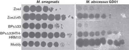
Dedrick, Rebekah M., Carlos A Guerrero-Bustamante, Rebecca A Garlena, Daniel A Russell, Katrina Ford, et al. 2019. Engineered bacteriophages for treatment of a patient with a disseminated drug-resistant Mycobacterium abscessus. Science 25 :730–733.
CHAPTER REVIEW
Review Questions
1. Compare and contrast the forms of icosahedral and filamentous (helical) viruses, citing specific examples. 2. How do viral genomes gain entry into cells in bacteria, plants, and animals?
3. Explain the key structural features that define the seven Baltimore groups of viral genomes. Explain the consequences of each structure for viral replication. 4. How do viral genomes interact with host genomes, and what are the consequences for host evolution?
5. Compare and contrast the lytic, lysogenic, and slow-release replication cycles of bacteriophages. What are the strengths and limitations of each?
6. Compare and contrast the replication cycles and transmission strategies of a coronavirus and a papillomavirus. What are the strengths and limitations of each?
7. Explain the plate count procedure for enumerating viable bacteriophages. How must this procedure be modified to measure the concentration of animal viruses? Oncogenic viruses?
8. Explain how a pure isolate of a virus can be obtained. How do the procedures differ from those used for isolating bacteria?
9. Explain the generation of the step curve of virus proliferation. Why is virus proliferation generally observed as a single step, or generation, in contrast to the life cycles of cellular microbes outlined in Chapter 4? 10. Explain the key contributions of viruses to natural ecosystems. What may happen in an ecosystem where viruses are absent or fail to cause significant infection?
Thought Questions
1. Discuss the functions of different structural proteins of a virion, such as capsid, nucleocapsid, tegument, and envelope proteins. How do these functions compare and contrast with functions of cell proteins?
2. What are the relative advantages of the virulent phage replication cycle of phage T4, the lysis/lysogeny options of phage lambda, and the slow-release replication of phage M13? Under what conditions might each strategy be favored over the others?
3. Given the basis of viral tropism, how might an animal evolve traits that confer resistance to a virus infection? 4. If viruses return a substantial fraction of marine CO 2 to the atmosphere, can you imagine any ways to modulate virus proliferation so as to divert the carbon into sedimenting biomass?
Key Terms
bacteriophage (202)
batch culture (242)
burst size (224, 243)
capsid (202, 210)
cell-surface receptor (222) core particle (210)
CRISPR (226)
eclipse period (242)
endocytosis (229)
endogenous virus (205)
envelope (210)
filamentous virus (211)
host range (208)
latent period (242)
lysate (243)
lysis (224)
lysogeny (224)
mechanical transmission (239) multiplicity of infection (MOI) (242) oncogenic virus (237)
ortholog (orthologous gene) (221) pararetrovirus (219)
phage (202)
plaque (203, 244)
plaque-forming unit (PFU) (245) plasmodesma (239)
polyprotein (231)
prion (215)
prophage (205)
protease (231)
provirus (205, 208)
retrovirus or reverse-transcribing virus (219) reverse transcriptase (219) rise period (243)
RNA-dependent RNA polymerase (218) segmented genome (217)
site-specific recombination (224) spike protein (211)
temperate phage (224)
transcytosis (227)
transduction (224)
transform (237)
tropism (229)
uncoating (229)
viral factory (233)
viral shunt (208)
virion (202, 203)
virocell (206)
viroid (213)
virome (207)
virulence (243)
virus (202)
Recommended Reading
Abergel, Chantal, Matthieu Legendre, and Jean-Michel Claverie. 2015. The rapidly expanding universe of giant viruses: Mimivirus, Pandoravirus, Pithovirus and Mollivirus. FEMS Microbiology Reviews 39 :779–796.
Chaturvedi, Anil K., Barry I. Graubard, Tatevik Broutian, Robert K. L. Pickard, Zhen-Yue Tong, et al. 2018. Effect of prophylactic human papillomavirus (HPV) vaccination on oral HPV infections among young adults in the United States. Journal of Clinical Oncology, 36 :262–267.
Dion, Moïra B., Frank Oechslin, and Sylvain Moineau. 2020. Phage diversity, genomics and phylogeny. Nature Reviews. Microbiology 18 :125–138.
DeLong, John P., Maitham A. Al-Sammak, Zeina T. Al-Ameeli, David D. Dunigan, Kyle F. Edwards, et al. 2022. Towards an integrative view of virus phenotypes. Nature Reviews Microbiology 20 :83–94.
Fridman, Svetlana, José Flores-Uribe, Shirley Larom, Onit Alalouf, Oded Liran, et al. 2017. A myovirus encoding both photosystem I and II proteins enhances cyclic electron flow in infected Prochlorococcus cells. Nature Microbiology 2 :1350– 1357.
Fu, Chi-yu, Kang Wang, Lu Gan, Jason Lanman, Reza Khayat, et al. 2010. In vivo assembly of an archaeal virus studied with whole-cell electron cryotomography. Structure 18:1579–1586.
Grigg, Patricia, Allison Titong, Leslie A. Jones, Tilahun D. Yilma, and Paulo H. Verardi. 2013. Safety mechanism assisted by the repressor of tetracycline (SMART) vaccinia virus vectors for vaccines and therapeutics. Proceedings of the National Academy of Sciences USA 110 :15407–15412.
Legendre, Matthieu, Julia Bartoli, Lyubov Shmakova, Sandra Jeudy, Karine Labadie, et al. 2013. Thirty-thousand-year-old distant relative of giant icosahedral DNA viruses with a pandoravirus morphology. Proceedings of the National Academy of Sciences USA 111 :4274–4279.
Leung, Nancy H. L. 2021. Transmissibility and transmission of respiratory viruses. Nature Reviews. Microbiology 19 :1–18. Manrique, Pilar, Michael Dills, and Mark J. Young. 2017. The human gut phage community and its implications for health and disease. Viruses 9 :141.
McBride, Alison A., and Alix Warburton. 2017. The role of integration in oncogenic progression of HPV-associated cancers. PLoS Pathogens 13 :e1006211.
Raoult, Didier, Stéphane Audic, Catherine Robert, Chantel Abergel, and Patricia Renesto. 2004. The 1.2-megabase genome sequence of Mimivirus. Science 306 :1344–1350.
Roossinck, Marilyn J. 2011. The good viruses: Viral mutualistic symbioses. Nature Reviews. Microbiology 9 :99.
Thurber, Rebecca Vega, Jérôme P. Payet, Andrew R.
Thurber, and Adrienne M. S. Correa. 2017. Virus-host interactions and their roles in coral reef health and disease. Nature Reviews. Microbiology 15 :205–216.
V’kovski, Philip, Annika Kratzel, Silvio Steiner, Hanspeter Stalder, and Volker Thiel. 2021. Coronavirus biology and replication: Implications for SARS-CoV-2. Nature Reviews. Microbiology 19 :155–170.
Webster, Craig G., Elodie Pichon, Manuella Van Munster, Baptiste Monsion, Maëlle Deshoux, et al. 2018.
Identification of plant virus receptor candidates in the stylets of their aphid vectors. Journal of Virology 92 :e00432-18. Wolf, Yuri I., Darius Kazlauskas, Jaime Iranzo, Adriana Lucía-Sanz, Jens H. Kuhn, et al. 2018. Origins and evolution of the global RNA virome. mBio 9 : e02329-18.
Glossary
virus A noncellular particle containing a genome that can replicate only inside a cell.
virion A virus particle.
capsid The protein shell that surrounds a virion’s nucleic acid. Within an enveloped virus, such as HIV, the capsid may be called a core particle.
bacteriophage Also called phage. A virus that infects bacteria.
phage See bacteriophage .
plaque A cell-free zone on a lawn of bacterial cells caused by viral lysis.
prophage A phage genome integrated into a host genome.
provirus A viral genome that is integrated into the host cell genome. endogenous virus A virus whose genome is encoded within the germ line of a host animal; may be considered a functional part of the host. virocell A cell infected by a virus that reprograms it to maximize production of virus particles.
virome The genomes of all the viruses that inhabit a particular organism or environment.
viral shunt The release by viral lysis of cell contents as organic material available for microbial consumers in the upper region of the ocean.
host range The species that can be infected by a given pathogen. envelope A structure external to the cell membrane, such as the cell wall or outer membrane of a bacterium. For a virus, the envelope is a membrane enclosing the capsid or core particle.
core particle A viral capsid that encloses its nucleic acid genome and is surrounded by an envelope.
spike protein A viral glycoprotein that connects the membrane to the capsid or the matrix and may be involved in viral binding to host cell receptors.
filamentous virus A viral structure type consisting of a helical capsid surrounding a single-stranded nucleic acid.
viroid An infectious naked nucleic acid.
prion An infectious agent that causes propagation of misfolded host proteins; usually consists of a defective version of the host protein.
segmented genome A viral genome that consists of more than one nucleic acid molecule.
RNA-dependent RNA polymerase An enzyme that produces an RNA complementary to a template RNA strand.
retrovirus Also called RNA reverse-transcribing virus. A single-stranded RNA virus that uses reverse transcriptase to generate a double-stranded DNA.
reverse transcriptase (RT)
An enzyme that produces a double-stranded DNA molecule from a single-stranded RNA template.
pararetrovirus Also called DNA reverse-transcribing virus. A virus with a double-stranded DNA genome that generates an RNA intermediate and thus requires reverse transcriptase to generate progeny DNA genomes.
ortholog or orthologous gene A gene present in more than one species that derived from a common ancestral gene and encodes the same function.
cell-surface receptor A transmembrane protein that senses a specific extracellular signal and may be the docking site for a specific virus. lysis Also called burst. The rupture of the cell by a break in the cell wall and membrane.
burst size The number of virus particles released from a lysed host cell. temperate phage A phage capable of lysogeny.
lysogeny A viral life cycle in which the viral genome integrates into and replicates with the host genome but retains the ability to initiate host cell lysis.
site-specific recombination Recombination between DNA molecules that do not share long regions of homology but do contain short regions of homology specifically recognized by the recombination enzyme.
transduction The transfer of host genes between bacterial cells via a phage head coat.
CRISPR Clustered regularly interspaced short palindromic repeats. CRISPR consists of short repeated DNA sequences in a bacterial or archaeal genome, derived from previous bacteriophage or viral infection and conferring protection from future infection; considered a prokaryotic “immune system.” In biotechnology, provides a means of editing the human genome.
transcytosis The movement of a cell or substance from one side of a polarized cell to the other side, using an intracellular route. tropism The ability of a virus to infect a particular tissue type. endocytosis The invagination of the cell membrane to form a vesicle that contains extracellular material.
uncoating The release of a viral genome from its capsid, following entry of the virion into a host cell.
polyprotein A long peptide translated from one open reading frame but later cleaved into separate proteins with different functions. protease An enzyme that cleaves protein.
viral factory Also called replication complex. An intracellular membrane compartment with viral proteins that synthesize progeny virions.
oncogenic virus A virus that causes cancer.
transform To cause bacteria to take up exogenous DNA. In eukaryotes, to convert cultured cells into cancer cells.
mechanical transmission A nonspecific means of transfer of a pathogen to a host, such as the transmission of a virus through a wound or of a bacterial pathogen from the body surface of a vector (such as a fly). plasmodesma pl. plasmodesmata A membrane channel in plants that connects adjacent plant cells.
batch culture The growth of bacteria in a closed system without additional input of nutrients.
multiplicity of infection (MOI)
The ratio of infecting virions to host cells.
eclipse period The time in the viral life cycle after viral genome injection into a host cell but before complete virions are formed.
latent period The time in the viral life cycle when progeny virions have formed but are still within the host cell.
rise period The period of time in the viral life cycle when cells lyse and viral progeny are liberated.
lysate The contents of broken cells; may include virus particles. virulence A measure of the severity of a disease caused by a pathogenic agent.
plaque-forming unit (PFU)
A measure of the concentration of phage particles in liquid culture.