Chapter introduction
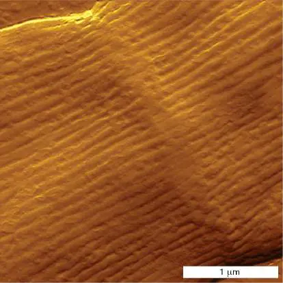
Cell division junction of an electrically wired cable bacterium. Cable bacteria conduct electron current across a long distance over wires through the periplasm. The wires contain nickel-sulfur cofactors.
ROB CORNELISSEN, UNIVERSITY OF HASSELT
Chapter Sections Learning Objectives
14.1 Electron » Transport Explain how an electron Systems and transfer system (ETS) carries the Proton energy through a series of Motive Force membrane-embedded molecules and generates a proton motive force.
14.2 The » Respiratory ETS Show how the ETS of cellular and ATP respiration generates proton Synthase motive force and drives synthesis of ATP.
14.3 Anaerobic » Respiration Describe how bacterial respiration may use metals and inorganic ions as terminal electron acceptors and explain the environmental importance of anaerobic respiration.
Chapter Sections Learning Objectives
14.4 Nanowires, » Electron Explain how bacterial Shuttles, and nanowires and electron Fuel Cells shuttles donate electrons to extracellular substrates and how bacterial current generates electricity in a fuel cell.
14.5 Lithotrophy » and Describe how oxidation of Methanogenesis inorganic minerals (lithotrophy) yields energy and how methanogenesis affects the environment.
Special Topic 14 Bacteria with “Locs” Rule the Bay 14.6 Phototrophy » Explain diverse forms of bacterial phototrophy, including sulfur photooxidation, cyclic photophosphorylation, and oxygenic photosynthesis (Z pathway).
eResearch Activity 14 Can Silver Extensions Amplify
Chapter Sections Learning Objectives
Bacterial Electricity?
Microbes move electrons—the equivalent of an electric current. Electrons move from reduced food molecules onto energy carriers, from energy carriers onto membrane proteins called cytochromes, and to organic electron carriers. This serial transfer of electrons is called an electron transport system (ETS). Electrons from the ETS ultimately reduce oxygen or other oxidized molecules. ETS electron flow drives protons across the cell membrane and generates a proton motive force. The proton motive force powers synthesis of the molecule ATP, which stores energy for later use.
Some microbes, such as cable bacteria (see the chapter-opening image), transfer electrons across a surprisingly long distance within the redox gradient of marine sediment. Others transfer electrons within biofilms. A fuel cell can harness these biofilms to generate electricity that can run devices. Microbial electricity offers exciting future opportunities for sustainable energy technology.
Chapter 14 presents electron flow in several different kinds of metabolism, including the respiration of organic substrates described in Chapter 13, as well as chemolithotrophy (oxidation of minerals) and photoautotrophy (light absorption by chlorophylls). Each of these pathways includes an ETS to store the energy and use it in the microbial cell.
14.1 Electron Transport Systems and the Proton Motive ForceUnit 4 · Metabolism
Electric current in living organisms has fascinated scientists ever since the eighteenth century, when Luigi Galvani (1737–1798) showed that a voltage caused a dead frog limb to flex. The idea of biological electricity inspired Mary Shelley’s famous novel Frankenstein: or, the Modern Prometheus, in which a physician is imagined to “create life” by jolting body parts with an electric shock. As we learned in Chapter 13, energy to support life is obtained through reactions that transfer electrons from a reduced electron donor to an oxidized electron acceptor. The simplest path of electron flow is found in fermentation, where electrons from the fermented substrate, such as glucose, are transferred onto NAD + to make NADH (reduction), and then returned to the glucose breakdown products, such as pyruvate. More complex kinds of metabolism, such as aerobic respiration, transfer electrons through a series of membrane-soluble carriers called an electron transport system (ETS), also known as an electron transport chain (ETC). For a review of fundamental redox chemistry, see eAppendix 1.
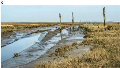
Today we know that electric current flows in all cells, including those of bacteria. For example, the most amazing kind of bacterial electricity was discovered in a salt marsh by Filip Meysman, Sairah Malkin, and their colleagues, then at the Free University of Brussels, Belgium (Fig. 14.1). The entire surface sediment of a marsh stores energy by a voltage potential via chains of bacteria. These bacterial chains, or filaments, are called cable bacteria because they transmit electrons through bundles of protein wires like an electric cable, as seen in the chapter-opening image. The wires extend in parallel through the periplasm, the outer compartment that envelopes the cytoplasm of Gram-negative bacteria. (Recall the outer membrane and periplasm, presented in Chapter 3.)
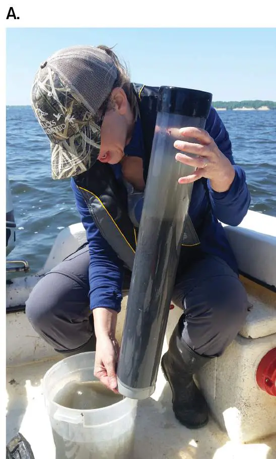
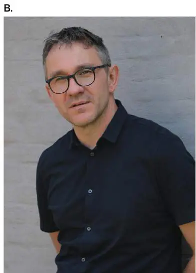
FIGURE 14.1 ■ Discoverers of electric bacteria. A. Sairah Malkin, University of Maryland Center for Environmental Science. B. Filip Meysman, University of Delft. C. Rattekaai salt marsh, Netherlands, where cable bacteria were first characterized.
ANDREW MCCARTHY
COURTESY OF FILIP MEYSMAN
PETER BRAAKMANN/SHUTTERSTOCK
Cable bacteria, most commonly the Desulfobulbaceae, are filaments of more than 10,000 cells that can transfer electrons across several centimeters (Fig. 14.2). Wires of protein with nickel cofactors extend through the periplasm, even across the septa between cells. Within marine sediment, cells on the deep end of the cable collect electrons from reduced molecules such as sulfides (HS − and H 2 S). Oxidation of sulfide (by removal of electrons) generates pure sulfur, deposited as precipitate. The electrons are captured by the bacteria, where they flow upward through the connected periplasm of the cell chain into the end cells exposed to oxygen gas. The upper-end bacteria then transfer the electrons to oxygen, which reacts with hydrogen ions to form water. The overall oxidation of sulfide by this bacterial battery removes forms of sulfur toxic to plants
and invertebrates, thus enriching the coastal ecosystem. This “electrified” ecosystem offers us a source of green energy.
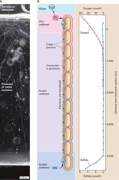
FIGURE 14.2 ■ Cable bacteria generate a voltage potential across 4 mm of marine sulfidic sediment. A.
Filaments (chains) of cable bacteria extend from the reduced sulfidic zone (bottom) to the oxidized zone (top; dark-field microscopy). B. Concentration gradients of sulfide (blue line) and oxygen (red line) were recorded across the microscopic chamber. Throughout the chain, electrons flow through the periplasm toward the more oxidized region.
Source: Part B modified from J. T. Bjerg et al. 2018. PNAS 115 :5786.
J. T. BJERG ET AL. 2018. PNAS 115 :5786, FIG. 1
An ETS Obtains Energy in Small Units
In Chapter 13, we learned that life obtains energy by converting substrates to products through reactions for which Δ G (free energy change) is negative. The major classes of energy-yielding metabolism are summarized in Table 13.1. But what would happen if all the energy were transferred at once? Most of it would be lost as heat. To minimize heat loss, a cell’s metabolism must transfer energy in small packets that are just energetic enough to power the cell’s biosynthesis and other processes one step at a time. The energy yield from transfer of electrons can be subdivided in successive redox steps within an electron transport system (ETS) of membrane-soluble carriers. The energy-yielding redox reactions are coupled to transport of ions across the membrane, primarily hydrogen ions, also called protons (H +). The proton potential stores energy for cell use, as explained shortly.
Classes of metabolism that use an ETS to yield energy include organotrophy (organic electron donors), lithotrophy (inorganic electron donors), and phototrophy (light absorption excites electrons). Some bacteria and archaea are specialists, whereas others use more than one of these types of metabolism. For example, in cable bacteria the oxidation of sulfide, an inorganic electron donor, is a kind of lithotrophy. Even organisms with very different forms of metabolism show common themes in their mechanisms of electron transfer. In fact, the protein complexes for processes as different as organotrophy and phototrophy show homology, indicating that they evolved from a common ancestral ETS.
Note: Recall the following prefixes.
Energy source: Photo-: Light absorption captures energy and excites electrons. Chemo-: Chemical electron donors are oxidized.
Electron source: Litho-: Inorganic molecules donate electrons.
Organo-: Organic molecules donate electrons.
Electron Carriers
In an electron transport system, electrons are carried by proteins with small organic cofactors and metal centers that can gain or lose an electron with small amounts of energy. The protein components of an ETS were first discovered in the 1930s at Cambridge University by Russian-born entomologist David Keilin (1887–1963), who studied insect mitochondria. The mitochondrial inner membranes contain proteins called cytochromes, which were named for their deep colors, typically red to brown. The colors derive from absorption of visible light. Like mitochondria, bacteria and archaea have many kinds of cytochromes (Fig. 14.3).
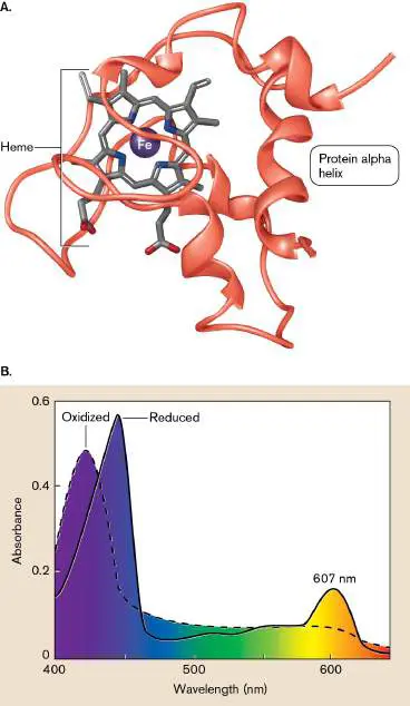
FIGURE 14.3 ■ Cytochromes. A. Cytochrome c protein containing one heme cofactor, from the pathogen Pseudomonas aeruginosa. (PDB code: 2PAC) B. Light absorption peaks shift between the oxidized and reduced forms of a cytochrome from Haloferax volcanii, a halophilic archaeon.
The changes in reduction state of cytochromes usually require a buried cofactor containing an iron ion (Fe 3+ or Fe 2+). The protein shown in Figure 14.3A, cytochrome c of Pseudomonas aeruginosa, transfers electrons for nitrate respiration. Its reddish color derives from its cofactor, heme, a ring of conjugated double bonds (bonds with delocalized electrons) surrounding the iron. The heme plays a key role in acquiring and transferring electrons, with a transition between Fe 3+ and Fe 2+. A metal-reducing bacterium such as Geobacter may make more than a hundred different types of cytochromes in its envelope. The envelope may thus accumulate electrons until the bacterium finds an exogenous metal oxidant to accept them.
Microbial cytochromes show various colors. Figure 14.3B shows the absorbance spectrum of a cytochrome from the cell membrane of Haloferax volcanii, a halophilic archaeon isolated from the Dead Sea. In the reduced cytochrome, light absorption peaks in the blue range (440 nm) and in the orange (607 nm). Upon oxidation, the cytochrome loses its absorption peak at 607 nm, and the 440-nm peak shifts to a shorter wavelength.
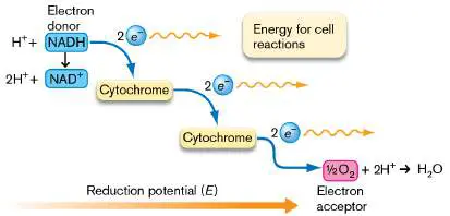
An ETS typically includes several different cytochromes with different reduction potentials (ability to gain electrons). Keilin proposed that the cytochromes pass electrons sequentially from each protein complex to the next-stronger electron acceptor, with each step providing a small amount of energy to the organism (Fig. 14.4 ). By dividing the cell’s energy into small packets, the ETS makes it possible to “spend” small amounts of energy for cell processes without wasting the excess.
FIGURE 14.4 ■ Electron transport system. David Keilin’s model for electron transfer through cytochromes. Each cytochrome in sequence receives electrons from a stronger electron donor and transfers them to a stronger electron acceptor. The electron transport proteins are called oxidoreductases because they oxidize one substrate (removing electrons) and reduce another (donating electrons). Oxidoreductases consist of multiprotein complexes that include cytochromes as well as noncytochrome proteins. The structure and function of ETS oxidoreductase complexes are discussed in Section 14.2.
Energy Storage
In Chapter 13 we showed how the free energy change Δ G determines whether energy can be obtained from a given reaction of substrates to products. Similar considerations of Δ G govern the reactions of electron flow through an ETS and the storage of energy in transmembrane ion gradients.
The reduction potential. To obtain energy, biochemical reactions require a negative Δ G. In oxidation-reduction reactions (redox reactions), the Δ G values are proportional to the reduction potential (E) between the oxidized form of a molecule (electron acceptor) and
its reduced form (electron donor). The reduction potential represents the tendency of a compound to accept electrons, measured in volts (V) or millivolts (mV). A positive value of E has a negative Δ G , so the gain of electrons yields energy. A negative value of E means that the reverse reaction (loss of electrons) yields energy. The oxidized and reduced states of a compound are called a redox couple. An example of a redox couple is the electron acceptor O 2 and the electron donor H 2 O. Because O 2 is a strong electron acceptor (readily gains 2 e −), the redox couple ½O /H O (that is,
2 2
reduction of O 2 to H 2 O) has a high positive value of E. So, when ½O (one oxygen atom) plus 2H + + 2 e − forms H O, the reaction
2 2
yields a large amount of energy.
Standard values of E (E °) are known as the standard reduction potential. The standard reduction potential for biochemical reactions at 25°C, E °′, assumes a concentration of 1 M for all components at pH 7. For example, the value of E °′ for H formation from 2H + + 2 e
2
− is −420 mV. This large negative reduction potential means that it takes a lot of energy to add the two electrons to 2H +. Thus, the reverse reaction donating 2 e − from H to an appropriate electron
2
acceptor yields a lot of energy. An example of microbes that donate electrons from H 2 for energy is hydrogen-oxidizing bacteria growing in oral biofilms, where they cause gum disease.
Reduction potentials represent a form of the standard free energy change, Δ G °′. The value of Δ G °′ (in kilojoules per mole, or kJ/mol) for an electron transfer reaction with a given reduction potential E °′ is given by Δ G °′ = − nFE °′ where n is the number of electrons transferred, F is Faraday’s constant [F = 96.5 kJ/(V • mol)], and E °′ is the standard reduction potential (in volts, V). Thus, E °′ represents the standard reduction potential per electron, whereas Δ G °′ gives the overall free energy change. Note that the reaction is favored for positive values of E, which correspond to negative values of Δ G.
The standard reduction potential E °′ gives the potential difference between the oxidized and reduced states under standard conditions, when both oxidized and reduced forms are at equal concentrations; namely, 1 M. Assuming equal concentrations at 1 M, we may compare E °′ values of various molecules available in the environment for microbial respiration. Such a comparison generates an “electron tower” representing the reduction potential E °′ of molecules that may act as electron acceptors or donors (Table 14.1 ). The oxidized state of each redox couple in the table is shaded red; the reduced state is shaded blue.
TABLE “Electron Tower” of Standard
14.1 Reduction Potentials
Electron → Electron E °′ (mV) a Δ G °′ (kJ) acceptor donor CO + 4H + [CH 2 O] −430 +166
2
− → glucose + + 4 e H 2 O 2H + + 2 e − H 2 −420 +81 → NAD + + 2H + NADH + H −320 +62 + 2 e − → + TABLE “Electron Tower” of Standard
14.1 Reduction Potentials
S 0 + 2H + + H 2 S −280 +27 2 e − → CO + 2H + CH 4 + 2H 2 −240 +46
2
+ 3H + 2 → O
2
e − SO 2− + 10H H 2 S + 4H −220 +170
4
+ − → O + 8 e 2 FAD + 2H + + FADH 2 −220 +42 2 e − → b FMN + 2H + FMNH 2 −190 +37 + 2 e − → b Menaquinone Menaquinol −74 +14 + 2H + + 2 → e − Fumarate + Succinate +33 −6 2H + + 2 e →
−
Ubiquinone + Ubiquinol +110 −21 2H + + 2 e →
−
TABLE “Electron Tower” of Standard
14.1 Reduction Potentials
Fe 3+ + e − Fe 2+ (at +200 −19 → pH 7)
NO − + 2H + NO − + H +420 −81
3 2
− → O + 2 e 2 NO − + 8H + NH + + +440 −255
2 4
− → 2H O + 6 e 2 MnO + 4H + Mn 2+ + 2H +460 −89
2
− → O + 2 e 2 NO − + 6H + ½N 2 + 3H +740 −357
3
− → O + 5 e 2 Fe 3+ + e − Fe 2+ (at +770 −74 → pH 2)
½O + 2H + H 2 O +820 −158
2
− → + 2 e
Note: A more positive E °′ means that reducing the electron
acceptor yields more energy. A more negative value of E °′ means that oxidizing the electron donor yields more energy.
In the electron tower, the more negative values of E °′ represent couples (half reactions) with a stronger electron donor (H 2, NADH), whereas the more positive values represent stronger electron acceptors (O, NO −). For example, in the redox couple NAD +
2 3
/NADH + H +, the oxidized form is NAD + and the reduced form is NADH. The reduction potential is strongly negative (−320 mV), so NADH is a strong electron donor; that is, the reverse reaction, NADH + H + → NAD +, releases a lot of energy (+320 mV).
A complete redox reaction combines two redox couples: one accepting electrons (red column in Table 14.1), the other donating electrons (blue column, arrow reversed). Redox couples with more negative values of E °′ can provide electron donors (blue column) for electron acceptors with more positive values of E °′ (red column). The E °′ for the overall reaction is then given by adding the reversed reaction of the electron donor E °′ to the electron acceptor E °′. A positive value of E °′ (thus, negative Δ G °′) means that the reaction may proceed to provide energy. For example, the oxidation of NADH by O 2 results from combining two couples: O 2 /H 2 O (forward) and NADH/NAD + (reversed from Table 14.1). For the reversed couple, the sign of E °′ changes: E °′ Δ G °′ Electron acceptor is reduced: ½O + 2 e − + 2H + → H O +820 mV –158 kJ/mol
2 2
Electron donor is oxidized: NADH + H + → NAD + + 2e − –(–320 mV) –(+62 + 2H + kJ/mol)
NADH + H + + ½O → H O +1,140 mV –220 kJ/mol
2 2
+ NAD + The aerobic oxidation of NADH pairs a strong electron donor (NADH) with a strong electron acceptor (O 2). Thus, NADH oxidation via the ETS provides the cell with a huge amount of potential energy (comparable to a 1-V battery cell) to make ATP and generate ion gradients. NADH is oxidized by many electron transport systems of bacteria and archaea, as well as mitochondria. The food we eat donates electrons to make NADH for our mitochondria to oxidize. Bacteria and archaea have evolved many alternative donor-acceptor systems; the one they use depends on what their environment provides. For example, in anoxic (low-oxygen) marine sediment, the proteobacterium Shewanella can donate electrons to many alternative oxidized minerals as terminal electron acceptors, including iron, lead, and nitrate. The use of terminal electron acceptors other than oxygen is called anaerobic respiration (discussed in Section 14.3).
Electron donors can also be oxidized by organic molecules. In the human colon, which has limited molecular oxygen, Escherichia coli can oxidize NADH by transferring 2 e − onto fumarate, which is reduced to succinate. Table 14.1shows that: NADH + H + + fumarate → NAD + + succinate E °′ = 320 mV + 33 mV = 353 mV Δ G °′ = −68 kJ/mol The fumarate reduction potential is small but yields energy for growth. When oxygen reappears in the environment, the reverse reaction is favored—succinate can donate electrons to O 2, forming fumarate and water: ½O 2 + succinate → H 2 O + fumarate E °′ = 820 mV − 33 mV = 787 mV Δ G °′ = −152 kJ/mol
Thought Questions
14.1 Pseudomonas aeruginosa, a cause of pneumonia in cystic fibrosis patients, oxidizes NADH with nitrate (NO −) to nitrite (NO
3 2
−) at neutral pH. What is the value of E °′?
14.2 Could a bacterium obtain energy from succinate as an electron donor with nitrate (NO −) as an electron acceptor? Explain.
3
14.3 Use Table 14.1to write the chemical reaction used by cable bacteria. What is its standard reduction potential? Explain why electrons must travel such a long distance within the bacterial cable. Concentrations of electron donors and acceptors. A reaction with a small value of E °′ can provide more energy if the concentration of an electron donor is high. A high donor concentration often drives lithotrophic reactions such as iron oxidation. For example, iron-mine drainage can fill a stream with a thick yellow biofilm of iron-oxidizing Gallionella.
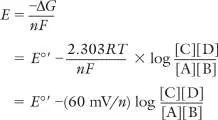
The standard reduction potential E °′ assumes 1-M concentrations of reactants and products at 25°C and pH 7, but actual values of E within cells depend on actual reactant concentrations. In Chapter 13 we saw that Δ G includes a term incorporating the ratio of product concentrations (C and D) to reactant concentrations (A and B): where R is the gas constant [R = 8.315 × 10 −3 kJ/(mol • K)], and T is the absolute temperature in kelvins (298 K, or 25°C). Similarly, the reduction potential E of a redox reaction depends on the log ratio of products to reactants: where n is the number of electrons transferred, and F is Faraday’s constant. As the ratio of products to reactants increases, E decreases; in other words, there is less potential to do work. For a tenfold ratio of products to reactants, assuming moderate temperatures (25°C– 40°C), the constants in the concentration term (2.303 RT / F) combine to yield approximately 60 mV per electron. Thus, a tenfold ratio of products to reactants subtracts about 60 mV from E (decreases energy yield), whereas a tenfold excess of reactants adds 60 mV to E (increases energy yield).
An ETS Functions within a Membrane
An electron transport system transfers electrons onto membrane-embedded carriers in a reaction series of increasing reduction potential (that is, lower on the electron tower) ending at a terminal electron acceptor such as O, Fe 3+, or NO −. Some electron
2 3
transfer steps yield energy to pump ions across the membrane. To store this energy, the ETS must maintain an ion gradient across a membrane that fully separates two aqueous compartments. In bacteria, the cytoplasmic membrane separates the cytoplasm and external medium.
Gram-negative bacteria such as E. coli have their ETS in the inner (cytoplasmic) membrane, which separates the cytoplasm from the periplasm (see Chapter 3). The Gram-negative outer membrane surrounding the periplasm is permeable to protons and other small molecules; thus, the outer membrane does not store energy. Figure 14.5Ashows the inner membrane of Helicobacter pylori, a Gram-
negative gastric pathogen. The inner membrane contains the ETS complexes; the cell wall and outer membrane do not participate in respiration. Some respiratory bacteria, such as the nitrite/ammonia oxidizer Nitrospira, pack their ETS within intracytoplasmic pockets called lamellae.
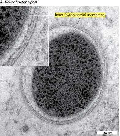
FIGURE 14.5 ■ Respiratory membranes. A. Electron transport occurs in the inner (cytoplasmic) membrane of Helicobacter pylori. The cytoplasmic membrane and cell wall are surrounded by periplasm and outer membrane. B. Mitochondrion within the brain stem neuron of a cat, modeled by cryo-electron tomography, shown with one electron microscopy section through the cell. The mitochondrial outer membrane (blue) encloses inner membrane organized in pockets called cristae (other colors).
YANPING LIU ET AL. 2006. J. GASTROENTEROL. 41 :569–574. © SPRINGER-VERLAG
2006
GUY A. PERKINS ET AL. 2010. J. NEUROSCI. 30 :1015
Respiratory membranes similar to bacterial lamellae are found within our own mitochondria. Mitochondrial respiration uses only O 2 as a terminal electron acceptor. The ETS proteins are embedded in folds of the mitochondrial inner membrane called cristae (singular:
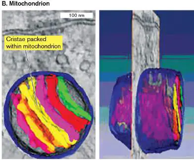
crista; Fig. 14.5B ). The inner membrane separates the inner mitochondrial space from the intermembrane space (between the inner and outer membranes). The mitochondrial inner membrane, including its electron transport proteins, evolved from the cell membrane of an endosymbiotic bacterial ancestor. Mitochondrial evolution is discussed further in Chapter 17.
The Proton Motive Force
The sequential transfer of electrons from one ETS protein to the next yields energy to pump ions (in most cases H +) across the membrane. Proton pumping generates a proton motive force, PMF (or proton potential) composed of the H + concentration difference plus the charge difference across the membrane. The proton motive force stores energy for most microbes and for the mitochondria of animals and plants.
The PMF (Δ p) drives many different cellular processes, such as ATP synthesis and flagellar rotation. In pathogens, the PMF drives drug efflux pumps that confer resistance to antibiotics such as tetracycline.
Note that in water, H + never occurs as such, because it combines with a water molecule to form hydronium ion, H O +. Current data,
3
however, are consistent with the passage of hydrogen nuclei (protons) through the proton pumps of the electron transport system. Within the pump complex, the proton associates with one chemical group after another; for example, H + may combine with the amine of an amino acid (RNH) to form an ammonium ion (RNH +), and
2 3
then transfer to a different proton-accepting group within the protein.
Note: In this book we use “hydrogen ions” and “H + ”
interchangeably with “protons.”
Discovery of the proton motive force. The discovery of the proton motive force radically changed the field of biochemistry. Early in the twentieth century, David Keilin and other scientists knew that the energy acquired by electron transport proteins was used to make ATP, but they did not know how. Most biochemists were convinced that electron transport proteins were directly coupled to ATP synthesis. The actual means of coupling by proton motive force was proposed and demonstrated by one of Keilin’s students—Peter Mitchell (1920–1992)—and Mitchell’s colleague Jennifer Moyle (1921– 2016) at Cambridge University (Fig. 14.6A). In 1961, Mitchell and Moyle proposed an astonishing explanation, called the chemiosmotic theory, for the coupling of electron transport to ATP synthesis. The chemiosmotic theory states that the energy from electron transfer between membrane proteins is used to pump protons across the membrane, leading to a higher H + concentration outside the cell. The pump generates the proton motive force, Δ p, which stores energy that can be used to make ATP (Fig. 14.6B ).
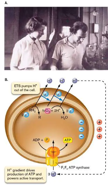
FIGURE 14.6 ■ Jennifer Moyle and Peter Mitchell discovered the proton motive force (Δ p). A. Moyle and Mitchell proposed and tested the chemiosmotic theory. B. An electron transport system pumps protons out of the cell. The resulting electrochemical gradient of protons (proton motive force) drives conversion of ADP to ATP through ATP synthase.
FROM THE PHOTOGRAPH COLLECTION OF THE FORMER GLYNN RESEARCH
FOUNDATION LTD. REPUBLISHED WITH PERMISSION OF PORTLAND PRESS, LTD.,
FROM BIOSCIENCE REPORTS 11 :293
Other biologists questioned how a proton potential could be coupled to ATP synthesis at an enzyme complex separate from the ETS, at a distant location on the membrane. They were skeptical that the H + concentration gradient and charge difference could exist everywhere on the membrane separating the two compartments. Because the membrane has very low permeability for hydrogen ions, the H + current can flow back only through a proton-driven complex such as the membrane ATP synthase (discussed further in Section 14.2). In effect, the proton potential is a “proton battery,” analogous to the electron potential of an electrical battery.
The chemiosmotic theory proved so controversial that Mitchell and Moyle left Cambridge University to found their own laboratory, Glynn House. There, they conducted experiments to test the hypothesis that the ETS-derived proton potential drives ATP synthesis. A key requirement was to show that the ETS generates a proton potential. Moyle’s experiment showed that respiration of mitochondria is associated with proton efflux: Mitochondria isolated from rat livers were exposed to oxygen, causing efflux of hydrogen ions. The H + efflux occurred as electrons were transferred across the ETS and hydrogen ions were expelled by the proton pumps. The number of protons extruded per electron transferred down the ETS was consistent with the chemiosmotic model. Similar results were obtained with vesicles made first from mitochondrial membranes and later from the membranes of chloroplasts and bacteria.
A greater challenge was to demonstrate that a proton motive force could, in fact, drive the ATP synthase and other ion transporters without any undiscovered intermediate. A friend of Mitchell and Moyle, André Jagendorf, at Johns Hopkins University, tested the effect of a proton gradient imposed on spinach chloroplasts. Chloroplasts contain ATP synthase directed outward (that is, driven by proton flow from inside to outside; Fig. 14.7A). The chloroplasts were partly opened by osmotic shock and then suspended in medium containing concentrated hydrogen ions (pH 4). With the osmotic balance restored, the chloroplast membranes closed again, with increased [H +] trapped inside. When the pH outside was raised to pH 8.3—that is, lower [H +]—protons from the acidic interior of the vesicles flowed out through the ATP synthase, and ADP and inorganic phosphate were converted to ATP. Jagendorf interpreted this result as consistent with the chemiosmotic theory for the mechanism of phosphorylation.
Other researchers showed that ATP synthesis could be driven by a charge difference across a vesicle membrane (Fig. 14.7B ). A charge difference (electrical potential, Δψ) was applied by loading vesicles with potassium ions (K +), which add charge without affecting the chemical concentration of hydrogen ions. The potassium ions were conducted across the membrane by the ionophore valinomycin, a small hydrophobic peptide that binds K + and solubilizes it in the membrane. The potassium ions flow down their concentration gradient, along with their associated positive charge. In the experiment shown in Figure 14.7B , the vesicles are “inside-out” bacterial membrane vesicles, in which the ATP synthase points outward instead of inward. The K + influx adds positive charge, which drives H + out through ATP synthase, catalyzing formation of ATP. Thus, Δψ drives the formation of ATP.
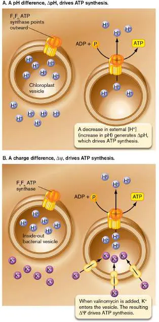
FIGURE 14.7 ■ Either ΔpH or Δψ drives ATP synthesis. A. A pH difference imposed across the chloroplast inner membrane drives a proton current through an outwardly directed ATP synthase. B. A charge difference generated by K + influx drives a proton current through ATP synthase.
Δ p includes Δψ and ΔpH. The transfer of H + through a proton pump generates a H + concentration difference across the membrane. Because H + carries a positive charge, the proton transfer also generates a charge difference across the membrane. The H + concentration difference (ΔpH) plus the charge difference (Δψ) make a proton potential (Δ p), also called a proton motive force (PMF). Thus, when protons are pumped across the membrane, the proton motive force stores energy in two different forms—the separation of charge (electrical potential) and the gradient of H + concentration (pH difference)—as shown in Figure 14.7. Either form (or both) can drive Δ p -dependent cellular processes: The electrical potential (Δψ) arises from the separation of charge between the cytoplasm (more negative) and the solution outside the cell membrane (more positive). For many bacteria, this “battery” potential is about −50 to −150 mV.
The pH difference (ΔpH) is the difference between internal and external pH (pH int − pH ext). For example, if the bacterial internal pH is 7.5 and the external pH is 6.5, the ΔpH is 1.0, and the ratio of [H +] to [H +] is 10. A ΔpH of 1.0 corresponds
ext int
to a proton potential of approximately −60 mV when the temperature is 25°C.
The relationship between electrical and chemical components of the proton potential Δ p (in millivolts) is given by: Δ p = Δψ − (2.3 RT / F)ΔpH or approximately: Δ p = Δψ − 60ΔpH For cells grown at neutral pH, all three terms (Δ p, Δψ, and −60ΔpH) usually have a negative value, meaning that their force drives protons inward from outside.
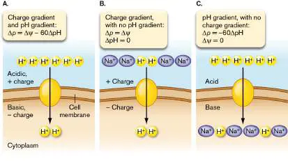
In living cells, the relative contributions of Δψ and −60ΔpH vary, depending on other sources of charge difference and protons, as shown in Figure 14.8. Either Δψ, −60ΔpH, or both can drive a device such as ATP synthase or a flagellar motor.
FIGURE 14.8 ■ Electrical potential and pH difference. Proton motive force drives protons into the cell, via a proton-powered device (red icon) such as ATP synthase or a flagellar motor. A. The proton motive force Δ p is composed of the transmembrane electrical potential Δψ (the charge difference) plus the transmembrane pH difference ΔpH (the chemical concentration gradient of H +). B. If the pH inside and outside the cell is equal, then Δ p = Δψ. C. If the electrical charge inside and outside the cell is equal, then Δ p = −60ΔpH.
Both the Δψ and ΔpH components of Δ p are influenced by other factors besides ETS proton transport. For example:
The charge difference Δψ includes charges on other ions, such as K + and Na +. These ions are pumped or exchanged by ion-specific membrane transport proteins.
The pH difference Δ pH is affected by metabolic generation of acids, such as fermentation acids, or by pH changes outside the cell. Permeant acids can run down the gradient through the membrane, collapsing the ΔpH to zero.
Overall, the cell uses its various membrane pumps and metabolic pathways to adjust and maintain its proton motive force at a size sufficient to drive ATP synthesis but not so great as to disrupt the membrane.
Thought Questions
14.4 What do you think happens to Δψ as the cell’s external pH increases or decreases? What could happen to the Δ p of bacteria that are swallowed and enter the extremely acidic stomach? 14.5 How could a simple experiment provide evidence that a proton pump in the bacterial cell membrane drives efflux of antibiotics such as tetracycline?
What about other ions, such as the sodium ions in Figure 14.8? Can a sodium gradient drive cell processes? For some bacteria, a gradient of Na + concentration can drive transport of a nutrient through a transporter protein embedded in the cell membrane. Bacteria such as Vibrio cholerae (the cause of cholera) can use a Na + concentration gradient to power ATP synthesis by a Na + -dependent ATP synthase.
Dissipation of proton motive force. What happens if something disrupts the membrane so that protons leak through? Even a small leak can quickly dissipate all the energy stored as ΔpH.
Many fermentation products are weak acids, such as acetic acid, whose protonated form can dissolve in the membrane and then dissociate on the other side (Fig. 14.9, top; discussed also in Chapter 3). A membrane-permeant weak acid conducts protons through the membrane until the ΔpH is dissipated. Other weak acids cross the membrane in both charged and uncharged forms. The weak acid 2,4-dinitrophenol (DNP) dissociates to an anion whose charge is relatively evenly distributed around the molecule; thus, it remains hydrophobic enough to penetrate the membrane (Fig. 14.9, bottom). Because both protonated (uncharged) and unprotonated (negatively charged) forms cross the membrane, DNP can cyclically bring protons into the cell and collapse both Δψ and ΔpH. Such molecules are called uncouplers because they uncouple electron transport from ATP synthesis through the membrane ATP synthase. With uncoupling, electron transport accelerates, but the energy dissipates as heat.
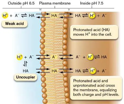
FIGURE 14.9 ■ Membrane-permeant weak acids and uncouplers. A. A weak acid crosses the membrane in the protonated form and then dissociates, acidifying the cell. B. An uncoupler crosses the membrane in both protonated and unprotonated forms, cyclically acidifying the cell and dissipating the charge difference (electrical potential, Δψ).
When the bacterial PMF is uncoupled from energy-spending reactions, the ETS pumps protons at an accelerated rate, while losing the energy as heat. Thus, uncouplers inhibit bacterial growth. They are highly toxic for human cells because the acceleration of electron transport causes overheating and brain damage.
Δ p Drives Many Cell Functions
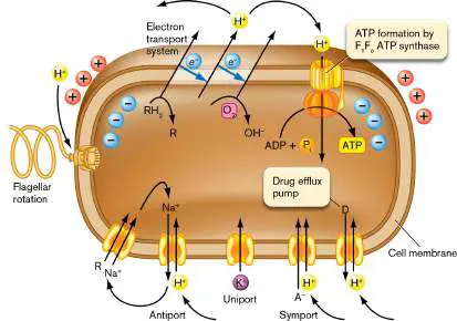
Besides ATP synthesis, Δ p drives many cellular processes directly ( Fig. 14.10). Processes driven by the proton motive force include the rotation of flagellar motors (see Chapter 3) and the secretion of toxins and signaling molecules across the cell membrane (see Chapter 8). Proton flux is also coupled to transport of ions such as K + or Na + through parallel transport (symport) or oppositely directed transport (antiport), as discussed in Chapter 4. Ion flux then drives uptake of nutrients such as amino acids or efflux of molecules such as antibacterial drugs. Drug efflux pumps are a major problem in hospitals, where the pump proteins are encoded by genes carried on plasmids that spread among virulent strains (see Chapter 9). FIGURE 14.10 ■ Processes driven by the proton motive force. Processes powered by proton potential include ATP synthesis through the F 1 F o ATP synthase, flagellar rotation, uptake of nutrients, and efflux of toxic drugs. “R” represents an organic nutrient; “D” represents a toxic drug.
Thought Question
14.6 Suppose that de-energized cells of E. coli (Δ p = 0) with an internal pH of 7.6 are placed in a solution at pH 6. What do you predict will happen to the cell’s flagella? What does this effect demonstrate about the function of Δ p?
Initial Electron Donors and Terminal Electron Acceptors
Each class of metabolism requires a specific electron transport system. Metabolism using an ETS is classified by the nature of the initial electron donors and terminal electron acceptors. The remaining sections of this chapter cover three major classes of prokaryotic energy acquisition involving an ETS. Their relationships were summarized in Table 13.1.
Organotrophy (or chemo organo trophy) is a form of metabolism in which organic “food” molecules donate electrons and generate reduced carriers such as NADH. If the electrons are donated to a membrane-embedded ETS and ultimately reduce a terminal electron acceptor, the overall pathway is called “respiration.” The terminal electron acceptor may be O 2 for aerobic respiration or an alternative electron acceptor such as nitrate (NO −) for anaerobic respiration.
3
Lithotrophy (or chemo litho trophy) requires initial electron donation by inorganic molecules such as reduced iron or the sulfides for cable bacteria (see the chapter-opening image and Fig. 14.2). The terminal electron acceptor may be O 2 or an anaerobic alternative, such as NO −.
3
Phototrophy requires light capture by chlorophyll or another photopigment. The absorption of light energy may enable splitting of H 2 S or H 2 O (photolysis), which then donates electrons to an ETS. Photoautotrophs couple photolysis to CO 2 fixation to form biomass (discussed in Chapter 15).
Photoheterotrophs may use photoexcited electrons for an ETS to generate ATP and supplement catabolism of organic electron donors.
Note: The term respiration refers to metabolism in which chemical
electron donors yield energy through an ETS. In this book we reserve the term “respiration” for an ETS with organic electron donors (organotrophy). The use of inorganic electron donors for an ETS is called lithotrophy. Other fields use the term “respiration” differently. In medical physiology, “respiration” refers to catabolism with an ETS or to the process of carbon dioxide exchange with oxygen through breathing. In ecology, “respiration” refers to carbon flux in an ecosystem or throughout the atmosphere.
To Summarize
An electron transport system (ETS) consists of a series of electron carriers that sequentially transfer electrons to the carrier of next-higher reduction potential E (that is, the next-stronger electron acceptor). Electron flow through the ETS begins with an initial electron donor and ultimately transfers all electrons to a terminal electron acceptor.
The ETS includes protein complexes and cofactors.
Protein complexes called oxidoreductases include cytochromes and noncytochrome proteins. Cytochromes are iron-bearing proteins whose absorbance spectrum shifts with a change in redox state.
The reduction potential E for a complete redox reaction must be positive to yield energy for metabolism. The standard reduction potential for biochemistry, E °′, assumes that all reactant concentrations equal 1 M at 25°C and pH 7.
Concentrations of electron donors and acceptors in the environment influence the actual reduction potential E experienced by the cell.
The ETS is embedded in a membrane that separates two compartments. Two aqueous compartments must be separate to maintain an ion gradient generated by the ETS. The chemiosmotic theory states that ETS complexes generate a proton motive force (Δ p). The force is usually directed inward, driving back into the cell protons that have accumulated outside the cell as a result of the ETS. The proton potential drives ATP synthase and other functions, such as ion transport and flagellar rotation.
The proton potential (Δ p, measured in millivolts) is composed of the electrical potential (Δψ) and the hydrogen ion chemical gradient (ΔpH): Δ p = Δψ − 60ΔpH.
Uncouplers are molecules taken up by cells in both protonated and unprotonated forms. Uncouplers can collapse the entire proton potential, thus uncoupling respiration from ATP synthesis.
Glossary
electron donor Also called reducing agent. A reduced molecule (e.g., NADH) that can donate electrons.
electron acceptor An oxidized molecule (e.g., NAD +) that can accept electrons. electron transport system (ETS) or electron transport chain (ETC) Also called cytochrome system. A series of membrane-embedded proteins that converts the energy of redox reactions into a proton potential.
electron transport system (ETS) or electron transport chain (ETC) Also called cytochrome system. A series of membrane-embedded proteins that converts the energy of redox reactions into a proton potential.
organotrophy Also called chemoorganotrophy or chemoheterotrophy. The metabolic oxidation of organic compounds to yield energy without absorption of light.
lithotrophy Also called chemolithotrophy. The metabolic oxidation of inorganic compounds to yield energy and fix single-carbon compounds into biomass.
phototrophy The use of chemical reactions powered by the absorption of light to yield energy.
cytochrome A membrane protein that donates and receives electrons. heme An organic molecule containing a ring of conjugated double bonds surrounding an iron atom. It is involved in redox reactions and oxygen binding.
oxidoreductase An electron transport system protein that accepts electrons from one molecule (oxidizing that molecule) and donates electrons to a second molecule, thereby reducing the second molecule. redox couple The oxidized and reduced states of a compound. For example, NAD + and NADH form a redox couple.
standard reduction potential (E °)
The reduction potential (tendency of a chemical to gain electrons and thereby become reduced) under standard conditions of 1 M concentration, 25°C temperature, and 1 atm pressure.
proton potential or proton motive force (PMF)
The potential energy of the concentration gradient of protons (hydrogen ions; H +) plus the charge difference across a membrane.
proton potential or proton motive force (PMF)
The potential energy of the concentration gradient of protons (hydrogen ions; H +) plus the charge difference across a membrane.
uncoupler A molecule that makes a membrane permeable to protons, dissipating the proton motive force and uncoupling electron transport from ATP synthesis.
chemoorganotrophy See organotrophy .
chemolithotrophy See lithotrophy .
respiration The oxidation of reduced organic electron donors through a series of membrane-embedded electron carriers to a final electron acceptor. The energy derived from the redox reactions is stored as an electrochemical gradient across the membrane, which may be harnessed to produce ATP.
lithotrophy Also called chemolithotrophy. The metabolic oxidation of inorganic compounds to yield energy and fix single-carbon compounds into biomass.
Endnotes
1. Note a: Reduction potentials for redox couples when all concentrations are 1 M at 25°C and pH 7. Return to reference a 2. Note b: Values are for free FAD or FMN. FAD or FMN bound to a specific flavoprotein has a different E °′ that depends on its protein environment. Return to reference b 3. Note b: Values are for free FAD or FMN. FAD or FMN bound to a specific flavoprotein has a different E °′ that depends on its protein environment. Return to reference b
14.2 The Respiratory ETS and ATP SynthaseUnit 4 · Metabolism
The electron transport system (ETS) of oxidative respiration consists of various carrier molecules and proteins. For aerobic bacteria, and for the mitochondrial inner membrane, the respiratory ETS receives electrons from NADH and FADH 2 and transfers electrons ultimately to O 2, producing H 2 O. In between, the series of membrane-embedded carriers harvests the reducing potential of electrons in small steps.
Note: This section presents the classic respiratory ETS that
resides in the inner membrane of Gram-negative bacteria (such as Escherichia coli) or the cell membrane of Gram-positive bacteria (such as Bacillus subtilis). In addition, many species extend electron transfer to components in the outer membrane or outside the cell (see Section 14.4).
Cofactors Allow Small Energy Transitions
ETS proteins such as cytochromes associate electron transfer with energy transitions that are small and reversible. The energy transitions are mediated by cofactors such as heme, small molecules that associate with the protein (Fig. 14.11). Cofactors allow small, reversible redox changes. The structure of each cofactor must allow transition of an electron between closely spaced energy levels to avoid “spending it all in one place.” If all of the energy were spent in one transition, most of it would be lost as heat instead of being converted to several small processes, such as pumping H + across the membrane.
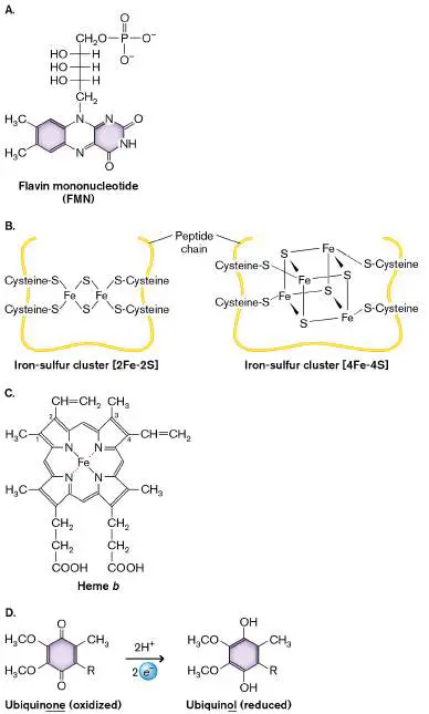
FIGURE 14.11 ■ Cofactors for electron transport. A. Flavin mononucleotide (FMN). B. Iron-sulfur clusters: [2Fe-2S] and [4Fe-4S]. C. Heme b. The side chains of the ring vary among hemes, yielding different levels of redox potential. D. Ubiquinone, which is reduced to ubiquinol.
Small energy transitions typically involve these kinds of molecular structures: Metal ions such as iron or copper , coordinated (and hence held in place) with amino acid residues. Iron is often coordinated by sulfur atoms of cysteine residues in the protein; examples shown in Figure 14.11B are [2Fe-2S] and [4Fe-4S]. Transition metals make useful electron carriers because their outer electron shell has several closely spaced energy levels, facilitating small energy transitions.
Conjugated double bonds and heteroaromatic rings , such as the nicotinamide ring of NAD + /NADH, also provide narrowly spaced energy transitions. Membrane-soluble carriers such as quinones (reduced to quinols) allow even smaller energy transitions than does NAD + /NADH.
The major protein complexes of electron transport each have one or more redox centers containing either metal ions or conjugated double bonds or both. In flavin mononucleotide (FMN), the conjugated double bonds allow a small energy transition (Fig. 14.11A). In iron-sulfur clusters—[2Fe-2S] and [4Fe-4S]—the metal atoms provide the site for a small energy transition, its size dependent on the cluster’s connections within the associated protein (Fig. 14.11B ). The heme group found in cytochromes and other oxidoreductases contains extensive conjugated double bonds, coordinated around the metal Fe 3+ (Fig. 14.11C ). Reduction by a transferred electron converts the iron to Fe 2+. The branching of side chains on the ring varies among different hemes, altering the magnitude of E °′ for the redox couple Fe 3+ /Fe 2+.
Some electron carriers are small molecules that associate loosely with a protein complex and then come off to diffuse freely within the membrane. Mobile electron carriers include quinones (Q) such as ubiquinone (Fig. 14.11D ), which can be reduced to quinols (QH 2 ): Q + 2H + + 2 e − → QH
2
Quinols carry electrons and protons laterally within the membrane between the proton-pumping protein complexes of the ETS. The hydrophobic quinols never leave the membrane; thus their electrons are kept in the membrane until transfer out of the ETS. After transferring their electrons to the next protein complex, quinols revert to quinones, capable of accepting electrons again.
Note: Dehydrogenases, reductases, and oxidases are all
oxidoreductases, which oxidize one substrate (remove electrons) and reduce another (donate electrons). Oxidoreductases that accept electrons from NADH or FADH 2 are also called dehydrogenases, because their reaction releases hydrogen ions.
Oxidoreductase Protein Complexes
A respiratory electron transport system includes at least three functional components: an initial substrate oxidoreductase (or dehydrogenase), a mobile electron carrier, and a terminal oxidase. Microbes make alternative versions of each component, using alternative electron-donating substrates and terminal electron acceptors, depending on what is available in the environment. Here we present a typical bacterial ETS receiving electrons from NADH and transferring them to oxygen.
Initial substrate oxidoreductase. A respiratory ETS begins with an initial oxidoreductase that receives a pair of electrons from an organic substrate such as NADH. Note that NADH forms by receiving two electrons plus 2H + from an organic product of catabolism (designated RH; Fig. 14.12A). The 2H + are ultimately balanced
2
by 2H + from the cytoplasm combining with O (or another terminal
2
electron acceptor) at the end of the ETS. The two electrons (2 e −) from NADH enter an ETS protein complex embedded in the membrane.
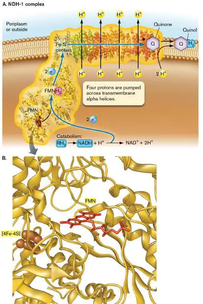
FIGURE 14.12 ■ NADH:quinone oxidoreductase complex (NDH-1). A. NDH-1 transfers two electrons from NADH onto NDH-1. The energy from oxidizing NADH is coupled to pumping 4H + across the cell membrane. B. Within the NDH-1 complex, FMN lies adjacent to the first iron-sulfur center, [4Fe-4S]. (PDB code: 1NOX)
Source: Part A modified from R. Efremov and L. Sazanov. 2010. Nature 476:414; part B modified from L. A. Sazanov and P. Hinchliffe. 2006. Science 311:1430–1436.
NADH donates electrons to NADH dehydrogenase (NADH:quinone oxidoreductase, NDH-1; Fig. 14.12A). In E. coli, the NDH-1 complex has 14 different subunits, including the cofactor FMN, as well as several iron-sulfur clusters—typically 7[4Fe-4S] and 2[2Fe-2S]. The cofactors “hand off” electrons to each other through adjacent connections; see, for example, the placement of FMN and the first [4Fe-4S] within the peptide coils of NDH-1 (Fig. 14.12B ). Each electron from NADH travels through FMN and the iron-sulfur series. At the end of the chain, the electrons and 2H + from solution are transferred to a quinone (Q), which is thus reduced to a quinol (QH 2).
Within the NDH-1 complex, the oxidation of NADH and reduction of Q to QH yields energy to pump up to 4H + across the
2
membrane. A crystallographic model shows four apparent proton channels through transmembrane alpha helices of the protein subunits (Fig. 14.12A). Within each channel, a hydrogen ion hops along a series of amino acid residues. The H + translocation is driven by a conformational change in the alpha helices throughout the protein, arising from the initial two-electron reduction by NADH. The hydrogen ions pumped across the membrane contribute to the proton potential Δ p. In human mitochondria, the NADH dehydrogenase (aka “complex I”) is critical for health; genetic defects in complex I are associated with diseases such as Parkinson’s and some forms of diabetes.
Note that the 4H + pumped across the membrane are distinct from the 2H + acquired by the quinone (Q → QH). Some halophilic
2
bacteria pump 4Na + instead of 4H +, while their quinones acquire 2H + + 2 e −.
Note: In our figures, protons that cross the membrane by the end
of the ETS (and thus contribute to Δ p) are highlighted yellow. Not all substrate oxidoreductases pump protons. For example, E. coli has an alternative NADH dehydrogenase (NDH-2) that transfers two electrons to Q without pumping additional protons across the membrane. (The unused energy is lost as heat.) NDH-2 functions during rapid growth, when the cell must limit its proton potential to avoid membrane breakdown. Other complexes, such as succinate dehydrogenase, transfer electrons from substrates in a reaction that lacks sufficient energy to pump extra protons. However, these electrons can be transferred to another ETS complex at lower voltage potential where redox reactions do pump protons.
Quinone pool. A quinone can receive 2 e − from the substrate oxidoreductase, along with 2H + from solution, to balance the negative charges, yielding a quinol (Fig. 14.12A). The quinols diffuse within the membrane and carry reduction energy to other ETS components. After transferring 2 e − to the next protein complex, the quinol releases its 2H +. Usually the 2H + released are on the opposite side of the membrane from where 2H + were originally picked up (Fig. 14.13). Thus, besides transferring two electrons, a quinol may contribute two protons to the transmembrane proton potential. The reoxidized carriers then recycle back as quinones.
FIGURE 14.13 ■ Cytochrome bo quinol oxidase complex. Each quinol (QH 2) transfers two electrons to heme b. The two H + from each quinol are expelled to the periplasm (or outside the bacterial cell). The 2 e − from the quinols are transferred through the complex onto 2H + plus an oxygen atom from O 2, forming water.
Source: Based on the structure determined by Jeff Abramson et al. 2000. Nat. Struct. Biol. 7 :910–917.
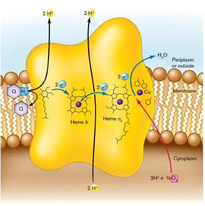
Each quinone can bind to a substrate dehydrogenase, pick up a pair of electrons and hydrogen ions, and then diffuse within the membrane and carry the electrons to a reductase. The quinones and quinols, referred to as the quinone pool, diffuse freely within the membrane. Thus, the quinones/quinols are able to transfer electrons between many different redox enzymes.
Different oxidoreductase complexes interact with slightly different quinones, such as ubiquinone and menaquinone. The reduction potentials of ubiquinone and menaquinone are given in Table 14.1. For clarity, in this chapter we refer to all of them as quinones (Q) and to their reduced forms as quinols (QH 2). Terminal oxidase. A terminal oxidase complex receives electrons from a quinol (QH 2) and transfers them to a terminal electron acceptor, such as O 2 (Fig. 14.13). The complex usually includes a cytochrome that accepts electrons from quinols. Cytochromes of comparable function are designated by letters; for example, cytochrome b (E. coli and mitochondria) and cytochrome c (mitochondria). The cytochrome is bound to an oxidase complex containing a series of electron-transferring carriers: two iron-centered hemes and three copper atoms. This unique center couples electron transfer and proton pumping.
The cytochrome bo quinol oxidase of E. coli consists of cytochrome b plus oxidase complex o. The cytochrome b subunit receives two electrons from a quinol (QH 2 → Q) and releases the 2H + out to the periplasm. Each electron from quinol travels through the two hemes of the oxidase complex. Because the two quinol hydrogens originated from 2H + in the cytoplasm (see Fig. 14.12) and 2H + were released outside by a quinol, there is net efflux of 2H +. In addition, the transfer of 2 e − between the two oxidase hemes is coupled to pumping 2H + from the cytoplasm across to the periplasm (Fig. 14.13). The hydrogen ions then leak through the outer membrane, effectively outside the cell.
The second heme of the oxidase (heme o 3) acts to reduce an atom of oxygen from O 2. Each oxygen atom receives two electrons and combines with two protons (2H +) from the cytoplasm to form H O. The 2H + consumed balance the 2H + released by catabolism
2
to make NADH + H +.
Note that the oxidase is conventionally shown as obtaining two electrons from the cytoplasm and donating them to an atom of an oxygen molecule (½O 2). A full reaction cycle of cytochrome bo quinol oxidase actually puts four electrons from two quinols (originally 2 NADH) onto O, taking up 4H + from the cytoplasm to
2
make two molecules of H 2 O.
Besides cytochrome bo quinol oxidase, bacteria express different terminal oxidases that differ with respect to the ratio of cytoplasmic protons pumped to electrons transferred. For example, the alternative cytochrome bd quinol oxidase reduces O 2 to water but pumps no extra protons. Although it pumps no protons, cytochrome bd quinol oxidase can bind O 2 at much lower concentrations and donate electrons, completing the respiratory circuit. Thus, the bd oxidase enables E. coli to respire within low-oxygen habitats such as the mammalian intestine.
Figure 14.14summarizes a complete ETS for oxidation of NADH by ½O 2 in the inner membrane of E. coli. Overall, an ETS for respiration on organic substrates includes at least three phases of electron transfer, such as: (1) NADH from an organic substrate donates electrons to an initial oxidoreductase; (2) the electrons are transferred to a quinone, which is reduced to a quinol; (3) the quinol transfers electrons to a terminal oxidase and releases 2H + outside the cell. Both enzymes and quinones show considerable complexity and diversity among different species in different environments.
FIGURE 14.14 ■ A bacterial ETS for aerobic NADH oxidation. In E. coli, electrons from NDH-1 are transferred to quinones, generating quinols, which transfer electrons onto cytochrome bo (Cyt bo) quinol oxidase complex. For each NADH oxidized, up to 8H + may be pumped across the membrane.
The entering carrier NADH carries two electrons with protons obtained from catabolized food molecules. The two electrons (2 e − ) from NDH-1 and two protons (2H +) from solution are transferred onto Q (quinone), converting it to QH 2 (quinol). The transfer of 2 e − from NADH yields sufficient energy to pump as many as 4H + across the membrane. The exact number depends on cellular conditions, such as the concentrations of NADH and the terminal electron acceptor.
The QH 2 diffuses within the membrane until it reaches a terminal oxidase complex, such as the cytochrome bo quinol oxidase. The 2H + from QH are released outside the cell, while the
2
2 e − enter the oxidase, reducing the two hemes. The two electrons join 2H + from the cytoplasm, combining with an oxygen atom to make H O. The reaction is coupled to pumping of 2H + across the
2
membrane plus a net increase of 2H + outside through redox eactions.
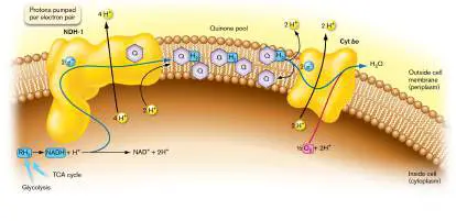
Overall, the oxidation of NADH exports about 8H + per 2 e − transferred through the ETS to make H 2 O. The export of protons generates a proton potential Δ p.
Note: Protons (hydrogen ions, H +) can have three different
fates in the ETS: 1. Protons are pumped across the membrane (H +) by an oxidoreductase complex. Contributes to Δ p.
2. Protons are consumed from the cytoplasm by quinone/quinol, while other protons are released outside the membrane (H + ). Contributes to Δ p.
3. Protons are consumed by combining with the terminal electron acceptor (O 2). If the loss balances protons released by catabolism, it does not affect Δ p.
Thought Questions
14.7 In Figure 14.14, what is the advantage of the oxidoreductase transferring electrons to a pool of mobile quinones, which then reduce the terminal reductase (cytochrome complex)? Why does each oxidoreductase not interact directly with a cytochrome complex?
14.8 In Figure 14.14, why are most electron transport proteins fixed within the cell membrane? What would happen if they “got loose” in aqueous solution?
Environmental modulation of the ETS. The ETS just described represents optimal conditions, when food and oxygen are unlimited. What happens when food (that is, electron donors) or oxygen is scarce? When the environment changes, bacteria adjust the efficiency of their ETS by expressing alternative oxidoreductases. For example, at low concentrations of oxygen (microaerophilic conditions, discussed in Chapter 5), the reduction potential E is decreased. So the ETS may be unable to reduce O 2 to H 2 O while pumping four protons. Instead, E. coli uses cytochrome bd quinol oxidase, which has higher affinity for oxygen but pumps no protons. (Protons are still pumped by the NADH oxidoreductase.) Thus, the bacteria will gain less energy, but they will still be able to grow. Bacteria also have alternative oxidoreductases to serve different electron donors and acceptors. Some enzymes take electrons from donors lacking the potential of NADH; for example, succinate dehydrogenase catalyzes the one step of the tricarboxylic acid (TCA) cycle that yields FADH 2 —a step that provides not quite enough energy to produce NADH (discussed in Chapter 13). Succinate dehydrogenase is the only TCA enzyme embedded in the membrane as an ETS component. In anoxic environments, where O 2 concentration is so low as to be thermodynamically unavailable, some bacteria can use other electron acceptors, such as nitrate (anaerobic respiration, discussed in Section 14.3) or even a metal electrode (see Section 14.4). Yet another alternative is to oxidize inorganic electron donors (lithotrophy; see Section 14.5). Mitochondrial respiration. In contrast to E. coli, mitochondria have only a single ETS, optimized for a relatively uniform intracellular environment (Fig. 14.15). Protected by the constant environment of the eukaryotic cytoplasm, mitochondria do not need to use alternative versions of their ETS to respire under different conditions. Instead, a set of just four electron-carrying complexes has evolved so as to maximize the energy obtained from NADH, using oxygen as the terminal electron acceptor, and minimizing energy lost as heat. This mitochondrial ETS is nearly universal throughout the cells of animals, plants, and most eukaryotic microbes. In addition, some bacteria, such as Paracoccus denitrificans, have an ETS whose organization resembles that of mitochondria.
FIGURE 14.15 ■ Mitochondrial electron transport. In addition to NADH dehydrogenase, succinate dehydrogenase, and a terminal cytochrome oxidase, mitochondria possess ubiquinol:cytochrome c oxidoreductase, which provides an intermediate electron transfer step. As a result, mitochondrial membranes export 10–12 H + per NADH.
The mitochondrial ETS has homologs (proteins encoded by genes with a common ancestor) of bacterial ETS components, including NADH dehydrogenase, succinate dehydrogenase, and cytochrome c oxidase. However, the mitochondrial ETS differs from that of E. coli in the following respects: An intermediate cytochrome oxidoreductase complex transfers electrons. Besides NADH dehydrogenase (complex I) and cytochrome c oxidase (complex IV), mitochondria show an intermediate step of electron transfer to ubiquinol:cytochrome c oxidoreductase (complex III, shown in Fig. 14.15). The intermediate electron transfer step pumps an additional 2H +. Another 2 e − and 2H + come from succinate
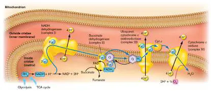
dehydrogenase (complex II), which forms FADH 2 through the TCA cycle.
The mitochondrial ETS pumps more protons per NADH.
As many as 10–12 protons may be pumped per NADH, in contrast to 2–8 protons in E. coli.
Homologous complexes have numerous extra subunits.
Mitochondria have evolved additional nonhomologous subunits specific to eukaryotes. For example, the homologous subunits of cytochrome c oxidoreductase are enveloped by a series of eukaryotic proteins.
The Proton Potential Drives ATP Synthase
The proton potential drives synthesis of ATP by the membrane ATP synthase, also known as the F 1 F o ATP synthase. The proton-driven synthesis of ATP completes the cycle of oxidative phosphorylation, in which hydrogen ions pumped by the ETS drive phosphorylation of ADP to ATP by the ATP synthase. The same ATP synthase can use the proton potential generated by lithotrophy (see Section 14.5) or by phototrophy (see Section 14.6). Surprisingly, despite the homology of ATP synthase across all forms of life, the complex is a target for antibiotics. For example, the ATP synthase of Mycobacterium tuberculosis, the cause of tuberculosis, is inhibited specifically by bedaquiline, an antibiotic approved in 2012 for patients whose disease resists all other drugs.
The F 1 F o ATP synthase is a protein complex highly conserved in the bacterial cell membrane, the mitochondrial inner membrane, and the chloroplast thylakoid membrane. An elegant molecular machine, the ATP synthase is composed of two complexes—F o and F 1 —that rotate relative to each other (Fig. 14.16). The F o complex translocates protons across the membrane. Twelve c subunits form a cylinder embedded in the membrane, where the cylinder is stabilized by subunits a and b.
FIGURE 14.16 ■ Bacterial membrane-embedded ATP synthase (F 1 F o ATP synthase). The F o complex is embedded in the bacterial plasma membrane, whereas the F 1 complex catalyzes ATP synthesis. (PDB codes: 1B9U, 1C17, 1E79, 2CLY)
The F 1 complex consists of six alternating subunits of types alpha and beta surrounding a gamma subunit, which drives the F 1
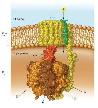
complex to rotate one-third of a turn. Each “third” of the F 1 complex (an alpha plus a beta subunit) interconverts ADP + P i with ATP + H 2 O. The gamma subunit connects the tripartite “knob” of F 1 to the membrane-embedded F o. Proton transport through F o drives ATP synthesis by F 1.
One proton at a time enters the subunit-a channel and moves into a c subunit of F o (Fig. 14.17). The proton potential directed inward ensures that protons more often enter from the outside than from the cytoplasm. Each entering proton causes a c subunit to rotate around the center, causing rotation of the long gamma subunit. Rotation of approximately one-third turn releases a bound proton to the cytoplasm. The flux of three protons through F o is coupled to formation of one molecule of ATP by one alpha-beta unit of F 1 (Fig. 14.17A). During each cycle generating ATP, the ring of c subunits of the F o rotor rotates in the membrane one-third of one turn relative to the F 1 in the cytoplasm (Fig. 14.17B ). FIGURE 14.17 ■ H + flux drives ATP synthesis. A. Three protons enter c subunits of the F o complex. The number of c subunits varies from 8 to 15 among bacterial species; 12 are shown here. B. The ring of c subunits rotates one-third turn relative to F 1. Flux of three protons through F o is coupled to F 1 converting ADP + P i to ATP.
Note that the generation of ATP is completely reversible, so ATP hydrolysis by F 1 can pump protons back through F o across the membrane. In the absence of a proton potential, a high ATP concentration can drive the F o in reverse, actually pumping protons to generate Δ p. This reversal of ATP synthase is used, for example, by Enterococcus faecalis, intestinal Gram-
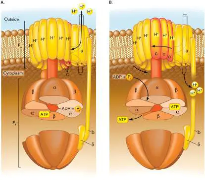
positive cocci that generate ATP mainly by fermentation. E. faecalis can operate the membrane-embedded F 1 F o ATP synthase in reverse, consuming ATP and thus generating a proton potential for nutrient uptake and ion transport.
Thought Question
14.9 Would E. coli be able to grow in the presence of an uncoupler that eliminates the proton potential supporting ATP synthesis?
Na + Pumps: An Alternative to H + Pumps
While a proton potential provides primary energy storage for most species, some bacteria generate an additional potential of sodium ions. A sodium motive force (ΔNa +) is analogous to the proton motive force in that it includes the electrical potential Δψ plus the sodium ion concentration gradient (log ratio of the Na + concentration difference across the membrane). For extreme halophilic archaea, which grow in concentrated NaCl, the sodium potential entirely substitutes for the proton potential to drive ATP synthesis. These “haloarchaea” make use of the high external Na + concentration to store energy in the form of a sodium potential. In some bacteria, an ETS oxidoreductase pumps Na + instead of H +. For example, the proton-pumping NADH dehydrogenase can be supplemented by an NADH dehydrogenase that pumps Na + out of the cell. This primary sodium pump is found in many pathogens, including Vibrio cholerae (the cause of cholera) and Yersinia pestis (the cause of bubonic plague). These pathogens use the sodium-rich blood plasma to store energy in a sodium potential.
To Summarize
Cofactors allow small energy transitions. Electron carriers containing metal ions and/or conjugated double-bonded ring structures are used for electron transfer. For example, cytochrome bo quinol oxidase has two hemes and three copper ions.
A substrate dehydrogenase receives a pair of electrons from a particular reduced substrate, such as NADH. NADH dehydrogenase (NADH:quinone oxidoreductase) typically has an FMN carrier and nine iron-sulfur clusters.
Quinones receive electrons from the substrate dehydrogenase and become reduced to quinols.
Typically, a quinol receives 2H + from the cytoplasm, and then releases 2H + across the membrane upon transfer of 2 e − to an electron acceptor complex.
Protons are pumped by substrate dehydrogenases (oxidoreductases) and terminal oxidases. The number of protons pumped by a bacterial ETS is determined by environmental conditions, such as the concentrations of substrate and terminal electron acceptor.
Protons are consumed by combining with the terminal electron acceptor , such as combining with oxygen to make H 2 O.
The proton potential drives ATP synthesis through the membrane-embedded F 1 F o ATP synthase. Three protons drive each F 1 F o cycle, synthesizing one molecule of ATP. Some bacteria use a similar ATP synthase driven by Na +.
Glossary
quinone An oxidized electron carrier that can diffuse laterally within membranes.
quinol A reduced electron carrier that can diffuse laterally within membranes.
oxidoreductase An electron transport system protein that accepts electrons from one molecule (oxidizing that molecule) and donates electrons to a second molecule, thereby reducing the second molecule.
oxidative phosphorylation A process of an electron transport chain that uses diatomic oxygen as a final electron acceptor and generates a proton gradient across a membrane for the production of ATP via ATP synthase.
14.3 Anaerobic RespirationUnit 4 · Metabolism
Most multicellular animals and plants must transfer electrons to oxygen. Likewise, some bacteria are called obligate aerobes because they grow only using O 2 as a terminal electron acceptor; examples include important nitrogen-fixing bacteria, such as Sinorhizobium meliloti and Azotobacter vinelandii. However, other organotrophic bacteria and archaea can use a wide range of terminal electron acceptors, including metals, oxidized ions of nitrogen and sulfur, and chlorinated organic molecules. The use of terminal electron acceptors other than O 2 is called anaerobic respiration. Most electron acceptors are weaker oxidants than O 2, so anaerobic respiration generally takes place in anoxic environments, where O 2 concentrations are too low to support aerobic respiration. Such anoxic conditions can be found in wetland soil and water and in the human digestive tract.
For anaerobic respiration, each type of terminal electron acceptor requires a different terminal oxidoreductase. The Escherichia coli genome encodes several different terminal oxidoreductases, each of whose expression is induced by the presence of a specific electron acceptor (Fig. 14.18). These enzymes serve a function equivalent to that of cytochrome quinol oxidase, but they reduce an alternative electron acceptor, such as nitrate (NO −) reduced to nitrite (NO −) or NO − reduced to NO
3 2 2
(nitric oxide). Because the electron acceptor stays outside the cytoplasm, even a toxic molecule may be used. For example, at the gut epithelium, E. coli can donate electrons to hydrogen peroxide (H 2 O 2), a toxic molecule formed by the host immune system as a defensive response.
FIGURE 14.18 ■ Alternative electron donors and electron acceptors. Escherichia coli can oxidize various foods (electron donors) while reducing various electron acceptors. Each electron donor may use alternative substrate oxidoreductases, depending on the environmental conditions.
Other electron acceptors can be organic products of catabolism; for example, the TCA cycle intermediate fumarate can be reduced to succinate. Organic electron acceptors play important roles in food decomposition. For example, the substance trimethylamine oxide, used by fish as an osmoprotectant against sea salt, is reduced by bacteria to trimethylamine—the main cause of the “fishy” smell. At the top of the ETS, alternative dehydrogenases (or substrate oxidoreductases) receive electrons from different organic electron donors, as well as from molecular hydrogen (H 2). The enzymes “connect” to various terminal oxidoreductases through the pool of quinones. Note that the various electron donors differ greatly in their reduction potential and hence in their capacity to generate proton potential (see Table 14.1). In a given environment, bacteria use the strongest electron donor and the strongest electron acceptor available. The best donor and best acceptor usually induce the expression of genes encoding their respective redox enzymes.
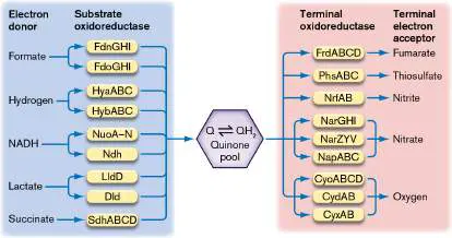
For example, in the presence of nitrate, genes encoding nitrate reductase are expressed. At the same time, nitrate represses the expression of reductases for poorer electron acceptors, such as fumarate. (The mechanisms of gene induction and repression are discussed in Chapter 10.)
Oxidized Forms of Nitrogen
Respiration using oxidized forms of nitrogen is widespread among bacteria and archaea. Most eukaryotes must breathe oxygen, but some eukaryotic microbes respire on nitrate and nitrite. In anoxic soil, many yeasts and filamentous fungi can reduce nitrate to nitrite and nitrite to nitrous oxide.
The nitrogen series offers an abundant source of strong electron acceptors. Reduction of oxidized states of nitrogen for energy yield is called dissimilatory denitrification. Dissimilatory nitrate reduction to ammonium (DNRA) contributes to respiration, whereas assimilatory reduction of nitrate generates ammonium ion for fixation into biomass (discussed in Chapters 15 and 22). Some pathogens are dissimilatory denitrifiers, such as Neisseria meningitidis (a cause of meningitis) and Brucella species that cause brucellosis in cattle, sheep, and dogs. In the dissimilatory nitrogen redox series, a given oxidation state can serve as an acceptor in one redox couple but as a donor in the next. The redox couples are summarized here: Each nitrogen state requires a specialized reductase, such as nitrate reductase or nitrite reductase, to receive electrons from the ETS.
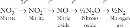
During the reactions, oxygen atoms are removed and combined with protons to form water. The full series of nitrate reduction to N 2 plays a crucial role in producing the nitrogen gas of Earth’s atmosphere (discussed in Chapter 22).
An alternative option for many soil bacteria, such as Bacillus species, is to reduce nitrite to ammonium ion (NH +):
4
NO − + 8H + + 6 e − → NH + + 2H O
2 4 2
Dissimilatory nitrate and nitrite reduction to ammonium ion can increase the soil pH. The high pH will precipitate metals such as iron.
The presence of a particular reductase can be used as a diagnostic indicator for clinical isolates of bacteria. For example, the chemical test for nitrate reduction is a key step in the diagnosis of Neisseria gonorrhoeae, the causative agent of gonorrhea. N.
gonorrhoeae happens to lack the terminal reductase for nitrate; hence, it tests negative, whereas several closely related species test positive.
Oxidized Forms of Sulfur
The redox potentials for sulfur oxyanions are generally lower than those for oxidized nitrogen. Nevertheless, with the appropriate oxidoreductases, sulfate and sulfite receive electrons from many kinds of electron donors, including acetate, hydrocarbons, and H 2. Sulfate-reducing bacteria and archaea are widespread in the ocean, from the Arctic waters to submarine thermal vents. The ubiquity of sulfate reduction is related to the high sulfate content of seawater, in which SO 2− is the most common anion after chloride.
4
The major oxidized forms of sulfur that serve as bacterial electron acceptors include: An important function of sulfate-reducing bacteria, such as the Desulfuromonadales group, is to help metabolize methane hydrates. The methane is oxidized by anaerobic methane-oxidizing archaea (ANME), which require sulfate-reducing bacteria (SRB) as symbiotic partners. This microbial partnership is vital for Earth’s global climate because it dissipates large amounts of methane before the greenhouse gas reaches the atmosphere. The ANME-SRB microbial partnership is discussed further in Chapter 19.
Dissimilatory Metal Reduction
An important class of anaerobic respiration involves the reduction of metal cations, or dissimilatory metal reduction. The term “dissimilatory” indicates that the metal reduced as a terminal electron acceptor is excluded from the cell. This is in contrast to minerals reduced for the purpose of incorporation into cell components (assimilatory metal reduction).
The metals most commonly reduced through anaerobic respiration are iron (Fe 3+ → Fe 2+) and manganese (Mn 4+ → Mn 2+), but virtually any metal with multiple redox states can be reduced or oxidized by some bacteria. For example, Geobacter can reduce uranium (U 6+ → U 4+) to respire on acetate. Reduced uranium is insoluble in water, precipitating as uranite (UO 2). Thus, Geobacter respiration with uranium can remediate uranium-contaminated water. The U.S. Department of Energy uses uranium-reducing bacteria for remediation at Rifle, Colorado, where uranium contamination threatens the Colorado River. In this process, acetate is pumped into the water table, where Geobacter metallireducens oxidizes the acetate to CO by reducing U 6+ to U 4+. The reduced
2
uranium then precipitates out of the water into the soil, where it can be collected and removed, while the cleansed water flows through. Anoxic or low-oxygen environments, such as the sediment of a lake or wetland, offer a series of different electron acceptors (Fig. 14.19). The stronger electron acceptors are consumed, in turn, by species that have the terminal oxidases to use them. For example, as oxygen grows scarce, denitrifying bacteria reduce nitrate to nitrogen gas (NO − → N).
3 2
As nitrate is used up, other species reduce manganese (Mn 4+ → Mn 2+), iron (Fe 3+ → Fe 2+), and sulfate (SO 2− → H S). At the
4 2
bottom of the lake or sediment, methanogenic archaea reduce carbon dioxide to methane (see Section 14.5). Microbial reduction of minerals plays a critical role in every ecosystem and participates in the geochemical cycling of elements throughout Earth’s biosphere (discussed in Chapter 22).
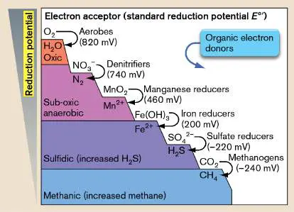
FIGURE 14.19 ■ Anaerobic respiration in a lake water column. As each successive terminal electron acceptor is used up, its reduced form appears. The next-best electron acceptor is then used, generally by a different species of microbe.
To use metals as terminal electron acceptors requires electron delivery to solid materials outside the cell. How can the cell’s ETS reach such materials? Electron delivery requires unique structures such as shuttle molecules, vesicles, and protein nanowires. Microbial communities may work collaboratively across a range of voltage potentials. Such communities have surprising applications for microbial fuel cells, as described in the next section and in eResearch Activity 14.
Thought Question
14.10 The scheme for uranium removal requires injection of acetate under highly anoxic conditions, with less than 1 part per million (ppm) dissolved oxygen. Why must the acetate be anoxic?
To Summarize
Anaerobic terminal electron acceptors , such as nitrogen and sulfur oxyanions, oxidized metal cations, and oxidized organic substrates, accept electrons from a specific reductase complex of an ETS.
Because O 2 is the strongest oxidant, anaerobic respiration usually occurs only when oxygen concentration is extremely low.
Nitrate is successively reduced by bacteria to nitrite, nitric oxide, nitrous oxide, and ultimately nitrogen gas.
Alternatively, nitrate and nitrite may be reduced to ammonium ion, a product that alkalinizes the environment. Sulfate is successively reduced by bacteria to sulfite, thiosulfate, elemental sulfur, and hydrogen sulfide. Sulfate reducers are especially prevalent in seawater.
Oxidized metal ions such as Fe 3+ and Mn 4+ are reduced by bacteria in anoxic soil and aquatic habitats—a form of anaerobic respiration also known as dissimilatory metal reduction.
The geochemistry of natural environments is shaped largely by anaerobic bacteria and archaea.
Glossary
anaerobic respiration The use of a molecule other than oxygen as the final electron acceptor of an electron transport chain.
dissimilatory denitrification Metabolic reduction of nitrate or nitrite to yield energy; anaerobic respiration of nitrate or nitrite.
dissimilatory metal reduction A type of anaerobic respiration that uses metal cations as terminal electron acceptors.
14.4 Nanowires, Electron Shuttles, and Fuel CellsUnit 4 · Metabolism
All electron transfer systems require a terminal electron acceptor to complete metabolism and yield energy. Soluble electron acceptors such as O or NO − can reach the cell by diffusion, but oxidized
2 3
metals such as Fe 3+ are often barely soluble in water. How does a bacterial membrane oxidoreductase interact with an insoluble metal particle outside the cell? It turns out that bacteria have evolved myriad ways to reach insoluble or distant electron acceptors. If bacteria can transfer electrons to metals, why not to a human-made electrode? Metal-reducing bacteria form electrogenic biofilms — biofilms that generate electricity in a human-made device.
Nanowires: Electrically Conductive Pili
Over the past decade, several laboratories have pursued the question of how bacteria reach outside their cytoplasm to deposit electrons. Derek Lovley at the University of Massachusetts, Amherst (Fig. 14.20), and Gemma Reguera at Michigan State University investigate the cytochromes and conductive pili of Geobacter species that reduce numerous metals. Similar experiments by Kenneth Nealson and Mohamed El-Naggar, at the University of Southern California, focus on the marine bacterium Shewanella oneidensis, which reduces iron, chromium, and uranium. An intriguing silver extension of Shewanella biofilms for electricity is described in eResearch Activity 14.
FIGURE 14.20 ■ Electron-conducting pili, or nanowires, of Geobacter sulfurreducens. A. Geobacter sulfurreducens possesses pili that conduct electric current. B. Electron-conductive pili transfer electrons from cell to cell in a biofilm. The biofilm can generate a voltage potential at an electrode. C. Derek Lovley directed pioneering studies of Geobacter nanowires and applications for fuel cells.
Source: Part B adapted from G. Reguera. 2018. FEMS Microbiol. Ecol. 94 (7), fig. 5.
COURTESY OF DEREK LOVLEY
RGB VENTURES/SUPERSTOCK/ALAMY STOCK PHOTO
Lovley and colleagues observed how metal-reducing bacteria such as Geobacter sulfurreducens grow adherent to particles of iron oxide or other metals. They developed conditions to culture Geobacter in a fuel cell, where the bacteria formed a biofilm over the anode (the electrode that receives electrons). The biofilm grew only when the anode was connected to a cathode in the fuel cell,
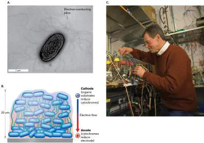
allowing completion of an electrical circuit. When provided with an electron donor substrate, acetate, the biofilm generated electric power.
Lovley noticed that Geobacter formed long protein filaments called pili (Fig. 14.20A). A pilus consists of a tube of helically stacked protein subunits (presented in Chapter 3). Lovley proposed that the pili of Geobacter conduct electrons, and that they connect the bacteria in a biofilm, allowing the relay of electric current across the biofilm to the anode (Fig. 14.20B ). The pili thus serve as nanowires, completing an electrical circuit through the biofilm.
Note: The word “filament” may refer to a cellular filament (chain
of cells), such as that of cable bacteria, or a protein filament (tubular complex of subunits) such as a pilus. The meaning must be inferred from the context.
The model of pili as conducting current was controversial because protein generally has the property of an insulator, resisting current, unlike metals, which conduct current. Nevertheless, Lovley’s students and colleagues obtained strong evidence that pili act as nanowires: G. sulfurreducens that is provided with acetate (organic electron donor) generates electric current in a fuel cell. A strain genetically altered for increased number of pili generates greater electric current.
With the electrical circuit open (no current possible), the bacteria grow a biofilm with few pili and low conductivity (the measured ability to conduct electric current).
Pili isolated from G. sulfurreducens show conductivity measurements comparable to those of metal wires. Pili conductivity shows a temperature dependence (conductivity increases at lower temperature) comparable to that of metal wires.
The conductivity of isolated G. sulfurreducens pili increases at low pH, similar to the pH dependence of organic metal compounds.
While the evidence for pili nanowires is strong, it raises the question: How do insulator-like proteins conduct electric current? Gemma Reguera addressed this question by investigating the amino acid residue content of Geobacter conductive pili (Fig. 14.21), which includes a high proportion of the aromatic amino acids tyrosine and phenylalanine. Aromatic rings contain conjugated pi bonds (bonds between p orbitals) that can readily transfer electrons via stacking of molecules whose orbitals overlap. Reguera showed that the tyrosines and phenylalanines are positioned at distances ideal for “electron hopping” from one aromatic ring to the next. Furthermore, a mutant was constructed in which one tyrosine was replaced by alanine, an amino acid with only a methyl R group. The loss of the one tyrosine broke the electrical connection. This mutant showed pili with normal molecular structure but substantially decreased electrical conductivity.
FIGURE 14.21 ■ Conductive pili transfer electrons via aromatic amino acid residues. A. Electron-conducting pilus protein includes aromatic amino acid residues (yellow = tyrosine; green = phenylalanine). The aromatic residues are spaced at distances that allow overlapping pi bond orbitals to
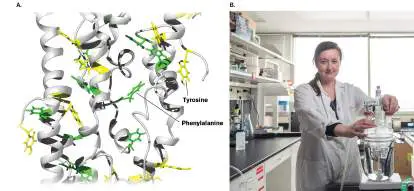
transfer electrons. B. Gemma Reguera demonstrated electron conductivity in Geobacter pili.
Source: Part A modified from G. Reguera. 2018. FEMS Microbiol. Ecol. 94 (7), fig. 4C.
MICHIGAN STATE UNIVERSITY
Extracellular Cytochromes, Shuttles, and Cell Extensions
Further investigations reveal a virtual hardware store of diverse extracellular electrical devices (Fig. 14.22A). These devices are found on Geobacter, on the Gram-negative pathogen Pseudomonas, and even on Gram-positive bacteria such as Thermincola, whose thick cell walls were once thought to preclude electron transfer. Electron-transferring cytochromes, once thought to reside only in the plasma membrane (or inner membrane), have now been found in bacterial outer membranes, as well as in the periplasm. The outer membrane–associated proteins (named Omc) contact extracellular oxidized minerals such as ferric oxide. Certain Omc proteins participate in porin-cytochrome complexes that connect via periplasmic cytochromes with the classic NADH-oxidizing ETS of the inner membrane (Fig. 14.22A). Other Omc proteins decorate the tips of conductive pili. The Omc contacts the electron acceptor, thus completing a circuit via the conductive pili all the way back to the inner membrane ETS.
FIGURE 14.22 ■ Bacterial mechanisms for donating electrons outside the cell. A. Various mechanisms of Gram-negative bacteria include cytochromes that reside in the outer membrane or periplasm, electrically conductive pili (nanowires), and extracellular electron shuttles. B. Cytoplasmic extension of Shewanella oneidensis that is proposed to transmit electric current.
Source: Part A modified from G. Reguera. 2018. PNAS 115 :5632–5634, fig. 1.
S. PIRBADIAN ET AL. 2014. PNAS 111 :12883–12888, FIG. 5B
The conductive pili are positioned by an extension-retraction apparatus embedded in the membrane. The apparatus is known as a type IV secretion system, similar to those found in pathogens for virulence attachment (discussed in Chapter 25). In the presence of electron acceptors, Geobacter bacteria build the pilus complex and
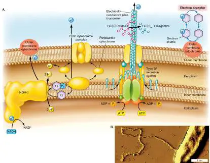
extend it through the secretion apparatus. When electron acceptors are absent, pili are retracted.
The species G. sulfurreducens was shown to use extracellular electron shuttles. Electron shuttles are aromatic molecules similar to quinones that allow low-energy transitions for gaining or losing an electron. Unlike quinones, however, which remain embedded in the cell membrane, an electron shuttle dissolves in the aqueous medium and diffuses away from the cell. Thus, bacteria such as G. sulfurreducens can transfer electrons from their ETS to an Omc on the outer membrane, which then reduces the shuttle and allows it to diffuse away. The shuttle ultimately transfers its electron to a metal or some other electron acceptor at a distance. Do electron shuttles return to the bacteria that make them? The answer is unclear, but often electron shuttles are used in a biofilm where many cells share nutrients and a redox gradient.
Pathogens such as Pseudomonas aeruginosa use electron shuttles to transfer electrons to distant oxygen. Dianne Newman at the California Institute of Technology and Lars Dietrich at Columbia University have shown how electron shuttles enable P. aeruginosa to conduct aerobic respiration within biofilms buried by thick mucus. The shuttles used are polycyclic aromatics called phenazines. Phenazines enable bacteria under anoxic conditions to pick up electrons from a cell-surface Omc and let them diffuse away to reach distant oxygen. Thus, phenazine electron shuttling favors Pseudomonas infections of cystic fibrosis patients.
In the soil, bacteria may shuttle electrons to extracellular metals using quinone-like degradation products of lignin, a complex aromatic substance that forms the bulk of wood and woody stems. Lignin degradation products are also called “humics” because of their presence in humus, the organic components of soil (discussed in Chapter 21). Humics accumulate in anoxic environments, where decomposition is slow.
Yet another possible means of electron transfer is that of cytoplasmic extensions, studied by El-Naggar and Nealson in Shewanella (Fig. 14.22B ). These cytoplasmic extensions are composed of chains of membrane vesicles whose properties are still under investigation. They may be the extracellular electrical conductors used by Shewanella to reduce iron and cobalt in clay sediments—and electrodes in a fuel cell.
Thought Question
14.11 What conditions might favor electron transfer by pili versus that by electron shuttles?
Fuel Cells: Bacterial Electric Power
How can we harness bacterial electric current to run human devices? In an electrolytic fuel cell, the bacteria form a biofilm on the anode (electron-attracting electrode; Fig. 14.23). As the bacteria oxidize organic substrates such as acetate, they transfer electrons to an anode. The result is charge separation between electrons and hydrogen ions. The electrons then pass through a circuit to the cathode, generating current that can run a device.
FIGURE 14.23 ■ A microbial fuel cell. A. A bacterial fuel cell. B. Reaction cycle of a bacterial fuel cell.
Source: Part B modified from MURI Microbial Fuel Cell Project, University of Southern California (http://mfc-muri.usc.edu).
The “fuel” for the cell can be a mixture of organic substances derived from any kind of food waste or sewage. Organic waste includes small organic molecules such as lactate, acetate, and even
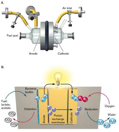
formaldehyde. The biofilm bacteria on the anode can remove hydrogens from these organic molecules, separating the electrons and hydrogen ions (Fig. 14.23B ). The hydrogen ions migrate through a polymer membrane, whereas the electrons enter the anode leading to an electrical wire. The remaining carbon and oxygen atoms of the fuel are released as CO 2. The process is similar to natural respiration, except that instead of molecular oxygen, the electron acceptor is an electrode made of graphite. To complete the circuit, the electrons from the wire current ultimately react with oxygen and hydrogen ions to form water, as in aerobic respiration.
So far, microbial fuel cells have been able to generate milliamps of current, enough to drive small devices, such as clocks and marine data sensors. eResearch Activity 14 describes the use of silver extensions to increase the power density (power per unit surface area) of bacterial electricity.
Thought Question
14.12 An alternative mechanism for a fuel cell involves the use of a bacterium that receives electrons from an electrode instead of donating electrons. How might this work?
To Summarize
Metal electron acceptors require extracellular devices to donate electrons.
Electrically conductive pili transmit electrons via pi bond orbital interactions between closely spaced aromatic amino acid residues in the pilus protein.
Outer membrane cytochromes and periplasmic cytochromes extend the reach of the Gram-negative inner membrane–embedded ETS.
Electron shuttles reversibly transfer electrons to electron acceptors at a distance from the cell.
Extracellular electron transmission devices enable formation of electrogenic biofilms.
Electrogenic biofilms can power a fuel cell.
Glossary
electrogenic biofilm A biofilm that generates electricity in a human-made device. nanowire In microbiology, a bacterial appendage that conducts electric current.
outer membrane–associated protein A protein linked to or embedded in the outer membrane. electron shuttle An aromatic molecule that allows low-energy transitions for gaining or losing an electron.
14.5 Lithotrophy and MethanogenesisUnit 4 · Metabolism
Many reduced minerals and single-carbon compounds can serve as electron donors for an ETS in the energy-yielding form of metabolism known as lithotrophy (or chemolithotrophy). Some organotrophs have alternative oxidoreductases that conduct lithotrophy by oxidizing H or Fe 2+; for example, the gastric pathogen
2
Helicobacter pylori oxidizes hydrogen gas released by mixed-acid-fermenting bacteria in the colon. The cable bacteria, such as Desulfobulbus, oxidize sulfide or organic substrates. Other species are “obligate lithotrophs”; that is, they oxidize only inorganic molecules. All lithotrophs are bacteria or archaea. Thus, bacteria and archaea fill many key niches in ecosystems that eukaryotes cannot. Lithotrophy includes many kinds of electron donors, from metals and anions to single-carbon groups (Table 14.2). Each type of electron donor, such as H, NH +, or Fe 2+, requires a specialized
2 4
electron-accepting oxidoreductase. Most inorganic substrates other than H 2 are relatively poor electron donors compared to organic donors such as glucose. Therefore, the terminal electron acceptor is usually a strong oxidant, such as O, NO −, or Fe 3+. Obligate
2 3
lithotrophs (organisms that conduct only lithotrophy) consume no organic carbon source; they build biomass by fixing CO 2 (a process discussed in Chapter 15).
Lithotrophy: Electron Donors
TABLE 14.2
and Acceptors
Type of Species example Electron Electron lithotrophy donor acceptor
Lithotrophy: Electron Donors
TABLE 14.2
and Acceptors
Hydrogenotrophy Aquifex aeolicus H 2 → O 2 → H 2 2H + + O 2 e − Sulfate reduction Desulfovibrio H 2 → SO 2−
4
(hydrogenotrop vulgaris 2H + + → S 0, hy) 2 e − − HS Methanogenesis Methanocaldococcus H 2 → CO 2 → (hydrogenotrop jannaschii 2H + + CH 4 hy) 2 e − Iron oxidation Acidithiobacillus Fe 2+ → O 2 → H 2 ferrooxidans Fe 3+ O + e − Ammonia Nitrosomonas NH 3 → O 2 → H 2 oxidation europaea NO − O
2
(nitrosification)
Nitrification Nitrobacter NO − O 2 → H 2
2
winogradskyi → NO O
−
3
Anammox “ Candidatus NH 3 + NO 2 → N Kuenenia 4H + + 2 stuttgartiensis ” 4 e − → N 2
Lithotrophy: Electron Donors
TABLE 14.2
and Acceptors
Carboxidotrophy Carboxydothermus CO → H 2 O → hydrogenoformans CO 2 + H 2 e − Sulfide oxidation Sulfolobus HS − → O 2 → H 2 solfataricus, S 0 + O Desulfobulbus H + + (cable bacteria) 2 e − Sulfur oxidation Acidithiobacillus S 0 + 2H O 2 → H 2 thiooxidans + + 4 O e − → H 2 SO
4
Iron Oxidation
Reduced metal ions such as Fe 2+ and Mn 2+ provide energy through oxidation by O or NO −. Bacteria perform these lithotrophic
2 3
reactions in soil where weathering exposes reduced minerals. They generate metal ions with higher oxidation states (such as Fe 3+ or Mn 4+), which other bacteria use for anaerobic respiration. Environments such as ponds and wetlands that experience frequent shifts between oxygen availability and oxygen depletion are likely to host a variety of metal-oxidizing lithotrophs, as well as metal-reducing anaerobic heterotrophs. Iron oxidizers form orange mats of twisted stalks encrusted by Fe 3+ oxyhydroxides. Alternatively, newly discovered species sculpt their iron into other unusual forms found in coastal wetlands (Special Topic 14). The roles of lithotrophy in ecology are discussed further in Chapters 21 and 22.
SPECIAL TOPIC 14 Bacteria with “Locs” Rule the Bay
The Chesapeake Bay is the United States’ largest estuary, a partly enclosed region where the Potomac and Susquehanna Rivers meet the Atlantic Ocean. Famous for blue crabs and bald eagles, the bay also hosts a complex microbial community. The brackish (mixed-salt) sediments support heterotrophs as well as diverse chemolithotrophs, such as iron oxidizers (described in Section 14.5). Bacteria that oxidize iron have key roles in making iron available to coastal ecosystems—but they were thought to be limited to sedimentary biofilms at low pH. Clara Chan’s lab at the University of Delaware studies iron oxidation throughout the bacterial domain. She found surprising evidence of iron oxidizers at neutral pH where the reduced iron (Fe 2+) concentration is relatively low. Chan’s team isolated iron-oxidizing Zetaproteobacteria in culture using plugs of iron carbonate (Fig. ST 14.1 ). The growing bacteria collected in suspension, forming bands colored bright orange by iron oxyhydroxides [Fe(III)OOH].
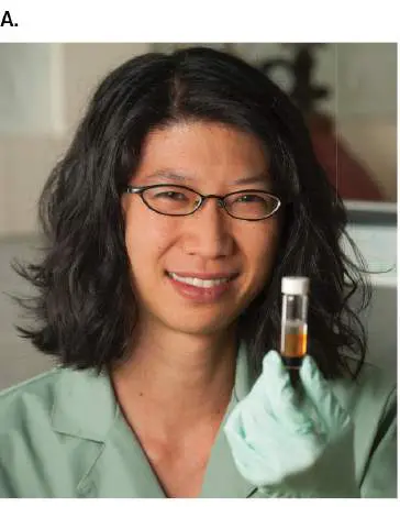
FIGURE ST 14.1 ■ Iron-oxidizing bacteria cultured from Chesapeake Bay. A. Clara Chan, School of Marine Science and Policy, University of Delaware. B. Mariprofundus aestuarium bacteria form yellow bands during growth in medium containing iron II carbonate (FeCO 3). Control tube at left contains no bacteria.
EVAN KRAPE/UNIVERSITY OF DELAWARE
B. K. CHIU ET AL. 2017. FRONT MICROBIOL. 8 :1280
In Chan’s cultures, the bacteria remain suspended. How is this possible, if the oxidized minerals encrust the bacterial filaments and weigh them down into mats?
The answer is that Chan’s Zetaproteobacteria shape oxidized iron into an unexpected form described by the researchers as
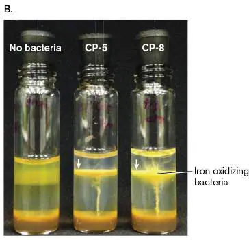
“dreadlocks,” or “locs,” due to their resemblance to the hairstyle (Fig. ST 14.2 ). Like hair, the iron salts extend away from their source—in this case, the bacterial cell. However, the iron salts can also fall away from the bacteria that formed them. Thus, the iron-oxidizing bacteria can keep themselves in suspension throughout the water column, where they oxidize even low levels of reduced iron. Once oxidized (Fe 3+), the iron then serves as a terminal electron acceptor for anaerobic respirers in the nutrient-rich sediment. With their flexible metabolism, these “bacteria with locs” may rule the iron levels for a wide range of marine habitats.
FIGURE ST 14.2 ■ Bacteria form “locs” of oxidized iron minerals. A. Mariprofundus aestuarium CP-5 bacteria form precipitates of Fe(III) oxyhydroxide (phase-contrast and fluorescence microscopy). B. Fe(III) oxyhydroxides isolated from the bacteria that formed them (SEM).
Source: Beverly Chiu et al. 2017. Front. Microbiol. 8 :1280.
B. K. CHIU ET AL. 2017. FRONT MICROBIOL. 8 :1280
B. K. CHIU ET AL. 2017. FRONT MICROBIOL. 8 :1280
RESEARCH QUESTION
How could you identify other kinds of iron-oxidizing bacteria in another marine ecosystem? What tools would you use?
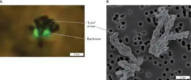
Chiu, Beverly K., Shingo Kato, Sean M. McAllister, Erin K. Field, and
Clara S. Chan. 2018. Novel pelagic iron-oxidizing zetaproteobacteria from
the Chesapeake Bay oxic–anoxic transition zone. Frontiers in Microbiology 8
:1280.
Bacteria such as Acidithiobacillus ferrooxidans oxidize Fe 2+ (ferrous ion) using the reduction potential between Fe 3+ /Fe 2+ and O 2 /H 2 O: ETS overall reaction E ° (pH 2) Δ G ° (pH 2)
2Fe 2+ → 2Fe 3+ + 2 e − (pH 2) −(+770) +149 mV kJ/mol ½O + 2H + + 2 e − → H O (pH +1,100 mV −212
2 2
2) kJ/mol 2Fe 2+ + ½O + 2H + → 2Fe 3+ + +330 mV −63 kJ/mol
2
H 2 O The removal of electrons from Fe 2+ requires an input of energy, which is compensated for by the larger yield of energy from reducing oxygen to water. The net reduction potential is small; thus, A. ferrooxidans must cycle large quantities of iron in order to grow. Because the reaction consumes H +, it goes forward most efficiently at low pH, such as that of acid mine drainage where other microbes oxidize sulfur to sulfuric acid. The bacterial cell must keep its cytoplasmic pH considerably higher (pH 6.5) by transmembrane exchange of H + with cations and by inversion of the electrical potential Δψ (positive inside). Thus, the proton potential Δ p exists entirely in the form of ΔpH. The ΔpH drives ATP synthesis.
Unlike organic electron donors, metals such as iron must be accessed from insoluble particles outside the cell (Fig. 14.24). In A. ferrooxidans and other iron-oxidizing bacteria, the electrons are collected from external iron by an unusual form of cytochrome c 2 designated Cyc2 (Fig. 14.24, blowup). The Cyc2 protein actually consists of two parts: a beta-barrel outer membrane porin (described in Chapter 3) and a heme-bearing cytochrome domain, the two attached by a flexible linker peptide. The cytochrome is proposed to collect electrons from Fe 2+ outside and carry them through the porin to reach periplasmic components of the ETS.
FIGURE 14.24 ■ Iron oxidation ETS. Outside the cell, Fe 2+ is oxidized to Fe 3+ by the Cyc2 cytochrome oxidase, an unusual porin with a linker-fused cytochrome. The external pH and periplasm are about pH 2. Electrons from Cyc2 are transferred to
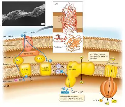
rusticyanin (in the periplasm), which feeds electrons to the cytochrome aa 3 complex to reduce O 2 to H 2 O. Alternatively, some electrons from rusticyanin reduce the cytochrome bc 1 complex. ΔpH drives reverse electron flow through an NDH complex to form NADPH. OMP = outer membrane porin. Inset: Twisted stalk of iron-oxidizing bacteria encrusted in Fe(III) oxyhydroxides produced by iron oxidation. From a microbial mat on the wall of a silver mine, Germany.
Source: Cyc2 modified from Raquel Quatrini et al. 2009. BMC Genomics 10
:394.
A. PICARD ET AL. 2015. NAT COMMUN. 6 :6277
The periplasmic protein that accepts electrons from Cyc2 is called rusticyanin. From rusticyanin, electrons are transferred to an inner membrane cytochrome complex that reduces oxygen to water.
Overall, the system enables generation of proton motive force and stores energy for the cell while keeping toxic iron outside. The oxidized iron precipitates, encrusting the cells into twisted chains that clog streams of mine drainage (Fig. 14.24, inset).
For iron oxidation (Fe 3+ /Fe 2+), the reduction potential is too high to reduce NADP + to NADPH; that is, Fe 2+ is a weaker electron donor than NADPH (Table 14.1). So, how do cells obtain enough energy to form NADPH for biosynthesis? An alternative pathway directs electrons to an enzyme complex that uses proton influx (spends Δ p) to convert NADP + to NADPH. This pathway is called reverse electron flow because it reverses the flux of electrons seen in an ETS that spends NADH (or NADPH) to form NAD + (or NADP +). In reverse electron flow, a relatively poor electron donor (such as Fe 2+ ) reduces an ETS with an unfavorable reduction potential, requiring input of energy to generate NADH or NADPH. For iron oxidation, the energy input comes from the large ΔpH across the inner membrane (pH 6.5 inside, and pH 2 outside). Reverse electron flow is seen in some kinds of lithotrophy, in syntrophy (discussed in Chapter 13), and in phototrophy (discussed in Section 14.6).
Thought Question
14.13 Use Figures 14.14 and 14.15 to propose a pathway for reverse electron flow in an organism that spends ATP from fermentation to form NADH. Draw a diagram of the pathway.
Nitrogen Oxidation
One kind of lithotrophy essential for the environment is oxidation of nitrogen compounds: Reduced forms of nitrogen, such as ammonium ion derived from fertilizers, support growth of nitrifiers, bacteria that oxidize ammonia and nitrite. For example Nitrosomonas species are Betaproteobacteria that oxidize ammonia to nitrite, and Nitrobacter species are Alphaproteobacteria that oxidize nitrite to nitrate. Bacteria generate nitr ite or nitr ate in the form of nitr ous acid or nitr ic acid, respectively. This acid production can degrade environmental quality. In soil treated with artificial fertilizers, nitrifiers decrease the ammonium ions obtained from such fertilizer and produce toxic concentrations of nitrites, which leach into groundwater. Still, nitrifiers can be useful in sewage treatment, where they eliminate ammonia that would harm aquatic life (discussed in Chapter 22).
Ammonia/ammonium and nitrite are relatively poor electron donors compared to organic molecules. Thus, their oxidation through the ETS pumps fewer protons, and the bacteria
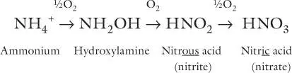
must cycle relatively large quantities of substrates to grow. As we saw for iron oxidation, reduced cofactors such as NADPH must be obtained through reverse electron flow involving redox carriers with different amounts of energy.
What happens to ammonium ion from detritus that accumulates in anoxic regions, such as the bottom of a lake? Surprisingly, NH + can
4
yield energy through oxidation by nitr ite (a product of nitr ate respiration): NH + + NO − → N + 2H O Δ G °′ = −357 kJ/mol
4 2 2 2
Under conditions of high ammonium and extremely low oxygen, nitrite oxidation of ammonium ion supports growth of bacteria. Known as the anammox reaction, anaerobic ammonium oxidation plays a major role in wastewater treatment, where it eliminates much of the ammonium ion from sewage breakdown. In the oceans, anammox bacteria cycle as much as half of all the nitrogen gas returned to the atmosphere.
Anammox is conducted by planctomycetes, irregularly shaped bacteria with unusual membranous organelles that fill much of the cell (Fig. 14.25A). The central compartment is called the anammoxosome. The anammoxosomal membrane is composed of unusual ladder-shaped lipids called ladderanes. An enzyme of the anammoxosomal membrane reduces nitrite to NO plus H 2 O (Fig. 14.25B , step 1). Another membrane-embedded enzyme, hydrazine synthase, catalyzes NO reduction by NH + to form hydrazine (N H
4 2 4
; step 2). Hydrazine is a high-energy compound that engineers use for rocket fuel; the substance is highly toxic, so the planctomycete keeps it sequestered within the specialized anammoxosomal membrane.
FIGURE 14.25 ■ Anammox ETS within a planctomycete. A. A planctomycete with anammoxosomal membranes (cryo-electron tomography). B. An enzyme of the anammoxosomal membrane reduces nitrite to NO plus H 2 O. NO is reduced by NH 4 + to form hydrazine (N H). Hydrazine is a toxic molecule that
2 4
gets trapped in the anammoxosome by the membrane composed of ladderanes. Hydrazine donates electrons to reduce nitrite, and gives off N 2. Protons may be pumped by the hydrazine synthase complex.
LAURA VAN NIFTRIK ET AL. 2008. J. STRUCT. BIOL. 161 :401
The hydrazine is further oxidized to N 2 (Fig. 14.25B , step 3), and its protons are released within the anammoxosome compartment. The protons then drive an ATP synthase embedded in the anammoxosomal membrane. The hydrazine oxidation enzyme obtains additional electrons from catabolism of organic substrates. Thus, anammox bacteria are examples of microbes whose metabolism combines lithotrophy and organotrophy.
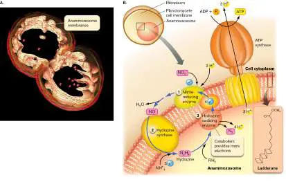
Note: Distinguish between lithotrophy (oxidation of reduced
minerals, usually by O 2, nitrate, or nitrite) and anaerobic respiration (reduction of oxidized minerals, usually by organic food molecules).
Sulfur and Metal Oxidation
Major sources of lithotrophic electron donors are minerals containing reduced sulfur, such as hydrogen sulfide and sulfides of iron and copper. As we saw for nitrogen compounds, each sulfur compound that undergoes partial oxidation may serve as an electron donor and be further oxidized: Sulfur oxidation produces the strong acid sulfuric acid (H 2 SO 4), which dissociates to produce an extremely high H + concentration. In the early twentieth century, no one would have believed that living organisms could grow in concentrated sulfuric acid, much less produce it. The Star Trek science fiction episode “The Devil in the Dark” portrayed an imaginary alien creature, the Horta, that produced corrosive acid in order to eat its way through solid rock ( Fig. 14.26A). In actuality, no creatures beyond the size of a microbe are yet known to grow in sulfuric acid. But archaea such as Sulfolobus species oxidize hydrogen sulfide to sulfuric acid and grow at pH 2, often in hot springs at near-boiling temperatures (Fig. 14.26B and C ). Sulfolobus makes irregular Horta-shaped cells without even a cell wall to maintain shape; how it protects its cytoplasm from disintegration is not yet understood.
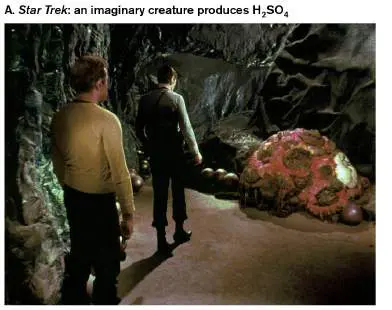
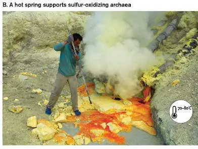
FIGURE 14.26 ■ Organisms produce sulfuric acid: science and science fiction. A. In the Star Trek episode “The Devil in the Dark,” starship officers encounter an imaginary creature, called the Horta, that tunnels through rock by producing corrosive acid. B. Volcanic rocks and hot springs support growth of sulfur-oxidizing thermophilic archaea such as Sulfolobus species, whose growth at pH 2 colors the rocks in this photo. C. Sulfolobus acidocaldarius grows as irregular spheres (TEM).
PARAMOUNT/COURTESY: EVERETT COLLECTION
TUUL & BRUNO MORANDI/GETTY IMAGES
R.L. WEISS. 1974. J. BACTERIOL. 118 :275. REPRODUCED WITH PERMISSION FROM
AMERICAN SOCIETY FOR MICROBIOLOGY
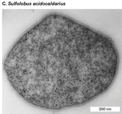
Microbial sulfur oxidation can cause severe environmental acidification, eroding concrete structures and stone monuments. The problem is compounded by sulfur oxidation in the presence of iron, such as in iron-mine drainage. For example, the sulfur-oxidizing archaeon Ferroplasma acidarmanus was discovered in the abandoned mine Iron Mountain in Northern California by Katrina Edwards (1968– 2014; University of Southern California) and colleagues from the University of Wisconsin–Madison. Ferroplasma oxidizes ferrous disulfide (pyrite) with ferric iron (Fe 3+) and water: FeS + 14Fe 3+ + 8H O → 15Fe 2+ + 2SO 2− + 16H +
2 2 4
This reaction generates large quantities of sulfuric acid. The acidity of the mine water is near pH 0—one of the most acid environments found on Earth. As Ferroplasma grows, it forms biofilms of thick streamers in the mine drainage, which poison freshwater streams. Anaerobic reactions between sulfur and iron cause hidden hazards for human technology, such as the corrosion of steel in underwater bridge supports. Anaerobic corrosion was long considered a mystery, as iron was known to rust by means of spontaneous oxidation by O 2. In anoxic conditions, however, sulfur-reducing bacteria can corrode iron (Fig. 14.27). In one pathway, the bacteria reduce elemental sulfur (S 0) with H to hydrogen sulfide (H S). H S then combines
2 2 2
with iron metal (Fe 0), which gives up two electrons to form Fe 2+, precipitating as iron sulfide (FeS). The displaced 2H + combine with the 2 e − from iron, regenerating H —now available to reduce sulfur
2
once again. In an alternative mechanism, bacteria use Fe 0 to reduce sulfate directly to FeS. These damaging processes may resemble the iron-based metabolism of Earth’s most ancient life forms.
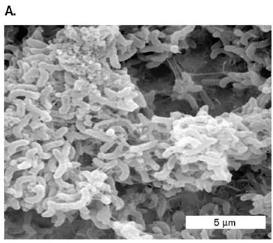
FIGURE 14.27 ■ Anaerobic iron corrosion. A. Sulfate-reducing bacteria corrode iron. B. Anaerobic corrosion of iron is accelerated by sulfur-reducing bacteria. Alternatively, in other bacteria, iron reduces sulfate. Source: Part B modified from Derek Lovley. 2000. Environmental Microbe-Metal Interactions, p. 163.
ENVIRONMENTAL MICROBE-METAL INTERACTIONS BY D. LOVLEY, 2000, ASM PRESS
Nevertheless, like the imaginary Horta that ended up helping miners with their excavations, acid-producing microbes are now used to supplement commercial mining—a process called biomining. Lithotrophs such as Acidithiobacillus ferrooxidans oxidize sulfides of iron and copper found in minerals such as chalcopyrite (CuFeS 2), chalcocite (Cu S), and covellite (CuS). The oxidation of Cu + to Cu
2
2+, as well as the acidification resulting from production of sulfate, dissolves the metal from the rock. Cu + can be oxidized aerobically with O or anaerobically with NO − from the soil. Other metals
2 3
oxidized by A. ferrooxidans include selenium, antimony, molybdenum, and uranium. The process of metal dissolution from ores is called
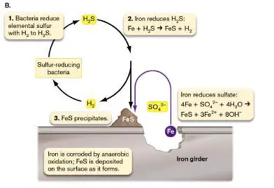
leaching. Leaching of minerals has been a part of mining since ancient times, long before the existence of microbes was known. Today, over 10% of the copper supply in the United States is provided by biomining ores too low in copper to smelt directly (Fig. 14.28). A similar process is being developed to biomine gold.
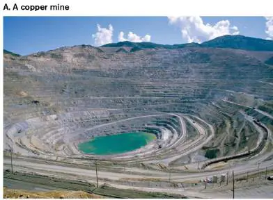
FIGURE 14.28 ■ Copper mining. A. Bingham Canyon copper mine near Salt Lake City, Utah. B. Acidithiobacillus ferrooxidans, a Gram-negative rod that oxidizes copper and iron sulfides (TEM). Such bacteria are used to leach copper from low-grade ores.
ROBERT HARDING/ALAMY
REPRINTED BY PERMISSION FROM SPRINGER NATURE: L. E. MURR. 2006. JOM 58
:23. HTTPS://DOI.ORG/10.1007/S11837-006-0136-3
Hydrogenotrophy Uses H 2 as an Electron Donor
The use of molecular hydrogen (H 2) as an electron donor is called hydrogenotrophy (Table 14.3). An example is oxidation of H 2 by sulfur to form H 2 S, performed by Pyrodictium brockii, an archaeon that grows at thermal vents above 100°C. Hydrogen gas is a stronger electron donor than most organic foods, so it can be oxidized with the full range of electron acceptors. It is available as a fermentation
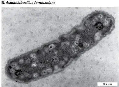
product in anoxic communities, such as our intestinal microbiome and wetland sediment.
Hydrogenotrophy:
TABLE 14.3
Examples
Specific reaction General description O 2 + 2H 2 → 2H 2 O Aerobic oxidation of H 2 Fumarate + H 2 → succinate Organic + H 2 → organic 2CO 2 + 4H 2 → CH 3 COOH + Mineral + H 2 → organic 2H 2 O 2H + + SO 2− + 4H → H Mineral + H 2 → mineral
4 2 2
S + 4H 2 O CO 2 + 4H 2 → CH 4 + 2H 2 O Methanogenesis Hydrogenotrophy is hard to categorize. Because H 2 is inorganic, oxidation by O 2 is considered lithotrophy; yet many species that oxidize H 2 with molecular oxygen are organotrophs that also catabolize organic foods. When hydrogen reduces an organic electron acceptor, such as fumarate (− O C–
2
CH⚌CH–CO −), the process may be considered either fermentation
2
or anaerobic respiration. When hydrogen reduces a mineral such as sulfur or sulfate, the process is anaerobic lithotrophy. A form of hydrogenotrophy with enormous potential for bioremediation is “dehalorespiration,” also known as organohalide respiration. In dehalorespiration, halogenated organic molecules serve as electron acceptors for H 2 (Fig. 14.29A). Chlorinated molecules such as chlorobenzenes, perchloroethene, and polyvinyl chloride (known as PVC) are highly toxic environmental pollutants. In dehalorespiration, the chlorine is removed as chloride anion and replaced by hydrogen—a reaction requiring input of two electrons from H 2. Many soil bacteria respire on organohalides; researchers have discovered a particularly unusual cell wall–less bacterium, Dehalococcoides (Fig. 14.29B ), that dechlorinates chlorobenzene, a highly stable aromatic molecule.
FIGURE 14.29 ■ Dehalorespiration. A. H 2 reduction of a chlorinated substrate yields energy for organohalide respiration while dechlorinating a toxic pollutant (tetrachloroethene, also known as perchloroethene) to a nontoxic form (ethene). B.
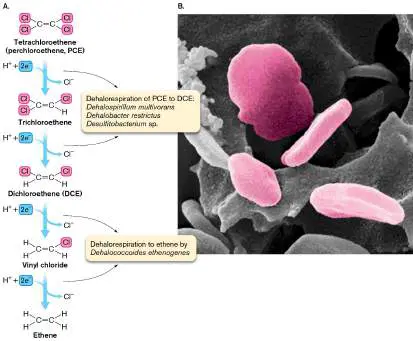
Dehalococcoides strain CBDB1, a cell wall–less bacterium that reductively dechlorinates chlorobenzene (SEM).
LORENZ ADRIAN
In most anoxic habitats, H 2 is one of numerous electron donors. But deep underground, hydrogen may be the key source of electrons. Subsurface H 2 comes from radiolysis, the bombardment of water by nuclear decay radiation including alpha, beta, and gamma rays. As radioactive metals in Earth’s crust decay, they release these high-energy particles that collide with H 2 O, forming ions that eventually react to H 2. Thus, radioactivity indirectly drives a vast underground ecosystem of microbial hydrogenotrophy.
Methanogenesis
Hydrogen is such a strong electron donor that it can even reduce the highly stable carbon dioxide to methane. Reduction of CO 2 and other single-carbon compounds, such as formate, to methane is called methanogenesis. Methanogenesis supports a major group of archaea known as methanogens, many of which grow solely by autotrophy, generating methane.
The simplest form of methanogenesis involves hydrogen reduction of CO 2: CO 2 + 4H 2 → CH 4 + 2H 2 O E °′ = 180 mV Because both sides of the equation contain a weak electron acceptor (CO 2, H 2 O) and a strong electron donor (H 2, CH 4), it was surprising that such a reaction could yield energy for growth. But the presence of sufficient carbon dioxide and hydrogen drives the reaction forward and supports enormous communities of methanogens. Such conditions prevail wherever bacteria grow by fermentation and their gaseous products are trapped, such as in landfills, where methane can be harvested as natural gas, as well as in the digestive systems of cattle and humans. Variants of methanogenesis also include pathways by which H 2 reduces various one-carbon and two-carbon molecules, such as methanol, methylamine, and acetic acid. Diverse methanogens are found in all kinds of environments (discussed in Chapter 19).
A simplified pathway of methanogenesis from CO 2 is shown in Figure 14.30. The CO 2 undergoes stepwise hydrogenation, and each oxygen is reduced to H 2 O. The increasingly reduced carbon is transferred through a series of unique cofactors (methanofuran, tetrahydromethanopterin, coenzyme M-SH). Three of the hydrogenation steps involve a membrane ETS complex that includes the carrier coenzyme F 420, whose reaction generates a proton potential. Note, however, that the final step of methane production generates a transmembrane sodium potential (ΔNa +), which drives ATP synthesis by a sodium-powered ATP synthase embedded in the cell membrane. Further details of methanogenesis are presented in Chapter 19.
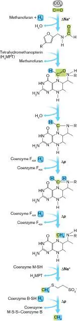
FIGURE 14.30 ■ Methanogenesis. A methanogen reduces carbon dioxide with hydrogen, generating methane (CH 4). The incorporation of hydrogen contributes to both a proton potential and a sodium potential (discussed in Chapter 19).
Thought Question
14.14 Hydrogen gas is so light that it rapidly escapes from Earth. Where does all the hydrogen come from to be used for hydrogenotrophy and methanogenesis?
Methylotrophy: Oxidation of Single-Carbon Substrates
Many forms of catabolism release reduced single-carbon molecules that other microbes can oxidize for energy, such as methanol (CH 3 OH), methylamine (CH 3 NH 2), methane (CH 4), and even carbon monoxide (CO). Oxidation of single-carbon molecules via an ETS is called meth yl otrophy, a form of metabolism outside the definitions of lithotrophy and organotrophy. The oxidant may be O 2 or anaerobic electron acceptors such as nitrite, sulfate, or metals. Methylotrophs are found in soil, marine, and freshwater sediments.
The methane released by methanogenesis provides a niche for meth an otrophy, a form of methylotrophy in which bacteria and archaea oxidize methane. In deep-ocean sediment, the activity of methanogens is so high that enormous quantities of methane become trapped on the seafloor in the form of water-based crystals known as methane hydrates. If all the methane from these crystals were released at once, global warming would be greatly accelerated. Anaerobic methane oxidation may be critical for the global carbon cycle, as it suggests a mechanism for removal of deep-sea methane (discussed in Chapters 21 and 22).
To Summarize
Lithotrophy (chemolithotrophy) is the acquisition of energy by oxidation of inorganic electron donors.
Reverse electron flow powered by Δ p can generate NADH or NADPH.
Nitrogen oxidation includes successive oxidation of ammonia to hydroxylamine, nitrous acid, and nitric acid. Anammox, the oxidation of ammonium ion by nitrite, returns half the ocean’s N 2 to the atmosphere.
Sulfur oxidation includes oxidation of H 2 S to sulfur or to sulfuric acid by sulfur-oxidizing bacteria, often accompanied by iron oxidation. Sulfuric acid production leads to extreme acidification.
Hydrogenotrophy uses hydrogen gas as an electron donor. Hydrogen (H 2) has sufficient reducing potential to donate electrons to nearly all biological electron acceptors, including chlorinated organic molecules (through dehalorespiration). Methanogenesis is the oxidation of H 2 by CO 2, releasing methane. Methanogenesis is performed only by the methanogen group of archaea.
Methylotrophs use O 2, nitrite, or sulfate to oxidize single-carbon compounds such as methane, methanol, or methylamine. A class of methylotrophs called methanotrophs specifically oxidize methane.
Glossary
lithotrophy Also called chemolithotrophy. The metabolic oxidation of inorganic compounds to yield energy and fix single-carbon compounds into biomass.
chemolithotrophy See lithotrophy .
reverse electron flow An enzyme-catalyzed redox reaction that couples an electron transfer with a positive Δ G (such as NAD + reduction to NADH) to a source of energy with a larger negative Δ G (such as a proton potential generated by an electron transport system). nitrifier An organism that converts reduced nitrogen compounds to nitrite or nitrate.
anammox reaction The anaerobic oxidation of ammonium to nitrogen gas, using nitrite as electron acceptor; yields energy.
hydrogenotrophy The use of molecular hydrogen (H 2) as an electron donor for a variety of electron acceptors.
methanogenesis An energy-yielding metabolic process that releases methane, commonly from hydrogen gas and oxidized one-or two-carbon compounds. It is unique to archaea.
methylotrophy The metabolic oxidation of single-carbon compounds such as methanol, methylamine, or methane to yield energy.
methanotrophy The metabolic oxidation of methane to yield energy.
Figure 14.14 FIGURE 14.14 ■ A bacterial ETS for aerobic NADH oxidation. In E. coli, electrons from NDH-1 are transferred to quinones, generating quinols, which transfer electrons onto cytochrome bo (Cyt bo) quinol oxidase complex. For each NADH oxidized, up to 8H + may be pumped across the membrane.
Figure 14.15 FIGURE 14.15 ■ Mitochondrial electron transport. In addition to NADH dehydrogenase, succinate dehydrogenase, and a terminal cytochrome oxidase, mitochondria possess ubiquinol:cytochrome c oxidoreductase, which provides an
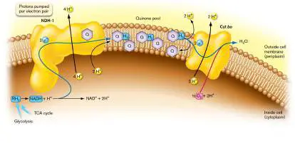
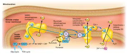
intermediate electron transfer step. As a result, mitochondrial membranes export 10–12 H + per NADH.
Figure 14.14 FIGURE 14.14 ■ A bacterial ETS for aerobic NADH oxidation. In E. coli, electrons from NDH-1 are transferred to quinones, generating quinols, which transfer electrons onto cytochrome bo (Cyt bo) quinol oxidase complex. For each NADH oxidized, up to 8H + may be pumped across the membrane.
Figure 14.15
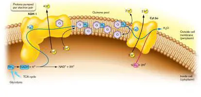
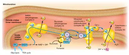
FIGURE 14.15 ■ Mitochondrial electron transport. In addition to NADH dehydrogenase, succinate dehydrogenase, and a terminal cytochrome oxidase, mitochondria possess ubiquinol:cytochrome c oxidoreductase, which provides an intermediate electron transfer step. As a result, mitochondrial membranes export 10–12 H + per NADH.
Fig. 14.11D
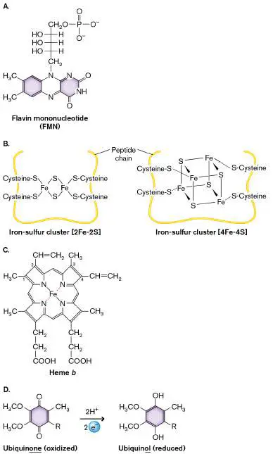
FIGURE 14.11 ■ Cofactors for electron transport. A. Flavin mononucleotide (FMN). B. Iron-sulfur clusters: [2Fe-2S] and [4Fe-4S]. C. Heme b. The side chains of the ring vary among hemes, yielding different levels of redox potential. D. Ubiquinone, which is reduced to ubiquinol.
14.6 PhototrophyUnit 4 · Metabolism
On Earth today, the ultimate source of electrons driving metabolism is phototrophy, the use of photoexcited electrons to power cell growth. Every year, photosynthesis converts more than 10% of atmospheric carbon dioxide to biomass, most of which then feeds microbial and animal heterotrophs. Most of Earth’s photosynthetic production, especially in the oceans, comes from microbes (discussed in Chapter 21). Even the frigid seas of Antarctica support vast communities of phototrophic algae and bacteria, at −2°C beneath pack ice (Fig. 14.31). Microbial phototrophs and land plants (with chloroplasts evolved from microbes) form the foundation of Earth’s biosphere.
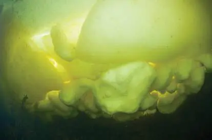
FIGURE 14.31 ■ Phototrophy beneath Antarctic pack ice. Underwater view of the underside of ice, showing yellow-green algae illuminated by sunlight.
NATURE PICTURE LIBRARY/ALAMY STOCK PHOTO
In phototrophy, the energy of a photoexcited electron is used to pump protons. Different kinds of phototrophic systems include the bacteriorhodopsin proton pump; the single-cycle chlorophyll-based photosystems I and II; and the double-cycle Z pathway of oxygenic photosynthesis in cyanobacteria and chloroplasts.
Note: Oxygenic photosynthesis is coupled directly to fixation of
CO 2 into biomass. The process of CO 2 fixation is discussed in Chapter 15.
Retinal-Based Proton Pumps
In most ecosystems, the dominant source of carbon and energy is photosynthesis based on chlorophyll. At the same time, many halophilic archaea and marine bacteria supplement their metabolism with a simpler, more ancient form of phototrophy that is based on a single-protein, light-driven proton pump containing the pigment retinal. Many varieties of the retinal-based proton pump have evolved, known as bacteriorhodopsin in haloarchaea and as proteorhodopsin in bacteria. We present bacteriorhodopsin as a relatively simple form of phototrophy, followed by the more complex ETS-based photolysis in bacteria and chloroplasts.
Bacteriorhodopsin and proteorhodopsin. Bacteriorhodopsin is a small membrane protein commonly found in halophilic archaea (or haloarchaea) such as Halobacterium salinarum, a single-celled archaeon that grows in evaporating salt flats containing concentrated NaCl (discussed in Chapter 19). For many years, bacteriorhodopsin-like proton pumps were thought to be limited to extreme halophilic archaea. As bacterial genomes were sequenced, however, homologs of the protein appeared in several species of proteobacteria; the homologs were termed proteorhodopsin. The proteorhodopsin genes appear to have entered bacteria by horizontal transfer from halophilic archaea. In 2005, Oded Béjà and colleagues from Israel, Austria, Korea, and the United States surveyed the genomes of unculturable bacteria from the upper waters of the Mediterranean and Red Seas. They found that 13% of the marine bacteria contain proteorhodopsins, accounting for a substantial—and previously unrecognized—fraction of marine phototrophy.
Bacteriorhodopsin absorbs light with a broad peak in the green range (500–550 nm). Thus the organisms containing large amounts of bacteriorhodopsin reflect blue (450–490 nm) and red (650–700 nm), appearing purple. The protein consists of seven hydrophobic alpha helices surrounding a molecule of retinal, the same cofactor bound to light-absorbing opsins in the vertebrate retina (Fig. 14.32A). In bacteriorhodopsin, the retinal is attached to the nitrogen end of a lysine residue (Fig. 14.32B ).
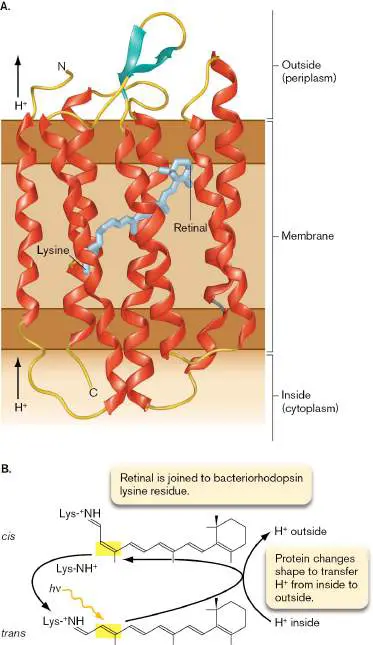
FIGURE 14.32 ■ The light-driven cycle of bacteriorhodopsin. A. Bacteriorhodopsin contains seven alpha helices that span the membrane in alternating directions and surround a molecule of retinal, which is linked to a lysine residue. (PDB code: 1FBB) B. A photon (h ν) is absorbed by retinal, which shifts the configuration from trans to cis. The relaxation back to the trans form is coupled to pumping 1H + across the membrane. (PDB code: 1M0L)
Source: Purple Membrane: Theoretical Biophysics Group, VMD Image Gallery, NIH Resource for Macromolecular Modeling and Bioinformatics. Retinal has a series of conjugated double bonds that absorb visible light. Upon absorbing a photon, an electron in one of the double bonds is excited to a higher energy level. This process is called photoexcitation. As the electron falls back to the ground state, the double bond shifts position from trans (substituents pointing opposite) to cis (substituents pointing in the same direction). This change in shape of the retinal alters the conformation of the entire protein, causing it to pick up a proton from the cytoplasm. Eventually, the retinal switches back to its original trans configuration. The reversion to trans is coupled to the release of a proton from the opposite end of the protein facing outside of the cell. Thus, photoexcitation of bacteriorhodopsin is coupled to the pumping of one H + across the membrane.
The proton gradient generated by bacteriorhodopsin drives ATP synthesis by a typical F 1 F o ATP synthase. Light capture by bacteriorhodopsin supplements catabolism for energy and heterotrophy for carbon source. This combination of light absorption and heterotrophy is a form of photoheterotrophy.
Purple membrane captures light rays. One problem every phototroph needs to solve is how to “capture” light rays. For chemotrophy, food molecules diffuse in solution and can be picked up by receptors for transport into a cell. Phototrophy, however, requires a photon to impinge on one point of the cell, where the photon either is absorbed or passes through. Thus, the only way to absorb a high percentage of photons is to spread light-absorbing pigments over a wide surface area. To maximize light absorption, Halobacterium salinarum archaea pack their entire cell membranes with bacteriorhodopsin. The protein assembles in trimers that pack in hexagonal arrays, forming the “purple membrane” (Fig. 14.33). FIGURE 14.33 ■ Bacteriorhodopsin purple membrane. Trimers of bacteriorhodopsin (monomers shown red, blue, and green) are packed in hexagonal arrays, forming the “purple membrane.” (PDB code: 1M0L)
Source: Purple Membrane: Theoretical Biophysics Group, VMD Image Gallery, NIH Resource for Macromolecular Modeling and Bioinformatics.
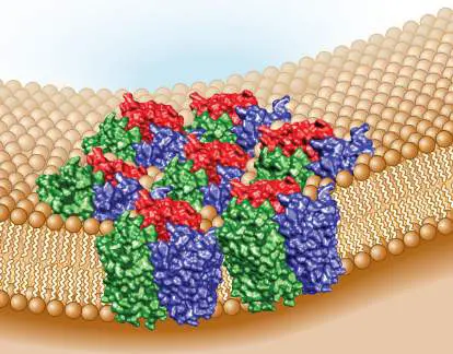
Although the bacteriorhodopsin cycle is much simpler than chlorophyll-based photosynthesis (discussed next), it nevertheless illustrates several principles that apply to more complex forms of phototrophy: A photoreceptor absorbs light, causing excitation of an electron to a higher energy level, followed by return to the ground state. To maximize light collection, large numbers of photoreceptors are packed throughout a membrane.
The photocycle (absorption and relaxation of the light-absorbing molecule) is coupled to energy storage in the form of a proton gradient.
Note: Distinguish among these terms of phototrophy:
Photo excitation means light absorption that raises an electron to a higher energy state, as in bacteriorhodopsin. Photo ionization means light absorption that causes electron separation.
Photo lysis means light absorption coupled to splitting a molecule.
Photo synthesis means photolysis with CO 2 fixation and biosynthesis.
Chlorophyll Photoexcitation and Photolysis
Cyanobacteria and chloroplasts, as well as other kinds of bacteria, obtain energy by photoexcitation of chlorophylls. Figuring out their “light reactions”—the fundamental source of energy for Earth’s biosphere—was one of the most exciting projects of the twentieth century. Among hundreds of important contributors, we note two major figures: the married couple Roger Stanier (1916–1982) and Germaine Cohen-Bazire (1920–2001; Fig. 14.34Aand B ). Stanier, a Canadian microbial physiologist at UC Berkeley, clarified the nature of cyanobacteria as phototrophic prokaryotes distinct from eukaryotic algae, and he helped distinguish the water-based photosynthesis of cyanobacteria from the sulfide metabolism of purple bacteria. Cohen-Bazire was a French bacterial geneticist who had studied lac operon regulation with Nobel laureate Jacques Monod at the Pasteur Institute in Paris. Cohen-Bazire applied her genetics skills to phototrophs, and she conducted the first genetic analysis of photosynthesis in purple bacteria and cyanobacteria. FIGURE 14.34 ■ Germaine Cohen-Bazire and Roger Stanier studied cyanobacterial photosynthesis. A. Stanier pioneered the study of cyanobacterial physiology. B. Cohen-Bazire performed the first studies of genetic regulation of
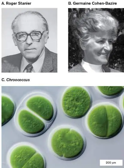
bacterial photosynthesis. C. Chroococcus, a genus of cyanobacterium studied by Stanier.
REPUBLISHED WITH PERMISSION OF THE ROYAL SOCIETY (U.K.), P. H. CLARKE.
1982. BIOGRAPHICAL MEMOIRS OF FELLOWS OF THE ROYAL SOCIETY 32 :542
REPRINTED BY PERMISSION FROM SPRINGER NATURE: GOVINDJEE, J. F. ALLEN, AND
J. T. BEATTY. 2004. PHOTOSYNTHESIS RESEARCH 80 :1
GERD GUENTHER/SCIENCE SOURCE
Cyanobacteria are the only bacteria that split water to make oxygen. Most cyanobacterial species appear green, like algae or plants (Fig. 14.34C ). Their green color arises from their chlorophyll, which absorbs blue and red but reflects green. Some species appear orange or brown because of secondary pigments. Cyanobacteria include a wide range of species, such as one of the ocean’s major producers, the tiny Prochlorococcus marinus, barely visible under a light microscope. Other cyanobacteria have cells as large as eukaryotic algae and form complex developmental structures with important symbiotic associations (see Chapters 18 and 21). Cyanobacteria are among the most widely distributed and diverse groups of life on Earth. Along with the chloroplasts of algae and plants, cyanobacteria produce all the oxygen available for aerobic life.
Overview of photolysis. The energy for photosynthesis derives from the photoexcitation of a light-absorbing pigment. The light-absorbing molecule with an electron in an excited state becomes a strong electron donor. The photoexcited molecule can thus donate an electron to an electron acceptor molecule, which is coupled to an ETS. The components of the ETS are often homologous to those of respiratory electron transport, and they share common electron carriers, such as cytochromes.
In plant chloroplasts and in cyanobacteria, photolysis is known as the “light reactions,” which are coupled to the “light-independent reactions” of carbon dioxide fixation. Note, however, that many of the sulfur-or organic-based bacterial phototrophs, such as Rhodospirillum rubrum, combine photolysis with heterotrophy instead of with CO 2 fixation. At the same time, lithotrophic bacteria such as Nitrospira and Acidithiobacillus fix CO 2 using energy from mineral oxidation instead of photolysis. In this chapter we focus on photolysis in cyanobacteria. We discuss CO 2 fixation along with other biosynthetic pathways in Chapter 15.
In ETS-based photosynthesis, photoexcitation leads to separation of an electron from a donor molecule such as H 2 O or H 2 S. Each electron is then transferred to an ETS, whose components show common ancestry with respiratory ETS proteins. The ETS generates a proton potential and the reduced cofactor NADPH. The proton potential drives ATP synthesis through an F 1 F o ATP synthase similar to the one for respiration.
Chlorophylls absorb light. The main light-absorbing pigments are chlorophylls. Each type of chlorophyll contains a characteristic chromophore, a light-absorbing electron carrier. The chlorophyll chromophore consists of a heteroaromatic ring complexed to a magnesium ion (Mg 2+; Fig. 14.35A). As we saw for ETS electron carriers, aromatic molecules offer electrons with relatively narrow energy transitions. The chromophore absorbs a photon through a reversible energy transition, such that the chlorophyll can alternate between excited and ground states.
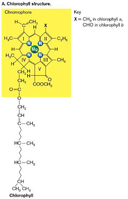
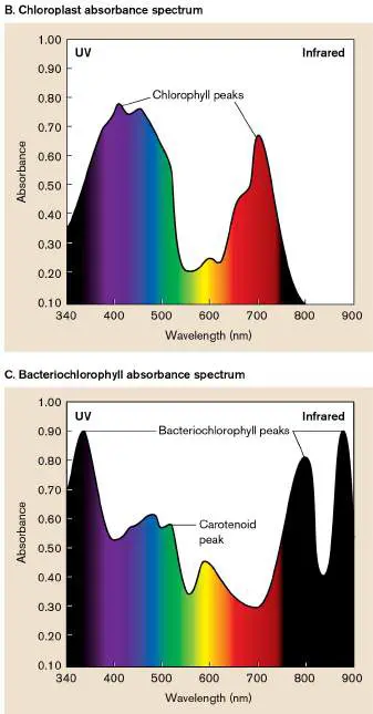
FIGURE 14.35 ■ Chlorophyll structure and absorbance. A. Chlorophyll molecular structure. B. Absorption by chloroplasts, including chlorophyll a, chlorophyll b, and carotenoid accessory pigments. The middle range (green) is reflected. C. Absorption by purple photosynthetic bacteria, including bacteriochlorophyll and carotenoids.
Chlorophyll molecules differ slightly in their substituent groups around the ring; for example, chlorophyll a of chloroplasts has a methyl group in ring II, whereas chlorophyll b has an aldehyde. Both chlorophylls a and b are made by chloroplasts and by cyanobacteria, their nearest bacterial relatives. Because they absorb red and blue, they reflect the middle range of the spectrum and so appear green ( Fig. 14.35B ).
By contrast, the chlorophylls of anaerobic phototrophs, or “purple bacteria,” such as Rhodobacter and Rhodospirillum, absorb most strongly in the far-red (infrared) and, in some cases, ultraviolet ( Fig. 14.35C ). Their chlorophylls are specifically named bacteriochlorophylls. The purple bacteria grow in pond water or sediment. Bacteriochlorophyll absorption over an extended range of wavelengths helps capture light missed by the cyanobacteria and algae at the water’s surface. In purple bacteria, bacteriochlorophylls are supplemented by accessory pigments called carotenoids, which absorb light of green wavelengths and transfer the energy to bacteriochlorophyll. The combination of green-absorbing carotenoids and infrared-absorbing bacteriochlorophylls makes cultures appear deep purple or brown.
The infrared radiation absorbed by bacteriochlorophylls is too weak to permit splitting H 2 O to produce oxygen. Thus, purple bacteria are limited to photolysis of H 2 S and small organic molecules; many are photoheterotrophs. On the other hand, infrared rays are available in water below the oxygenic phototrophs absorbing red and blue. Thus, anaerobic phototrophs grow at depths where their more high-powered oxygenic relatives do not.
Note: Distinguish among these classes of photopigments:
Bacteriorhodopsin is a retinal-containing proton pump.
Chlorophyll is a charge-separating photopigment (usually referring to chloroplasts and cyanobacteria).
Bacteriochlorophyll is a charge-separating chlorophyll of anaerobic purple and green bacteria (also known generically as chlorophyll).
Carotenoid is an accessory pigment that absorbs the midrange of light wavelengths but does not directly conduct photolysis. Antenna complex and reaction center. Light photons cannot be transported or concentrated in a compartment. Instead, they must be captured by absorption. The larger the array of absorptive molecules, the more photons will be captured.
To maximize light collection, many molecules of chlorophyll are grouped in an antenna complex. These antenna complexes are arranged like a satellite dish within the plane of the membrane in an elaborate cluster around accessory proteins. This cluster is called a light-harvesting complex (LH). Light-harvesting complex 2 (LH2) of Blastochloris viridis is shown in Figure 14.36A. B. viridis is a photoheterotroph, an alphaproteobacterium that absorbs an exceptionally broad range of light, into the infrared. The LH2 clusters of B. viridis associate in a ring around light-harvesting complex 1 (LH1), shown in Figure 14.36B . In the center, surrounded by LH2 complexes, LH1 contains the reaction center ( RC; Fig. 14.36B ). The reaction center is the protein complex in which chlorophyll photoexcitation connects to the ETS.
FIGURE 14.36 ■ Antenna complexes surround the reaction center. A. An antenna complex (LH2) of Blastochloris viridis contains 9 chlorophylls with chromophores facing parallel to the membrane (gold) and 18 with chromophores facing out from the ring (red). B. Multiple rings of chlorophyll-protein antenna complexes surround the reaction center (RC), like a funnel collecting photons. (PDB codes: 1PYH, 2FKW) C. Light-harvesting complex 1 (LH1), with reaction center. (PDB code: 6ET5)
Sources: Parts A and B modified from Quantum Biology of the PSU, NIH Resource for Macromolecular Modeling and Bioinformatics—BChl Antenna; part C modified from P. Qian et al. 2018. Nature 556 :203.
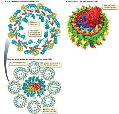
Throughout this antenna complex of bacteriochlorophylls and accessory pigments, whichever pigment molecule happens to be in the right place at the right time captures the photon. The energy from the photon then transfers from one chromophore to the next, until it arrives at the reaction center for electron transfer to the ETS. Other kinds of bacteria have other forms of antenna complexes, such as the “phycobilisome” of cyanobacteria (discussed shortly). Purple bacteria and cyanobacteria increase their efficiency of photon uptake by extensive backfolding of the photosynthetic membranes in oval pockets stacked like pita breads (Fig. 14.37). These oval pockets are called thylakoids. The extensive packing of thylakoids gives an incident photon hundreds of chances to meet a chlorophyll at just the right angle for absorption. The hollow “thylakoid pockets” also store H + ions to generate a transmembrane Δ p for ATP synthesis.
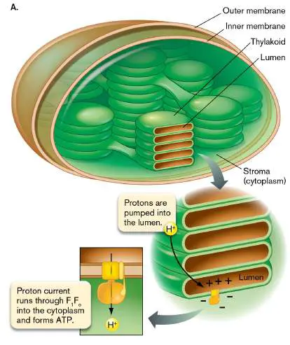
FIGURE 14.37 ■ Photosynthetic membranes. A. The photosynthetic membranes of bacteria and chloroplasts appear as hollow disks with tubular interconnections. The disks are called thylakoids. Topologically, the membrane separates the cytoplasm (stroma) from the interior space (lumen). B.
Transmission electron microscopy (TEM) section of a chloroplast, showing the stacked thylakoids.
BIOPHOTO ASSOCIATES/SCIENCE SOURCE
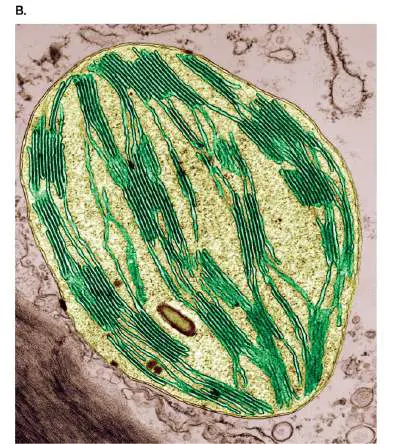
The thylakoids are connected by tubular extensions, so that there exists one interior space, the lumen, separated topologically from the regular cytoplasm, or stroma. Protons are pumped from the stroma across the thylakoid membrane into the lumen. The F 1 F o complex is embedded in the thylakoid, where it makes ATP using the proton current running through it into the cytoplasm. The F 1 knob of ATP synthase appears to face “outward” in photosynthetic organelles (as opposed to “inward” in respiratory chains). In each case, however, the proton current and ATP motor face in the same direction with respect to the cytoplasm (stroma). The proton potential is more negative in the cytoplasm (stroma), thus drawing protons through the ATP synthase to generate ATP.
The photolytic electron transport system. In photolysis, the absorption of light by chlorophyll or bacteriochlorophyll drives the separation of an electron. The chlorophyll may then gain an electron from the ETS, as in Rhodospirillum or Rhodobacter, or it may remove an electron from H 2 S or H 2 O, depending on the photosystem of a given bacterial species. In either case, the excited electron carries energy gained from the light absorbed. The excited electron enters a membrane-embedded ETS of oxidoreductases and quinones/quinols, as we saw for the ETS of respiration and lithotrophy.
Diverse kinds of photosynthesis in different environmental niches include oxygenic, sulfur-based, iron-dependent, and even heterotrophic photolysis. Nevertheless, all forms of photolysis share a common design: 1. Antenna system. The antenna system (Fig. 14.36)
maximizes photon capture. A phototrophic antenna system is a large complex of chlorophylls that captures photons and transfers their energy among the photopigments until it reaches a reaction center. The complex of chlorophylls and accessory pigments resides in the photosynthetic membrane where it collects photons. Energy from each photon is transferred among antenna pigments and eventually to the reaction center.
2. Reaction center complex. In the reaction center, the photon energy is used to separate an electron from chlorophyll. The electron is replaced by one from a small molecule, such as H 2 S (photosystem I, or PS I) or from H 2 O through a light-harvesting antenna complex (photosystem II, or PS II).
3. Electron transport system. Each photoexcited electron enters an ETS. In PS I, electrons separated from chlorophyll are transferred to NADP + to form NADPH. In PS II, the electron separated from bacteriochlorophyll is replaced by an electron returned from the ETS. In the oxygenic Z pathway (H 2 O photolysis), electrons flow from PS II into PS I, ultimately releasing O 2 from H 2 O.
4. Energy carriers. In PS I, electrons are used to make NADPH. In PS II, electron transfer provides energy to pump protons and drive the synthesis of ATP. The Z pathway makes both NADPH and ATP, which are used to fix CO 2.
Photosystems I and II
The steps of photolysis and electron transport occur in three different kinds of systems, in different classes of bacteria: Anaerobic photosystem I receives electrons associated with hydrogens from H S, HS −, or H, or even from reduced iron
2 2
(Fe 2+). Anaerobic PS I is found in chlorobia (“green sulfur” bacteria) and in chloroflexi (filamentous green bacteria). Anaerobic photosystem II returns an electron from the ETS to bacteriochlorophyll. Anaerobic PS II is found in alphaproteobacteria, “purple nonsulfur” bacteria, and other proteobacteria.
The oxygenic Z pathway includes homologs of photosystems I and II. Two pairs of electrons are received from two water molecules to generate O 2. The Z pathway is found in cyanobacteria and in the chloroplasts of green plants. The components of photosystems I and II (PS I and PS II) share common ancestry. Each system runs anaerobically, producing sulfur or oxidized organic by-products, but not O 2. Each photosystem shows more recent homology with the respective PS I and PS II components of the oxygenic Z pathway (so called because the electron path through a diagram of the two photosystems traces a Z). The Z pathway ultimately generates O 2 —the source of nearly all the oxygen we breathe.
Note: In photolysis, each quantum of light excites a single
electron. Some ETS components, such as the quinones/quinols (Q → QH 2), process two of these electrons in completing their redox cycle. The single-electron intermediate states of quinones are not shown here.
Photosystem I in chlorobia. Bacteria such as Chlorobium species use PS I (Fig. 14.38). The reaction center (RC) contains bacteriochlorophyll P840, named for its peak absorption at 840 nm; that is, the near-infrared, actually beyond the range that humans can see. But P840 and the chlorophylls of the antenna complex also absorb light over shorter wavelengths, in the range of 400–550 nm.
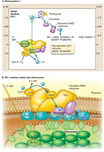
FIGURE 14.38 ■ Photosystem I separates electrons from sulfides and organic molecules. A. In green sulfur bacteria, photoexcitation of P840 transfers e − to a quinone (phylloquinone; PQ), at high reduction potential E. From PQ the electron is transferred to ferredoxin (Fd). Ferredoxin is oxidized by ferredoxin-NAD + reductase (FNR), donating 2 e − to NAD + (or the energetically equivalent NADP +) to form NADH (or NADPH). B. The chlorosome antenna complex transfers photon energy to the PS I reaction center. BChl a = bacteriochlorophyll a; BChl c = bacteriochlorophyll c; BChl P840 = bacteriochlorophyll P840; Cyt c = cytochrome c.
The Chlorobium antenna complex consists of a membrane compartment called a chlorosome (Fig. 14.38B ). A single chlorosome may contain 200,000 molecules of bacteriochlorophyll, harvesting photons with nearly 100% efficiency. The chlorosome is so sensitive that some chlorobia actually harvest thermal radiation from deep-sea thermal vents.
When any one bacteriochlorophyll absorbs a photon, the energy transfers among the photopigments until it reaches the PS I reaction center (Fig. 14.38). The photon yields sufficient energy to donate the high-potential electron to a high-potential quinone: phylloquinone/phylloquinol (PQ). Phylloquinol donates the electron to ferredoxin, an FeS protein. Ferredoxin transfers the electron to the enzyme ferredoxin-NAD + reductase; two of these electrons then reduce NAD + or the energetically equivalent NADP +. The reduced carrier (NADH or NADPH) provides reductive energy for CO 2 fixation and biosynthesis (discussed in Chapter 15).
Chlorobium is a true autotroph, fixing CO 2 for biosynthesis (see Chapter 15). However, the phototrophic ETS is supplemented by lithotrophy, the donation of electrons from H 2 S or H 2. The PS I electron flow generates a net proton gradient by consuming H + inside and generating H + outside the cell, thus providing proton motive force to drive ATP synthesis.
Photosystem II in alphaproteobacteria. Phototrophic alphaproteobacteria such as Rhodospirillum rubrum and Blastochloris viridis are typically found in wetlands and streams, where they capture light not used by other phototrophs. R. rubrum uses both PS I and PS II, whereas B. viridis has a simplified photosystem comprising only PS II, generating ATP. The antenna complex of B. viridis was shown earlier, in Figure 14.36. The peak wavelength absorbed by its bacteriochlorophyll P870 lies so far into the infrared (800–1,100 nm) that the photon energy is insufficient to reduce NAD(P) to NAD(P)H. The electrons from P870 are transferred by low-potential quinols to a terminal cytochrome oxidoreductase ( Fig. 14.39). The quinols pass their electrons to cytochromes, while moving 2H + across the membrane.
FIGURE 14.39 ■ Photosystem II separates an electron from bacteriochlorophyll. A. In purple bacteria, bacteriochlorophyll P870 (BChl P870) donates an energized electron to a quinone (Q). Two of these donated electrons complete the conversion of quinone to quinol (QH 2). Electrons flow through cytochrome bc (Cyt bc), coupled to pumping of protons. The proton potential drives synthesis of ATP. The cytochrome bc complex transfers the electrons back to P870. B. PS II reaction center.
When the electrons reach cytochrome c, they flow back to bacteriochlorophyll, where they can be reexcited by photon energy from the antenna complex. Because the electron path traces back to its source, the ETS of photosystem II leading to ATP synthesis is called cyclic photophosphorylation.
Because the reduction potential is too small to reduce NADP + to NADPH, photosystem II requires reverse electron flow. As we saw for iron oxidation (see Section 14.5), in reverse electron flow a low-potential electron donor reduces an ETS, requiring input of energy. Purple bacteria obtain this energy by spending ATP to increase the reduction potential or from a pathway outside photolysis, such as catabolism of organic compounds. The organic compounds also
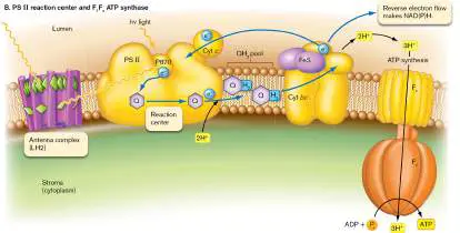
provide substrates for biosynthesis. Thus, most purple bacteria are photoheterotrophs.
Purple bacteria such as Rhodopseudomonas palustris are the ultimate generalists, often combining several major classes of metabolism. Caroline Harwood and colleagues at the University of Washington showed that R. palustris is a “photolithoheterotroph,” capable of photosynthesis, catabolism on many substrates, and lithotrophy. Photolithoheterotrophs are of interest for possible applications as solar-driven microbial fuel cells.
Thought Question
14.15 Suppose you discover bacteria that require a high concentration of Fe 2+ for photosynthesis. Can you hypothesize what the role of Fe 2+ might be? How would you test your hypothesis? Oxygenic photolysis. The Z pathway of photolysis found in cyanobacteria and chloroplasts combines key features of both PS II and PS I (Fig. 14.40). Both reaction centers, however, contain chlorophylls that absorb at shorter wavelengths (higher energy) than those of the respective purple or green homologs: P680 instead of P870 (PS II), and P700 instead of P840 (PS I). Thus, the cyanobacterial reaction centers can split water—a highly stable molecule that cannot be photolyzed by anaerobic phototrophs. Photolysis of water requires greater energy input but ultimately yields greater energy overall and produces molecular oxygen. The energy potentials are high enough to generate NADPH and fix CO 2 into biomass (presented in Chapter 15). Oxygenic phototrophs dominate the shallow water depths, whereas anaerobes grow at lower depths, using light at wavelengths unused by cyanobacteria and algae near the surface.
FIGURE 14.40 ■ Oxygenic photosynthesis in cyanobacteria and chloroplasts. A. Each H 2 O is photolyzed via the Z pathway of PS II and PS I. The 2 e − from each water molecule (4 e − in all) are transferred to the quinone pool, which transfers 4 e − to cytochrome bf (Cyt bf). Cytochrome bf pumps 4 × 2H + across the membrane and transfers 4 e − via plastocyanin (PC) to a PS I containing chlorophyll P700 (Chl P700). A second photon excites P700, enabling transfer of e − to
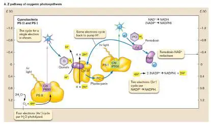
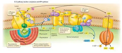
ferredoxin (4 e − per O formed), and from there to NADP + →
2
NADPH. B. The Z pathway within a photosynthetic membrane. Chl P680 = chlorophyll P680.
Source: Part B based on crystallographic data from thermophilic cyanobacteria, modified from Genji Kurisu et al. 2003. Science 302 :1009. Cyanobacteria harvest light via an exceptionally efficient antenna complex called the phycobilisome (Fig. 14.40B ). In the PS II reaction center, the photoexcitation of chlorophyll P680 yields enough energy to split H 2 O. The entire cycle of water splitting involves 4H + removed from 2H O, 4 e − transferred to carriers,
2
and the formation of O 2. The net reaction forming oxygen is: 2H O → 4H + + 4 e − + O
2 2
Through the ETS, the electrons are transferred to quinones. As in respiration, each 2 e − reduction of quinone to quinol requires pickup of 2H + from the stroma (equivalent to cytoplasm). Thus, the four electrons transferred generate a net change in the proton gradient of 4H +. Furthermore, the energy of electron transfer to cytochrome bf enables pumping of an additional 2H + across the membrane. Thus, in all, for each conversion of 2H 2 O to O 2, the net protons transferred across the thylakoid membrane include 4H + (water photolysis) plus 4 × 2H + (quinones to quinols) through cytochrome bf, to yield a total of 12H + for the proton gradient. The proton gradient drives the ATP synthase to make approximately 3 ATP per O 2 formed.
The electrons from PS II do not cycle back to the PS II reaction center, as they do in purple bacteria. Instead they are transferred to PS I by a copper-containing protein called plastocyanin. The energy of the electron transferred by plastocyanin is augmented through absorption of a second photon by the chlorophyll of PS I. Subsequent electron flow through ferredoxin can now generate NADH or NADPH. Some of the electron flow instead cycles back to cytochrome bf, where it contributes to pumping protons.
The overall equation for energy yield of oxygenic photolysis can be represented as: 2H O + 2 NADP + + 3 [ADP + P] →
2 i
O + 2 [NADPH + H +] + 3 ATP + 3H O
2 2
To release one O 2 and fix one CO 2 requires absorption of between 8 and 12 photons. The efficiency—that is, the proportion of photon energy converted to CO 2 fixation—is estimated to be 20%– 30%. To generate one molecule of glucose by CO 2 fixation, we need six rounds of the photolysis equation—one per CO 2 molecule “fixed” into sugar (C 6 H 12 O 6; discussed in Chapter 15). The CO 2 fixation equation works out to: A recurring theme of this chapter is that all forms of metabolism involve electron transfer reactions that yield energy for cell function. As molecules are rearranged by transfer of electrons from one substrate to another, energy is provided to form ion gradients and energy carriers. The energy carriers always need to be balanced between redox-neutral carriers, such as ATP, and reducing carriers such as NADH or NADPH. Whatever the means of obtaining energy, ultimately cells must spend energy for biosynthesis; for example, the Z pathway “light reactions” are tightly coupled to CO 2 fixation in biomass. Chapter 15 will present how microbes construct the
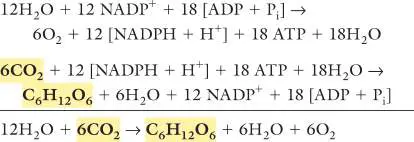
fundamental “nuts and bolts” of their cells—including products surprisingly useful for biotechnology.
To Summarize
Bacteriorhodopsin and proteorhodopsin —found in haloarchaea and in bacteria, respectively—are forms of a light-driven proton pump that contains retinal. The energy gained from light absorption supplements heterotrophy. Chlorophylls and bacteriochlorophylls are photopigments that absorb light and transfer energy to an ETS. Chlorophylls in cyanobacteria and in plant chloroplasts participate in oxygenic photosynthesis, whereas bacteriochlorophylls participate in phototrophy that does not produce oxygen.
The antenna complex of chlorophylls and other photopigments captures light energy for transfer to the reaction center in chlorophyll-based photosynthesis. Thylakoids are folded membranes within phototrophic bacteria or chloroplasts. The membranes extend the area for chlorophyll light absorption, and they separate two compartments to form a proton gradient.
Photosystem I obtains electrons from H S or HS −. The
2
electrons are transferred through an ETS to form NADH or NADPH.
Photosystem II transfers an electron through an ETS and pumps H + to generate ATP. An electron ultimately returns to bacteriochlorophyll through cyclic photophosphorylation. Reverse electron flow generates NADPH or NADH.
The oxygenic Z pathway in cyanobacteria and chloroplasts includes homologs of photosystems I and II. Eight photons are absorbed, and two electron pairs are removed from 2H 2 O, ultimately producing O 2.
Oxygenic photosynthesis generates 3 ATP + 2 NADPH per 2H 2 O photolyzed and O 2 produced. The ATP and NADPH are used to fix CO 2 into biomass.
Glossary
bacteriorhodopsin A haloarchaeal membrane-embedded protein that contains retinal and acts as a light-driven proton pump; it is homologous to the bacterial proteorhodopsin.
proteorhodopsin A bacterial membrane-embedded protein that contains retinal and acts as a light-driven proton pump; it is homologous to the archaeal protein bacteriorhodopsin.
retinal A vitamin A–related cofactor in opsin proteins; it undergoes a conformational change upon absorbing a photon.
photoheterotrophy Metabolism that includes gain of energy from light absorption with biosynthesis from preformed organic compounds. Usually also includes organotrophy, gain of energy from reactions of organic compounds.
chlorophyll A magnesium-containing porphyrin pigment that captures light energy at the start of photosynthesis.
chromophore A light-absorbing redox cofactor.
bacteriochlorophyll The chlorophyll of anaerobic phototrophic bacteria; it absorbs photons most strongly in the far-red end of the light spectrum. carotenoid An accessory photosynthetic pigment that absorbs photons in the green wavelengths of the spectrum.
antenna complex A complex of chlorophylls and accessory pigments in the photosynthetic membrane that collects photons and funnels them to a reaction center.
reaction center (RC)
The complex containing a chlorophyll molecule that donates its excited electron to an electron transport system.
reaction center (RC)
The complex containing a chlorophyll molecule that donates its excited electron to an electron transport system.
thylakoid An intracellular chlorophyll-containing membrane folded within a phototrophic bacterium or a chloroplast.
photosystem I (PS I)
A protein complex that harvests light from a chlorophyll or bacteriochlorophyll, donates an electron to an electron transport system, receives an electron from a small molecule such as H 2 S or H 2 O, and stores energy in the form of NADPH. photosystem I (PS I)
A protein complex that harvests light from a chlorophyll or bacteriochlorophyll, donates an electron to an electron transport system, receives an electron from a small molecule such as H 2 S or H 2 O, and stores energy in the form of NADPH. photosystem II (PS II)
A protein complex that harvests light from a chlorophyll or bacteriochlorophyll, donates an electron to an electron transport system, and stores energy in the form of a proton potential.
photosystem II (PS II)
A protein complex that harvests light from a chlorophyll or bacteriochlorophyll, donates an electron to an electron transport system, and stores energy in the form of a proton potential.
oxygenic Z pathway An ATP-producing photosynthetic pathway consisting of photosystems I and II. Water serves as the initial electron donor (generating O), and NADP + is the final electron
2
acceptor (generating NADPH).
ferredoxin An iron-and sulfur-containing protein that transfers electrons in electron transport systems.
cyclic photophosphorylation A photosynthetic process in which chlorophyll serves as both the initial electron donor and the final electron acceptor. ATP is produced via the proton potential from an electron transport system, but no NADPH is generated.
eResearch Activity 14
Can Silver Extensions Amplify Bacterial Electricity?
Microbial electricity offers exciting opportunities as a source of energy for human use. Bacteria such as Shewanella and Geobacter can donate electrons directly to electrodes, acting as a battery supply. But the amounts of current generated are generally low compared to what our devices require. So how can we engineer bacterial systems to concentrate and scale up their power? To understand the problem requires understanding the structure of the bacterial cell (discussed in Chapter 3). The bacteria that most effectively convert organic chemical energy to electricity are Gram-negative, possessing an outer membrane with periplasm that envelops the inner membrane. A series of reactions, beginning with the cytoplasmic NADH that carries electrons from organic “food” molecules, must transfer electrons to the inner-membrane electron transport system (ETS), and from there to periplasmic and outer-membrane cytochromes. The outer-membrane cytochromes must then transfer electrons to the surface of the electrode, where the cells are growing as a biofilm. Overall, the series of electron transfers is inefficient, losing energy as heat along the way. At UCLA, the research groups of Xiangfeng Duan and Yu Huang are taking a unique approach to amplifying bacterial electricity ( Fig. ERA 14.1 ). Their approach is to supplement the electrode with materials that increase electron transfer. One effective material is silver metal. Surprisingly, the bacteria grow well on silver-supplemented electrodes—and actually incorporate silver metal particles into their cell envelope.
FIGURE ERA 14.1 ■ Silver particles embedded in Shewanella bacteria. A. Xiangfeng Duan (left) and Yu Huang (right) led the research group that made silver-embedded Shewanella. B. Sections through Shewanella reveal silver nanoparticles embedded in the bacterial envelope (scanning TEM and EDX).
Source: Bocheng Cao et al. 2021. Science 373 :1336–1340.
XIANGFENG DUAN
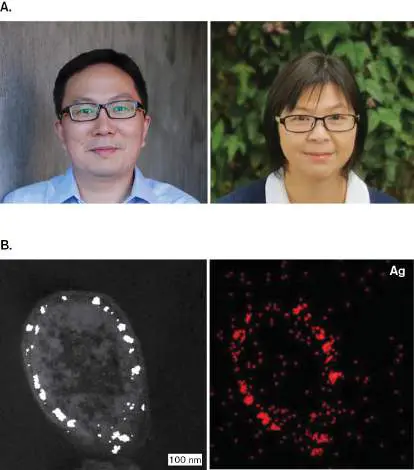
YU HUANG
B. CAO ET AL. 2021. SCIENCE 373:1336–1340
In Figure ERA 14.1B (left), the silver (Ag) nanoparticles are visualized by the method of scanning transmission electron microscopy (scanning TEM). This method differs from scanning electron microscopy (SEM) described in Chapter 2, in which electron beams scan a three-dimensional surface. In scanning TEM, the electron beam scans across a thin section of the bacterium, providing a TEM image with exceptional contrast and resolution. The highly electron-dense Ag nanoparticles appear embedded in the cell envelope.
How do we know the envelope-embedded particles consist of silver? Silver is identified by energy-dispersive X-ray spectroscopy (EDX). EDX measures the X-ray intensities emitted by particles under bombardment by the electron beam. The X-ray intensities have a spectrum characteristic of a specific element, such as silver. In Figure ERA 14.1B (right), the silver signal maps to the position of electron-dense nanoparticles visualized by scanning TEM. The research team proposed that the silver particles, with their high electrical conductivity, could bypass some of the bacterial envelope’s electron transfer reactions and thus increase the efficiency of electrical output. Figure ERA 14.2 shows the results of an experiment in which the power density (wattage output per square centimeter) was tested for three different kinds of electrode containing the Shewanella biofilm. A standard carbon electrode (black) and an electrode containing oxidized graphene nanoparticles (blue) were tested against the experimental electrode with nanoparticles containing silver metal (red).
FIGURE ERA 14.2 ■ Power density of Shewanella fuel cell using electrodes with or without silver. Electrodes were composed of graphene oxide/silver nanoparticles (red), graphene oxide nanoparticles (blue), or carbon paper (black). The electrode containing silver nanoparticles, which Shewanella takes up into its envelope, showed threefold higher power density than that of the electrode without silver. Moreover, the power density obtained was twice the amount reported by colleagues for other Shewanella fuel cells. Thus, silver extensions offer a step forward for commercial development of biobatteries to run our consumer devices.
Further Exploration
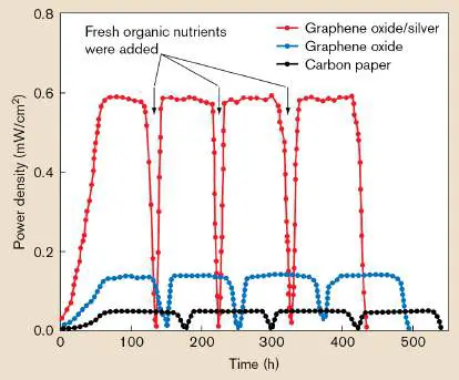
Could other kinds of metals—perhaps copper or nickel—increase the current density? Could Shewanella fuel cells be built to run consumer electronics such as fans and holiday lights? Or desalination units converting salt water to drinking water? Cao, Bocheng, Zipeng Zhao, Lele Peng, Hui-Ying Shiu, Mengning Ding, et al. 2021. Silver nanoparticles boost charge-extraction efficiency in Shewanella microbial fuel cells. Science 373 :1336–1340.
CHAPTER REVIEW
Review Questions
1. Explain the source of electrons and the sink for electrons (terminal electron acceptor) in respiration, lithotrophy, and photolysis.
2. How do bacteria combine redox couples for a metabolic reaction that yields energy? Cite examples, calculating the reduction potential.
3. How do environmental conditions affect the reduction potential of a metabolic reaction?
4. Explain the role of cytochromes and redox cofactors in electron transport systems. What features of a molecule make it useful for redox biochemistry?
5. Explain how a proton potential is composed of a chemical concentration difference plus a charge difference. Explain how each component of Δ p can drive a cellular reaction. 6. Explain the role of the substrate dehydrogenase (oxidoreductase), the quinones, and the cytochrome oxidase (oxidoreductase) in the respiratory ETS.
7. Compare the ETS function in lithotrophy with that in respiration.
8. Summarize the inorganic redox couples that can be used in anaerobic respiration and those that can be used in lithotrophy. What constraints determine whether a given molecule can serve as electron acceptor or as electron donor?
9. How do diverse forms of anaerobic respiration and lithotrophy contribute to ecosystems?
10. Explain the differences and common features of bacteriorhodopsin phototrophy and chlorophyll phototrophy.
11. Explain the differences and common features of photosystems I and II. Explain how the two photosystems combine in the Z pathway. Why can the Z pathway generate oxygen, whereas PS I and PS II cannot?
Thought Questions
1. The lung pathogen Pseudomonas aeruginosa, which also grows in soil, can respire aerobically or else anaerobically using nitrate. Under what conditions would P. aeruginosa use each form of metabolism? What part of its ETS would need to change to accommodate the different forms? 2. What environments favor oxygenic photosynthesis versus sulfur phototrophy? Explain.
3. In pathogens, which components of the ETS do you think would make good targets for new antibiotics, and why? 4. Devise a form of energy-yielding metabolism in which fumarate is converted to succinate; and a different form, in which succinate is converted to fumarate. Explain why the two reactions are reasonable and, on the Internet, try to find actual organisms that obtain energy through these reactions.
Key Terms
anaerobic respiration (562)
anammox reaction (573)
antenna complex (582)
bacteriochlorophyll (582)
bacteriorhodopsin (578, 582)
carotenoid (582)
chlorophyll (581, 582)
chromophore (581)
cyclic photophosphorylation (585) cytochrome (546)
dissimilatory denitrification (563) dissimilatory metal reduction (563) electrogenic biofilm (565)
electron acceptor (544)
electron donor (544)
electron shuttle (567)
electron transport system (ETS) (electron transport chain, ETC) ( 544)
ferredoxin (585)
heme (546)
hydrogenotrophy (575)
lithotrophy (chemolithotrophy) (546, 554, 569) methanogenesis (576)
methanotrophy (577)
methylotrophy (577)
nanowire (565)
nitrifier (571)
organotrophy (chemoorganotrophy) (546, 554) outer membrane–associated protein (567) oxidative phosphorylation (560) oxidoreductase (547, 556)
oxygenic Z pathway (583)
photoexcitation (580)
photoheterotrophy (579)
photoionization (580)
photolysis (580)
photosynthesis (580)
photosystem I (PS I) (583)
photosystem II (PS II) (583)
phototrophy (546)
proteorhodopsin (578)
proton motive force (PMF) (proton potential) (550) quinol (555)
quinone (555)
reaction center (RC) (582)
redox couple (547)
respiration (554)
retinal (579)
reverse electron flow (571)
standard reduction potential (E °) (547) thylakoid (582)
uncoupler (553)
Recommended Reading
Beatty, J. Thomas, Jörg Overmann, Michael T. Lince, Ann K. Manske, Andrew S. Lang, et al. 2005. An obligately photosynthetic bacterial anaerobe from a deep-sea hydrothermal vent. Proceedings of the National Academy of Sciences USA 102 :9306–9310.
Bjerg, Jesper T., Henricus T. S. Boschker, Steffen Larsen, David Berry, Markus Schmid, et al. 2018. Long-distance electron transport in individual, living cable bacteria. Proceedings of the National Academy of Sciences USA 115:5786–5791.
Boschker, Henricus T. S., Perran L. M. Cook, Lubos Polerecky, Raghavendran Thiruvallur Eachambadi, Helena Lozano, et al. 2021. Efficient long-range conduction in cable bacteria through nickel protein wires. Nature Communications 12 :1–12.
Cornelissen, Rob, Andreas Bøggild, Raghavendran Thiruvallur Eachambadi, Roman I. Koning, Anna Kremer, et al. 2018. The cell envelope structure of cable bacteria. Frontiers in Microbiology 9 :3044.
Farha, Maya A., Chris P. Verschoor, Dawn Bowdish, and Eric D. Brown. 2013. Collapsing the proton motive force to identify synergistic combinations against Staphylococcus aureus. Chemistry & Biology 20 :1168–1178.
Glasser, Nathaniel R., Scott H. Saunders, and Dianne K. Newman. 2017. The colorful world of extracellular electron shuttles. Annual Reviews of Microbiology 71 :731–751. Jones, Shari A., Fatema Z. Chowdhury, Andrew J. Fabich, April Anderson, Darrel M. Schreiner, et al. 2009.
Respiration of Escherichia coli in the mouse intestine. Infection and Immunity 75 :4891–4899.
Kaila, Ville R. I., and Mårten Wikström. 2021. Architecture of bacterial respiratory chains. Nature Reviews. Microbiology 19:319–330.
Kracke, Frauke, Igor Vassilev, and Jens O. Krömer. 2015. Microbial electron transport and energy conservation—the foundation for optimizing bioelectrochemical systems. Frontiers in Microbiology 6 :575.
Kuenen, J. Gijs. 2008. Anammox bacteria: From discovery to application. Nature Reviews. Microbiology 6 :320–326. Liu, Xinying, David J. F. Walker, Stephen S. Nonnenmann, Dezhi Sun, and Derek R. Lovley. 2021. Direct observation of electrically conductive pili emanating from Geobacter sulfurreducens. mBio 12 :e02209-21.
Lobritz, Michael A., Peter Belenky, Caroline B. M. Porter, Arnaud Gutierrez, Jason H. Yang, et al. 2015. Antibiotic efficacy is linked to bacterial cellular respiration. Proceedings of the National Academy of Sciences USA 112 :8173–8180.
Romero, Elisabet, Vladimir I. Novoderezhkin, and Rienk van Grondelle. 2017. Quantum design of photosynthesis for bio-inspired solar-energy conversion. Nature 543 :355–365. Shrestha, Pravin M., and Amelia-Elena Rotaru. 2014. Plugging in or going wireless strategies for interspecies electron transfer. Frontiers in Microbiology 5 :237.
Steidl, Rebecca J., Sanela Lampa-Pastirk, and Gemma Reguera. 2016. Mechanistic stratification in electroactive biofilms of Geobacter sulfurreducens mediated by pilus nanowires. Nature Communications 7 :12217.
Wang, Fengbin, Yangqi Gu, J. Patrick O’Brien, M. Yi Sophia, Sibel Ebru Yalcin, et al. 2019. Structure of microbial nanowires reveals stacked hemes that transport electrons over micrometers. Cell 177 :361–369.
Glossary
electron donor Also called reducing agent. A reduced molecule (e.g., NADH) that can donate electrons.
electron acceptor An oxidized molecule (e.g., NAD +) that can accept electrons. electron transport system (ETS) or electron transport chain (ETC) Also called cytochrome system. A series of membrane-embedded proteins that converts the energy of redox reactions into a proton potential.
organotrophy Also called chemoorganotrophy or chemoheterotrophy. The metabolic oxidation of organic compounds to yield energy without absorption of light.
lithotrophy Also called chemolithotrophy. The metabolic oxidation of inorganic compounds to yield energy and fix single-carbon compounds into biomass.
phototrophy The use of chemical reactions powered by the absorption of light to yield energy.
cytochrome A membrane protein that donates and receives electrons. heme An organic molecule containing a ring of conjugated double bonds surrounding an iron atom. It is involved in redox reactions and oxygen binding.
oxidoreductase An electron transport system protein that accepts electrons from one molecule (oxidizing that molecule) and donates electrons to a second molecule, thereby reducing the second molecule.
redox couple The oxidized and reduced states of a compound. For example, NAD + and NADH form a redox couple.
standard reduction potential (E °)
The reduction potential (tendency of a chemical to gain electrons and thereby become reduced) under standard conditions of 1 M concentration, 25°C temperature, and 1 atm pressure.
proton potential or proton motive force (PMF)
The potential energy of the concentration gradient of protons (hydrogen ions; H +) plus the charge difference across a membrane.
uncoupler A molecule that makes a membrane permeable to protons, dissipating the proton motive force and uncoupling electron transport from ATP synthesis.
respiration The oxidation of reduced organic electron donors through a series of membrane-embedded electron carriers to a final electron acceptor. The energy derived from the redox reactions is stored as an electrochemical gradient across the membrane, which may be harnessed to produce ATP.
quinone An oxidized electron carrier that can diffuse laterally within membranes.
quinol A reduced electron carrier that can diffuse laterally within membranes.
oxidative phosphorylation A process of an electron transport chain that uses diatomic oxygen as a final electron acceptor and generates a proton gradient across a membrane for the production of ATP via ATP synthase.
anaerobic respiration The use of a molecule other than oxygen as the final electron acceptor of an electron transport chain.
dissimilatory denitrification Metabolic reduction of nitrate or nitrite to yield energy; anaerobic respiration of nitrate or nitrite.
dissimilatory metal reduction A type of anaerobic respiration that uses metal cations as terminal electron acceptors.
electrogenic biofilm A biofilm that generates electricity in a human-made device. nanowire In microbiology, a bacterial appendage that conducts electric current.
outer membrane–associated protein A protein linked to or embedded in the outer membrane. electron shuttle An aromatic molecule that allows low-energy transitions for gaining or losing an electron.
reverse electron flow An enzyme-catalyzed redox reaction that couples an electron transfer with a positive Δ G (such as NAD + reduction to NADH) to a source of energy with a larger negative Δ G (such as a proton potential generated by an electron transport system). nitrifier An organism that converts reduced nitrogen compounds to nitrite or nitrate.
anammox reaction The anaerobic oxidation of ammonium to nitrogen gas, using nitrite as electron acceptor; yields energy.
hydrogenotrophy The use of molecular hydrogen (H 2) as an electron donor for a variety of electron acceptors.
methanogenesis An energy-yielding metabolic process that releases methane, commonly from hydrogen gas and oxidized one-or two-carbon compounds. It is unique to archaea.
methylotrophy The metabolic oxidation of single-carbon compounds such as methanol, methylamine, or methane to yield energy.
methanotrophy The metabolic oxidation of methane to yield energy.
bacteriorhodopsin A haloarchaeal membrane-embedded protein that contains retinal and acts as a light-driven proton pump; it is homologous to the bacterial proteorhodopsin.
proteorhodopsin A bacterial membrane-embedded protein that contains retinal and acts as a light-driven proton pump; it is homologous to the archaeal protein bacteriorhodopsin.
retinal A vitamin A–related cofactor in opsin proteins; it undergoes a conformational change upon absorbing a photon.
photoheterotrophy Metabolism that includes gain of energy from light absorption with biosynthesis from preformed organic compounds. Usually also includes organotrophy, gain of energy from reactions of organic compounds.
chlorophyll A magnesium-containing porphyrin pigment that captures light energy at the start of photosynthesis.
chromophore A light-absorbing redox cofactor.
bacteriochlorophyll The chlorophyll of anaerobic phototrophic bacteria; it absorbs photons most strongly in the far-red end of the light spectrum. carotenoid An accessory photosynthetic pigment that absorbs photons in the green wavelengths of the spectrum.
antenna complex A complex of chlorophylls and accessory pigments in the photosynthetic membrane that collects photons and funnels them to a reaction center.
reaction center (RC)
The complex containing a chlorophyll molecule that donates its excited electron to an electron transport system.
thylakoid An intracellular chlorophyll-containing membrane folded within a phototrophic bacterium or a chloroplast.
photosystem I (PS I)
A protein complex that harvests light from a chlorophyll or bacteriochlorophyll, donates an electron to an electron transport system, receives an electron from a small molecule such as H 2 S or H 2 O, and stores energy in the form of NADPH. photosystem II (PS II)
A protein complex that harvests light from a chlorophyll or bacteriochlorophyll, donates an electron to an electron transport system, and stores energy in the form of a proton potential.
oxygenic Z pathway An ATP-producing photosynthetic pathway consisting of photosystems I and II. Water serves as the initial electron donor (generating O), and NADP + is the final electron
2
acceptor (generating NADPH).
ferredoxin An iron-and sulfur-containing protein that transfers electrons in electron transport systems.
cyclic photophosphorylation A photosynthetic process in which chlorophyll serves as both the initial electron donor and the final electron acceptor. ATP is produced via the proton potential from an electron transport system, but no NADPH is generated.
photoexcitation Light absorption that raises an electron to a higher energy state, as in bacteriorhodopsin or in chlorophyll.
photoionization Light absorption that causes electron separation.
photolysis The first energy-yielding phase of photosynthesis; the light-driven separation of an electron from a molecule coupled to an electron transport system.
photosynthesis The metabolic ability to absorb and convert solar energy into chemical energy for biosynthesis. Autotrophic photosynthesis, or photoautotrophy, includes CO 2 fixation.