Chapter introduction
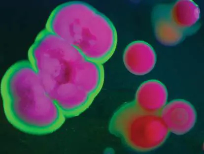
Plate culture of a marine actinomycete. Actinomycetes synthesize many secondary products that have antimicrobial and anticancer activity. New actinomycete species are discovered as symbionts of marine invertebrates. Their genomes can be mined for operons that specify modular enzyme complexes for synthesis of novel products that have potential clinical applications.
Some bacteria and archaea can build themselves entirely from carbon dioxide and nitrogen gas, plus a few salts. The cell assimilates carbon and nitrogen into small molecules and then assembles more complex structures. How do cells organize their components to build precisely the forms they need? How do bacteria Every life form on Earth consists of biomass—a body built of carbon and other elements. Each atom of every molecule had to get there through biosynthesis, the process of an organism building itself. To build a complex body from simple molecules, an organism must spend energy. In Chapters 13 and 14 we described how microbes gain energy from chemical reactions and store that energy in ion gradients and in small molecules such as ATP and NADPH. These energy-rich molecules can now drive biosynthesis.
Chapter 15 presents the fundamental ways by which microbes build themselves. First, autotrophs must assimilate elements such as carbon and nitrogen to build useful carbon skeletons. We say that the organisms fix these elements; that is, combine the inorganic compounds into biomass, the organic molecules that form the cell. Microbial enzyme factories form simple organic building blocks such as acetyl groups and amino acids, and then they assemble these simple building blocks into remarkably complex biomolecules, including vitamins and antibiotics important for human health. Commercial and biomedical uses of microbial biosynthesis are described in Chapter 16.
15.1 Overview of BiosynthesisUnit 4 · Metabolism
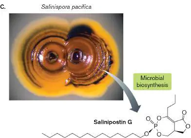
Biosynthesis is the building of complex biomolecules, also known as anabolism, the reverse of catabolism. Biosynthesis builds an extraordinary range of cell parts, from simple sugars and amino acids to exotic secondary products having bizarre forms that we cannot make in the laboratory. Secondary products are often antimicrobial agents that enable the producer to outcompete other microbes for scarce nutrients. An example of a secondary product is salinipostin G, a bicyclic phosphotriester (molecule with three ester-oxygen links) that inhibits Plasmodium, the malaria parasite. This antimalarial agent was discovered as a product of the marine actinomycete Salinispora pacifica (Fig. 15.1). At the Scripps Institution of Oceanography, UC San Diego, Paul Jensen’s lab explores marine environments for novel products of bacteria. In Jensen’s lab, Kaitlin Creamer mines actinomycete genomes for the operons that synthesize antimicrobial products, and she shows how related products mediate microbial communication. Marine actinomycetes offer an enormous resource for biosynthesis of previously unknown products that could treat human diseases.
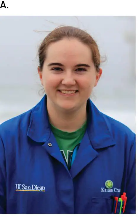
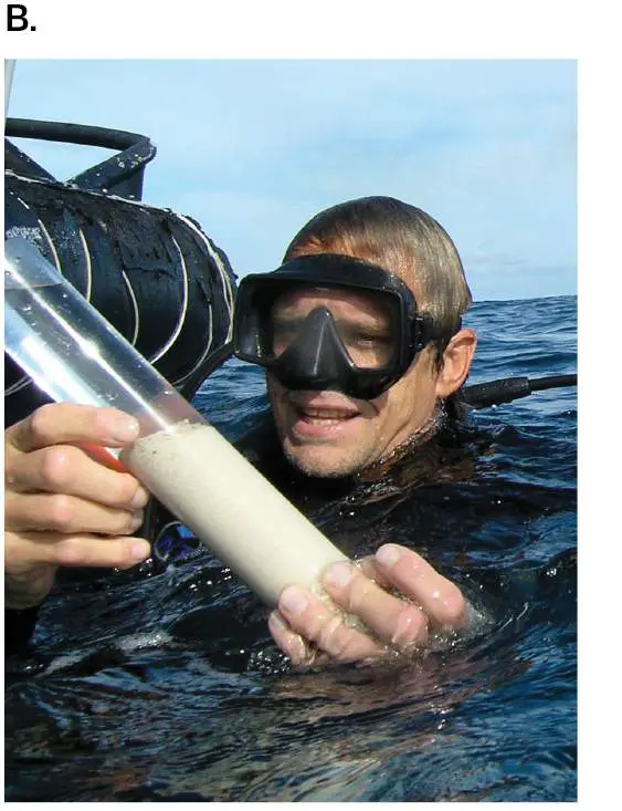
FIGURE 15.1 ■ Discovering new drugs from marine actinomycetes. A. Kaitlin Creamer mines actinomycete genomes for new antimicrobial and anticancer agents. B. Paul Jensen’s laboratory investigates marine actinomycetes. C. Salinispora pacifica produces the antimalarial agent salinipostin G (inset) and the anticancer agent marizomib (salinosporamide A). Source: Salinipostin G structure modified from Gregory Amos et al. 2017. PNAS 114 : E11121–E11130, fig. 7.
KRYSTLE CHAVARRIA
COURTESY OF PAUL JENSEN
SCRIPPS INSTITUTE OF OCEANOGRAPHY AT UC SAN DIEGO
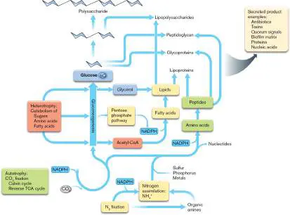
Figure 15.2presents an overview of biosynthesis and shows how it relates to catabolic pathways we saw in Chapter 13. Enzyme pathways such as the tricarboxylic acid (TCA)
cycle run in reverse to build carbon skeletons, which then incorporate nitrogen to form amino acids. Ultimately, sugar monomers and amino acids condense in chains to form polysaccharides, polypeptides, and
the cell wall material peptidoglycan. Besides the biomass of the cell proper, cells synthesize and secrete antibiotics, toxins for pathogenesis, quorum signals for cooperation, and matrix for biofilm. All this biosynthesis requires substrates and energy.
FIGURE 15.2 ■ Biosynthesis: an overview. Microbes obtain carbon skeletons by fixing CO 2 or by breaking down compounds formed by other organisms. For nitrogen, microbes fix N 2 or take up nitrates or organic amines. All biosynthesis requires energy from ATP and from reducing cofactors such as NADPH.
Biosynthesis Requires Substrates
For their biosynthesis, where do microbes get their organic substrates, such as acetyl-CoA? Some microbes synthesize all their organic parts from minerals such as carbonate and nitrate (autotrophy). Two fundamental classes of autotrophy are
photosynthesis and chemosynthesis. In photosynthesis or photoautotrophy, microbes fix (incorporate) single-carbon molecules (usually CO 2) into organic biomass, using energy from light absorption (discussed in Chapter 14). Chemosynthesis (or chemoautotrophy) fixes carbon dioxide via similar pathways, but without light. The energy for chemosynthesis usually comes from oxidation of minerals (lithotrophy; see Chapter 14). Other microorganisms must obtain multicarbon organic substrates from their environment (heterotrophy). Many soil microbes can conduct both autotrophy and heterotrophy, depending on what their environment provides.
For all organisms, biosynthesis requires: Essential elements. Biosynthesis requires carbon, oxygen, hydrogen, nitrogen, and other essential elements. Carbon is obtained either through CO 2 fixation (autotrophy) or through acquisition of organic molecules made by other organisms (heterotrophy). Autotrophy assembles carbon and water into small molecules such as acetyl-CoA, which then serve as substrates or building blocks for the cell. By contrast, heterotrophy breaks down larger molecules such as carbohydrates and peptides to release acetyl-CoA and other small substrates. In addition to carbon, biosynthesis must assimilate nitrogen and sulfur for proteins, phosphorus for DNA, and metals for metal-containing enzymes.
Reduction. Cell components such as lipids and amino acids are highly reduced. Their biosynthesis requires reducing a low-energy oxidized substrate, such as CO 2. A reducing agent such as NADPH hydrogenates the substrate and, in some cases, removes oxygen.
Energy. Assembling small molecules into complex ones requires spending energy. Biosynthetic enzymes spend energy by coupling their reactions to ATP hydrolysis, to NADPH oxidation, or to ion flux down a transmembrane ion gradient.
Heterotrophs such as Escherichia coli and actinomycetes obtain many substrates from glucose catabolism and the TCA cycle, central catabolic pathways discussed in Chapter 13 (Fig. 15.3). For example, the succinyl-CoA molecules from the TCA cycle serve as the foundation of several kinds of amino acids and of vitamin B 12. Glycerol 3-phosphate provides the glyceride backbone of lipids. Pyruvate provides the backbone of several amino acids with aliphatic side chains. From the pentose phosphate pathway, erythrose 4-phosphate contributes to the ring structures of aromatic amino acids. Other amino acids derive their carbon skeleton from TCA cycle intermediates oxaloacetate and 2-oxoglutarate, which incorporate nitrogen in the form of ammonium ions (NH +).
4
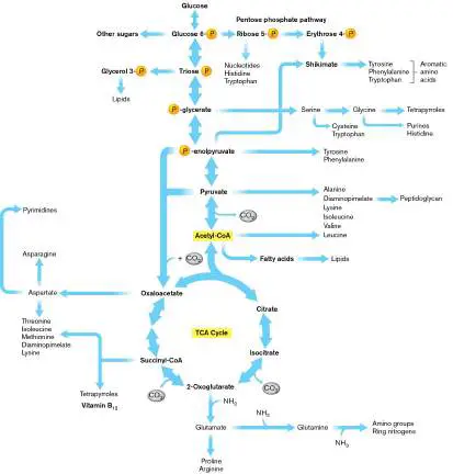
FIGURE 15.3 ■ Substrates for biosynthesis. Glucose catabolism and the TCA cycle provide substrates for biosynthesis of lipids and amino acids. Acetyl-CoA is a key substrate for biosynthesis.
Note that glucose catabolism and the TCA cycle are both reversible. Some autotrophs can synthesize entire sugar molecules, starting with CO 2 and working up through the reverse TCA cycle to build glucose. The synthesis of glucose, called gluconeogenesis, reverses most of the enzymes of glycolysis. Thus, many common metabolites are available both to autotrophs and to heterotrophs, as well as to microbes capable of mixed metabolism.
Thought Question
15.1 To run the TCA cycle and glycolysis in reverse, does a cell use the same enzymes or different ones? Explain why some enzymes might be used in both directions, whereas other steps require different enzymes for catabolic and anabolic directions.
Biosynthesis Spends Energy
Biosynthesis costs energy in several ways. The organism’s genome must maintain the DNA that encodes all the enzymes that catalyze all the steps of the pathway. The ribosomes spend energy to make enzymes. And the enzymes couple synthetic reactions to energy-releasing reactions. Collectively, these genomic and energetic costs generate enormous selective pressure for microbes to evolve mechanisms that control these costs. The diversity of these cost-cutting mechanisms generates surprising ecological relationships. Cost-cutting mechanisms include gene regulation, predation, and interspecies cooperation.
Regulation. Microbes regulate biosynthesis at multiple levels, such as gene transcription, protein synthesis, and allosteric enzyme control. Energy-expensive products block the expression or function of their biosynthetic enzymes. For example, as we saw in Chapter 10, if the cell’s tryptophan concentration increases, the molecule binds a corepressor to block transcription of the genes for tryptophan biosynthesis, and the aminoacyl-tRNA stalls the ribosome midtranslation. In this way, microbes avoid making more than they need under given conditions.
Microbes regulate their biosynthesis at the levels of transcription and translation of enzymes, as well as through feedback inhibition of enzyme activity. An example of molecular regulation is nitrogen fixation, discussed in Section 15.4.
Competition and predation. Free-living bacteria secrete antimicrobial agents that inhibit or kill competitors. The victors scavenge the spoils for raw materials. Actinomycetes, such as those discussed earlier (see the chapter-opening photo and Fig. 15.1), possess large genomes that encode enzymes for biosynthesis of several antibiotics. High nutrient levels inhibit antibiotic production, but as the bacterial filaments grow more slowly in stationary phase, they start making antibiotics to fight for resources.
Genome loss and cooperation. Gene maintenance and gene expression are energy expensive. A common way to avoid the expense of biosynthesis is to lose the enzymes through reductive evolution (discussed in Chapter 17). That is why human bodies require nine essential amino acids that we cannot make, as well as multiple vitamins, and cofactors such as vitamin B 12 (made by bacteria). But the organism then depends on trophic relations with other community members. Parasites and mutualists that grow only within a host cell show the most extensive loss of biosynthetic genes. Every species takes an evolutionary path either of maintaining the expense of a particular biosynthetic pathway, such as N 2 fixation, or of losing the pathway and becoming dependent on partner organisms.
Surprisingly, many marine phototrophs no longer encode certain biosynthetic pathways and depend on synthesis by partner heterotrophs. For example, Antarctic diatoms show a mutualism with bacteria (Fig. 15.4A). The Southern Ocean is highly productive for biomass and oxygen because of the high solar irradiance and iron availability. Many of the sea ice phytoplankton (diatoms and other phototrophic algae) fail to synthesize vitamin B 12, a fundamental cofactor for enzymes but one of the most complicated biomolecules to synthesize. So, the Antarctic diatoms obtain vitamin B 12 from adherent bacteria.
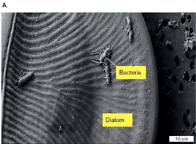
FIGURE 15.4 ■ Microbes share products of biosynthesis. A. The Antarctic diatom Amphiprora sp. hosts bacteria producing vitamin B 12 (SEM). B. E. coli strains connect via nanotubes to obtain the amino acid that one strain lacks but the other strain overproduces. Source: Part A from Eric Bertrand and Andrew Allen. 2012. Front. Microbiol. 3: 375. Part B from S. Pande et al. 2015. Nat. Commun. 6: 6238.
GREG WANGER AND THE ALLEN LAB AT JCVI
S. PANDE ET AL. 2015. NAT. COMMUN. 6 :6238, FIG. 5A
Closer to home, our own gut bacteria evolve ingenious ways to share substrates and minimize biosynthesis. Samay Pande and co-workers at the Max Planck Institute for Chemical Biology, Jena, Germany, investigated resource sharing in E.
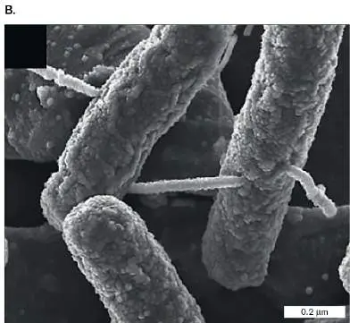
coli by constructing a complementary pair of mutants in which each overproduced an amino acid (histidine or tryptophan) but lacked a biosynthetic pathway for the other. Pande found that when the growth medium lacks the two amino acids (His and Trp), the two bacterial strains form nanotubes of phospholipids derived from the cell envelope (Fig. 15.4B ). But when the medium provides the amino acids needed, the nanotubes fail to form. Thus, even within the competitive gut microbial community, bacteria find ways to share excess carbon substrates for biosynthesis—and regulate their sharing to avoid helping a competitor when a ready source is available. Despite the expense, however, most microbes must possess certain essential pathways of biosynthesis. We will present the pathways by which autotrophs assimilate carbon and nitrogen into simple building blocks such as acetyl-CoA. We will also present the pathways used to build the cell’s key parts such as fatty acids and amino acids. Throughout the chapter, we discuss exciting discoveries in the biosynthesis of products useful to humans such as industrial chemicals and antibiotics.
To Summarize
Biosynthesis requires organic substrates. Microbes obtain carbon skeletons by CO 2 fixation or by breaking down compounds formed by other organisms.
Biosynthesis spends energy. Energy must be spent to build simple substrates into complex products.
Microbes prefer investment in energy-yielding reactions and limit their investment in energy-spending reactions that build products. Energy expense may be limited by gene regulation, by competition and predation, or by genome loss and cooperation.
Glossary
biosynthesis Also called anabolism. The building of complex biomolecules from smaller precursors.
anabolism See biosynthesis .
secondary product or secondary metabolite An organic product of biosynthesis that does not have essential functions but enhances nutrient uptake under certain conditions or inhibits competing species (e.g., an antibiotic). Often produced during stationary phase.
photosynthesis The metabolic ability to absorb and convert solar energy into chemical energy for biosynthesis. Autotrophic photosynthesis, or photoautotrophy, includes CO 2 fixation.
photoautotrophy The fixation of single-carbon compounds into organic biomass, using light as an energy source.
fix To incorporate an inorganic compound into biomass, the organic molecules that form the cell.
chemosynthesis The fixation of single-carbon molecules (usually carbon dioxide) into organic biomass, using energy from oxidation of inorganic electron donors. Also called chemoautotrophy.
chemoautotrophy See chemosynthesis .
gluconeogenesis The biosynthesis of glucose from single-carbon compounds.
15.2 CO 2 Fixation: The Calvin Cycle and Other PathwaysUnit 4 · Metabolism
The fundamental significance of carbon dioxide fixation by green plants and microbes was recognized in the early twentieth century. Fixing carbon into biomolecules requires tremendous energy input, as well as a large degree of reduction to incorporate hydrogen atoms. Bacteria have evolved a variety of cycles that fix CO 2 (Table 15.1). CO 2 fixation plays essential roles in soil, aquatic, and wetland ecosystems, as well as animal digestive systems. Globally, CO 2 fixation removes our atmosphere’s most abundant greenhouse gas—a major form of carbon sequestration. Our planet’s survival requires CO 2 fixation. TABLE 11.1 Carbon Dioxide Fixation Pathways Organisms in which pathways occur Pathway Bacteria Archaea Eukaryotes Photoautotrophs, photoheterotrophs, and lithoautotrophs Calvin cycle Cyanobacteria; Archaea have Chloroplasts purple phototrophs; Rubisco lithotrophs homologs, but their function is unclear Lithoautotrophic bacteria and archaea Reductive Green sulfur Hyperthermophilic Anaplerotic (reverse) TCA phototrophs (sulfur oxidizers (reactions fix cycle Chlorobium); Thermoproteus CO 2 to thermophilic and Pyrobaculum regenerate epsilonproteobacteria)
TABLE 11.1 Carbon Dioxide Fixation Pathways TCA intermediates Reductive acetyl-Anaerobes: Methanogens; None known CoA pathway acetogenic bacteria other anaerobes and sulfate reducers 3-Green phototrophs (Aerobic sulfur None known Hydroxypropionate Chloroflexus) oxidizers ( cycle Sulfolobus)
The Calvin Cycle The majority of the biomass on Earth consists of carbon fixed by chloroplasts and bacteria through the reductive pentose phosphate cycle, which recycles a pentose phosphate intermediate. This cycle is also known as the Calvin cycle, for which Melvin Calvin (1911–1997) was awarded the 1961 Nobel Prize in Chemistry. The full Calvin cycle is found only in bacteria and in chloroplasts, which evolved from bacteria. It has not been found in archaea or in the cytoplasm of eukaryotes. Archaea and some lithoautotrophic bacteria fix carbon by other pathways (Table 15.1). Thus, the Calvin cycle appears to have evolved after the divergence of the three domains of life.
The mechanism of the pentose phosphate cycle was solved by Calvin with colleagues Andrew Benson and James Bassham, at UC Berkeley. The importance of the Calvin cycle to the global ecosystem can scarcely be overestimated; it plays a major role in removing atmospheric CO 2. The Calvin cycle’s responsiveness to CO 2, temperature, and other factors must be considered in all models of global warming.
Note: The Calvin cycle is also known as the Calvin-Benson cycle, the Calvin-Benson-Bassham cycle, or the CBB cycle. This book uses the terms “Calvin cycle” and “CBB cycle.”
Several categories of bacteria conduct the Calvin cycle (Fig. 15.5and Table 15.1). These include photo auto trophs, photo hetero trophs, and some lithotrophs (chemoautotrophs).
Photo auto trophs. Oxygenic phototrophic bacteria (cyanobacteria) and the chloroplasts of algae and multicellular plants fix CO 2 by the Calvin cycle coupled to oxygenic photosynthesis (presented in Section 14.6). The Calvin cycle is also called the “dark reactions,” or “light-independent reactions,” of oxygenic photosynthesis. Cyanobacteria generate a large portion of the oxygen gas in Earth’s atmosphere. An example is the cyanobacterium Microcoleus vaginatus (Fig. 15.5A). M. vaginatus forms large plate-shaped cells that are stacked in filaments and packed with chlorophyll for photosynthesis. M. vaginatus forms microbial mats in lakes and also in desert crusts where it serves as an essential primary producer.
Photo hetero trophs. Some photo hetero trophs, including Alphaproteobacteria and Betaproteobacteria such as Rhodospirillum rubrum, use the Calvin cycle only when light is available (Fig. 15.5B ). In the dark, R. rubrum catabolizes organic molecules (organotrophy) and incorporates them into biomass (heterotrophy). These photoheterotrophs do not produce oxygen gas.
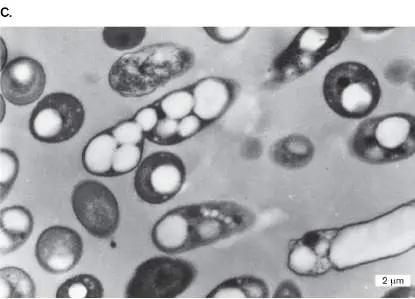
Lithotrophs (chemoautotrophs). Lithotrophic bacteria fix CO 2 using energy from oxidation of minerals (see Chapter 14). For example, Cupriavidus necator (Fig. 15.5C ) oxidizes hydrogen (hydrogenotrophy). C. necator incorporates CO 2 into polyhydroxybutyrate, a storage polymer of interest as an industrial plastic. These bacteria consume oxygen; they do not produce it.
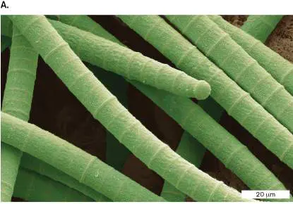
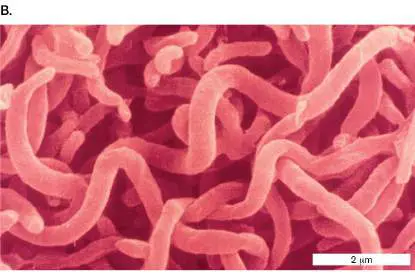
FIGURE 15.5 ■ Phototrophs and lithotrophs fix carbon via the Calvin cycle. A. Microcoleus vaginatus, a green filamentous cyanobacterium, fixes CO 2 by oxygenic photosynthesis (LM). B.
Rhodospirillum rubrum, purple photoheterotrophs with photosynthetic membranes (colorized SEM). C. Cupriavidus necator, a copper-tolerant lithotroph, uses the Calvin cycle to fix CO 2 and make polyhydroxyalkanoates (plastic) (TEM).
STEVE GSCHMEISSNER/SCIENCE SOURCE
SCIMAT/SCIENCE SOURCE
J. TIAN ET AL. 2005. J BACTERIOL. 187 :3814–3824
The Calvin Cycle: Rubisco Catalyzes CO 2 Reduction The Calvin cycle is a metabolic pathway by which each CO 2 becomes one “corner” of a glucose molecule. Alternatively, the fixed carbon can enter biosynthetic pathways for amino acids, vitamins, and other essential components of cells.
Early in the twentieth century, biochemists tried to figure out the mechanism of CO 2 fixation, believing that agricultural photosynthesis could be made more efficient. With the tools then available, however, researchers had no hope of
sorting out the intermediate products through which CO 2 was fixed. A fundamental breakthrough was the use of tracer radioisotopes, which are specific compounds labeled with radioactivity. The discovery of the carbon isotope 14 C by Martin Kamen (1913–2002) in 1940 revolutionized biochemistry, enabling the discovery of all kinds of cell metabolism. Another key technique was paper chromatography, a means of separating labeled compounds on the basis of differential migration in a solvent. This technique reveals short-lived intermediate compounds of a cycle. Calvin used paper chromatography for his Nobel-winning experiments to figure out the cycle that bears his name.
CO 2 fixation by Rubisco. The cycle begins with CO 2 fixation by ribulose 1,5-bisphosphate, a key intermediate that can derive from the pentose phosphate pathway (presented in Chapter 13). The addition of CO 2 is catalyzed by the enzyme Rubisco (ribulose 1,5-bisphosphate carboxylase-oxygenase) (Fig. 15.6 ). Rubisco is believed to be the most abundant protein on Earth. The CO 2 -fixing enzyme is unique to bacteria and chloroplasts and is not found in archaea. (Some archaeal genomes show a homolog of Rubisco, but the archaeal enzyme does not fix CO 2.)
FIGURE 15.6 ■ The mechanism of Rubisco. A. Rubisco from Alcaligenes eutrophus consists of eight large subunits (L) crowned by eight small subunits (S). Only the upper four L and S subunits are shown. On each L subunit, a catalytic site contains two phosphates (orange), which compete with the substrates for binding. (PDB code: 1BXN) B. Rubisco adds CO 2 to ribulose 1,5-bisphosphate to give an unstable six-carbon intermediate, a ketone, bound to the enzyme. The bound intermediate hydrolyzes to form
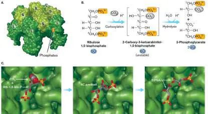
two molecules of 3-phosphoglycerate (PGA). C. The mechanism of CO 2 fixation at the catalytic site. CO 2 adds to the ketone, generating the 6C intermediate, which splits into two molecules of PGA. Rib-1,5-bis-P = ribulose 1,5-bisphosphate. (PDB code: 1RUS)
The structure of Rubisco is highly conserved across bacteria and chloroplasts. It consists of two types of subunits, designated small and large (Fig. 15.6A). The large (L) subunit contains the active (catalytic) site. The function of the small (S) subunit remains unclear; some bacteria, such as Rhodospirillum rubrum, have a Rubisco with no small subunits. Different species contain different multiples of the small and large subunits: eight (in lithotrophs, green phototrophs, and some algae), six (in plant chloroplasts and some algae), or two (in purple phototrophs). Nevertheless, the fundamental mechanism of CO 2 fixation in all these organisms appears to be similar.
Initially, Rubisco adds CO 2 to ribulose 1,5-bisphosphate to give an unstable six-carbon intermediate that remains bound to the enzyme (Fig. 15.6B and C ). The intermediate hydrolyzes into two molecules of 3-phosphoglycerate (PGA), each held at the active site by its phosphate. Only one PGA molecule contains a carbon from the newly fixed CO 2; nevertheless, as both molecules dissociate from the enzyme, they enter the cellular pool of PGA and behave equivalently in the rest of the cycle.
Rubisco has a high affinity for CO 2, and the typical concentration of Rubisco active sites (one per large subunit, six per complex) in plant chloroplasts is 4 mM —about 500 times greater than the concentration of CO 2. Thus, considerable energy is invested in CO 2 absorption. Yet the efficiency of carbon fixation by Rubisco is lowered by the existence of a competing reaction with O 2 that leads to 2-phosphoglycolate instead of 3-phosphoglycerate. This oxygenation reaction is called photorespiration. The function of photorespiration has been studied intensively but remains unclear.
Thought Question 15.2 Speculate on why Rubisco catalyzes a competing reaction with oxygen. Why might researchers be unsuccessful in attempting to engineer a Rubisco molecule that lacks the ability to catalyze this reaction?
CO 2 concentration for Rubisco. The concentration of CO 2 is a special problem because CO 2 diffuses readily through phospholipid membranes. Thus, cells cannot concentrate this substrate across the cell membrane to reach the level needed for Rubisco catalysis. Some carbon-fixing bacteria solve the gas concentration problem by enzymatic conversion of CO 2 to bicarbonate (HCO 3 − ), which is trapped in the cytoplasm, unable to leak out of the cell membrane. This enzyme system is called the carbon-concentrating mechanism (CCM). Other bacteria use alternative CO 2 -fixing systems adapted to different CO 2 concentrations.
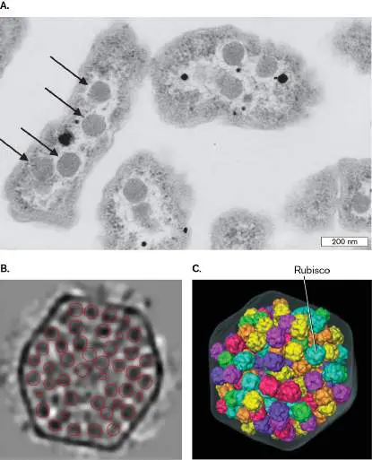
Many organisms that fix CO 2 contain the Rubisco complex within subcellular structures called carboxysomes. Figure 15.7Ashows carboxysomes within the cell of a sulfur-oxidizing lithotroph. Carboxysomes are found also within cyanobacteria and chloroplasts. A carboxysome consists of a polyhedral shell of protein subunits (Fig. 15.7B ) surrounding tightly packed molecules of Rubisco ( Fig. 15.7C ). The carboxysome takes up bicarbonate (converted from CO 2). Once inside the carboxysome, the bicarbonate is immediately converted to CO 2 by the enzyme carbonic anhydrase. The CO 2 is then fixed by Rubisco to PGA—the first step of CO 2 fixation. The PGA exits the carboxysome to complete the Calvin cycle in the cytoplasm. Mutant strains of bacteria lacking carboxysomes can fix CO 2 only at high concentration (5%), much higher than the atmospheric CO 2 concentration (0.04%).
FIGURE 15.7 ■ Carboxysomes. A. Thin section of Halothiobacillus neapolitanus, a sulfur-oxidizing lithoautotroph (TEM), showing polyhedral carboxysomes (arrows). B. Carboxysome isolated from Synechococcus cyanobacterium is packed with Rubisco complexes (cryo-electron tomography). C. A 3D model of Rubisco complexes packed inside a carboxysome.
Source: Part A modified from Y. Tsai et al. 2007. PLoS Biol. 5 :E144.
COURTESY OF SABINE HEINHORST AND DR. GORDON C. CANNON
C. V. IAN ET AL. 2007. J MOL BIOL. 372 :764–773
C. V. IAN ET AL. 2007. J MOL BIOL. 372 :764–773
Instead of carboxysomes, some phototrophs possess alternative systems of CO 2 uptake and concentration adapted to different levels of CO 2. For example, the purple photoheterotroph Rhodobacter sphaeroides has two unlinked operons, each encoding a different set of CO 2 fixation genes, called form I and form II. Form I and form II each encode all the key enzymes, such as Rubisco. When CO 2 is limiting, form I enzymes work best; when CO 2 levels are saturating, form II enzymes work best.
Ribulose 1,5-Bisphosphate Incorporates CO 2 Beginning with Rubisco as the catalyst, the Calvin cycle condenses CO 2 and H 2 O with the intermediate ribulose 1,5-bisphosphate. Overall, three molecules each of CO 2 and of H 2 O are fixed by three molecules of ribulose 1,5-bisphosphate and split into six molecules of 3-phosphoglycerate (PGA). The six PGA are reduced by 6 NADPH (with 6 ATP) to six glyceraldehyde 3-phosphate (G3P) molecules (Fig. 15.8). Each step requires a specific enzyme (not shown). An additional 3 ATP are consumed during sugar exchange reactions to regenerate three molecules of ribulose 1,5-bisphosphate.
FIGURE 15.8 ■ The Calvin cycle: overview. The Calvin cycle condenses CO 2 and H 2 O with the intermediate ribulose 1,5-bisphosphate. Overall, three molecules each of CO 2 and of H 2 O are fixed and split into six molecules of 3-phosphoglycerate (PGA), which are reduced by 6 NADPH (with 6 ATP) to six glyceraldehyde 3-phosphate (G3P) molecules. An additional 3 ATP are consumed during sugar exchange reactions to regenerate three molecules of the recycled 5C intermediate ribulose 1,5-bisphosphate. Labels “3C,” “5C,” and “6C” indicate the number of carbon atoms per molecule. Figure 15.8sums the result of three “turns” of the cycle fixing 3CO 2 into one G3P. Each “turn” of the Calvin cycle fixes one molecule of CO 2 into biomass and regenerates one ribulose 1,5-bisphosphate. The cycle involves these three stages: 1. Carboxylation and splitting: 6C → 2[3C]. Ribulose 1,5-bisphosphate condenses with CO 2 and H 2 O, mediated by Rubisco. Rubisco generates a six-carbon intermediate, which immediately hydrolyzes (splits into two parts by incorporating H 2 O). The split produces two molecules of PGA, one of which contains the CO 2 fixed by this cycle.
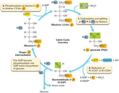
2. Reduction of PGA to G3P. The carboxyl group of each PGA molecule is phosphorylated by ATP. The phosphorylated carboxyl group is then reduced by NADPH, forming G3P.
3. Regeneration of ribulose 1,5-bisphosphate. Each incorporation of CO 2 completes the regeneration of one molecule of ribulose 1,5-bisphosphate. This regeneration requires an intricate series of sugar conversions (Fig. 15.9). The net conversion of five G3P molecules to three molecules of ribulose 1,5-bisphosphate releases 2H 2 O, restoring two of the 3H 2 O fixed with 3CO 2. The remaining sixth G3P exits the cycle, available to be used in the biosynthesis of sugars and amino acids. Thus, three fixed carbons lead to one three-carbon product.
FIGURE 15.9 ■ The Calvin cycle in detail. The Calvin cycle assimilates three CO 2 molecules, forming six 3-phosphoglycerate (PGA) molecules reduced to glyceraldehyde 3-phosphate (G3P), and converts five G3P into three molecules of ribulose 1,5-bisphosphate. Labels “3C,” “5C,” “6C,” and so on indicate the number of carbon atoms per molecule. Some sugars may enter alternative pathways to build amino acids.
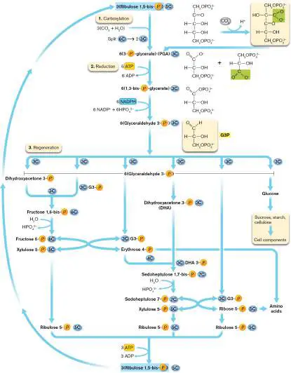
Regeneration of ribulose 1,5-bisphosphate. After PGA is reduced to G3P (step 2), all G3P molecules must participate in carbon exchanges that regenerate ribulose 1,5-bisphosphate—the key substrate that incorporates the next CO 2 ( Figure 15.9). Overall, the Calvin cycle assimilates three CO 2 molecules, forming six 3-phosphoglycerate (PGA) molecules reduced to glyceraldehyde 3-phosphate (G3P), and converts five G3P into three molecules of ribulose 1,5-bisphosphate.
To maintain the cycle, for each G3P provided to biosynthesis, five other molecules of G3P must be recycled into three five-carbon molecules of ribulose 1,5-bisphosphate. The regeneration pathway is summarized here: Two molecules of G3P condense to form the six-carbon sugar fructose 6-phosphate.
Fructose 6-phosphate condenses with a third G3P. The nine-carbon molecule splits to form a five-carbon sugar (xylulose 5-phosphate) and a four-carbon sugar (erythrose 4-phosphate). Xylulose 5-phosphate rearranges to ribulose 5-phosphate.
Erythrose 4-phosphate condenses with a fourth G3P (rearranged to dihydroxyacetone 3-phosphate) to make a seven-carbon sugar (sedoheptulose 7-phosphate).
The seven-carbon sugar condenses with the fifth G3P and splits into two molecules of ribulose 5-phosphate (via xylulose 5-phosphate and ribose 5-phosphate).
Each of the three five-carbon sugars receives a second phosphoryl group from ATP, generating ribulose 1,5-bisphosphate. Each ribulose 1,5-bisphosphate is now ready for Rubisco to fix another CO 2.
Why would a cycle have evolved requiring so many enzymatic steps to so many different intermediates? First, a cycle with many steps breaks down the energy flow into numerous reversible conversions with near-zero values of Δ G. The nearer to equilibrium, the more energy is conserved in the conversion. Second, the multiple different intermediates provide substrates for biosynthesis. For example, some molecules of erythrose 4-phosphate and ribose 5-phosphate are withdrawn from the cycle to build aromatic amino acids and nucleotides. These amino acids provide the plant proteins vital for human consumption of plant foods.
Summary of the Calvin cycle. For every three turns of the cycle, fixing three molecules of CO, the cycle feeds one molecule of G3P (C 3 H 5 O 3 –PO 3 2−)
2
into biosynthesis: 3CO 2 + 6 NADPH + 6H + + 9 ATP + 9H 2 O → C H O –PO 2− + 6 NADP + + 9 ADP + 8 P i
3 5 3 3
The H 2 O and the phosphoryl group of G3P are ultimately recycled during biosynthetic assimilation of G3P. Two molecules of G3P may condense (that is, form a new C–C bond) in a pathway to synthesize glucose. The overall condensation of 6CO 2 → 2 G3P → glucose looks like this: 6CO 2 + 12 NADPH + 12H + + 18 ATP + 18H 2 O → C 6 H 12 O 6 + 12 NADP + + 18 ADP + 18 P i + 6H 2 O Each carbon fixed requires reduction by 2H + + 2 e − from NADPH + H +. Recall from Chapter 13 that NADPH is a phosphorylated derivative of NADH commonly associated with biosynthesis. The phosphoryl group of NADPH, however, does not participate in energy transfer. Glyceraldehyde 3-phosphate is the fundamental unit of carbon assimilation into sugars or amino acids.
Thought Questions 15.3 Why does ribulose 1,5-bisphosphate have to contain two phosphoryl groups, whereas the other intermediates of the Calvin cycle contain only one? 15.4 Which catabolic pathway (see Chapter 13) includes some of the same sugar-phosphate intermediates that the Calvin cycle has? What might these intermediates in common suggest about the evolution of the two pathways? Industrial Products of Photosynthesis The Calvin cycle of phototrophs plays a starring role in human affairs as our foremost means of atmospheric carbon sequestration. Removing CO 2, and thereby curbing the greenhouse effect, requires trapping the carbon into biomass, mainly the trunks of trees. But photosynthesis also provides a way of “green production” of industrial products, using light energy to fix carbon and release oxygen. A simplified view: CO 2 + H 2 O → Product + O 2 These products would otherwise be made from petroleum by use of processes of manufacturing that spend energy by releasing CO 2. Instead, a phototroph such as a plant or alga can be engineered to produce the product, thus replacing a carbon source with a net carbon sink.
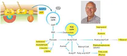
The most energy-efficient phototrophs are cyanobacteria such as Synechococcus and Synechocystis. These bacteria start out with naturally evolved pathways to produce and excrete a number of chemicals we use in industry (Fig. 15.10).
FIGURE 15.10 ■ The Calvin cycle fixes CO 2 into products useful for industry. Peter Lindblad (inset) and his laboratory engineer cyanobacteria to produce butanol and other products (highlighted yellow) from sunlight and CO 2. ACP = acyl carrier protein; PEP = phosphoenolpyruvate; PS I, photosystem I; PS II, photosystem II.
PETER LINDBLAD
Figure 15.10shows how cyanobacteria such as Synechococcus harness photosynthesis to produce organic products. First, the “light reactions” of photosynthesis (presented in Chapter 14) yield energy and provide NADPH and ATP to fix carbon into G3P. Natural enzymes in the cyanobacterial cytoplasm interconvert G3P with pyruvate, which in turn provides the carbon skeleton for amino acids (leucine and valine), acetaldehyde (a substrate to make adhesives), and isobutanol (automobile products). Alternatively, the pyruvate is decarboxylated to acetyl-CoA, which then may condense to form polyhydroxybutyrate (a plastic polymer) or other industrial substrates 1-butanol or isopropanol. Fatty acids are used in foods, and alkanes are used for fuels and solvents.
The wild-type strains of bacteria produce limited amounts of these substances, which drain organic carbon from the cell, so economical use of
“green” production requires engineering of the natural strain for industrial purposes (discussed in Chapter 16). Peter Lindblad’s laboratory at Uppsala University, Sweden, focuses on the design and construction of cyanobacteria as “photosynthetic microbial cell factories.” Genetic engineering and culture manipulation increase efficiency and the output of the desired product. For an example of this approach, see Special Topic 15.
The Reductive, or Reverse, TCA Cycle Several alternatives to the Calvin cycle fix carbon in various bacteria and archaea. An ancient pathway of biosynthesis is the TCA cycle run in reverse, also known as the reductive TCA cycle. All major groups of organisms run the TCA cycle, or some part of it (discussed in Section 13.4). Most of the individual reaction steps of the TCA cycle are reversible because each has a relatively small Δ G of reaction. Thus, the steps that release CO 2 in the forward direction may assimilate small amounts of CO 2 in the reverse direction. Furthermore, all organisms, including humans, fix small amounts of CO 2 through special reactions that regenerate TCA cycle intermediates. These regeneration steps are called anaplerotic reactions. Common anaplerotic reactions are the formation of oxaloacetate from phosphoenolpyruvate (PEP), catalyzed by PEP carboxylase; and the formation of malate from pyruvate, catalyzed by malic enzyme. In some anaerobic bacteria and archaea, the entire TCA cycle runs “backward,” reducing CO 2 to generate acetyl-CoA and build sugars (Fig. 15.11A). The reductive, or reverse, TCA cycle is used by bacteria such as Chlorobium tepidum, a green sulfur bacterium originally isolated from a New Zealand hot spring (Fig. 15.11B ). Chlorobium conducts anoxygenic photosynthesis by photolyzing H 2 S to produce elemental sulfur, which collects in extracellular granules. Sulfur granule formation is of interest for the development of a process to remove H 2 S from sulfide-generating industries such as refining of coal and oil. The reductive TCA cycle is also used by epsilonproteobacteria of hydrothermal vent communities and by sulfur-reducing archaea, such as Thermoproteus and Pyrobaculum.
FIGURE 15.11 ■ The reductive, or reverse, TCA cycle for CO 2 fixation. A. Anaerobic phototrophs and archaea use the reverse TCA cycle to fix carbon into biomass. CO 2 is fixed by several intermediates, including succinyl-CoA, 2-oxoglutarate, and acetyl-CoA. Reduction (addition of 2H + + 2 e −) is performed by NADPH or NADH and by reduced ferredoxin (FdH 2). B. Chlorobium tepidum, a green sulfur bacterium that fixes CO 2 by the
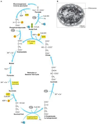
reductive TCA cycle (TEM). Chlorosome membranes contain the reaction centers of photolysis.
NIELS-ULRIK FRIGAARD ET AL. 2002. J. BACTERIOL. 184 :3368
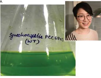
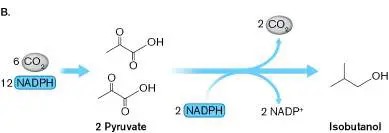
SPECIAL TOPIC 15 Sunlight Makes Industrial Chemicals So many consumer devices are made with petrochemical-derived plastics and adhesives—materials produced in ways that spew greenhouse gases (discussed in Chapter 22). What if we could make these components by a “green” process powered by sunlight? Microbial cell factories that do just that are the dream of cyanobacterial engineers. In Peter Lindblad’s laboratory, Rui Miao and colleagues are pursuing this dream with the cyanobacterium Synechocystis PCC 6803 (Fig. ST 15.1 ). The first cyanobacterium to have its genome sequenced, Synechocystis grows readily in the laboratory as small clusters of planktonic cells powered by efficient, Rubisco-driven photosynthesis. The bacterium naturally uses light energy to convert a net 4CO 2 to isobutanol, a substrate used to make adhesives and paint thinners.
FIGURE ST 15.1 ■ Cyanobacteria produce isobutanol. A. The cyanobacterium Synechocystis PCC 6803 conducts oxygenic photosynthesis. Inset: Rui Miao optimizes photosynthesis for industrial production. B. Photosynthetic CO 2 fixation forms pyruvate, which is converted to isobutanol.
V. K. PATEL ET AL. 2016. ALGAL RESEARCH. 16 :36–45
RUI MIAO
Cyanobacterial manufacturing, however, takes lots of research to realize. While cyanobacteria have evolved naturally to produce many kinds of organic molecules, to compete in nature they can’t afford to give up so
much organic material and spend so much energy. To make cyanobacterial manufacturing useful for us, we need to engineer their genetics and optimize their culture conditions.
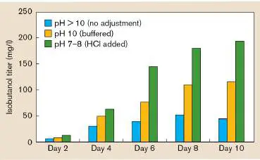
Miao worked with the known pathway for isobutanol production to generate mutant strains of Synechocystis that would produce greater amounts of isobutanol. Strains with mutations in a given gene may produce more or less of the desired product than that produced by the original parent. The importance of genetic engineering of industrial microbes is discussed in Chapter 16. In the case shown in Figure ST 15.2A , Miao added copies of various genes in the pathway, in the hope of overriding a “bottleneck” effect somewhere. Unfortunately, none of Miao’s gene-amplified strains showed an increase in isobutanol output over that observed in the parent strain (pEEK2-ST).
A B
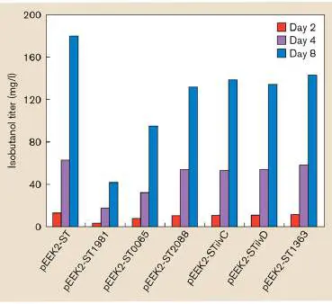
FIGURE ST 15.2 ■ Optimizing microbial production of desired product. A. Mutant strains of Synechocystis were designed and tested for increased levels of isobutanol. B. pH conditions were optimized for growth.
Eventually, Miao greatly increased the isobutanol output by manipulating the culture conditions (Fig. ST 15.2B ). A key determinant of growth and biosynthetic output is the pH. Photosynthesis commonly results in net acid consumption, raising the pH above 10. As CO 2 dissolves in water, it combines with H 2 O to form carbonic acid, H 2 CO 3. At pH 10, the carbonic acid equilibrium favors deprotonation to carbonate, CO 3 2−, a molecule that is not taken up by the bacteria. So, Miao tried to lower the pH by adding HCl. Adding HCl to the culture decreased the pH to a level that maintained carbonic acid in the protonated state. Cells were then able to access carbonic acid, increasing the production of isobutanol. Much further work is needed, but the bioengineers are on their way toward achieving their dream of a green chemical product.
RESEARCH QUESTION On the basis of your study of genetics in Chapters 7 and 8, what kind of gene(s) might we mutate in order to increase isobutanol output? Consider the cyanobacterial metabolism diagrammed in Figure 15.10.
Miao, Rui, Hao Xie, and Peter Lindblad. 2018. Enhancement of photosynthetic
isobutanol production in engineered cells of Synechocystis PCC 6803. Biotechnology for
Biofuels 11 :1–9.
Reversal of the TCA cycle uses four or five ATP to fix four molecules of CO 2 and generates one molecule of oxaloacetate. The enzymes used are the same as for the “forward” cycle, except for three key enzymes that spend ATP to drive the reaction in reverse: ATP citrate lyase, 2-oxoglutarate:ferredoxin oxidoreductase, and fumarate reductase. For example, ATP citrate lyase catalyzes the cleavage of citrate into acetyl-CoA and oxaloacetate. CO 2 assimilation requires reduction by NADPH. The reductive TCA cycle is believed to be the most ancient means of CO 2 fixation and amino acid biosynthesis, the original cycle of biomass generation in the ancestors of all three living domains.
From CO 2 to acetyl-CoA, the reverse TCA cycle essentially reverses the overall cycle of catabolism: 2CO 2 + 2 ATP + 8H + + 8 e − + HS-CoA → CH 3 CO-S-CoA + 3H 2 O + 2 ADP + 2 P i The coenzyme A (CoA) is recycled as the acetyl group enters biosynthesis. For example, acetyl-CoA can assimilate another CO 2 with reduction to pyruvate. Pyruvate then builds up to glucose and related sugars by gluconeogenesis (reverse glycolysis), with expenditure of ATP and NADPH. Alternatively, acetyl-CoA can enter biosynthetic pathways to produce fatty acids or amino acids. Unlike the Calvin cycle, the reverse TCA cycle provides several different molecular intermediates that can assimilate CO 2. Here are the key steps: Succinyl-CoA assimilates CO 2 to form 2-oxoglutarate (alpha-ketoglutarate). 2-Oxoglutarate assimilates CO 2 to form isocitrate.
Acetyl-CoA (produced by the reverse TCA cycle) assimilates CO 2 to form pyruvate.
Each CO 2 assimilation requires one or more reduction steps. 2-Oxoglutarate is reduced by the reduced form of the protein ferredoxin (FdH 2). FdH 2 also mediates acetyl-CoA reduction to pyruvate. Other reduction steps may be accomplished by NADPH or NADH.
The Reductive Acetyl-CoA Pathway The earliest route for CO 2 assimilation into acetyl-CoA and pyruvate may have been the reductive acetyl-CoA pathway (Fig. 15.12). The acetyl-CoA pathway is used by methanogens such as Methanocaldococcus jannaschii (Fig. 15.12B ). It is also used by anaerobic bacteria and thermophiles, such as the soil bacterium Clostridium thermoaceticum and the autotrophic sulfate reducer Desulfobacterium autotrophicum. A remarkable finding is that transition metals such as iron, nickel, and cobalt can catalyze reduction of CO 2 to acetate and pyruvate—in the absence of any living organism. Thus, it is argued that CO 2 reduction to acetate may have been used for biosynthesis by the earliest cells to evolve.
FIGURE 15.12 ■ The reductive acetyl-CoA pathway of CO 2 fixation. A. The first CO 2 is reduced to formate and is transferred onto the cofactor tetrahydrofolate (THF). After two further reduction steps, the methyl group is transferred to a vitamin B 12 –like cofactor (TB). The second CO 2 is reduced to carbon monoxide (CO) by the enzyme carbon monoxide dehydrogenase, and then incorporated into acetyl-CoA. B.
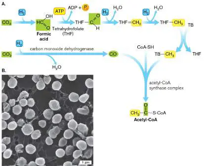
Methanocaldococcus jannaschii, a thermophilic marine methanogen that fixes CO 2 by the reductive acetyl-CoA pathway (SEM).
C. B. PARK ET AL. 2002. APPL. ENVIRON. MICROBIOL. 68 :1458–1463. DOI: 10.1128/AEM.68.3.1458-
1463.2002. REPRODUCED WITH PERMISSION FROM AMERICAN SOCIETY FOR MICROBIOLOGY
In the acetyl-CoA pathway, two CO 2 molecules are condensed through converging pathways to form the acetyl group of acetyl-CoA. The substrates and ultimate products of the acetyl-CoA pathway are the same as those of the reverse TCA cycle, except that the reducing agent is H 2 instead of NADPH. The reductive acetyl-CoA pathway is slightly more efficient than the reductive TCA cycle, requiring one less ATP: 2CO 2 + ATP + 4H 2 + HS-CoA → CH 3 CO-S-CoA + 3H 2 O + ADP + P i Nevertheless, the order of the pathway and its intermediate compounds are completely different from those of the TCA cycle. The reductive acetyl-CoA pathway is linear, with no recycled intermediates.
Reduction of the first CO 2. The first CO 2 enters the linear pathway by reduction to formate (Fig. 15.12A). The formate is transferred onto a complex carrier cofactor. In bacteria, the cofactor is tetrahydrofolate (THF), a reduced form of folate. Folate (folic acid) is a heteroaromatic cofactor; it is an essential vitamin required by many organisms, including humans. A different cofactor, methanopterin (MPT), carries the formate in methanogenic archaea. In either case, the formate carbon is reduced in successive steps to a methyl group (–CH 3 ).
Reductive CO 2 incorporation into acetyl-CoA. The second CO 2 is reduced to carbon monoxide (CO) by the enzyme carbon monoxide dehydrogenase. This same enzyme is used in reverse reaction by some lithotrophs to gain energy from carbon monoxide (CO) as an electron donor. For fixation, the CO is condensed with the methyl group carried by the vitamin B 12 –like cofactor TB to form acetyl-CoA. The acetyl-CoA then enters pathways of biosynthesis, as it does when formed by the reverse TCA cycle.
Still other pathways of CO 2 uptake have evolved in other autotrophs. For example, Chloroflexus bacteria, known as “green nonsulfur” phototrophs, fix CO 2 using a cycle that involves 3-hydroxypropionate.
To Summarize The Calvin cycle fixes CO 2 by reductive condensation with ribulose 1,5-bisphosphate. The Calvin cycle is used by cyanobacteria and chloroplasts and by some lithotrophs and photoheterotrophs.
Carboxysomes sequester and concentrate CO 2 for fixation by the Calvin cycle.
Rubisco catalyzes the condensation of CO 2 with ribulose 1,5-bisphosphate. The six-carbon intermediate immediately splits into two molecules of 3-phosphoglycerate (PGA), which are activated by ATP and reduced by NADPH to glyceraldehyde 3-phosphate (G3P).
One of every six G3P molecules is converted to glucose or amino acids. The other five molecules of G3P undergo reactions to regenerate ribulose 1,5-bisphosphate.
The reductive, or reverse, TCA cycle fixes CO 2 in methanogens and in some anaerobic bacteria.
Anaplerotic reactions in all organisms regenerate TCA cycle intermediates, sometimes by fixing CO 2.
The acetyl-CoA pathway in anaerobic bacteria and methanogens fixes CO 2 by condensation to form acetyl-CoA.
Glossary
carbon dioxide fixation The enzymatic reduction and covalent incorporation of inorganic carbon dioxide (CO 2) into an organic compound.
carbon sequestration In environmental science, the fixation of carbon into biomass, resulting in removal of a greenhouse gas from the atmosphere.
reductive pentose phosphate cycle See Calvin cycle .
Calvin cycle or Calvin-Benson cycle or Calvin-Benson-Bassham (CBB) cycle Also called pentose phosphate cycle or reductive pentose phosphate cycle. The metabolic pathway of carbon fixation in which the CO 2 -condensing step is catalyzed by Rubisco. Found in chloroplasts and in many bacteria. Calvin cycle or Calvin-Benson cycle or Calvin-Benson-Bassham (CBB) cycle Also called pentose phosphate cycle or reductive pentose phosphate cycle. The metabolic pathway of carbon fixation in which the CO 2 -condensing step is catalyzed by Rubisco. Found in chloroplasts and in many bacteria. Calvin cycle or Calvin-Benson cycle or Calvin-Benson-Bassham (CBB) cycle Also called pentose phosphate cycle or reductive pentose phosphate cycle. The metabolic pathway of carbon fixation in which the CO 2 -condensing step is catalyzed by Rubisco. Found in chloroplasts and in many bacteria. CBB cycle Also called pentose phosphate cycle or reductive pentose phosphate cycle. The metabolic pathway of carbon fixation in which the CO 2 -condensing step is catalyzed by Rubisco. Found in chloroplasts and in many bacteria. Rubisco Ribulose 1,5-bisphosphate carboxylase/oxygenase, the enzyme that catalyzes the carbon fixation step in the Calvin cycle.
carboxysome A protein-enclosed compartment containing Rubisco to fix CO 2.
anaplerotic reaction A type of metabolic reaction, occurring in all organisms, that fixes small amounts of CO 2 to regenerate TCA cycle intermediates.
reductive acetyl-CoA pathway A carbon assimilation pathway in which two CO 2 molecules are condensed and reduced by two H 2 molecules to form an acetyl group.
reductive (reverse) TCA cycle A CO 2 fixation pathway that generates acetyl-CoA through reversal of TCA cycle reactions. It requires ATP and NADPH.
folate Folic acid, a heteroaromatic cofactor that is required by some enzymes.
15.3 Fatty Acids and Antibioticsnot assigned
Fatty acid production exemplifies the construction of cell components from repeating units (Fig. 15.13). The construction of molecules based on repeating units requires a modular pathway that feeds its products back repeatedly as substrates for further synthesis. The advantage of a repeating or modular process is that large polymers can be made with a limited number of enzymes; for example, the mycolic acids of Mycobacterium tuberculosis are extended to lengths of over 100 carbons. Such a repeating process is called modular synthesis. By contrast, other cell components, such as amino acids, have more complex, nonrepeating structures whose biosynthetic enzymes require long operons (presented in Section 15.5).
FIGURE 15.13 ■ Fatty acid biosynthesis. A. Stepwise elongation of a saturated fatty acid. Units of acetyl-CoA are carboxylated to malonyl-CoA, and then successively condense to form the long chain of a fatty acid. Each two-carbon unit added (yellow highlight) requires reduction by 2 NADPH, until the final unit that retains COOH. B. An alkene “kink” can be left unsaturated during chain extension. The double bond then forms between the third and fourth carbons, rather than the second
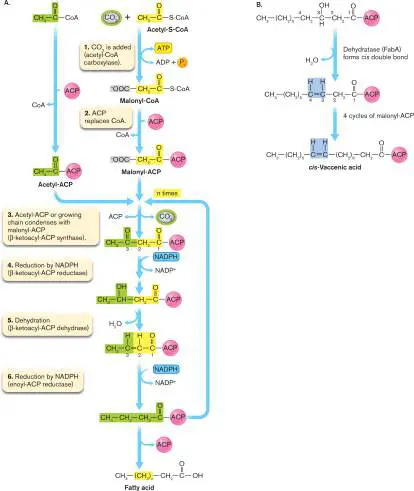
and third. In this position, the double bond escapes reduction as the chain lengthens through subsequent cycles of malonyl addition. ACP = acyl carrier protein.
Modular synthesis also builds more complex products with repeating units that have various R groups. For example, polyketides consist of a hydrocarbon backbone equivalent to that of fatty acids, with a unique R group attached to each two-carbon unit. Polyketides include a major class of antibiotics such as erythromycin. Another vital class of antibiotics made by bacteria is that of nonribosomal peptides such as vancomycin. The mechanism of modular synthesis facilitates the evolution of an extraordinary diversity of antibiotics, which microbes use in natural environments for competition and predation. Researchers mine these environmental microbial communities to discover new antibiotics.
Fatty Acids Are Built from Repeating Units
The structure of fatty acids was presented in Chapter 3. Pathways for biosynthesis of fatty acids start with the small, versatile substrate acetyl-CoA. Acetyl-CoA is a product of catabolic pathways such as glycolysis and benzoate catabolism (see Sections 13.5 and 13.6). The acetyl group also forms anabolically from the reverse TCA cycle (Fig. 15.11). Fatty acid biosynthesis provides targets for antimicrobial drugs such as triclosan, an inhibitor of enoyl-ACP reductase.
The fatty acid synthase complex. The modular process of fatty acid synthesis is managed by the fatty acid synthase complex. The complex contains all the enzymes and component binding proteins bound together in proximity, so that all steps proceed in one place without “losing” the unfinished molecule. The main bacterial version of this complex is designated FASII. All of the components are essential for viability and thus are potential drug targets. Analogous multienzyme complexes are used by actinomycete bacteria to synthesize other long-chain products, such as polyketide antibiotics (discussed next).
Activation of acetyl-CoA. Most acetyl groups within the cell are “tagged” with coenzyme A (CoA), a cofactor that directs acetate into catabolic pathways such as the TCA cycle. To redirect acetyl groups into fatty acid biosynthesis, acetyl-CoA molecules are first tagged at the “back end” by condensation with CO 2 (Fig. 15.13A, step 1), catalyzed by acetyl-CoA carboxylase. The addition of CO 2 in this step does not count as carbon fixation, because the CO 2 added is not permanently incorporated into the carbon skeleton. Instead, like phosphate or CoA, the CO 2 exists to be displaced when its function is no longer required. The CO 2 -tagged acetyl-CoA is called malonyl-CoA. The coenzyme A is replaced by acyl carrier protein (ACP), making malonyl-ACP (step 2).
Malonyl-ACP condenses with the growing chain. In step 3 (see Fig. 15.13A), malonyl-ACP hooks onto the head of an acetyl-ACP or a longer-growing chain. The process of hooking onto the new unit involves displacement of CO 2 from the back end of malonyl-ACP, which replaces ACP from the front end of the growing chain. The growing chain now contains a ketone (at carbon 3, from the former acetyl group). The ketone is reduced by NADPH and dehydrated, forming an alkene (C⚌C, unsaturated double bond between adjacent carbons 2 and 3). The alkene is further reduced by a second NADPH (Fig. 15.13A, steps 4–6). In all, the chain gains 4H + + 4 e − and is now fully hydrogenated (saturated). Once hydrogenated, the chain is ready to take on the next malonyl-ACP, which, as before, loses CO 2 and replaces the ACP from the front of the growing chain (return to step 3 in Fig. 15.13A). Successive addition can continue many times to build a saturated fatty acid.
Desaturation. Certain fatty acids require unsaturated “kinks” in the chain (alkenes) to serve a structural purpose, such as to increase the fluidity of the membrane. Desaturation can be generated during a cycle of elongation (Fig. 15.13A, step 5). During the first four cycles of acetyl addition, an alkene bond forms between the second and third carbons of the fatty acid. After the fifth two-carbon addition, however, a special dehydratase enzyme generates the alkene double bond between the third and fourth carbons (Fig. 15.13B ). This alkene fails to be hydrogenated further. Instead, several additional malonyl units are added, generating a long-chain fatty acid with a kink of a cis double bond. Regulation of fatty acid synthesis. The synthesis of fatty acids consumes enormous quantities of reducing energy; thus, cells must regulate it to avoid waste. From a structural standpoint, production of fatty acids incorporated into membranes must be balanced with growth of the cytoplasm. Furthermore, the many different variants of fatty acids are regulated in response to particular environmental needs. Some mechanisms of regulation include the following: Acetyl-CoA carboxylase represses its own transcription.
Transcription of an operon encoding two subunits of acetyl-CoA carboxylase (AccB, AccC) is repressed by one of its subunits, protein AccB. As AccB increases in concentration, it binds the promoter of the accBC operon, repressing further transcription. Thus, initiation of fatty acid biosynthesis (Fig. 15.13A, step 1) is always limited by the number of AccB and AccC enzyme subunits that are present.
Starvation blocks fatty acid biosynthesis. Starvation for carbon sources blocks fatty acid biosynthesis through the “stringent response.” Blockage is mediated by the polyphosphorylated nucleotide ppGpp (guanosine tetraphosphate), the global regulator of the stringent response (discussed in Section 10.4).
Temperature regulates fatty acid composition. Bacterial fatty acid composition is regulated by environmental factors such as temperature. In Escherichia coli, low temperature favors unsaturated fatty acids because they are less rigid and maintain membrane flexibility. Low temperature induces expression of the gene fabA, which encodes the dehydratase enzyme that desaturates the fatty acid bond. As the dehydratase activity is increased, more unsaturated fatty acids are made.
Thought Question
15.5 For a given species, uniform thickness of a cell membrane requires uniform chain length of its fatty acids. How do you think chain length may be regulated?
Modular (repeating addition) pathways of elongation comparable to fatty acid biosynthesis are used to build other kinds of polymers for energy storage. For example, many bacteria synthesize polyesters such as polyhydroxybutyrate. The term “polyester” indicates the multiple ester groups that are formed by repeated esterification of the carboxylic acid group of the chain with the hydroxyl group of a new alkanoate unit. Polyesters are insoluble in water, so they collect as storage granules within the bacterial cell, ready to release stored energy when needed. Polyester granules are synthesized by human pathogens such as Legionella pneumophila, the cause of legionellosis. Polyester storage helps L. pneumophila survive in water sources such as air-conditioning units.
Commercially, polyesters produced by soil bacteria such as Ralstonia eutropha are used to manufacture biodegradable surgical sutures.
Antibiotic Biosynthesis: Mining the Microbiomes
Antibiotics since the twentieth century have been isolated largely from soil microbes, especially fungi and actinomycete bacteria such as Streptomyces species. Soil samples from all over the world provide novel sources of bacteria that synthesize their distinctive agents of “microbial warfare.” Genome sequences reveal homologs of known enzymes for antibiotic biosynthesis, and these homologs turn out to mediate pathways of previously unknown antibiotics, as well as antiviral and antitumor agents. The genomic analysis is conducted by increasingly sophisticated computational pipelines, such as anti-SMASH. More recently, such analysis reveals antibiotic biosynthesis in a surprising range of bacteria, including cyanobacteria, as well as Gram-negative genera such as Burkholderia.
Genomic analysis also reveals antibiotic production in environments beyond the soil, most excitingly the oceans. Marine ecosystems yield an extraordinary diversity of bacteria and habitats, evolved under extreme conditions of temperature and pressure. Marine microbes also show complex interactions with host animals, particularly invertebrates such as corals and sponges. In these host relationships, the bacterial antibiotics may actually protect their host animals from predation or infection. An example of antibiotic discovery from a marine invertebrate mutualist is presented in eResearch Activity 15.
Polyketide Antibiotics
The modular biosynthesis of fatty acids provides a model for biosynthesis of the polyketide antibiotics. As an example, we present here the well-known polyketide erythromycin, a broad-spectrum antibiotic prescribed for bacterial pneumonia and chlamydia infections (Fig. 15.14). Erythromycin blocks bacterial translation at the step of peptide elongation (see Chapter 8). The antibiotic was discovered in 1949 by scientists at the Eli Lilly company from a soil sample obtained by Filipino scientist Abelardo Aguilar. Aguilar’s soil sample contained a previously unknown species of actinomycete, Saccharopolyspora erythraea, which produced a molecule that inhibits bacteria. The molecule, called erythromycin, showed bactericidal activity against a wide range of Gram-negative and Gram-positive bacteria.
FIGURE 15.14 ■ Synthesis of the erythromycin ring. A. An acyltransferase (AT) transfers R 1 -acetyl-CoA onto an acyl carrier protein (ACP), with release of coenzyme A. B. The R 1 - acetyl group is then transferred onto a ketosynthase (KS). The R 1 -acetyl group condenses with R 2 -malonyl-ACP, with release of CO 2. C. Modular subunits of the polyketide synthase complex elongate the polyketide to form the ring precursor of erythromycin. In this example, all R groups are methyl (–CH 3). Some modules include reducing enzymes such as ketoreductase (KR), dehydratase (DH), and enoyl reductase (ER). Elongation is terminated by thioesterase (TE).
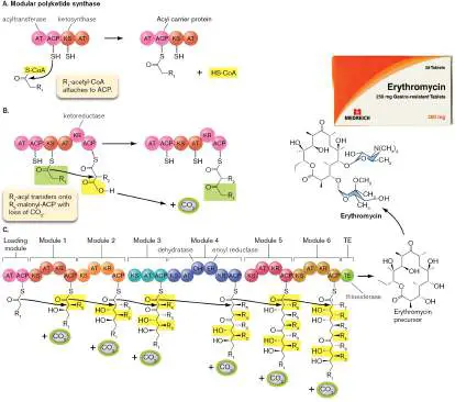
Source: Parts A–C modified from David E. Cane et al. 1998. Science 282 :63.
PACKSTOCK/ALAMY STOCK PHOTO
The synthesis of a polyketide involves repeated elongation by an enormous enzyme complex called a modular enzyme. A modular enzyme consists of multiple modules that add similar but nonidentical units to a growing chain (Fig. 15.14). Like fatty acids, polyketides are built by the successive condensation of malonyl-ACP units, with a CO 2 tag to be replaced by the growing chain (acyl). For a polyketide, each malonyl group carries a unique extension, or R group. In erythromycin (a relatively simple polyketide), the R groups are all methyl groups. In more complex polyketides, the R groups range from hydroxyl groups and amides to aromatic rings, some of which undergo secondary reactions and interconnections.
How is the order of different R groups determined? The modular enzyme contains a series of active sites, or modules, each of which catalyzes the addition of one of a series of units. Each module contains all the enzyme activities needed to add the unit and recognizes a unit with one specific R group. The modular polyketide synthase may consist of a single protein with a series of domains or it may consist of a complex of several protein subunits. Either the multidomain enzyme or the complex acts as an assembly line to generate the specific polyketide.
Figure 15.14Aand B show the chain extension synthesis of a polyketide. The initial two domains of the modular enzyme are acyltransferase (AT) and an acyl carrier protein (ACP) domain. The acyl group is analogous to the growing chain of fatty acid biosynthesis, but it is specialized to accept one component of the polyketide. The acyltransferase (AT) transfers the acyl group (R 1 - acetyl) from R 1 -acetyl-CoA onto the ACP, with release of coenzyme A (HS-CoA). The R 1 -acetyl is then transferred to a ketosynthase domain (KS).
Meanwhile, a malonyl group with its own R group (R 2 -malonyl) has been transferred by a second AT onto the second ACP domain ( Fig. 15.14B ). Recall from fatty acid biosynthesis that the malonyl group is a two-carbon acetyl group, with an added “tag” of CO 2; however, this malonyl has a distinctive R 2 group, depending on the polyketide to be made. For erythromycin, all R groups are methyl; other polyketides have R groups that are more complex. The R 1 - acetyl group then leaves KS and condenses with R 2 -malonyl-ACP, releasing the CO 2.
The initial AT and ACP domains constitute a “loading module” for the first acyl group, whereas subsequent sets of domains constitute “extender modules” that each add a different R-malonyl group. Extender modules can include secondary activities such as dehydratase (DH; removes H 2 O) and ketoreductase (KR; hydrogenates a ketone to OH). In principle, the modular approach can construct a limitless range of products with diverse antibiotic properties.
After the first two acyl groups condense (R 1 R 2), the acyl chain subsequently undergoes a series of comparable extensions by R-malonyl groups, with each cycle removing a CO 2 tag (Fig. 15.14C ). The chain condenses and undergoes reduction of ketones, analogous to fatty acid extension. Finally, elongation of the polyketide is terminated by thioesterase (TE), which hydrolyzes the thioester bond to the final ACP. This polyketide chain is an erythromycin precursor. The precursor requires additional enzymes (not shown) to add extra components, including two sugars (glycosylation). Sugar addition completes the erythromycin.
Nonribosomal Peptide Antibiotics
Bacteria synthesize a vast array of peptide products that serve the functions of development, communication, and combat. These peptides have unique modifications that depart from the standard 20 amino acid residues. Some peptides are translated by ribosomes using the standard amino acids, and then undergo posttranslational modification. These are called ribosomally synthesized and posttranslationally modified peptides (RiPPs). Many RiPPs have antimicrobial activity; some are called “lantibiotics.”
Other peptides, however, are synthesized without ribosomes. The nonribosomal peptides are synthesized entirely by modular enzymes comparable to those that make polyketides. Nonribosomal peptides include powerful antibiotics such as vancomycin and immunosuppressant drugs such as cyclosporine.
We present here the example of the nonribosomal peptide vancomycin (Fig. 15.15), the drug of last resort for combating life-threatening Clostridioides difficile and “flesh-eating” methicillin-resistant Staphylococcus aureus (MRSA) infections. Vancomycin was originally isolated in 1953 from a soil sample sent by a missionary in the jungle of Borneo to a friend at the Eli Lilly company. The Lilly biochemists isolated a new actinomycete, now called Amycolatopsis orientalis. This actinomycete produced a substance that killed Gram-positive pathogens. The active molecule was isolated and called vancomycin, for its ability to vanquish tough microbes. Vancomycin has a peptide backbone (shaded yellow in Fig.
15.15 ) equivalent to that of a ribosomal peptide, but its aminoacyl residues are nonstandard and show atypical secondary connections. Like polyketides, polypeptide antibiotics are synthesized by an enormous modular enzyme complex.
FIGURE 15.15 ■ Vancomycin, a nonribosomal peptide antibiotic. Vancomycin is produced by Amycolatopsis orientalis
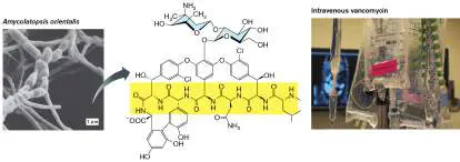
(left). The peptide backbone of the molecule is shaded yellow. Source: Modified from Christopher Walsh. 2003. Antibiotics: Actions, Origins, Resistance. ASM Press.
THE SOCIETY FOR ACTINOMYCETES (BY Y. GYOBU), HTTP://ATLAS.ACTINO.JP
DR. BARRY SLAVEN/VISUALS UNLIMITED, INC.
The complex is called a nonribosomal peptide synthetase (NRPS). The vancomycin peptide backbone is built by an NRPS that contains seven repeating modules. Each module includes the following key domains (Fig. 15.16): FIGURE 15.16 ■ Elongation of a nonribosomal peptide. A. Enzyme domain A adenylylates (activates) an amino acid and
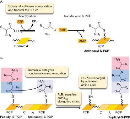
then transfers it onto S-PCP (peptidyl carrier protein). B. Domain C catalyzes transfer of the R 2 aminoacyl group to the R 3 amino group, forming a peptide bond.
Source: Modified from Christopher Walsh. 2003. Antibiotics: Actions, Origins, Resistance. ASM Press.
Domain A: adenylylation and transfer. The A domain catalyzes adenylylation (addition of adenosine monophosphate; AMP) to the carboxylate of the amino acid (Fig. 15.16A). The adenylylation reaction is driven by ATP releasing pyrophosphate (PP i). Next, release of AMP drives transfer of the aminoacyl group onto a sulfur atom of the peptide carrier protein (S-PCP). Each S-PCP recognizes only one specific aminoacyl group.
Domain C: condensation and elongation. The C domain catalyzes transfer of the peptidyl-S-PCP from one module onto the amine of the next aminoacyl-S-PCP (Fig. 15.16B ). In each subsequent round, the nascent peptide is transferred onto the amino group of the next aminoacyl PCP. As each peptide travels down all seven modules, it accretes seven aminoacyl groups in all.
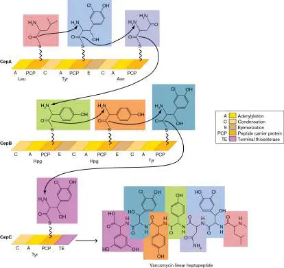
Domain E: epimerization. Some (not all) modules include an E domain to epimerize the aminoacyl group (change its configuration from L to D).
The vancomycin NRPS complex includes three multidomain proteins: CepA, CepB, and CepC (Fig. 15.17). Each protein provides one to three modules for chain extension. The seven aminoacyl groups include leucine (Leu), asparagine (Asn), three tyrosines (Tyr), and two hydroxyphenylglycines (Hpg); hydroxyphenylglycine is a nonstandard derivative of glycine. While the peptide grows, additional enzymes (not shown) catalyze chlorination, cross-linking of hydroxyls, and sugar transfer to complete the structure shown in Figure 15.15. In all, vancomycin biosynthesis requires about 30 different genes, as predicted by bioinformatic analysis of the vancomycin gene cluster.
FIGURE 15.17 ■ Elongation of the vancomycin heptapeptide. The nascent peptide from Figure 15.16 is transferred repeatedly onto the amino group of the next amino acid bound to PCP. Overall, the vancomycin synthesis complex includes seven modules for peptide elongation, which thus form a peptide of seven amino acid residues. The E domains catalyze epimerization (convert an L -aminoacyl to the D isomer).
Source: Modified from Christopher Walsh. 2003. Antibiotics: Actions, Origins, Resistance. ASM Press.
Modular enzyme biosynthesis of peptide antibiotics offers exciting prospects for the discovery and design of new antibiotics,
anticancer agents, and other therapeutic molecules. Such research requires large-scale screening of microbial communities and mining of genomic data, as described in Special Topic 15.
To Summarize
Fatty acid biosynthesis involves successive condensation of malonyl-ACP groups formed from acetyl groups tagged with acyl carrier protein (ACP) and a carboxylate. Each successive malonyl group is transferred onto the growing acyl chain, with release of CO 2.
The growing acyl chain is hydrogenated. Each added unit is hydrogenated by two molecules of NADPH, unless an unsaturated kink is required.
Some fatty acids are partly unsaturated. An unsaturated kink may be generated by a special desaturase, which generates the alkene double bond between the third and fourth carbons.
Fatty acid biosynthesis is regulated by the levels of acetyl-CoA carboxylase and by the stringent response to carbon starvation. Bond saturation is regulated by temperature and other environmental factors.
Polyketide antibiotics are synthesized by modular enzymes. The repeating units consist of acetyl groups with added functional groups.
Nonribosomal peptide antibiotics are also made by modular enzymes. The repeating units consist of amino acids, many of which are outside the 20 amino acids of proteins assembled by ribosomes.
Glossary
modular synthesis The construction of a polymeric molecule based on incorporation of repeating units.
polyketide A polymer that consists of alternating carbonyl and CHR groups (–CO–CHR–) in which many diverse R groups are possible; often has antimicrobial properties.
nonribosomal peptide A peptide with antimicrobial activity synthesized by modular enzymes and not by ribosomes.
acyl carrier protein (ACP)
A protein that can carry an acetyl group for anabolic pathways such as fatty acid synthesis.
modular enzyme A multifunctional enzyme in which several domains or subunits conduct sequential steps to generate a product.
nonribosomal peptide synthetase (NRPS)
A modular enzyme that synthesizes a peptide without using a ribosome.
15.4 Nitrogen Fixation and Regulationnot assigned
A cell contains DNA, amino acids, cell walls—think how much of this biomass is nitrogen. Although nitrogen gas makes up more than three-quarters of our atmosphere, our own bodies cannot assimilate or use any of it. The dinitrogen molecule (N 2) with its triple bond requires an enormous input of energy to split and reduce to two molecules of ammonia. Ammonia, protonated to ammonium ion (NH +), is incorporated into carbon skeletons.
4
Early in evolution, all cells may have fixed their own N 2, but today only certain species of bacteria and archaea retain the ability. All other organisms depend on reduced or oxidized forms of nitrogen, which ultimately derive from N 2. Thus, all living organisms, directly or indirectly, depend on N 2 -fixing prokaryotes within the biosphere (discussed in Chapters 21 and 22). Nitrogen limits microbes in many ecosystems, such as algae in freshwater lakes.
Given the limitations, nitrogen use by cells is subject to several levels of regulation. In this section we present some important molecular mechanisms of nitrogen regulation, which serves as an example of how cells regulate all biosynthesis and catabolism, depending on their relative needs for energy and materials.
Note: Bacteria and archaea play essential roles in global cycling
of nitrogen, sulfur, and phosphorus. The geochemical cycling of these elements is discussed in Chapter 22.
Nitrogen Fixation
To fix nitrogen into biomass, bacteria or archaea must reduce the nitrogen completely to ammonia (NH 3). At pH 7, ammonia is mostly protonated to ammonium ion (NH +). Unlike carbon,
4
nitrogen rarely appears in oxidized form in complex biomolecules. Inorganic forms, such as nitric oxide (NO), are used as defense mechanisms against invading pathogens (see Chapter 23) or as signaling molecules. In macromolecules, however, virtually all the nitrogen is reduced; organic compounds containing oxidized nitrogen are generally toxic.
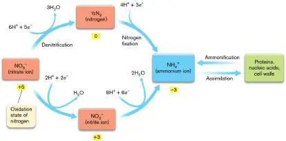
While N 2 is the ultimate source and sink of biospheric nitrogen, several oxidized or reduced forms are found in the environment, produced by living organisms (Fig. 15.18). Most free-living bacteria can acquire nitr a te (NO −) or nitr i te (NO −) for
3 2
reduction to ammonium ion. Even nitrogen-fixing legume symbionts, such as Rhizobium, can use nitrate.
FIGURE 15.18 ■ Nitrogen assimilation. Different oxidation states of nitrogen require different amounts of reducing energy for assimilation into biomass.
In natural environments, most potential sources of nitrogen are subject to competition from dissimilatory metabolism in which the
molecule is oxidized or reduced for energy, as discussed in Chapter 14. For example, anaerobic respirers convert nitrate and nitrite to N (denitrification), whereas lithotrophs oxidize NH + to nitrite and
2 4
nitrate (nitrification). An important consequence of nitrification is that most of the commercial ammonia fertilizer spread on agricultural fields is soon oxidized by lithotrophs to nitrates and nitrites. High concentrations of nitrates in water are harmful because they combine with hemoglobin, generating a form that cannot take up oxygen. When infants drink water with high nitrite, they may become ill with “blue baby syndrome.”
Nitrogen Fixation: Early Discoveries
The first N 2 fixers, discovered by Martinus Beijerinck and colleagues in the late 1800s, were soil and wetland bacteria such as Beggiatoa and Azotobacter. Species of rhizobia such as Bradyrhizobium japonicum were discovered to fix nitrogen as endosymbionts of leguminous plants (see Chapter 21). For several decades, it was believed that N 2 was fixed only by a few special bacteria in the soil or in symbiotic plant bacteria. But in the 1940s, Martin Kamen and colleagues noticed that phototrophs such as Rhodospirillum rubrum produce hydrogen gas, a known by-product of nitrogen fixation. Nitrogen fixation was hard to demonstrate in the laboratory because the energy-intensive process is repressed by the presence of alternative nitrogen sources such as ammonia. When traces of ammonia and other nitrogen sources were eliminated, Kamen’s student Herta Bregoff found that these photosynthetic bacteria fix nitrogen. Nitrogen is now known to be fixed by most phototrophic bacteria (green and purple bacteria, as well as cyanobacteria) and by many archaea. Marine cyanobacteria fix a large proportion of both the nitrogen and carbon dioxide assimilated by our biosphere. To fix N 2, aquatic cyanobacteria such as Anabaena develop special cells called heterocysts, in which photosynthesis is turned off to maintain anaerobic conditions (Fig. 15.19A). Land ecosystems require nitrogen-fixing bacteria and archaea in the soil, often in mutualistic relationships with plants. For example, the bacterium Bradyrhizobium japonicum associates with the roots of soybean plants, causing them to grow nodules. The nodule cells form symbiosomes, organelles that contain “bacteroids,” forms of the bacteria that grow within the root cells (Fig. 15.19B ). The bacteroids release extra reduced nitrogen into the soil; for this reason, farmers alternate soy crops with nitrogen-intensive crops such as corn. Nitrogen-fixing mutualism is discussed in Chapter 21. FIGURE 15.19 ■ Nitrogen fixation requires specialized structures. A. Anabaena spiroides, a filamentous cyanobacterium, segregates N 2 fixation in heterocysts, cells that inactivate photosystem II and thus maintain anoxic conditions. B. Soybean root nodule (TEM) shows bacteroids of Bradyrhizobium japonicum within symbiosomes of the plant cells.
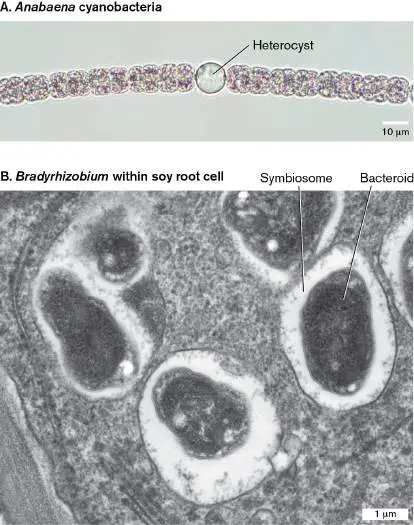
ELIF BAYRAKTAR/SHUTTERSTOCK
DARTMOUTH ELECTRON MICROSCOPE FACILITY
Bacterial nitrogen fixation, however, has not sufficed to drive high-yield agriculture. Industrial nitrogen fixation uses the Haber process, in which nitrogen gas is hydrogenated by methane (natural gas) under extreme heat and pressure to form ammonia. Scientists estimate that the Haber process reduces more atmospheric N 2 than all of Earth’s other processes combined. While this amount of nitrogen represents a small fraction of the atmosphere, the environmental effects are huge, polluting waterways and causing marine areas of hypoxia called “dead zones” (discussed in Chapters 21 and 22). The anthropogenic (human-caused) influx of nitrogen into the biosphere is an astonishing example of the influence of human society on our planet.
The Mechanism of Nitrogen Fixation
In living cells, nitrogen fixation is an enormously energy-intensive process. The mechanism is largely conserved across species: N + 8H + + 8 e − + 16 ATP → 2NH + H + 16 ADP + 16 P
2 3 2 i
The electrons are donated by NADH, H 2, or pyruvate or obtained through photosynthesis. The total energy investment includes approximately 3 ATP-equivalents per 2 e −, plus 16 ATP molecules, as shown. That makes 12 + 16 = 28 ATP in all—a large part of the energy gained from oxidation of glucose. The production of H 2 is surprising, as it consumes extra ATP. Hydrogen loss results from initiating the cycle of nitrogen reduction (discussed shortly). Some bacteria have secondary reactions to reclaim the lost hydrogen with part of its lost energy.
Note: Although nitrogen fixation is commonly represented as
producing ammonia (NH 3), under most conditions of living cells the predominant form is the protonated ammonium ion (NH +).
4
Nitrogenase reaction mechanism. The overall conversion of nitrogen gas to two molecules of ammonia is catalyzed in four cycles by nitrogenase, an enzyme highly conserved in nearly all nitrogen-fixing species. Nitrogenase probably evolved once in a common ancestor of all nitrogen-fixing organisms.
The mechanism of nitrogenase is of intense interest to agricultural scientists because of the potential benefits of improving the efficiency of plant growth and of extending nitrogen-fixing symbionts to nonleguminous plants such as corn. In 1960, scientists at the DuPont laboratory first isolated the nitrogenase complex from a bacterium, Clostridium pasteurianum. They measured its activity by incorporating heavy-isotope nitrogen gas, 15 N, into 15 NH.
2 3
The detailed structure of nitrogenase has been solved (Fig. 15.20). The complex catalyzes an extraordinary series of electron transfer reactions involving metal cofactors like those described in Chapter 14. Today, researchers continue to work out the details; for example, Hannah Rutledge and Akif Tezcan, at UC San Diego, show how amino acid residues of the FeMo protein interact with the P cluster to allow electron transfer.
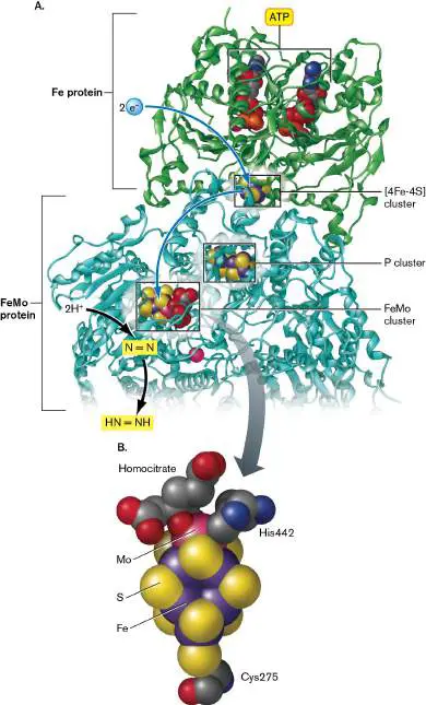
FIGURE 15.20 ■ Structure of the nitrogenase complex. A. In each active site of the Fe protein–FeMo protein complex, the Fe protein binds ATP and receives electrons from the electron donors. The electrons are subsequently channeled down through the [4Fe-4S] cluster and the P cluster (Fe-S cluster) to the FeMo cluster, where they reduce the N 2. (PDB code: 1N2C) B. The metal cluster (Fe 7 -S 9 Mo) is held in place by coordination with a molecule of homocitrate plus two amino acid residues of nitrogenase: His442 and Cys275.
The active nitrogenase complex includes two kinds of subunits encoded by different genes: Fe protein (shaded green in Fig. 15.20 ), containing a [4Fe-4S] center; and FeMo protein (shaded cyan), containing iron and molybdenum. The Fe protein contains a typical [4Fe-4S] structure to facilitate electron transfer. As we learned in Chapter 14, metal atoms help to transfer electrons because their orbitals are closely spaced, and transitions between these orbitals involve relatively small amounts of energy. The electrons are funneled through a second Fe-S cluster (the P cluster) to the FeMo (iron-molybdenum) cluster. The FeMo cluster is an unusual structural characteristic of nitrogenase, in which trios of sulfur atoms alternate with trios of iron atoms. One end of the cluster is capped with another iron, and the other end is capped with an atom of molybdenum (Mo). A consequence of the nitrogenase structure is that most nitrogen-fixing organisms (and their plant hosts, for leguminous symbionts) require the element molybdenum for growth. Some bacteria make an alternative nitrogenase that substitutes vanadium for molybdenum.
Four cycles of reduction. Nitrogen fixation requires four reduction cycles through nitrogenase (Fig. 15.21). To initiate the first cycle of N reduction, 2 e − from an electron donor such as
2
NADH or H 2 are transferred by a ferredoxin to Fe protein. The reduced Fe protein transfers each electron to the FeMo center, with energy supplied by four molecules of ATP (Fig. 15.21, step 1). The FeMo protein binds 2H +, which are reduced to H (step 2).
2
Only then does N 2 bind to the active site, by displacing the H 2 (step 3).
FIGURE 15.21 ■ Nitrogen fixation by nitrogenase. The enzyme nitrogenase successively reduces nitrogen by electron transfer, ATP hydrolysis, and H + incorporation.
In the next reduction cycle, two electrons are transferred to Fe protein, where they reduce the iron. The reduced Fe protein transfers each electron to the FeMo center, near the binding site for N 2. The electron transfer requires expenditure of 2 ATP per electron, or 4 ATP per 2 e −. Two hydrogen ions join the N and
2
receive the two electrons, forming HN⚌NH.
A third pair of electrons enters the Fe protein, which then joins another 2H +, reducing N to H N–NH. The cycle again requires
2 2 2
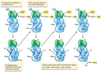
hydrolysis of 4 ATP. A final cycle of electron transfer and H + uptake reduces H 2 N–NH 2 to 2NH 3. At typical cytoplasmic pH values (near pH 7), NH is protonated to NH +.
3 4
The loss of H 2 during nitrogen fixation is puzzling because it represents lost energy in a highly energy-intensive process. The actual fate of the H 2 varies among species. Klebsiella pneumoniae, a Gram-negative bacterium, gives off the H 2 without further reaction. On the other hand, Azotobacter uses an irreversible hydrogenase enzyme to convert H back to 2H + and recover the
2
transferred electrons. In leguminous rhizobial symbionts such as Sinorhizobium species, H 2 recovery varies surprisingly among strains and can affect the efficiency of plant growth.
Nitrogen fixation is expensive, both in terms of protein synthesis and in terms of reducing energy and ATP. Even bacteria capable of nitrogen fixation repress the process in the presence of other nitrogen sources, such as nitrate (NO −), nitrite (NO −),
3 2
ammonia (NH 3), and nitrogenous organic molecules acquired from dead cells.
Anaerobiosis and N 2 Fixation
The reductive action of nitrogenase is extremely sensitive to oxygen because of the large reducing power needed to make NH +. Thus,
4
cells can fix nitrogen only in an anaerobic environment. This is no problem for anaerobes. But oxygenic phototrophs such as cyanobacteria face an obvious problem, as do bacterial symbionts of oxygenic plants. Aerobic and oxygenic organisms have developed several solutions to the problem of fixing nitrogen in aerobic environments: Protective proteins. Aerobic Azotobacter species synthesize protective proteins that stabilize nitrogenase and prevent attack by oxygen. For rhizobia, on the other hand, the plant hosts produce leghemoglobin (named for “legume” plants), a form of hemoglobin that sequesters oxygen away from the bacteria. Temporal separation of photosynthesis and N 2 fixation.
Some species of cyanobacteria fix nitrogen only at night, when they do not conduct photosynthesis and thus release no oxygen.
Specialized cells for N 2 fixation inactivate photosystem II to avoid releasing O 2. Filamentous cyanobacteria develop specialized nitrogen-fixing cells called heterocysts ( Fig. 15.19A). The heterocysts lose their photosynthetic capacity entirely and specialize in nitrogen fixation. Heterocyst development is directed by a complex genetic program induced by nitrogen starvation. In natural environments, the heterocysts leak organic acids that attract heterotrophic bacteria, which use up all the oxygen around the heterocyst, generating ideal anaerobic conditions for nitrogen fixation.
Molecular Regulation of N 2 Fixation
Nitrogen fixation costs substantial energy, and therefore the process is regulated with extraordinary fine-tuning. Multiple factors determine the expression of genes encoding nitrogenase (nifHDKTY ) and other nitrogen fixation proteins. Oxygen represses the expression of nif genes because nitrogenase is inactivated by oxygen, and high NH + depresses expression because enough fixed
4
nitrogen is already available to the cell. Responses are mediated by several molecular regulators, including a nitrogen starvation sigma factor (sigma-54) and the NtrB-NtrC two-component signal transduction system. (Sigma factors and two-component regulators are discussed in Chapter 10.)
The NtrB-NtrC system was dissected in Klebsiella pneumoniae by Sydney Kustu and colleagues at UC Berkeley. When NH + is low (
4
Fig. 15.22), the cell needs to fix nitrogen. The nitrogen sensor kinase NtrB autophosphorylates (obtains a phosphoryl group from ATP). NtrB-P then phosphorylates NtrC, forming NtrC-P, the nitrogenase activator. The NtrC-P phosphoprotein binds an upstream enhancer sequence to activate expression of nifLA, which endode NifL and NifA proteins. Expression of nifLA is coactivated by sigma-54. Sigma-54 and the NifA protein together activate expression of nifHDKTY to make nitrogenase. Thus, low NH + concentration
4
turns on nitrogen fixation.
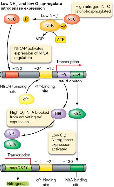
FIGURE 15.22 ■ Regulation of nitrogen fixation. When cellular levels of NH + are low, NtrC is phosphorylated and
4
activates expression of nifLA. Expression is coactivated by the nitrogen starvation sigma factor, sigma-54. The NifA protein and sigma-54 activate expression of nif genes encoding nitrogenase. When oxygen levels are high, however, nitrogen fixation cannot occur, so NifA is blocked by binding NifL. (Numbers refer to positions upstream of the transcription start site.)
When cellular levels of NH + are high, NtrC remains
4
unphosphorylated. The nifLA operon is not transcribed, no activators bind the nifHDKTY promoter, and the nitrogen fixation genes are not expressed. Nitrogenase activity is stopped by oxygen, so high oxygen levels block expression of the gene. When oxygen is high, NifA binds NifL and is prevented from binding the nitrogenase promoter. Low oxygen allows NifA to bind the promoter and express nitrogenase to fix nitrogen. Overall, NtrC governs response to NH +
4
, whereas NifA governs response to oxygen. Being responsive to multiple environmental signals is typical of enzymes in energy-expensive biosynthetic pathways.
NtrB and NtrC also regulate nitrogen usage at the level of biosynthesis of the amino acids glutamate and glutamine. Pathways of amino acid and nucleobase biosynthesis are discussed in the next section.
To Summarize
O xidized or reduced forms of nitrogen , such as nitrate, nitrite, and ammonium ions, can be assimilated by bacteria and plants. Assimilation competes with dissimilatory reactions that obtain energy.
Nitrogen gas (N) is fixed into ammonium ion (NH +)
2 4
only by some species of bacteria and archaea—never by eukaryotes.
Nitrogenase enzyme includes a protein containing an iron-sulfur core (Fe protein) and a protein containing a complex of molybdenum, iron, and sulfur (FeMo protein). Electrons acquired by Fe protein (with energy from ATP) are transferred to FeMo protein to reduce nitrogen.
Four cycles of reduction by NADPH or an equivalent reductant reduce one molecule of N 2 to two molecules of NH. At neutral pH, NH is protonated to NH +.
3 3 4
Oxygen inhibits nitrogen fixation. Bacteria have various means of separating nitrogen fixation from aerobic respiration, such as heterocyst development or temporal separation.
Nitrogen and oxygen regulate transcription of nitrogenase.
Glossary
heterocyst In filamentous cyanobacteria, a specialized nitrogen-fixing cell that maintains a reducing environment and excludes O 2.
Haber process Industrial nitrogen fixation, in which dinitrogen is hydrogenated by methane (natural gas) under extreme heat and pressure to form ammonia.
nitrogenase The enzyme that catalyzes nitrogen fixation.
15.5 Amino Acids and Nucleobasesnot assigned
Where do microbes obtain amino acids to make their proteins and cell walls, as well as nitrogenous bases to synthesize DNA and RNA? When possible, microbes obtain these molecules from their environment through membrane-embedded transporters. But competition for such valuable nutrients is high, especially for free-living microbes in soil or water. Most free-living microbes and plants have the ability to make all the standard amino acids and bases of the genetic code, as well as nonstandard variants used for cell walls and transfer RNAs. We can use microbial biosynthesis of amino acids in the industrial production of food supplements, for ourselves and for farm animals.
Amino Acid Synthesis
Like fatty acid biosynthesis, synthesis of amino acids and nitrogenous bases requires the input of large amounts of reducing energy. These compounds pose additional challenges because of their unique and diversified forms, which cannot be made by the modular processes that generate molecules from repeating units. Synthesis of complex, asymmetrical molecules such as amino acids requires many different conversions, each mediated by a different enzyme. Nevertheless, some economy is gained by an arrangement of branched pathways in which early intermediates are used to form several products (Fig. 15.23). For example, oxaloacetate is converted to aspartate, which can be converted to four other amino acids.
The carbon skeletons of amino acids arise from diverse intermediates of metabolism (Fig. 15.23). As in fatty acid biosynthesis, precursor molecules are channeled into amino acid biosynthesis by specialized cofactors and reducing energy carriers such as NADPH. Note that certain amino acids arise directly from key metabolic intermediates (for example, glutamate from 2-oxoglutarate), whereas others must be synthesized from preformed amino acids (for example, glutamine, proline, and arginine from glutamate).
FIGURE 15.23 ■ Major pathways of amino acid biosynthesis. Some amino acids arise from key metabolic
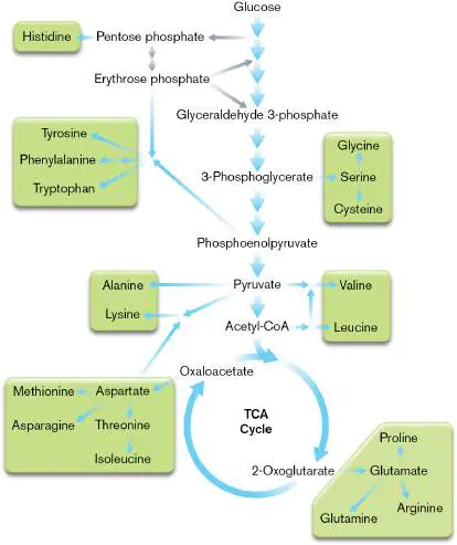
intermediates, whereas others must be synthesized out of other amino acids.
It has been hypothesized that the amino acids arising in just one or two steps from central intermediates are more ancient in cell evolution than those requiring more complex pathways. These amino acids are the same as those detected in meteorites, whose composition resembles that of prebiotic Earth. For example, a famous meteorite that fell in Sutter’s Mill, California, in 2012 was found to contain glycine and alanine (Fig. 15.24). Five of the amino acids that appear in meteorites—glutamate, aspartate, valine, alanine, and glycine—also appear in early-Earth simulation experiments in which methane, ammonia, and water are heated under reducing conditions and subjected to electrical discharge. Thus, we speculate that the first amino acids that early cells evolved to make were the same as those that arose spontaneously in the prebiotic chemistry of our planet.
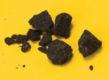
FIGURE 15.24 ■ The Sutter’s Mill meteorite. Fragments of a meteorite that fell in Sutter’s Mill, California, in 2012 contain the amino acids alanine and glycine.
NASA/ERIC JAMES
A more subtle effect of evolution has been to adjust the amino acid composition of proteins on the basis of the energetic cost of biosynthesis. Proteins that are secreted or that project outside the cell cannot be recycled; their amino acids are ultimately lost to the cell. Thus, secreted and externally projecting proteins have evolved to contain the “cheaper” amino acids; that is, the amino acids whose synthesis requires spending fewer molecules of ATP and NADPH. This effect can be seen in the diagram of a bacterial flagellum and its attached motor (Fig. 15.25), which contains extracellular as well as cytoplasmic components, colored in the figure on a scale that is based on biosynthetic “expense” of amino acids in a given protein. The external and secreted components favor less expensive amino acids compared to the cytoplasmic components.
FIGURE 15.25 ■ External proteins and proteins extending outside the cell use less expensive amino acids. Diagram of flagellar proteins (FlgE, FlgK, FlgL, FlgG, FliC) attached to the motor complex. The external and secreted
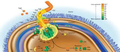
structures (FlgE, FliC, FlgM) are composed of amino acids that are less energy-expensive in terms of ATP spent per amino acid. The color scale represents the percentage of lower-expense amino acids in a given protein.
Source: Modified from Daniel Smith and Matthew Chapman. 2010. MBio 1:e00131.
Assimilation of NH 4 +
Unlike sugars and fatty acids, amino acids must assimilate another key ingredient: nitrogen. The NH + produced by N fixation or by
4 2
nitrate reduction is the key source of nitrogen for biosynthesis. But NH + always exists in equilibrium with NH, which raises pH to
4 3
toxic levels. Moreover, NH 3 travels freely through membranes, making it difficult to store NH + within a cell. Deprotonation to NH
4
3 increases as pH rises; by pH 9.2, deprotonation reaches 50%. Even at neutral pH, a very small equilibrium concentration of NH 3 can drain NH + out of the cell. Thus, cells avoid storing high levels
4
of NH +; instead, the fixed nitrogen is incorporated immediately
4
into organic products.
2-Oxoglutarate and glutamate condense with NH 1. In
4
most bacteria, the main route for NH + assimilation is the
4
condensation of NH + either with 2-oxoglutarate to form glutamate
4
or with glutamate to form glutamine (Fig. 15.26).
FIGURE 15.26 ■ Assimilation of NH + into glutamate
4
and glutamine. The key TCA cycle intermediate 2-oxoglutarate (alpha-ketoglutarate) incorporates one molecule of NH + at the
4
ketone to form glutamate. Glutamine can combine with oxoglutarate to produce two molecules of glutamate. These reactions provide sources of nitrogen to feed other pathways of amino acid synthesis.
Three key enzymes interconvert these substrates: Glutamate dehydrogenase (GDH), actually named for its reverse activity, condenses NH + with 2-oxoglutarate to form
4
glutamate. The condensation requires reduction by NADPH. Glutamine synthetase (GS), or GlnA, condenses a second NH 4 + with glutamate to form glutamine—a process driven by spending one ATP.
Glutamate synthase (GOGAT, or glutamine: 2-oxoglutarate aminotransferase) converts 2-oxoglutarate plus glutamine into two molecules of glutamate. Different variants of this enzyme use different reducing agents: NADPH, NADH, or ferredoxin. The NADPH variant is shown in Figure 15.26.
Through these reactions, all three substrates exchange amino groups readily. High nitrogen levels induce GDH to take up NH +,
4
but repress GS and GOGAT. GS has a higher affinity than GDH for NH
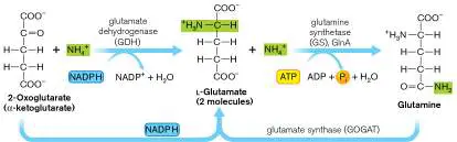
+. Low nitrogen levels induce GS (to make glutamine) and GOGAT
4
(to convert some glutamine to glutamate as needed).
Both glutamate and glutamine contribute an amine, as well as their carbon skeletons, to the synthesis of other amino acids in the biosynthetic “tree.” The transfer of ammonia between two metabolites such as glutamate and glutamine is called transamination. Many other pairs of amino acids and metabolic intermediates undergo transamination. For example, glutamate transfers NH 3 to oxaloacetate, making aspartate and 2-oxoglutarate; the reaction can also be reversed. In another example, valine transfers an amine group to pyruvate, generating alanine and 2-ketoisovalerate.
Thought Question
15.6 Suggest two reasons why transamination is advantageous to cells.
Glutamate and Glutamine Signal Nitrogen Availability
The cellular levels of glutamate and glutamine act as indicators of nitrogen availability. When nitrogen is scarce, NtrC is phosphorylated to NtrC-P as we saw earlier, in Figure 15.22. Along with nitrogenase, NtrC-P up-regulates the expression of glutamine synthetase (Fig. 15.27), as well as a high-affinity ammonia transporter, and transporters for organic sources of nitrogen such as amino acids, oligopeptides, and cell wall fragments containing amino sugars. All these molecules can be “scavenged” to obtain nitrogen for biosynthesis.
FIGURE 15.27 ■ Glutamine regulation. The NtrB-NtrC two-component system regulates glutamine synthetase (GlnA)
expression. GlnB, D, and E regulate GlnA activity.
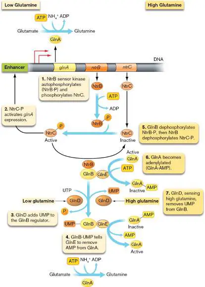
When ammonium ion (NH +) is abundant, cells incorporate the
4
nitrogen into glutamine, via glutamine synthetase (GlnA; Fig. 15.27). GlnA uses the energy released from ATP hydrolysis to assimilate NH + into glutamic acid and produce glutamine, which
4
stores the nitrogen while keeping pH neutral. But what if the cell makes too much active GlnA? Then all of the cell’s glutam ate will be converted to glutam ine, and not enough glutamate will remain to build proteins. To prevent a glutamate shortage, excess glutamine signals the cell to stop making GlnA and inactivate whatever GlnA is already present. To maintain the balance requires an intricate series of regulators responding to low glutamine or to high glutamine.
Glutamine acts at the levels of transcriptional and posttranslational control. Here we describe only a portion of this very complex regulatory system. As you study the system, imagine how such molecular controls interconnect all the enzymes and regulators throughout a bacterium.
Low glutamine. NtrB and NtrC form a two-component signal transduction system (see Chapter 10). When glutamine levels are low (Fig. 15.27, left), the sensor kinase NtrB autophosphorylates (NtrB-P) and then phosphorylates the response regulator NtrC (Fig. 15.27, step 1). NtrC-P regulator binds to an enhancer sequence, which can influence target gene expression from great distances (1 or 2 kb; step 2). As a result of NtrC-P binding to its enhancer, the bacterium makes glutamine synthetase, assimilates nitrogen (in the form of ammonium ion), and incorporates the ammonium ion as glutamine. Maintaining GlnA requires active NtrC-P and active NtrB-P. When the nitrogen level increases, NtrB-P gets dephosphorylated by GlnB (discussed shortly).
Glutamine synthetase (GlnA) control is so important that after the enzyme is synthesized, its activity is also regulated. To regulate GlnA activity, GlnB is modified by another protein, GlnD, which adds uridine monophosphate (UMP; Fig. 15.27, step 3). GlnB-UMP then activates GlnA by removing AMP from the deactivated form, GlnA-AMP (step 4).
High glutamine. When glutamine levels are in excess (Fig. 15.27, right), the cell needs to stop making GlnA and inactivate whatever still remains. To halt glnA transcription, GlnB dephosphorylates NtrB-P, and then NtrB dephosphorylates NtrC-P (step 5). NtrC cannot bind the enhancer, and thus glnA transcription stops.
Meanwhile, GlnB also compels GlnE to add an AMP to GlnA (adenylylation; Fig. 15.27, step 6). This modification inactivates whatever GlnA remains in the cell. The reaction is amplified by GlnD, which, under high glutamine, reverses its reaction, removing UMP from GlnB (step 7). The result is that no more glutamine synthase (GlnA) makes glutamine, ammonium ion no longer condenses with glutamate, and the glutamate/glutamine ratio is maintained.
Building Complex Amino Acids
Seven “fundamental” amino acids have relatively simple biosynthetic pathways. Others require longer pathways involving numerous enzymes. One amino acid produced by a complex pathway is arginine (Fig. 15.28). Bacterial arginine biosynthesis generally involves about a dozen different enzymes distributed among four to eight operons. Operon expression is regulated by an arginine repressor that binds the promoter of each operon to prevent transcription in the presence of sufficient arginine for cell needs. In some species, the arginine repressor also activates enzymes of arginine catabolism, making the excess amino acid available as a carbon source.
FIGURE 15.28 ■ Arginine biosynthesis in E. coli. In arginine biosynthesis, two glutamate molecules react with acetyl-CoA, and nitrogen is donated by glutamate and glutamine. Arginine biosynthesis. Arginine synthesis begins with the condensation reaction of glutamate with acetyl-CoA (Fig. 15.28, step 1), transferring an amino group (–NH 2) to the arginine precursor. Three more amino groups are transferred subsequently by glutamate, glutamine, and aspartate. A second glutamate transfers an amino group to the arginine precursor (step 2), while its own carbon skeleton cycles back as 2-oxoglutarate (alpha-
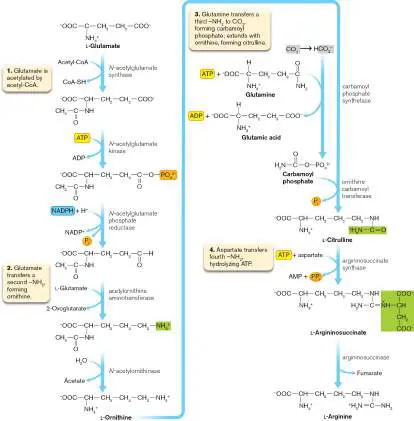
ketoglutarate). This transfer of NH + between two organic
4
intermediates is an example of transamination. The original acetyl group from acetyl-CoA (in step 1) is then hydrolyzed, producing ornithine. Ornithine is a central intermediate, used by cells to synthesize proline and various polyamines, as well as arginine.
Thought Question
15.7 Which energy carriers (and how many) are needed to make arginine from 2-oxoglutarate?
Ornithine receives a third amino group (Fig. 15.28, step 3) from glutamine via conversion of CO 2 to carbamoyl phosphate [H 2 N–CO(PO) 2−]:
4
CO + H O → H + + HCO −
2 2 3
HCO − + ATP + glutamine →
3
H N–CO(PO) 2− + ADP + glutamate
2 4
Carbamoyl phosphate provides a way to assimilate one nitrogen plus one carbon into a structure.
The fourth nitrogen is acquired from aspartate in a two-step process requiring ATP release of pyrophosphate (PP i; Fig. 15.28, step 4). The release of fumarate, a TCA cycle intermediate, yields arginine.
Aromatic amino acids. The complexity of aromatic amino acids requires particularly energy-expensive biosynthesis. The number of different enzymes required, however, is minimized by the presence of a common core pathway branching to several different amino acids (Fig. 15.29).
FIGURE 15.29 ■ Biosynthesis of aromatic amino acids. Aromatic amino acids are assembled out of various carbon skeletons. In E. coli, the pathways to chorismate and tryptophan are encoded by operons of contiguous genes, aro and trp. The three aromatic amino acids—phenylalanine, tyrosine, and tryptophan—each require a common precursor, chorismate. The pathway to chorismate starts with simple intermediates of sugar catabolism (phosphoenolpyruvate and erythrose 4-phosphate), but its synthesis requires 10 enzymes encoded in a single operon, aroABCDEFGHKL. From chorismate to phenylalanine or tyrosine requires three additional enzymatic steps. From chorismate to
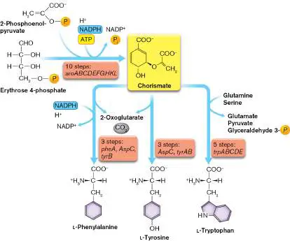
tryptophan, the most complex amino acid specified by the genetic code, requires another five enzymes, expressed by trpABCDE. Thus, at least 15 enzymes encoded by different genes are required to make tryptophan out of simple substrates.
Not surprisingly, the presence of tryptophan in the cell represses expression of its own biosynthetic enzymes. Repression of the trp operon includes two mechanisms: the Trp repressor, effective over moderate to high levels of tryptophan, and an RNA loop mechanism called attenuation, sensitive to lower levels of tryptophan (discussed in Chapter 10). Attenuation involves the destabilization of the transcription complex by formation of a specific stem loop on the nascent trp mRNA. The mechanism of attenuation in Escherichia coli was discovered in 1981 by Charles Yanofsky (1925–2018), winner of a National Medal of Science in 2003. Several other amino acids commonly show attenuation in bacteria—particularly histidine, threonine, phenylalanine, valine, and leucine.
Purine and Pyrimidine Synthesis
Nitrogenous bases include purines and pyrimidines, the essential coding components of DNA and RNA. The accuracy of the entire genetic code requires accurate synthesis of these bases. Besides their role in nucleic acids, purine and pyrimidine nucleotides such as ATP serve as energy carriers.
Bases are built onto ribose 5-phosphate. The purine and pyrimidine bases are not built as isolated units; rather, they are constructed on a ribose 5-phosphate substrate, forming a nucleotide (sugar-base-phosphate). Note that ribonucleotides are synthesized first and then converted to deoxyribonucleotides by enzymatic removal of the 2′ OH.
As we have seen, ribose 5-phosphate and related sugars participate in numerous metabolic pathways. Ribose 5-phosphate is directed into nucleotide synthesis when it is tagged with pyrophosphate at the carbon 1 (C-1) position, forming 5-phosphoribosyl-1-pyrophosphate (PRPP; Fig. 15.30). PRPP is a major metabolic intermediate, the starting point for the synthesis of purine and pyrimidine nucleotides. PRPP is also produced by “scavenger” pathways, in which excess nitrogenous bases are broken down and recycled. Its synthesis is regulated by feedback inhibition to avoid overproduction of purines. In humans, overproduction of PRPP is one mechanism that leads to gout, a condition in which the purine breakdown product uric acid precipitates in the joints, causing painful swelling of wrists and feet.
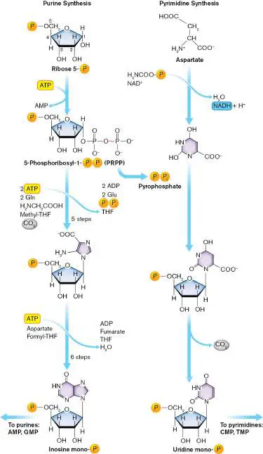
FIGURE 15.30 ■ Biosynthesis of purines and pyrimidines. Both purines and pyrimidines are built onto ribose 5-phosphate. Ribose 5-phosphate is directed into purine and pyrimidine biosynthesis by the activating step of ATP → AMP, converting the sugar to 5-phosphoribosyl-1-pyrophosphate (PRPP). The purine ring is built up out of successive additions of amines, one-carbon units, plus a formyl group from formyltetrahydrofolate (formyl-THF). The purine ring is modified to yield AMP and GMP. Likewise, the pyrimidine ring is modified to yield CMP or TMP.
Purine synthesis. The hydrolysis of PRPP releases pyrophosphate. This irreversible reaction drives forward the subsequent reactions to make the purine (Fig. 15.30, left). To construct the purine, the pyrophosphate at carbon 1 (C-1) is replaced by an amine from glutamine. The purine is built up by addition of a series of single-carbon groups (formyl, methyl, and CO 2) alternating with nitrogens from glutamine and glycine. The number of single-carbon assimilations, including assimilation of CO 2, is striking. It may reflect the ancient origin of the purine synthetic pathway, which evolved in CO 2 -fixing autotrophs.
The first purine constructed is inosine monophosphate. Inosine is the “wobble” purine found at the third position of tRNA anticodons. Inosine monophosphate is subsequently converted to adenosine monophosphate (AMP) or guanosine monophosphate (GMP).
Pyrimidine synthesis. The pyrimidine is built by a slightly different route (Fig. 15.30, right). First, the six-membered pyrimidine ring forms from aspartate plus carbamoyl phosphate. The pyrimidine ring then displaces the pyrophosphate of PRPP, attaching by a nitrogen to the ribosyl carbon 1. The first pyrimidine built is uracil (as UMP), which can be converted to cytosine or thymine—as cytidine monophosphate (CMP) or thymidine monophosphate (TMP), respectively.
Thought Questions
15.8 Why are purines synthesized onto phosphoribosyl diphosphate (PRPP)?
15.9 Why are the ribosyl nucleotides synthesized first and then converted to deoxyribonucleotides as necessary? What does this order suggest about the evolution of nucleic acids?
To Summarize
Amino acid biosynthesis requires numerous different enzymes to catalyze many unique conversions. Structurally related amino acids branch from a common early pathway. Metabolic intermediates from glycolysis and the TCA cycle initiate amino acid biosynthetic pathways.
Ammonium ion is assimilated by TCA intermediates , such as 2-oxoglutarate into glutamate. Glutamate assimilates ammonium ion to form glutamine. Transamination is the donation of NH + from one amino acid to another, such as
4
the transfer of ammonia from glutamine to 2-oxoglutarate to make aspartate.
Arginine biosynthesis requires multiple steps of NH 3 transfer and carbon skeleton condensation.
Aromatic amino acids are built from a common pathway that branches out. Their biosynthesis is regulated tightly at both transcriptional and translational levels.
Purines are built as nucleotides attached to a ribose phosphate. Several single-carbon groups are assimilated, including CO 2 —a phenomenon suggesting an ancient pathway.
Pyrimidines are made from aspartate , and then added onto PRPP.
Glossary
glutamate dehydrogenase (GDH)
An enzyme that condenses NH + with 2-oxoglutarate to form
4
glutamate. The condensation requires reduction by NADPH. glutamine synthetase (GS)
An enzyme that condenses NH + with glutamate to form
4
glutamine.
glutamate synthase Glutamine:2-oxoglutarate aminotransferase (GOGAT), an enzyme that converts 2-oxoglutarate plus glutamine into two molecules of glutamate.
GOGAT See glutamate synthase .
transamination The transfer of an ammonium ion between two metabolites. eResearch Activity 15
How Does Coculture Reveal a New Antibiotic from Sea Squirt Bacteria?
A remarkable source of novel antimicrobials can be found in the bacterial mutualists of marine invertebrates such as ascidians (sea squirts and tunicates) and sponges (Fig. ERA 15.1 ). Ascidians are sessile animals with soft bodies and simple digestive systems. As much as 10% of a marine animal may consist of bacteria such as actinomycetes. These bacteria receive nutrients from the host and may defend it from predation. Their defense involves producing secondary products that have potent activity against bacterial invaders and grazers.
FIGURE ERA 15.1 ■ New antibiotic from an invertebrate bacterial symbiont. A. Tim Bugni of University of Wisconsin– Madison. B. Keyicin, an anthracycline antibiotic produced by a cryptic operon of the ascidian-associated bacterium Micromonospora WMMB-235. C. Ascidians and sponges, Florida
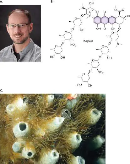
island marine invertebrates that commonly harbor antibiotic-producing bacteria.
TIM BUGNI
AMAR AND ISABELLE GUILLEN — GUILLEN PHOTO LLC/ALAMY STOCK PHOTO
A mutualistic actinomycete may have a genome that contains 10 or 20 biosynthetic gene clusters (BGCs) that encode products of medical interest. But how do we identify such gene clusters, many of which are cryptic (that is, lack expression) under known conditions? Navid Adnani, a graduate student with Tim Bugni at University of Wisconsin–Madison, found an intriguing approach to mining the genome: Culture the bacterial producer together with a target microbe. The presence of the target microbe induces expression of the cryptic gene cluster, causing production of the antibiotic. The mystery antibiotic was produced in sufficient quantity to deduce its structure by a combination of chemical tools, and then its chemistry provided clues to the gene cluster responsible. The antibiotic was originally produced by Micromonospora WMMB-235, an actinomycete symbiont of an ascidian host from coastal waters off the Florida Keys (Fig. ERA 15.1B and C ). The molecule was named keyicin, for the island chain. The producer actinomycete was screened by coculture with various potential target bacteria, model pathogens such as Mycobacterium and Staphylococcus aureus. Several Gram-positive organisms showed a zone of inhibition (clearing) on agar in the presence of WMMB-235. The largest clearing zone appeared with the bacterium Rhodococcus sp. On the basis of the results of this screening process, a setup was devised for large-scale coculture of Micromonospora and Rhodococcus (Fig. ERA 15.2 ).
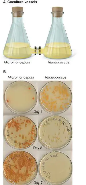
FIGURE ERA 15.2 ■ Coculture of Micromonospora sp. WMMB-235 with its target bacterium Rhodococcus sp. A.
Coculture vessels are connected by a 0.2-μm filter that blocks bacteria but permits passage of antibiotic molecules. B. Plated aliquots of Micromonospora and of Rhodococcus over time.
N. ADNANI ET AL. 2017. ACS CHEM BIOL. 12 :3093–3101
N. ADNANI ET AL. 2017. ACS CHEM BIOL. 12 :3093–3101
The coculture setup consisted of two flasks, each with an open side-arm. The two side-arms were connected by a filter with 0.2-μm pore size, small enough to exclude bacteria but large enough to allow diffusion of chemical products (Fig. ERA 15.2A ). Over several days of coculture, samples were plated on agar and colonies were counted (Fig. ERA 15.2B ). After the first day, Rhodococcus outgrew the actinomycete. But by day 3, the growth of Rhodococcus had dwindled, whereas Micromonospora increased. By day 7, the target bacterium was gone. What had happened? Chemical signals from Rhodococcus had passed the filter, reaching Micromonospora, which was then induced to produce keyicin. The keyicin then passed through the filter, reaching Rhodococcus, where it inhibited growth. The coculture thus produced enough keyicin for the researchers to probe its structure by several techniques of two-dimensional nuclear magnetic resonance (2D NMR), some of which required isotope labeling with 13 C. These tools of chemistry revealed the highly unusual structure of an anthracycline (the three fused aromatic rings) decorated with nitroglycosyl groups (chains of nitrated sugars, or nitroglycans) (Fig. ERA 15.1B ).
Keyicin production was then used to identify the biosynthetic gene cluster that produces the molecule. The BGC was identified by analysis of the proteome. Proteomes were compared for Micromonospora monocultures versus Micromonospora-Rhodococcus cocultures. Certain proteins overexpressed in the cocultures were then sequenced by mass spectrometry, and their sequences provided reverse genetic clues to gene clusters in the genome. By these means, the researchers identified a unique and effective antibiotic for Gram-positive pathogens—a promising new tool for medical therapies.
Further Exploration
How might the discovery of keyicin enable us to investigate other sources for nitroglycosylated antibiotics? How could we test these antibiotics to determine their mechanism of action?
Adnani, Navid, Marc G. Chevrette, Srikar N. Adibhatla, Fan Zhang, Qing Yu, et al. 2017. Coculture of marine invertebrate-associated bacteria and interdisciplinary technologies enable biosynthesis and discovery of a new antibiotic, keyicin. ACS Chemical Biology 12 :3093–3102.
CHAPTER REVIEW
Review Questions
1. What are the sources of substrates for biosynthesis? From what kinds of pathways do they arise?
2. How do microbial species economize by synthesizing only the products they need? Cite long-term as well as short-term mechanisms.
3. Compare and contrast the different cycles of carbon dioxide fixation. What classes of organisms conduct each type?
4. How is ribulose 1,5-bisphosphate consumed and re-formed through the Calvin cycle? What key products emerge to form sugars and amino acids?
5. How do oxygenic phototrophs maintain CO 2 at sufficient levels to conduct the Calvin cycle?
6. Explain the repeating process of chain extension in fatty acid biosynthesis. Explain the generation of occasional unsaturated “kinks” in the chain.
7. Explain the different kinds of regulation of fatty acid biosynthesis.
8. Explain how modular synthesis generates a polyketide antibiotic, and how it generates a nonribosomal polypeptide antibiotic.
9. What are the different environmental sources of nitrogen? Explain how and why microbes use these different sources.
10. Explain the process by which nitrogenase converts N 2 to 2NH +. Why is H formed?
4 2
11. Explain the different ways that microbes maintain anaerobic conditions for nitrogenase.
12. Explain the molecular basis for regulation of nitrogen fixation and nitrogen scavenging.
13. Compare and contrast the general scheme of the biosynthesis of amino acids with that of fatty acids. 14. Outline the interconversions of 2-oxoglutarate, glutamate, and glutamine that provide nitrogen for amino acid biosynthesis.
15. Compare and contrast the processes of purine and pyrimidine biosynthesis. What is the role of the sugar ribose in each case?
Thought Questions
1. Why do some soil microbes fix N 2, whereas others depend on available nitrate, ammonium ion, or organic nitrogen? What environmental conditions would favor each strategy?
2. Disease-causing bacteria vary widely in their ability to synthesize amino acids. What kinds of pathogens would be likely to make their own amino acids, and what kinds would not?
3. Mycoplasma genitalium, an organism growing in human skin, lacks the ability to synthesize fatty acids. How do you think it makes its cell membrane? How could you test this?
Key Terms
acyl carrier protein (ACP) (608) anabolism (592)
anaplerotic reaction (603)
biosynthesis (592)
Calvin cycle (Calvin-Benson cycle, Calvin-Benson-Bassham cycle, CBB cycle) (597)
carbon dioxide fixation (596)
carbon sequestration (597)
carboxysome (599)
chemosynthesis (chemoautotrophy) (593) fix (593)
folate (606)
gluconeogenesis (595)
glutamate dehydrogenase (GDH) (621) glutamate synthase (GOGAT) (621) glutamine synthetase (GS) (621) Haber process (615)
heterocyst (615)
modular enzyme (611)
modular synthesis (607)
nitrogenase (616)
nonribosomal peptide (607)
nonribosomal peptide synthetase (NRPS) (611) photoautotrophy (593)
photosynthesis (593)
polyketide (607)
reductive acetyl-CoA pathway (605) reductive pentose phosphate cycle (597) reductive (reverse) TCA cycle (603) Rubisco (598)
secondary product (592)
transamination (621)
Recommended Reading
Bartholomae, Maike, Andrius Buivydas, Jakob H. Viel, Manuel Montalbán-López, and Oscar P. Kuipers. 2017.
Major gene-regulatory mechanisms operating in ribosomally synthesized and post-translationally modified peptide (RiPP) biosynthesis. Molecular Microbiology 106 :186–206.
Gogineni, Vedanjali, and Mark T. Hamann. 2018. Marine natural product peptides with therapeutic potential: Chemistry, biosynthesis, and pharmacology. Biochimica et Biophysica Acta 1862 :81–196.
Iancu, Cristina V., H. Jane Ding, Dylan M. Morris, D. Prabha Dias, Arlene D. Gonzales, et al. 2007. The structure of isolated Synechococcus strain WH8102 carboxysomes as revealed by electron cryotomography. Journal of Molecular Biology 372 :764–773.
Kerfeld, Cheryl A., William B. Greenleaf, and James N. Kinney. 2010. The carboxysome and other bacterial microcompartments. Microbe 5 :257–263.
Koller, Martin. 2017. Production of polyhydroxyalkanoate (PHA) biopolyesters by extremophiles. MOJ Polymer Science 1 :1–19. Kudo, Yuta, Takayoshi Awakawa, Yi-Ling Du, Peter A.
Jordan, Kaitlin E. Creamer, et al. 2020. Expansion of gamma-butyrolactone signaling molecule biosynthesis to phosphotriester natural products. ACS Chemical Biology 15 :3253–3261.
Pande, Samay, Shraddha Shitut, Lisa Freund, Martin Westermann, Felix Bertels, et al. 2015. Metabolic cross-feeding via intercellular nanotubes among bacteria. Nature Communications 6 :6238.
Rutledge, Hannah L., and F. Akif Tezcan. 2020. Electron transfer in nitrogenase. Chemical Reviews 120:5158–5193. Schirmer, Andreas, Rishali Gadkari, Christopher D. Reeves, Fadia Ibrahim, Edward F. DeLong, et al. 2005.
Metagenomic analysis reveals diverse polyketide synthase gene clusters in microorganisms associated with the marine sponge Discodermia dissoluta. Applied and Environmental Microbiology 71 :4840–4849.
Singh, Rahul K., Shree P. Tiwari, Ashwani K. Rai, and Tribhuban M. Mohapatra. 2011. Cyanobacteria: An emerging source for drug discovery. Journal of Antibiotics 64 :401–412. Xu, Ying, Roland D. Kersten, Sang-Jip Nam, Liang Lu, Abdulaziz M. Al-Suwailem, et al. 2012. Bacterial biosynthesis and maturation of the didemnin anticancer agents. Journal of the American Chemical Society 134 :8625–8632.
Yang, Yu-Liang, Yuquan Xu, Paul Straight, and Pieter C. Dorrestein. 2009. Translating metabolic exchange with imaging mass spectrometry. Nature Chemical Biology 5 :885–887.
Glossary
biosynthesis Also called anabolism. The building of complex biomolecules from smaller precursors.
anabolism Also called biosynthesis. The building of complex biomolecules from smaller precursors.
secondary product or secondary metabolite An organic product of biosynthesis that does not have essential functions but enhances nutrient uptake under certain conditions or inhibits competing species (e.g., an antibiotic). Often produced during stationary phase.
photosynthesis The metabolic ability to absorb and convert solar energy into chemical energy for biosynthesis. Autotrophic photosynthesis, or photoautotrophy, includes CO 2 fixation.
photoautotrophy The fixation of single-carbon compounds into organic biomass, using light as an energy source.
fix To incorporate an inorganic compound into biomass, the organic molecules that form the cell.
chemosynthesis The fixation of single-carbon molecules (usually carbon dioxide) into organic biomass, using energy from oxidation of inorganic electron donors. Also called chemoautotrophy.
gluconeogenesis The biosynthesis of glucose from single-carbon compounds. carbon dioxide fixation The enzymatic reduction and covalent incorporation of inorganic carbon dioxide (CO 2) into an organic compound. carbon sequestration In environmental science, the fixation of carbon into biomass, resulting in removal of a greenhouse gas from the atmosphere. reductive pentose phosphate cycle Also called Calvin cycle or pentose phosphate cycle. The metabolic pathway of carbon fixation in which the CO 2 - condensing step is catalyzed by Rubisco. Found in chloroplasts and in many bacteria.
Calvin cycle or Calvin-Benson cycle or Calvin-Benson-Bassham (CBB) cycle Also called pentose phosphate cycle or reductive pentose phosphate cycle. The metabolic pathway of carbon fixation in which the CO 2 -condensing step is catalyzed by Rubisco. Found in chloroplasts and in many bacteria.
Rubisco Ribulose 1,5-bisphosphate carboxylase/oxygenase, the enzyme that catalyzes the carbon fixation step in the Calvin cycle. carboxysome A protein-enclosed compartment containing Rubisco to fix CO 2.
anaplerotic reaction A type of metabolic reaction, occurring in all organisms, that fixes small amounts of CO 2 to regenerate TCA cycle intermediates.
reductive acetyl-CoA pathway A carbon assimilation pathway in which two CO 2 molecules are condensed and reduced by two H 2 molecules to form an acetyl group.
reductive (reverse) TCA cycle A CO 2 fixation pathway that generates acetyl-CoA through reversal of TCA cycle reactions. It requires ATP and NADPH. folate Folic acid, a heteroaromatic cofactor that is required by some enzymes.
modular synthesis The construction of a polymeric molecule based on incorporation of repeating units.
polyketide A polymer that consists of alternating carbonyl and CHR groups (–CO–CHR–) in which many diverse R groups are possible; often has antimicrobial properties.
nonribosomal peptide A peptide with antimicrobial activity synthesized by modular enzymes and not by ribosomes.
acyl carrier protein (ACP)
A protein that can carry an acetyl group for anabolic pathways such as fatty acid synthesis.
modular enzyme A multifunctional enzyme in which several domains or subunits conduct sequential steps to generate a product.
nonribosomal peptide synthetase (NRPS)
A modular enzyme that synthesizes a peptide without using a ribosome.
heterocyst In filamentous cyanobacteria, a specialized nitrogen-fixing cell that maintains a reducing environment and excludes O 2. Haber process Industrial nitrogen fixation, in which dinitrogen is hydrogenated by methane (natural gas) under extreme heat and pressure to form ammonia.
nitrogenase The enzyme that catalyzes nitrogen fixation.
glutamate dehydrogenase (GDH)
An enzyme that condenses NH + with 2-oxoglutarate to form
4
glutamate. The condensation requires reduction by NADPH. glutamine synthetase (GS)
An enzyme that condenses NH + with glutamate to form
4
glutamine.
glutamate synthase Glutamine:2-oxoglutarate aminotransferase (GOGAT), an enzyme that converts 2-oxoglutarate plus glutamine into two molecules of glutamate.
transamination The transfer of an ammonium ion between two metabolites.