Chapter introduction
Alphaproteobacteria (yellow), gammaproteobacteria (pink), and other bacteria (red) colonize the marine sponge Sarcotragus spinosulus. Bacteria are identified by FISH fluorescent probe hybridized to SSU rRNA. Image
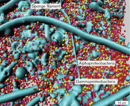
Bacteria vary tremendously in their cell structure and metabolism. They include heterotrophs, phototrophs, and lithotrophs—and some species are all three. Bacterial cell shapes include rods, cocci, spirals, and budding forms. They range in size from a centimeter long down to cells that pass through a 0.2-μm filter. Ecologically, bacteria include mutualists, pathogens, and organisms that cannot be cultured in our laboratories. You might think that, by now, we would know at least all the major phyla of bacteria, just as we know the major kinds of vertebrate animals and vascular plants. Yet we are constantly discovering new clades of bacteria never seen before, in locations ranging from the bodies of marine sponges, as seen in How do we begin to describe bacterial diversity, when even in familiar habitats the vast majority of species remain unknown? Chapter 18 surveys bacteria that we do know something about. Many kinds of bacteria grow in the ocean by photosynthesis, or colonize plants and animals, or grow in soil, competing with thousands of species for organic and inorganic nutrients. Well-known bacteria are generally those we can culture in the laboratory and subject to experiments under controlled conditions. The sequences of their genomes show how major groups of bacteria are related. But sequencing uncultured bacteria from metagenomes (see Chapter 21) reveals entire new realms of bacteria that were Chapter 18 emphasizes taxa that are of physiological, ecological, and medical importance. We organize our discussion by phylogeny (evolutionary relatedness of taxa), as well as key traits of a taxon, such as the oxygenic photosynthesis of Cyanobacteria and the multilayered cell wall of Firmicutes (Bacillota). For each major taxonomic group, we describe a few key species to represent the spectrum of diversity. We show how diverse bacteria contribute to communities. Microbial communities and ecology are explored further in Chapter 21, and their roles in global biogeochemical cycles are discussed in Chapter 22.
18.1 Bacterial Diversity at a GlanceUnit 2 · Genomes
To survey bacterial diversity in one chapter is like touring all the countries of a continent in a single day. Like countries, bacterial taxa have complex traits and histories, and often contested borders. But overall, bacteria share major traits in common. Here we review these common traits, and we then go on to explore the unique traits of selected clades and species.
Bacteria: Common Traits and Diverging Phylogeny
Chapter 17 summarized the differences and similarities of the three major domains—Bacteria, Archaea, and Eukarya (see Table 17.2). A common feature of bacteria is their central apparatus for gene expression, particularly their sigma factors for transcription, ribosomal RNAs, and translation factors. Bacterial gene expression complexes differ more from those of Archaea or Eukarya than the complexes of Archaea or Eukarya differ from each other. This subtle point of molecular biology has a profound consequence for human medicine and agriculture: It underlies the selective activity of many antibiotics, such as streptomycin, that attack only bacteria, without affecting animals or plants.
Another trait distinguishing bacteria from archaea and eukaryotes is that most bacterial cells possess a cell wall of peptidoglycan (discussed in Chapter 3). Peptidoglycan is composed of disaccharide-peptide chains that can cross-link in three dimensions; key enzymes that build the peptide links are blocked by antibiotics such as penicillin and vancomycin. Some archaea possess analogous sugar-peptide structures called “pseudopeptidoglycan” (discussed in Chapter 19), but their structural details and antibiotic sensitivity differ fundamentally from those of bacterial peptidoglycan. Eukaryotes such as fungi and plants have cell walls of polysaccharides such as cellulose and chitin (discussed in Chapter 20 ).
In bacteria, variant forms of peptidoglycan distinguish different species. For instance, the Gram-positive pathogen Staphylococcus aureus has cell wall peptides cross-linked by pentaglycine (a chain of five glycine residues). Some species, such as mycoplasmas, lack peptidoglycan altogether, although they arose by reductive evolution from bacteria that possess it.
A phylogeny of known bacteria is presented in Figure 18.1. The tree includes sequence data obtained from cultured and uncultured microorganisms conducted by Jillian Banfield’s group at UC Berkeley. The tree was computed by sequence comparison of genes encoding conserved ribosomal proteins, as discussed in Chapter 17. Some branch positions were modified on the basis of trees from other laboratories that incorporate additional highly conserved proteins. Our understanding of bacterial phylogeny is surprisingly fluid, dependent on the choice of molecular clock genes. Even among highly conserved genes, perhaps 30% sometimes undergo horizontal transfer—in some cases between domains Bacteria and Archaea. While all life shares an ancient ancestor, it is harder to define all the branches of vertical descent.
FIGURE 18.1 ■ Bacterial phylogeny. A phylogenetic tree of representative Bacteria that is based on comparison of ribosomal RNA (rRNA) and ribosomal protein sequences. The tree is rooted with respect to Archaea and Eukarya. Black labels indicate the three domains of life. Blue labels indicate phyla and deep-branching classes discussed in this chapter. Unit of branch length represents the average number of substitutions per base. For International Code of Nomenclature of Prokaryotes (ICNP)
phylum names, see Figure A3.26 in eAppendix 3. Inset: Jillian Banfield.
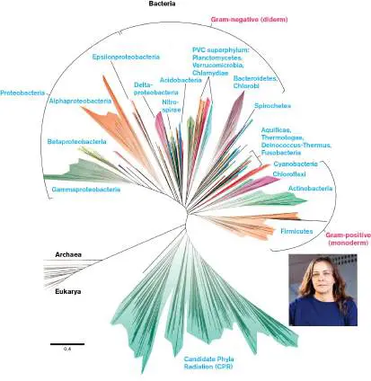
Sources: Modified from Laura Hug et al. 2016. Nat. Microbiol. 1 :1–6; Ehdieh Khaledian et al. 2020 Microorganisms 8 :312.
JILLIAN BANFIELD
In Figure 18.1, the names of selected phyla and classes are lettered in blue. A phylum (plural, phyla) is defined as a group of organisms sharing a common ancestor that diverged early from other bacteria. Increasingly, whole-genome data reveal new kinds of bacteria faster than we can figure out how to culture or characterize them. We continually discover very different new kinds of bacteria, known as emerging clades (see Chapter 17). Apart from their genome sequences, we know little about those newly discovered bacteria. Lineages whose genome sequences diverged early are called deep-branching taxa. In addition, other environmental DNA sequences of uncultivated bacteria (not shown) branch from all parts of this tree.
Phyla and other major divisions are also defined on the basis of historical convention and consensus of the research community. Before the advent of DNA sequencing, the Gram stain (see Chapter 2) was considered a fundamental basis for dividing all bacterial species into two groups, those that retained the crystal violet stain (Gram-positive) and those that lost the stain in the classic procedure (Gram-negative). In general, bacteria that test Gram-positive possess a single membrane with a thick, multilayered peptidoglycan cell wall, whereas those that test Gram-negative usually have an outer membrane but a thin layer of peptidoglycan. The latter arrangement may be referred to as “diderm,” meaning “two-skinned.” DNA sequence analysis reveals that the Proteobacteria (Pseudomonadota) are diderm, as are several other phyla such as Bacteroidetes, Cyanobacteria, and Spirochetes. Diderm bacteria generally stain Gram-negative. By contrast, the Actinobacteria and Firmicutes have thick cell walls and stain Gram-positive. Other phyla do not fit these patterns and are considered Gram-variable. Generally, a well-studied bacterial phylum comprises species that share key traits as well as genetic ancestry. While sharing key traits, the member species often show remarkable diversity in other ways. For example, members of the phylum Cyanobacteria share a unique form of metabolism (oxygenic photosynthesis) yet have evolved many diverse cell shapes. Other phyla, such as Spirochetes, share a unique cell form while diverging in habitat and host interactions. The designation “phylum,” however, is subject to change, as emerging DNA sequence data often reveal new depths of divergence. For example, within the Proteobacteria, the Greek-lettered “classes” (Alphaproteobacteria, Betaproteobacteria, and so on) are in fact as deeply divergent as many of the phyla designated in Figure 18.1.
Bacterial Phyla
We present here certain phyla and classes of bacteria that have been studied extensively. The traits of many species are known in detail. Most of the groups presented here include cultured isolates. Table 18.1summarizes key information about these groups. TABLE 18.1 Bacterial Diversity * Cyanobacteria (Cyanophyta). Oxygenic photoautotrophs with thylakoid membranes. Share ancestry with chloroplasts.
Chroococcales. Square colonies based on two division planes.
Gloeobacterales. Lack thylakoids; conduct photosynthesis in cell membrane.
Nostocales. Filamentous chains with N 2 -fixing heterocysts. Some grow symbiotically with corals or plants. Nostoc sp.
TABLE 18.1 Bacterial Diversity * Oscillatoriales. Filamentous chains with motile hormogonia (short chains). Oscillatoria sp.
Pleurocapsales. Globular colonies; reproduce through baeocytes.
Prochlorales. Tiny single cells, elliptical or spherical (0.5 μm). Prochlorococcus sp.
Firmicutes, Mycoplasma, and Actinobacteria (Gram-positive). Peptidoglycan multiple layers, cross-linked by teichoic acids.
Firmicutes (Bacillota). Low-GC, Gram-positive rods and cocci.
Bacillales. Aerobic or facultative anaerobes. Bacillus subtilis.
Clostridia, order Clostridiales. Anaerobic rods. Clostridium botulinum and C. tetani.
Lactobacillales. Non–spore formers. Facultative anaerobes. Ferment, producing lactic acid. Lactobacillus lactis.
Mycoplasma (Mycoplasmatota). Lack cell wall; require animal host. Mycoplasma pneumoniae.
Actinobacteria (Actinomycetota). High-GC, Gram-positive bacteria with moderate salt tolerance.
Actinomycetales.
Actinomycetaceae. Filamentous, forming aerial hyphae and spores. Streptomyces coelicolor.
Bifidobacteriaceae. Ferment without gas.
Bifidobacterium sp. are important residents of the human infant microbiome.
Corynebacteriaceae. Irregularly shaped rods.
Corynebacterium diphtheriae.
TABLE 18.1 Bacterial Diversity * Micrococcaceae. Small, airborne cocci. Micrococcus luteus.
Mycobacteriaceae. Exceptionally complex cell walls. Mycobacterium tuberculosis.
Propionibacteriaceae. Propionic acid fermentation. Propionibacterium shermanii.
Proteobacteria (Pseudomonadota). Gram-negative bacteria with diverse cell forms and metabolism. Alphaproteobacteria Caulobacterales. Aquatic oligotrophs; alternate stalk and flagellum. Caulobacter crescentus.
Rhizobiales. Plant mutualists and pathogens.
Sinorhizobium meliloti.
Rhodobacterales, Rhodospirillales. Flagellated photoheterotrophs.
Rickettsiales. Intracellular parasites; related to mitochondria. Rickettsia rickettsii causes Rocky Mountain spotted fever.
Sphingomonadales. Heterotrophs and photoheterotrophs. Sphingomonas sp.
Betaproteobacteria Burkholderiales. Burkholderia pseudomallei causes melioidosis.
Neisseriales. Diplococci. Neisseria gonorrhoeae and N. meningitidis.
Gammaproteobacteria Acidithiobacillales. Lithotrophs. Acidithiobacillus ferrooxidans oxidizes iron and sulfur.
Aeromonadales. Aquatic heterotrophs such as Aeromonas hydrophila.
TABLE 18.1 Bacterial Diversity * Enterobacteriales. Facultative anaerobes; colonize the human colon.
Legionellales. Legionella pneumophila causes legionellosis pneumonia.
Pseudomonadales. Rods; aerobic or respire on nitrate; catabolize aromatics. Pseudomonas aeruginosa infects lungs in cystic fibrosis patients.
Thiotrichales. Lithotrophs and heterotrophs.
Vibrionales. Marine heterotrophs. Vibrio cholerae causes cholera.
Deltaproteobacteria Bdellovibrionales. Periplasmic predators.
Desulfobacterales. Reduce sulfate.
Myxococcales. Gliding bacteria that form fruiting bodies. Epsilonproteobacteria Campylobacterales. Spirillar pathogens. Campylobacter jejuni causes gastroenteritis. Helicobacter pylori causes gastritis.
Deep-branching Gram-negative phyla. Spirochetes, Bacteroidetes, and other phyla whose cells possess an outer membrane and usually stain Gram-negative.
Acidobacteria (Acidobacteriota). Acidophiles and thermophiles.
Bacteroidetes (Bacteroidota). Anaerobes that feed on diverse carbon sources, in gut or soil (Bacteroides thetaiotaomicron); facultative aerobic soil heterotrophs ( Cytophaga).
Chlorobi (Chlorobiota). Green sulfur-oxidizing phototrophs. Chlorobium tepidum.
TABLE 18.1 Bacterial Diversity * Fusobacteria (Fusobacteriota). Gram-negative anaerobic bacteria found in septicemia and in skin ulcers. Fusobacterium nucleatum.
Nitrospirae (Nitrospirota). Oxidize nitrite; aerobic or facultative. Nitrospira marina.
Spirochetes (Spirochaetota). Narrow, coiled cells with axial filaments, encased by sheath. Polar flagella beneath sheath double back around cell.
Borrelia. B. burgdorferi causes Lyme disease, transmitted by ticks.
Hollandina. Termite gut endosymbionts.
Leptospira. L-shaped animal pathogens; cause leptospirosis.
Spirochaeta. Aquatic, free-living heterotrophs.
Treponema. T. pallidum causes syphilis.
Planctomycetes-Verrucomicrobia-Chlamydiae (PVC)
superphylum. Irregular cells lacking peptidoglycan, with subcellular structures analogous to those of eukaryotes. Chlamydiae (Chlamydiota). Intracellular cell wall–less pathogens of animals or protists. Chlamydia trachomatis causes sexually transmitted disease and trachoma (eye infection).
Planctomycetes (Planctomycetota). Some species have double membrane analogous to eukaryotic nuclear membrane. Brocadia species anaerobically oxidize ammonium and release N 2 (anammox reaction). Pirellula species inhabit marine sediment.
Verrucomicrobia (Verrucomicrobiota). Stalk-like appendages contain tubulin. Aquatic oligotrophs. TABLE 18.1 Bacterial Diversity * Prosthecobacter sp.
Deep-branching thermophiles. Thermophilic bacteria that diverged early from archaea and eukaryotes. Many genes transferred laterally from archaea Aquificae (Aquificota). Hyperthermophiles (70°C–95°C). Oxidize H 2. Aquifex.
Chloroflexi (Chloroflexota). Filamentous phototrophs, often with chlorosomes. Chloroflexus aurantiacus. Deinococcus-Thermus (Deinococcota). Radiation-resistant species and thermophiles. Deinococcus radiodurans.
Thermotogae (Thermotogota). Thermophiles (55°C– 100°C). Anaerobic heterotrophs. Thermotoga sp.
Candidate Phyla Radiation (CPR). Group of approximately 30–100 phyla known by DNA sequence, with no cultured isolates. Ultrasmall cells pass through a 0.2-μm filter. Anaerobic fermenters; have lost respiration and TCA cycle. Probably obligate symbionts.
Note: In 2021, the International Committee on Systematics of
Prokaryotes proposed to standardize phylum-level scientific names in the International Code of Nomenclature of Prokaryotes (ICNP). This decision would change the long-standing names of many prokaryotic phyla, mainly bacteria. eAppendix 3 presents a conversion table of ICNP phylum names (see Table A3.1) and a version of Figure 18.1 using ICNP names (see Figure A3.26). In Chapter 18, we present ICNP names in parentheses.
Cyanobacteria. The phylum Cyanobacteria (Cyanophyta) is a fundamental part of our biosphere, whose members conduct photosynthesis by splitting water and releasing oxygen (O 2). Marine cyanobacteria such as Prochlorococcus and Synechococcus are ubiquitous in Earth’s oceans and produce a large portion of the oxygen we breathe (discussed in Chapter 21). The cell shape and physiology of cyanobacterial species show an immense range of different forms, including chains of cells, square arrays, and globular colonies (see Section 18.2).
Firmicutes, Mycoplasma, and Actinobacteria. The classic Gram-positive phylum is the Firmicutes (Bacillota), or “hard skin” bacteria. Firmicutes have an exceptionally thick cell wall, with several layers of peptidoglycan threaded by supporting molecules such as teichoic acids or mycolic acids. The thick, reinforced cell wall is what retains the Gram stain (discussed in Chapter 2). In addition, most Firmicutes possess a well-developed S-layer of glycoproteins (discussed in Chapter 3). Many Firmicutes form endospores, inert and heat-resistant spores that can remain viable for thousands of years. Endospores are the most durable type of spore formed by bacteria.
Mycoplasma (Mycoplasmatota) is a phylum of bacteria related to Firmicutes that fail to stain Gram-positive because they lack a cell wall. Mycoplasmas lost their cell walls by reductive evolution as protected symbionts of host organisms.
Actinobacteria (Actinomycetota) have a thick peptidoglycan cell wall, but some species possess a thick waxy coat that excludes the Gram stain. Actinobacteria differ genetically from Firmicutes in their high GC content; that is, the proportion of their genomes consisting of guanine-cytosine base pairs (as opposed to adenine-thymine). Actinobacteria were thus formerly known as the “high-GC Gram-positives.” These bacteria include the actinomycetes (order Actinomycetales), which undergo complex life cycles, forming filamentous hyphae and arthrospores. Other groups closely related to actinomycetes grow as isolated rods or cocci or with variable shape, such as the corynebacteria. Actinomycete relatives include the well-known causative agents of tuberculosis (Mycobacterium tuberculosis) and leprosy (M. leprae). Actinobacteria are ubiquitous in soil and water.
Proteobacteria. Proteobacteria (Pseudomonadota) comprise several classes that are called “Gram-negative” because they possess an outer membrane of lipopolysaccharides (LPS), and their single layer of peptidoglycan fails to retain the Gram stain (see Section 18.4). Some researchers consider these “classes” to be separate phyla and Proteobacteria to be a “superphylum.” The five main classes are the Alphaproteobacteria, Betaproteobacteria, Gammaproteobacteria, Deltaproteobacteria, and Epsilonproteobacteria. In contrast to Cyanobacteria, the Proteobacteria show an immense range of diverse metabolism (heterotrophy, lithotrophy, and anaerobic phototrophy). Most species are aerobic or facultative anaerobes. Proteobacteria include famous model organisms and pathogens, such as Escherichia coli, Salmonella enterica, and Yersinia pestis. Others are human or animal symbionts, either as mutualists or as pathogens—including Rickettsia, the genus most closely related to mitochondria. Deep-branching Gram-negatives. Several phyla with an outer membrane or sheath diverge distantly from the Proteobacteria but stain Gram-negative. The Spirochetes (Spirochaetota) have evolved a unique cell form of a flexible, extended spiral, resembling an old-style telephone cord. The cytoplasm and cell membrane are contained within an outer membrane called the sheath. Between the sheath and the cell membrane extend flagella doubled back from each pole. The rotation of these flagella is coordinated so as to twist and flex the helical body, generating motility and chemotaxis. Spirochetes include many free-living forms in aquatic systems, as well as digestive endosymbionts and pathogens.
Other Gram-negative phyla show diverse metabolism and morphology (see Section 18.5). Members of widely different clades often coexist in multispecies biofilms, such as the biofilm growing on a colon tumor that is pictured in Figure 18.2. The biofilm includes Bacteroidetes (Bacteroidota), common residents of the gut and soil, as well as members of the Fusobacteria (Fusobacteriota), which include human pathogens. Besides these two Gram-negative clades, the tumor biofilm includes the family Lachnospiraceae, which are Firmicutes (Gram-positive, introduced earlier). These biofilms, which differ in composition from the normal colon microbiome, are studied for their possible role in causing cancer.
FIGURE 18.2 ■ Multispecies biofilm on a colon tumor. Fluorescence in situ hybridization (FISH) of specific DNA probes
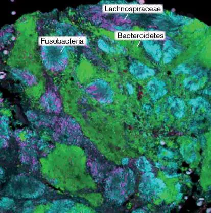
shows surprisingly diverse bacteria growing on a tumor of the right ascending colon. Three different DNA probes hybridize with 16S rRNA of distinct bacterial taxa: Bacteroidetes (green), Lachnospiraceae (magenta), Fusobacteria (cyan).
CHRISTINE M. DEJEA ET AL. 2014. PNAS 111 :18321
Most members of the phyla Bacteroidetes and Chlorobi (Chlorobiota) are obligate anaerobes. Within Bacteroidetes, Bacteroides species ferment complex carbohydrates, serving as the major mutualists of the human gut. By contrast, the closely related Chlorobi species are anaerobic “green sulfur” phototrophs that photolyze sulfides or hydrogen.
The Nitrospirae (Nitrospirota) largely resemble proteobacteria in form. Most oxidize nitr i te (NO −) to nitr a te (NO −). Nitrite
2 3
oxidation is a lithotrophic conversion essential for ecosystems (see Chapter 22). Another Gram-negative clade is the Acidobacteria (Acidobacteriota), an important phylum of soil bacteria.
Planctomycetes-Verrucomicrobia-Chlamydiae (PVC)
superphylum. The three related phyla of the PVC superphylum have unusual compartmentalized cells, with complex structural adaptations and development. These bacteria were thought to have lost their peptidoglycan cell walls, but peptidoglycan is now detected in some species.
Planctomycetes (Planctomycetota) are free-living aquatic bacteria, with stalked cells that reproduce by budding. Each planctomycete cell contains an extra double membrane surrounding its nucleoid, analogous to a eukaryotic nuclear membrane, though it evolved independently. In the case of anammox bacteria (discussed in Chapter 14), the membrane compartment protects the cell from toxic intermediates of ammonia oxidation. Verrucomicrobia (Verrucomicrobiota), also free-living, are bacteria with wart-like protruding structures containing tubulin. They are found in soil and water.
The Chlamydiae (Chlamydiota) are intracellular parasites that lose most of their cell envelope during intracellular growth. The replicating parasites generate multiple spore-like “elementary bodies” that escape to infect the next host. Chlamydia trachomatis causes one of the most common sexually transmitted infections of humans, as well as the eye disease trachoma.
Deep-branching thermophiles. Deep-branching thermophiles show unusually large genetic divergence from other bacterial clades. This large divergence makes the thermophiles appear to have separated earlier from other kinds of bacteria. Other evidence, however, implicates high mutation rates and gene transfer between distant relatives as the source of genetic divergence of thermophiles.
The deep-branching thermophile taxa include extremophiles such as those in the phylum Aquificae (Aquificota) (growing at up to 95ºC at marine thermal vents). These organisms also show rapid growth and high mutation rates, which may have accelerated their molecular clock. These hyperthermophilic bacteria also share their high-temperature habitats with Archaea and show surprising archaeal traits, such as archaeal ether-linked membrane lipids (discussed in Chapters 3 and 19). The genes encoding these “archaeal” lipids appear to have entered bacteria by horizontal transfer from archaea to bacteria sharing the high-temperature habitat. Aquifex pyrophilus, a flagellated rod, was first discovered in a submarine hydrothermal vent north of Iceland by extremophile microbiologist Karl Stetter of the University of Regensburg. Most members of the Aquificae are hydrogenotrophs, oxidizing hydrogen gas with molecular oxygen to make water.
Another deep-branching phylum of thermophiles (growing at 50°C–80°C) is Thermotogae (Thermotogota). Thermotoga maritima is a sulfur-reducing respirer, originally isolated from a geothermal vent in Vulcano, Italy. The cells have a loosely bound sheath, or “toga,” for which the genus is named. As discussed in Chapter 17, both Aquificae and Thermotogae show remarkable mosaic genomes. Nearly a quarter of the T. maritima genome derives from archaea. This degree of mosaicism leads some researchers to argue that Aquificae and Thermotogae cannot be said to branch from one clade; or if they do, that we can never know, statistically, which clade that is.
Thought Question
18.1 What taxonomic questions are raised by the apparent high rate of gene transfer between archaea and thermophilic bacteria? Chloroflexi (Chloroflexota) are filamentous photoheterotrophs, supplementing heterotrophy with photosystem II (PS II) to generate ATP (discussed in Chapter 14). Together with other thermophiles, Chloroflexi species form massive microbial mats in the hot springs of Yellowstone (Fig. 18.3). Most species of Chloroflexus contain their photosynthetic apparatus within membranous organelles called chlorosomes. The process of photosynthesis by chlorosomes is presented in Chapter 14. Chloroflexi are informally called “green nonsulfur bacteria” to distinguish them from Chlorobi, a phylum of green phototrophs that are strict anaerobes (introduced earlier, with the deep-branching Gram-negative phyla). However, Chloroflexus species may appear red or yellow, owing to accessory pigments.
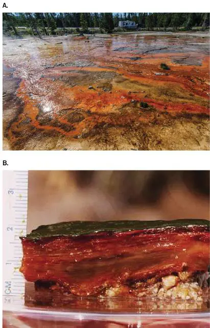
FIGURE 18.3 ■ A deep-branching thermophile: Chloroflexus. A. This hot-spring bacterial mat in Yellowstone National Park contains Chloroflexus species and other thermophilic bacteria. B. Bacterial mat section showing layers of Chloroflexus.
DURK TALSMA/ALAMY STOCK PHOTO
L. L. JAHNKE/NASA
The phylum Deinococcus-Thermus (Deinococcota) features a unique structural trait: the substitution of L -ornithine for diaminopimelic acid in the peptidoglycan cross-bridge. Thermus species (growing at 70°C–75°C) are heterotrophs commonly isolated from hot tap water. Deinococcus species, however, are not thermophilic. D. radiodurans bacteria resist extremely high doses of ionizing radiation. Their unusual chromosome structure and DNA repair system are described in Section 5.6. A heterotroph, D. radiodurans was originally isolated from cans of meat supposedly sterilized with a radiation dose of several megarads. Deinococcus bacteria are found suspended in air, both indoors and high in the atmosphere; they resist drought.
Emerging Clades
Clades of microbes that are recently defined or characterized are referred to as emerging. Emerging clades are discovered by field microbiologists who devise ever-more-creative screens, finding new bacteria with unexpected traits. For instance, in 2015, Jill Banfield’s group at UC Berkeley discovered bacteria so small that they pass through a 0.2-μm filter—barely big enough for ribosomes. These ultrasmall bacteria were originally found in the Rifle, Colorado, aquifer; related bacteria have since been found throughout soil and water habitats. The DNA sequences of these tiny cells show deeply branching phylogeny; that is, very distant relatedness to all the bacteria known (Fig. 18.1). The sequenced genomes “radiate”— that is, diverge from each other—so much that they may include more than 100 new candidate phyla. As a group, they are called the Candidate Phyla Radiation (CPR).
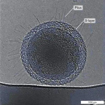
Single-cell sequencing and transmission electron microscopy (TEM) identify some CPR bacteria as cells that contain about 50 ribosomes and have a cell wall enclosed by an S-layer and long pili ( Fig. 18.4). The tiny cells’ genomes are smaller than those of most known bacteria. Most lack genes for respiration and biosynthesis, as well as certain ribosomal proteins that are found in all other bacteria. Bacteria in the CPR probably are obligate symbionts of other species. Some of their 16S rRNA sequences are so distant from those of other taxa that they cannot be amplified by so-called universal bacterial primers for polymerase chain reaction (PCR). Their extreme divergence may reflect an accelerated rate of evolution commonly found in dependent symbionts (such as Buchnera; see Fig. 17.16). We know little about these bacteria—or others yet unknown, which future microbe hunters may discover. FIGURE 18.4 ■ Ultrasmall bacteria. Bacteria capable of passing through a 0.2-μm filter were discovered by Jill Banfield and her colleagues in 2015 in an aquifer in Rifle, Colorado. The tiny cells possess approximately 50 ribosomes, a cell wall surrounded by an S-layer, and long pili (cryo-TEM).
BIRGIT LUEF ET AL. 2015. NAT. COMMUN. 6 :6372
Note: The term “emerging” can refer to a newly discovered clade
of organisms for which data are accumulating or to an outbreak of
disease in a location where the pathogen has not been seen recently. The meaning is inferred from the context.
Thought Questions
18.2 Which taxonomic groups in Table 18.1stain Gram-positive, and which stain Gram-negative? Which group contains both Gram-positive and Gram-negative species? For which groups is the Gram stain undefined, and why?
18.3 Which groups of bacterial species share common structure and physiology within the group? Which groups show extreme structural and physiological diversity?
Note: Formal names for taxa such as phyla, orders, and genera
are capitalized (for example, phylum Cyanobacteria, genus Streptococcus). Informal forms are lowercase and roman (cyanobacteria, streptococci, and so on), as are adjectival forms (cyanobacterial, streptococcal).
Following our brief tour of the bacterial domain, we now explore some of the diverse species within the well-studied phyla of Table 18.1.
To Summarize
Cyanobacteria conduct oxygenic photosynthesis and interact closely with heterotrophs. Species vary widely in their cell shape and ecological niche.
Firmicutes and Actinobacteria have a thick cell wall and generally stain Gram-positive. Mycoplasma are related to Firmicutes but have lost their cell wall.
Proteobacteria have a thin cell wall and an LPS-containing outer membrane. They show diverse metabolism and ecological adaptations.
Spirochetes have flexible, spiral-shaped cells with complex intracellular architecture.
Other deep-branching Gram-negative phyla include Acidobacteria, Bacteroidetes, Chlorobi, Fusobacteria, and Nitrospirae.
Chlamydiae, Planctomycetes, and Verrucomicrobia have irregularly shaped cells with complex intracellular form and development.
Deep-branching thermophiles such as Aquifex and Thermotoga species share traits and habitats with thermophilic archaea. Their genomes are highly mosaic, including many archaeal genes. Deinococcus species are highly resistant to ionizing radiation. Chloroflexus species are thermophilic photoheterotrophs.
Emerging organisms continually reveal previously unknown clades of bacteria, such as the Candidate Phyla Radiation (CPR).
Note: The main organizing principle used in Chapters 18–20 is
that of phylogeny based on DNA relatedness. Characteristic traits described for each branch apply to the majority of its known species, though many exceptions have evolved, such as members of Spirochetes that lack spiral form.
Glossary
phylum pl. phyla The taxonomic rank one level below domain; a group of organisms sharing a common ancestor that diverged early from other groups.
deep-branching taxon A lineage whose genome sequences diverged early, at or before the well-known phyla.
Cyanobacteria or Cyanophyta A phylum of oxygen-producing photoautotrophic bacteria containing chlorophylls a and b. They share an ancient ancestor of chloroplasts.
Firmicutes (Bacillota)
A phylum of Gram-positive bacteria with relatively low GC content.
endospore A durable, inert, heat-resistant spore that can remain viable for thousands of years.
Mycoplasma (Mycoplasmatota)
A phylum of bacteria related to Firmicutes and which lack a cell wall. Most known species are class Mollicutes, genus Mycoplasma.
Actinobacteria (Actinomycetota)
A phylum of Gram-positive bacteria with high GC content. GC content The proportion of an organism’s genome consisting of guanine-cytosine base pairs.
actinomycete A member of the Actinomycetales, an order of Actinobacteria that includes branched spore formers such as Streptomyces, as well as irregularly shaped corynebacteria.
Proteobacteria (Pseudomonadota)
A large, metabolically and morphologically diverse group of Gram-negative bacteria; possess an outer membrane containing LPS.
Alphaproteobacteria A class of Proteobacteria (Pseudomonadota) that includes, for example, rhizobia and rickettsias.
Betaproteobacteria A class of Proteobacteria (Pseudomonadota) that includes, for example, species of Neisseria and Burkholderia.
Gammaproteobacteria A class of Proteobacteria (Pseudomonadota) that includes, for example, the Enterobacteriales (enteric facultative anaerobes) and the pseudomonads.
Deltaproteobacteria A class of Proteobacteria (Pseudomonadota) that includes, for example, the Desulfobacterales (sulfate reducers) and the myxobacteria.
Epsilonproteobacteria A class of Proteobacteria (Pseudomonadota) that includes, for example, species of Campylobacter and Helicobacter.
Spirochetes or Spirochaetota A phylum of bacteria with a unique morphology: a flexible, extended spiral that twists via intracellular flagella. Bacteroidetes (Bacteroidota)
A phylum of Gram-negative bacteria; nearly all members are obligate anaerobes.
Fusobacteria (Fusobacteriota)
A phylum of anaerobic Gram-negative bacteria with an outer membrane, related to the Proteobacteria; includes human pathogens.
Chlorobi (Chlorobiota)
A phylum of Gram-negative bacteria. They are obligate anaerobes, “green sulfur” phototrophs that photolyze sulfides or H 2.
Nitrospirae (Nitrospirota)
A phylum of Gram-negative bacteria, many of which are lithotrophs, oxidizing nitrite to nitrate or ammonia to nitrate. Acidobacteria (Acidobacteriota)
A phylum of Gram-negative bacteria with an outer membrane, related to the Proteobacteria; often found in soil habitats. Planctomycetes (Planctomycetota)
A phylum of free-living bacteria that have stalked cells and reproduce by budding. Their nucleoid is surrounded by a membrane.
Verrucomicrobia (Verrucomicrobiota)
A phylum of free-living aquatic bacteria with wart-like, protruding structures containing tubulin.
Chlamydiae (Chlamydiota)
A phylum of intracellular parasitic bacteria that grow only within a host cell and generate multiple spore-like structures that escape to infect the next host.
Aquificae (Aquificota)
A phylum of hyperthermophilic bacteria.
Thermotogae (Thermotogota)
A phylum of thermophilic bacteria; in some species, the cell is enclosed by a toga-shaped outer covering.
Chloroflexi (Chloroflexota)
A phylum of bacteria that are filamentous phototrophs having chlorosomes.
chlorosome A membranous photosynthetic organelle found in some “green” bacteria of the phyla Chloroflexi and Chlorobi.
Deinococcus-Thermus (Deinococcota)
A phylum of bacteria that are resistant to ionizing radiation and high temperature.
emerging Describing an organism or other entity that is newly isolated, defined, or recognized, as in “emerging clade,” “emerging pathogen,” or “emerging disease.”
Candidate Phyla Radiation (CPR)
A recently described group of bacterial phyla whose members represent broad genetic diversity but small genome size and limited metabolic capabilities.
Mycoplasma (Mycoplasmatota)
A phylum of bacteria related to Firmicutes and which lack a cell wall. Most known species are class Mollicutes, genus Mycoplasma.
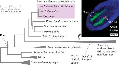
Fig. 17.16 FIGURE 17.16 ■ Intestinal bacteria and related proteobacteria. The phylogenetic tree was derived from concatenated sequences of highly conserved “housekeeping” proteins. The scale bar corresponds to 5% amino acid sequence divergence. Inset: Aphid embryo contains bacterial symbionts (DNA labeled by fluorescence in situ hybridization; FISH). Green = Buchnera; pink = Regiella; blue = aphid nuclei.
Sources: Phylogeny modified from Morgan Price et al. 2008. Genome Biol. 9 :R4; and from Fabia Battistuzzi et al. 2004. BMC Evol. Biol. 4 :44.
RYUICHI KOGA, NATIONAL INSTITUTE OF ADVANCED INDUSTRIAL SCIENCE
AND TECHNOLOGY, JAPAN
Endnotes
1. Note *: *Bulleted terms are representative orders within the phylum (unless stated otherwise). Return to reference *
18.2 Cyanobacteria: Oxygenic Phototrophsnot assigned
All of the oxygen gas in Earth’s atmosphere comes from Cyanobacteria and from plant
chloroplasts that evolved from an ancient cyanobacterium. The phylum Cyanobacteria (also
called Cyanophyta) is named for the blue phycocyanin accessory pigments possessed by
some genera, giving them a bluish tint. Cyanobacteria and chloroplasts (eukaryotic
organelles that evolved from Cyanobacteria) have a unique two-photosystem apparatus for
oxygenic photosynthesis arranged in lamellar arrays of membranes called thylakoids
(discussed in Chapter 14). The predominant blue and red absorption by their chlorophylls a
and b gives rise to the green color of some cyanobacteria and most plants.
Most cyanobacteria possess additional accessory pigments such as phycoerythrin, which
absorbs blue-green light in a range missed by cyanobacterial chlorophylls (Fig. 18.5 ).
Different accessory pigments color some species red or brown. In the marine
cyanobacterium Synechococcus, Laurence Garczarek and colleagues at the French National
Center for Scientific Research showed that the accessory pigments for photosynthesis have
all evolved peak absorption at ranges outside the wavelengths of water vibrational
absorption (Fig. 18.5 ). Thus the oxygenic phototrophs that fill our oceans have evolved
an extremely fine-tuned apparatus to maximize light absorption.
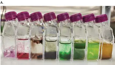
FIGURE 18.5 ■ Photosynthetic pigments of Synechococcus. A. Various
accessory pigments absorb different wavelengths of light and thus transmit different
colors. B. The photosynthetic pigments have all evolved peak absorption at ranges
outside the wavelengths of water vibrational absorption (dashed lines).
JOÃO MORAIS
Another source of cyanobacterial diversity is sulfur reduction. Under anoxic conditions,
many cyanobacteria can also reduce sulfur compounds and organic compounds. Use of H 2 S
allows flexibility in habitats such as wetlands that alternate between aerobic and anoxic
conditions. Different species of cyanobacteria colonize all kinds of habitats, even forming
symbiotic associations with mosses and fungi.
Cyanobacterial Cell Structure
The photosynthetic apparatus of cyanobacteria is organized within thylakoids, pockets of
membrane resembling flattened spheres packed with reaction centers. The thylakoids may
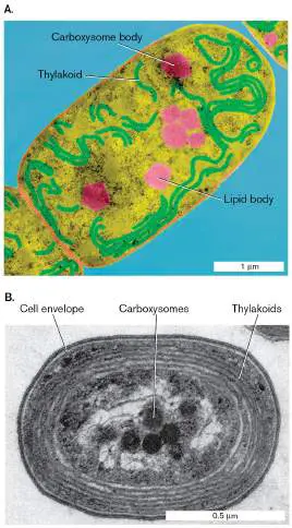
be distributed through the cell, as in filamentous genera such as Nostoc (Fig. 18.6A ), or
they may encircle the cell in concentric layers, as in the single-celled marine species
Prochlorococcus marinus (Fig. 18.6B ). Prochlorococcus is one of the smallest and most
abundant oxygen producers in the biosphere, accounting for 40%–50% of all marine
phototrophic biomass. In both large cells and small, the thylakoids are completely separate
from the plasma membrane, unlike the attached chlorosomes of Chloroflexi and the plasma
membrane extensions of “purple” Proteobacteria. Cyanobacterial thylakoids resemble the
thylakoids of eukaryotic chloroplasts; they are the most complex and specialized form of
photosynthetic apparatus.
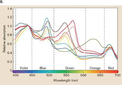
FIGURE 18.6 ■ Cyanobacterial cell structure. A. Intracellular organelles of
Nostoc, a typical filamentous cyanobacterium (colorized TEM). B. Intracellular
organelles of Prochlorococcus, a prochlorophyte cyanobacterium, the smallest known
phototroph. Prochlorococcus accounts for 40%–50% of marine phototrophic biomass.
DENNIS KUNKEL MICROSCOPY/SCIENCE SOURCE
REPUBLISHED WITH PERMISSION OF JOHN WILEY AND SONS INC., P. W. JOHNSON AND J. MCN. SIEBURTH.
1979. LIMNOLOGY AND OCEANOGRAPHY 24 :928
Cyanobacteria have several other subcellular structures (Fig. 18.6 ). Carboxysomes
(also known as polyhedral bodies) are rich in the enzyme Rubisco, and they fix CO 2
(discussed in Chapter 15). Cyanobacteria store energy-rich compounds in lipid bodies. To
maintain height in the water column and thus access to sunlight, cyanobacteria have gas
vesicles, whose buoyancy enables cells to float. Their external structures include a thick
peptidoglycan cell wall, similar to that of Gram-positive cells, plus several external layers
that vary with different species. Many species move by “gliding,” a form of motility whose
mechanism is poorly understood.
Besides fixing CO 2, many cyanobacteria fix N 2. Because nitrogen fixation requires the
absence of oxygen, cyanobacteria have to solve the problem of maintaining anaerobic
biochemistry while producing huge quantities of highly toxic O 2. Different species solve
this problem in different ways:
Formation of specialized nitrogen-fixing cells called heterocysts. Heterocysts exclude
oxygen, which associated heterotrophic bacteria consume.
Temporal separation, alternating between photosynthesis during daylight and nitrogen
fixation at night.
Accumulation of large aggregates of cells in which the interior becomes sufficiently
anaerobic for nitrogenase to function, while the exterior continues oxygenic
photosynthesis.
Symbiosis with fungi (as in lichens) or plants that consume oxygen or otherwise
maintain anoxic conditions.
Single-Celled, Filamentous, and Colonial Cyanobacteria
Single-celled species include Synechococcus and Prochlorococcus, the most abundant
phototrophs in the oceans. Another single-celled cyanobacterium is Microcystis, a colonial
microbe found in freshwater that produces dangerous toxins (microcystins). Blooms of
Microcystis form in the spring and summer when nitrogen and phosphorus runoff from
agricultural fertilizer and animal waste enters lakes and rivers. A Microcystis bloom in Lake
Erie in 2014 resulted in microcystin levels great enough that the water supply of Toledo was
contaminated (discussed in Chapter 21).
Other cyanobacterial genera, such as Oscillatoria and Nostoc, form multicellular
filaments. Such filaments may contain hundreds or even thousands of cells (Fig. 18.7 ).
Oscillatoria cells are stacked like plates, wider than they are long. To disseminate their cells
beyond the biofilm, the filaments produce hormogonia (singular, hormogonium), short
motile chains of three to five cells. Many filamentous species, such as Nostoc, develop
heterocysts to fix nitrogen (Fig. 18.7A ; discussed in Chapter 15).
FIGURE 18.7 ■ Pond cyanobacteria. A. Nostoc filamentous cyanobacteria form
heterocysts that fix nitrogen. B. Oscillatoria filaments consist of platelike cells.
DR. ROBERT CALENTINE/VISUALS UNLIMITED, INC.
M. I. WALKER/SCIENCE SOURCE
Under environmental stress, such as light limitation or phosphate starvation, filamentous
cyanobacteria such as Anabaena form specialized spore cells called akinetes. An akinete
forms as a long, oval cell adjacent to a heterocyst, where it stores nitrogen and develops a
thickened envelope. Like other types of spores, akinetes resist desiccation and remain
viable for long periods. Table 18.2 compares the properties of akinetes with those of spore
types of other taxa. Akinetes lie dormant but viable until improved conditions permit
germination and growth of new vegetative filaments. In lake water, akinete germination
may cause toxic blooms of Anabaena.
TABLE Spore Types in Bacteria 18.2
Initiation Formation
Spore Bacteria that of spore of the Properties
type produce the spore formation spore of the spore
Akinete Filamentous Light An akinete Desiccation
cyanobacteria limitation develops and cold
Cold next to a resistant
temperatu heterocyst Viable for
re as a large decades
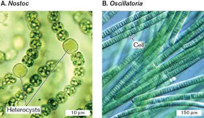
TABLE Spore Types in Bacteria 18.2
Initiation Formation
Spore Bacteria that of spore of the Properties
type produce the spore formation spore of the spore
Phosphate oval cell
starvation with a
multilayer
ed
envelope.
Arthrospore Actinomycetes Carbon At the tip of Desiccation
starvation an aerial and heat
Phosphate mycelium, resistant
starvation cells
undergo
vegetative
division
and pinch
off as
arthrospor
es.
Elementary Chlamydias Completion Intracellular Survives
body of chlamydia outside
intracellula reticular host
r life cycle bodies Desiccation
replicate resistant
and then
develop
into
elementar
y bodies
with cross-
linked
outer
membrane
proteins.
Elementar
y bodies
survive
outside
the host
cell.
TABLE Spore Types in Bacteria 18.2
Initiation Formation
Spore Bacteria that of spore of the Properties
type produce the spore formation spore of the spore
Endospore Firmicutes Carbon Individual Highly heat
starvation cell and
Nitrogen develops desiccation
starvation mother resistant
Phosphate cell and Viable for
starvation forespore. centuries,
Low pH The possibly
Peptide forespore thousands
antibiotics develops of years
into an
endospore
with a
spore coat
reinforced
by keratin
and
calcium
dipicolinat
e.
Myxospore Myxobacteria Nutrient Myxobacteria Desiccation
starvation aggregate and UV
Heat to form a resistant
shock fruiting
Glycerol body in
Dimethyl which
sulfoxide vegetative
cell
division
forms a
mass of
myxospore
s.
Some filamentous cyanobacteria, such as Lyngbya and Trichodesmium, form algal
blooms in the ocean. Trichodesmium can form giant blooms visible from outer space,
covering many square kilometers of ocean surface. Such a bloom can be triggered by an
influx of iron carried by wind from a dust storm blowing off the Sahara desert (discussed in
Chapter 22).
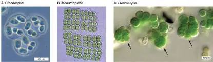
Yet other species, called colonial cyanobacteria, divide to form small groups or larger
colonies (Fig. 18.8 ). Gloeocapsa and Chroococcus form doublets or quartets, encased in a
thick protective mucous slime (Fig. 18.8A ). Others, such as Merismopedia, continue cell
division in two planes, extending to form long, square sheets of attached cells (Fig. 18.8B
). Colonial genera such as Myxosarcina and Pleurocapsa reproduce by multiple fission,
forming large cell aggregates (Fig. 18.8C ). As the aggregate matures, some cells
continue to divide and release single cells called baeocytes. Each baeocyte reproduces and
develops into a new cell aggregate. The aggregate group maintains anoxic conditions at
the center for nitrogen fixation.
FIGURE 18.8 ■ Colonial cyanobacteria. A. Gloeocapsa is surrounded by mucus.
Cells grow as single cells, doublets, or quartets. B. Merismopedia forms extended
quartets, octets, and so on. C. Unicellular cyanobacterium Chroococcidiopsis sp. with
baocytes (arrows) formed by successive multiple divisions.
MICHAEL ABBEY/SCIENCE SOURCE
MICHAEL ABBEY/SCIENCE SOURCE
BURKHARD BÜDEL
Thought Questions
18.4 What are the relative advantages and disadvantages of propagation by hormogonia
compared with propagation by akinetes?
18.5 What are the relative advantages and disadvantages of the different strategies for
maintaining separation of nitrogen fixation and photosynthesis?
Multicellularity
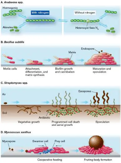
Cyanobacterial filaments and colonies, in effect, represent multicellular organisms. Different
bacteria have evolved diverse means of generating multicellularity (Fig. 18.9 ). As we
have seen, filamentous cyanobacteria such as Anabaena or Nostoc form chains by serial cell
division. At intervals, a cell differentiates into a heterocyst, which performs nitrogen fixation
—a specific function for the filament as a whole. In other clades, we will see very different
mechanisms of multicellularity (discussed in Sections 18.3 and 18.4).
FIGURE 18.9 ■ Bacterial multicellularity. Multicellularity arises by various
means. A. Filamentous cyanobacteria form chains by serial cell division. At intervals, a
cell differentiates into a heterocyst. B. Bacillus species assemble by attachment to a
substrate. They grow as a biofilm that releases endospores. C. Actinomycetes form
branching filaments that produce antibiotics, then undergo programmed cell death.
Aerial filaments generate exospores. D. Myxobacteria assemble by chemotaxis of
swarmer cells. They differentiate into a fruiting body and release myxospores.
Cyanobacteria in Microbial Communities
Cyanobacteria share many kinds of associations with animals, plants, fungi, and protists.
Sponges growing on coral reefs may harbor communities of cyanobacteria that provide the
sponges with nutrients from photosynthesis. The products of photosynthesis supplement
the nutrients obtained by the sponges from their filter feeding, and these extra nutrients
greatly augment the sponge growth rate within the competitive coral reef environment.
Sponge symbionts produce many pharmaceutically active compounds.
In salt marshes and sand flats, cyanobacteria participate in multilayered microbial mats
with other kinds of phototrophs, such as the section shown in Figure 18.10 , cut from the
sand flats of Great Sippewissett Salt Marsh, on Cape Cod, Massachusetts. The high
concentration of sulfides in the sediment supports growth of high populations of sulfur
phototrophs. Typically, cyanobacteria and eukaryotic algae such as diatoms (see Chapter 20
) form the upper green layer. Below, the purple layer consists of “purple sulfur”
proteobacteria (see Section 18.4), whose bacteriochlorophyll photopigments absorb at
longer wavelengths (Table 18.3 ). The pale-colored layer below the purple layer consists
of proteobacteria with bacteriochlorophyll that absorbs farther into the infrared.
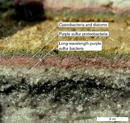
FIGURE 18.10 ■ Cyanobacteria in microbial mats. Cutaway through a
multilayered microbial mat from the sand flats of Great Sippewissett Salt Marsh (Cape
Cod, Massachusetts). Cyanobacteria and diatoms form the upper green layer, above
layers of purple sulfur proteobacteria.
JORG OVERMANN AND FERRAU GARCIA-PICHEL
TABLE 18.3 Phyla That Include Phototrophic Bacteria
Absorption
spectrum
Cell structure (nm;
and whole Reaction
Taxon Energy generation photopigments cell) center
Chloroflexi +O 2 Heterotrophy PS II
“Green nonsulfur” –O 2
Chloroflexus Photoheterotrophy
or use reduced sulfur
Cyanobacteria +O 2 PS I and
“Blue-greens” Oxygenic PS II
Anabaena phototrophy
–O 2
Photolithoautotrophy
on
reduced sulfur
Firmicutes +O 2 PS I
“Sun bacteria” –O 2
Heliobacterium Photoheterotrophy
Proteobacteria +O 2 PS II
“Purple nonsulfur” Heterotrophy
(Alpha and
Beta phyla)
BChl a
Rhodospirillum
BChl b
Blastochloris
–O 2
Photoheterotrophy
TABLE 18.3 Phyla That Include Phototrophic Bacteria
Absorption
spectrum
Cell structure (nm;
and whole Reaction
Taxon Energy generation photopigments cell) center
“Purple sulfur” +O 2 PS II
(Gamma –O 2
phylum) Photolithoautotrophy
Chromatium on
reduced sulfur
“Proteorhodopsin” +O 2
(Alpha and Heterotrophy
Gamma phyla) –O 2
Pelagibacter Photoheterotrophy
(SAR11)
SAR86
Chlorobi –O 2 only PS I
“Green sulfur” Photolithoautotrophy
Chlorobium on
reduced sulfur
BChl = bacteriochlorophyll; Chl = chlorophyll; PS = photosystem.
The cyanobacterial chlorophylls a and b are distinct from the bacteriochlorophylls used
by non-oxygenic bacterial phototrophs (Table 18.3 ). Most bacteriochlorophylls absorb
lower-energy photons at longer wavelengths. Because they absorb only lower-energy
photons, bacteriochlorophylls cannot break down water, the most stable molecule to be
photolyzed. Table 18.3 summarizes key features of phototrophy in clades throughout the
bacterial domain, such as the thermophilic Chloroflexi or the sulfur-based phototrophs
among the Proteobacteria. All light-harvesting complexes descend from one of two
ancestral sources: the chlorophyll/bacteriochlorophyll with electron transport (PS I, PS II) or
the proteorhodopsin proton pump. These photosystems have diverged through vertical
inheritance, as well as by horizontal transfer between different clades.
To Summarize
Cyanobacteria and plant chloroplasts supply all the oxygen we breathe .
Cyanobacterial chlorophylls and other photopigments have evolved to maximize light
absorption at those wavelengths that most strongly penetrate water.
Cyanobacteria conduct photosynthesis in thylakoids and fix CO 2 in
carboxysomes . Waterborne cyanobacteria maintain buoyancy using gas vesicles.
Some species exhibit gliding motility.
Single-celled cyanobacteria such as Prochlorococcus are among the smallest
and most abundant phototrophic producers in the oceans.
Microcystis species produce microcystins that poison lakes polluted by
agricultural runoff.
Filamentous cyanobacteria such as Nostoc and Oscillatoria are common in
freshwater lakes. They form heterocysts to fix nitrogen and reproduce by hormogonia
or by akinetes.
Colonial cyanobacteria such as Myxosarcina produce large cell aggregates with an
anaerobic core for nitrogen fixation. The colonies reproduce through baeocytes.
Symbiotic associations of cyanobacteria occur with animals, fungi, and plants.
Glossary
carboxysome
A protein-enclosed compartment containing Rubisco to fix CO 2.
gas vesicle
An organelle that traps gases to increase the buoyancy of aquatic microbes.
heterocyst
In filamentous cyanobacteria, a specialized nitrogen-fixing cell that maintains a
reducing environment and excludes O 2.
hormogonium pl. hormogonia
A short, motile chain of three to five cells produced by filamentous cyanobacteria to
disseminate their cells.
18.3 Firmicutes, Mycoplasma, and Actinobacterianot assigned
Which bacteria have the toughest, thickest cell walls? Bacteria of the phylum Firmicutes, meaning “tough skin” bacteria, have thick peptidoglycan cell walls that retain the Gram stain (discussed in Chapter 3). The thick cell wall helps exclude antibiotics and antibacterial agents from competitors in the environment. Bacteria of the closely related phylum Mycoplasma, however, have lost their cell wall entirely. Members of another phylum, Actinobacteria, have a thick cell wall, and some species stain Gram-positive. Species in both phyla have thick cell walls reinforced by teichoic acids, cross-threading phosphodiester chains of glycerol and ribitol (discussed in Chapter 3).
The Firmicute cell is enclosed by a single phospholipid membrane (the cell membrane), so the bacteria are sometimes called monoderm. Monoderm bacteria are distinguished from diderm bacteria, those with an outer membrane (discussed in Chapter 3), such as the several phyla of Proteobacteria (discussed later). Most actinomycetes, too, are monoderm—except for those with an exceptionally complex cell wall and envelope, such as Mycobacterium tuberculosis (discussed in Chapter 3).
Many firmicutes, such as Bacillus and Clostridium species, survive unfavorable environmental conditions by forming durable endospores. Non–spore formers such as Lactobacillus and Streptococcus may have evolved from a common Firmicutes ancestor that formed endospores. In many cases, the machinery to form endospores is discovered in the genomes of firmicutes previously thought to be non–spore formers, such as Carboxydothermus, soil bacteria that oxidize CO (carbon monoxide) to CO 2. On the other hand, actinobacteria of the order Actinomycetales (actinomycetes), such as Streptomyces, do not form endospores, but they develop filaments that disperse arthrospores (Table 18.2).
Firmicutes Include Endospore-Forming Rods
Endospore-forming bacteria are common in soil and air because their spore forms resist desiccation and can remain viable in a dormant state for thousands of years. Well-known Firmicutes include the order Bacillales (mainly aerobic respirers) and the class Clostridia, order Clostridiales (obligate anaerobes). Both groups include species of environmental and economic importance, as well as causative agents of well-known diseases. Many emerging firmicutes are found in the gut microbial community, with extraordinary diversity of metabolism and genetics. Some firmicutes of the primate gut have even evolved different codon assignments for protein synthesis, as described in eResearch Activity 18.
Bacillales. The genus Bacillus was one of the first bacterial genera to be classified, in the nineteenth century (Fig. 18.11). Colonies soon appear on a nutrient agar plate exposed to air. Bacillus species can be isolated from soil or food by suspending a sample in water and heating at 80°C for half an hour. Vegetative cells (that is, cells undergoing binary fission) and non–spore formers are killed at that temperature. The remaining endospores will germinate and grow on a beef broth agar plate at 25°C–30°C. Bacillus species isolated in this way include over a thousand characterized strains, all but a few of them harmless to humans.
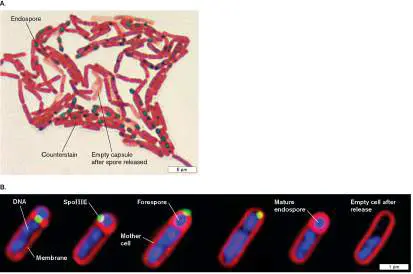
FIGURE 18.11 ■ Bacillus species: Gram-positive endospore formers. A. Gram-stained Bacillus sp., sporulating culture. Endospores stain green with malachite green. B.
Correlated fluorescence imaging of membrane migration, protein translocation, and chromosome localization during B. subtilis sporulation. Membranes were stained with red fluorescent FM4-64. Chromosomes were localized with the blue fluorescent nuclear counterstain 4′,6-diamidino-2-phenylindole (DAPI). The small, green fluorescent patches indicate the localization of a green fluorescent protein (GFP) gene fusion to Spo III E, a protein essential for both initial membrane fusion and forespore engulfment by the mother cell. Progression of the engulfment is shown from left to right.
CDC
MARC D. SHARP AND KIT POGLIANO. 1999. PROCEEDINGS OF THE NATIONAL
ACADEMY OF SCIENCES USA 96 :14553. © (1999) NATIONAL ACADEMY OF
SCIENCES, U.S.A.
The large, rod-shaped vegetative cells (growing and replicating form) of Bacillus species are easily stained and visualized. A species
of particular scientific importance is Bacillus subtilis, the best-studied Gram-positive organism, a “model system” for Firmicutes. The B. subtilis genome sequence reveals a large number of transporters for carbon sources and many secretory complexes for industrially important enzymes and drug resistance proteins. It also reveals several integrated prophages (integrated phage genomes). Phage genomes contribute to bacterial evolution by transferring genes between different strains and species, as discussed in Chapter 17.
B. subtilis is used as a model system to study stress response. Enormous changes in protein expression accompany starvation and general stress conditions. As the vegetative cells run short of nutrients on an agar plate, they begin a program to “sporulate”; that is, develop inert endospores (Fig. 18.11A). The life cycle of endospore production (see Chapter 4) involves a coordinated developmental plan, in which the cell divides near the pole instead of at the cell equator (Fig. 18.11B ). The polar compartment develops as the forespore, directed by unique regulatory proteins such as SpoIIIE. The larger compartment, called the mother cell, provides DNA and nutrients to the growing forespore and disintegrates after release of the mature endospore. When the released endospore encounters favorable conditions of moisture and nutrients, it germinates and restarts vegetative growth.
A Bacillus species of economic importance is B. thuringiensis, the most successful biological control agent yet produced. B.
thuringiensis was discovered in 1901 by Japanese bacteriologist Shigetane Ishiwata as a cause of disease in silkworms. The organism proved easy to culture on agar-based medium, and its spores are now applied as an insecticide against the gypsy moth caterpillar. During sporulation, B. thuringiensis generates an insecticidal protein known as delta endotoxin. The toxin is activated only at high pH in the digestive tracts of insect larvae; thus, it is safe for animals with acidic digestive tracts.
Many Bacillus species are extremophiles, growing at high pH (B. alkalophilus), at high temperature (B. thermophilus), or in high salt (B. halodurans). Some species combine alkaliphily with thermophily, as in the case of the “alkalithermophile” B. alkalophilus. Genomic research has focused on the surprisingly subtle differences that distinguish an extremophile from a closely related mesophile. For example, thermostability of proteins can be determined by comparing the protein sequences of a thermophile with those of a related mesophile. The thermophilic sequence shows specific patterns of amino acid residues that confer thermostability. Such amino acid substitutions provide useful information for industrial engineering of enzymes.
Clostridia. This diverse class includes anaerobic spore formers such as the genus Clostridium (Fig. 18.12A), species of which cause botulism (C. botulinum) and tetanus (C. tetani). The botulism toxin (botulinum, or “Botox”) is famous for its therapeutic use to relax muscle spasms and smooth wrinkles in skin (discussed in Chapter 26). Other species, such as C. acetobutylicum, have economic importance as producers of industrial solvents such as butanol and acetone (for industrial microbiology; see Chapter 16). The butanol pathway is an example of the diverse fermentative strategies found among clostridia. Unlike Bacillus (a monophyletic clade with a common ancestor), the Clostridium group is polyphyletic, representing many clades that branch among different genera.
FIGURE 18.12 ■ Clostridia are spore-forming anaerobes. A. Clostridium botulinum cells sporulating. The endospore swells, forming a characteristic “drumstick”
appearance (SEM). B. Clostridioides difficile, a major drug-resistant hospital-acquired pathogen (colorized TEM).
DENNIS KUNKEL MICROSCOPY/SCIENCE SOURCE
DR. KARI LOUNATMAA/SCIENCE SOURCE
The best-known Clostridium species, such as C. botulinum and C. tetani, sporulate in a form distinct from that of Bacillus (Fig. 18.12A). The growing Clostridium endospore swells the end of the cell, forming a “drumstick” appearance. Clostridium spores are found in soil and water, ready to germinate and grow when the environment becomes anoxic. Soil-borne spores of C. tetani cause tetanus, which should be prevented by vaccination (discussed in Chapter 24). Pathogenic C. botulinum can grow within the colon of very young infants, and infant botulism has been implicated in some cases of sudden infant death syndrome. Nevertheless, surprisingly many harmless species of clostridia are found in the human colon.
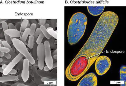
A closely related organism, Clostridioides difficile (formerly classified Clostridium difficile), is a life-threatening intestinal pathogen, resistant to most antibiotics (Fig. 18.12B ).
Clostridioides difficile grows in patients treated with antibiotics that eliminate normal enteric bacteria, allowing growth of the pathogen. The pathogen can be so hard to eradicate that patients require fecal bacteriotherapy, or “fecal transplant” from a healthy person’s colon. The healthy colon residents recolonize the gut and outcompete the pathogen.
A group of clostridia outside the order Clostridiales are the Heliobacteriaceae, or “Sun bacteria,” the only known photoheterotrophs in Firmicutes. Their name derives from their yellowish color, caused by the shift of their red-peak absorbance into the infrared, which allows transmission of red light plus green light (perceived as yellow).
Note: Distinguish Heliobacterium from Helicobacter, helical
species of the Gammaproteobacteria.
Variant sporulation and “live birth.” The order Clostridiales includes species of exceptionally large bacteria that grow only within the digestive tract of specific animal hosts. These species have evolved intriguing variations of the endospore former life cycle, as revealed by Esther Angert and colleagues at Cornell University (Fig. 18.13A). Metabacterium polyspora (size 15–20 μm) grows throughout the digestive tract of guinea pigs (Fig. 18.13B ). Endospores ingested from feces germinate in the upper intestine but rarely undergo binary fission. Instead, the growing cell forms forespores at both poles (Fig. 18.13C ). The forespores actually multiply within the mother cell to form several endospores, which are released in the colon before defecation.
FIGURE 18.13 ■ Multiple endospore formation. A. Esther Angert characterized unusual forms of sporulation and reproduction in exceptionally large firmicute bacteria. B.
Metabacterium polyspora forms multiple endospores (phase-contrast LM). C. A forespore forms at each pole. Forespores fission and multiply within the mother cell and then are released. Germinated cells undergo limited or no binary fission.
ESTHER ANGERT, CORNELL UNIVERSITY
ESTHER ANGERT, CORNELL UNIVERSITY
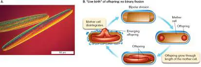
An even larger enteric endosymbiont is Epulopiscium fishelsoni, found in the digestive tract of surgeonfish (Fig. 18.14). These bacteria are large enough to be seen by eye—about the size of the period at the end of this sentence. As in M. polyspora, Angert showed that E. fishelsoni reproduction is synchronized with the digestive cycle of its host, but it has gone even further in transformation of the sporulation cycle. Binary fission is eliminated; the cell must fission at both poles. Each polar fission generates an intracellular daughter cell that grows to nearly the full length of the mother cell. From two to seven intracellular offspring ultimately emerge in “live birth” from the mother cell, which then disintegrates.
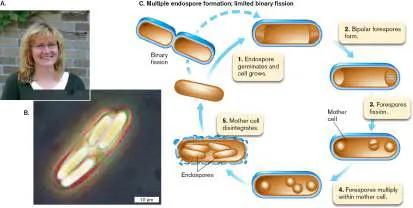
FIGURE 18.14 ■ “Live birth” in Epulopiscium. A. Epulopiscium fishelsoni forms offspring cells that grow internally. B. An offspring cell forms by fission at each pole. The cells grow internally until released. No binary fission occurs outside the mother cell.
ESTHER ANGERT, CORNELL UNIVERSITY
An even more bizarre “live birth” species related to clostridia was discovered in the mammalian intestine. These bacteria, provisionally named “ Candidatus Savagella,” grow as filaments attached to the intestinal epithelial cells (Special Topic 18).
SPECIAL TOPIC 18 Gut Bacterial Hairballs
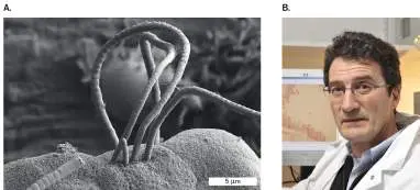
A bizarre kind of bacteria sit attached to cells of mammalian gut epithelium (Fig. ST 18.1A ). Related to clostridia, these bacteria form long, segmented filaments that interact symbiotically with their host cell. These “segmented filamentous bacteria” were first described by American microbiologist Dwayne Savage (1934–2016). They were mistakenly identified as Arthromitus (a different kind of bacterium) but have now been provisionally named for their discoverer as “ Candidatus Savagella.” Because the name is
provisional, microbiologists have a rule that “ Candidatus ” (Latin for “candidate”) is italicized, whereas the provisional name (“Savagella”) is not.
FIGURE ST 18.1 ■ Segmented filamentous bacteria (Savagella) adhere to mouse gut epithelium. A.
Savagella filaments adhere to mouse gut epithelium (SEM). B. Philippe Sansonetti, at the Pasteur Institute, Paris.
I. I. IVANOV ET AL. 2011. CELL 139 (3):485–498; FROM IVAYLO IVANOV
(COLUMBIA UNIV. MEDICAL CENTER), DAN LITTMAN (NYU LANGONE MEDICAL
CENTER), AND DOUG WEI (CARL ZEISS SMT, INC.)
PATRICK ALLARD/REA/REDUX
For over 50 years, microscopists have seen these segmented filamentous bacteria in the gut of mice and humans. The bacteria require their host for growth, and in turn, they provide essential services by inducing development of the host immune system. Attached to the epithelium above gut lymph tissues, the bacteria actually help protect their host from infection.
Philippe Sansonetti (Fig. ST 18.1B ) at the Pasteur Institute cultured Savagella filaments attached to mouse gut cells in tissue culture. To do this, Sansonetti isolated the multicell filaments by filtration at 5 μm, a pore size large enough to exclude most single-celled gut bacteria. The filaments were inoculated into germ-free mice. They were also
used to inoculate cultured TC7 cells, a cell line of gut epithelial tumor cells. The bacteria attached to the cultured cells and grew massive “hairballs” of filaments.
The in vitro system enabled Sansonetti to observe the bacteria’s unique process of differentiation and production of intracellular offspring (Fig. ST 18.2 ). Electron microscopy showed that each filament begins with a single “newborn” cell whose holdfast attaches to the surface of the host epithelial cell. The cell then grows and divides, forming a multicellular filament (primary segment cells). When the filament length exceeds 50 μm, the distal-segment cells start to differentiate into mother and daughter cells. Each daughter cell becomes engulfed by a mother cell. The engulfed daughter cell then divides and differentiates into two intracellular offspring. Finally, the mother cell breaks open, releasing the two offspring. Each offspring cell possesses a holdfast to attach to a new site on the gut epithelium.
FIGURE ST 18.2 ■ Savagella growth and reproduction. A. Primary segments divide and differentiate until offspring bacteria develop within each cell. Offspring are released, to attach to an epithelial cell at a
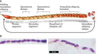
new position and grow a new filament. B. Intracellular offspring develop within cells of the filament (Gram stain).
PAMELA SCHNUPF ET AL. 2015. NATURE 520 :99–103
How do the attached bacterial filaments influence our gut epithelium? Pamela Schnupf, at the Institut Necker-Enfants Malades, has shown that the filament-attached epithelial cells have elevated transcription of genes that govern innate immune responses (discussed in Chapter 23). A more recent study by Bo Chen and colleagues at Zhejiang University, China, finds that segmented filamentous bacteria specifically colonize human children. In children, the bacteria may modulate T cells regulating adaptive immunity. Future research may reveal how this bacterium’s unusual life cycle plays a key role in the function of its mammalian host.
RESEARCH QUESTION
How do you think Savagella filaments communicate with the host immune system? Can you design an experiment to test your hypothesis?
Chen, Bo, Huahai Chen, Xiaoli Shu, Yeshi Yin, Jia Li, et al. 2018. Presence of segmented filamentous bacteria in human children and its potential role in the modulation of human gut immunity. Frontiers in Microbiology 9 :1403.
Schnupf, Pamela, Valérie Gaboriau-Routhiau, Marine Gros, Robin Friedman, Maryse Moya-Nilges, et al. 2015. Growth and host interaction of mouse segmented filamentous bacteria in vitro. Nature 520:99–103.
Non-Spore-Forming Firmicutes
Many Firmicute species do not form spores. In non-spore-forming firmicutes, endospore formation was probably lost by reductive evolution. An important order of non–spore formers is the Lactobacillales, or lactic acid bacteria, which are important for food production.
Other non–spore formers are human pathogens such as Listeria and Streptococcus species. Listeria monocytogenes are facultative anaerobic bacilli, named for the British surgeon Joseph Lister (1827– 1912), who was the first to promote antisepsis during surgery. L. monocytogenes contaminates cheese and sauerkraut (discussed in Chapter 16). Unlike other food-associated organisms, Listeria grows at temperatures as low as 4°C. Under preindustrial conditions of food preparation, L. monocytogenes was generally outcompeted by other flora. The era of refrigeration led to the emergence of Listeria as the cause of listeriosis, a severe gastrointestinal illness that can progress to the nervous system. L. monocytogenes cells are taken up by macrophages into phagocytic vesicles, but they avoid digestion and escape the vesicles. The bacteria then multiply as they travel through the host cytoplasm, generating “tails” of actin ( Fig. 18.15). The actin tails eventually project the cells of Listeria out of the original host cell and enable it to penetrate a neighboring host cell.
FIGURE 18.15 ■ Listeria monocytogenes: intracellular pathogen that travels on tails of actin. A. Fluorescent phalloidin marks the tails of polymerized actin (green) behind the Listeria monocytogenes, which are labeled using an antibody against a bacterial surface protein (ActA) (yellow) and are traveling within an infected PtK2 potoroo kidney epithelial cell. B. Invading bacteria encapsulate themselves in actin. Actin tails
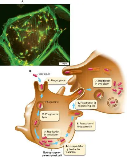
propel the bacteria through the host cytoplasm and out through the cell membrane to invade a neighboring cell.
Source: Part B from U. South Carolina, Microbiology and Immunology On line.
JULIE A. THERIOT AND TIMOTHY J. MITCHISON
Lactic acid bacteria. The lactic acid bacteria (order Lactobacillales) are aerotolerant (capable of growth in the presence of oxygen), though they do not use oxygen to respire. Most lactic acid bacteria are obligate fermenters; that is, they generate ATP by substrate-level phosphorylation (discussed in Chapter 13). They ferment primarily by converting sugars to lactic acid (a fermentation pathway discussed in Chapter 13). As the acid builds up, the pH decreases until it halts bacterial growth; thus, the carbon source retains much of its food value for human consumption. This is the basis of yogurt and cheese production. Lactococcus and Lactobacillus species are extremely important for the dairy industry (discussed in Chapter 16). Other common genera of lactic acid bacteria include Leuconostoc, which often spoils meat, and Pediococcus, found in sauerkraut and fermented bean products, as well as meat products such as sausage.
The shape of lactate-producing bacteria varies among species, from long, thin rods to curved rods and cocci. Most lactic acid bacteria have fastidious growth requirements and need many amino acids and vitamins. They can be isolated from pasture grasses incubated anaerobically in moderate acid (pH 5). The human intestinal flora include species of lactic acid bacteria. Certain species, particularly Lactobacillus acidophilus, are believed to play a positive role in human health by inhibiting the growth of pathogens. For this reason L. acidophilus may be ingested as a probiotic therapy.
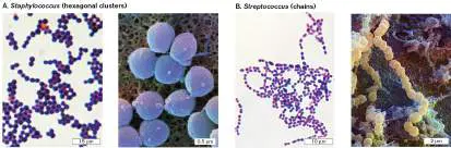
Staphylococcus and Streptococcus. The staphylococci are facultative aerobic cocci that grow in clusters, often packed in hexagonal arrays (Fig. 18.16A). They include common skin flora such as Staphylococcus epidermidis. The staphylococci are generally salt tolerant, and their fermentation generates short-chain fatty acids that inhibit growth of skin pathogens. Certain species, however, are themselves serious pathogens. Staphylococcus aureus causes impetigo and toxic shock syndrome, as well as pneumonia, mastitis, osteomyelitis, and other diseases (discussed in Chapter 26 ). It is a major cause of nosocomial (hospital-acquired) infections, especially contamination of surgical wounds. The most dangerous strains, now resistant to most known antibiotics, are termed MRSA (methicillin-resistant S. aureus).
FIGURE 18.16 ■ Staphylococci and streptococci. A. Staphylococcus species: Gram stain (left); colorized SEM (right). B. Streptococcus species: Gram stain (left); colorized SEM (right).
EYE OF SCIENCE/SCIENCE SOURCE
DAVID SCHARF/SCIENCE SOURCE
EYE OF SCIENCE/SCIENCE SOURCE
DAVID SCHARF/SCIENCE PHOTO LIBRARY/SCIENCE SOURCE
Streptococcus species generally form chains instead of clusters, because their cells divide in a single plane (Fig. 18.16B ). They are aerotolerant (grow in the presence of oxygen) but metabolize by fermentation. Many live on oral or dental surfaces, where they cause caries (tooth decay). Their fermentation of sugars produces such high concentrations of lactic acid that the pH at the tooth surface can fall to pH 4. Streptococcus species cause many serious diseases,
including pneumonia (S. pneumoniae), strep throat, erysipelas, and scarlet fever (S. pyogenes or group A streptococci).
The streptococci are less salt tolerant than Staphylococcus species, which tolerate as much as 5%–15% NaCl. Another genus whose size and fermentative metabolism resembles that of streptococci is Enterococcus. E. faecalis is a common member of the intestinal flora, and related strains are enteric pathogens. E. faecium is an exceptionally virulent pathogen with multiple drug resistance, one of the ESKAPE pathogens transmitted in hospitals.
Anaerobic dechlorinators. Some firmicutes from the soil show promising abilities to degrade chlorinated pollutants, such as dry-cleaning solvents that are biodegraded very slowly in the environment. The chlorinated molecules are reduced as alternative electron acceptors, a process called organohalide respiration. Dehalobacter restrictus, a flagellated rod, was isolated as an anaerobe capable of respiring by donating electrons to chlorine atoms in tetrachloroethene (Fig. 18.17). Organohalide respiration is discussed further in Section 14.5.
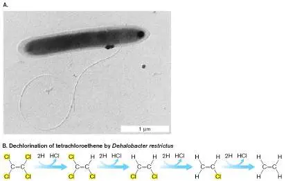
FIGURE 18.17 ■ Dehalobacter restrictus conducts anaerobic respiration by dechlorination. A. Dehalobacter restrictus, a flagellated rod related to clostridia. B. D. restrictus donates electrons to remove chlorine from tetrachloroethene, a major industrial pollutant.
C. HOLLIGER ET AL. 1998. ARCH. MICROBIOL. 169 :313
While genetic analysis places D. restrictus among the clostridia, the bacterium actually stains Gram-negative, perhaps because its peptidoglycan layer is relatively thin. Nevertheless, the species shows no Gram-negative outer membrane. It does possess a thick S-layer of hexagonally tiled proteins, typical of Gram-positive bacteria.
Mycoplasmas Lack a Cell Wall
The mycoplasmas are cell wall-less bacteria, now classified as phylum Mycoplasma (Mycoplasmatota). Mycoplasmas have completely lost their cell wall and S-layer through reductive evolution, retaining only their cell membrane. Presumably, the loss of these energy-expensive structures enhanced the reproductive rate of cells in a protected host environment. The mycoplasmas include many genera of flexible wall-less cells that maintain a shape through some kind of cytoskeleton (Fig. 18.18Aand B ). On agar they form colonies that have a characteristic “fried-egg” appearance (Fig. 18.18C ).
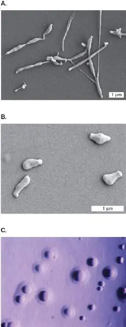
FIGURE 18.18 ■ Mycoplasmas: parasites without cell walls. A. Mycoplasma penetrans cells have an elongated tip used for attachment to the host (SEM). B. Mycoplasma mobile cells (SEM). C. Mycoplasmas cultured on agar show a “fried-egg” colony shape.
MITCHELL F. BALISH, MIAMI UNIVERSITY (OHIO)
MITCHELL F. BALISH, MIAMI UNIVERSITY (OHIO)
MICHAEL GABRIDGE/VISUALS UNLIMITED
The best-known genus is Mycoplasma, although genome sequences reveal that its species include several distantly related branches. Mycoplasmas are found as parasites of every known class of multicellular organism, including vertebrates, insects, and vascular plants; in humans, they cause pneumonia and meningitis. Another medically important mycoplasma is Ureaplasma urealyticum, an opportunistic pathogen inhabiting the genital tract. Most individuals are unaware that they harbor the organism, but U. urealyticum has been associated with urethritis, amniotic infections, and pulmonary infections.
Some Mycoplasma species remain adherent to the host cell, whereas others penetrate and grow intracellularly. Mycoplasma motility involves gliding with attachment to the oligosaccharide surfaces of host tissues. Most mycoplasma cells have a rounded cell shape with one or two extended tips. In Mycoplasma penetrans, an opportunistic pathogen infecting AIDS patients, the cell’s attachment tip is coated with adhesion molecules that enable attachment to a host cell surface (Fig. 18.18A). The attachment tip penetrates deep into epithelial tissues. By contrast, the fish pathogen M. mobile has no attachment tip, but glides along a surface at a rate of seven cell lengths per second (Fig. 18.18B ).
Mycoplasma genomes are among the smallest in known cellular organisms. They also lack biosynthetic pathways for amino acids and phospholipids, which instead must be acquired from the host. Mycoplasmas have unique nutritional requirements, such as cholesterol, a membrane component typical of eukaryotes but rare for prokaryotes. For these reasons, mycoplasmas are difficult to grow in pure culture, although they readily infect tissue cultures. In fact, mycoplasma contamination of tissue culture is so prevalent that it has compromised major studies of cancer and AIDS.
Actinomycetes Form Multicellular Filaments
The phylum Actinobacteria (Actinomycetota), the “high-GC Gram-positives,” includes several orders of branched filamentous bacteria such as Actinomycetales and Frankiales. Members of the order Actinomycetales are called “actinomycetes.” Actinomycetes include filamentous spore formers such as Streptomyces that produce antibiotics, as well as marine actinomycetes such as Salinispora, isolated from sediment and from sponges. Other filamentous Actinobacteria such as Frankia are often nitrogen-fixing symbionts of plants.
The actinomycetes form complex multicellular filaments superficially resembling the branched “fuzzy” form (mycelium) of fungi (fungi are eukaryotes, discussed in Chapter 20). Profoundly important for medicine, actinomycetes and fungi produce most of our antibiotics. Researchers mine the soil of remote environments to find actinomycetes producing antibiotics for which resistance genes are not yet widespread.
Streptomyces: filamentous spore formers. The best-studied actinomycetes are Streptomyces bacteria, whose filaments generate dispersible spores (see the streptomycete life cycle, Fig. 4.40). Streptomycetes play a major role in the ecosystems of soil (discussed in Chapter 21). Decaying Streptomyces cells produce the compound geosmin, which causes the characteristic odor of soil and can affect the taste of drinking water. In culture, the best-known species, S. coelicolor (Latin for “sky color”), forms strikingly blue colonies (Fig. 18.19A). The blue color derives from several pigments, including actinorhodin, a polyketide antibiotic. Other species produce filaments that are red, orange, green, or gray, depending on their distinctive products, many of which are antibiotics.
FIGURE 18.19 ■ Streptomyces bacteria. A. Colonies of Streptomyces coelicolor show sky-blue mycelia. B. Streptomyces cells form coiled filaments (filament width approx. 0.5 μm; SEM). C. Close-up of a coiled filament, showing individual cells (SEM).
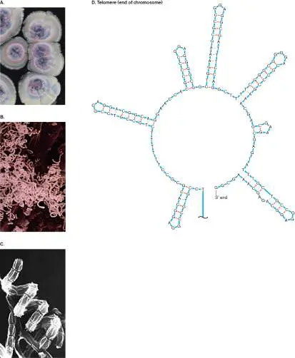
D. Hairpin-looped telomere end of the linear chromosome of S. griseus.
Source: Part D modified from Yasuo Ohnishi et al. 2008. J. Bacteriol. 190:4050, fig. 2B.
COURTESY OF JUAN PABLO GOMEZ-ESCRIBANO, JOHN INNES CENTRE
NCRS/USDA
K. FURIHATA, A. SHIMAZU & T. SHOMURA, HTTP://ATLAS.ACTINO.JP/
Thought Question
18.6 Why would Streptomyces produce antibiotics targeting other bacteria?
Streptomyces species are obligate aerobes, requiring access to air to complete their multicellular life cycle (see earlier, Fig. 18.9C). When a Streptomyces spore germinates, it extends vegetative mycelia, branched filaments that grow into the substrate. Some of the filaments then grow upward into the air, where they develop into aerial mycelia. The aerial mycelia in some species grow in tightly coiled spirals (Fig. 18.19B and C ). As the mycelial colony runs out of nutrients, older cells of the filament age and lyse, releasing nutrients that are absorbed by the younger cells. The nutrients also attract other scavengers, which may be killed by antibiotics produced by the aging streptomycete cells. The dead scavengers, too, release nutrients that feed the growing tip of the mycelium. As mycelia mature, they fragment into smaller cells called exospores (also called arthrospores). Exospores are vegetative cells, not dormant like Bacillus endospores (Table 18.2). The exospores separate and are dispersed by the wind, enabling them to colonize a new location. Streptomycete mycelia can be obtained from natural habitats by burying a glass slide in soil and then waiting several days for spores to germinate, covering the slide with mycelia. They are challenging to isolate in pure culture, however, because their coiled filaments trap cells of other bacteria.
The S. coelicolor genome is one of the largest prokaryotic genomes, containing over 8 million base pairs. Streptomycete chromosomes are linear with special “telomeres,” single-strand end sequences that double back to form hairpin loops (Fig. 18.19D ). Much of the lengthy genome of a streptomycete encodes catabolism of a rich array of diverse organic components of decaying plant and animal matter, including even lignin. Other genes encode extensive operons for production of diverse secondary products (see Chapter 15), including antibiotics. More than half of the antibiotics currently used in medicine derive from Streptomyces species.
Note: Distinguish Streptomyces species, filamentous rod-shaped
actinomycetes, from nonactinomycete Streptococcus species, Gram-positive cocci that form short, unbranched chains.
Actinobacteria associated with animals and plants. Many marine actinobacteria associate with invertebrate animals such as sponges, corals, mollusks, and ascidians (Fig. 18.20). In some cases the animal hosts a specific bacterium, such as the sponge Hymeniacidon perleve, which harbors Actinoalloteichus bacteria. More commonly, a sponge or coral supports a diverse community of many actinobacterial taxa. These bacterial symbionts often make up a substantial portion of the biomass of their host animal. The bacteria produce antibiotics and other secondary products that may help the animal resist pathogens or predators. They represent a vast, untapped potential source of novel therapeutic agents— antibiotics, antiparasitic agents, antitumor agents, immunomodulators, and agricultural agents. Mining the marine microbiomes is a growing aspect of the pharmaceutical industry—an aspect that depends on the global health and quality of our oceans. FIGURE 18.20 ■ Marine animals harbor actinobacteria that produce antibiotics.
ROBERTO NISTRI/ALAMY STOCK PHOTO
ALEXIS ROSENFELD/GETTY IMAGES
© IN-DEPTH IMAGES KWAJALEN
AVALON.RED/ALAMY STOCK PHOTO
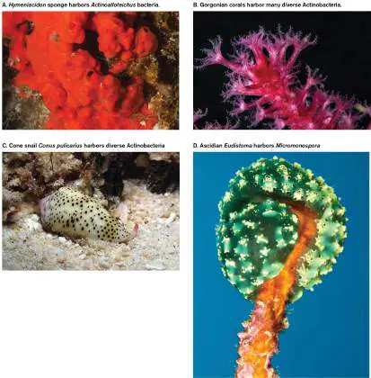
On land, actinobacteria maintain symbiotic relationships with animals and plants. For example, Frankia bacteria form nitrogen-fixing nodules on the alder tree. Certain Streptomyces species have evolved a highly structured mutualism with leaf-cutter ants. The ants culture the bacteria on special organs to produce antibiotics against parasites of their fungal gardens. A few actinomycetes are animal pathogens; for example, Actinomyces species cause actinomycosis, a form of skin abscesses in humans and cattle.
Nonmycelial Actinobacteria: Bifidobacterium, Mycobacterium, and Corynebacterium
Several groups of actinobacteria share the thick cell wall of Streptomyces but lack mycelial development. The Bifidobacteriales form rod-shaped cells that may form short branches but not extended filaments. Bifidobacterium species are essential to our gut microbiome. Among the first colonizers of the breast-fed infant digestive tract, Bifidobacterium species promote development of a healthy gut microbiome. They digest fiber and convert amino acids to neurotransmitters such as GABA (discussed in Chapter 13). The neuroactive catabolites of many gut bacteria are now associated with neurological illness and well-being.
Two nonfilamentous actinomycete genera that cause dreaded diseases are Mycobacterium (Fig. 18.21) and Corynebacterium. Both genera have thick cell envelopes containing mycolic acids and phenolic glycolipids. Mycolic acids are extremely diverse and include some of the longest-chain acids known, up to 90 carbons (Fig. 18.21C ). The mycolic acids are linked to arabinogalactan, a polymer of arabinose and galactose built on the peptidoglycan (discussed in Chapter 3). The mycolyl-arabinogalactan-peptidoglycan complex forms a waxy coat that impedes the entry of nutrients through porins and thus limits growth rate, but it also protects the bacterium from host defenses and antibiotics. For this reason, to cure tuberculosis requires an exceptionally long course of antibiotic therapy.
FIGURE 18.21 ■ Mycobacterium tuberculosis causes tuberculosis. A. Acid-fast stain of tissue sample containing Mycobacterium tuberculosis (chains of pink rods). B. Crinkled appearance of M. tuberculosis colonies. C. Mycolic acids and phenolic glycolipids coat the cell wall of M. tuberculosis.
CDC/DR. GEORGE P. KUBICA
CDC
Mycobacterium includes the species M. tuberculosis and M. leprae as well as lesser-known pathogens such as M. ulcerans. Cells of M. tuberculosis can be detected by the acid-fast stain as tiny rods associated with sloughed cells in sputum (Fig. 18.21A). In the acid-fast stain, cells are penetrated with a dye that is retained under treatment with acid alcohol (discussed in Chapter 2). The acid-fast property is associated with unusual cell wall lipids, such as mycolic
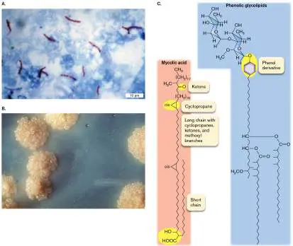
acids. M. tuberculosis bacteria are challenging to culture; they form crinkled colonies after 2 weeks of growth on agar-based media ( Fig. 18.21B ).
The closely related species M. leprae causes the disfiguring disease leprosy (Fig. 18.22A). The species has one of the longest known doubling times of any pathogen (about 14 days), and it can take a year to grow enough cells in the laboratory for observation. Growth of M. leprae requires lower temperature; for this reason, leprosy attacks the extremities (hands and feet) whose temperature is lower than that of the body core. Culture on artificial media is impossible; the bacteria can be grown only within low-temperature animals, such as armadillos, or within genetically immunodeficient mice.
FIGURE 18.22 ■ Mycobacterium leprae causes leprosy. A. Hand disfigured by leprosy. B. The genome of M. leprae shows
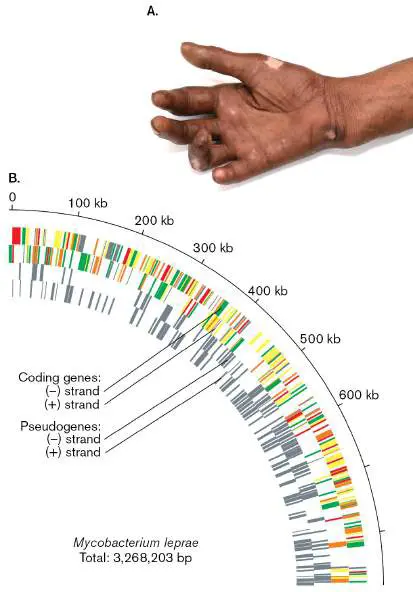
a high content of decaying pseudogenes (gray bars), most of which correspond to functional genes in the genome of M.
tuberculosis.
SCIENCE PHOTO LIBRARY/SCIENCE SOURCE
The genomes have been sequenced for both M. tuberculosis and M. leprae. M. tuberculosis has surprisingly few recognizable pathogenicity genes but a large number of environmental stress components, including 16 environmental sigma factors (discussed in Chapter 8), as well as 250 genes for its complex lipid metabolism. Over half of the M. leprae genome consists of pseudogenes, homologs of M. tuberculosis genes undergoing reductive evolution ( Fig. 18.22B ). Thus, M. leprae appears to be an evolving pathogen “caught in the act” of losing many genes no longer needed in its sheltered host environment. How it lost the need for so many genes preserved in M. tuberculosis remains a mystery. M. leprae causes disease worldwide, including 250 cases of leprosy annually in the United States. In 2013, a new test was approved that reveals leprosy infection a year before symptoms appear—enabling the disease to be cured with antibiotics before nerve damage is irreversible.
Mycobacteria also include a much larger number of harmless commensals, such as Mycobacterium smegmatis, isolated from human skin. Species of mycobacteria can be isolated from soil and water, as well as from various animal sources. Their culture is difficult because of their slow growth rates, but isolation can be enhanced by treatment with a base (NaOH or KOH) at concentrations that kill most other bacteria.
Note: Distinguish Mycobacterium species (rods whose cell walls
contain mycolic acids) from Mycoplasma species (bacteria that lack cell walls).
Irregularly shaped actinomycetes. Several nonmycelial actinomycetes show unusual cell shapes. Members of the genus Corynebacterium include soil bacteria, as well as pathogens such as C. diphtheriae, the cause of the lung disease diphtheria.
Corynebacterium species grow as irregularly shaped rods, which may divide by a “half-snapping” mechanism in which one side of the cell remains attached like a hinge (Fig. 18.23A). Related soil bacteria include the genera Nocardia and Rhodococcus.
FIGURE 18.23 ■ Irregularly shaped actinomycetes: Corynebacterium and Arthrobacter. A. Corynebacterium diphtheriae divides by snapping off one side while remaining attached at the other; the result is a typical V shape or “Chinese letter” arrangement (colorized SEM). B. Arthrobacter globiformis cultures form coccoid cells in stationary phase. With added nutrients, the coccoid cells grow out as rods.
DR. GARY GAUGLER/SCIENCE SOURCE
J. J. GERMIDA AND L. E. CASIDA, JR., DEPARTMENT OF MICROBIOLOGY,
PENNSYLVANIA STATE UNIVERSITY
Soil bacteria of the genus Arthrobacter exhibit an unusual cell cycle in which coccoid stationary-phase cells sprout into rods, which
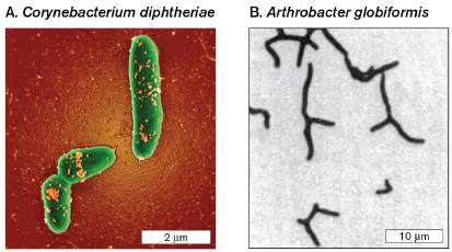
eventually run out of nutrients and revert to the coccoid form. The growing rods form irregular branched filaments (Fig. 18.23B ). An Arthrobacter species was discovered conducting anaerobic respiration by reduction of hexavalent chromium (Cr 6+), a toxic metal pollutant, to a less toxic oxidation state. Arthrobacter now shows potential as an agent of bioremediation of hexavalent chromium.
Micrococcaceae. Relatives of Arthrobacter are nonfilamentous cocci such as Micrococcus. M. luteus is one of the most widespread of soil bacteria, appearing readily as yellow colonies on agar plates exposed to air. Historically, the genus Micrococcus in the family Micrococcaceae was classified with Firmicutes such as Staphylococcus, but its DNA sequence data now place Micrococcus and most of the Micrococcaceae within the actinomycetes.
Micrococci are aerobic heterotrophs, commonly isolated from air and dust, although their habitat of choice is human skin. Micrococci grow in square or cuboid formations, dividing in two or three planes; cuboid clusters are known as sarcinae (singular, sarcina). M. luteus is harmless to humans and grows well at room temperature, so it makes an excellent laboratory organism for observation by students.
To Summarize
Firmicutes, the low-GC Gram-positive bacteria, include endospore-forming genera such as Bacillus and Clostridium. Endospore formation was probably present in the common ancestor of this phylum.
Nonsporulating firmicutes include pathogenic rods such as Listeria, as well as food-producing bacteria such as Lactobacillus and Lactococcus.
Staphylococcus and Streptococcus are Firmicutes, Gram-positive cocci that include normal human flora, as well as serious pathogens causing toxic shock syndrome, pneumonia, and scarlet fever.
Mycoplasma are related to Firmicutes but lack the cell wall and S-layer. They have flexible cytoskeletons and show gliding motility. Mycoplasma species cause diseases such as meningitis and pneumonia.
Actinobacteria (order Actinomycetales) include mycelial spore-forming soil bacteria, such as the actinomycete Streptomyces. Other actinobacteria have irregularly shaped cells, such as Arthrobacter.
Nonmycelial actinobacteria include Bifidobacterium , an important genus of the gut microbiome, as well as Mycobacterium and Corynebacterium species. C. diphtheriae is the cause of diphtheria.
Mycobacteria are actinobacterial rods whose cell envelope contains a diverse assemblage of complex mycolic acids. They stain acid-fast. Species of Mycobacterium cause tuberculosis and leprosy. Corynebacterium species also contain mycolic acids.
Glossary
monoderm Enclosed by a single phospholipid membrane.
diderm Enclosed by two phospholipid membranes—an inner membrane and an outer membrane.
vegetative cell A metabolically active, replicating bacterial cell.
forespore In sporulation of Gram-positive bacteria, the smaller cell compartment formed through asymmetrical cell division; it develops into the endospore.
mother cell In sporulation of Gram-positive bacteria, the larger cell that forms during the asymmetrical cell division leading to spore formation. The mother cell will engulf the forespore, but then disintegrates as the forespore matures.
aerial mycelium A mass of hyphae (branched filaments) that extend above the surface and produce spores at the tips.
mycolic acid One of a diverse class of sugar-linked fatty acids found in the cell envelopes of mycobacteria such as Mycobacterium tuberculosis.
acid-fast stain A diagnostic stain for mycobacteria, which retain the dye fuchsin because of mycolic acids in the cell wall.
sarcina pl. sarcinae A cubical octad cluster of cells formed by septation at right angles to the previous cell division.
vegetative mycelium A mass of hyphae (branched filaments) produced by vegetative cells that expand into the substrate.
Fig. 4.40 FIGURE 4.40 ■ Developmental cycle of Streptomyces coelicolor.
Fig. 18.9C
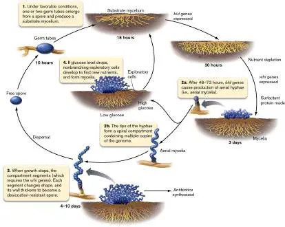
FIGURE 18.9 ■ Bacterial multicellularity. Multicellularity arises by various means. A. Filamentous cyanobacteria form chains by serial cell division. At intervals, a cell differentiates into a heterocyst. B. Bacillus species assemble by attachment to a substrate. They grow as a biofilm that releases endospores. C. Actinomycetes form
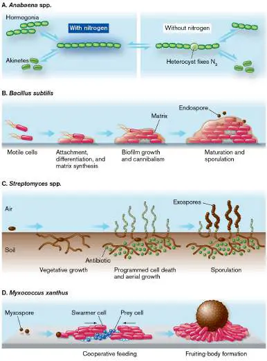
branching filaments that produce antibiotics, then undergo programmed cell death. Aerial filaments generate exospores. D. Myxobacteria assemble by chemotaxis of swarmer cells. They differentiate into a fruiting body and release myxospores.
18.4 Proteobacteria: From Photoheterotrophs to Lithotrophsnot assigned
Proteobacteria—the “protean,” or “many-formed,” bacteria—show highly diverse
metabolism. Their lifestyles range from plant and animal pathogens to soil lithotrophs
that reduce nearly any oxidized metal. Nevertheless, all proteobacteria share a common
feature, the diderm (two-membrane) envelope, which causes their cells to stain Gram-
negative. The Gram-negative cell envelope consists of an outer membrane,
peptidoglycan cell wall permeated by the periplasm, and inner membrane (plasma
membrane; discussed in Chapter 3). The outer membrane is packed with receptor
proteins and porins, and it comprises two-thirds of the mass of the membrane. Porins
evolved so as to admit nutrients while excluding antibiotics. The outer membrane lipids
contain long sugar polymer extensions (lipopolysaccharide, or LPS). In pathogens, LPS
repels phagocytosis and has toxic effects when released by dying cells.
The five classes of Proteobacteria are designated Alpha through Epsilon (Table 18.1
). Even within each class, we see nearly as wide a range of cell shape and metabolism as
we see in Proteobacteria as a whole.
The Protean Metabolism of Proteobacteria
The Proteobacteria excel at metabolic diversity. Many soil bacteria, such as Pseudomonas
species, can metabolize hundreds of different organic substrates, including intractable
materials such as lignin (see Chapter 13). Geobacter species can donate catabolic
electrons to numerous metals via extracellular cytochromes (see Chapter 14). Many
Proteobacteria oxidize H 2 (hydrogenotrophy) as well as complex organic molecules that
have aromatic rings. Some photoheterolithotrophs, such as Rhodopseudomonas palustris
, carry out nearly all the fundamental classes of metabolism, depending on
environmental conditions such as availability of light, oxygen, and nutrients.
A closer look at proteobacterial metabolism shows that processes that at first seem
very different actually connect through linked biochemical modules (Fig. 18.24 ). In
such a “protean” organism, the core of all proteobacterial energy acquisition is a
respiratory chain of electron donors and acceptors (discussed in Chapter 14). Electrons
may enter the chain from a photoexcited chlorophyll, from organic electron donors such
as sugars or benzoates, or from a mineral electron donor such as reduced sulfur or iron
(lithotrophy). In facultative anaerobes, the electron acceptor may be a mineral such as
nitrate or sulfate (anaerobic respiration). The capabilities of a given organism depend on
which oxidoreductases its genome encodes.
FIGURE 18.24 ■ The protean metabolism of Proteobacteria. In many Gram-
negative species, metabolic diversity arises through minor “add-ons” of biochemical
modules such as light absorption by bacteriochlorophyll, use of sulfide or organic
electron donors, and use of oxygen or alternative (anaerobic) electron acceptors. Cyt
= cytochrome.
Photoheterotrophy: light-supplemented heterotrophy. In the Alpha-, Beta-, and
Gammaproteobacteria, diverse forms of light absorption have evolved from a common
ancestor of photosystems I and II (Table 18.3 ). The various bacteriochlorophylls of
Proteobacteria peak in two ranges—in the blue and in the red or infrared.
Bacteriochlorophyll b, in Blastochloris viridis, peaks well beyond 1,000 nm. Species with
different photopigments often grow together in stratified layers of sediment or wetland—
the infrared absorbers below, where they capture the longer wavelengths “left over” from
the shorter-wavelength absorbers above.
The prevalence of homologous photosystems suggests that the common ancestor of
the Gammaproteobacteria was a photoheterotroph. At the same time, other evidence
supports horizontal transfer of photosystems among proteobacterial branches. The
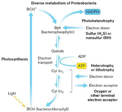
evidence is particularly strong in the case of proteorhodopsin, a homolog of the retinal-
protein light-driven proton pump first characterized in halophilic archaea (discussed in
Chapters 14 and 19). The proteorhodopsin light pump in Pelagibacter, a marine relative
of Rickettsia, absorbs green and yellow (wavelength 500–600 nm), the midrange of solar
radiation reaching Earth’s surface. Different species have proteorhodopsins with very
different absorption ranges, apparently adapted to different niches in the marine
ecosystem.
Proteobacterial phototrophs are historically called “purple bacteria.” The actual colors
of all the phototrophs range from purple-red through yellow and brown. “Purple sulfur
bacteria” are species that photolyze reduced forms of sulfur, such as H 2 S, HS −, S 0, or
S 2 O 3 2− (thiosulfate), whereas “purple nonsulfur bacteria” photolyze H 2 or use
photosystem II for cyclic photophosphorylation (discussed in Chapter 14). However, it
turns out that many purple sulfur and non-sulfur bacteria also use photosystem II. Most
proteobacterial phototrophs conduct photosynthesis only in the absence of oxygen,
reverting to aerobic heterotrophy when oxygen is present. Some, however, are obligate
anaerobes. Still others, found in marine environments, surprisingly photolyze sulfide or
organic compounds only in the presence of oxygen. Making sense of this extraordinary
diversity is the job of marine and freshwater microbiologists, particularly those
calculating global cycles of carbon and oxygen (discussed in Chapters 21 and 22).
Thought Question
18.7 Why might genes for the proteorhodopsin light-powered proton pump be more
likely to transfer horizontally than the genes for bacteriochlorophyll-based photosystems
PS I and PS II?
Lithotrophy: inorganic electron donors. Many different Proteobacteria oxidize
inorganic electron donors, obtaining energy from the redox reaction (discussed in Chapter
14). Examples of lithotrophy are given in Table 18.4 . Different species oxidize reduced
forms of nitrogen, iron, sulfur, or manganese. The ability to oxidize or reduce minerals
evolved multiple times in different clades by modification of the electron transport chain.
Many lithotrophic reactions generate acid, which conveniently breaks down rock
containing additional reduced minerals. Bacterial and archaeal lithotrophy has
tremendous significance for global cycling of elements in the biosphere (discussed in
Chapter 22). Some lithotrophs are autotrophs, fixing carbon dioxide into biomolecules by
the Calvin cycle (in carboxysomes) or the reverse tricarboxylic acid (TCA) cycle. Others
have alternative pathways of heterotrophy when organic foods are available.
Lithotrophy and Methylotrophy TABLE 18.4 (Examples)
Δ G °
′/2 e
Example −
Class/phylum species Reaction (kJ) Class of reaction
Alphaproteobacteria Nitrobacter NO 2 − + −54 Nitrite oxidation
winogradskyi ½O 2 → (nitrification to
NO − nitrate)
3
Paracoccus H 2 S + Sulfide oxidation
pantotrophus 2O 2 → to sulfate
2H + +
SO 4 2−
Roseobacter CO + H 2 CO oxidation
litoralis O → CO
2 + 2H
+ + 2 e
−
Hyphomicrobium CH 3 OH + Methylotrophy
O 2 →
CO 2 +
H 2 O +
2H + +
2 e −
Lithotrophy and Methylotrophy TABLE 18.4 (Examples)
Δ G °
′/2 e
Example −
Class/phylum species Reaction (kJ) Class of reaction
Betaproteobacteria Nitrosomonas NH 3 + O 2 Ammonia
europaea → HNO oxidation
2 − + (nitrification to
3H + + nitrite)
2 e −
Ralstonia 2H 2 + −237 Hydrogen
eutropha ½O 2 → oxidation
2H 2 O
Thiobacillus 3S 0 + Anaerobic sulfur
denitrificans 4NO 3 − oxidation
→ 3SO 4
2− + 2N
2
Lithotrophy and Methylotrophy TABLE 18.4 (Examples)
Δ G °
′/2 e
Example −
Class/phylum species Reaction (kJ) Class of reaction
Gammaproteobacteria Acidithiobacillus 4FeS 2 + −164 Iron-sulfur
ferrooxidans 15O 2 + oxidation
14H 2 O
→
4Fe(OH)
3 + 16H
+ + 8SO
4 2−
Lithotrophy and Methylotrophy TABLE 18.4 (Examples)
Δ G °
′/2 e
Example −
Class/phylum species Reaction (kJ) Class of reaction
Acidithiobacillus 2S 0 + 3O −196 Sulfur oxidation
thiooxidans 2 + 2H 2
O →
2SO 4 2−
+ 4H +
Methylococcus CH 4 + 2O −203 Methanotrophy
capsulatus 2 →
HCO 3 −
+ H + +
H 2 O
Beggiatoa alba 2H 2 S + −210 Sulfide oxidation
O 2 →
2S 0 +
2H 2 O
Chromatium 4Fe 2+ + Iron phototrophy
CO 2 + (photoferrotrop
11H 2 O hy)
+ h ν →
4Fe(OH)
3 + [CH
2 O] +
8H +
Deltaproteobacteria Desulfovibrio 4S 0 + 4H −11.3 Sulfur oxidation
2 O → (at
SO 4 2− pH
− 8)
+ 3HS
+ 5H +
Lithotrophy and Methylotrophy TABLE 18.4 (Examples)
Δ G °
′/2 e
Example −
Class/phylum species Reaction (kJ) Class of reaction
Nitrospirae Nitrospira NO 2 − + −54 Nitrite oxidation
½O 2 → (nitrification to
NO − nitrate)
3
Planctomycetes Brocadia NH 4 + + −238 Anaerobic
anammoxidans NO 2 − ammonium
→ N + oxidation
2 (anammox)
2H 2 O
Alphaproteobacteria: Photoheterotrophs, Methylotrophs, and Endosymbionts
The Alphaproteobacteria include photoheterotrophs, heterotrophs, and bacteria that
metabolize single-carbon compounds, as well as intracellular mutualists and pathogens.
Photoheterotrophs. Most alphaproteobacterial photoheterotrophs are unicellular. Their
cell shapes range from flagellated spirilla to rounded rods (Rhodobacter sphaeroides;
Fig. 18.25A ), wide spirals (Rhodospirillum rubrum), and stalked cells (
Rhodomicrobium; Fig. 18.25B ). The cryo-electron microscopy model of Rhodobacter
sphaeroides in Figure 18.25A shows that the cell is packed with photomembranes
(membranes containing the photosynthetic complex) arranged in vesicles invaginated
from the cytoplasmic membrane. These intracellular photomembranes expand the
surface area for photon capture, but during growth with oxygen, the photomembranes
disappear. The outer surface of the cell shows LPS filaments extending from the outer
membrane.
FIGURE 18.25 ■ Alphaproteobacterial photoheterotrophs. A. Rhodobacter
sphaeroides, showing intracellular photosynthetic vesicles (cryo-electron
tomography). B. Rhodomicrobium vannielii with stalked cells (phase contrast). C.
Citromicrobium species, an aerobic photoheterotroph, forms highly pleomorphic
shapes, including this Y shape (TEM).
JADE M. NOBLE ET AL. 2018. MOL. MICROBIOL. 109 :812–825
N. PFENNING. 1968. J. BACTERIOL. 2 :597
V. V. YURKOV ET AL. 1969. J. BACTERIOL. 181 :4517
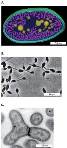
The metabolism of Rhodospirillum rubrum shifts drastically with oxygen.
Anaerobically, R. rubrum bacteria grow dark red because they synthesize membrane
containing bacteriochlorophyll a, which absorbs green and infrared and reflects primarily
red. With oxygen, however, R. rubrum fails to synthesize bacteriochlorophyll a, and the
cells grow white as their metabolism switches to straight heterotrophy. Heterotrophy is
supplemented by the oxidation of small molecules such as carbon monoxide.
Alphaproteobacterial photoheterotrophs that require oxygen (but do not photolyze
water) were found unexpectedly in genomic surveys of marine bacteria. Originally
thought to be straight heterotrophs, these organisms revealed genes encoding
bacteriochlorophyll. Their aerobic heterotrophy was shown to be driven by light
absorption. The reason for the oxygen requirement is unclear, because their
photosystems do not involve oxygen. Examples of O 2 -requiring phototrophs have since
been found in most of the proteobacterial clades, and species have been isolated from all
major habitats, including freshwater, marine, and soil ecosystems. Some show unusual
cell morphology, such as the Y shape of Citromicrobium (Fig. 18.25A ).
The aerobic photoheterotroph Erythromicrobium ramosum can reduce toxic metal
compounds such as tellurite ion (TeO 3 2−). The cells generate tellurium crystals that
take up 30% of their cell weight—a trait that we might use to remove tellurite from liquid
waste. Other toxic metals reduced by aerobic photoheterotrophs include selenium and
arsenic. Thus, these obscure bacteria now have a promising future in remediation of
metal-contaminated industrial wastes.
Aquatic and soil oligotrophs. Alphaproteobacteria also include many
nonphototrophic heterotrophs of soil and water. Many aquatic and soil
heterotrophs are oligotrophs adapted to extremely low nutrient concentrations; an
example is Caulobacter crescentus, the stalk-to-flagellum organism discussed in Chapter
3. Oligotrophic bacteria often have unusual extended shapes enhancing nutrient uptake,
such as the starlike cell aggregates of Seliberia stellata. In Seliberia species, the
individual tightly coiled rods generate oval or spherical reproductive cells by a budding
process. The budding reproductive cells germinate into rods, which then form new
aggregates.
Some heterotrophs common in soil are pathogens. Brucella species are intracellular
pathogens of animals that can also infect humans. The soil pathogen Granulibacter
bethesdensis is associated with chronic granulomatous disease, an inherited disorder of
the phagocyte oxidase system that leaves patients susceptible to infection. Other
pathogens are carried by insects or animal hosts; for example, Bartonella henselae
causes cat scratch disease.
Methylotrophy and methanotrophy. Methylotrophy is the ability of an organism to
oxidize reduced single-carbon compounds such as methanol, methylamine, or methane.
The Alphaproteobacteria include several genera of methylotrophs, which are found in all
environments, including soil, freshwater, and the ocean. Most methylotrophs can grow on
both single-carbon and organic compounds. One such versatile genus, Methylobacterium,
is equally at home in soil and water, on plant surfaces, and as a contaminant of facial
creams and purified water for silicon chip manufacture.
Other species are restricted to single-carbon compounds, incapable of metabolizing
organic compounds with carbon-carbon bonds. Methylotrophs that grow solely on
methane (CH 4) are called methanotrophs , conducting methanotrophy. Methane-
oxidizing species of Alpha- and Gammaproteobacteria and Verrucomicrobia contribute to
aquatic ecosystems, serving as major food sources for zooplankton. They eliminate much
of the methane produced by methanogens before it reaches the atmosphere, where it
has a potent greenhouse effect (see Chapter 22).
An interesting methylotroph is Hyphomicrobium, a bacterium with an unusual stalk-
to-flagellum transition similar to that of the alphaproteobacterium Caulobacter crescentus
, whose life cycle is described in Chapter 3. Hyphomicrobium species are found in
environments as diverse as wastewater sludge and Antarctic island soil. For example, the
Antarctic soil species H. sulfonivorans metabolizes sulfur compounds such as dimethyl
sulfone (Fig. 18.26A ). Like Caulobacter, a flagellated cell of Hyphomicrobium species
may lose its flagellum to form a stalk (also called “hypha”). But unlike Caulobacter,
Hyphomicrobium then forms a daughter cell from the opposite end of the stalk! As the
DNA replicates, one daughter nucleoid must migrate all the way through the stalk to
reach the daughter cell. The daughter cell forms a flagellum and septates, separating
from the parent (Fig. 18.26B ).
FIGURE 18.26 ■ Hyphomicrobium sulfonivorans from Signy Island,
Antarctica. A. These bacteria with coiled stalks catabolize dimethyl sulfone. B. Life
cycle of Hyphomicrobium species.
S. AZRA MOOSVI ET AL. 2005. SYST. APPL. MICROBIOL. 28 :541.
Thought Question
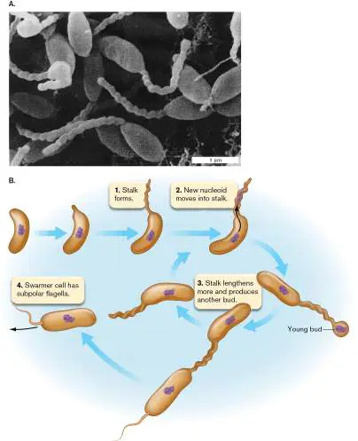
18.8 Can you hypothesize a mechanism for migration of the daughter nucleoid of
Hyphomicrobium through the stalk to the daughter cell? For possibilities, see Chapter 3
and consider the various molecular mechanisms of cell division and shape formation.
Plant endosymbionts. The Alphaproteobacteria include many highly evolved
intracellular symbionts of plants. As isolated bacteria, they are generally rod-shaped and
have aerobic metabolism, but their shape is transformed within the host cell.
Intracellularly, they need to solve special problems, such as the exclusion of oxygen from
nitrogen fixation (a process requiring anaerobiosis) or, in the case of pathogens, the need
to resist host defenses.
Nitrogen-fixing endosymbionts of plants include genera such as Rhizobium,
Bradyrhizobium, and Sinorhizobium. The nomenclature has undergone many changes,
but the species are generally referred to as rhizobia. Though they can live freely in the
soil, rhizobia prefer to colonize plants, usually legumes such as peas or alfalfa, where
they form distinctive nodule structures (Fig. 18.27 ). The host plant cells provide the
bacteroids with nutrients, as well as protective components such as leghemoglobin, an
oxygen-binding protein that maintains anaerobiosis within infected cells. Leghemoglobin
turns the nodule interior pink (Fig. 18.27A ). Leghemoglobin is the molecule now used
in “Impossible Burger” to imitate the color and taste of ground beef (Fig. 18.27B ).
FIGURE 18.27 ■ Rhizobia: legume endosymbionts. A. Legume nodules cut
open to show pink regions where the plant cells produce leghemoglobin to maintain
anaerobic conditions for bacteroid nitrogen fixation. B. Leghemoglobin is the source
of beef-like color and flavor of the “Impossible Burger.” C. Clover root hair curls
around infecting Sinorhizobium meliloti. The bacteria enter the curl and grow down
the root hair as an infection thread that penetrates the legume cells, enabling the
bacteria to colonize in the form of bacteroids.
MARIANGELA HUNGRIA
JOHN D. IVANKO/ALAMY STOCK PHOTO
J. FOURNIER ET AL. 2008. PLANT PHYSIOL. 148 :1985-1995. © 2008 AMERICAN SOCIETY OF PLANT
BIOLOGISTS
Each bacterial species colonizes and infects a particular host range. Complex
chemosensory processes attract the bacteria to the surface of the host root, where their
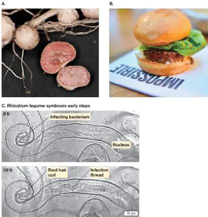
presence induces root hairs to form curls around the bacteria (Fig. 18.27C ). The
curling of the root hairs enables the bacteria to form infection threads, chains of rod-
shaped bacteria that invade the root cells. Within the host cells, the bacteria lose their
cell wall and become rounded bacteroids, specialized for nitrogen fixation.
A close relative of the rhizobia is the plant pathogen Agrobacterium tumefaciens. A.
tumefaciens has the remarkable ability to convert host plant cells to a form that produces
tumors. These plant tumors are called galls (Fig. 18.28A ). The tumor-inducing plant
pathogen conducts natural genetic engineering on plants—and is now used for industrial-
scale plant engineering. Long before scientists invented “recombinant DNA,” A.
tumefaciens had evolved a gene transfer system by which it induces infected plant cells
to generate food molecules to feed the pathogen. This highly efficient gene transfer
system is readily modified to insert genes conferring traits of interest, such as herbicide
resistance, into plant genomes.
FIGURE 18.28 ■ Agrobacterium tumefaciens transforms plant cells. A. A
crown gall induced by Agrobacterium tumefaciens on a chrysanthemum plant. B.
Left: A. tumefaciens transfers T-DNA containing opine synthesis genes into a plant,
which then produces opines to feed the bacteria. Right: A. tumefaciens can be
engineered to transfer T-DNA containing a recombinant gene of interest into the
plant genome. A recombinant strain of A. tumefaciens has the Ti plasmid divided
into two separate plasmids: one containing the vir operon conducting DNA transfer,
the other containing T-DNA with most of its genes substituted by a desired
recombinant gene.
NIGEL CATTLIN/SCIENCE SOURCE
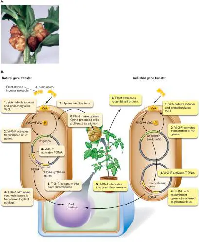
Tumorigenic strains of A. tumefaciens possess a special plasmid for engineering plant
cells, called the Ti plasmid (tumor-inducing plasmid). The Ti plasmid is the source of the
genetic material that gets transferred into a plant cell through a process mediated by
bacterial proteins, similar to conjugation (see Chapter 9). The Ti plasmid includes the vir
operons, which encode a virulence system, as well as the T-DNA (transferable DNA), a
set of genes that will be transferred to the host plant and recombined into its genome. T-
DNA encodes tumor induction genes, such as auxin synthesis genes, as well as enzymes
for biosynthesis of a carbon and nitrogen source called an opine. Opines are specialized
amino acids made by a one-step synthesis from arginine, typically by amination of a
central metabolite such as pyruvate or 2-oxoglutarate. A given strain of A. tumefaciens
typically provides one type of opine synthesis enzyme and has the ability to metabolize
the corresponding opine.
The Agrobacterium vector transfers its DNA to the plant by means of the vir gene
products. The vir gene products from the Ti plasmid detect the presence of a plant host
and stimulate plasmid transfer (Fig. 18.28B , left). In the cell envelope, VirA protein
detects a chemical signal from a wounded plant, which is capable of being infected. VirA
then activates VirG to induce expression of other vir genes, encoding proteins that direct
DNA transfer into the plant cell. Within the plant cell nucleus, the T-DNA becomes
integrated into the plant genome, where it induces opine production. The opine-
producing cells proliferate, forming a tumor.
For industrial use (Fig. 18.28B , right), a recombinant strain of A. tumefaciens has
the Ti plasmid divided into two separate plasmids. One contains the vir operons
conducting DNA transfer, while the other contains T-DNA with its left and right ends intact
but its genes substituted by the desired recombinant genes. Upon infection, the virulence
system induces transfer of the T-DNA to the plant cell without any tumor-inducing genes,
enabling genomic integration of the recombinant genes with their desired traits, without
tumor induction or opine production.
Thought Question
18.9 Why would an herbicide resistance gene be desirable in an agricultural plant?
What long-term problems might be caused by microbial transfer of herbicide resistance
genes into plant genomes?
Intracellular parasites. The Alphaproteobacteria include rickettsias, famous
intracellular pathogens of humans. Short coccoid rods, rickettsias lack flagella and can
grow only within a host cell. The best-known rickettsia is Rickettsia rickettsii, the cause
of Rocky Mountain spotted fever, a disease spread by ticks throughout the United States (
Fig. 18.29A ). Rickettsia species parasitize human endothelial cells (Fig. 18.29B ).
The bacteria induce phagocytosis and then dissolve the phagocytic vesicle and escape
into the cytoplasm. Some rickettsias propel themselves through the host cell by
polymerizing cytoplasmic actin behind them (Fig. 18.29C )—a process similar to that of
Listeria (described earlier with the Firmicutes). The actin tails eventually project outward
as filopodia, extensions of host cytoplasm and membrane that protect the bacteria from
host defenses while enabling them to invade adjacent cells.
FIGURE 18.29 ■ Rickettsias are obligate intracellular parasites. A. The
Rocky Mountain tick carries Rickettsia rickettsii, the cause of Rocky Mountain
spotted fever. B. Rickettsia species parasitize human endothelial cells (TEM). C. The
rickettsias propel themselves through the host cell by polymerizing cytoplasmic actin
behind them (TEM).
DR. JAMES L. CASTNER/VISUALS UNLIMITED
SCIENCE VU/VISUALS UNLIMITED
VSEVOLOD POPOV AND DAVID H. WALKER, UNIVERSITY OF TEXAS MEDICAL BRANCH AND THE MICROBE
LIBRARY
The rickettsias show a remarkable genetic relatedness to mitochondria. All eukaryotic
mitochondria appear to be descendants of an ancient rickettsial parasite whose
respiratory apparatus ultimately became essential to power eukaryotic cells. The
evolution of mitochondria and their role in microbial eukaryotes are discussed in Chapters
17 and 20.
Betaproteobacteria: Photoheterotrophs, Lithotrophs, and Pathogens
The Betaproteobacteria include many environmental photoheterotrophs such as
Rhodocyclus, as well as a diverse range of lithotrophs (Table 18.4 ). High
Betaproteobacteria levels are considered a sign of a healthy aquatic stream or pond
(discussed in Chapter 21). At the same time, the Beta class includes a few important
pathogens.
Lithotrophs: nitrifiers and sulfur oxidizers. An important betaproteobacterial group
of nitrogen lithotrophs consists of the nitrifiers. Nitrifiers oxidize ammonia (NH 3) to
nitrite (NO 2 −) or nitrite to nitrate (NO 3 −). Typically, different species conduct the two
reactions separately while coexisting in soil and water. Nitrifiers are of enormous
economic and practical importance for wastewater treatment because they decrease the
reduced nitrogen content of sewage. Special systems have been developed to retain
nitrifier bacteria behind filters as one stage of water treatment. In the system shown in
Figure 18.30A , nitrifying bacteria are encapsulated in pellets to retain them within the
bioreactor while the treated water flows through a filter.
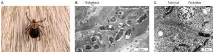
FIGURE 18.30 ■ Nitrifiers. A. Wastewater treatment uses nitrifier bacteria to
remove ammonia. B. Nitrosomonas europaea, of the Betaproteobacteria, oxidizes
ammonia to nitrite (TEM). Internal membranes contain the electron transport
complexes.
SCIENCE VU/S. WATSON/VISUALS UNLIMITED, INC.
Commonly isolated ammonia oxidizers in Betaproteobacteria are species of
Nitrosomonas and Nitrosovibrio. Nitrosomonas cells conduct electron transport through
extensive internal membranes that are either stacked or invaginated (Fig. 18.30B ).
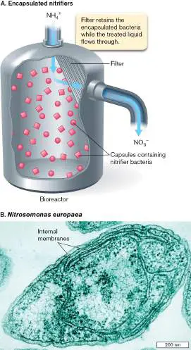
Note that outside the Proteobacteria, nitrite oxidizers include the phylum Nitrospirae
(Nitrospirota). Although distantly related, the unusual spiral-shaped cells of Nitrospirae
have a Gram-negative cell envelope with outer membrane similar to that of
Proteobacteria.
Pathogens. The Betaproteobacteria include aerobic heterotrophic cocci such as
Neisseria species. The cocci of Neisseria species form distinctive pairs known as
diplococci. Most Neisseria species are harmless commensals of the nasal or oral mucosa,
but N. gonorrhoeae causes the sexually transmitted disease gonorrhea. N. gonorrhoeae
is actually a microaerophile, requiring a narrow range of oxygen concentration; it has
fastidious growth requirements, necessitating cultivation on a special blood-based
medium. A related organism, N. meningitidis, may be carried asymptomatically by as
much as a quarter of the human population, but occasionally it causes meningitis, which
can be fatal. Other neisserias, such as N. sicca, are easily isolated from skin and often
presented to undergraduates as an unknown for identification.
Other members of the Beta class are important animal and plant pathogens, such as
Burkholderia. B. cepacia was originally isolated from onions as a cause of bulb rot; it is
now known to be a major opportunistic invader of the lungs of cystic fibrosis patients.
Gammaproteobacteria: Photolithotrophs, Enteric Flora, and Pathogens
The Gammaproteobacteria include vast numbers of marine organisms, such as Colwellia
and Oceanospirillales species, that catabolize petroleum hydrocarbons and other complex
organic pollutants. Their role in remediating oil spills is presented in Chapter 21. But the
most well-known Gammaproteobacteria are the Enterobacteriaceae family of facultative
anaerobes found in the human colon—the family that includes the famous research
model and industrial workhorse, Escherichia coli. Gammaproteobacteria also include
photolithotrophs that oxidize iron and nitrite. Still other lithotrophs such as Nitrosococcus
species oxidize ammonia.
Sulfur lithotrophs. The sulfur-oxidizing genus Beggiatoa was one of the first kinds of
lithotrophs described by pioneer microbial ecologist Sergei Winogradsky (discussed in
Chapter 1). Beggiatoa species oxidize H 2 S to elemental sulfur, which collects as sulfur
granules within the periplasm. Beggiatoa also stores carbon in cytoplasmic granules of
polyhydroxybutyrate—a common strategy among proteobacteria. The cells of Beggiatoa
grow as extended filaments with sulfur granules, forming biofilms on sulfide-rich
sediment (Fig. 18.31 ).
FIGURE 18.31 ■ Beggiatoa species oxidize sulfur in marine sediment.
JANE E. ROCHE/NATURE PL.COM
Bacteria of the genus Acidithiobacillus oxidize iron or sulfur (Fig. 18.32A ). Iron-
oxidizing bacteria commonly form the brown stains found inside plumbing. Most species
are short rods or vibrios (comma-shaped). Acidithiobacillus and other sulfur-oxidizing
genera can undergo a number of different reactions oxidizing H 2 S to S 0 and S 0 to SO 4
2− (Table 18.4 ). Sulfate production makes an environment acidic enough to erode
stone monuments and the interior surface of concrete sewer pipes. Sulfur oxidation is
often coupled to oxidation of iron, Fe 2+ → Fe 3+. The bacterium A. ferrooxidans is
known for its role in acidification of mine water, which contributes to leaching of iron,
copper, and other minerals (Fig. 18.32B ).

FIGURE 18.32 ■ Acidithiobacillus: iron oxidizers. A. Acidithiobacillus
ferrooxidans leaches copper and iron from molybdenite ore (SEM). The hexagonal
object is a molybdenite crystal. B. A copper mine in Utah, where Acidithiobacillus
species can oxidize copper ores, leaching the copper into solution for retrieval.
P. ROMANO ET AL. 2001. J. CHEM. TECHNOL. BIOTECHNOL. 76 :723
ROYCE BAIR/GETTY IMAGES
Sulfur and iron phototrophs. The gammaproteobacterial phototrophs, such as
Chromatium species (Fig. 18.33A ), mainly utilize sulfide and produce sulfur, which is
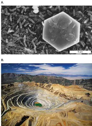
deposited as intracellular granules visible within the cytoplasm. Their phototrophy is
entirely anaerobic. Some of the Gamma class are true autotrophs and do not use organic
substrates. Some of these actually conduct phototrophy using iron (Fe 2+) to donate
electrons. Iron phototrophy, or “photoferrotrophy” (Fig. 18.33B ), is considered an
intriguing possibility for the metabolism of early life (discussed in Chapter 17). Thiocapsa
uses NO 2 − as an electron donor and was the first nitrogen-based phototroph discovered.
FIGURE 18.33 ■ Chromatium: sulfur and iron phototrophs. A. Chromatium
forms single-flagellated rods full of sulfur granules. B. Photoferrotrophy. Under
illumination, color develops over time (tubes 1–5) as a Chromatium isolate oxidizes
Fe 2+ to Fe 3+.
H. G. SCHLEGEL AND N. PFENNIG. 1961. ARCH. MICROBIOL. 38 :1–39
A. EHRENREICH ET AL. 1994. APPL. ENVIRON. MICROBIOL. 60 (12):4517–26. REPRODUCED WITH PERMISSION
FROM AMERICAN SOCIETY FOR MICROBIOLOGY
Enterobacteriaceae: intestinal fermenters and respirers. The family
Enterobacteriaceae, facultative anaerobes of the Gammaproteobacteria, include some of
the most intensively studied species of all bacteria. Species are readily isolated from the
contents of the human digestive tract and easily grown on laboratory media that are
based on human food. The best-known species of Enterobacteriaceae—indeed, the most
studied of all bacterial species—is the model organism Escherichia coli. Some strains of
E. coli grow normally in the human intestine, feeding on our mucous secretions and
producing vitamins, such as vitamin K. They may grow in symbiosis with anaerobic
fermenters such as Bacteroides species, which release short-chain sugars that E. coli
digests (discussed in Chapter 21).
Other strains of E. coli, such as E. coli O157:H7, cause serious illness. A large
proportion of the world’s children die of diarrheal infections caused by E. coli and related
pathogens such as Salmonella, Shigella, and Campylobacter. The genus Cronobacter is a
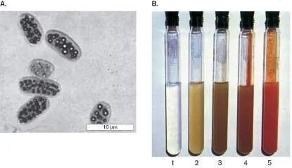
rare contaminant of powdered baby formula and can cause fatal illness in infants. In
2022, in the United States, Cronobacter contamination led to a shutdown of a major
formula producer, which then caused a nationwide shortage of baby formula.
The Enterobacteriaceae are Gram-negative rods, although as nutrients
diminish, their size dwindles almost to a coccoid form. They grow singly, in
chains, or in biofilms. Many species are motile, with numerous flagella. Most
strains grow well with or without oxygen, by either respiration (aerobic or anaerobic) or
fermentation. They ferment rapidly on carbohydrates, generating fermentation acids,
ethanol, and gases (CO 2 plus H 2) in varying proportions, depending on the species.
Their presence in the intestine supports the growth of organisms utilizing these gases,
including methanogens (discussed in Chapter 19). Many strains form biofilms. Biofilm
formation explains the persistence of drug-resistant infections, such as those associated
with urinary catheters in long-term hospital patients.
Thought Question
18.10 Why do you think it took many years of study to realize that Escherichia coli and
other Proteobacteria can grow as a biofilm?
Because they are easy to cultivate and have been studied extensively in clinical
laboratories, many genera of Enterobacteriaceae are familiar to students in introductory
microbiology laboratory courses. A common laboratory exercise is to distinguish
Enterobacter and Klebsiella species from E. coli by their fermentation to the pH-neutral
product butanediol, which tests positive in the Voges-Proskauer test. (Fermentation is
discussed in Chapter 13.) Enterobacter species occur more frequently in freshwater
streams than in the human body, although a few species colonize the intestine and cause
illness.
Proteus mirabilis and P. vulgaris cause bladder and kidney infections, particularly as a
complication of surgical catheterization. Proteus species are heavily flagellated and
display a remarkable swarming behavior (Fig. 18.34 ). In response to an
environmental signal, the flagellated rods grow into long-chain swarmer cells. The
swarmers gather together, forming “rafts” that swim together and grow into a complex
biofilm.
FIGURE 18.34 ■ The enteric rod Proteus mirabilis: isolated swimmer or
cooperative swarmer. A. A thickly flagellated swarmer cell (TEM). Cell length can
reach 20 μm. B. Karine Gibbs, at UC Berkeley, studies Proteus mirabilis swarming
behavior. C. Swarmer rafts of P. mirabilis migrate through blood agar.
REPRINTED FROM R. BELAS, 2014. TRENDS MICROBIOL. 22 :517. WITH PERMISSION FROM ELSEVIER
KARINE GIBBS
The swarming behavior of P. mirabilis is studied by Karine Gibbs at UC Berkeley. Gibbs
is interested in how bacterial cells distinguish self from nonself. The video associated with
Figure 18.34 shows how different populations of the swarming bacteria behave when
their migration fronts make contact. The two different strains of P. mirabilis were
inoculated at different locations on a blood agar plate. The strains establish and retain a
boundary; the two different populations recognize each other as nonself and do not
merge.
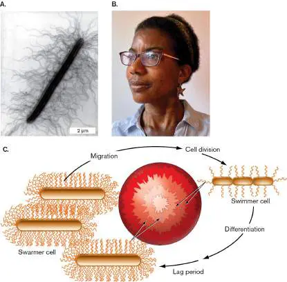
Besides symbionts of animals, Enterobacteriaceae include plant pathogens, such as
Erwinia carotovora and related species. Erwinia species cause wilts, galls, and necrosis of
a wide variety of plants, including bananas, tomatoes, and orchids.
Related facultative rods in soil and water conduct anaerobic respiration by donating
electrons from organic substrates to a variety of metals, such as iron and magnesium.
Shewanella oneidensis and other metal reducers are used to make electricity in fuel cells
(discussed in Chapter 14).
Aerobic rods. Closely related to Enterobacteriaceae are several genera of rod-shaped
bacteria that are obligate respirers. Many are obligate aerobic respirers (requiring O 2 for
growth), although some can use alternative electron acceptors, such as nitrate. These
genera catabolize an extraordinary range of natural compounds, including aromatic
derivatives of lignin; thus, they have important roles in natural recycling and soil
turnover.
The Pseudomonadaceae are a large and amorphous group of Gammaproteobacteria.
As the “pseudo-” prefix suggests, their taxonomic unit is poorly defined and includes
species whose DNA sequence has necessitated their reassignment to other groups. The
pseudomonads, such as Pseudomonas aeruginosa and P. fluorescens, respire on oxygen
or nitrate and are vigorous swimmers with single or multiple polar flagella. P. aeruginosa
can swim throughout a standard agar plate (much to the chagrin of students attempting
to isolate colonies).
Nevertheless, under appropriate environmental conditions, pseudomonad cells give up
their motility and develop biofilms (Fig. 18.35 ; discussed in Chapter 4). Hassan
Sakhtah and Lars Dietrich at Columbia University have studied how aromatic electron
carriers in the biofilm increase oxygen access for the bacteria. Biofilms of P. aeruginosa
cause lethal infections of the pulmonary lining in cystic fibrosis patients.
FIGURE 18.35 ■ Pseudomonas species form biofilms. A. Pseudomonas
aeruginosa phenazine mutant forms a crinkly biofilm that maximizes access to
oxygen. B. Hassan Sakhtah, Aranta Bio, now develops microbial applications for
industry. C. Lars Dietrich, at Columbia University, studies aromatic electron carriers in
biofilms of Pseudomonas aeruginosa.
COLONY BIOFILM OF A PSEUDOMONAS AERUGINOSA ISOLATE. HASSAN SAKHTAH, DIETRICH LAB
HASSAN SAKHTAH
LARS DIETRICH, DIETRICH LAB
Some pseudomonad pathogens have the unusual ability to infect both plants and
animals. For example, P. aeruginosa commonly infects plants as well as humans. P.
fluorescens infects seedlings and causes rotting of citrus fruit, while it also appears as an
opportunistic pathogen of immunocompromised cancer patients. Other pseudomonad
species, however, are harmless residents of soil or sewage.
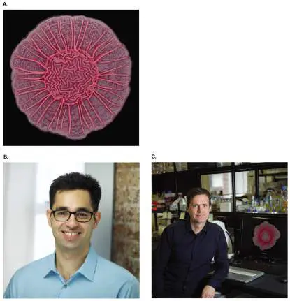
Legionella pneumophila is a well-publicized pathogen related to the pseudomonads.
Incapable of growth on sugars, L. pneumophila requires oxygen to respire on amino
acids. The organism exhibits an unusual dual lifestyle, alternating between intracellular
growth within human macrophages and intracellular growth within freshwater amebas (
Fig. 18.36 ). Growth within amebas facilitates transmission through aerosols into the
human lung. L. pneumophila is an environmental pathogen that takes advantage of our
lifestyle (the prevalence of large-scale air-conditioning units that contain unfiltered
water).
FIGURE 18.36 ■ Legionella pneumophila colonizes an ameba. A. Legionella
pneumophila cell caught by an ameba’s pseudopod (colorized SEM). B. L.
pneumophila cells have colonized the ameba (TEM).
CDC/DR. BARRY S. FIELDS
A related bacterium, Coxiella burnetii, causes Q fever, a respiratory illness of
livestock and humans. C. burnetii converts to a spore-like form that persists in soil. C.
burnetii resembles a rickettsia in some of its strategy of pathogenesis, but it is
genetically closer to Legionella.
Gammaproteobacteria include important plant pathogens, such as Xanthomonas
species. Xanthomonas is a flagellate rod that colonizes a wide range of agricultural
plants, such as tomatoes, potatoes, onions, broccoli, and citrus fruits (Fig. 18.37 ). The
disease it causes may mottle the leaves and fruit, and it may spread throughout the
plant.
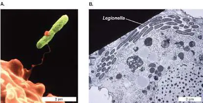
FIGURE 18.37 ■ Bacterial spot disease of orange fruit, caused by
Xanthomonas species.
NIGEL CATTLIN/ALAMY STOCK PHOTO
Deltaproteobacteria: Lithotrophs and Multicellular Communities
The Deltaproteobacteria include important sulfur and iron reducers, such as the fuel-cell
bacterium Geobacter metallireducens (discussed in Chapter 14). Other species have
complex life cycles that include multicellular developmental forms. The best-studied
example is the myxobacteria. Myxobacteria can have exceptionally large genomes, such
as that of Sorangium cellulosum (12.6 Mb).
Myxobacteria. The myxobacteria, such as Myxococcus xanthus, are free-living soil
bacteria that can grow as isolated cells but come together to form a multicellular
structure for the purpose of spore dispersal (Fig. 18.38 ). When nutrients are plentiful,
the myxobacteria grow and divide as individual cells. As nutrients run out, the cells begin
to attract each other by quorum sensing, and they move into parallel formations.
Myxobacteria have no flagella, but they move along a surface by using a form of motility
called gliding. The aggregating cells coalesce to develop a fruiting body. For Myxococcus
, the fruiting body rises from the aggregating cells to form a globular mass. The fruiting
body then forms myxospores, which disperse on the wind or via insects.
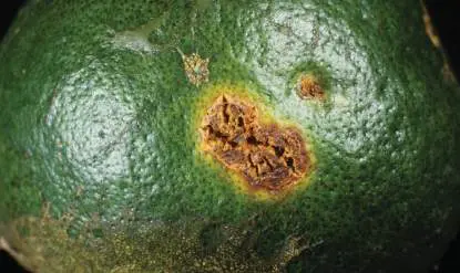
FIGURE 18.38 ■ Myxococcus xanthus fruiting body. Upon starvation,
swarming Myxococcus xanthus cells come together to generate a fruiting body
packed with spherical myxospores (SEM).
J. M. KUNER ET AL. 1982. J. BACTERIOL. 151 :458–461, FIG. 1
Other myxobacterial taxa form different kinds of fruiting bodies. For example,
swarming cells of Stigmatella aurantiaca form a fruiting body that is branched with
bulbous ends (Fig. 18.39 ). The bulbs produce myxospores for dispersal.
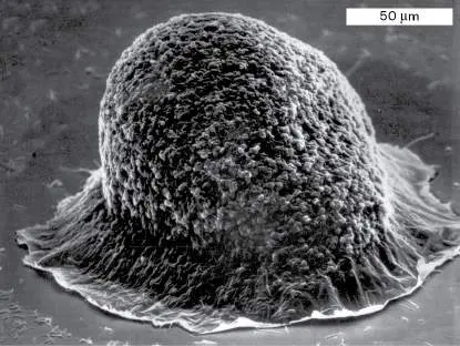
FIGURE 18.39 ■ The myxobacterium Stigmatella aurantiaca. A.
Stigmatella aurantiaca fruiting bodies. B. Life cycle: Starving Stigmatella cells glide
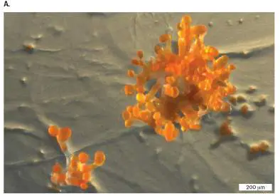
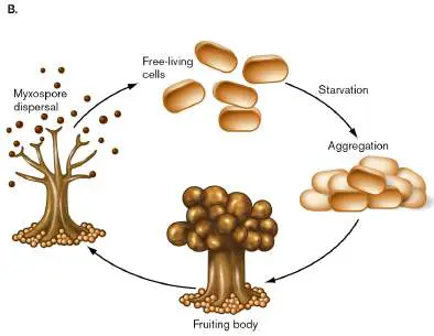
toward each other to aggregate. The aggregation generates a fruiting body with
bulges packed with small, spherical myxospores. The myxospores disperse on the
wind to colonize new locations in the soil.
DR. RONALD GARCIA AND PROF. ROLF MÜLLER, HELMHOLTZ INSTITUTE FOR PHARMACEUTICAL RESEARCH
SAARLAND, GERMANY
Thought Question
18.11 Compare and contrast the formation of firmicute endospores, actinomycete
arthrospores, and myxococcal myxospores.
Note: Distinguish my x obacteria from My c obacterium, the Gram-positive genus that
includes the causative agents of tuberculosis and leprosy.
Bdellovibrios parasitize bacteria. Bacteria can be parasitized or preyed on by
smaller bacteria. The Deltaproteobacteria include Bdellovibrio species, which attack
proteobacterial host cells. The structure of the “attack cell” is a small, comma-shaped rod
with a single flagellum. The attack cell attaches to the envelope of its host and then
penetrates the periplasm, where it uses host resources to grow (Fig. 18.40 ). The
growing cell produces enzymes that cross the inner membrane to degrade host
macromolecules and make their components available to the bdellovibrio. The entire
host cell loses its shape and becomes a protective incubator for the predator. This stage
is called the bdelloplast.
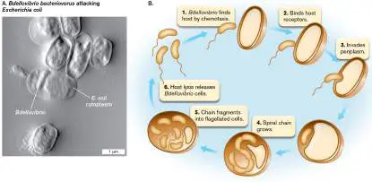
FIGURE 18.40 ■ Predators of bacteria: Bdellovibrio. A. Escherichia coli
under attack by Bdellovibrio bacteriovorus (note the Bdellovibrio cell within the E.
coli periplasm; atomic force microscopy). B. Life cycle of a bdellovibrio.
MEGAN E. NÚÑEZ ET AL. 2003. BIOPHYS. J. 84 :3379–88. © THE BIOPHYSICAL SOCIETY, PUBLISHED BY
ELSEVIER INC.
Within the periplasm, the invading bdellovibrio elongates as a spiral filament while
replicating several copies of its DNA. When most nutrients have been exhausted, the
filament septates into multiple short cells. The cells develop flagella, and the bdelloplast
bursts, releasing the newly formed attack cells (Fig. 18.40B ).
Bdellovibrios can be isolated from sewage, soil, or marine water—any environmental
source of Gram-negative prey bacteria. Different species infect E. coli and Pseudomonas,
as well as Agrobacterium and Rhizobium in their free-living state. They can be cultured
as plaques on a top-agar plate containing host bacteria—the same procedure used to
isolate bacteriophages (discussed in Chapter 6).
Epsilonproteobacteria: Helical Pathogens and Marine Sulfur Bacteria
Epsilonproteobacteria include the genera Campylobacter and Helicobacter. Helicobacter
pylori is well known today as the causative agent of gastritis and stomach ulcers (Fig.
18.41 ). Historically, microbiologists believed that bacteria could not live in the acidic
stomach. The discovery of H. pylori as the cause of gastritis earned Barry Marshall and J.
Robin Warren, of the University of Western Australia, the 2005 Nobel Prize in Physiology
or Medicine. Marshall famously swallowed a Helicobacter culture to prove that these
bacteria cause gastritis.
FIGURE 18.41 ■ Helicobacter, a neutralophile growing within the acidic
stomach. Helicobacter pylori is a short spirillum with unusual knobbed flagella
projecting from one end.
CAMR/A. BARRY DOWSETT/SCIENCE SOURCE
H. pylori and related species grow primarily on the stomach epithelium, at about pH
6, which is less acidic than the gastric contents (pH 2–4). The bacteria bury themselves
in the epithelial layer and neutralize their acidic surroundings by making urease enzyme,
which converts urea to ammonia and carbon dioxide. Helicobacter species form wide
spiral cells (spirilla) with an unusual grouping of flagella at one end. The metabolism of
H. pylori is microaerophilic, requiring a low level of oxygen. The bacteria can be isolated
through biopsy of the gastric mucosa.
Several groups of related Epsilonproteobacteria are sulfur oxidizers and sulfur
reducers, found in marine and freshwater habitats. Thiovulum species oxidize sulfides
aerobically, using oxygen available in marine water. In deep-ocean sediment, near
hydrothermal vents, hydrogenotrophs Nautilia and Hydrogenimonas oxidize H 2 using
sulfur or nitrate. These bacteria enrich the marine habitat by cycling carbon, nitrogen,
and sulfur. Surprisingly, genes from Epsilonproteobacteria are found in the sequenced
genomes of deep-branching taxa, the Aquifex species (see Section 18.1). Like other
hyperthermophiles, the Epsilons “get around,” with their genes migrating into the
genomes of distantly related organisms.
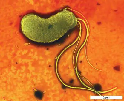
To Summarize
Proteobacteria stain Gram-negative and have a thin cell wall and an outer
membrane containing LPS. They show wide diversity of form and metabolism,
including phototrophy, lithotrophy, and heterotrophy on diverse organic substrates.
Alphaproteobacteria include photoheterotrophs (such as Rhodospirillum) and
heterotrophs, as well as methylotrophs. They include intracellular mutualists such
as rhizobia and pathogens such as the rickettsias. Rickettsias share ancestry with
mitochondria.
Betaproteobacteria include photoheterotrophs (Rhodocyclus), as well as
nitrifiers (Nitrosomonas) and iron-sulfur oxidizers (Thiobacillus). Pathogenic
diplococci include Neisseria gonorrhoeae, the cause of gonorrhea.
Gammaproteobacteria include sulfur and iron bacteria (Acidithiobacillus,
Chromatium). The most famous species are the Enterobacteriaceae found in the
human colon. Intracellular pathogens include Salmonella and Legionella. The
pseudomonads, aerobic rods, can respire on a wide range of complex organic
substrates.
Deltaproteobacteria include sulfur and iron reducers (Geobacter), fruiting-body
myxobacteria (Myxococcus), and bacterial predators (Bdellovibrio).
Epsilonproteobacteria include spirillar pathogens such as Helicobacter pylori,
the cause of gastritis. They also include marine sulfur oxidizers (Thiovulum) and
reducers (Hydrogenimonas).
Glossary
proteorhodopsin
A bacterial membrane-embedded protein that contains retinal and acts as a light-
driven proton pump; it is homologous to the archaeal protein bacteriorhodopsin.
methylotrophy
The metabolic oxidation of single-carbon compounds such as methanol,
methylamine, or methane to yield energy.
leghemoglobin
An iron-bearing plant protein that sequesters oxygen to maintain an anoxic
environment for nitrogenase within cells containing bacteroids.
bacteroid
A cell wall–less, undividing, differentiated rhizobial cell within a plant cell. The
bacteroid provides fixed nitrogen for the plant.
Ti plasmid
A plasmid found in tumorigenic strains of Agrobacterium tumefaciens that can be
used as a vector to introduce DNA into plant cells.
diplococcus
The paired cocci of Neisseria species.
swarming or swarming motility
A behavior in which some microbial cells differentiate into large swarmer cells and
swim together as a unit.
fruiting body
A multicellular fungal or bacterial reproductive structure.
myxospore
A durable spherical cell produced by the fruiting body of myxobacteria.
methanotrophy
The metabolic oxidation of methane to yield energy.
18.5 Spirochetes, Acidobacteria, Bacteroidetes, and Chlorobinot assigned
Besides Proteobacteria, members of several other phyla possess an outer membrane and generally stain Gram-negative. They have diverse lifestyles and habitats, ranging from aquatic phototrophs to human pathogens. The spirochete cell has a distinctive form of a tightly coiled spiral, with internalized flagella. The phylum Spirochetes includes famous human pathogens that cause Lyme disease and syphilis. Other important phyla of deep-branching Gram-negative bacteria include Acidobacteria, Bacteroidetes, Chlorobi, Fusobacteria, and Nitrospirae.
Spirochetes: Sheathed Spiral Cells with Internalized Flagella
Spirochetes (Spirochaetota) is a unique phylum of heterotrophic bacteria that form tightly coiled spirals. While different species of spirochetes conduct a broad range of heterotrophy, from aerobic to anaerobic, all spirochetes share a distinctive cell structure consisting of a long, tight spiral that is flexible like an old-style telephone cord (Fig. 18.42A). For many species, the spiral is so thin that its width cannot be resolved by bright-field microscopy, and the organisms can pass through a filter of pore size 0.2 μm.
FIGURE 18.42 ■ Spirochetes. A. Treponema azotonutricium fixes nitrogen in the termite gut (TEM). B. Periplasmic flagellum of T. azotonutricium. C. Borrelia recurrentis, shown here in a blood film, is the cause of relapsing fever (stained LM). D. Lyme disease rash, caused by tick-borne Borrelia burgdorferi.
JOSEPH GRABER ET AL. 2004. APPL. ENVIRON. MICROBIOL. 70 :1315
JOSEPH GRABER ET AL. 2004. APPL. ENVIRON. MICROBIOL. 70 :1315
MICHAEL ABBEY/VISUALS UNLIMITED
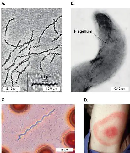
CDC/JAMES GATHANY
Spirochete diversity. Spirochetes grow in a wide range of habitats, from ponds and streams to the digestive tracts of animals. Aquatic systems carry free-living sugar fermenters of the genus Spirochaeta. A particularly interesting spirochete community is found in the termite gut, where the organisms form elaborate symbiotic associations with protists and assist in the digestion of cellulose. Treponema azotonutricium actually fixes nitrogen for its host termite (Fig. 18.42A and B ). Other members of the genus Treponema are normal residents of the human and animal oral, intestinal, and genital regions.
The best-known spirochetes are those that cause human and animal diseases. The causative agent of the sexually transmitted disease syphilis is the spirochete Treponema pallidum. The cell of T. pallidum is too narrow (0.2 μm) to visualize by bright-field microscopy; instead, dark-field, fluorescence, or electron microscopy must be used. T. pallidum is not culturable in the laboratory, but its genome sequence reveals much about its physiology. The genome of 1.1 million base pairs is highly degenerate, lacking nearly all components of biosynthesis and of the TCA cycle; its only system for ATP production is glycolysis.
The spirochete genus Borrelia includes two pathogens that cause serious tick-borne diseases in the United States. B. recurrentis causes relapsing fever (Fig. 18.42C ). The related species B. burgdorferi causes Lyme disease, known for the distinctive bull’s-eye rash (Fig. 18.42D ). A unique feature of Borrelia species is their multipartite genome. Each species possesses a linear main chromosome of less than a million base pairs, plus a number of linear and circular plasmids. B. burgdorferi, for example, has a linear chromosome of 910,725 base pairs, with at least 17 linear and circular plasmids that total an additional 533,000 base pairs. The reason for this unusual fragmentation of Borrelia genomes is unknown.
Spirochetes include important pathogens of animals, such as Leptospira, the cause of leptospirosis, a form of nephritis (kidney inflammation) with complications in the liver and other organs. Leptospira cells are known for the peculiar L shape of the cell, as each end of the spirochete turns out at an angle. Members of the genus Treponema also cause digital dermatitis in cattle and sheep. As in other bacterial phyla, however, the pathogenic species are far outnumbered by harmless organisms.
Note: Distinguish the spirochete Leptospira from the nitrite-
oxidizing proteobacterium Leptospirillum.
Cell structure of Spirochetes. The spirochete cell is considered diderm, as the plasma membrane and cell wall are surrounded by a thick outer sheath of lipopolysaccharides and proteins. The sheath is similar to a proteobacterial outer membrane, except that the periplasmic space completely separates the sheath from the plasma membrane. At each end of the cell, one or more polar flagella extend and double back around the cell body within the periplasmic space (Fig. 18.43). The periplasmic flagella (axial fibrils) rotate on proton-driven motors, as do regular flagella; but because they twine back around the cell body, their rotation forces the entire cell to twist around, corkscrewing through the medium.
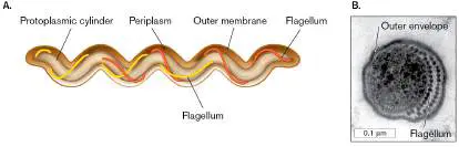
FIGURE 18.43 ■ Spirochete structure. A. Spirochete cell structure, showing the arrangement of periplasmic flagella. B. Cross section through a human gingival spirochete, showing outer envelope and flagella (axial fibrils; TEM).
M. A. LISTGARTEN AND S. S. SOCRANSKY. 1964. J. BACTERIOL. 88 :1087
This corkscrew motion of the cell body turns out to have a physical advantage in highly viscous environments, such as human mucous secretions or agar culture medium. Few nonspirochetes can swim through agar at standard culture concentrations (1.5% agar); thus, growth within agar provides a way to isolate anaerobic spirochetes from environmental sources. When an environmental sample is inoculated into agar, other bacteria grow and concentrate at the injection point, whereas spirochetes migrate outward in a “veil” through the agar.
Acidobacteria
Acidobacteria (Acidobacteriota) are abundant in soils, where they metabolize a wide range of organic substrates using diverse electron acceptors. Many species grow in extreme conditions, such as in the presence of acid and metals; for example, in soils contaminated by uranium mining. Others grow at high temperature, such as isolate K22 (Fig. 18.44A) obtained from the Taupo Volcanic Zone, New Zealand, by GNS Science, a geoscience prospecting company. The thin section of isolate K22 in Figure 18.44B shows its extensive outer membrane.
The candidate species Chloracidobacterium thermophilum is a thermophilic aerobic phototroph, isolated from Octopus Spring, at Yellowstone National Park. This acidobacterium is a photoheterotroph that supplements catabolism with light absorption by photosystem I (PS I). It grows chlorosomes similar to those of Chloroflexi (see Section 18.1). These distantly related organisms may have shared photosynthesis genes via horizontal gene transfer. FIGURE 18.44 ■ Acidobacteria isolate K22. A. K22 is a thermophilic acidophile, showing a Gram-negative outer membrane. B. Thin section of isolate K22 (TEM).
REPUBLISHED WITH PERMISSION OF JOHN WILEY AND SONS INC., P. F. DUNFIELD
ET AL. 2008. ENVIRON. MICROBIOL. 10 :2030
REPUBLISHED WITH PERMISSION OF JOHN WILEY AND SONS INC., P. F. DUNFIELD
ET AL. 2008. ENVIRON. MICROBIOL. 10 :2030
Bacteroidetes
The phylum Bacteroidetes (Bacteroidota) includes genera such as Bacteroides and Flavobacterium. Bacteroides species, such as B. fragilis and B. thetaiotaomicron, are the major inhabitants of the human colon (Fig. 18.45). Their envelope polysaccharides help the bacteria evade the immune system. Bacteroides species grow anaerobically under extremely low oxygen concentrations, such as those found in the human intestine (1 ppm), yet they can actually use the oxygen they find; thus they have been called “nanoaerobes.”
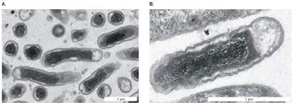
FIGURE 18.45 ■ Bacteroides fragilis cells colonize the human colon. B. fragilis is part of the normal gut community, but strains can cause intestinal infections (colorized SEM).
DENNIS KUNKEL MICROSCOPY/SCIENCE SOURCE
Their main source of energy is fermentation of a wide range of sugar derivatives from plant material, compounds that are indigestible by humans and potentially toxic (discussed in Chapter 13). Bacteroides species convert these substances into simple sugars and fermentation acids, some of which are absorbed by the intestinal epithelium; others feed associated gut bacteria such as Escherichia coli (discussed in Chapter 13). Thus, Bacteroides species serve important functions for their host: They break down potential toxins in plant foods, and their fermentation products make up as much as 15% of the caloric
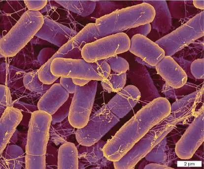
value we obtain from food. Yet another benefit of Bacteroides is their ability to remove side chains from bile acids, enabling the return of bile acids to the hepatic circulation. In effect, our gut bacteria, including Bacteroides, constitute a functional organ of the human body.
Bacteroides species cause trouble, however, when they reach parts of the body not designed to host them. During abdominal surgery, bacteria can escape the colon and invade the surrounding tissues. The displaced bacteria can form an abscess, a localized mass of bacteria and pus contained in a cavity of dead tissue. The interior of the abscess is anaerobic and often impenetrable to antibiotics.
Chlorobi
The Chlorobi (Chlorobiota) are known informally as green sulfur bacteria. While they are genetically close to Bacteroides, their metabolism is surprisingly different. Chlorobi are strict photolithotrophs, using PS I to split electrons from H 2, H 2 S, or other reduced sulfur compounds (Fig. 18.46; see also Table 18.3 ). During the oxidation of sulfide, Chlorobium species deposit elemental sulfur extracellularly, forming attached sulfur globules ( Fig. 18.46A). In contrast, the sulfur granules of Gammaproteobacteria such as Chromatium are deposited internally. FIGURE 18.46 ■ Chlorobium: Gram-negative green sulfur bacteria. A. Chlorobium sp. cells covered with sulfur
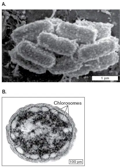
globules; cells form a cluster surrounding a nonphototrophic symbiotic bacterium. B. C. tepidum cell containing chlorosomes.
REPRINTED BY PERMISSION FROM SPRINGER NATURE: J. OVERMANN. 2010.
ADVANCES IN EXPERIMENTAL MEDICINE AND BIOLOGY, VOL. 675. SPRINGER, NEW
YORK
“CHLOROBIUM TEPIDUM MUTANT LACKING BACTERIOCHLOROPHYLL C MADE BY
INACTIVATION OF THE BCHK GENE, ENCODING BACTERIOCHLOROPHYLL C
SYNTHASE” BY N. FRIGAARD ET AL. 2002. J. BACTERIOL. 184 (12):3368–76; DOI:
10.1128/JB.184.12.3368-3376.2002. REPRODUCED WITH PERMISSION FROM
AMERICAN SOCIETY FOR MICROBIOLOGY
Chlorobium species require extensive membrane systems full of photopigments for light absorption. The chlorophyll reaction centers of Chlorobium are contained within chlorosomes associated with the cytoplasmic membrane (Fig. 18.46B ), similar to chlorosomes of the deep-branching phylum Chloroflexi. The photopigment of Chlorobium is predominantly bacteriochlorophyll c, which absorbs in the blue (460 nm) and near-infrared (750 nm), thus reflecting the middle range, brownish green.
Note: Distinguish phylum Chlorobi (sulfur photoautotrophs) from
phylum Chloro flexi (thermophilic filamentous photoheterotrophs). Both taxa are phototrophs containing green photopigment complexes arranged in chlorosomes, but they are deeply divergent genetically.
Fusobacteria
Fusobacteria (Fusobacteriota) is a phylum that includes virulent pathogens. Fusobacterium nucleatum was identified from dental plaque; this bacterium is the second most frequent cause of human abscesses (after Bacteroides). Within biofilms such as dental plaque, Fusobacterium species form carbohydrate bridges with numerous other kinds of bacteria, such as spirochetes, proteobacteria, and firmicutes, as well as eukaryotic pathogens such as fungi. These distantly related biofilm partners have transferred exceptional numbers of genes to the F. nucleatum genome.
Nitrospirae
The phylum Nitrospirae (Nitrospirota) consists of Gram-negative spiral bacteria that oxidize nitrite ion to nitrate (NO − to NO −).
2 3
Their cell structure resembles that of the Proteobacteria, although their phylogenetic branch is deep enough for assignment to a separate phylum. Most species, such as Nitrospira species (Tables 18.1 and 18.4 ), are lithotrophs that fix carbon in the form of carbon dioxide or carbonate using carboxysomes. Nitrospira species are generally found in freshwater or salt water. Their removal of excess nitrite makes a key contribution to aquatic ecosystems. Another important genus is Leptospirillum, which includes acidophilic iron oxidizers. Leptospirillum species are also strict autotrophs, fixing carbon by using Fe 2+ as their electron donor and O 2 as the electron acceptor. Their metabolism generates acid, contributing to acid mine drainage in iron mines at Iron Mountain, California, where they grow in massive pink biofilms.
To Summarize
The spirochete cell is a tight coil, enclosed by a sheath and periplasmic space containing periplasmic flagella.
Spirochete motility is driven by a flexing motion caused by rotation of the periplasmic flagella, propagated along the length of the coil.
Spirochetes grow in diverse habitats. Some are free-living fermenters in water or soil. Others are pathogens, such as Treponema pallidum, the cause of syphilis. Still others are endosymbionts of an animal digestive tract, such as the termite gut.
Acidobacteria are Gram-negative soil bacteria, including many acidophiles.
Bacteroidetes are anaerobes that ferment complex plant materials in the human colon. They may enter body tissues through wounds and cause abscesses.
Chlorobi are green sulfur phototrophs, obligate anaerobes incapable of heterotrophy.
Fusobacteria are pathogens that cause septicemia and skin ulcers.
Nitrospirae are Gram-negative spiral bacteria that oxidize nitrite to nitrate (Nitrospira).
Glossary
Spirochetes or Spirochaetota A phylum of bacteria with a unique morphology: a flexible, extended spiral that twists via intracellular flagella. Acidobacteria (Acidobacteriota)
A phylum of Gram-negative bacteria with an outer membrane, related to the Proteobacteria; often found in soil habitats. Bacteroidetes (Bacteroidota)
A phylum of Gram-negative bacteria; nearly all members are obligate anaerobes.
Chlorobi (Chlorobiota)
A phylum of Gram-negative bacteria. They are obligate anaerobes, “green sulfur” phototrophs that photolyze sulfides or H 2.
Fusobacteria (Fusobacteriota)
A phylum of anaerobic Gram-negative bacteria with an outer membrane, related to the Proteobacteria; includes human pathogens.
Nitrospirae (Nitrospirota)
A phylum of Gram-negative bacteria, many of which are lithotrophs, oxidizing nitrite to nitrate or ammonia to nitrate.
18.6 Planctomycetes, Verrucomicrobia, and Chlamydiaenot assigned
Several related phyla of bacteria—the P lanctomycetes, V errucomicrobia, and C hlamydiae—are known as the PVC superphylum. All major groups of the PVC superphylum possess compartmentalized cells with relatively diminished cell walls. Genomic evidence suggests they evolved from a common compartmentalized ancestor, yet their diverse cell form and metabolism show remarkable environmental adaptations.
Planctomycetes: A Nucleus-like Compartment
The Planctomycetes (Planctomycetota) evolved largely as free-living organisms. Planctomycetes are oligotrophs, heterotrophs requiring nutrients at extremely low concentrations. They grow in freshwater, marine, and saline environments. Their mechanism of osmoregulation remains poorly understood.
Some planctomycete cells possess multiple internal membrane compartments of unknown function (Fig. 18.47A). In these species, an internal membrane just inside the cell membrane divides the cytoplasm into concentric portions. A double membrane surrounds the entire nucleoid, analogous to the double membrane surrounding the eukaryotic nucleus—a remarkable example of independent analogous evolution between bacteria and the eukaryotes. The actual function of the nucleoid-surrounding membrane varies with different species; for example, in anammox planctomycetes, a single membrane enclosing the nucleoid contains the complexes that conduct the anammox reaction (anaerobic ammonium oxidation; discussed in Chapter 14).
Like eukaryotic protists (discussed in Chapter 20), the planctomycetes have flexible cell bodies that can assume diverse forms. Remarkably, in 2019, planctomycetes were found to engulf smaller microbes, analogous to the eukaryotic process of phagocytosis. Some Planctomyces species have rotary flagella (Fig. 18.47B ), whereas P. bekefii cells have stalks that attach to each other to generate a starlike aggregate. Most planctomycetes reproduce by budding, a strategy typical of eukaryotic yeasts.
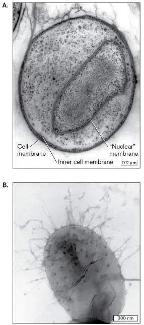
FIGURE 18.47 ■ Planctomyces: bacteria with a “nuclear membrane.” A. Section through the planctomycete Gemmata obscuriglobus, showing membrane compartmentalization (TEM). The DNA is contained within a double membrane analogous to a eukaryotic nuclear membrane. B. A swarmer cell of Planctomyces sp. with multiple flagella (TEM).
DR JOHN FUERST, UNIVERSITY OF QUEENSLAND
DR JOHN FUERST, UNIVERSITY OF QUEENSLAND
Verrucomicrobia: Warty Microbes
Verrucomicrobia (Verrucomicrobiota), or “warty microbes,” are irregularly shaped bacteria found in a wide variety of aquatic and terrestrial environments, as well as in the mammalian gastrointestinal tract (Fig. 18.48) . Most Verrucomicrobia are oligotrophs, growing heterotrophically in low-salt habitats; their wart-shaped protrusions may enhance nutrient uptake by increasing the cell’s surface area. Some Verrucomicrobia are ectosymbionts of protists, attached to the cell surface of the eukaryote, where they eject harpoon-like objects. They are rarely cultured, and in the past they failed to show up in PCR amplification of natural isolates, because their ribosomal DNA (rDNA) sequences are poorly amplified by the standard primer sequences. When appropriate primer pairs are used, Verrucomicrobia compose 5% of all surveyed microbial sequences in some natural environments.
FIGURE 18.48 ■ Verrucomicrobia: the “warty” bacteria. Verrucomicrobium spinosum with wart-like cell protrusions (colorized SEM).
DENNIS KUNKEL MICROSCOPY/SCIENCE SOURCE
Verrucomicrobia have peptidoglycan cell walls, but their shape is dominated by wart-like projections. The wart-like cytoskeleton appears to contain tubulin, a cytoskeletal protein previously believed to exist only in eukaryotes. In 2002, genes encoding tubulin were found in the partly sequenced genome of Prosthecobacter dejongeii, a free-living member of Verrucomicrobia. The genes appear so similar to those of eukaryotes that they must have undergone horizontal transfer from a eukaryotic genome. This horizontal transfer of a eukaryotic trait may be contrasted with the independent development of a nucleus-like structure in planctomycetes.
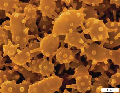
Chlamydiae: Intracellular Parasites
In the phylum Chlamydiae (Chlamydiota), the genus Chlamydia evolved complex developmental life cycles of parasitizing host cells. Chlamydia trachomatis causes the most prevalent sexually transmitted infection among young people in the United States. The same pathogen also causes trachoma, an eye disease dating back to records in ancient Egypt. The related species Chlamydophila pneumoniae causes pneumonia and has been implicated in cardiovascular disease.
Chlamydiae, or chlamydia, alternate between two developmental stages with different functions: elementary bodies and reticulate bodies (Fig. 18.49A). The form of chlamydia transmitted outside host cells is called an elementary body. Like endospores, elementary bodies are metabolically inert, with a compacted chromosome. While lacking a cell wall, they possess an outer membrane whose proteins are cross-linked by disulfide bonds, making a tough coat that provides osmotic stability. The elementary body adheres to a host cell surface and is endocytosed (Fig. 18.49B ).
FIGURE 18.49 ■ Chlamydia life cycle. A. Chlamydia trachomatis, multiplying within a human cell (colorized TEM). Infected cell, equivalent to step 5 in part B, contains reticulate
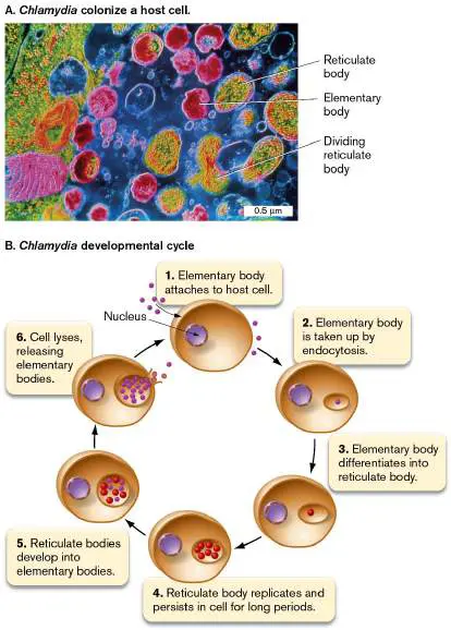
bodies (yellow) growing and dividing, as well as newly formed elementary bodies (red). B. Chlamydia spp. persist outside the host as a spore-like “elementary body.” Upon endocytosis, the elementary body avoids lysosomal fusion and develops into a “reticulate body” in which the DNA, now uncondensed, has a reticular (netlike) appearance. The reticulate body replicates within the host cytoplasm and then develops into new elementary bodies that are released when the cell lyses.
CNRI/SCIENCE SOURCE
To reproduce, the elementary body must transform itself into a reticulate body, named for the netlike appearance of its uncondensed DNA. The reticulate body has an active metabolism and divides rapidly, but outside the cell it is incapable of infection and vulnerable to osmotic shock. To complete the infection cycle, therefore, reticulate bodies must develop into new elementary bodies before exiting the host. When the host cell lyses, the elementary bodies are released to infect new cells. Chlamydiae infect a wide range of host cell types, from respiratory epithelium to macrophages.
Among the Bacteria, we have seen how some kinds evolved highly diverse metabolism and cell form. Others show relative uniformity in cell structure (Chlamydiae) or in metabolism (Cyanobacteria). Yet our attempts to generalize about any given clade inevitably run up against the unexpected appearance of exceptions, such as the heliobacteria, photosynthetic endospore formers that unaccountably branch from clostridia. Given the range of bacterial phenotypes, it is hard to imagine that yet more diverse forms of microbial life exist; but they do, as we will find among the Archaea (Chapter 19) and the microbial Eukarya (Chapter 20).
To Summarize
Planctomycetes include species that have evolved a membrane enclosing the nucleoid, analogous to the eukaryotic nuclear membrane.
Verrucomicrobia have cell projections containing tubulin. Their tubulin genes are thought to have arisen by horizontal transfer from a eukaryote.
Chlamydiae are obligate intracellular parasites that undergo a complex developmental progression, culminating in a spore-like form called an elementary body that can be transmitted outside the host cell.
Glossary
Planctomycetes (Planctomycetota)
A phylum of free-living bacteria that have stalked cells and reproduce by budding. Their nucleoid is surrounded by a membrane.
Verrucomicrobia (Verrucomicrobiota)
A phylum of free-living aquatic bacteria with wart-like, protruding structures containing tubulin.
chlamydia 1. A pathogenic bacterium lacking a cell wall; chlamydias (or chlamydiae) grow within human or animal cells, transmitting as elementary bodies to other cells. 2. A disease caused by chlamydia cells. The most frequently reported sexually transmitted disease in the United States. Symptoms range from none, to a burning sensation upon urination, to sterility. elementary body The endospore-like form of chlamydias transmitted outside host cells.
reticulate body The metabolically and reproductively active form of chlamydias. eResearch Activity 18
Do Fecal Firmicutes Rewrite the Genetic Code?
Among all the diverse bacteria, one kind of diversity we do not expect to find in nature is that of alternative genetic codes—codes that reassign a canonical codon to a different amino acid (see Chapter 8). While researchers might design such an organism, how could such codon reassignment naturally evolve, leading to thousands of mistaken proteins? In the relatively small genomes of mitochondria, such reassignments are known to include those of “sense” codons, which encode a specific amino acid. But in free-living bacteria, until now, only rare cases of stop-codon reassignments were known, such as the UGA stop codon being reassigned to glycine or tryptophan—never reassignment of a sense codon.
A Harvard graduate student, Yekaterina Shulgina (Fig. ERA 18.1 ) wondered whether sense-codon reassignments might be found among all the diversity of sequenced microbial genomes. Online databases now contain thousands of genomes and partial genomes of bacterial species, assembled computationally from the metagenomes of microbial communities. Shulgina and her doctoral advisor, Sean Eddy, devised a program, called Codetta, to screen 250,000 bacterial and archaeal genomes stored in the GenBank public database. These genomes have been sequenced from isolates and uncultured samples from all over the world. Shulgina screened these sequences by translating all six reading frames to possible peptide sequences (discussed in Chapter 9). The possible peptide sequences were aligned with 17,000 protein family sequences in the protein family database Pfam. The Codetta program is designed to flag genomes in which protein sequence alignments consistently mismatch one particular codon among homologous genes (orthologs) from different species.
FIGURE ERA 18.1 ■ Bacterial genomes show a reassigned codon. A. Yekaterina Shulgina, PhD student, Harvard University, discovered genomes of baboon and human fecal bacteria that contain AGG codons reassigned from arginine to methionine. B. Phylogenetic tree relates three genomes with AGG → Met reassignment, along with related outgroup genomes that show the standard genetic code.
Source: Y. Shulgina and S. R. Eddy. 2021. eLife 10 :p.e71402.
YEKATERINA SHULGINA
To the researchers’ surprise, Codetta found five clades of uncultured bacteria that reassigned an arginine codon to another amino acid. Three of the species reassigned arginine to methionine. Genome analysis classified the three species as Firmicutes (described in Section 18.3) of class Bacillales. All three genomes came specifically from fecal microbiomes of humans or baboons— why from these particular sources is unknown.
Three closely related genomes reassigned the arginine codon AGG; their phylogeny is shown in Figure ERA 18.1B . In the standard genetic code, arginine is specified by six alternate codons.

In bacteria, the AGG codon is usually the least often used, so the fewest proteins would lose function from its reassignment. Figure ERA 18.2A shows alignment of an orthologous protein sequence from three of the AGG → Met genomes, along with four closely related outgroup genomes and four standard well-known bacterial genomes. From this sample, and the full extended data, it is clear how AGG codons fill numerous positions that most often encode methionine.
FIGURE ERA 18.2 ■ Arginine codon reassignment in uncultured fecal Firmicutes. A. Partial protein sequences translated from genomes of uncultured bacteria (Fig. ERA 18.1B) obtained from baboon or human fecal microbiomes. Genomes with AGG → Met reassignment are aligned with outgroups and with more distant well-known bacterial species. Red “R” indicates AGG codons that appear to be reassigned from arginine to
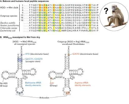
methionine. B. Structures of tRNA reassigned to methionine, compared with original tRNA assigned to arginine. Colored nucleotides indicate substitutions, numbered by tRNA sequence position. Each anticodon “CCU” base-pairs antiparallel to the codon “AGG.”
Source: Y. Shulgina and S. R. Eddy. 2021. eLife 10 :p.e71402.
GOODFOCUSED/SHUTTERSTOCK
What is the evolutionary mechanism of the reassignment? Did a methionine transfer RNA (Met-tRNA) gain a mutation in its anticodon? It would require only an A → C replacement to enable its anticodon to match AGG instead of the canonical codon AUG. In this study, however, no such anticodon mutation was found. Instead, the reassigned arginine tRNA actually mutated in the D-loop, a domain that mediates aminoacyl-tRNA synthetase recognition (Fig. ERA 18.2B ). The mutated D-loop now helps the synthetase attach methionine to the tRNA instead of arginine. It remains unknown—to us as well as the baboons—what selective pressures would favor such codon reassignment, adding yet another dimension of diversity in the bacterial domain.
Further Exploration
What aspects of the fecal bacterial genomes and their ecological context might favor codon reassignment? Would higher GC content favor replacement of AT-rich codons with GC-rich codons? Is codon reassignment more likely in organisms with smaller genomes? Could bacteria use such codon reassignment to foil phage infections, where the phage depend on a cell’s standard genetic code to synthesize their proteins?
Shulgina, Yekaterina, and Sean R. Eddy. 2021. A computational screen for alternative genetic codes in over 250,000 genomes. eLife 10 :e71402. CHAPTER REVIEW
Review Questions
1. Which deep-branching phyla include hyperthermophiles? Why is the actual branch position of these groups controversial?
2. Compare and contrast Chloroflexi and Cyanobacteria with respect to habitat, cell structure, and means of photosynthesis.
3. Compare and contrast the colonial and filamentous cyanobacteria with respect to life cycle and means of nitrogen fixation.
4. Name and describe three genera of Firmicutes that form endospores. Discuss the difference between single and multiple spore formers. Explain the existence of non-spore-forming species of Firmicutes.
5. Compare and contrast firmicute endospores and actinomycete arthrospores with respect to their means of production, their resistance properties, and their dispersal mechanisms.
6. Describe the diverse kinds of metabolism available to different species of Proteobacteria. Which Proteobacteria conduct lithotrophy? Anaerobic respiration? Which species are pathogens?
7. What do species of Bacteroidetes and Chlorobi have in common, and how do they differ?
8. Explain the structure and mechanism of motility typical in species of Spirochetes.
9. Discuss the properties of as many intracellular bacteria as you recall from various phyla. What do they have in common, and how do they differ?
10. Describe the unique cell structure of Planctomyces. Explain how the traits of planctomycete cells appear analogous to aspects of eukaryotic cells.
11. How do microbiologists seek to find previously unknown kinds of bacteria? What techniques reveal unknown microorganisms?
Thought Questions
1. Why are the Proteobacteria metabolically diverse? How is it possible that species of Alphaproteobacteria use so many kinds of molecules to yield energy?
2. How do you think Mycobacterium tuberculosis manages to grow, despite its thick envelope screening out most nutrients?
3. For motility, what are the relative advantages of external flagella versus the flexible spiral cells of spirochetes? 4. Why do different species of microbes grow together in layered biofilms (a) on a sand flat and (b) on the surface of human teeth?
Key Terms
acid-fast stain (739)
Acidobacteria (Acidobacteriota) (720, 756) Actinobacteria (Actinomycetota) (718) actinomycete (718)
aerial mycelium (736)
Alphaproteobacteria (718)
Aquificae (Aquificota) (720)
bacteroid (745)
Bacteroidetes (Bacteroidota) (720, 757) Betaproteobacteria (718)
Candidate Phyla Radiation (CPR) (722) carboxysome (724)
Chlamydiae (Chlamydiota) (720, 759) Chlorobi (Chlorobiota) (720, 757) Chloroflexi (Chloroflexota) (721) chlorosome (721)
Cyanobacteria (718)
deep-branching taxon (718)
Deinococcus-Thermus (Deinococcota) (721) Deltaproteobacteria (718)
diderm (728)
diplococcus (747)
elementary body (760)
emerging (721)
endospore (718)
Epsilonproteobacteria (718)
Firmicutes (Bacillota) (718)
forespore (729)
fruiting body (752)
Fusobacteria (Fusobacteriota) (720, 758) Gammaproteobacteria (718)
gas vesicle (724)
GC content (718)
heterocyst (724)
hormogonium (725)
leghemoglobin (744)
methanotrophy (744)
methylotrophy (743)
monoderm (728)
mother cell (729)
mycolic acid (738)
Mycoplasma (Mycoplasmatota) (718) myxospore (752)
Nitrospirae (Nitrospirota) (720, 758) phylum (717)
Planctomycetes (Planctomycetota) (720, 758) Proteobacteria (Pseudomonadota) (718) proteorhodopsin (742)
reticulate body (760)
sarcina (740)
Spirochetes (Spirochaetota) (720, 755) swarming (750)
Thermotogae (Thermotogota) (721) Ti plasmid (745)
vegetative cell (729)
vegetative mycelium (736)
Verrucomicrobia (Verrucomicrobiota) (720, 759)
Recommended Reading
Angert, Esther. 2006. Beyond binary fission: Some bacteria reproduce by alternative means. Microbe 1 :127–131.
Brown, Christopher T., Laura A. Hug, Brian C. Thomas, Itai Sharon, Cindy J. Castelle, et al. 2015. Unusual biology across a group comprising more than 15% of domain Bacteria. Nature 523 :208–211.
Claessen, Dennis, Daniel E. Rozen, Oscar P. Kuipers, Lotte Søgaard-Andersen, and Gilles P. van Wezel. 2014.
Bacterial solutions to multicellularity: A tale of biofilms, filaments and fruiting bodies. Nature Reviews Microbiology 12:115–124.
Gauthier, Anna E., Courtney E. Chandler, Valentina Poli, Francesca M. Gardner, Aranteiti Tekiau, et al. 2021. Deep-sea microbes as tools to refine the rules of innate immune pattern recognition. Science Immunology 6 :eabe0531.
Holtrop, Tadzio, Jef Huisman, Maayke Stomp, Levi Biersteker, Jeroen Aerts, et al. 2021. Vibrational modes of water predict spectral niches for photosynthesis in lakes and oceans. Nature Ecology & Evolution 5 :55–66.
Kindaichi, Tomonori, Tsukasa Ito, and Satoshi Okabe. 2004. Ecophysiological interaction between nitrifying bacteria and heterotrophic bacteria in autotrophic nitrifying biofilms as determined by microautoradiography-fluorescence in situ hybridization. Applied and Environmental Microbiology 70:1641–1650.
Luef, Birgit, Kyle R. Frischkorn, Kelly C. Wrighton, Hoi-Ying N. Holman, Giovanni Birarda, et al. 2015. Diverse uncultivated ultra-small bacterial cells in groundwater. Nature Communications 6 :6372.
Masaki, Toshihiro, Jinrong Qu, Justyna Cholewa-Waclaw, Karen Burr, Ryan Raaum, et al. 2013. Reprogramming adult Schwann cells to stem cell-like cells by leprosy bacilli promotes dissemination of infection. Cell 152 :51–67.
Pasolli, Edoardo, Francesco Asnicar, Serena Manara, Moreno Zolfo, Nicolai Karcher, et al. 2019. Extensive unexplored human microbiome diversity revealed by over 150,000 genomes from metagenomes spanning age, geography, and lifestyle. Cell 176 :649–662.
Schmitz, Alexa M., Brooke Pian, Sean Medin, Matthew C. Reid, Mingming Wu, et al. 2021. Generation of a Gluconobacter oxydans knockout collection for improved extraction of rare earth elements. Nature Communications 12:1–11.
Schnupf, Pamela, Valérie Gaboriau-Routhiau, Marine Gros, Robin Friedman, Maryse Moya-Nilges, et al. 2015. Growth and host interaction of mouse segmented filamentous bacteria in vitro. Nature 520 :99–103.
Shams ul Hassan, Syed, Komal Anjuma, Syed Qamar Abbas, Najeeb Akhter, Bibi Ibtesam Shagufta, et al. 2017.
Emerging biopharmaceuticals from marine actinobacteria. Environmental Toxicology and Pharmacology 49 :34–47.
Shiratori, Takashi, Shigekatsu, Suzuki, Yukako Kakizawa, and Ken-ichiro Ishida. 2019. Phagocytosis-like cell engulfment by a planctomycete bacterium. Nature Communications 10 :5529.
Soo, Rochelle M., James Hemp, Donovan H. Parks, Woodward W. Fischer, and Philip Hugenholtz. 2017. On the origins of oxygenic photosynthesis and aerobic respiration in Cyanobacteria. Science 355 :1436–1440.
Wiegand, Sandra, Mareike Jogler, and Christian Jogler. 2018. On the maverick Planctomycetes. FEMS Microbiological Reviews 42 :739–760. http://doi.org/10.1093/femsre/fuy029.
Glossary
acid-fast stain A diagnostic stain for mycobacteria, which retain the dye fuchsin because of mycolic acids in the cell wall.
Acidobacteria (Acidobacteriota)
A phylum of Gram-negative bacteria with an outer membrane, related to the Proteobacteria; often found in soil habitats. Actinobacteria (Actinomycetota)
A phylum of Gram-positive bacteria with high GC content. actinomycete A member of the Actinomycetales, an order of Actinobacteria that includes branched spore formers such as Streptomyces, as well as irregularly shaped corynebacteria.
aerial mycelium A mass of hyphae (branched filaments) that extend above the surface and produce spores at the tips.
Alphaproteobacteria A class of Proteobacteria (Pseudomonadota) that includes, for example, rhizobia and rickettsias.
Aquificae (Aquificota)
A phylum of hyperthermophilic bacteria.
bacteroid A cell wall–less, undividing, differentiated rhizobial cell within a plant cell. The bacteroid provides fixed nitrogen for the plant. Bacteroidetes (Bacteroidota)
A phylum of Gram-negative bacteria; nearly all members are obligate anaerobes.
Betaproteobacteria A class of Proteobacteria (Pseudomonadota) that includes, for example, species of Neisseria and Burkholderia.
Candidate Phyla Radiation (CPR)
A recently described group of bacterial phyla whose members represent broad genetic diversity but small genome size and limited metabolic capabilities.
carboxysome A protein-enclosed compartment containing Rubisco to fix CO 2.
Chlamydiae (Chlamydiota)
A phylum of intracellular parasitic bacteria that grow only within a host cell and generate multiple spore-like structures that escape to infect the next host.
Chlorobi (Chlorobiota)
A phylum of Gram-negative bacteria. They are obligate anaerobes, “green sulfur” phototrophs that photolyze sulfides or H 2.
Chloroflexi (Chloroflexota)
A phylum of bacteria that are filamentous phototrophs having chlorosomes.
chlorosome A membranous photosynthetic organelle found in some “green” bacteria of the phyla Chloroflexi and Chlorobi.
Cyanobacteria or Cyanophyta A phylum of oxygen-producing photoautotrophic bacteria containing chlorophylls a and b. They share an ancient ancestor of chloroplasts.
deep-branching taxon A lineage whose genome sequences diverged early, at or before the well-known phyla.
Deinococcus-Thermus (Deinococcota)
A phylum of bacteria that are resistant to ionizing radiation and high temperature.
Deltaproteobacteria A class of Proteobacteria (Pseudomonadota) that includes, for example, the Desulfobacterales (sulfate reducers) and the myxobacteria.
diderm Enclosed by two phospholipid membranes—an inner membrane and an outer membrane.
diplococcus The paired cocci of Neisseria species.
elementary body The endospore-like form of chlamydias transmitted outside host cells.
emerging Describing an organism or other entity that is newly isolated, defined, or recognized, as in “emerging clade,” “emerging pathogen,” or “emerging disease.”
endospore A durable, inert, heat-resistant spore that can remain viable for thousands of years.
Epsilonproteobacteria A class of Proteobacteria (Pseudomonadota) that includes, for example, species of Campylobacter and Helicobacter.
Firmicutes (Bacillota)
A phylum of Gram-positive bacteria with relatively low GC content.
forespore In sporulation of Gram-positive bacteria, the smaller cell compartment formed through asymmetrical cell division; it develops into the endospore.
fruiting body A multicellular fungal or bacterial reproductive structure. Fusobacteria (Fusobacteriota)
A phylum of anaerobic Gram-negative bacteria with an outer membrane, related to the Proteobacteria; includes human pathogens.
Gammaproteobacteria A class of Proteobacteria (Pseudomonadota) that includes, for example, the Enterobacteriales (enteric facultative anaerobes) and the pseudomonads.
gas vesicle An organelle that traps gases to increase the buoyancy of aquatic microbes.
GC content The proportion of an organism’s genome consisting of guanine-cytosine base pairs.
heterocyst In filamentous cyanobacteria, a specialized nitrogen-fixing cell that maintains a reducing environment and excludes O 2. hormogonium pl. hormogonia A short, motile chain of three to five cells produced by filamentous cyanobacteria to disseminate their cells. leghemoglobin An iron-bearing plant protein that sequesters oxygen to maintain an anoxic environment for nitrogenase within cells containing bacteroids.
methanotrophy The metabolic oxidation of methane to yield energy.
methylotrophy The metabolic oxidation of single-carbon compounds such as methanol, methylamine, or methane to yield energy.
monoderm Enclosed by a single phospholipid membrane.
mother cell In sporulation of Gram-positive bacteria, the larger cell that forms during the asymmetrical cell division leading to spore formation. The mother cell will engulf the forespore, but then disintegrates as the forespore matures.
mycolic acid One of a diverse class of sugar-linked fatty acids found in the cell envelopes of mycobacteria such as Mycobacterium tuberculosis.
Mycoplasma (Mycoplasmatota)
A phylum of bacteria related to Firmicutes and which lack a cell wall. Most known species are class Mollicutes, genus Mycoplasma.
myxospore A durable spherical cell produced by the fruiting body of myxobacteria.
Nitrospirae (Nitrospirota)
A phylum of Gram-negative bacteria, many of which are lithotrophs, oxidizing nitrite to nitrate or ammonia to nitrate. phylum pl. phyla The taxonomic rank one level below domain; a group of organisms sharing a common ancestor that diverged early from other groups.
Planctomycetes (Planctomycetota)
A phylum of free-living bacteria that have stalked cells and reproduce by budding. Their nucleoid is surrounded by a membrane.
Proteobacteria (Pseudomonadota)
A large, metabolically and morphologically diverse group of Gram-negative bacteria; possess an outer membrane containing LPS.
proteorhodopsin A bacterial membrane-embedded protein that contains retinal and acts as a light-driven proton pump; it is homologous to the archaeal protein bacteriorhodopsin.
reticulate body The metabolically and reproductively active form of chlamydias. sarcina pl. sarcinae A cubical octad cluster of cells formed by septation at right angles to the previous cell division.
Spirochetes or Spirochaetota A phylum of bacteria with a unique morphology: a flexible, extended spiral that twists via intracellular flagella. swarming or swarming motility A behavior in which some microbial cells differentiate into large swarmer cells and swim together as a unit.
Thermotogae (Thermotogota)
A phylum of thermophilic bacteria; in some species, the cell is enclosed by a toga-shaped outer covering.
Ti plasmid A plasmid found in tumorigenic strains of Agrobacterium tumefaciens that can be used as a vector to introduce DNA into plant cells.
vegetative cell A metabolically active, replicating bacterial cell.
vegetative mycelium A mass of hyphae (branched filaments) produced by vegetative cells that expand into the substrate.
Verrucomicrobia (Verrucomicrobiota)
A phylum of free-living aquatic bacteria with wart-like, protruding structures containing tubulin.