Chapter introduction
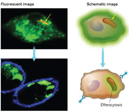
“I’m infected, eat me!” A phagocyte (macrophage; top panel, green fluorescent dye) infected with Mycobacterium tuberculosis (yellow arrow) undergoes a cell death program
Imagine two people swimming in the Gulf of Mexico—a healthy 12-year-old girl and a 66-year-old man with liver disease. While they swim, Gram-negative marine bacteria called Vibrio vulnificus enter their bodies through tiny cuts. Four days later, the girl returns to her Mississippi home, oblivious to the immunological battle recently waged in her bloodstream. The man, however, lies in a morgue, dead of an aggressive V. vulnificus blood infection. Why did one person live and the other die? A variety of nonspecific, innate immune factors present in the girl’s body killed the invading pathogen before it could multiply. The man with liver disease was deficient in several of those defense mechanisms—mechanisms that The human body teems with microbes vital to our existence (the human microbiome) but is also under constant attack from pathogens. How do we survive? Nonspecific defenses such as skin and stomach acid stop most microorganisms from infecting sensitive internal organs. But microbes that manage to breach those physical barriers are met with powerful innate and adaptive immune defenses. Chapter 23 begins by introducing the amazing, organ-like human microbiome, and it then describes the remarkable series of physical barriers and elaborate innate immune defenses (including phagocytes) that keep our microbiota and invading pathogens at bay.
23.1 The Human Microbiomenot assigned
From the moment of our birth to the time of our death, we are constantly populated by microbes. These include bacteria, archaea, fungi, viruses, and even some protozoa. The consortium of colonizing microbes has been dubbed the human microbiota, or microbiome. These microbes have a stake in keeping us, their hosts, alive. The fact that the human microbiome has been conserved over millennia is testament to their importance.
Note: “Microbiome,” “microbiota,” and “metagenome” were
all defined in Chapter 21. Chapter 23 presents the human microbiome as a special case. “Microbiota” refers to the community of microbes inhabiting a body site, whereas “microbiome” encompasses the community of microbes and their genes.
To illustrate the significance of the microbiome, consider that our bodies carry about as many bacterial cells (1−10 × 10 12) as human cells and about 100 times more nonredundant bacterial genes than human genes.
Microbes colonize wherever our body meets the external environment (for example, skin, mouth, gastrointestinal tract, and parts of the genitourinary tract; Fig. 23.1). Most internal organs, blood, and cerebrospinal fluid are considered sterile, and the presence of any bacteria at these sites is considered an infection. The majority of species that make up our microbiota are unknown and, as of now, have never been grown in the laboratory. Consequently, metagenomic strategies (described in Chapters 7 and 21) are being employed to identify our resident microbes. In 2016, the U.S. government announced the National Microbiome Initiative to advance understanding of how microbiomes contribute to our health and the environment.
FIGURE 23.1 ■ Relative amounts of microbial phyla and families present at various colonizing sites. Each pie chart presents the relative compositions of bacterial phyla, fungal genera, or viruses present in each microbiome as determined by the sequencing of genes encoding 16S ribosomal RNA (see Sections 7.6 and 17.5). Microbial taxa are identified by color in the legend. Note that this figure does not reflect the recent renaming
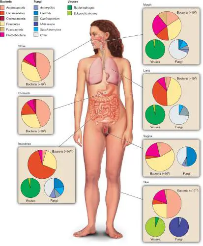
of bacterial phyla. The revised phylum names are presented in eAppendix 3. The total number of bacteria in the human microbiome is approximately 4 × 10 13. Estimates of bacteria present in each organ are shown.
Source: Data from Marsland and Gollwitzer. 2014. Nat. Rev. Immunol. 14 :827.
Note: Although our internal organs, blood, and cerebrospinal fluid
are devoid of bacteria, fungi, and most forms of viruses, tiny viruses such as anelloviruses (nonenveloped, single-stranded, circular DNA genomes of negative sense) have been found in the blood and some tissues of many healthy people.
Can members of our microbiome cause us harm? By and large, the body’s barriers of defense work well to prevent incursions by microbiota or, in the event of an incursion, to kill the invader. Unfortunately, these defenses break down when an immune system is compromised by medical treatments (such as anticancer drugs) or by diseases (for instance, a deficiency in complement factors, discussed in Section 23.6). Such a person is described as a compromised or immunocompromised host and can be repeatedly infected by certain normal biota. Organisms causing disease in this situation are called opportunistic pathogens. Later we will discuss how dysbiosis, an imbalance in the relative numbers of species in the microbiome, can negatively impact health.
We now understand that bacteria colonizing our bodies may be as important collectively as a kidney or liver. Microbiota in the gut, for instance, can communicate chemically with the brain and affect the function of the endocrine, nervous, and immune systems, while also affecting metabolism in a variety of ways (see Special Topic 13). The communication pathway (called the microbiome-gut-brain axis) takes place via the vagus nerve that connects the brain to the intestine and a number of other organs. Another more pervasive mode of communication was described by Gary Siuzdak and his colleagues at the Scripps Research Institute. They discovered that our body cells are continually bathed in, and respond to, microbial metabolites that circulate in our blood.
Are all of these host-microbe communications important? A variety of studies suggest that microbiota and their products have roles in allergies, liver disease, obesity, gastrointestinal syndromes (such as inflammatory bowel disease), and possibly psychiatric syndromes such as autism and depression. Its vast impact on human health has led some to call our microbiota a new organ-like system, and the term “holobiont” is used to describe a human paired with their microbiota.
Microbiome Acquisition and the Hygiene Hypothesis
Two seemingly unrelated questions have puzzled scientists for years: How does a microbiome take shape, and why do we experience more allergies and inflammatory diseases today than we did 100 years ago? It turns out that the answers to these questions are connected. When and how does a person develop a microbiome? A considerable controversy has emerged over when a human first develops a gut microbiome. Does it happen before or after birth? Studies that examined the first stool (meconium) passed by a newborn before its first meal did find some bacteria. However, a more recent study using sterile cotton swabs to collect feces directly from 20 fetuses during (not after) cesarean delivery found no evidence of bacteria. Whether meconium in the earlier reports may have been inadvertently contaminated is unknown.
However the controversy is resolved, it is clear that once babies break out of the embryonic membrane, they are exposed to a dizzying array of microbes residing in the birth canal and the outside world. Contributions to babies’ skin, mouth, gut, and genitourinary-tract microbiomes come from the food they eat, the air they breathe, and the people, places, and things they touch. The initial makeup of a neonate’s microbiome is shaped, in part, by the mode of delivery. Natural-birth babies are initially colonized by microbes acquired by passage through the mother’s vagina. In contrast, babies born by cesarean section start with microbes donated by the delivery room and by contact with the mother’s skin. However, the microbiome of a newborn is not static. Young babies have microbiomes that are less diverse than those of adults, but by 5 years of age, the species diversity increases in complexity to assume an adultlike composition.
Figure 23.1presents the major human body sites (skin, respiratory, digestive, and genitourinary tracts) that are colonized by microbes and illustrates the relative makeup of bacteria, fungi, and viruses that populate each system. As you view the figure, notice the dramatic difference in levels of Bacteroidetes found in nasal and intestinal microbiomes or of Candida in the microbiomes of the mouth and vagina. Although Archaea are missing from this figure, they too are present at various host sites (intestine, skin, nose, and lung). Representatives from five archaeal phyla have been identified, but they comprise only a small portion of the entire microbiome. Their contributions to human health are unknown but under investigation. With all of its complexity, a person’s microbiome remains relatively constant over time but can significantly fluctuate with diet, age, geography, or drug use.
The human virome. A significant component of the human microbiome is the human virome, comprised of phages that prey on bacteria and viruses that infect archaea and human cells. The number of viruses in the virome ranges between 0.1 and 10 times the number of bacterial cells, with most being bacteriophages of both lytic and temperate varieties. These include Caudovirales (tailed phages) and Microviridae (icosahedral, untailed phages). Viruses that infect human cells include those able to cause acute infection, others that establish latent infection, and some “long-term passengers” that are not associated with any disease. Metagenomic studies have exposed a number of new passenger viruses, such as the Anelloviridae family, that populate a number of tissues including blood but have yet to be grown in pure culture.
Much like bacteria, viruses begin to colonize newborns at birth. Infants born vaginally generally have a more diverse virome than babies delivered by cesarean. Not surprisingly, the first members are bacteriophages, usually arising from induction of prophages within bacterial lysogens. Human viruses come later. Surprisingly, human viruses associated with diarrheal disease (rotavirus, picornavirus, calicivirus, and adenovirus) are commonly detected in healthy infant guts. As with bacteria, virome diversity is influenced by diet, host genetics, geography, and medications. We are also learning that alterations in the virome can trigger some autoimmune diseases. For instance, a reproducible expansion of Caudovirales phage and a reduction of Microviridae are associated with inflammatory bowel disease. Whether the shift is a cause or effect of the disease is not clear.
The hygiene hypothesis. For millennia, human microbiota had been shaped by human contact with natural environments composed of animals, caves, dirt, poop, and bugs. This natural outdoor world harbored a vast array of microbial taxa that could compete to populate our skin and mucosal surfaces. Today we are mostly an indoor species, spending almost all of our time inside closed buildings, segregated from nature—an arguably less diverse microbial environment. Add to that our use of soaps, antibiotics, and disinfectants, and you can appreciate how severely we have restricted our access to microbes. As a result, our microbiota appear less diverse than those of our long-ago ancestors.
Studies that support this idea have compared the modern-day human microbiome with those of closely related wild African apes and of uncontacted Amerindians (see Chapter 27). Although improved hygiene limits exposure to pathogens, studies suggest that narrowing the diversity of our microbiome can contribute to inflammatory diseases such as asthma, inflammatory bowel disease, colorectal cancer, and obesity. Several intriguing studies have found that, even today, children who grow up on dairy farms are less likely to develop allergies and asthma. The reason is that our microbiome helps train our immune system (discussed later and in Chapter 24). Exposure to more microbes and other environmental antigens, especially early in life, may produce a more tolerant, well-controlled immune system less prone to inflammatory and autoimmune diseases (disorders in which the immune system reacts against the self).
The sections that follow describe the microbiota of various body sites and discuss the benefits and risks of a microbiome. Many of the bacterial taxa mentioned here, such as Gram-negative rods and Staphylococcus, are discussed individually in Chapter 18.
Skin
The average human adult is covered with 2 square meters (over 21 square feet) of skin (epidermis) populated by 10 12 –10 13 microorganisms. Skin microbiota include aerobes, anaerobes, and facultative bacteria. There are, for instance, approximately 10 4 –10 5 microbes per sweat gland, at a ratio of 1 aerobe to 10 facultative or anaerobic species. As with all colonized body sites, resident (normal) and transient members of the microbiota inhabit the skin. But even a resident microbe exhibits diversity as different strains colonize at different times.
Several features of epidermis make it difficult to colonize. The skin has an acidic pH (pH 4–6) owing to the secretion of organic acids by oil and sweat glands. Organic acids inhibit microbial growth by lowering bacterial cytoplasmic pH (see Section 5.3). Epidermal secretions are also high in salt and low in water activity (see Section 5.2), and they contain enzymes, such as lysozyme, that degrade bacterial peptidoglycan. Despite these hurdles, many species of bacteria manage to colonize the epidermal habitat. Most of these are Gram-positive organisms, because they tend to be more resistant to salt and dryness. Large expanses of dry skin tend to support the growth of Betaproteobacteria. Moister areas, such as scalp, ear, armpit, genital, and anal regions, are colonized primarily by Corynebacterium species and Staphylococcus epidermidis.
Skin microbiota benefit us by prompting the expression of epithelial cell tight-junction proteins, modulating functions of the immune system (immunomodulation), and secreting antibacterial peptides. For instance, Staphylococcus hominis and Staphylococcus epidermidis, friendly members of the skin microbiome, can kill Staphylococcus aureus, a pathogen that can form abscesses and a variety of other infections. S. epidermidis also helps the host produce ceramides, the main constituents in skin that prevent dehydration and aging.
Some members of the skin microbiome, however, can be problematic. One such member is the Gram-positive, anaerobic rod Cutibacterium acnes (formerly Propionibacterium acnes), which causes acne, a very visible plague of adolescence. Increased hormonal activity in teenagers stimulates oil production by the sebaceous glands (Fig. 23.2). C. acnes readily degrades the triglycerides in this oil, turning them into free fatty acids that then promote inflammation of the gland. One consequence of the inflammatory response is the formation of a blackhead, a plug of fluid and keratin that forms in the gland duct. The result is the typical skin eruptions of acne. Because of its microbial basis, treatments for acne include tetracycline (oral) or clindamycin (topically applied) to kill the bacteria.
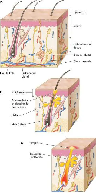
FIGURE 23.2 ■ Microbiology of skin and the development of acne. A. The location of sebaceous glands in normal skin. B. Dead cells and sebum can block the pore. C.
Cutibacterium acnes can infect, inflame, and further block the gland duct, leading to blackheads and acne. D. Acne.
SPL/SCIENCE SOURCE
Eye
The eye is exposed to the outside environment, so is it heavily colonized? Actually, it is not, because colonization is inhibited by the presence of antimicrobial factors, such as lysozyme, in the tears that continually rinse the eye surface (conjunctiva). Despite this protection, a few transient commensal bacteria can be found on the conjunctiva. Skin microbiota such as Staphylococcus epidermidis and diphtheroids (Gram-positive rods that look like clubs), as well as some Gram-negative rods, such as Escherichia coli, Klebsiella, and
Proteus, manage, at least temporarily, to make the eye their home without causing damage. (The traits of these genera are discussed in Chapter 18.)
Oral and Nasal Cavities
Within hours after birth, a human infant’s mouth becomes colonized with nonpathogenic Neisseria species (Gram-negative cocci), Streptococcus, Actinomyces, Lactobacillus (all Gram-positive), and some yeasts. These organisms come from the environment surrounding the newborn, such as the mother’s skin and garments. As teeth emerge in the newborn, the anaerobic space between teeth and gums supports the growth of anaerobes, such as Prevotella and Fusobacterium (Fig. 23.3B and C ). Colonizers of the oral cavity adhere to surfaces, like teeth and gums, to avoid mechanical removal and flushing into the acidic stomach. The teeth and gingival crevices are colonized by 500–700 species of bacteria.
FIGURE 23.3 ■ Oral microbiota and periodontal disease. A. Structures of the oral and nasal cavities with examples of normal microbiota listed. B, C. Two anaerobic bacteria associated with periodontal disease. B. Prevotella (colorized TEM; each cell
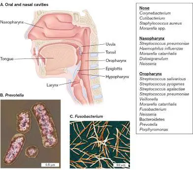
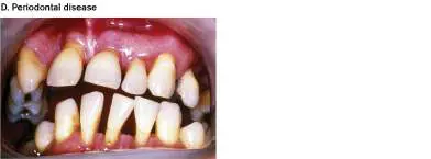
approx. 2 μm long). C. Fusobacterium (SEM; each cell approx. 1–5 μm long). D. Symptoms of periodontal disease include red and swollen gums, bleeding gums, gum shrinkage, and teeth drifting apart.
SCIENCE VU/VISUALS UNLIMITED
BSIP/SCIENCE SOURCE
BIOPHOTO ASSOCIATES/SCIENCE SOURCE
Organisms such as Streptococcus mutans (which attaches to tooth enamel) and Streptococcus salivarius (which binds gingival surfaces) form a glycocalyx that enables them to firmly adhere to oral surfaces and to each other. They are but two of the microbes that lead to dental plaque formation. The acidic fermentation products of these organisms demineralize teeth and cause dental caries (tooth decay). Important microbial habitats of the throat include the nasopharynx, which is the area leading from the nose to the oral cavity, and the oropharynx, which lies between the soft palate and the upper edge of the epiglottis (Fig. 23.3A). Figure 23.3includes a listing of common inhabitants of these sites.
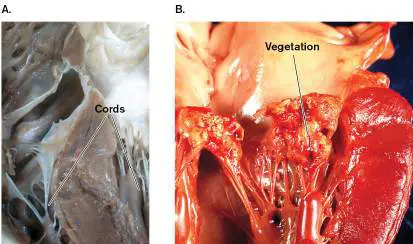
The oral microbiota are normally harmless, but they can cause disease. Dental procedures, for instance, will often cause these organisms to enter the bloodstream, producing what is called bacteremia (infection of the bloodstream). Normal immune mechanisms typically clear these transient bacteremias quite easily, but in patients who have a mitral valve prolapse (heart murmur), microbes can occasionally become trapped in the defective valve and form bacterial vegetations. Vegetations are biofilms that contain a large number of bacterial cells encased within glycocalyx (a polysaccharide or peptide polymer secreted by the organism) and fibrin (produced by clotting blood). Because the onset of disease is often insidious (slow), it is called subacute bacterial endocarditis ( Fig. 23.4). Once ensconced within a vegetation, the microbes are extremely difficult to kill with antibiotics. Fortunately, immunocompetent individuals are at very low risk of valve infection. Immunocompromised patients, however, are at much higher risk and receive prophylactic treatment with antibiotics before any dental procedure.
FIGURE 23.4 ■ Gross pathology of subacute bacterial endocarditis involving the mitral valve. A. An open, normal mitral valve shows cords that tether it to the heart wall. B. The left ventricle of the heart has been opened to show mitral valve fibrin vegetations due to infection. These growths are not present in a normal heart.
VIDEOSURGERY/SCIENCE SOURCE
SCIENCE SOURCE
Thought Question
23.1 How can an anaerobic microorganism grow on skin or in the mouth, both of which are exposed to air?
Respiratory Tract
The average adult respiratory tract has a surface area of approximately 75 square meters (800 square feet) directly exposed to the environment. Originally thought to be sterile, the lungs and trachea are now recognized to harbor normal microbiota. Estimates in the lower respiratory tract are 10–100 bacteria per 1,000 human cells, the most prominent members being Prevotella (Bacteroidetes), Streptococcus (Firmicutes), Veillonella (Firmicutes), and Sphingomonas (Proteobacteria).
Many organisms entering the nasopharynx become trapped in the nose by cilia that beat toward the pharynx. The microbes are propelled toward the acidic stomach and death. Microorganisms that slip into the trachea are trapped by mucus produced by ciliated epithelial cells lining the airways. Cilia usher the microbes up and away from the lungs. The ciliated mucous lining of the trachea, bronchi, and bronchioles makes up the mucociliary escalator (Fig. 23.5), which constantly sweeps foreign particles up and out of the lungs. Respiratory diseases such as asthma reduce the diversity of species in the lung microbiome.
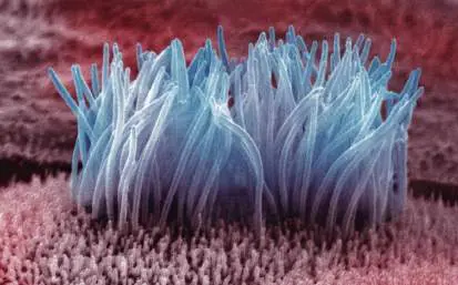
FIGURE 23.5 ■ Mucociliary escalator. Movement of these hairlike cilia ushers particles up and out of the trachea and lungs (colorized SEM; diameter from 0.5–1 μm).
DR. DAVID M. PHILLIPS/VISUALS UNLIMITED
The mucociliary escalator is extremely important for preventing respiratory infections. When it fails, as when it is covered with tar from years of heavy smoking, or when it is overwhelmed by the inhalation of too many infectious microbes, infections such as the common cold (for example, rhinovirus) or pneumonia (for example, Streptococcus pneumoniae) can result. Patients who smoke and develop chronic obstructive pulmonary disease (COPD) have a significantly altered lung microbiome. Decreased mucociliary clearance leads to enrichment with Pseudomonas species. In contrast, patients with asthma have a microbiome enriched with Proteobacteria. The significance of these changes is not clear.
Genitourinary Tract
Much of the genitourinary tract is normally free from microbes. These areas include the kidneys used to remove waste products from the blood and the ureters that remove urine from the kidneys. The urinary bladder, which holds urine until it is excreted, was thought to be sterile but is now known to harbor microbes, mainly anaerobes. The distal urethra, however—because of its proximity to the outside world—normally contains Staphylococcus epidermidis, Enterococcus species, and some members of Enterobacteriaceae. Some of these organisms can cause bladder disease (known as urinary tract infection, or UTI) if they make their way into the bladder (for example, via catheterization).
The large surface area and associated secretions of the female genital tract make it a rich environment for microbes, including bacteria and yeasts. Firmicutes are the largest bacterial contributor to the vaginal microbiome; however, the microbiome’s composition changes with the menstrual cycle, owing to changing nutrients and pH. The mildly acidic nature of vaginal secretions (approximately pH 4.5) discourages the growth of many bacteria. As a result, the acid-tolerant Lactobacillus crispatus is among the most populous vaginal species.
Healthy women appear to fall into two broad categories: 70% have lactobacillus as the primary member of their vaginal microbiota, while 30% have mixed species with few lactobacilli. Women in the latter group appear to be more susceptible to sexually transmitted infections. A study of the endometrium, where fertilized eggs are implanted, found that a Lactobacillus dominance of at least 90% correlates well with reproductive success.
The balance between different species that comprise vaginal and endometrial microbiota is crucial to preventing disease. Antibiotic therapy to treat an infection anywhere in the body can also affect the vaginal microbiota. The resulting imbalance can allow overgrowth of Candida albicans, otherwise known as a yeast infection. C. albicans, as a fungus, is not susceptible to antibiotics designed to kill bacteria. Lastly, some studies indicate that the microbiome of a sexual partner’s penis can also influence the incidence of bacterial vaginosis.
Stomach
We have known since 1824 that the stomach contents are acidic due to hydrochloric acid (pH 1–3) and since 1925 that gastric acidity can kill bacteria. Just how important that acidity is for protection against microbes is illustrated by the infection caused by Vibrio cholerae, the causative agent of cholera. Cholera is a severe diarrheal disease endemic to many of the poorer countries of the world. Although cholera actually affects the intestines, not the stomach, the bacteria must survive passage through the stomach to reach the intestines. The organism, however, is extremely acid sensitive. Healthy volunteers must ingest a trillion organisms before contracting disease.
Despite the high infectious dose, cholera epidemics in developing countries kill tens of thousands of people every year. Part of the reason is that the poor, malnourished populations in these countries suffer from hypochlorhydria (decreased stomach acid). The less acidic stomach gives ingested microbes more time to enter the intestine, where they can thrive and cause devastating disease. Compare the astronomical number of acid-sensitive V. cholerae needed to cause disease to the mere ten organisms needed to develop diarrheal disease from Shigella, a very acid-resistant pathogen.
Although the stomach contents are very acidic, the mucous lining of the stomach is much less so. It is there that some bacteria can take refuge, primarily Actinobacteria and Firmicutes (see Fig. 23.1). In fact, the stomach harbors a diverse microbiota, as detected by cultural and molecular techniques. Estimates are between 10 and 1,000 organisms per gram.
A classic stomach pathogen is Helicobacter pylori. This organism has a remarkable ability to resist acidic pH. (It survives at pH 1 using the enzyme urease to generate ammonia, which neutralizes acid.) H. pylori will not grow in strong acid pH conditions, but it can grow in the mucous lining of the stomach, where the pH is closer to 5 or 6 ( Fig. 23.6). The U.S. Centers for Disease Control and Prevention (CDC) estimates that Helicobacter colonizes the stomachs of half the world’s population. Most of the time, Helicobacter does not cause any apparent problem, but on occasion, the organism can produce gastric ulcers and even cancer. On the plus side, data suggest that H. pylori colonization in children is associated with a reduced risk for allergic disease.
FIGURE 23.6 ■ Helicobacter pylori defies stomach acidity. H. pylori growing in the mucus on stomach epithelium (colorized SEM). H. pylori bacteria attach to gastric epithelial cells and induce specific changes in cell function, such as an increase in
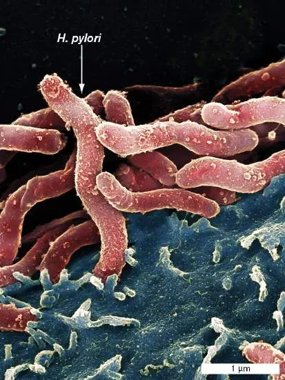
the expression of laminin receptor 1, a protein associated with malignancy. Peptic ulcers can result.
SPL/SCIENCE SOURCE
Intestine
The intestine is an extremely long tube (approximately 7.5 meters, or 25 feet) consisting of several sections, each of which supports the growth of different combinations of bacterial species. Pancreatic secretions (at pH 10) enter the intestine at a point just past the stomach (pH 1–3) and raise the intestinal pH to about pH 8. The relatively high pH and bile content of the duodenum and jejunum allow colonization by only a few resident, mostly Gram-positive, bacteria (enterococci, lactobacilli, and diphtheroids). These particular Gram-positive organisms (Firmicutes phylum) possess a bile salt hydrolase that helps them grow in the presence of intestinal bile salts.
The distal parts of the human intestine (ileum and colon) have a slightly acidic pH (pH 5–7) and a lower concentration of bile salts— conditions that support a more diverse ecosystem. The intestine actually contains roughly 10 11 –10 13 bacteria per gram of feces, mostly anaerobes (1,000 anaerobes to 1 facultative organism). Why is the intestinal lumen anaerobic? The small amount of oxygen that diffuses from the intestinal wall into the lumen is immediately consumed by the facultative bacteria, such as E. coli, thereby rendering the environment anaerobic (discussed in Section 21.4). Most of the data used to inventory members of the gut microbiome, as well as microbiota from other systems, come from culture-independent molecular techniques. Segments of 16S ribosomal RNA genes are randomly amplified by PCR from DNA extracted from fecal specimens. Sequences from individual amplicons are used to identify the phyla, species, or operational taxonomic unit (OTU) of bacteria in the gut. As noted earlier, culture-independent sequencing was necessary because most members of the microbiome were considered unculturable. However, microbial ecologists have devised novel techniques of culturing previously uncultured microbes from all kinds of environmental communities, including the human microbiome (see Section 21.2). For instance, a culture enrichment strategy devised by Michael Surette (McMaster University) and colleagues may change how we inventory a microbiome.
These scientists collected fecal material from volunteers and smeared dilutions from each sample onto 66 different kinds of highly enriched and pre-reduced agar media. Duplicate plates were incubated under aerobic and anaerobic conditions. The colonies that grew were removed from the agar plates and mixed together, and their DNA was extracted. PCR was used to amplify a common segment of the many bacterial 16S rRNA genes in the mixture, and the individual amplicons were sequenced to determine distinct species or OTUs. Using this culture enrichment strategy, Surette and his group managed to culture 95% of the gut microbiota predicted by culture-independent techniques to be at >0.1% abundance. Their work also revealed a much greater bacterial diversity than was observed from previous culture-independent techniques. Altogether, there are approximately 500–1,000 different species of bacteria in the human gut microbiome, over 90% of which are from the Proteobacteria, Firmicutes, Actinobacteria, and Bacteroidetes phyla (see eAppendix 3.9 for the list of recently renamed bacterial phyla). This new culture enrichment approach will enable scientists to grow previously unknown (or uncultured) microbiome members and test their importance to human health.
Acquisition of gut microbiota. Infant intestines are initially colonized by large numbers of E. coli and streptococci that quickly generate a reducing environment able to support growth of strict anaerobic species—mainly Bifidobacterium, Bacteroides, Clostridium, and Ruminococcus. The intestines of breast-fed babies are dominated by bifidobacteria, possibly because of growth factors present in breast milk. In contrast, the microbiota of formula-fed infants is more diverse, with high numbers of Enterobacteriaceae, enterococci, bifidobacteria, Bacteroides, and clostridia. As seen in Figure 23.1, by adulthood over 90% of the bacteria in the intestine are composed of just two phyla: Bacteroidetes (for example, Bacteroides species) and Firmicutes (for example, Clostridiales clusters XIV and IV), followed by Proteobacteria (for example, Escherichia coli) and Actinobacteria (for example, Bifidobacterium species). Besides bacteria, other inhabitants are the yeast species Candida albicans and protozoa such as Trichomonas hominis and Entamoeba hartmanni.
Some 1,000 different bacterial species and a few methanogenic archaea comprise the intestinal microbiome. One reason the intestine can support such a large and eclectic mix of species is that different bacteria attach to different host cell receptors. Another reason is that many different food sources are available to support diverse groups of microbes (discussed in Section 21.4). Even human breast milk evolved to encourage the growth of specific subpopulations of microbiota. Breast milk contains carbohydrates (lactose) that nourish babies but also includes complex carbohydrates that only our microbiota can digest.
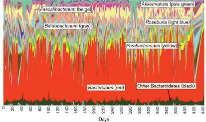
Makeup of the gut microbiome within a single adult individual is relatively constant but still varies over time. Figure 23.7follows the fecal biota from a single individual over a period of 14 months. The relative proportions of bacterial families fluctuate significantly but tend to return to typical adult composition. Across the spectrum of the human population, however, the composition of intestinal microbiota is a continuum of many different combinations of microbes (enterotypes). At one end of the spectrum are microbiomes dominated by Bacteroides, while at the other end are communities dominated by Prevotella (another Gram-negative anaerobic species). One study showed, for example, that people with high protein and animal-fat diets have fecal communities enriched with the Bacteroides -dominant enterotype, while those with carbohydrate-rich diets have fecal communities dominated by Prevotella. The significance of this difference is not yet clear.
FIGURE 23.7 ■ Variability of gut microbiota over time. Daily fecal samples taken from one individual were analyzed to identify and quantify resident bacterial families by sequencing 16S rRNA genes. Selected genera are identified. Colored segments at each time point represent the proportions of specific genera relative to the entire population. The depth of a colored segment along the y -axis reflects that microbe’s proportional contribution to the whole microbiome. Thus, the major genus at most time points is Bacteroides (red).
CAPORASO ET AL. 2011. GENOME BIOL. 12 :R50. © CAPORASO ET AL. LICENSEE
BIOMED CENTRAL LTD. 2011.
Does a host’s genetic makeup influence composition of the gut microbiome? Available evidence indicates that host genetics plays only a small role. Environmental considerations such as diet, cholesterol levels, and blood glucose are more predictive of gut microbiota. However, a small number of bacterial taxa are strongly heritable. For instance, Christensenella minuta (a Gram-negative anaerobe) is the most highly heritable bacterial species among identical twins (see Section 7.6). “Heritability” here does not mean that the organism was passed from mother to siblings. The term reflects the ability of a host’s genome to sustain the presence of this
organism. The organism is more likely to be maintained in two identical twins than in two non-twin siblings from the same mother. Keeping our microbiome at bay. Clearly, our intestines are packed with microbes. Why don’t they cause chronic intestinal inflammation? Part of the answer was discovered by Lora Hooper ( Fig. 23.8A) and colleagues at the University of Texas Southwestern Medical Center. Whenever bacteria contact epithelial cells, the human cells secrete lectins (carbohydrate-binding proteins) into the mucus layer lining the intestine. The lectins kill bacteria that get too close ( Fig. 23.8B ). Hooper’s group discovered that one of the antimicrobial lectins, RegIIIα (Fig. 23.8B inset; cryo-EM map), forms a membrane-penetrating pore in Gram-positive microbes. The enforced separation of microbiome and host mucosal cells minimizes the likelihood that members of the microbiome will activate an immune response that could severely damage the intestinal lining. Other immune mechanisms that control microbiota in the intestinal mucosa, such as secretory IgA antibodies, are described in Chapter 24. FIGURE 23.8 ■ Keeping the gut microbiome at bay. A. Lora Hooper discovered the mechanism of an antimicrobial lectin. B. Separation of the microbiome from the intestinal mucosal surface. Bacteria are green (FISH using a DNA probe that hybridizes to bacterial 16S rRNA genes), and the nuclei of intestinal mucosal cells are blue (DAPI stained). Inset: Model of the Reg III α pore that forms in Gram-positive cell membranes.
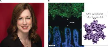
COURTESY OF LORA HOOPER
VAISHNAVA ET AL. 2011. SCIENCE 334 :255.
Thought Questions
23.2 Why do many Gram-positive microbes that grow on the skin, such as Staphylococcus epidermidis, grow poorly or not at all in the gut?
23.3 How might you provide evidence that the intestinal microbiome communicates with the brain via the vagus nerve? Hint: The vagus nerve includes afferent nerves that send information from organs to the brain and efferent nerves that regulate gastrointestinal secretion and gut endocrine activity.
To Summarize
Opportunistic pathogens infect only compromised hosts. The normal microbiota present on skin and mucosal surfaces is acquired at birth but changes over a lifetime. Skin microbiota consists primarily of Gram-positive microbes, including Cutibacterium acnes, which can cause acne. Oral and nasal surfaces are colonized by aerobic and anaerobic microbes. Vaginal microbiota influences susceptibility to sexually transmitted infections.
Areas of the body considered to be free of bacteria include blood, cerebrospinal fluid, and internal organs. However, some tiny, mostly harmless viruses can be found circulating in the blood.
Normal microbiota can cause disease if organisms gain access to the circulation or deeper tissues.
The intestine is populated by 10 11 –10 13 microbes per gram of feces. Principal phyla are Firmicutes, Bacteroidetes, Proteobacteria, and Actinobacteria. The ratio of anaerobes to facultative bacteria is 1,000:1. The gut microbiome includes bacteria, archaea, fungi, and viruses.
Glossary
microbiota or microbiome The total community of microbes associated with an organism (such as the human body) or with a defined habitat (such as soil or plants).
microbiota or microbiome The total community of microbes associated with an organism (such as the human body) or with a defined habitat (such as soil or plants).
compromised or immunocompromised host An animal with a weakened immune system.
opportunistic pathogen A microbe that normally is not pathogenic but can cause infection or disease in an immunocompromised host organism.
epidermis The outer protective cell layer in most multicellular animals. nasopharynx The passage leading from the nose to the oral cavity.
oropharynx The area between the soft palate and the upper edge of the epiglottis.
bacteremia A bacterial infection of the blood.
mucociliary escalator The ciliated mucous lining of the trachea, bronchi, and bronchioles that sweeps foreign particles up and away from the lungs.
23.2 Benefits and Risks of Microbiotanot assigned
Our colonizing microbes are not hostile armies camped at our body’s gates (also called portals of entry) waiting to invade. These microorganisms are of great benefit to us, and we, as good hosts, reciprocate. Microbes in our gut, for instance, have enzymes to catabolize foods that we cannot digest (as presented in Chapter 13). Some bacteria synthesize vitamins that we cannot make [E. coli, for instance, makes vitamin B 2 (riboflavin) and vitamin K]. In addition, chemical signals released by members of the skin microbiome have been shown to promote host tissue development and healing. Figure 23.9shows the results of using Vaseline or neomycin to treat damage left by circular punch biopsies in three volunteers. After 15 days of treatment, the wounds treated with Vaseline healed faster than wounds treated with antibiotic.
FIGURE 23.9 ■ Skin microbiome enhances wound healing in humans. Identically sized (4-mm) punch biopsies were removed from behind the knees of six patients (three are shown, bottom panels). One wound on each patient (top left photo for each) was treated with petroleum jelly (Vaseline), while the other wound (top right photo for each patient) was treated with the antimicrobial neomycin (Neosporin) to reduce the skin microbiome in the area. Wound healing after 15 days of treatment is shown in the upper panels. Unrepaired tissue is marked by
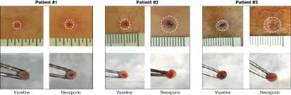
circles. In each case, the Vaseline-treated wound healed faster than the Neosporin-treated wound.
Source: Geofeng Wang et al. 2021. Cell Host and Microbe 29 :1–15; fig. 1A, top
panels. https://doi.org/10.1016/j.chom.2021.03.003
G. WANG ET AL. 2021. CELL HOST MICROBE 29 :777–791
G. WANG ET AL. 2021. CELL HOST MICROBE 29 :777–791
G. WANG ET AL. 2021. CELL HOST MICROBE 29 :777–791
A study from Rockefeller University found that certain lipids made by gut bacteria interact with host membrane receptors and affect host metabolism, immune cell differentiation, immune cell traffic through the body, and tissue repair. Even the short-chain fatty acid products of fermentation affect host gene expression and can influence host immune system function. Acetate, for instance, tends to promote inflammation, whereas butyrate tends to inhibit inflammation.
We, for our part, maintain our gut microbiome by secreting complex carbohydrates that members of the microbiome can use and chemical factors, such as hormones, that can alter microbiome gene expression.
Examples of Beneficial Gut Microbiota
An important gut microbe that promotes host tissue rejuvenation is Akkermansia muciniphila, a Gram-negative, anaerobic bacterium (Verrucomicrobia phylum) that degrades mucin, the glycosylated protein layer that covers gut epithelium. Mucin degradation by A. muciniphila fosters continuous rejuvenation of the protective mucin layer. The short-chain fatty acids produced by this organism feed epithelial metabolism and modulate inflammatory responses of immune cells.
Another important microbe, the anaerobe Bacteroides thetaiotaomicron, metabolizes many of the complex carbohydrates we eat, breaking them down into products that can be absorbed by the body or used by other members of the microbiome. We humans actually absorb 15%–20% of our daily caloric intake in this way.
Our gut microbiome also limits infections by competing with pathogens for food sources and for attachment receptors on host cells. Symbiotic bacteria can also make antimicrobial compounds that limit growth of potential pathogens. For example, nonpathogenic gut E. coli produces bacteriocin (a secreted protein toxin), which directly inhibits growth of the related pathogen enterohemorrhagic E. coli (EHEC; see Chapter 25).
Gut microbes also influence the development and efficiency of our immune system. The intestinal microbe Faecalibacterium prausnitzii, a member of the order Clostridiales (Firmicutes phylum), comprises up to 5% of total fecal microbiota in adults. The organism elicits powerful anti-inflammatory effects on human immune cells through secreted bacterial factors and interactions with Toll-like receptors (see Section 23.5). For example, bacterial factors can inhibit induction of the inflammatory cytokine IL-8 (cytokines are small, secreted host proteins that regulate immune cell function; see Section 23.5) and increase the population of anti-inflammatory T-cell lymphocytes (regulatory T cells; see Chapter 24). The absence of this organism in human intestines has been linked to painful inflammatory intestinal diseases such as Crohn’s disease.
Another important group of intestinal anaerobes consists of the segmented filamentous bacilli (SFBs, “ Candidatus Savagella”) in mice and humans (see Special Topic 18). These spore formers are important for developing a healthy immune system. We discuss the connections between gut microbiota and the immune system more fully in Chapter 24. The anti-inflammatory effects of these and other microbes protect the intestinal mucosa from an overzealous immune system.
The Microbiome-Gut-Brain Axis
Nerves that connect the human gastrointestinal tract to the central nervous system (CNS) form the gut-brain axis (Fig. 23.10). The axis is composed of the CNS, the neuroendocrine and autonomic nervous systems, the enteric nervous system, and the gut microbiota. The vagus nerve is an important part of the gut-brain axis. Neurochemicals made by the brain initiate efferent vagus nerve impulses that travel from the brain to the gut, whereas gut bacteria make neurotransmitter-like chemicals that can fire afferent signals back along the axis to the brain. Consequently, brain activity can control gut functions, but the microbiome can also influence brain function. This so-called microbiome-gut-brain axis can alter human health in several ways.
FIGURE 23.10 ■ The gut-brain axis. Signals through the gut-brain axis of nerves, such as the vagus nerve, pass back and forth from the central nervous system (CNS) to the gut.
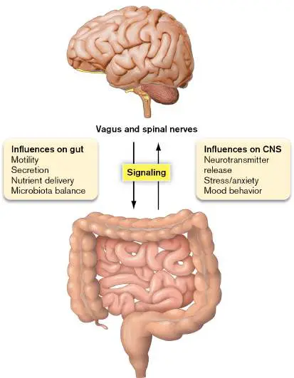
Gut microbes, for instance, may affect your mood by making compounds that mimic feel-good neurotransmitters such as dopamine or chemicals associated with fear (5-hydroxytryptamine) or anger (4-aminobutanoic acid, commonly referred to as GABA because of its former name, “gamma-aminobutyric acid”). Some microbial products can even influence the production of host receptors associated with depression. Metagenomic studies with humans have identified three genera of Firmicutes in the gut that correlate with depression. Faecalibacterium and Coprococcus were more prevalent in “happy” individuals, while people identified as depressed were depleted for Coprococcus and Dialister. (However, recall that correlation does not prove causation.)
The gut microbiome may also influence behaviors such as the craving for certain foods—even for chocolate! After a gastric bypass, for example, the gut microbiome undergoes a dramatic shift that can alter a patient’s food cravings. Most lose interest in treats and fatty foods. It is thought that altering the microbiome composition will change the cocktail of microbial neurosignaling compounds made and that the new microbial products will alter how the gut-brain axis is stimulated. The result is stimulation of brain areas that influence cravings.
And then there’s the famous phenomenon of strange food cravings during pregnancy, such as pickles and ice cream at 3:00 a.m. Hormones released by the mother and fetus during pregnancy can alter the mother’s gut microbiome and, consequently, tweak the gut signals sent along the vagus nerve. Taste bud receptors can be changed, for instance. While these studies are exciting, it is important to realize that definitive proof for gut bacteria affecting mood and behavior is lacking.
Intriguing research also suggests a link between gut dysbiosis (an imbalance in microbiome composition) and the development of certain neurological diseases. For instance, Parkinson’s disease is a neurodegenerative disease characterized by the accumulation of specific intracellular protein inclusion bodies (Lewy bodies) that destroy neurons in the CNS. The hypothesis is that intestinal dysbiosis can trigger a form of inflammation that initiates Lewy body formation in the intestinal section of the vagus nerve. These protein aggregates then travel up the nerve to eventually reach the brain stem. Similar potential links have also been made between dysbiosis of the gut microbiome and Alzheimer’s disease (see eResearch Activity 23).
Containment Breaches and Dysbiosis
Everything works well as long as the composition of your microbiome is balanced and stable. However, if certain organisms penetrate beyond a site of colonization or some form of dysbiosis develops, the result can be an infection or inflammatory disease, respectively. A cancerous lesion in the colon, for example, can provide a passageway for microbes to enter deeper tissues and cause infection. Bacteroides fragilis, usually a harmless anaerobe in the gut, and even Escherichia coli can invade tissues through surgical wounds, causing intra-abdominal abscesses and even gangrene after abdominal surgery. Other infections caused by certain gut microbes escaping the intestine include urinary tract infections (cystitis), septicemia, and meningitis.
Aside from causing infections after escaping the intestine, some bacteria in the gut can carry genes whose products stimulate cancerous growths. For example, Cynthia Spears from Johns Hopkins University (Fig. 23.11A) and her colleagues have been studying how strains of Bacteroides fragilis that produce the oncotoxin BFT ( Bacteroides fragilis toxin) stimulate the growth of microadenomas in mice (Fig. 23.11B and C ) and in the colons of people with the genetic disease familial adenomatous polyposis. In humans, immune responses to colon biofilms that contain oncotoxin-producing strains of B. fragilis or E. coli attract a myeloid cell type whose secreted growth factors accelerate tumor growth.

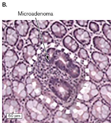
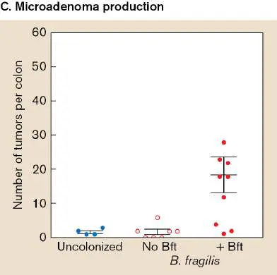
FIGURE 23.11 ■ Potential negative effects of intestinal dysbiosis. A. Cynthia Sears studies the role of oncotoxin-producing strains of Bacteroides fragilis and Escherichia coli on stimulating tumorigenesis in humans and mice. B. A microadenoma in a mouse colon. C. The incidence of microadenomas found in uncolonized mice, in mice colonized with B. fragilis lacking BFT toxin, and in BFT-producing B. fragilis. D. The firmicute Clostridioides difficile is often a minor component of the normal gut microbiome whose numbers are limited by competing gut microbiota. However, antibiotic treatment can kill
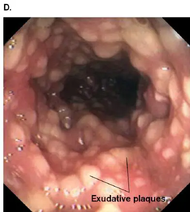
off the competing microbes, leaving the antibiotic-resistant C. difficile to grow unabated. Toxins made by C. difficile will kill host cells, causing small exudative plaques to form on the intestinal wall. Plaques coalesce to form a large, bloody, and painful pseudomembrane.
COURTESY OF CYNTHIA SEARS
CHUNG, ET AL. 2018. CELL HOST MICROBE 23 :203–214, FIG. 4F
DAVID M. MARTIN M.D./SCIENCE SOURCE
External factors that can alter the balance and diversity of the gut include emotional stress, a change in diet, and antibiotic therapy. The resultant dysbiosis can lead to poor digestion; to pseudomembranous enterocolitis, an infectious disease caused by Clostridioides difficile (formerly Clostridium difficile; Fig. 23.11D ); or to Crohn’s disease. Even non-antibiotic drugs such as proton pump inhibitors, nonsteroidal anti-inflammatory drugs (NSAIDs), anti-diabetics (metformin), and atypical antipsychotics can perturb microbiome balance. One study that screened 1,000 drugs found that 24% inhibited growth of one or more gut microbes.
Some people favor restoring natural microbial balance by orally ingesting living microbes such as the lactobacilli present in foods (yogurt, for instance) or supplements called probiotics (from the Greek meaning “for life”).The most commonly used probiotic genera are Lactobacillus and Bifidobacterium (Fig. 23.12). The potential mechanisms by which probiotics improve intestinal health include competitive bacterial interactions that prevent the growth of pathogenic bacteria, production of antimicrobial compounds, and immunomodulation (an ability to change the activity of cells of the immune system). One study even found that the probiotic organism Lactobacillus rhamnosus GG does not alter microbiome composition but orchestrates broad transcriptional changes in other members of the microbiome—functions that could promote anti-inflammatory pathways in the resident microbes.
FIGURE 23.12 ■ Commonly used probiotic microorganisms. A. Bifidobacterium (note the Y shape of some cells). B. Lactobacillus acidophilus. Colorized SEMs.
SCIMAT/SCIENCE SOURCE
SCIMAT/SCIENCE SOURCE
Probiotics are now being tested for treating several gastrointestinal disorders, including inflammatory bowel disease (IBD). Realize, though, that using probiotics such as live-culture yogurt to treat complex diseases like IBD is still controversial. An extreme but clinically proven form of delivering probiotic microbes to the intestine is the so-called fecal transplant, which is more precisely termed fecal bacteriotherapy or fecal microbiota transplantation (FMT). In its original forms, fecal transplants transferred the intestinal microbiome of a healthy person to a relative suffering from a severe intestinal disease such as “pseudomembranous enterocolitis.” Restoring a “normal” microbiome in this way has successfully cured patients suffering from repeated C. difficile infections that did not respond to antibiotics. Today the procedure uses a “superdonor” rather than relatives, and it may someday be a personalized bacterial cocktail delivered in pill form. Probiotics in the form of vaginal suppositories are also used to treat patients prone to vaginal infections.
The Link between Obesity and Dysbiosis
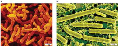
Obesity is epidemic in industrialized and developing countries. Over 500 million people worldwide are obese and predisposed to developing related diseases, such as diabetes, cardiovascular disease, nonalcoholic fatty liver disease, cancer, and some immune disorders. Obesity involves complex, puzzle-like interactions among genes, diet, and a long-term imbalance between energy intake and expenditure, which together yield an excessive increase in body fat. Compelling evidence now points to gut microbiota playing a prominent role in obesity by influencing nutrient acquisition, regulating energy metabolism, and affecting fat storage.
Many studies showing connections between gut microbiota and obesity use gnotobiotic animals. A gnotobiotic animal is a germ-free animal or an animal in which all the microbial species present are known because they were added by the experimenter. To develop a gnotobiotic colony of animals, offspring must be delivered by cesarean section under aseptic conditions in an isolator. The newborn is moved to a separate isolator where all entering air, water, and food are sterilized. Once gnotobiotic animals are established, the colony is maintained by normal mating between the members. Fecal organisms and fecal content can be transplanted orally to these animals by syringe and stomach tube (gavage). After the transplant, the effects on animal physiology and disease can be observed. Fecal transplants include mouse-to-mouse transfers or even human-to-mouse transfers (discussed shortly).
Tantalizing evidence suggesting a link between the gut microbiome and obesity goes back decades. For instance, livestock farmers used to add antibiotics in low doses to animal feed as a way to promote animal growth (a practice no longer permitted with medically important antibiotics because of the potential for generating antibiotic-resistant microbes). In another example, treating obese mice with high doses of antibiotics to remove microbiota reduces mouse body weight and improves glucose metabolism. Such studies suggested that altering an animal’s microbiome with antibiotics can increase or decrease weight, depending on circumstances.
Newer metagenomic studies find that obese humans and mice carry a less diverse gut microbiome and have an increased capacity to absorb energy compared to their lean counterparts. The ratios of major bacterial phyla in the colon also seem to differ between obese and lean individuals. For instance, the gut microbiomes of obese children have Firmicutes/Bacteroidetes phylum ratios that are higher than those of lean children. Studies looking at this ratio among adults are less clear. Some find that the Firmicutes/Bacteroidetes ratio is higher in obese adults than in lean adults. Other studies indicate the opposite, and some show no difference. The real key to microbiome effects on obesity may be the difference in species makeup and metabolic output rather than broad phylum differences.
Can your microbiome keep you thin? A landmark study from Jeffrey Gordon (Fig. 23.13; Washington University School of Medicine in St. Louis) and colleagues demonstrated that obesity in humans is clearly linked to their microbiomes. The scientists collected fecal contents from sets of monozygotic human twins, one of whom was obese and the other lean, and then orally transplanted the fecal preps into germ-free mice (Fig. 23.14). Mice receiving fecal microbiota from the lean human co-twin remained lean, as measured by change in fat mass (“Ln” in Fig. 23.14). However, mice receiving fecal microbiota from the obese co-twin gained adiposity over a mere 10-day period (“Ob” in Fig. 23.14). Even more remarkable, when Ob and Ln co-twin fecal-transplanted mice were cohoused (“ch”) in a single cage immediately after transplant, the Ob ch mice gained less weight than the control Ob mice.
FIGURE 23.13 ■ Jeffrey I. Gordon, MD. Gordon (second from left) is a recipient of the 2015 Keio Medical Science Prize. He stands here with some of the researchers who work in his lab.
JAMES BYARD/WUSTL PHOTOS
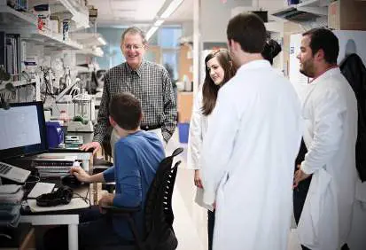
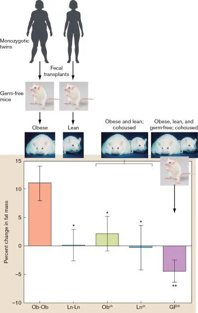
FIGURE 23.14 ■ Adiposity of germ-free mice transplanted with fecal preparations from human twins.
Fecal contents from sets of monozygotic human twins—one obese (Ob), the other lean (Ln)—were transplanted orally into germ-free (GF) mice. Mice were cohoused (ch) in different combinations. Changes in fat mass were measured after 10 days. * = P value ≤0.05; ** = P ≤0.01.
Source: (Germ-free mice) tiripero/Getty Images; (obese and lean mice) Human
Genome wall for SC99 at http://www.ornl.gov or Wikimedia.
Metagenomic analysis of fecal contents from the mice revealed that the microbiome of Ob ch mice came to resemble that of the Ln ch mice. Essentially, the microbiota of the Ln ch mouse invaded the Ob ch mouse and displaced the original microbiome. Transfer took place because mice are naturally coprophagous (eat feces). Even though Ln ch mice also consumed Ob ch mouse feces, the Ln ch fecal microbiome did not change. The most successful Ln ch invaders of the Ob ch microbiota were Bacteroides species.
Another aspect of this study asked what would happen to germ-free (GF) mice if they were cohoused with Ob and Ln fecal-transplanted mice. The authors found that the germ-free mice developed gut microbiota similar to their Ln ch cage mates and lost weight (see Fig. 23.14). This confirmed that the Ln-derived microbial taxa had greater fitness over the Ob-derived taxa. The scientists concluded that a higher Firmicutes/Bacteroidetes ratio favors obesity, while the opposite favors leanness. Their studies also found that as fat people lose weight, the proportion of Bacteroidetes to Firmicutes in the gut microbiome increases.
How might the microbiome influence obesity? Microbiota can influence obesity by affecting two major processes: the harvesting of energy from ingested foods, and the triggering of intestinal inflammation.
As noted in Chapter 21, the metabolic dexterity of intestinal microbes allows them to digest many foods that we cannot. In the process, these microbes produce short-chain fatty acids (SCFAs) that our cells use in a variety of ways. The major SCFAs are butyrate, propionate, and acetate. Numerous investigations suggest that the amounts and ratios of these SCFAs influence obesity (Fig. 23.15).
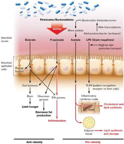
FIGURE 23.15 ■ Effects of microbiome products on weight gain. Members of the gut microbiome ferment ingested complex carbohydrates and produce short-chain fatty acids (SCFAs). SCFAs have varied effects on hunger, fat production, and inflammation. LPS from Gram-negative bacteria can be transported across the mucosal barrier and interact with pattern recognition receptor TLR4. The inflammatory cytokines made can affect liver metabolism. Arrows and terms in red indicate processes that promote weight gain.
Generally, acetate is thought to promote obesity. Acetate is absorbed well by intestinal epithelial cells, travels to the liver where it is used for lipid synthesis, and is distributed throughout the body to be used as a substrate for cholesterol synthesis (particularly in adipose tissues). Butyrate, however, seems to prevent obesity. It is used primarily by intestinal epithelial cells for energy and stimulates production of gut hormones that limit hunger. Butyrate also dampens synthesis of the pro-inflammatory cytokine interferon-gamma (IFN-gamma), described in Sections 23.5 and 24.3. Propionate, for its part, has effects that can either stimulate or prevent obesity. This SCFA stimulates IFN-gamma synthesis, which promotes inflammation associated with obesity. But propionate is also involved in gluconeogenesis, an anti-obesity process that diverts fatty acids away from cholesterol synthesis and toward glucose synthesis. In addition, propionate stimulates the production of anti-obesity gut hormones that limit hunger. So, is there a correlation between SCFA levels in the colon and obesity?
A study from Elena Comelli’s lab at the University of Toronto found that a cohort of obese or overweight patients generally had higher levels of SCFAs (propionate, butyrate, and acetate) than did healthy, lean patients on similar diets, and that SCFA levels did correlate with Firmicutes/Bacteroidetes ratios. The more Firmicutes there were, the more SCFAs were produced. However, obese and lean subjects actually had similar Firmicutes/Bacteroidetes ratios, suggesting that the most important factor is which species of Firmicutes and Bacteroidetes are present (and what they are doing), rather than the overall phylum ratios.
Archaea in the gut might also tilt metabolic balance toward obesity. About 50% of humans have significant numbers of methane-producing archaea, primarily Methanobrevibacter smithii (methanogens are discussed in Chapter 21). This archaeon can comprise up to 10% of the anaerobes in the colons of healthy adults. Bruce Rittmann and colleagues from Arizona State University found that a small group of obese patients had significantly more archaea than either lean or gastric-bypass patients had. This finding raised the possibility that archaea may play a role in obesity. But how? Jeffrey Gordon’s laboratory provides a possible explanation. His team discovered that the archaeon M. smithii dramatically altered the transcriptome of Bacteroides thetaiotaomicron when the two organisms co-colonized germ-free mice. The methanogen shifted the carbohydrate priority of Bacteroides from glucans to fructans, which stimulated more acetate production. Higher levels of acetate could enhance adiposity as outlined already. How the archaeon modulated the anaerobe’s metabolism is not known. Another hypothesis is that methanogens in the gut can rapidly consume the H 2 end product of bacterial fermentation. Removing H 2 removes feedback control over fermentation. The resultant increase in carbohydrate fermentation yields even more acetate that can contribute to weight gain. In addition to affecting the harvesting of energy, inflammation induced by gut microbes appears to influence the development of obesity and diabetes. Obese individuals are in a chronic state of low-grade inflammation, characterized by elevated blood and tissue levels of pro-inflammatory cytokines and markers of inflammation such as C-reactive protein (discussed in Section 23.6). The source of inflammation is thought to be outer membrane lipopolysaccharides (LPS), also called endotoxin, produced by Gram-negative members of the gut microbiome. A high-fat diet promotes absorption of endotoxin across the intestinal epithelium and its distribution to tissues via blood. LPS interacts with a cell receptor called TLR4, present in a variety of host cells (see Section 23.5). LPS-TLR4 interaction induces production of pro-inflammatory cytokines, such as IL-6, and reactive oxygen species that interfere with the normal metabolism of liver, adipose, and skeletal muscle, all of which are important to the regulation of glucose and lipid homeostasis.
While the presence of some bacterial groups in the gut correlate with energy intake, others, such as Faecalibacterium prausnitzii, influence obesity by altering inflammation. For example, the presence of F. prausnitzii, a major producer of butyrate in the gut, reduces the low-grade inflammation associated with obesity and diabetes. Even culture supernatants of F. prausnitzii fed to mice were able to lower inflammation in chemically induced colitis models. Butyrate in the supernatant was one anti-inflammatory factor, but others, including a 15-kDa protein called microbial anti-inflammatory molecule, are suspected.
Can individual members of a lean-associated microbiome treat obesity? Despite the tremendous insights we have gained about the link between obesity and the microbiome, the data suggest that different blends of microbiota will be necessary to decrease calorie harvesting from the diet, alter how derived energy is stored, and minimize chronic inflammatory processes. It is unlikely that a single “silver bullet” species exists. Research is under way to identify specific members of the microbiome that might treat diseases such as IBS (irritable bowel syndrome), IBD, or perhaps obesity. For example, Section 7.6 describes the identification of the intestinal Firmicute Christensenella minuta from human twins, which prevents weight gain when transferred to germ-free mice. The idea is that select species can be harvested from a healthy microbiome and administered in pill form to patients in a probiotic-like treatment. There is also great hope that we may one day cure or prevent various disorders of dysbiosis using phage to manipulate the microbiome. Phages naturally play a major role in maintaining gut homeostasis by regulating bacterial abundance, diversity, and metabolism; they are also key drivers of horizontal gene transfer and bacterial evolution. One way to modulate microbiome diversity would be to ingest phage that specifically target unwanted bacteria (see Chapter 5).
To Summarize
Benefits of the microbiome include interfering with pathogen colonization, producing immunomodulatory proteins, metabolizing foods that the host cannot process (energy harvesting), producing vitamins that the host cannot make, and honing our immune system.
Infections such as septicemia, abscesses, cystitis, and meningitis can be caused by some members of the microbiota if they breach the body’s containment mechanisms. Dysbiosis of the microbiome can contribute to infection, obesity, and inflammatory and autoimmune diseases.
Probiotics are foods or solutions containing helpful members of a microbiome. Different probiotics can restore balance to the intestinal or vaginal microbiomes.
Obesity has been linked to intestinal dysbiosis. An increase in species able to harvest energy from ingested foods and a decrease in species that temper inflammation are involved.
Glossary
microbiome-gut-brain axis A complex bidirectional network of communication between the central nervous system, the intestine, and intestinal microbiota. dysbiosis An imbalance in microbiome composition that can lead to disease.
probiotic A food or nutritional supplement that contains live microorganisms and aims to improve health by promoting beneficial bacteria.
gnotobiotic animal An animal that is germ-free or colonized by a known set of microbes.
23.3 Overview of the Immune Systemnot assigned
We are surrounded by, and a host to, trillions of bacteria. How are we not constantly infected? Part of the answer is that our bodies have numerous innate physical and chemical barriers that form an effective first line of defense against infection. These systems play a critical role in managing our resident ecosystems. But these barriers are not unbreachable. Organisms can still slip through. Consequently, humans, as well as other mammals, have a more aggressive defense called the immune system. The immune system is an integrated system of organs, tissues, cells (Fig. 23.16), and cell products that differentiates self from nonself and neutralizes potentially pathogenic organisms or substances. This complex collection of cells and soluble proteins is capable of responding to nearly any foreign molecular structure.
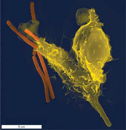
FIGURE 23.16 ■ Colorized SEM of phagocytosis. A single white blood cell (yellow) engulfs anthrax bacteria (orange). The white cell, called a neutrophil, is a member of the innate immune system that seeks out, captures, and kills bacterial pathogens.
IMAGE CREDIT: THE MICROGRAPH WAS TAKEN BY VOLKER BRINKMANN WITH A LEO 1550
SCANNING ELECTRON MICROSCOPE
Innate and Adaptive Immunity
There are two broad types of immunity: innate immunity (often called nonadaptive immunity) and adaptive immunity (discussed in detail in Chapter 24). Innate immunity is ancient, having evolved early in
invertebrates and then carried over to vertebrates, including humans. In contrast, adaptive immunity is more recent, evolving only in vertebrates. Innate immune mechanisms include physical barriers such as skin, chemical barriers such as stomach acid, and relatively nonspecific innate cellular defense mechanisms present from birth.
Immediately after a pathogen or other foreign substance breaches a physical or chemical barrier, innate cellular defenses are triggered by microbial structures such as peptidoglycan or lipopolysaccharides. The innate defenses then almost indiscriminately attack different types of foreign substances and organisms and can even damage innocent bystander host cells. The speed and broad targeting of innate immunity differentiates it from the slower but more focused adaptive immune system.
Adaptive immunity is slower to respond than innate immunity because it evolved to react to very specific structures called antigens. An antigen is any chemical, compound, or structure foreign to the body that elicits an adaptive immune response. Adaptive immunity to a specific antigen is not launched until the body “sees” that antigen and then builds a response to it over days. Amazingly, adaptive immune mechanisms can recognize at least 10 10 different antigenic structures and specifically launch a directed attack against each one. Once such an attack has been activated, the organism keeps a “memory” of the exposure in the form of specific memory cells. An encounter with the microbe years later will reactivate the memory cells specific to that antigen and produce an amplified response.
The two types of immunity are illustrated by the response of the immune system to infection by the microorganism Neisseria gonorrhoeae, which causes the sexually transmitted disease gonorrhea. A component of the innate immune response is complement, composed of several soluble protein factors constantly present in the blood. Within moments of an initial infection, especially upon entering the bloodstream, complement proteins form holes in the bacterial membrane, thereby killing the microbe (see Section 23.6). Later, but within a week of infection, the adaptive immune response will have generated specific antibodies against cells of N. gonorrhoeae that escaped the innate mechanisms. Together, innate and adaptive immunity can resolve disease caused by this organism. Unfortunately, N. gonorrhoeae infection does not generate long-lasting “memory,” so reinfection is possible. In contrast, other bacterial pathogens such as Streptococcus pneumoniae, a cause of pneumonia, generate long-lasting memory cells.
It is important to know that the innate and adaptive immune systems are also interconnected. For instance, adaptive immune system antibodies can help activate the complement cascade of the innate immune system.
Conversely, activation of innate resistance mechanisms can cause the release of small immunomodulatory peptides (cytokines or chemokines) that influence the type and strength of adaptive immunity brought to bear. In military terms, the cooperation between innate and adaptive immunities is similar to an army coordinating its actions with those of the air and naval forces.
Infection versus Disease
An infectious agent entering the body (infection) does not guarantee that a person will develop symptoms of the disease. If the number of invading organisms is small and both the innate and adaptive immune systems are effective, individuals may never realize they were infected. However, a clinician who knows that a patient has been exposed to certain microorganisms will treat the patient with antibiotics as a preventive measure. Chapter 26 more fully discusses the difference between being infected and developing disease.
Any microbe that causes disease must first breach the host’s physical and chemical barriers to gain entrance to the body. It must then survive the innate defense mechanisms and begin to multiply. Finally, the microbe must surmount the last line of defense—namely, the host’s adaptive immunity— that begins to respond as the microbe struggles to overcome innate immune defenses. The rest of this chapter will discuss the various facets of innate defense; adaptive immunity is explored in Chapter 24.
Although innate immunity and adaptive immunity are often treated as separate entities, certain components play a role in both types of immunity. We thus introduce the various cells and organs of the immune system as a whole before focusing on innate immunity.
Cells of the Immune System
Blood is composed of red blood cells (also called erythrocytes), white blood cells (also known as leukocytes), and platelets (Fig. 23.17). The many types of white blood cells are formed by the differentiation of hematopoietic stem cells produced in bone marrow (Fig. 23.18). Hematopoietic stem cells differentiate into two main lineages: myeloid cells that produce the leukocytes of innate immunity, and lymphoid cells that develop into the leukocytes (primarily lymphocytes) of adaptive immunity.
FIGURE 23.17 ■ Red blood cells, white blood cell, and a platelet. This colorized scanning electron micrograph illustrates the relative sizes and 3D morphologies of these components of blood.
EYE OF SCIENCE/SCIENCE SOURCE; INSET: SCIMAT/GETTY IMAGES
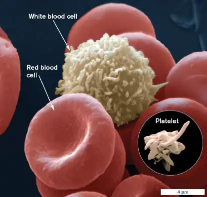
FIGURE 23.18 ■ Development of white blood cell components of the immune system. Pluripotent hematopoietic stem cells in bone marrow divide to form two lineages: myeloid cells and lymphoid cells. Myeloid stem cells develop into PMNs (the primary components of innate immunity), red blood cells, and platelets. Lymphoid stem cells differentiate into B cells, T cells, and natural killer cells. Final maturation into B cells and T cells (the principal cells involved in adaptive immunity) occurs in the bone marrow and thymus, respectively. Colors indicate a group of differentiated cells that arise from the same progenitor. The leukocytes of innate immunity include the following: Polymorphonuclear leukocytes (PMNs)
Monocytes Macrophages
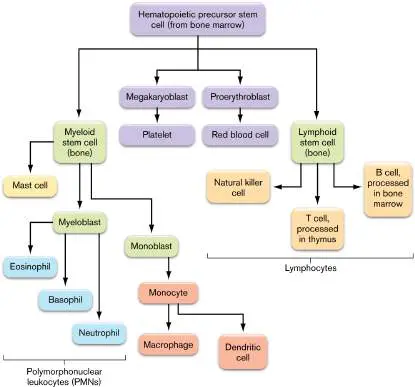
Dendritic cells Mast cells Natural killer cells (part of the lymphoid lineage but also part of innate immunity)
PMNs, also called granulocytes, are known for their conspicuous multilobed nuclei and enzyme-rich lysosome organelles. PMNs differentiate from an intermediate cell called the myeloblast. There are several types of PMNs, named for their different staining characteristics. Each cell type has a different function. Neutrophils (Fig. 23.19A), for example, make up the vast majority of white cells in the blood.
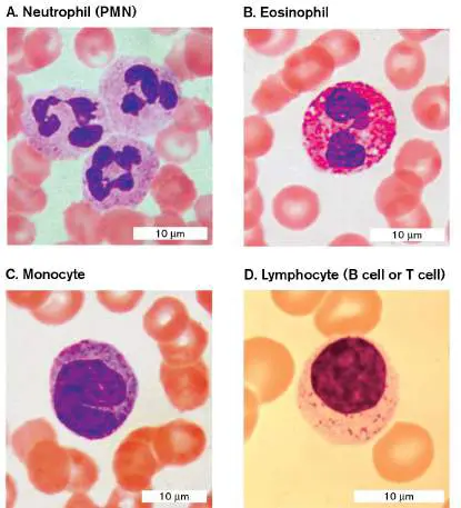
FIGURE 23.19 ■ Types of white blood cells. Basophils (not shown) look similar to eosinophils.
DR. JOHN D. CUNNINGHAM/VISUALS UNLIMITED
DR. FRED HOSSLER/VISUALS UNLIMITED
DR. FRED HOSSLER/VISUALS UNLIMITED
DR. FRED HOSSLER/VISUALS UNLIMITED
Neutrophils can engulf microbes by phagocytosis, which involves the extrusion of pseudopods (arm-like protrusions) that attach to and envelop the pathogen. Pseudopods have surface receptors that attach directly to the pathogen or attach indirectly to host complement or antibodies already bound to the pathogen. After the pathogen is engulfed, it ends up in a phagosome vacuole. The phagocyte then kills the organism by fusing enzyme-gorged lysosomes with the phagosomes (Fig. 23.20). Enzymes (described later) spilling from the lysosome into the phagosome will destroy various components of the microbe and, ultimately, the microbe itself (see Section 23.5). Approximately 100 million neutrophils are made in the bone marrow every day, but each cell lives for only about 24 hours.
FIGURE 23.20 ■ Phagocytosis and phagosome-lysosome fusion.
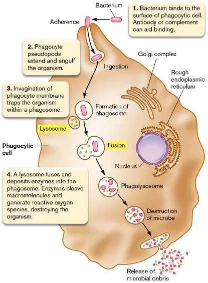
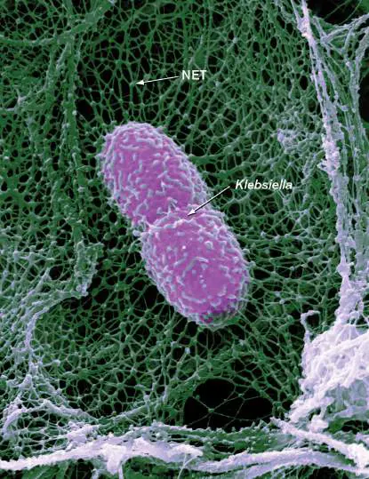
Neutrophils also “throw” neutrophil extracellular traps (NETs) around nearby pathogens. After interacting with bacteria, neutrophils can undergo NETosis, an unusual form of cell death, in which cells spew a latticework of DNA (chromatin) impregnated with antimicrobial compounds into the immediate area (Fig. 23.21). Much as a fishing net traps fish, NETs trap pathogens and prevent them from spreading. The antimicrobial compounds then kill the captured microbes. Some pathogens, such as group B Streptococcus and Neisseria gonorrhoeae, can escape NETs by secreting extracellular nucleases. NETs have also been implicated in certain autoimmune diseases, such as systemic lupus erythematosus.
FIGURE 23.21 ■ Neutrophil extracellular trap (NET). An infected mouse lung shows a Klebsiella pneumoniae bacterium (colorized; length approx. 1 μm) snared in a NET (green), a web of decondensed chromatin released by neutrophils to catch and kill pathogens.
SPL/SCIENCE SOURCE
Thought Question
23.4 Figure 23.21 shows how a neutrophil extracellular trap can ensnare a nearby pathogen. Which bacterial structure might blunt the microbicidal effect of NETs?
Two other types of PMNs are basophils, which stain with basic dyes, and eosinophils (Fig. 23.19B ), which stain with the acidic dye eosin. These white cells do not effectively phagocytose microbes or throw NETs but instead release products, such as major basic protein, that are toxic to the microbe. These two types of blood cells also release chemical mediators (called vasoactive agents) that affect the diameter and permeability of blood vessels (the significance of which is discussed in Section 23.5). Eosinophils play a major role in killing multicellular parasites such as helminth worms. Mast cells are similar to basophils in structure but differentiate in a lineage separate from PMNs (Fig. 23.18). Unlike PMNs, mast cells are residents of connective tissues and mucosa and do not circulate in the bloodstream. Basophils and mast cells have roles in inflammation and wound healing and also contain high-affinity receptors for a class of antibody, called immunoglobulin E (IgE), associated with allergic responses (detailed in Section 24.7).
Note: Because neutrophils constitute the vast majority of PMNs, the terms
“neutrophil” and “PMN” are often used interchangeably.
Monocytes (Fig. 23.19C ) are white blood cells with a single nucleus (not multilobed like a PMN); they engulf (phagocytose) foreign material. Monocytes circulating in the blood can migrate out of blood vessels into various tissues and differentiate into macrophages and dendritic cells (Fig. 23.22). Macrophages are phagocytic and form a major part of the amorphous mononuclear phagocyte system (MPS), previously called the reticuloendothelial system, involving connective tissues that surround kidney, liver, spleen, lymph nodes, Peyer’s patches and bone marrow. Widely distributed throughout the body, the MPS is composed of two cell types that can take up and sequester particles: monocytes that circulate in blood, and macrophages that roam through tissues. The MPS also includes immobile macrophages called Kupffer cells that line the special capillaries (sinusoids) of the liver. The function of macrophages and the MPS is to phagocytose microorganisms and other foreign particles, clearing them from the bloodstream. In fact, recent research indicates that a certain type of macrophage in skin contributes to the longevity of tattoos (Special Topic 23 ).
FIGURE 23.22 ■ The innate immune system depends on white blood cells called macrophages and dendritic cells. A. Membrane protrusions from a macrophage (20 μm long) detecting and engulfing bacteria (pink; E. coli, 1.5 μm long). B. Dendritic cell. Colorized SEMs.
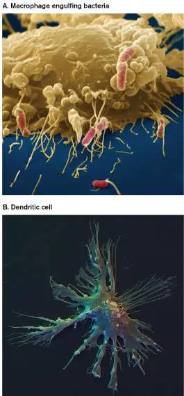
EYE OF SCIENCE/SCIENCE SOURCE
DAVID SCHARF/SCIENCE SOURCE
Macrophages are present in most tissues of the body and are the cells most likely to make first contact with invading pathogens. They have two functions. As part of innate immunity, they kill invaders directly. Protrusions from the macrophage surface extend and clasp nearby bacteria, pulling them into the cell (Fig. 23.22A). Once ingested by the macrophage, the bacteria are destroyed.
Subsequently, in the first step of adaptive immunity (called antigen processing), the remnants of dead bacteria are processed (degraded) by the macrophage into smaller peptide antigens that are presented on the macrophage cell surface. Thus, macrophages are called antigen-presenting cells (APCs; see Chapter 24). Specific white blood cells called T cells (a type of lymphocyte, discussed shortly) can bind to the antigens displayed on the macrophage and become activated. Once activated, these T cells will stimulate a specific subset of B cells (another type of lymphocyte) to synthesize specific antibodies that bind to the bacterial or viral antigen. Some T cells become cytotoxic T cells that can kill any host cell infected by the pathogen.
Macrophages are not the only APC cells; dendritic cells (Fig. 23.22B ) are also APCs. Present in skin, mucosal tissue, lymph nodes, and the spleen, they, like macrophages, can take up, process, and present small antigens on their cell surface. Dendritic cells are distinct from macrophages: They have a different structure, and they take up small soluble antigens from their surroundings in addition to phagocytosing whole bacteria.
Platelets are small, puzzle piece–shaped cell fragments that lack a nucleus and derive from megakaryoblasts, precursor cells that also differentiate from hematopoietic stem cells (see Fig. 23.18). Platelets, which circulate in the bloodstream, are required for efficient blood clotting. When activated by damaged endothelial cells (cells lining blood vessels), platelets clump, become trapped by fibrin, and form plugs that stop the bleeding. Several factors produced by platelets also help wound repair. In addition to forming blood clots and mediating wound repair, platelets are part of the innate immune system. Invading bacteria can bind platelets and trigger the release of antimicrobial peptides (see Section 23.4). Bacterially activated platelets can also induce NET formation by neutrophils.
Natural killer (NK) cells derive from lymphoid stem cells (see Fig. 23.18) and are also part of innate immunity. Instead of killing microbes, however, the mission of NK cells is to kill host cells that harbor microorganisms or that have been transformed into cancer cells (Fig. 23.23). Natural killer cells recognize changes in cell-surface proteins of infected or cancer cells (MHC class I molecules; described next). This recognition causes the NK cells to degranulate—that is, to release chemicals that kill the target host cells. FIGURE 23.23 ■ Natural killer cells. Natural killer (NK) cells attack eukaryotic cells infected by microbes, not the microbes themselves. Perforin produced by the NK cell punctures the membranes of target cells, causing them to burst (colorized SEM).
EYE OF SCIENCE/SCIENCE SOURCE
Natural killer cells recognize their targets in two basic ways: by the absence of major histocompatibility complex (MHC) class I molecules on host cells, or by the presence of antibodies on host cells. The major histocompatibility complex (MHC) consists of proteins found on the surfaces of cells that help the immune system recognize self versus foreign substances. A normal host cell displays two classes of MHC molecules on the outside of the cell membrane. MHC I is an indicator of “self.” All nucleated host cells have MHC I on their surface. Red blood cells, because they lack a nucleus, do not produce MHC I. (MHC I and II molecules are discussed in Chapter 24.) NK cells have specific receptors that bind to self MHC I molecules on the surfaces of other cells in the body. An NK cell that “touches” a self MHC I molecule–containing cell from the same person will not attack that cell. However, if a host cell lacks MHC class I molecules, NK cells
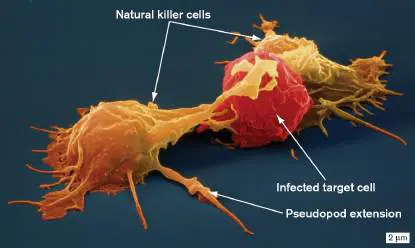
perceive the target as foreign and a potential threat. Host cells can lose their MHC molecules during infection or as a result of malignant transformation. Note that NK cells also require contact with activating signals on the surface of nucleated target cells in order to start an attack. Red blood cell membranes do not have these activating signals.
When an NK cell encounters a host cell that lacks self MHC I markers but has activating signals, granules in the NK cell move (polarize) to where the NK cell touches the target cell (Fig. 23.24). Degranulation then releases a pore-forming protein (perforin) that inserts into the membrane of the target cell. Degranulation also releases cytotoxic proteases (granzymes) that pass through the perforin pore into the target cell. The objective of the NK cell is not to lyse the target cell, which would release any intracellular pathogens present, but to coax the target cell into killing itself by a process called apoptosis.
FIGURE 23.24 ■ Polarization of NK cell granules at the interface between NK cell and target cell. A. First contact between cells. Granules are dispersed throughout the NK cell. B. Polarization of granules at the NK cell–target cell interface before they are released from
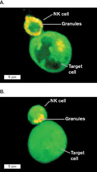
the NK cell. Cytoplasmic granules of NK cells were labeled with acridine orange.
MODIFIED FROM BILLADEAU, ET AL. 1998. J. EXP. MED 188 :549–559.
MODIFIED FROM BILLADEAU, ET AL. 1998. J. EXP. MED 188 :549–559.
Apoptosis is triggered when granzymes damage mitochondria.
Cytochromes spilled from damaged mitochondria activate cytosolic host proteases called caspases (c ysteine-dependent asp artate-directed prote ases). Activated caspases cleave a variety of host molecules, including signal transduction proteins, cytoskeletal proteins, DNA repair proteins, and inhibitors of endonucleases. The consequence of this complex process is that host cell DNA becomes fragmented, cell membranes bleb, and the cell breaks into small, membrane-encased blobs (apoptotic bodies) that can be cleared by neutrophils via a specialized form of phagocytosis (efferocytosis), all without causing unwanted inflammation, a major goal of apoptosis.
Phagocytosis of the apoptotic bodies also kills the infectious agent. The chapter-opening image shows the result of efferocytosis of an infected cell by a macrophage.
Leaving nothing to chance, another granzyme (granzyme B), also delivered by NK cells into infected cells, will enter intracellular bacteria and disrupt their macromolecular processes, thereby killing the pathogens directly. Farokh Dotiwala, Sriam Chandrasekaran, and Judy Lieberman discovered that granzyme B protease prevents protein synthesis in E. coli, Listeria monocytogenes, and Mycobacterium smegmatis (Fig. 23.25). The enzyme does so by digesting key ribosomal proteins and aminoacyl-tRNA synthetases.
FIGURE 23.25 ■ Granzyme B disrupts bacterial protein synthesis. Granzyme B and sublytic amounts of the pore-forming protein granulysin that delivers granzyme B into bacteria were added to Escherichia coli (A) , Listeria monocytogenes (B) , and Mycobacterium smegmatis (C) . Protein synthesis was measured as incorporation of 35 S-
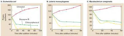
radiolabeled methionine into trichloroacetic acid precipitable material (percentage, compared to untreated controls). The protein synthesis inhibitor chloramphenicol was added as a positive control.
Source: Modified from Dotiwala et al. 2017. Cell 171 :1125–1137, fig. 3A.
SPECIAL TOPIC 23 Why Do Tattoos Last Forever?
CLEM ONOJEGHUO/PEXELS
Do you or your friends have a tattoo? It seems everyone under 40 has one. Many are beautiful, but some are unmitigated disasters, like the misspelled tattoo on one teenager’s arm ironically proclaiming “No Regr i ts.” Unfortunately for him, tattoos are forever. The question we address here is: Why don’t they fade?
In the past, fibroblasts were considered the primary long-term reservoir of tattoo pigment granules. But research groups headed by Sandrine Henri and Bernard Malissen (Fig. ST 23.1 ) at Aix-Marseille University in France uncovered a new culprit: a type of macrophage

called a melanophage. In mice, melanophages normally ingest melanin produced by melanocytes and deliver the melanin to keratinocytes that become colored hairs on the mouse’s body. The Henri and Malissen labs found that melanophages, not fibroblasts, are the cells that capture and retain tattoo pigment particles. Melanophages do not migrate from the skin to lymph nodes, but they will eventually die in place. If a melanophage is laden with dye particles when it dies, the particles are released and should be drained via the lymphatic vessels. But contrary to expectation, they are not drained. So, what keeps the particles there?

FIGURE ST 23.1 ■ Sandrine Henri (left) and Bernard Malissen (right).
COURTESY OF SANDRINE HENRI
COURTESY OF BERNARD MALISSEN
Figure ST 23.2 examines cells taken from the skin of a mouse tail that was tattooed with a green pigment. Pigment particles were engulfed only by melanophage cells (Fig. ST 23.2A ); the particles were not seen in other macrophages or dendritic cells (Fig. ST 23.2B and C ). Next, the scientists asked what would happen to the particle if they selectively killed the melanophages (Fig. ST 23.3 ). To delete, or ablate, melanophages, the mice used had been genetically engineered to express diphtheria toxin receptor (DTR), but only on mouse melanophages. Mouse cells normally lack the DTR gene. Figure ST 23.3A shows the green pigment particles residing in melanophages after tattooing. Diphtheria toxin was then injected into the same mice. Two days later the melanophages were gone and the green particles were seen free, not within any cells (Fig. ST 23.3B ). Why aren’t these

particles drained from the site, causing the tattoo to disappear? Do new melanophages enter the tattooed region and reingest the particles? FIGURE ST 23.2 ■ Cells sorted from green-tattooed mouse tail.
BARANSKA, A., ET AL. 2018. J. EXP. MED. 215 :1115.
BARANSKA, A., ET AL. 2018. J. EXP. MED. 215 :1115.
BARANSKA, A., ET AL. 2018. J. EXP. MED. 215 :1115.
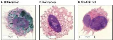
FIGURE ST 23.3 ■ Fate of green tattoo particles when melanophages are ablated. A, C. Melanophages contain green particles at the tattoo site before ablation. B. Forty-eight hours after ablation with diphtheria toxin, melanophages are absent and green particles are found only extracellularly. C. Ninety days after ablation, melanophages have returned to the tattoo site and recaptured green particles.
BARANSKA, A., ET AL. 2018. J. EXP. MED. 215 :1115.
BARANSKA, A., ET AL. 2018. J. EXP. MED. 215 :1115.
BARANSKA, A., ET AL. 2018. J. EXP. MED. 215 :1115.
BARANSKA, A., ET AL. 2018. J. EXP. MED. 215 :1115.
Figure ST 23.3C (control) again shows tail skin melanophages laden with green particles 3 weeks after tattooing. Melanophages were
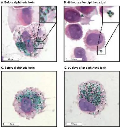
then ablated using diphtheria toxin for 2 days. After ablation, the toxin was allowed to dissipate for 90 days so that the mice could restore the melanophage population. Once again, the tattoo did not fade. After the 90-day period, skin cells taken from the tattoo site were examined. Figure ST 23.3D shows that, indeed, new melanophages had entered the area and reingested the particles.
The data indicate that a cycle of pigment capture by melanophages, pigment release, and then recapture by new melanophages can account for long-term tattoo persistence. These results also explain why the “No Regrits” tattoo will taunt our teenager for many years to come. The research conducted by this group was funded by the European Research Council.
RESEARCH QUESTION
Laser pulses are used to remove tattoos, but multiple treatments are often required. How might the research presented in this paper be used to explain this phenomenon and facilitate tattoo removal?
Anna Baranska, Alaa Shawket, Mabel Jouve, Myriam Baratin, Camille
Malosse, et al. 2018. Unveiling skin macrophage dynamics explains both tattoo
persistence and strenuous removal. Journal of Experimental Medicine 215 :1115–1133.
https://doi.org/10.1084/jem.20171608.
The second killing mechanism wielded by NK cells is called antibody-dependent cell-mediated cytotoxicity (ADCC). Besides receptors that detect MHC I molecules, NK cells also contain receptors on their cell surface that bind to a part of an antibody called the Fc region. An antibody is a Y-shaped protein that has two identical antigen-binding arms located at the top and an Fc region located at the tail. The Fc region does not bind antigen but can bind to specific membrane Fc receptors on other cells (discussed in Chapter 24, Fig. 24.8). ADCC is activated when the Fc receptor on the NK cell links to an antibody-coated host cell.
Why would host cells be coated with antibodies? During replication, many viruses place viral proteins in the membrane of the infected cell. Antibodies to those viral proteins will coat the compromised cell, tagging it for ADCC. Once the compromised cell has been targeted, it is killed by the NK cell in the same way as described for cells lacking MHC—namely, by insertion of a perforin molecule and injection of granzymes to initiate apoptosis. This killing mechanism is an example of cooperation between innate immunity (NK cells) and adaptive immunity (antibody-producing lymphocytes).
Diagnostic value of white blood cell ratios. Many diseases, including infections, can alter the ratios of white blood cells (WBCs). A WBC differential is a test that physicians often order to help them diagnose infections and other syndromes. Table 23.1 presents general guidelines for interpreting a WBC differential, indicating which cell types increase or decrease in response to infections with bacteria, viruses, or parasites (protozoa or worms). Notice that total WBC counts increase in each case, but the type of WBC that increases in number differs with the infectious agent.
TABLE Guidelines for Interpreting White Blood
23.1 Cell (WBC) Counts with Differential *
Acute bacterial Viral Parasitic Normal infection infection infection Allergy Total WBC 4,500– Elevated: Elevated Elevated Elevated count 11,000/ 12,000 mm 3 – 30,000 Differential Neutrophils 54%–62% Increased Can be number reduce s and d more immatu re forms (band cells)
TABLE Guidelines for Interpreting White Blood
23.1 Cell (WBC) Counts with Differential *
Acute bacterial Viral Parasitic Normal infection infection infection Allergy Eosinophils 1%–3% Increased Basophils 0%–0.75% Increased Lymphocytes 25%–33% Increased Monocytes 3%–7% Typically increase with chronic infections (for example, tuberculosis, brucellosis, subacute bacterial endocarditis, Rickettsia, many protozoan infections)
Lymphoid Organs
As Figure 23.18shows, lymphoid stem cells produce lymphocytes as well as natural killer cells, described earlier. Lymphocytes (Fig. 23.19D ) are the main participants in adaptive immunity. These cells are present in blood at about 2,500 cells per microliter, accounting for about one-third of all peripheral white blood cells. However, an individual lymphocyte spends most of its life within specialized solid tissues (lymphoid organs) and enters the bloodstream only periodically, where it migrates from one place to another, surveying tissues for possible infection or foreign antigens. Consequently, most lymphocytes are found in the lymph nodes or spleen. No more than 1% of the total lymphocyte population circulates in the blood at any one time. The tissues of the immune system, where the great majority of lymphocytes are found, are classified as primary or secondary lymphoid organs or tissues, depending on their function (Fig. 23.26). The primary lymphoid organs and tissues are where immature lymphocytes made in bone marrow mature into antigen-sensitive B cells and T cells. B cells that ultimately produce antibodies (see Chapter 24) develop in b one marrow tissue. T cells, which modulate various facets of adaptive immunity, develop in the t hymus, an organ located above the heart.
FIGURE 23.26 ■ Lymphoid organs.
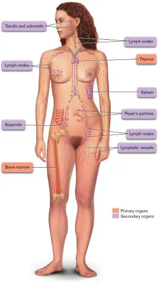
The secondary lymphoid organs serve as stations where lymphocytes can encounter antigens. These encounters lead to the differentiation of B cells into antibody-secreting plasma cells and of T cells into antigen-specific helper cells, as discussed in Chapter 24. The spleen is an example of a secondary lymphoid organ that filters blood to detect microorganisms. Macrophages in the spleen engulf these organisms and destroy them and then migrate to other secondary lymphoid organs to present pieces of the microbe (called antigens) to the B and T cells, which then become activated.
The lymph nodes, another kind of secondary lymphoid organ, are positioned at strategic locations to trap organisms that drain from local (nearby) tissues. These nodes are sometimes called draining lymph nodes. Organisms traveling from infected tissues to a draining lymph node are carried in lymph by lymphatic vessels rather than through blood vessels. Lymphoid tissues are also present in the mucosal regions of the gut and respiratory tracts (for example, Peyer’s patches and gut-associated lymphoid tissue, or GALT, discussed in the next section). Other secondary lymphoid organs are tonsils, adenoids, and the appendix.
To Summarize
The immune system consists of both innate and adaptive mechanisms that recognize and eliminate pathogens.
Innate immunity includes physical and chemical barriers and some cellular responses to various microbial structures.
Adaptive immunity is a cellular response to specific structures (antigens) in which a memory of exposure is produced.
Myeloid bone marrow stem cells differentiate to form cells of the innate immune system—namely, phagocytic PMNs, monocytes, macrophages, antigen-presenting dendritic cells, and mast cells. Platelets are derived from a different cell line.
Lymphoid stem cells differentiate into natural killer cells (part of the innate immune system) and B cells and T cells (part of the adaptive immune system). B cells ultimately produce antibodies, whereas T cells regulate adaptive immunity.
Natural killer (NK) cells are a class of white blood cells that continually patrol tissues for cancer cells, cells infected with microbes, or host cells coated with antibody (antibody-dependent cell-mediated cytotoxicity, ADCC). NK cells kill by inserting pores of perforin and granzymes into target cell membranes.
Primary lymphoid organs include bone marrow (where B cells develop) and the thymus (where T cells develop). Secondary lymphoid organs (spleen, lymph nodes, Peyer’s patches, tonsils, appendix) are where lymphocytes encounter antigens.
Glossary
immune system An organism’s cellular defense system against pathogens.
innate immunity Also called nonadaptive immunity. Nonspecific mechanisms for protecting against pathogens.
nonadaptive immunity See innate immunity .
adaptive immunity Immune responses activated by a specific antigen and mediated by B cells and T cells.
antigen A compound, recognized as foreign by the cell, that elicits an adaptive immune response. See also immunogen .
complement Innate immunity proteins produced by the liver; they circulate in the blood, where they can form holes in bacterial membranes, killing the bacteria.
leukocyte White blood cell.
neutrophil A white blood cell of the innate immune system that can phagocytose and kill microbes.
phagosome A large intracellular vesicle that forms as a result of phagocytosis. neutrophil extracellular trap (NET)
A net of chromatin (including DNA) and antimicrobial peptides expelled by dying neutrophils to trap and injure nearby pathogenic bacteria. basophil A white blood cell, stained by basic dyes, that secretes compounds that aid innate immunity.
eosinophil A white blood cell that stains with the acidic dye eosin and secretes compounds that facilitate innate immunity.
mast cell A white blood cell that secretes proteins that aid innate immunity. Mast cells reside in connective tissues and mucosa and do not circulate in the bloodstream.
monocyte A white blood cell with a single nucleus that can differentiate into a macrophage or a dendritic cell.
macrophage A mononuclear, phagocytic, antigen-presenting cell of the immune system.
dendritic cell An antigen-presenting white blood cell that primarily takes up small soluble antigens from its surroundings.
mononuclear phagocyte system (MPS)
An amorphous part of the immune system that consists mostly of monocytes and macrophages present in connective tissue that surrounds kidney, liver, spleen, lymph nodes, Peyer’s patches, and bone marrow. antigen-presenting cell (APC)
An immune cell that can process antigens into antigenic determinants and display those determinants on the cell surface for recognition by other immune cells.
APC See antigen-presenting cell .
platelet A small cell fragment without a nucleus found in blood that is involved in clotting.
natural killer (NK) cell A lymphocyte that does not need antigen stimulation to kill tumor or infected host cells by inserting granules containing perforin. major histocompatibility complex (MHC)
Transmembrane cell proteins important for recognizing self and for presenting foreign antigens to the adaptive immune system.
perforin A cytotoxic protein, secreted by T cells, that forms pores in target cell membranes.
apoptosis A cell death program triggered during tissue differentiation or in certain damaged or infected cells.
antibody-dependent cell-mediated cytotoxicity (ADCC)
The process by which natural killer cells destroy antibody-coated host cells that are infected by a virus.
lymphocyte A mononuclear leukocyte (white blood cell) that is a product of lymphoid tissue and participates in immunity (e.g., B cell and T cell). B cell An adaptive immune cell, developed in bone marrow tissue, that can give rise to antibody-producing cells.
T cell An adaptive immune cell, developed in the thymus, that can give rise to antigen-specific helper cells and cytotoxic T cells.
lymph node A secondary lymphatic organ, formed by the convergence of lymphatic vessels, that traps foreign particles from local tissue and presents them to resident immune cells.
Fig. 24.8
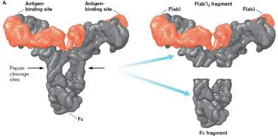
FIGURE 24.8 ■ Basic antibody structure. A. The 3D structure of an antibody. (PDB code: 1R70) Every antibody contains two heavy chains (grey) and two smaller light chains (red) held together by disulfide bonds (shown in part B). The Y-shaped structure contains two antigen-binding sites, one at each arm [F(ab) region] of the molecule. These two sites are formed by the amino-terminal regions of the heavy-and light-chain pairs. The Fc portion points downward and is used to attach the antibody to different cell-surface molecules. Pepsin can be used to cleave a site in each heavy chain that releases the two F(ab) regions as one piece [F(ab′) 2] from the Fc region. B. Constant and variable regions in antibody structure. This schematic representation of IgG shows the various constant and variable regions of the heavy and light chains. Combined, the sequences V H and V L form antigen-binding sites at the amino termini of the heavy and light chains. An antigen is shown binding to one of the antigen-binding sites. Areas of the Fc region that can bind complement and cell-surface receptors are marked. Papain cleaves a site in each heavy chain that releases two F(ab) fragments, each one carrying one antigen-binding site.
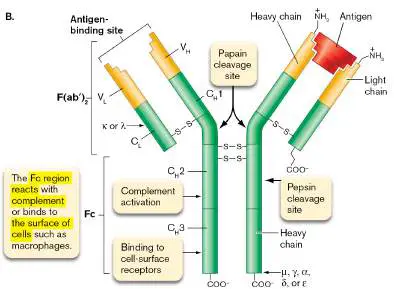
Endnotes
1. Note *: This table provides general guidelines. Individual organisms or certain noninfectious medical conditions or immunological defects can alter the findings. A blank space indicates little change in the parameter. Return to reference *
23.4 Physical and Chemical Defenses against Infectionnot assigned
In defending a mythical medieval castle, the first lines of defense might include physical barriers (the castle wall), chemical barriers (boiling oil tossed onto invaders trying to scale the wall), and, finally, hand-to-hand combat once the wall was breached. Similarly, the body’s initial defenses against infectious disease are composed of physical, chemical, and cellular barriers designed to prevent a pathogen’s access to host tissues. Although generally described as nonspecific, some innate defense systems are more specific than others.
Physical Barriers to Infection
The first line of defense against any potential microbial invader (either commensal or pathogenic) is found where parts of the body interface with the environment. These interfaces (skin, lung, gastrointestinal tract, genitourinary tract, and oral cavities) have similar defense strategies, although each has unique characteristics. A defense common to all host surfaces involves tight junctions — watertight adhesions that link adjacent epithelial cells at mucosal surfaces and endothelial cells lining blood vessels. Tight junctions prevent bacteria, and even host cells, from moving between internal and external host compartments. The “glue” that holds tight junctions together is a series of interconnecting glycoprotein molecules (Fig. 23.27).
FIGURE 23.27 ■ Tight junctions hold adjacent cells together. Tight junctions are formed by an interconnected series of glycoproteins (blowup). By tightly linking adjacent cell membranes together, tight junctions produce a barrier through which bacteria, viruses, and other host cells cannot easily pass. Skin. Few microorganisms can penetrate skin because of the thick keratin armor produced by closely packed cells called keratinocytes. Keratin protein is a hard substance (hair and fingernails are made of it) that is not degraded by known microbial enzymes. An oily substance (sebum) produced by the sebaceous glands will cover and protect the skin. Its slightly acidic pH inhibits bacterial growth. Skin secretions also contain antimicrobial peptides such as cathelicidin LL-37 that disrupt membrane integrity and antimicrobial RNases such as RNase 3 that can destabilize and penetrate bacterial membranes to cleave intracellular mRNA. Competition between species also limits
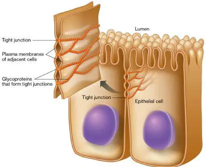
colonization by pathogens, and microorganisms that manage to adhere to skin are continually removed by the constant shedding of outer epithelial skin layers.
Other more specialized cells just under the skin can recognize microbes managing to slip through the physical barrier. They are part of a consortium of cells called skin-associated lymphoid tissue (SALT). Langerhans cells make up a significant portion of SALT. They are specialized dendritic cells that can phagocytose microbes. Once a lymphoid Langerhans cell has ingested a microbe, the cell migrates by ameboid movement to nearby lymph nodes and presents parts of the microbe to the immune system (antigen presentation) to activate antimicrobial immunity.
Note: Distinguish phagocytic Langerhans cells from the pancreatic
“islets of Langerhans,” which secrete insulin.
Mucous membranes. Mucosal surfaces form the largest interface (200–300 square meters, or 2,000–3,000 square feet) between the human host and the environment (the intestine alone is 7–8 meters, or 23–26 feet, long). Mucous membranes in general present a containment problem. They must be selectively permeable in order to exchange nutrients as well as to export products and waste components. At the same time, they must constitute a barrier against invading pathogens. Mucosal membranes are covered with special tightly knit epithelial layers that support this barrier function. The mucus secreted from stratified squamous epithelial cells coats mucosal surfaces and traps microbes. Compounds within the mucus can serve as a food source for some microbiome organisms, but other secreted compounds—like the enzyme lysozyme (which cleaves cell wall peptidoglycan) and lactoperoxidase (which uses hydrogen peroxide [H 2 O 2] to produce short-lived bactericidal agents such as hypothiocyanite)—can kill an organism trapped in the mucus. Semispecific innate immune mechanisms are also associated with mucosal surfaces. Host cells, even epithelial cells, in mucosa have evolved mechanisms to distinguish harmless compounds from microorganisms. Patterns of conserved structures on microbes, called microbe-associated molecular patterns (MAMPs), are recognized by host cell-surface receptors such as various Toll-like receptors and CD14 (discussed in Section 23.5). Once a MAMP has been recognized, chemicals secreted by the affected host cell activate immune system cells to perform innate and adaptive immune functions.
Note: MAMPs were previously called PAMPs, for “pathogen-
associated molecular patterns.” Because the structures recognized are also present on nonpathogenic bacteria and viruses, the term was changed to MAMP.
Like skin, the gastrointestinal system possesses an innate mucosal immune system, in this case called gut-associated lymphoid tissue (GALT). GALT includes tonsils, adenoids, and Peyer’s patches ( Fig. 23.28A). These tissues contain specialized M cells that dot the intestinal surface and are wedged between epithelial cells. “M” stands for “microfold,” which describes their appearance (Fig. 23.28B ). These are immobilized cells that take up microbes (microbiota or pathogens) from the intestine and release them, or pieces of them, into a pocket formed on the opposite, or basolateral, side of the cell. Other cells of the innate immune system, such as macrophages (which migrate through tissues), gather here and collect the organisms that emerge. Macrophages engulf and try to kill the organism. If successful, the macrophage will place small, degraded components of the microbe onto cell-surface MHC I and MHC II molecules (antigen presentation). Other immune system cells can recognize the presented antigens and initiate adaptive immune functions such as antibody production (described in Chapter 24). As a result, M cells are extremely important for the development of mucosal immunity to pathogens. However, M cells can also serve as a portal for some pathogens to gain entry to the body and cause disease.
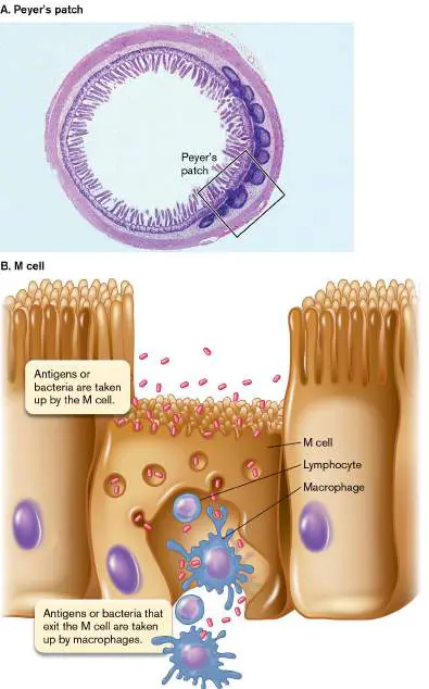
FIGURE 23.28 ■ Gut-associated lymphoid tissue (GALT). A. A Peyer’s patch located on the small intestine. B. Diagram of an M cell (microfold cell).
COURTESY OF DR. DEBORAH W. VAUGHAN
The lungs. The lungs also have a formidable defense. In addition to the mucociliary escalator discussed in Section 23.1, microorganisms larger than 100 μm become trapped by hairs and cilia lining the nasal cavity and trigger a forceful expulsion of air from the lungs (a sneeze). The sneeze is designed to clear the organism from the respiratory tract. Organisms that make it to the alveoli are met by phagocytic cells called alveolar macrophages. These cells can ingest and kill most bacteria.
Another important factor that prevents lung infections is an epithelial membrane protein called cystic fibrosis transmembrane conductance regulator (CFTR). CFTR is a membrane chloride channel that regulates chloride movement across the membrane—an essential part of hydrating mucus in healthy individuals. Cystic fibrosis patients have a defective CFTR and are much more susceptible to lung infections, especially those caused by Pseudomonas aeruginosa (Fig. 23.29A). Diminished chloride secretion due to a defective CFTR produces highly viscous airway mucus that impedes the mucociliary escalator. The P. aeruginosa strains posing the most serious threat produce a thick slimy material (alginate) that further impedes lung clearance mechanisms (Fig. 23.29B ).
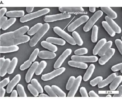
FIGURE 23.29 ■ Mucoid strains of Pseudomonas aeruginosa commonly infect cystic fibrosis patients. A. SEM of P. aeruginosa. B. Nonmucoid and mucoid forms of P.
aeruginosa. The organism can grow as biofilms in tissue and secrete an extracellular polymeric substance (EPS) that gives colonies of these strains a very mucoid appearance. Source: International Journal of Nanomedicine. 2015. 10 :5025–5034.
Originally published by and used with permission from Dove Medical Press Ltd.
REPUBLISHED WITH PERMISSION OF DOVE PRESS. DOSUNMU, E., ET AL. 2015. INT J
NANOMEDICINE 10 :5025–5034.
J. RAO ET AL. 2011. FRONT MICROBIOL. 63 :3054–3061
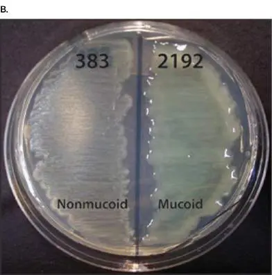
Chemical Barriers to Infection
Some examples of chemical barriers were mentioned earlier, such as the acidic pH of the stomach, lysozyme in tears, and generators of superoxide. In addition, a variety of human cells produce small antimicrobial, cationic (positively charged) peptides called defensins ( Table 23.2). These antimicrobial peptides are important components of innate immunity against microbial infections.
Categories of Natural
TABLE 23.2
Antimicrobial Peptides
Major presence in humans Class (Examples) (Source)
Alpha-defensins (-1, -2, -3, -4) Neutrophils (Stored in a granules)
Alpha-defensins (-5, -6) Paneth cells, small intestine (Stored in granules)
Cathelicidins (LL-37, hCAP18) Neutrophils (Secreted)
Histatins Saliva (Secreted)
Beta-defensins (HBD-1, -2) Epithelia (Secreted)
Kinocidins (tPMP, b PF-4) Platelets (Secreted)
Maganins Frogs Protegrins Pigs Indolicidins Cattle Defensins range in length from 29 to 47 amino acids and are found in mammals, birds, amphibians, and plants. Defensins (and other antimicrobial peptides) destroy an invading microbe’s cytoplasmic membrane and are effective against Gram-positive and Gram-negative bacteria, fungi, and even some viruses (those with membranes, like HIV). To kill Gram-negative bacteria, the peptides must first bind the negatively charged outer membrane lipopolysaccharides (LPS) of Gram-negative bacteria and move into the periplasm. Defensins are then pulled into the cytoplasmic membrane of either Gram-positive or Gram-negative bacteria by the transmembrane electrical potential, about −150 mV in bacteria (that is, the cell interior is more negative than the exterior). The peptides assemble into channels that destroy the cytoplasmic membrane barrier, killing the bacterial cell. Defensins generally do not affect eukaryotic cells, which have a much lower membrane potential (−15 mV). Antimicrobial peptides are produced by many human cells, including cells of the skin, lungs, genitourinary tract, and gastrointestinal tract (Fig. 23.30).
Vertebrate defensins of the alpha variety are stored in membrane-enclosed granules within neutrophils and in Paneth cells in the small intestine (Fig. 23.30A). When stimulated, these cells degranulate (release their granule contents) by fusing their granule membranes to cytoplasmic or vacuolar membranes, dumping their contents into the surroundings or into phagocytic vacuoles, where the alpha-defensins can destroy engulfed microbes by forming pores in the bacterial membrane (Fig. 23.30B and C ). In contrast, the beta-defensins are not stored in cytoplasmic granules. The synthesis of beta-defensins is activated only after contact with bacteria or their products.
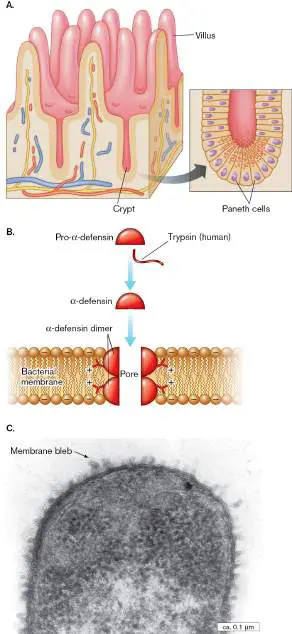
FIGURE 23.30 ■ Defensins. A. Certain defensins are produced in the crypts of the intestine. The crypts contain granule-rich Paneth cells (blowup) that discharge their granules into the crypt lumen in response to the entry of bacteria or as a result of food-related stimulation by acetylcholine. B. Precursor of human alpha-defensin is cleaved by trypsin into an active form. The defensin permeabilizes the bacterial membrane by forming a dimeric pore stabilized by electrostatic forces between cationic defensin residues (positive charge) and anionic phospholipid groups (negative charge) on the membrane. C. The effect of cationic peptides on E. coli O111. Polymyxin B is a small cationic peptide antibiotic that mimics the action of defensins. In this micrograph, polymyxin B causes blebs of membrane to ooze from the surface of the cell.
G. MARTINEZ DE TEJADA ET AL. 1995. INFECT. IMMUN. 63 :3054. © AMERICAN
SOCIETY FOR MICROBIOLOGY, HTTP://IAI.ASM.ORG/CGI/REPRINT/63/8/3054.PDF.
Thought Question
23.5 Why do defensins have to be so small? Do defensins kill normal microbiota?
To Summarize
Skin defenses against invading microbes include closely packed keratinocytes and a SALT lymphoid system made up largely of phagocytic Langerhans cells.
Mucous-membrane defenses involve secreted enzymes, cytokines, and GALT tissues, such as Peyer’s patches, that contain phagocytic M cells.
Microbe-associated molecular patterns (MAMPs) are recognized by Toll-like receptors found on many host cells, such as macrophages. Binding triggers release of chemical signaling molecules that activate innate and adaptive immune mechanisms.
M cells in gut-associated lymphoid tissues sample bacterial cells at their surface and release pieces of them to awaiting immune system cells.
Phagocytic alveolar macrophages inhabit lung tissues, contributing to nonspecific (innate) defense.
Chemical barriers against disease include cationic defensins, acid pH in the stomach, and superoxide produced by certain cells.
Glossary
tight junction A type of junction between the membranes of two adjacent vertebrate cells that form an impermeable barrier.
skin-associated lymphoid tissue (SALT)
Immune cells, such as dendritic cells, located under the skin that help eliminate bacteria that have breached the skin surface. Langerhans cell A specialized, phagocytic dendritic cell that is the predominant cell type in skin-associated lymphatic tissue.
microbe-associated molecular pattern (MAMP)
Formerly called pathogen-associated molecular pattern (PAMP). Molecules associated with groups of microbes, both pathogenic and nonpathogenic, that are recognized by cells of the innate immune system.
gut-associated lymphoid tissue (GALT)
Lymphatic tissues such as tonsils and adenoids that are found in conjunction with the gastrointestinal tract and contain immune cells.
M cell A phagocytic innate immune cell (microfold cell) found between intestinal epithelial cells.
alveolar macrophage A type of macrophage, located in the lung alveoli, that phagocytoses foreign material.
cystic fibrosis transmembrane conductance regulator (CFTR) A chloride channel found in respiratory epithelia. Mutations in the CFTR gene lead to cystic fibrosis.
defensin A type of small, positively charged peptide, produced by animal tissues, that destroys the cell membranes of invading microbes. degranulate To release antimicrobial granule contents by fusing granule membranes to cytoplasmic or vacuolar membranes.
Endnotes
1. Note a: Alpha-defensins are named alpha-defensin-1, alpha-defensin-2, and so on. Return to reference a 2. Note b: tPMP = thrombin-induced platelet microbicidal protein. Return to reference b
23.5 Innate Immunity: Surveillance, Cytokines, and Inflammationnot assigned
The boil shown in Figure 23.31 is an acute (quickly developing) inflammatory response
triggered by infection with the organism Staphylococcus aureus. Inflammation is a critical
innate defense in the war between microbial invaders and their hosts. It provides a way for
phagocytic cells (such as neutrophils) normally confined to the bloodstream to gain access to
infected sites within tissues. Movement of these cells out of blood vessels is called
extravasation or diapedesis, which we discuss shortly. Once at the infection site, the
neutrophils begin engulfing microbes. The white pus associated with an infection is teeming
with these white blood cells. In this section we describe the pathophysiology of inflammation
that leads to the five cardinal (major) signs of inflammation, first described more than 2,000
years ago and listed here:
FIGURE 23.31 ■ Inflammation caused by infection. Boil resulting from infection
of a hair follicle by Staphylococcus aureus (size 0.5–1.0 μm). Blowup: Colorized SEM.
SPL/SCIENCE SOURCE
IMAGE SOURCE/ALAMY STOCK PHOTO
Heat (warmth at the site from increased blood flow)
Edema (swelling from fluid accumulating outside of blood vessels)
Redness (due to dilated blood vessels)
Pain (pharmacological stimulation of nerve endings)
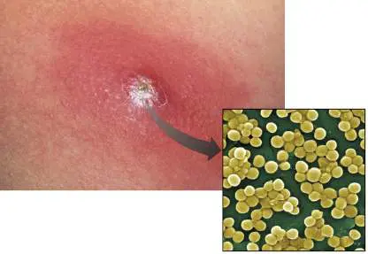
Altered function at the affected site (due to pain or damage to an inflamed organ)
Although many things can trigger inflammation, we focus here on how microbes cause the
response. The process begins with the infection itself. Microorganisms introduced into the
body—for example, on a wood splinter—will begin to grow and produce compounds that host
cells sense or that damage host cells (Fig. 23.32 ). Resident macrophages that wander
into the infected area engulf these organisms and then release inflammatory mediators that
orchestrate the inflammatory response. These mediators include chemokines
(chemoattractants) that “call out” to neutrophils for more help, and vasoactive factors such
as leukotrienes, platelet-activating factor, and prostaglandins, which act on blood vessels of
the microcirculation, increasing blood volume and capillary permeability to help deliver white
blood cells to the area. In addition, small protein molecules called cytokines are secreted.
Cytokines diffuse to the vasculature and stimulate the expression of specific receptors
(selectins) on the endothelial cells of capillaries and venules. But how are cytokines made in
response to an infection?
FIGURE 23.32 ■ Basic inflammatory response. Neutrophils (a type of phagocyte)
circulate freely through blood vessels and can squeeze between cells in the walls of a
capillary (extravasation) to the site of infection. They then engulf and destroy any
pathogens they encounter.
Sensing the Invader: Pattern Recognition Receptors and Cytokines
The faster the body can detect a pathogen, the more quickly it can deal with it. The more
quickly it deals with the pathogen, the better the outcome of an infection. It takes time,
however, for the adaptive immune system to make antibodies specific for a microbe (see
Chapter 24). All the while, the pathogen can grow and cause disease. Fortunately, bacteria
and viruses possess unique structures that immediately tag them as foreign. Structures such
as peptidoglycan, flagellin, lipoteichoic acids, and double-stranded RNA are not present in
tissues unless bacteria or viruses are present. These structures have microbe-associated
molecular patterns (MAMPs) that can be recognized by Toll-like or NOD-like receptors (TLRs
or NLRs) present on or in various host cell types (Fig. 23.33 ; Table 23.3). TLRs and NLRs
are tantamount to burglar alarm systems that activate upon encountering an intruder.
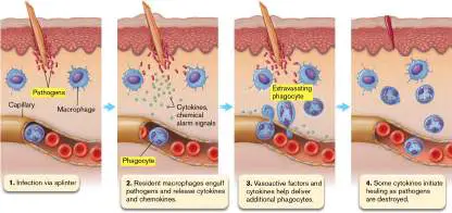
Collectively, TLRs, NLRs, and similar proteins are called pattern recognition receptors (PRRs)
because they recognize MAMPs.
FIGURE 23.33 ■ TLRs, NLRs, and inflammasomes. Stimulation of Toll-like
receptors (TLRs) activates transcription factors to induce production of cytokines and
other factors. (Note: TLR3, TLR7, and TLR9, not shown in this figure, are located in
endosomal membranes). Cytoplasmic NOD-like receptors (NLRs) bind MAMPs generated
primarily by intracellular pathogens and trigger a signal cascade different from that used
by TLRs. Some cytokines are directly released (for instance, IL-1 and IL-8). Others are
made as inactive precursors (procytokines) that must be cleaved by inflammasomes.
Inflammasome assembly is also orchestrated by certain NLRs.
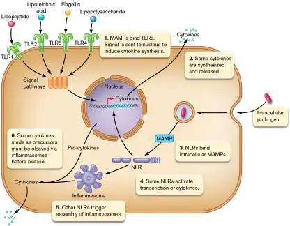
TABLE Examples of Toll-like Receptors and NOD-like 23.3 Receptors
MAMPs
Receptor recognized a Source Host cells Location
TLR1 Lipopeptides Bacteria Monocytes/macrophages, Cell surface
dendritic cells, B cells
TLR2 Glycolipids, Bacteria, Monocytes/macrophages, Cell surface
lipoteichoic viruses dendritic cells, mast
acids, viral cells
capsid
TLR3 Double-stranded Viruses Dendritic cells, B cells Cell
RNA compartmen
t
TLR4 Lipopolysaccharide, Bacteria Monocytes/macrophages, Cell surface
heat-shock dendritic cells, mast
proteins cells, intestinal
epithelium
TLR5 Flagellin Bacteria Monocytes/macrophages, Cell surface
dendritic cells,
intestinal epithelium
TLR6 Diacyl lipopeptides Mycoplasma Monocytes/macrophages, Cell surface
mast cells, B cells
TLR9 Unmethylated CpG Bacteria Monocytes/macrophages Cell
(cytosine- compartmen
phosphate- t
guanine)
residues in DNA
NOD 1 Component of Gram- Many cell types Inflammasome
Gram-negative negative
peptidoglycan bacteria
NOD 2 Peptidoglycan Bacteria Macrophages, dendritic Inflammasome
component cells, epithelia of lung
and GI tract
NLRP-3 Peptidoglycan Bacteria Many cell types Inflammasome
TABLE Examples of Toll-like Receptors and NOD-like 23.3 Receptors
MAMPs
Receptor recognized a Source Host cells Location
NLRP-4 Flagellin, CpG, ATP, Bacteria Many cell types Inflammasome
dsRNA
Toll-like receptors. First discovered in insects and named Toll receptors, Toll-like receptors
(TLRs) are evolutionarily conserved cell-surface glycoproteins present on the cells of many
eukaryotic genera (Fig. 23.33 ). TLRs enable the innate immune system to respond rapidly
to infection, giving the adaptive immune system the time it needs to gear up. The term “Toll”
came from Christiane Nüsslein-Volhard’s 1985 exclamation, “That’s crazy!” (in German, “Das
ist ja toll!”), when shown the fungus-destroyed posterior of a mutant fruit fly (Drosophila).
Thus the gene was dubbed “ toll.” TLRs in mammals display sequence similarities to the toll
genes involved with insect embryogenesis (Nüsslein-Volhard was co-winner of the 1995 Nobel
Prize in Physiology or Medicine for her research on the control of embryonic development).
TLRs are transmembrane proteins found mostly on the cell surface, although some are
found in endosomes (Table 23.3). TLRs have an extracellular (or intravesicular) MAMP-
binding domain and an intracellular Toll/interleukin 1 receptor domain (TIR domain). Humans
have numerous Toll-like receptors, each of which recognizes different MAMPs present on
pathogenic microorganisms, making them an innate defense mechanism with some degree of
specificity. For example, TLR2 binds to lipoarabinomannan from mycobacteria, zymosan from
yeasts, lipopolysaccharide (LPS) from spirochetes, and peptidoglycan. TLR4, on the other
hand, binds LPS from Gram-negative bacteria, as well as host proteins released at sites of
infection (for example, heat-shock protein 60). CD14, another host cell-surface protein,
serves as a coreceptor for LPS. Note that these receptors bind fragments of structures after
they are released from the microbe. They do not interact with the whole organism.
Once bound to a MAMP, the TLRs trigger an intracellular transcription regulatory cascade
via their TIR domain, causing the host cell to make and release cytokines that diffuse away
from the site, bind to receptors on various cells of the immune system (see Table 23.3), and
direct them to engage the invader. Cytokines are discussed further here and in Chapter 24.
The cells that respond to cytokines can be part of innate immunity, adaptive immunity, or
both. TLR recognition of MAMPs can also trigger autophagy in infected cells. Bruce Beutler
and Jules Hoffman shared the 2011 Nobel Prize in Physiology or Medicine for their work on
the role of TLRs in immunity.
NOD-like receptors. While TLRs are important sensors of external MAMPs (or of MAMPs
inside endosomes), NOD-like receptor (NLR) proteins are important cytoplasmic sensors of
MAMPs (see Table 23.3). NLRs are structurally similar to a family of plant proteins called
NODs (n ucleotide-binding o ligomerization d omains) that provide resistance to pathogens.
In mammals, some NLRs bound to a MAMP send a signal to the nucleus to activate cytokine
production. Other NLRs become part of large, intracellular, multimeric, disklike complexes of
proteins, called inflammasomes (Fig. 23.34 ), on which a key protease called caspase-1
oligomerizes. When activated, caspase-1 processes (cleaves) inactive cytokine precursors
(procytokines) to make a smaller but active cytokine (for instance, IL-1β and IL-18). The
cytokine array that is ultimately secreted at the site of infection stimulates inflammation and
activates adaptive immune mechanisms.
FIGURE 23.34 ■ Inflammasome structure. Eleven NLR monomers form an
inflammasome. Caspase-1 (not shown) oligomerizes at the base.
Source: Modified from Zhang et al. 2015.
Source: 350 :404–409.
Inflammasome assembly starts with a single NOD-like receptor (NLR) protein binding to
its cognate MAMP. The NLR-MAMP initiates the circular assembly of 10–11 monomeric blades
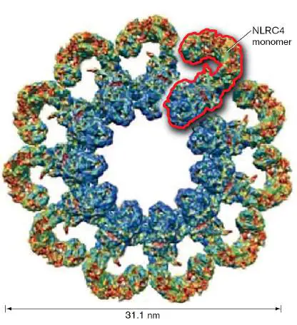
of another NLR to form a disk. Caspase-1 protease oligomerizes at the base of the disk.
Type I Interferons Are “Intruder-Alert” Cytokines
When a community is threatened by a thief, a neighbor who has been robbed alerts others to
take precautions. Something similar happens during viral infections. In 1957, it was
discovered that cells exposed to inactivated viruses produce at least one soluble factor that
can “interfere” with viral replication when applied to newly infected cells. The term interferon
was coined to represent these molecules. Interferons are low-molecular-weight cytokines
(14–20 kDa) produced by many eukaryotic cells in response to intracellular infection. The
action of interferons is usually host-species specific but virus non specific; that is, an
interferon from mice works only on mice but will protect mice from different viruses.
There are three general types of interferons, which differ in the receptors they bind and
the responses they generate. Type I interferons are made by almost all cells in the body and
have high antiviral potency. Type I interferons include IFN-alpha (IFN-α), IFN-beta (IFN-β),
and others (κ, ε, ω). The lone type II interferon, IFN-gamma (IFN-γ), has more of an
immunomodulatory function by activating various white blood cells (macrophages, natural
killer cells, and T cells) to, among other things, increase the number of major
histocompatibility complex (MHC) antigens on their surfaces (discussed further in Section
24.3). Type III interferons (IFN-λ) also induce antiviral responses but primarily in epithelial
cells.
How do interferons work? Once released by infected cells, type I interferons will bind to
specific receptors on uninfected host cells and either render those cells resistant to viral
infection or cause them to sacrifice themselves if they become infected. Type I interferons
induce dozens of interferon-stimulated genes (ISGs) that inhibit viral entry, destroy RNA, or
prevent translation of RNA. Different ISGs have specificity for different viruses. Consequently,
the sheer number of ISGs explains how a type I interferon can impose broad, nonspecific
antiviral protection.
Two classes of ISGs are particularly important. One class encompasses double-stranded,
RNA-activated endoribonucleases, such as RNase L, that cleave all cellular RNAs and, as a
result, trigger apoptosis. RNase L remains inactive as long as the cell remains uninfected. If
the cell becomes infected, however, the appearance of virus double-stranded RNA will
activate RNase L. Active RNase L will then cleave viral and host DNA, thereby killing the cell
and stopping viral spread. Another class of ISGs is made up of protein kinases that become
activated by binding viral double-stranded RNA. One of the activated protein kinases
phosphorylates eukaryotic initiation factor 2 (eIF2), rendering ribosomes unable to translate
viral or cellular RNA. These mechanisms can affect both RNA and DNA viruses because
protein synthesis is required for the propagation of all viruses. However, RNA viruses will
induce more interferon I than DNA viruses induce. Type I interferons are also used medically
to treat certain viral infections, such as chronic hepatitis C.
Type II interferon functions by activating various white blood cells—for example,
macrophages, natural killer cells, and T cells—to, among other things, increase the number of
MHC antigens on their surfaces. MHC proteins are important for recognizing self and for
presenting foreign antigens to the adaptive immune system. They are discussed more fully in
Chapter 24.
Does interferon production explain why children are more resistant to COVID-19?
It is known that the original SARS-CoV-2 virus, the cause of the COVID-19 pandemic, usually
causes worse symptoms in adults than in children. The reason, however, was a mystery. A
group of scientists scattered among several research institutions in Germany may have
discovered an answer that involves interferons and the nose.
We have seen that interferons function as an important innate viral defense mechanism.
But because interferon production typically requires activation of host pattern recognition
receptors, a host cell must be attacked by the virus before interferons can be produced.
However, if a viral pathogen can quickly shut down interferon synthesis, the virus wins.
Indeed, one of the pathogenic mechanisms of SARS-CoV-2 is to quickly shut down type I
interferon synthesis.
The scientists from Germany inventoried the innate immune system in the upper airways
of adults and children and discovered that, even without having contact with the virus, the
epithelial and immune cells in the noses of children were pre-activated and primed for virus
sensing. However, adults were much slower to respond. A more efficient and earlier
production of interferons in the infected airways of children shuts down viral replication faster
than the virus can stop host production of antiviral interferons. As a result, children are less
likely to develop COVID-19 disease than are adults.
Acute Inflammation
In medicine, the term “acute” means “rapid onset.” Acute inflammation, for instance, is
inflammation that develops rapidly, usually within a day, after a foreign object such as a
splinter or infectious agent is introduced into the body. Chronic inflammation, in contrast,
develops over long periods of time. The function of acute inflammation is to wall off, kill, or
digest the intruder, thereby quickly resolving an infection and promoting healing of the
affected area. Neutrophils play a central role in the inflammatory process.
Neutrophils, however, normally reside in the circulation. How do they find the infection
site? Once a macrophage encounters an infectious agent in tissues (Fig. 23.35 , steps 1 and
2), MAMPs located on, in, or shed by the pathogen bind to macrophage PRRs (pattern
recognition receptors), triggering the synthesis of cytokines (step 3); some of which are
chemokines (examples are IL-8 and monocyte chemoattractant protein, MCP-1). Chemokines
diffuse from a site of infection toward nearby capillaries and form a concentration gradient.
Then, like a fox following a prey’s scent, neutrophils and macrophages leave the bloodstream
and follow the gradient back to the infection. But how do these white blood cells pass
through blood vessel walls?
FIGURE 23.35 ■ The acute inflammatory response. Step 1: Traumatic
introduction of microbes through skin. Damaged host cells release factors that help
initiate inflammation. Step 2: Local macrophages engulf microbes and release a variety
of cytokines and chemokines. Step 3: Some cytokines initiate loosening of endothelial
tight junctions. Step 4: Cytokines initiate synthesis of selectins on endothelial cells.
Step 5: Selectins snag passing neutrophils to slow them down. Neutrophils start to roll.
Step 6: Other cytokines induce neutrophils to synthesize integrins that more tightly bind
the endothelial adhesion molecules ICAM-1 and VCAM-1. Step 7: Neutrophils squeeze
through the more permeable endothelium (extravasation). Step 8: Bradykinin made by
damaged host cells contributes to the increase in vascular permeability and activates
nearby mast cells to degranulate. Step 9: Mast cells release histamine to further
increase capillary permeability. Step 10: Bradykinin will also stimulate prostaglandin
synthesis in endothelial cells. Prostaglandins stimulate nearby nerve cells to register
pain.
The events leading to extravasation begin when the cytokines interleukin 1 (IL-1) and
tumor necrosis factor alpha (TNF-alpha), released by macrophages, stimulate the production
of adhesion molecules (selectins) on the inner lining of the capillaries (Fig. 23.35 , step 4).
P-selectin is produced first, followed by E-selectin. The selectins snag neutrophils zooming by
in the bloodstream, slow them down, and cause them to roll along the endothelium (step 5).
Rolling neutrophils that encounter inflammatory mediators are activated to produce and
display integrin adhesion molecules on their surface (step 6). Integrins on neutrophils lock
onto the endothelial adhesion molecules ICAM-1 (intercellular adhesion molecule 1) and
VCAM-1 (vascular cell adhesion molecule 1). Binding to these endothelial adhesion molecules
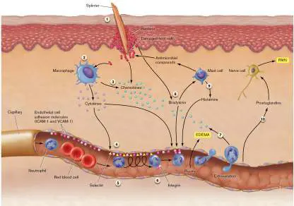
stops the neutrophils from rolling and initiates extravasation in which the white blood cells
squeeze through the endothelial wall and into the tissues (step 7).
The passage of neutrophils through vascular walls also requires loosening adhesions
between endothelial cells. Some cytokines released in step 3 of Figure 23.35 started this
process, but vasoactive factors such as bradykinin, a nine-amino-acid polypeptide released
by damaged tissue cells and macrophages, further increase vascular permeability so that
neutrophils can pass through vessel walls (step 8). Increased vascular permeability also
allows blood plasma to escape into tissues, causing swelling (edema). Even though tight
junctions are loosened, in order to actually pass between endothelial cells, activated
neutrophils need the enzyme sialidase to temporarily break carbohydrate linkages that hold
tight junctions together. Bradykinin also triggers degranulation of mast cells. The histamine
released from mast cells further loosens the endothelial cell junctions (step 9). More fluid
enters tissues and accumulates.
In addition to increasing vascular permeability, bradykinin and histamine relax smooth
muscles within blood vessel walls so that vessel diameter increases (vasodilation).
Vasodilation slows blood flow and, as a result, increases blood volume in the affected area.
Increased vascular permeability and vasodilation cause the localized swelling, redness, and
heat associated with inflammation.
Thought Question
23.6 As illustrated in Figure 23.35 , integrin is important for neutrophil extravasation.
Some individuals, however, produce neutrophils that lack integrin. What is the likely
consequence of this genetic disorder?
Once neutrophils have passed through the vascular wall, the chemokines released in step
3 of Figure 23.35 lure them to the proper location. In addition, neutrophils can sense
certain microbial chemoattractants—fMet-Leu-Phe peptide, for instance. Many bacterial
proteins have fMet (N -formylmethionine) as their N-terminal amino acid, but mammalian
proteins, in general, do not. Bacteria will often cleave off the fMet peptide, which can then
diffuse away from the bacterium. (Chapter 8 describes posttranslational processing in
bacteria.) The fMet peptide binds to neutrophil receptors and stimulates pseudopod
projections aimed toward the microbe. As a result, the white blood cell migrates in the
direction of the infection. Once phagocytes arrive at the site of infection, they begin
devouring microbes. Realize, however, that much of the damage caused by an infection is not
due directly to the microbe but is the result of the body’s inflammatory reaction to the
microbe’s presence.
What causes the pain of inflammation? Bradykinin induces capillary cells to make
prostaglandins that cause pain by stimulating nerve endings in the area (Fig. 23.35 , step
10). A key enzyme involved in prostaglandin synthesis is cyclooxygenase (COX). Aspirin,
ibuprofen, and the anti-inflammatory agent naproxen are COX inhibitors that prevent the
synthesis of prostaglandins and thus reduce inflammatory pain.
Thought Question
23.7 What happens to all the neutrophils that enter a site of infection once the infection has
resolved?
Phagocytes Recognize Alien Cells and Particles
For phagocytosis to proceed safely, macrophages and neutrophils must first recognize the
surface of a particle as foreign (Fig. 23.36 ). When a phagocyte surface interacts with the
surface of another body cell, the phagocyte becomes temporarily paralyzed (unable to form
pseudopods). Paralysis allows the phagocyte to evaluate whether the other cell is friend or
foe, self or nonself. To recognize self, glycoproteins located on the white blood cell membrane
must bind to inhibitory glycoproteins present on other host cell membranes. The inhibitory
glycoprotein on human cells is called CD47. Because invading bacteria lack these inhibitory
surface molecules, they can readily be engulfed.
FIGURE 23.36 ■ Images of phagocytosis. A. A white blood cell phagocytosing
Mycobacterium cells (green, 2 μm long; colorized SEM). B. Contacts between phagocyte
and target microbe, illustrating how phagocytes “grab” the target bacterium. C. A
macrophage engulfing bacteria on the outer surface of a blood vessel (SEM,
magnification 1,315×). D. Streptococcus pneumoniae and capsule (India ink
preparation). The slippery nature of the polysaccharide capsule makes phagocytosis
more difficult.
S. KAUFMANN & J. GOLECKI/SCIENCE SOURCE
VOLKER BRINKMANN/VISUALS UNLIMITED, INC.
DENNIS KUNKEL MICROSCOPY/SCIENCE SOURCE
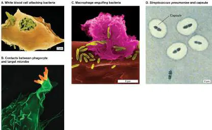
CDC
Although many bacteria, such as Mycobacterium or Listeria species, are easily recognized
and engulfed by phagocytosis (Fig. 23.36A–C ), others, such as Streptococcus pneumoniae
, possess polysaccharide capsules that are too slippery for pseudopods to grab (Fig. 23.36D
). This is where innate immunity and adaptive immunity join forces. Adaptive immunity
produces antibodies that bind to bacterial capsules. These anticapsular antibodies can aid the
innate immune mechanism of phagocytosis through a process known as opsonization (Fig.
23.37 ). An opsonin is any factor, such as antibodies, that can promote phagocytosis. The
anticapsular antibodies coat the surface of the bacterium, leaving the tail end of the
antibodies, called the Fc region (see Section 24.2), pointing outward. These bacteria are said
to be “opsonized.” The Fc regions of these antibodies are recognized and bound by specific
receptors on phagocyte cell surfaces. As a result, the antibodies link the opsonized bacteria to
phagocytic Fc receptors, thereby helping the phagocyte to more easily engulf the invader.
FIGURE 23.37 ■ Opsonization. Opsonization is a process that facilitates
phagocytosis. Here, macrophage Fc receptors bind to the Fc region of antibodies
attached to bacteria.
Thought Question
23.8 If NK cells can attack infected host cells coated with antibody, can neutrophils do the
same?
Oxygen-Independent and -Dependent Killing Pathways
During phagocytosis, the cytoplasmic membrane of the phagocyte flows around the
bacterium and then engulfs it, producing an intracellular phagosome, as described earlier
(see Fig. 23.20). Subsequent fusion between the phagosome and a lysosome produces a
phagolysosome that generates both oxygen-independent and oxygen-dependent killing
pathways. Mechanisms independent of oxygen include enzymes like lysozyme to destroy the
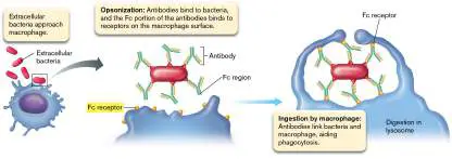
cell wall; compounds such as lactoferrin to sequester iron away from the microbe; and
defensins, small cationic antimicrobial peptides (described in Section 23.4).
Oxygen-dependent mechanisms are activated through Toll-like receptors (discussed
earlier) and kill by producing various oxygen radicals that can damage macromolecules.
NADPH oxidase, myeloperoxidase, and nitric oxide synthetase in the phagosome membrane
are extremely important. NADPH oxidase yields superoxide ion (• O 2 −), and superoxide
dismutase converts • O 2 − to hydrogen peroxide (H 2 O 2). Ferrous ion (Fe 2+) reacts with H
2 O 2 to produce hydroxyl radicals (• OH) and hydroxide ions (OH −). Myeloperoxidase,
present only in neutrophils, converts hydrogen peroxide and chloride ions to hypochlorous
acid (HOCl) and OH −.
Macrophages, mast cells, and neutrophils also generate reactive nitrogen intermediates
that serve as potent cytotoxic agents. Nitric oxide (NO) is synthesized from arginine by NO
synthetase. Further oxidation of NO by oxygen yields nitrite (NO 2 −) and nitrate (NO 3 −)
ions.
All of these reactive oxygen and nitrogen species attack bacterial membranes and
proteins. The mechanisms generating these molecules greatly increase oxygen consumption
during phagocytosis and produce what is called the oxidative burst. The reactive chemical
species formed during the oxidative burst do little to harm the phagocyte because the burst is
limited to the phagosome (where the bacteria are) and because the various reactive oxygen
species, such as superoxide, are very short-lived. Although phagocytes are very good at
clearing infectious agents, many bacteria have developed ways to outsmart this aspect of
innate immunity.
Autophagy and Intracellular Pathogens
Intracellular pathogens that grow in eukaryotic cytoplasm can be a serious problem for the
host. Many pathogens, such as Mycobacterium tuberculosis, the cause of tuberculosis, enter
the host cell in ways that bypass endosome formation or, if they do enter via an endosome,
can escape from that compartment. These pathogens block normal host cell clearance
pathways. To circumvent this problem, eukaryotic host cells (not just phagocytes) use a
process that normally degrades damaged organelles (called autophagy) to clear themselves
of intracellular pathogens. During autophagy, the cell constructs a double membrane around
the organism (or damaged organelle). This structure, called the autophagosome, sequesters
the microbe from the nutrient-rich cytosol. Lysosomes then fuse with the autophagosome,
depositing degradative enzymes that digest the organism. Ever-adapting intracellular
microbes, however, have found ways to suppress autophagy and survive.
Chronic Inflammation Causes Permanent Damage
Inflammation that persists over months or years is called chronic inflammation and is
provoked by the long-term presence of a causative stimulus. Chronic inflammation inevitably
causes permanent tissue damage, even though the body attempts repair. The causes of
chronic inflammation are many. For example, infectious organisms such as Mycobacterium
tuberculosis, Actinomyces bovis, and various protozoan parasites can avoid or resist host
defenses (Fig. 23.38 ). As a result, they persist at the site and continually stimulate the
basic inflammatory response. The continual stimulation of an inflammatory response leads to
chronic inflammation.
FIGURE 23.38 ■ Chronic inflammation. A. The fluorescent green organisms shown
are Mycobacterium tuberculosis (2 μm long; fluorescence microscopy) within
macrophages in a tuberculosis abscess. B. Fish tank granuloma. Mycobacterium marinum
, the cause of a tuberculosis-like infection in fish, can accidentally enter an open wound
or abrasion of a person cleaning out an aquarium. The infection is first noted as a slowly
healing lesion on the hand or forearm. Once it does heal, it often forms a granuloma at
the site that contains live organisms. C. Cross section of liver showing a necrotizing
granuloma caused by M. tuberculosis organisms that spread from the lung to the liver
through the bloodstream. D. Healthy colon. E. Intestinal granulomas of Crohn’s disease.
ALISSA ROTHCHILD
ISM/SCIENCE PHOTO LIBRARY
D. G. RUSSELL ET AL. 2010. CELL HOST MICROBE 8 :68–76
DAVID M. MARTIN, MD/SCIENCE SOURCE
GASTROLAB/SCIENCE SOURCE
Nonliving, irritant material like wood splinters, inhaled asbestos particles, or surgical
implants can also cause chronic inflammation. Autoimmune diseases are another important
cause. Autoimmunity (reaction against self) occurs when there is a failure to regulate some
aspect of adaptive immunity (see Chapter 24). As a result, the immune system recognizes a
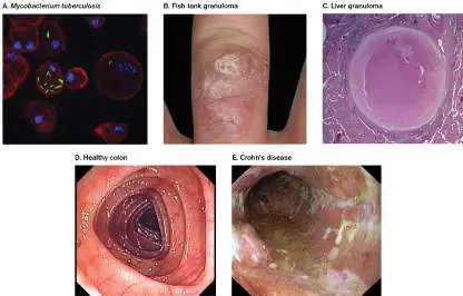
part of the body as foreign (not self) and begins to react against it. Rheumatoid arthritis is
one example of the body attacking itself.
Whatever causes chronic inflammation, macrophages and lymphocytes are continually
recruited from the circulation. The body may attempt to “wall off” the site of inflammation by
forming a granuloma. A granuloma begins as an aggregation of the mononuclear
inflammatory cells surrounded by a rim of lymphocytes. The body then deposits
fibroconnective tissue around the lesion, causing tissue hardening known as fibrosis.
Several forms of granulomas are shown in Figure 23.38 . M. tuberculosis, for example,
has a thick, waxy cell wall that protects mycobacteria against the mechanisms used by
macrophages to destroy microorganisms. As a result, the organisms live for prolonged periods
within macrophages (Fig. 23.38A ). Resistance to host defenses can produce long-term
chronic infections and granulomas. For example, skin infections caused by Mycobacterium
marinum will produce skin granulomas (Fig. 23.38B ), while M. tuberculosis can cause liver
granulomas (Fig. 23.38C ). Another disease thought to involve granuloma formation is
Crohn’s disease, which commonly manifests as abdominal pain, frequent bowel movements,
and rectal bleeding. Crohn’s disease has been attributed to an autoimmune reaction possibly
activated by intestinal microbiota. In this case, the intestinal bacteria are thought to cause a
chronic inflammation resulting in characteristic granulomas (compare Fig. 23.38D and E ).
To Summarize
Acute inflammation begins when host cells are damaged or infected. The process
walls off and digests invading microbes and initiates the healing of affected areas.
Damaged cells or tissue macrophages release vasoactive factors that produce
vasodilation and increased vascular permeability, cytokines that stimulate the
production of blood vessel selectin receptors, and chemoattractant molecules that
cause neutrophil movement (extravasation) from the bloodstream into infected
tissues. Bradykinin causes the release of prostaglandins, which produce pain in the
affected area. The cardinal signs of inflammation include redness, warmth, swelling,
pain, and loss of function.
Surface Toll-like receptors (TLRs) and cytoplasmic NOD-like receptors (NLRs)
in most host cells recognize microbe-associated molecular patterns (MAMPs) and
synthesize cytokine proteins that diffuse and activate other cells of the immune
system.
Interferons are one group of cytokines that can nonspecifically interfere with viral
replication (type I and type III) or modulate the immune system (type II).
Phagocytic cells have oxygen-independent and oxygen- dependent
mechanisms of killing that are initiated by the fusion of lysosomes and bacteria-
containing phagosomes.
The oxidative burst , a large increase in oxygen consumption during phagocytosis,
results in the production of superoxide ions, nitric oxide, and other reactive oxygen
species.
Autophagy is a process by which intracellular bacteria can be sequestered from the
cytoplasm (via an autophagosome) and killed following fusion with a lysosome.
Chronic inflammation results from the persistent presence of a foreign object.
Glossary
extravasation
Also called diapedesis. The movement of cells of the immune system out of blood
vessels and into surrounding infected tissue.
chemokine
An attractant for white blood cells that is produced by damaged tissues.
vasoactive factor
A cell signaling molecule that increases capillary permeability.
cytokine
A small, secreted host protein that binds to receptors on various endothelial and immune
system cells, regulating the cells’ responses.
pattern recognition receptor (PRR)
A protein receptor that recognizes microbe-associated molecular patterns (MAMPs) and
signals production of cytokines.
Toll-like receptor (TLR)
A member of a eukaryotic transmembrane glycoprotein family that recognizes a
particular microbe-associated molecular pattern (MAMP) present on pathogenic
microorganisms.
NOD-like receptor (NLR)
A eukaryotic cytoplasmic protein that recognizes particular microbe-associated molecular
patterns (MAMPs) present on microorganisms.
inflammasome
A cytoplasmic multiprotein complex that promotes the maturation of inflammatory
cytokines IL-1β and IL-18. Inflammasome assembly is triggered by NLR interactions with
microbe-associated molecular patterns (MAMPs).
interferon
A host-secreted immunomodulatory protein that inhibits viral replication.
interleukin 1 (IL-1)
A cytokine released by macrophages.
tumor necrosis factor alpha (TNF-alpha)
A cytokine involved in systemic inflammation.
selectin
One of a family of cell adhesion molecules.
bradykinin
A cell signaling molecule that promotes extravasation, activates mast cells, and
stimulates pain perception.
opsonization
The coating of pathogens with antibodies that aid pathogen phagocytosis by innate
immune cells.
opsonin
An antibody that renders its target (e.g., bacteria) susceptible to phagocytosis.
oxidative burst
A large increase in the oxygen consumption of immune cells during phagocytosis of
pathogens as the immune cells produce oxygen radicals to kill the pathogen.
autophagy
Eukaryotic cell function normally used to degrade damaged organelles. Also used to kill
intracellular pathogens.
chronic inflammation
Inflammation that has persisted over long periods of time, usually months or years.
granuloma
A thick lesion formed around a site of infection.
Fig. 23.20
FIGURE 23.20 ■ Phagocytosis and phagosome-lysosome fusion.
Endnotes
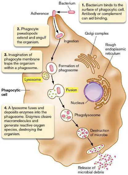
1. Note a: CpG = cytosine-phosphate-guanine sequence in DNA. Return to reference a
23.6 Complement and Fevernot assigned
White blood cells (WBCs) engulf and kill pathogens, but can simple serum proteins also kill microbes? Yes, a series of 20 serum proteins (complement factors) that make up the complement cascade can also attack bacterial invaders. Complement was first discovered as a heat-labile component of blood that enhances (or complements) the killing effect of antibodies on bacteria. Unlike antibodies, the complement cascade is a form of innate immunity, ready for full response without adaptation.
Several complement factors are proteases that sequentially form and then cleave other complement factors. (The liver is the main source of complement proteins.) Once a complement cascade is triggered, several things happen. Pores are inserted into bacterial membranes, causing cytoplasmic leaks (Fig. 23.39), while pieces of some complement proteins attract WBCs and facilitate phagocytosis (opsonization). Complement has also been implicated in the killing of pathogens trapped by neutrophil NETs, as described earlier (see Section 23.3).
FIGURE 23.39 ■ Pore structure of the membrane attack complex (MAC). Multimer assembly of complement factor 9 forms a pore in a bacterial membrane that destroys membrane integrity. Image shows a model of the MAC complex derived from cyo-EM studies. Components are colored as follows: C5b (brown), C6 (light green), C7 (dark green), C8 subunits (orange,
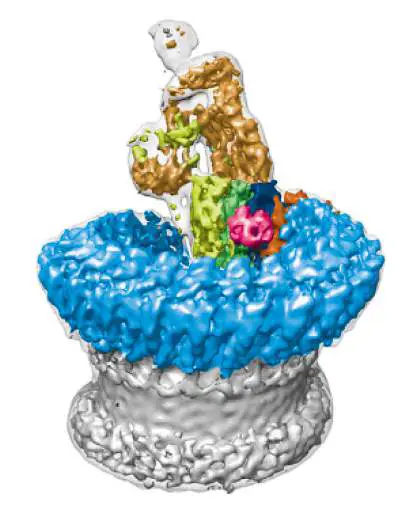
red, and dark blue), and finally C9 (light blue). Gray indicates the pore.
M. SERNA ET AL. 2016. NAT COMMUN. 7 :10587
Complement Activation Pathways
The three routes to complement activation are officially known as the classical pathway, the alternative pathway, and the lectin pathway. The classical complement pathway is discussed in the next chapter (see Section 24.4) because triggering the pathway depends on antibody, so it is part of both adaptive immunity and innate immunity. The lectin pathway requires the synthesis of mannose-binding lectin by the liver in response to certain macrophage cytokines. Lectin coats the surfaces of invading microbes and activates complement without needing antibody. However, the lectin pathway has more in common with the classical complement pathway and is also described in Chapter 24. We focus here in this chapter on the alternative pathway, as it is called, because this pathway does not involve adaptive immunity and can attack invading microbes long before a specific immune response can be launched.
The alternative complement pathway begins with the complement factor C3. In blood, C3 slowly cleaves into C3a and C3b (Fig. 23.40, step 1). C3b, under normal circumstances, is rapidly degraded—a process that thwarts inadvertent complement activation. However, if C3b meets LPS on an invading Gram-negative microbe, the bound C3b becomes stable and binds another factor, designated factor B (step 2), and makes factor B susceptible to cleavage by yet another protein, factor D (step 3). The resulting complex, called C3bBb, has two roles: It can quickly cleave more C3 to amplify the cascade and is changed by another serum protein (properdin) into what is called C5 convertase (step 4).
FIGURE 23.40 ■ The alternative complement pathway. Although called “alternative,” this complement cascade is part of the first-line innate defense system.
From this point on, the three complement pathways (alternative, classical, and lectin) work in the same way: C5 convertase cleaves C5 in serum to C5a and C5b (Fig. 23.40, step 5), and C5b then forms a prepore complex by binding to C6 and C7 (step 6). The resulting C5bC6C7 complex binds to target membranes. Finally, C8 and C9 factors join in to form the membrane attack complex (MAC), becoming a destructive pore (step 7).
In Gram-negative bacteria, MAC pores first form in the outer membrane (Fig. 23.40, step 7). Lysozyme (present in serum) enters through the MAC outer membrane pores and cleaves peptidoglycan, making the cytoplasmic membrane more susceptible
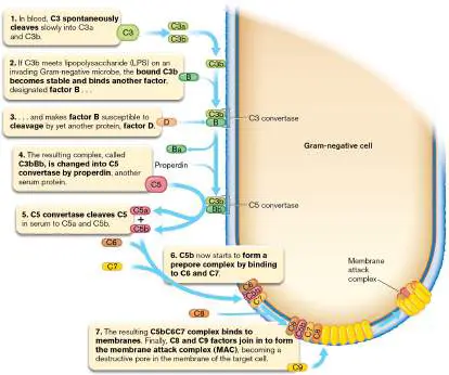
to the membrane attack complex. The inner membrane MAC pores destroy membrane integrity and, with it, proton motive force. Gram-positive bacteria are resistant to complement because they lack an outer membrane (and therefore have no LPS to efficiently start the cascade) and have a thick peptidoglycan layer that hinders access of complement components. Even in the absence of LPS, however, there are ways to activate complement that involve antigen-antibody complexes (see Section 24.4).
Thought Question
23.9 Figure 23.40 shows how the complement cascade can destroy a bacterial cell. Factor H (not shown) is a blood protein that regulates complement activity. Factor H binds host cells and inhibits complement from attacking our cells by accelerating degradation of C3b and C3bBb. How could bacteria take advantage of factor H?
Other Roles for Complement Peptides in Innate Immunity
Factor C3b, in addition to initiating the complement cascade, is a potent opsonin. As described earlier, an opsonin is any factor that can promote phagocytosis. PMNs (neutrophils) have specific C3b receptors on their surface. Thus, when C3b binds to a bacterial cell surface, it tags that cell and makes it easier for PMNs to grab and engulf the organism.
The complement fragments C5a and C3a have many roles in immune function. They are anaphylatoxins, which trigger degranulation of vasoactive factors such as histamine from endothelial cells, mast cells, or phagocytes. They can also stimulate chemotaxis of immune cells. C5a and C3a bind to separate receptors on WBC plasma membranes and activate separate signal cascade pathways. C5a triggers Ca 2+ release from intracellular stores and stimulates the actin polymerization needed for cell migration. Conversely, C3a triggers Ca 2+ influx, which facilitates extravasation. These peptide mediators also stimulate the release of certain cytokines, such as IL-4 from monocytes and mast cells, that prepare capillary endothelium for the rolling and adhesion of neutrophils. C5a will also up-regulate P-selectin and ICAM-1. Thus, some complement factors not only help directly destroy target bacterial cells but also facilitate phagocytosis and contribute directly to inflammation.
Acute-Phase Reactants and Complement
As noted earlier, inflammation is associated with the production of various cytokines by macrophages. Some of these cytokines (such as IL-1, TNF-alpha, and IL-6) travel to the liver, where they stimulate synthesis of several so-called acute-phase reactant proteins, including C-reactive protein. Acute-phase reactants circulate in the bloodstream and can be detected during laboratory testing. Named for its ability to activate complement, C-reactive protein will bind to components of bacterial cell surfaces but not to host cell membranes. Once linked to the bacterial cell surface, C-reactive protein will bind complement factor C1q of the classical complement pathway (see Chapter 24), ultimately converting factor C3 to C3b, propagating the complement cascade. C-reactive protein accelerates C3b production at the bacterial surface, where it can do the most damage. Note that an elevated level of C-reactive protein in serum is considered a general indicator of inflammation, not just inflammation caused by infection.
Fever
What is fever, and why is it a good thing? To understand fever, we first need to know how the body controls its temperature (thermoregulation). Heat in a human body is produced as a consequence of metabolic reactions. The liver and muscles are the major generators of heat and will warm the blood that passes through them. Body heat is generated (thermogenesis) by lipolysis in certain adipose tissues through the combustion of lipid substrates and the ability to uncouple the electron transport chain in mitochondria from the synthesis of ATP. Protons then leak from a high concentration at the mitochondrial intermembrane space into the matrix, releasing heat energy as the protons make new bonds. In a healthy person, body temperature is kept between 36°C and 38°C (97°F and 100°F), despite large differences in surrounding temperature and physical activity. Fever is defined as an oral temperature above 100.4°F (38°C).
How do we normally maintain a body temperature of 37°C? Heat sensors located throughout the skin and large organs and along the spinal cord send information about the body’s temperature to the thermoregulatory center in the hypothalamus, a small structure in the brain near the brain stem. The hypothalamus acts as a thermostat by controlling lipolysis in adipose tissue, as well as blood flow through the skin and subcutaneous areas. Vasoconstriction (tightening of blood vessel diameter) prevents the release of body heat when we are cold, whereas vasodilation secures its quick release when we are hot. If skin temperature is too high, the hypothalamus directs vasodilation to accelerate heat release. If body temperature is too low, blood flow will decrease to conserve heat, and shivering begins as a way to generate more heat. Fever is a natural reaction to infection and is usually accompanied by general symptoms such as sweating, chills, and the sensation of being cold. Substances that cause fever are known as pyrogens. Exogenous pyrogens (for example, certain bacterial toxins) originate outside the body, whereas internal or endogenous pyrogens (such as tumor necrosis factor and the interferon IL-6) are made by the body itself. External pyrogens generally cause fever by inducing the release of internal cytokine pyrogens.
Pyrogenic cytokines cross the blood-brain barrier and bind to neurons in the thermoregulatory center of the anterior hypothalamus. Cytokine-receptor interaction stimulates the production of phospholipase A2, an enzyme required to make prostaglandins. Prostaglandin E2 is made and changes the responsiveness of thermosensitive neurons. In other words, prostaglandin E2 turns up the thermostat. The body “thinks” it is cold, so the hypothalamus sends signals via the autonomic nervous system (which acts below the level of consciousness) to increase lipolysis and constrict peripheral blood vessels (vasoconstriction). Heat is not released and builds up to cause fever. Involuntary muscle contractions (shivering and chills) also generate heat. What are the advantages of fever? Because the ideal growth temperature for many microbes is 37°C, elevated temperature can place the pathogenic organism outside its “comfort zone” of growth. There is also evidence that fever decreases iron availability to bacteria (cytokine release causes an increase in iron storage protein). Slower growth of the pathogen “buys” the immune system time to subdue the infection before it is too late. Consequently, interventions that reduce a moderate fever caused by infection may actually slow recovery.
Thought Question
23.10 If increased fever limits bacterial growth, why do bacteria make pyrogenic toxins?
To Summarize
Complement is a series of 20 proteins naturally present in serum.
Activation of the complement cascade results in a pore being introduced into target membranes.
The three pathways for activation are the classical, alternative, and lectin pathways.
The alternative activation pathway begins when complement factor C3b is stabilized by interaction with the LPS of an invading microbe.
The cascade of protein factors , C3b ⟶ factor B ⟶ factor D ⟶ properdin ⟶ C5 ⟶ C6 ⟶ C7 ⟶ C8 and C9, results in the formation of a membrane attack complex in target membranes.
C-reactive protein in serum is activated when bound to microbial structures and will convert C3 to C3b, which can start the complement cascade.
The hypothalamus acts as the body’s thermostat.
Exogenous and endogenous pyrogens elevate body temperature by stimulating prostaglandin production. Prostaglandins change the responsiveness of thermosensitive neurons in the hypothalamus.
Glossary
membrane attack complex (MAC)
A cell-destroying pore produced in the membrane of invading bacteria by the host cell complement cascade.
C-reactive protein A peptide that stimulates the complement cascade, induced by cytokines in the liver. Elevated levels in the blood are associated with inflammatory processes such as heart disease. pyrogen Any substance that induces fever.
eResearch Activity 23
Can Altering the Gut Microbiome Affect Memory?
Dementia is a terrifying proposition for anyone, especially for older people. In fact, a major risk factor for developing dementia is aging itself. The precise reasons are unclear, but aging is associated with an increased secretion of senescence-linked mediators, such as the inflammatory cytokine TNF-alpha, and a decrease in brain-derived neurotrophic factor (BDNF) that is linked to learning and memory. Aging also alters the composition of the gut microbiome, and we know that gut dysbiosis can elevate production of inflammatory bacterial by-products, such as LPS, that can cause neuroinflammation in the brain. These and many other observations suggest that the composition of the microbiome could be connected to cognitive decline. If so, does the vagus nerve, a vital part of the gut-brain axis, serve as the conduit through which cognition-altering bacterial products travel to the brain?
Dong-Hyun Kim and collaborators at the Neurobiota Research Center in Seoul, South Korea, have explored these questions by taking fecal microbiota from young people (on average 20 years old) and from older individuals (on average 62 years old) and transplanting them into young mice. The scientists then tested the cognitive function of these mice using a series of behavioral tests. The results of the new object recognition test (NOR) are shown in Figure ERA 23.1 . During the familiarization phase of the NOR test, mice are presented with two similar objects that they can examine via touch. In a second session 24 hours later, one of the two objects is replaced by a new object. An index of recognition memory is calculated as the amount of time the mice take exploring (touching) the new object relative to the familiar object. The index is the number of times the new object is touched relative to the total touches made to both objects. Figure ERA 23.1 reveals that fecal transplantation from older humans (AF) significantly impaired recognition capability of the mice relative to mice receiving no transplant or to those receiving a transplant from young humans (YF).
FIGURE ERA 23.1 ■ Effects of transplanting young adult and elderly feces on cognitive impairment. Fecal suspensions were transplanted into young mice by oral gavage for 5 days. NOR cognitive tests were administered on the fifth day after treatment. NC= control mice, no transplant; YF = mice transplanted with microbiota from a young human; AF= mice transplanted with microbiota from an elderly human. The letters a and b indicate insignificant and significant (p >0.05) differences.
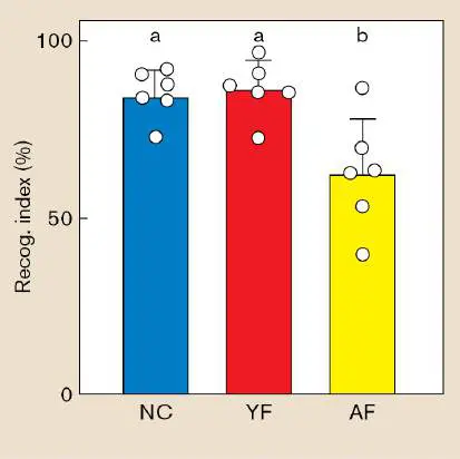
Next, the scientists examined the hippocampus of each mouse to determine if the fecal transplants affected the number of BDNF-positive cells (essential for learning and memory) and NeuN-positive cells (NeuN is a marker for mature neurons). Figure ERA 23.2 reveals that mice receiving fecal transplants from older people exhibited decreases in both cell types (compare Fig. ERA 23.2C to Fig. ERA 23.2A and B ). These data support the notion that microbiomes of aged people can potentially diminish cognition.
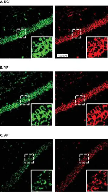
FIGURE ERA 23.2 ■ Effects of young adult and elderly fecal transplants on BDNF (green) and NeuN (red) cell counts in the hippocampus. BDNF and NeuN cells were detected by immunofluorescence. Brighter staining of cells equals greater quantity of corresponding cell type (A. , B. ). Transplants from older people (C. ) diminished fluorescence. NC= control mice, no transplant; YF = mice transplanted with microbiota from a young human (20 years old); AF= mice transplanted with microbiota from an older (62 years old) human.
K. E. LEE ET AL. 2020. MICROBIOME 8 :107
K. E. LEE ET AL. 2020. MICROBIOME 8 :107
K. E. LEE ET AL. 2020. MICROBIOME 8 :107
Paenalcaligenes hominis, a member of the Proteobacteria, has frequently been found in the gut microbiomes of elderly people but not in children or young adults. Escherichia coli, a common species found in the gut microbiome, is reportedly present at higher numbers in older people relative to those who are younger. So, Kim’s group separately transplanted these two microorganisms into young specific pathogen free (SPF) mice. In contrast to germ-free mice, SPF mice have a gut microbiome that is verified to be free of known pathogens. Both sets of transplanted SPF mice exhibited significant cognitive impairment by the NOR test relative to control mice (Fig. ERA 23.3A ). The results in Figure ERA 23.3C and D show that transplantation with these organisms also decreased the numbers of BDNF and NeuN cells relative to control mice (Fig. ERA 23.3B ). The fecal population of both organisms eventually fell to control levels after 30 days, which, for E. coli, also paralleled recovery of cognitive abilities. Curiously, however, the cognitive ability of mice transplanted with P. hominis hardly recovered.
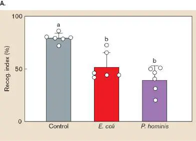
Figure ERA 23.3 ■ Effects of Paenalcaligenes hominis and Escherichia coli on cognitive impairment in specific pathogen free (SPF) mice. SPF mice transplanted with these bacteria were subjected to the NOR test (A ) as described in Figure ERA 23.1 and were examined for BDNF and NeuN cells (B , C , D ) in the hippocampus as described in Figure ERA 23.2.
K. E. LEE ET AL. 2020. MICROBIOME 8 :107
K. E. LEE ET AL. 2020. MICROBIOME 8 :107
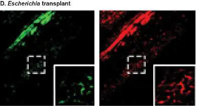
K. E. LEE ET AL. 2020. MICROBIOME 8 :107
How did the transplanted P. hominis and E. coli cause cognitive impairment? The mechanism between the two seems to differ. The researchers suspected one route involved communication between the gut microbiome and the brain via the vagus nerve, the conduit of the microbiome-gut-brain axis. The other route was thought to involve LPS made in the gut passing through blood to enter the brain, where it triggered inflammation. The researchers repeated the earlier experiments, but this time they included mice in which the vagus nerve was surgically severed (vagotomy). After this surgery, the transplantation of P. hominis no longer affected cognitive ability. When the same experiment was done with E. coli, however, vagotomy did not prevent the cognition effect. These results indicate that the gut-brain axis is important for the diminished cognition caused by P. hominis but not by E. coli.
The researchers then suspected that bacterial extracellular vesicles (BEV) shed from P. hominis might be traveling along the vagus nerve to affect the brain. Numerous studies have shown that BEVs from gut microbiota can carry and shelter cargo such as bacterial enzymes, signal molecules, DNA, RNA, and even psychoactive molecules. As they predicted, administering P. hominis BEV by oral gavage resulted in cognitive decline, and when P. hominis fluorescein isothiocyanate (FITC)–labeled BEVs were used, FITC accumulated in the hippocampus (Fig. ERA 23.4A ). Severing the vagus nerve, however, prevented BEV-induced cognitive decline, and it prevented the accumulation of FITC in the hippocampus (Fig. ERA 23.4B ). Vagotomy also prevented the accumulation of bacterial 16S rRNA in the hippocampus, suggesting that BEVs containing P. hominis RNA are carried along the vagus nerve (Fig. ERA 23.4C ). Intact BEVs, however, were never found in the hippocampus, probably because BEVs must fuse with host membranes to deliver cargo. FIGURE ERA 23.4 ■ Vagotomy inhibited the accumulation of Paenalcaligenes hominis bacterial extracellular vesicles (BEVs) in the mouse hippocampus.
A. Mice orally gavaged with FITC-labeled BEVs from P. hominis accumulated the contents of those BEVs in the hippocampus (green). B. Vagotomy prevented that accumulation. Green indicates FITC-labeled EVs; red indicates macrophages. C.
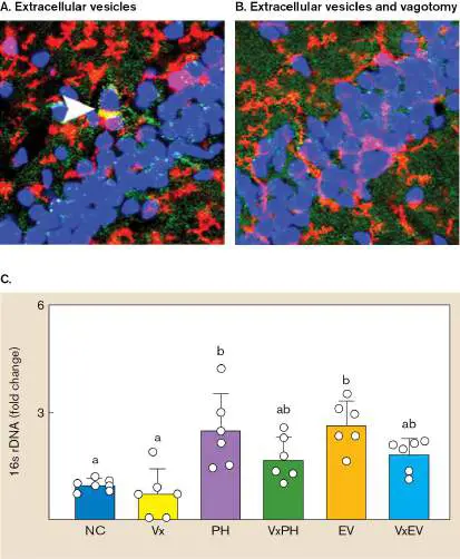
Accumulation of P. hominis 16S rRNA in the hippocampus of control mice (NC), vagotomized control mice (Vx), P. hominis - treated mice (PH), vagotomized PH-treated mice (VxPH), EV-treated mice (EV), and vagotomized BEV-treated mice (VxEV). Quantitative real-time PCR was used to determine fold change of P. hominis 16S rRNA relative to control mice (set to 1).
K. E. LEE ET AL. 2020. MICROBIOME 8 :107
K. E. LEE ET AL. 2020. MICROBIOME 8 :107
The researchers concluded that the BEVs of P. hominis may cause cognitive decline by translocating into the brain through the vagus nerve. How P. hominis BEVs cause cognitive decline remains under investigation.
The second pathway leading to cognitive decline involves LPS. LPS extracted from P. hominis or E. coli, administered by oral gavage, affected cognition whether or not the vagus nerve was severed. These results suggest that LPS disseminated from the gut, traveled through the bloodstream, and entered the brain to cause inflammation. But how inflammation led to cognitive decline is not yet known.
Further Exploration
How might this research eventually impact strategies to prevent Alzheimer’s or other forms of dementia?
Kyung-Eon Lee, Jeon-Kyung Kim, Sang-Kap Han, Dong Yun Lee, Hae-Ji
Lee, et al. 2020. The extracellular vesicle of gut microbial Paenalcaligenes hominis is a risk factor for vagus nerve-mediated cognitive impairment. Microbiome 8 :107.
https://doi.org/10.1186/s40168-020-00881-2.
CHAPTER REVIEW
Review Questions
1. Name some sterile body sites.
2. Which body sites are colonized by normal microbiota? 3. Under what circumstances can microbiota cause disease? 4. Why are commensal organisms beneficial to the host? 5. Name and describe various types of innate immunity. 6. What are probiotics? How do they help maintain health? 7. How do the lungs avoid being colonized?
8. What is a gnotobiotic animal?
9. Describe GALT and SALT.
10. Describe some chemical barriers to infection.
11. Discuss the different types of white blood cells. 12. What is a lymphoid organ?
13. Outline the process of inflammation.
14. Explain why phagocytes do not indiscriminately phagocytose body cells.
15. What is interferon?
16. Describe antibody-dependent cell-mediated cytotoxicity. 17. How does complement kill bacteria?
18. Why might fever be helpful in fighting infection?
Thought Questions
1. Is it common for microbial pathogens to pass the placental barrier (called transplacental transmission) and infect the fetus? Consider papillomavirus, Listeria monocytogenes, Escherichia coli, HIV, Treponema pallidum, Neisseria gonorrhoeae, and Staphylococcus aureus.
2. The vagina contains competing, commensal microbes that contribute to the health of the organ. So, is “cleaning” the vagina by douching actually unhealthy and likely to lead to an increased chance of infection (vaginosis)?
3. Why have microbes not altered their structures to avoid being recognized by Toll-like receptors?
4. The inflammatory response kills invading pathogens but can also damage bystander host cells and tissues. To prevent excessive damage, how does the host reset its innate immune system once an infection resolves?
Key Terms
adaptive immunity (970)
alveolar macrophage (982)
antibody-dependent cell-mediated cytotoxicity (ADCC) (976) antigen (971)
antigen-presenting cell (APC) (974) apoptosis (975)
autophagy (991)
B cell (979)
bacteremia (958)
basophil (973)
bradykinin (989)
C-reactive protein (994)
chemokine (984)
chronic inflammation (991)
complement (971)
compromised (immunocompromised) host (954) cystic fibrosis transmembrane conductance regulator (CFTR) ( 982)
cytokine (985)
defensin (982)
degranulate (983)
dendritic cell (974)
dysbiosis (965)
eosinophil (973)
epidermis (957)
extravasation (984)
gnotobiotic animal (966)
granuloma (991)
gut-associated lymphoid tissue (GALT) (981) immune system (970)
inflammasome (986)
innate immunity (970)
interferon (987)
interleukin 1 (IL-1) (989)
Langerhans cell (980)
leukocyte (971)
lymph node (979)
lymphocyte (978)
M cell (981)
macrophage (974)
major histocompatibility complex (MHC) (975) mast cell (973)
membrane attack complex (MAC) (993) microbe-associated molecular pattern (MAMP) (981) microbiome-gut-brain axis (964)
microbiota (microbiome) (954)
monocyte (973)
mononuclear phagocyte system (MPS) (974) mucociliary escalator (959)
nasopharynx (958)
natural killer (NK) cell (974)
neutrophil (972)
neutrophil extracellular trap (NET) (973) NOD-like receptor (NLR) (986)
nonadaptive immunity (970)
opportunistic pathogen (954)
opsonin (990)
opsonization (990)
oropharynx (958)
oxidative burst (991)
pattern recognition receptor (PRR) (985) perforin (975)
phagosome (972)
platelet (974)
probiotic (966)
pyrogen (995)
selectin (989)
skin-associated lymphoid tissue (SALT) (980) T cell (979)
tight junction (980)
Toll-like receptor (TLR) (985)
tumor necrosis factor alpha (TNF-alpha) (989) vasoactive factor (984)
Recommended Reading
Al Bataineh, Mohammad T., Rifat A. Hamoudi, Nihar R. Dash, Rakhee K. Ramakrishnan, Mohamad A. Almasalmeh, et al. 2020. Altered respiratory microbiota composition and functionality associated with asthma early in life. BMC Infectious Diseases 20 :697. https://doi.org/10.1186/s12879-020-05427-3.
Arneth, Borros. 2021. Trained innate immunity. Immunological Research 69 :1–7. https://doi.org/10.1007/s12026-021-09170-y.
Azzouz, Louiza, Ahmed Cherry, Magdalena Riedl, Meraj Khan, Fred G. Pluthero, et al. 2018. Relative antibacterial functions of complement and NETs: NETs trap and complement effectively kills bacteria. Molecular Immunology 97 :71–81. Birkle, Tim, and G. C. Brown. 2021. I’m infected, eat me! Innate immunity mediated by live, infected cells signaling to be phagocytosed. Infection and Immunity 89 : e00476-20.
https://doi.org/10.1128/IAI.00476-20.
Cebriá-Mendoza, María, Cristina Arbona, Luís Larrea, Wladimiro Díaz, Vicente Arnau, et al. 2021. Deep viral blood metagenomics reveals extensive anellovirus diversity in healthy humans. Nature Scientific Reports 11 :6921.
https://doi.org/10.1038/s41598-021-86427-4.
Gul, Muhammad Hamdan, Zin Mar Htun, and Asad Inayat.
2021. Role of fever and ambient temperature in COVID-19. Expert Review of Respiratory Medicine 15 :171–173.
https://doi.org/10.1080/17476348.2020.1816172.
Hadjadj, Jérôme, Nader Yatim, Laura Barnabei, Aurélien Corneau, Jeremy Boussier, et al. 2020. Impaired type I interferon activity and inflammatory responses in severe COVID-19 patients. Science 369 :718–724.
https://doi.org/10.1126/science.abc6027.
Koskinen, Kaisa, Manuela R. Pausan, Alexandra K. Perras, Michael Beck, Corinna Bangd, et al. 2017. First insights into the diverse human archaeome: Specific detection of Archaea in the gastrointestinal tract, lung, and nose and on skin. mBio 8:e00824-00817.
Lee-Sarwar, Kathleen A., Jessica Lasky-Su, Rachel S. Kelly, Augusto A. Litonjua, and Scott T. Weiss. 2020 .
Metabolome–microbiome crosstalk and human disease.
Metabolites 10 :181. https://doi.org/10.3390/metabo10050181. Leiva-Juárez, Miguel Martin, Jay K. Kolls, and Scott E. Evans. 2018. Lung epithelial cells: Therapeutically inducible effectors of antimicrobial defense. Mucosal Immunology 11 :21–34.
Li, Danyang, and Minghua Wu. 2021. Pattern recognition receptors in health and diseases. Signal Transduction and Targeted Therapy 6 :291. https://doi.org/10.1038/s41392-021-00687-0.
Liang, Guanxiang, and Frederic D. Bushman. 2021. The human virome: Assembly, composition and host interactions. Nature Reviews Microbiology 19 :S4.
https://doi.org/10.1038/s41579-021-00536-5.
Lindell, Anna E., Maria Zimmermann-Kogadeeva, and Kiran R. Patil. 2022. Multimodal interactions of drugs, natural compounds and pollutants with the gut microbiota. Nature Reviews Microbiology 20 :431–443.
https://doi.org/10.1038/s41579-022-00681-5.
Loske, J., J. Röhme, S. Lukassen, S. Stricker, V. G.
Magalhães, et al. 2021. Pre-activated antiviral innate immunity in the upper airways controls early SARS-CoV-2 infection in children. Nature Biotechnology 40 :319–324.
https://doi.org/10.1038/s41587-021-01037-9.
Maier, Lisa, Mihaela Pruteanu, Michael Kuhn, Georg Zeller, Anja Telzerow, et al. 2018. Extensive impact of non-antibiotic drugs on human gut bacteria. Nature 555 :623–628.
Morales-Primo, Abraham U., Ingeborg Becker, and Jaime Zamora-Chima. 2021. Neutrophil extracellular trap-associated molecules: A review on their immunophysiological and inflammatory roles. International Reviews of Immunology 41:253–274. https://doi.org/10.1080/08830185.2021.1921174. Moustafa, Ahmed, Chao Xie, Ewen Kirkness, William Biggs, Emily Wong, et al. 2017. The blood DNA virome in 8,000 humans. PLoS Pathogens 13 :e1006292.
https://doi.org/10.1371/journal.ppat.1006292.
Rathinam, Vijay A. K., and Francis Ka-Ming Chan. 2018. Inflammasome, inflammation, and tissue homeostasis. Trends in Molecular Medicine 24 :304–318.
Roswall, Josefine, Lisa M. Olsson, Petia Kovatcheva-Datchary, Staffan Nilsson, Valentina Tremaroli, et al. 2021. Developmental trajectory of the healthy human gut microbiota during the first 5 years of life. Cell Host and Microbe 29 :1–12. https://doi.org/10.1016/j.chom.2021.02.021.
Tao, Sha, and Ingo Drexler. 2020. Targeting autophagy in innate immune cells: Angel or demon during infection and vaccination? Frontiers in Immunology 11 :460.
https://doi.org/10.3389/fimmu.2020.00460.
von Mutius, Erika. 2021. The “hygiene hypothesis” and the lessons learnt from farm studies. Frontiers in Immunology 12 : article 635522. https://doi.org/10.3389/fimmu.2021.635522. Wikoff, William R., Andrew T. Anfora, Jun Liu, Peter G. Schultz, Scott A. Lesley, et al. 2009. Metabolomics analysis reveals large effects of gut microflora on mammalian blood metabolites. Proceedings of the National Academy of Sciences USA 106 :3698–3703.
Glossary
microbiota or microbiome The total community of microbes associated with an organism (such as the human body) or with a defined habitat (such as soil or plants).
compromised host An animal with a weakened immune system.
opportunistic pathogen A microbe that normally is not pathogenic but can cause infection or disease in an immunocompromised host organism. epidermis The outer protective cell layer in most multicellular animals. nasopharynx The passage leading from the nose to the oral cavity. oropharynx The area between the soft palate and the upper edge of the epiglottis.
bacteremia A bacterial infection of the blood.
mucociliary escalator The ciliated mucous lining of the trachea, bronchi, and bronchioles that sweeps foreign particles up and away from the lungs.
microbiome-gut-brain axis A complex bidirectional network of communication between the central nervous system, the intestine, and intestinal microbiota. dysbiosis An imbalance in microbiome composition that can lead to disease.
probiotic A food or nutritional supplement that contains live microorganisms and aims to improve health by promoting beneficial bacteria.
gnotobiotic animal An animal that is germ-free or colonized by a known set of microbes.
immune system An organism’s cellular defense system against pathogens. innate immunity Also called nonadaptive immunity. Nonspecific mechanisms for protecting against pathogens.
nonadaptive immunity See innate immunity .
adaptive immunity Immune responses activated by a specific antigen and mediated by B cells and T cells.
antigen A compound, recognized as foreign by the cell, that elicits an adaptive immune response. See also immunogen .
complement Innate immunity proteins produced by the liver; they circulate in the blood, where they can form holes in bacterial membranes, killing the bacteria.
leukocyte White blood cell.
neutrophil A white blood cell of the innate immune system that can phagocytose and kill microbes.
phagosome A large intracellular vesicle that forms as a result of phagocytosis.
neutrophil extracellular trap (NET)
A net of chromatin (including DNA) and antimicrobial peptides expelled by dying neutrophils to trap and injure nearby pathogenic bacteria.
basophil A white blood cell, stained by basic dyes, that secretes compounds that aid innate immunity.
eosinophil A white blood cell that stains with the acidic dye eosin and secretes compounds that facilitate innate immunity.
mast cell A white blood cell that secretes proteins that aid innate immunity. Mast cells reside in connective tissues and mucosa and do not circulate in the bloodstream.
monocyte A white blood cell with a single nucleus that can differentiate into a macrophage or a dendritic cell.
alveolar macrophage A type of macrophage, located in the lung alveoli, that phagocytoses foreign material.
dendritic cell An antigen-presenting white blood cell that primarily takes up small soluble antigens from its surroundings.
mononuclear phagocyte system (MPS)
An amorphous part of the immune system that consists mostly of monocytes and macrophages present in connective tissue that surrounds kidney, liver, spleen, lymph nodes, Peyer’s patches, and bone marrow.
antigen-presenting cell (APC)
An immune cell that can process antigens into antigenic determinants and display those determinants on the cell surface for recognition by other immune cells.
platelet A small cell fragment without a nucleus found in blood that is involved in clotting.
natural killer (NK) cell A lymphocyte that does not need antigen stimulation to kill tumor or infected host cells by inserting granules containing perforin.
major histocompatibility complex (MHC)
Transmembrane cell proteins important for recognizing self and for presenting foreign antigens to the adaptive immune system. perforin A cytotoxic protein, secreted by T cells, that forms pores in target cell membranes.
apoptosis A cell death program triggered during tissue differentiation or in certain damaged or infected cells.
antibody-dependent cell-mediated cytotoxicity (ADCC) The process by which natural killer cells destroy antibody-coated host cells that are infected by a virus.
lymphocyte A mononuclear leukocyte (white blood cell) that is a product of lymphoid tissue and participates in immunity (e.g., B cell and T cell).
B cell An adaptive immune cell, developed in bone marrow tissue, that can give rise to antibody-producing cells.
T cell An adaptive immune cell, developed in the thymus, that can give rise to antigen-specific helper cells and cytotoxic T cells. lymph node A secondary lymphatic organ, formed by the convergence of lymphatic vessels, that traps foreign particles from local tissue and presents them to resident immune cells.
tight junction A type of junction between the membranes of two adjacent vertebrate cells that form an impermeable barrier.
skin-associated lymphoid tissue (SALT)
Immune cells, such as dendritic cells, located under the skin that help eliminate bacteria that have breached the skin surface.
Langerhans cell A specialized, phagocytic dendritic cell that is the predominant cell type in skin-associated lymphatic tissue.
microbe-associated molecular pattern (MAMP)
Formerly called pathogen-associated molecular pattern (PAMP). Molecules associated with groups of microbes, both pathogenic and nonpathogenic, that are recognized by cells of the innate immune system.
gut-associated lymphoid tissue (GALT)
Lymphatic tissues such as tonsils and adenoids that are found in conjunction with the gastrointestinal tract and contain immune cells.
M cell A phagocytic innate immune cell (microfold cell) found between intestinal epithelial cells.
alveolar macrophage A mononuclear, phagocytic, antigen-presenting cell of the immune system.
cystic fibrosis transmembrane conductance regulator (CFTR) A chloride channel found in respiratory epithelia. Mutations in the CFTR gene lead to cystic fibrosis.
defensin A type of small, positively charged peptide, produced by animal tissues, that destroys the cell membranes of invading microbes. degranulate To release antimicrobial granule contents by fusing granule membranes to cytoplasmic or vacuolar membranes.
extravasation Also called diapedesis. The movement of cells of the immune system out of blood vessels and into surrounding infected tissue.
chemokine An attractant for white blood cells that is produced by damaged tissues.
vasoactive factor A cell signaling molecule that increases capillary permeability. cytokine A small, secreted host protein that binds to receptors on various endothelial and immune system cells, regulating the cells’ responses.
pattern recognition receptor (PRR)
A protein receptor that recognizes microbe-associated molecular patterns (MAMPs) and signals production of cytokines.
Toll-like receptor (TLR)
A member of a eukaryotic transmembrane glycoprotein family that recognizes a particular microbe-associated molecular pattern (MAMP) present on pathogenic microorganisms. NOD-like receptor (NLR)
A eukaryotic cytoplasmic protein that recognizes particular microbe-associated molecular patterns (MAMPs) present on microorganisms.
inflammasome A cytoplasmic multiprotein complex that promotes the maturation of inflammatory cytokines IL-1β and IL-18. Inflammasome assembly is triggered by NLR interactions with microbe-associated molecular patterns (MAMPs).
interferon A host-secreted immunomodulatory protein that inhibits viral replication.
interleukin 1 (IL-1)
A cytokine released by macrophages.
tumor necrosis factor alpha (TNF-alpha)
A cytokine involved in systemic inflammation.
selectin One of a family of cell adhesion molecules.
bradykinin A cell signaling molecule that promotes extravasation, activates mast cells, and stimulates pain perception.
opsonization The coating of pathogens with antibodies that aid pathogen phagocytosis by innate immune cells.
opsonin An antibody that renders its target (e.g., bacteria) susceptible to phagocytosis.
oxidative burst A large increase in the oxygen consumption of immune cells during phagocytosis of pathogens as the immune cells produce oxygen radicals to kill the pathogen.
autophagy Eukaryotic cell function normally used to degrade damaged organelles. Also used to kill intracellular pathogens. chronic inflammation Inflammation that has persisted over long periods of time, usually months or years.
granuloma A thick lesion formed around a site of infection.
membrane attack complex (MAC)
A cell-destroying pore produced in the membrane of invading bacteria by the host cell complement cascade.
C-reactive protein A peptide that stimulates the complement cascade, induced by cytokines in the liver. Elevated levels in the blood are associated with inflammatory processes such as heart disease. pyrogen Any substance that induces fever.