Chapter introduction
COVID-19 immunotherapy: A “gripping” story.
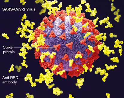
Despite vaccinations, variants of the SARS-CoV-2 virus have prolonged the COVID-19 pandemic. Antibodies elicited by a COVID-19 vaccine block virus entry by binding to a section of the virus spike protein that ordinarily binds to ACE2 receptors in host cell membranes. Virus variants with
As recently as 200 years ago, infectious diseases were thought to result from inhaling poisonous vapors, called “miasmas,” produced by decaying organic matter. According to theory, disease resulted when miasmas invaded the body and disturbed vital functions. Although physicians up until the nineteenth century were wrong about this, they did recognize that humans reacted to disease with fevers and “humors” that somehow overcame the miasmas—at least most of the time. Today we know that “miasmas” are actually infectious pathogens and that the innate and adaptive immune In this chapter we describe antibody-dependent immunity, in which B cells differentiate into plasma cells that make antibodies. The chapter also explores cell-mediated immunity, whereby specific T-cell lymphocytes develop to directly kill infected host cells. Along the way we explore how gut mucosal immunity coexists with the microbiome and how vaccines train the immune system. Ultimately, you will appreciate that adaptive immunity is a major reason why
24.1 Overview of Adaptive Immunitynot assigned
The immune response is a stunningly complex biological system controlled by many checks and balances that prevent overreaction. Unfortunately, with complexity comes the potential for catastrophic genetic defects. Consider the following case: A visibly ill 9-month-old baby boy was admitted to the hospital with intractable diarrhea, inappropriate weight loss, and oral thrush caused by Candida albicans (Fig. 24.1). This infection was just the latest of several that the boy had suffered during his short life. Laboratory results found that he was deficient in all classes of his antibodies (a syndrome called hypogammaglobulinemia). And, most disturbingly, his antigen-presenting cells (monocytes, macrophages, and B cells) lacked major histocompatibility complex (MHC) II molecules—protein complexes that are critically important to adaptive immunity.
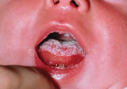
FIGURE 24.1 ■ Baby with thrush. The yeast Candida albicans can infect the oral cavities of babies, especially children with genetic immunodeficiencies.
DR. P. MARAZZI/SCIENCE SOURCE
MHC II molecules are essential for generating protective levels of antibodies, so their absence explained the boy’s hypogammaglobulinemia. The boy had bare lymphocyte syndrome, a disease caused by a defect in the gene that activates the transcription of MHC II genes. The only treatment is a bone marrow transplant to replace “bare” lymphocytes with compatible donor lymphocytes covered with MHC II molecules. This case dramatically illustrates the importance and fragility of the human immune system: All it takes is a small defect in a single gene to subvert the entire process.
Recall from Chapter 23 that the immune system has both nonadaptive and adaptive mechanisms. Nonadaptive (innate)
immune mechanisms are present from birth, whereas adaptive mechanisms are triggered as the need arises. For instance, an adaptive immune response to malaria does not develop until the individual has encountered the plasmodial parasite that causes the disease. The adaptive immune response—that is, adaptive immunity —is a complex, interconnected, and cross-regulated defense network.
Note: The terms “adaptive immune response” and “immune
response” are often used interchangeably. It is important to remember that the innate immune response always precedes the adaptive immune response.
Triggering Adaptive Immunity
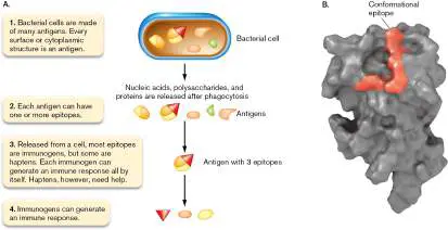
What triggers an adaptive immune response, and how quickly does it arise? After exposure to an invading microbe, adaptive immunity takes at least 3 or 4 days to develop. However, the immune system does not recognize the whole microbe all at once; rather, it recognizes innumerable pieces of it called antigens (Fig. 24.2). An antigen is any molecule, such as a protein, that elicits an immune response when introduced into a person. When the antigen by itself can elicit antibody production, it is termed an immunogen. Many antigens have even smaller segments, called epitopes or antigenic determinants, that also elicit an immune response (Fig. 24.2). Besides proteins, antigenic structures in the cell include complex polysaccharides, nucleic acids, and lipids. From birth, the naive immune system is capable of recognizing and responding to billions of possible foreign antigens while avoiding a response to self antigens. FIGURE 24.2 ■ Antigens and epitopes. A. Every protein, nucleic acid, lipid, and carbohydrate in (or on) the cell can be recognized by the immune system, so they are all antigens.
Antigens can have one or many different epitopes, and a large protein will have many epitopes. Some epitopes are immunogenic, while other epitopes act as haptens—so, all immunogens are antigens, but not all antigens are immunogens. B. A 3D protein structure, showing (in red) four amino acids that form a conformational epitope. Native proteins fold into a 3D shape, where several regions separated in the linear sequence can reside next to each other to form a conformational epitope.
ERIKA GUSTAFSSON ET AL. 2009. BMC IMMUNOL. 10 :1471–2172.
The adaptive immune response begins when cells of the innate immune system (macrophages and dendritic cells) engulf a microbe and break it down into component antigens (Fig. 24.3, step 1). These phagocytic macrophages and dendritic cells are also called antigen-presenting cells (APCs) because they place antigens and epitopes on their cell surface in order to “present” them to T lymphocytes. Imagine a stolen automobile entering a “chop shop,” where the car is taken apart and individual parts are sold (presented) to someone waiting outside. For presentation, APCs place the antigens on cell-surface proteins called major histocompatibility complex (MHC) proteins.
FIGURE 24.3 ■ Overview of the adaptive immune system.
Adaptive immunity comes in two forms: humoral and cell mediated. In humoral immunity (also called antibody-dependent immunity), B cells bind to free-floating microbial antigens released from phagocytic cells or from the microbe itself. However, each B cell
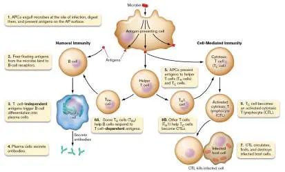
binds to only one antigenic structure. When an antigen binds to a matched B cell, the B cell divides to make many copies (Fig. 24.3, steps 2 and 3). Most of the copies become plasma cells that secrete large amounts of an antibody specific to the triggering antigen (step 4). An antibody is a Y-shaped protein that circulates in the bloodstream, waiting to bind to a matched antigen on an infecting pathogen. In fact, the system’s name “humoral” comes from a Latin word meaning “related to body fluids.” Note that plasma cells are terminally differentiated, so they cannot divide.
The other arm of adaptive immunity, called cell-mediated immunity (or cellular immunity), begins when an APC presents an antigen to an inactive cytotoxic T cell, or T C cell (Fig. 24.3, step 5). That interaction converts a T C cell into an activated cytotoxic T lymphocyte (CTL), which will seek and destroy host cells infected with microbes that express the same antigen (steps 6 and 7). In truth, the humoral and cellular immune responses are not separate but intertwined, each relying on some facet of the other to work efficiently. The cells that link the two forms of adaptive immunity are called helper T cells (T H cells). T H cells determine whether antibody-dependent or cell-mediated mechanisms will predominate in response to an antigen. APCs activate T H cells by presenting foreign antigens to a T H cell whose receptor can bind to the same antigen (Fig. 24.3, step 5). One class of helper T cells helps B cells become plasma cells (step 8A). A second class of helper T cells (T H 1) secretes cytokines that activate cytotoxic T cells (step 8B). So, the immune response to a microbe is really a cluster of responses made by thousands of different B cells, cytotoxic T cells, and helper T cells toward many different epitopes that came from a microbe digested by an APC. We present clarifying details of these steps later.
Where in the body does all of this happen? Antigen-presenting cells such as macrophages and dendritic cells encounter microbes at the site of an infection (Fig. 24.4, step 1). After engulfing the pathogen, the APCs migrate along lymphatic vessels to regional secondary lymph nodes where B cells and T cells await (step 2). Along the way, the APCs digest the microbe and present the antigenic pieces on their cell-surface MHC proteins. We reveal later how APCs actually present antigens.
FIGURE 24.4 ■ Staging areas of the immune response. APCs engulf pathogens at the infection site (1) and travel to the lymph node (2). In the lymph node (3), APCs present the antigens to T cells, interactions between B cells and T helper cells generate antibody-secreting plasma cells and memory B cells, and APCs present antigens to T C cells triggering differentiation to CTLs. The activated cells leave the lymph node and enter the bloodstream
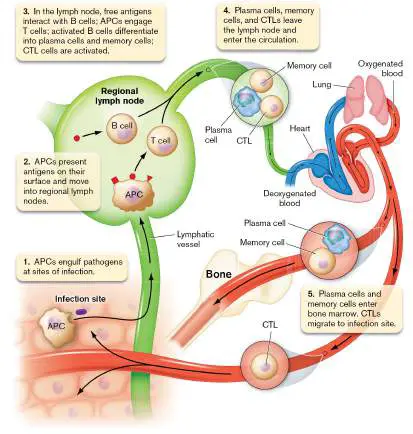
(4). Plasma cells and memory cells travel to and enter bone marrow, while CTLs travel to the infection (5).
Some antigens from the pathogen float freely in the lymph. Once they reach the lymph node, these free-floating antigens bind to B-cell receptors (BCRs) that are specific for that antigen (Fig. 24.4, step 3). Each B cell is programmed to bind to only one unique antigen. Meanwhile, APCs adorned with many different MHC-antigen complexes will present those antigens to the various T cells stationed in the lymph node. Each T cell can bind to a limited number of different antigens. Once activated by an APC, a helper T cell, called a follicular helper T cell (T FH cell), will engage a B cell already bound through its B-cell receptor to a different copy of the same antigen (see Fig. 24.3, step 8A). The interaction, which is described in Section 24.3, causes the B cell to proliferate and differentiate into plasma cells or memory cells.
Once plasma cells and memory cells are made, they migrate into the bloodstream and then to bone marrow where they remain, although some of these cells periodically recirculate through blood. Plasma cells release antibodies into blood, while memory cells await a second encounter with the antigen at some later date (Fig. 24.4, steps 4 and 5). Meanwhile, the activated cytotoxic T lymphocytes (CTLs) also leave the lymph node and migrate through the bloodstream to tissues where they attack infected host cells. We share details of these processes later.
Fluorescence-Activated Cell Sorting
How do scientists identify, count, and sort the many different cell types that make up the immune system? The instrument used to achieve this feat, a flow cytometer, was introduced in Chapter 4 (see Fig. 4.20) and detailed in Chapter 21 (Fig. 21.9). The technique, called fluorescence-activated cell sorting (FACS), uses fluorescently tagged antibodies to detect unique cell-surface proteins or glycoproteins present on different immune cells. Like identifying a dog by its spots, immune cell types can be identified by the presence or absence of unique antigen markers. Two examples are the CD4 marker on helper T cells and the CD8 marker on cytotoxic T cells. Figure 24.5shows the results of passing a patient’s lymphocytes through the flow cytometer to identify and count the CD4-positive and CD8-positive T cells. Each dot represents a single cell. In this figure, high values on the x -axis indicate the presence of CD8-positive lymphocytes, whereas high values on the y -axis indicate the CD4-positive lymphocytes. Few cells had both markers. By changing the detection antibodies, researchers can collect different cell types that pass through the flow cytometer and perform additional experiments with them. One such experiment is shown in Section 24.6 (see Fig. 24.29).
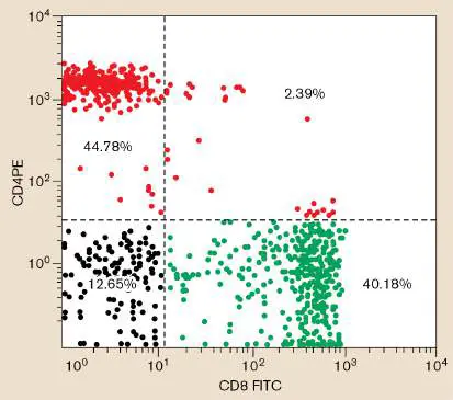
FIGURE 24.5 ■ Fluorescence-activated cell sorting: Identification of immune cell types. FACS analysis data used to identify and count CD4-positive helper T cells (T H cells) and CD8-positive cytotoxic T cells (T C cells). Fluorescently tagged antibodies to each marker (PE is Texas Red; FITC is fluorescein isothiocyanate) were added to lymphocytes collected from a patient’s blood sample. Separate detectors in the flow cytometer counted red cells (CD4 positive, y -axis) and green cells (CD8 positive, x -axis). Black dots indicate lymphocytes that had neither marker. Percentages indicate the fraction of cells in each quadrant relative to the total cell number.
Thought Question
24.1 Two different stretches of amino acids in a single protein form a 3D antigenic determinant. Will the specific immune response to that 3D antigen also recognize the same two amino acid stretches if they are removed from the whole protein?
Immunogenicity
Immunogenicity, or antigenicity, measures the relative effectiveness by which an antigen elicits an immune response—for example, how much antibody is produced. The strongest antigens are proteins, but carbohydrates can also elicit immune reactions. Nucleic acids and lipids are usually weaker antigens, in part because both molecules are made of relatively uniform repeating units that are very flexible. The flexible units present a variable 3D structure that does not easily interact with antibodies. Proteins are more effective antigens for three reasons: Different proteins have different shapes, they maintain their tertiary (3D) structure, and they are made of many different amino acids that can be assembled in many different combinations. These features provide stronger interactions with antibodies in the bloodstream and enable better recognition by lymphocytes, the cellular workhorses of the immune system.
Antigen presentation by antigen-presenting cells (described earlier) also plays a role in how immunogenic an antigen is. Antigen presentation is a necessary step before most immune responses can occur (Fig. 24.3, steps 1 and 5). For an APC to present antigen to a T cell, the antigenic determinant must first bind to a major histocompatibility complex on the APC. The more tightly an antigen can bind to an MHC surface protein, the more immunogenic the antigen is. The stronger the binding, the easier it is for T cells to recognize the complex.
A hapten, however, is a special form of antigen. Haptens are very small molecules, generally less than 1,000 daltons (a measure of mass). Haptens are so small, in fact, that they cannot bind to MHC molecules on APCs; therefore, they cannot be presented to T cells, and they will not elicit an immune response unless they are attached to a larger carrier protein. An example of a hapten is the antibiotic penicillin, a serious cause of allergic reactions in some individuals. The tiny penicillin molecule must bind to another, larger protein before the immune system can recognize it. Thus, there are two types of antigens: immunogens that elicit an immune response by themselves and haptens that must be attached to an immunogen in order to generate an immune response.
As you might expect, the body must regulate the immune system carefully so that a response is not leveled against itself. In effect, the immune system must become “blind” to its own antigens; as a result, the host will often be blind to foreign antigens that resemble epitopes of its own cells. Therefore, the more complex the foreign protein is, the more likely it will possess antigenic determinants that a lymphocyte can recognize as nonself. The farther an antigen is from “self,” the greater its immunogenicity will be.
Immunological Specificity
Note that antibodies, as well as B-and T-cell receptors, demonstrate varying levels of immunological specificity, also referred to as antigenic specificity. Immunological specificity is the degree to which an antibody, or receptor, distinguishes between similar-looking antigens. An antibody that can bind any one of several similar-looking but different antigens is said to have low immunological specificity. In contrast, an antibody that binds only one of those antigenic structures has high immunological specificity.
The earliest clues about immunological specificity came from smallpox. Smallpox is a devastating disease, caused by the variola virus, that inflicted enormous suffering and killed millions of people in the seventeenth and eighteenth centuries (see Chapter 1 and Fig. 24.6A). There was no cure, and the only available preventive treatment was to take dried material from the lesions of a previous smallpox sufferer, place it on a healthy person, and hope the person survived. Survivors were protected from subsequent bouts of smallpox but were still susceptible to other diseases. In other words, the immune response to smallpox will not protect someone against the plague bacillus (Yersinia pestis), because Y. pestis is antigenically different from the smallpox virus.
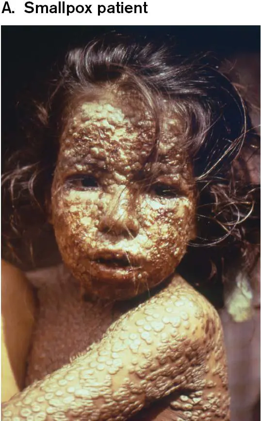
FIGURE 24.6 ■ Immunological specificity is the basis of vaccination. A. Smallpox patient covered with pox pustules. B. The smallpox virus, variola major (300 nm long; TEM). The photo
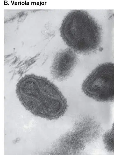
shows the dumbbell-shaped, membrane-enclosed nucleic acid core. Edward Jenner recognized the similarity in symptoms between the deadly smallpox and less severe cowpox diseases and used cowpox scrapings to vaccinate humans against smallpox.
CDC/JAMES HICK
CDC/DR. FRED MURPHY, SYLVIA WHITFIELD
While immunological specificity is important, it is not absolute. As described in Chapter 1, an English country physician named Edward Jenner (see Fig. 1.20B) in the late eighteenth century (long before viruses were discovered) learned to protect townsfolk from deadly smallpox disease (Fig. 24.6B ) by inoculating them with scrapings from lesions produced by cowpox (a tamer disease caused by the genetically related vaccinia virus). This story illustrates that an immune reaction against a less virulent organism or virus may be sufficient to cross-protect against an antigenically related but more virulent pathogen. The technique of protecting individuals from virulent microbes by exposing them to a less virulent version of the pathogen, now generally called vaccination, has been used to safeguard humans against many bacterial and viral pathogens (Table 24.1). Most vaccinations today involve administering one of the following: crippled (live but attenuated) strains of the pathogenic microbe, inactivated microbial toxins (for example, diphtheria toxin), or most recently fragments of RNA or DNA encoding a viral attachment protein (SARS-CoV-19).
Examples of Vaccines against
TABLE 24.1 Viral and Bacterial Parasite
Pathogens
Disease Vaccine Vaccination recommended for: Viral diseases Chickenpox Attenuated strain Children 12–18 (will still replicate) months COVID-19 Viral RNA or DNA Currently, everyone based >6 months old Hepatitis A Inactivated virus Children 12 months (will not replicate)
Hepatitis B Viral antigen Newborns Influenza Inactivated virus or Everyone, after 6 antigen months old, yearly Measles, mumps, Attenuated viruses; Children 12 months rubella (MMR) MMR combined vaccine Polio Inactivated Children 2–3 (injection, Salk) months Rabies Inactivated virus Persons in contact with wild animals Yellow fever Attenuated virus Military personnel Bacterial diseases Anthrax Bacillus anthracis, Agricultural and toxin components; veterinary unencapsulated personnel; key strain health care workers Cholera Killed Vibrio cholerae Travelers to, toxin endemic areas components Diphtheria Toxoid (inactivated Children 2–3 toxin) months Lyme disease Borrelia burgdorferi, Canines; human lipoproteins OspA vaccine and OspC surface discontinued antigens Meningitis Bacterial capsular Children under 5 caused by polysaccharide years Haemophilus influenzae type b (Hib)
Meningococcal Neisseria Children >2 years; disease meningitidis, adults >50 years bacterial capsular polysaccharides Pertussis Acellular Bordetella Children 2–3 pertussis months Pneumococcal Streptococcus Children; adults pneumonia pneumoniae, >50 years bacterial capsular polysaccharides Tetanus Toxoid Children 2–3 months Tuberculosis (Attenuated Exposed individuals Mycobacterium Mycobacterium tuberculosis) bovis [BCG (Bacille Calmette-Guérin)
vaccine] Typhoid fever Killed Salmonella Individuals in Typhi endemic areas Typhus Killed Rickettsia Medical personnel prowazekii in endemic areas; scientists; discontinued Parasite Disease Malaria Circumsporozoite Children 5–17 protein (CSP) months antigen (endorsed by WHO)
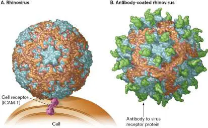
Cross-protection, in which immunization against one microbe can protect against a second microbe, works only if two proteins critical to the pathogenesis of the two different microorganisms share key antigenic determinants. Cross-protection will not take place if the structures of these determinants differ too much from one another. A good example is the common cold, which is caused by several viruses, including hundreds of closely related rhinovirus strains (rhinitis, a runny nose, is one of the symptoms of this viral disease). Infection with one strain of rhinovirus will not immunize the victim against a second strain. The reason is that the structures of rhinovirus proteins that attach to the ICAM-1 surface protein on host cells differ dramatically between different strains of rhinovirus (Fig. 24.7A). Antibodies called neutralizing antibodies, which bind to the attachment protein on one strain of rhinovirus, will prevent infection by that strain (Fig. 24.7B ) but will not bind an antigenically distinct ICAM-1 receptor protein from a different strain. A key to one lock will not work in a different lock.
FIGURE 24.7 ■ Antibodies prevent rhinovirus attachment to cell receptors. A. The complex rhinovirus capsid is pictured here attaching to the cell-surface molecule ICAM-1 (intercellular adhesion molecule, shown in reddish brown). (PDB code: 1rhi) B. Rhinovirus coated with protective (neutralizing) antibodies (green) block the ICAM-1 receptors on the virus. As a result, the virus fails to attach to and infect the host cell. (PDB code: 1RVF)
Thought Questions
24.2 How does a neutralizing antibody that recognizes a viral coat protein prevent infection by the associated virus?
24.3 The attachment proteins of different rhinovirus strains all bind to ICAM-1. How can all these proteins be immunologically different if they find the same target (ICAM-1)? Why won’t antibodies directed against one rhinovirus strain block the attachment of other rhinovirus strains?
To Summarize
An antigen that can elicit an immune response is also called an immunogen. An antigen usually consists of many different epitopes (antigenic determinants), each of which binds to a different, specific antibody.
Humoral (antibody-dependent) immunity against infection is the result of antibody production originated by B cells.
Cell-mediated (cellular) immunity involves a subgroup of T cells called cytotoxic T cells that can directly kill infected host cells.
Antigen-presenting cells (APCs) , such as macrophages, degrade microbial pathogens and present distinct antigens on cell-surface MHC proteins. T cells scanning the presented antigens become activated when they find a compatible antigen.
Different subgroups of T-cell lymphocytes stimulate B cells to become antibody-secreting plasma cells and activate T C cells to become cytotoxic T cells.
Proteins are better immunogens than nucleic acids and lipids because proteins have more diverse chemical forms. A hapten is a small antigenic compound that must be conjugated to a larger carrier antigen to elicit an immune response.
Immunological specificity refers to whether or not an antibody made to one epitope will bind to a different epitope. Highly specific antibodies bind to only one epitope. However, less specific antibodies can cross-bind to similar epitopes; for example, antibody to cowpox virus will bind to a similar epitope on smallpox virus.
Glossary
adaptive immunity Immune responses activated by a specific antigen and mediated by B cells and T cells.
antigen A compound, recognized as foreign by the cell, that elicits an adaptive immune response. See also immunogen .
immunogen An antigen that, by itself, can elicit antibody production. epitope See antigenic determinant .
antigenic determinant Also called epitope. A small segment of an antigen that is capable of eliciting an immune response. An antigen can have many different antigenic determinants.
antigen-presenting cell (APC)
An immune cell that can process antigens into antigenic determinants and display those determinants on the cell surface for recognition by other immune cells.
major histocompatibility complex (MHC)
Transmembrane cell proteins important for recognizing self and for presenting foreign antigens to the adaptive immune system. humoral immunity (antibody-dependent immunity)
A type of adaptive immunity mediated by antibodies.
antibody-dependent cell-mediated cytotoxicity (ADCC)
The process by which natural killer cells destroy antibody-coated host cells that are infected by a virus.
B cell An adaptive immune cell, developed in bone marrow tissue, that can give rise to antibody-producing cells.
plasma cell A short-lived antibody-producing cell.
antibody A host defense protein produced by B cells in response to a specific antigenic determinant. Antibodies, a type of immunoglobulin, bind to their corresponding antigenic determinants.
cell-mediated (cellular) immunity A type of adaptive immunity employing mainly T-cell lymphocytes.
cell-mediated (cellular) immunity A type of adaptive immunity employing mainly T-cell lymphocytes.
cytotoxic T cell (T C cell)
A T cell that expresses CD8 on its cell surface and can secrete toxic proteins such as perforin and granzymes.
helper T cell (T H cell)
A T cell that expresses CD4 on its cell surface and secretes cytokines that modulate B-cell isotype, or class, switching. immunogenicity (antigenicity)
A measure of the effectiveness of an antigen in eliciting an immune response.
immunogenicity (antigenicity)
A measure of the effectiveness of an antigen in eliciting an immune response.
hapten A small compound that must be conjugated to a larger carrier antigen in order to elicit production of an antibody that binds to it.
immunological specificity (antigenic specificity)
The ability of antibodies produced in response to a particular epitope to bind that epitope almost exclusively. Antibodies made to one epitope bind only weakly, if at all, to other epitopes. immunological specificity (antigenic specificity)
The ability of antibodies produced in response to a particular epitope to bind that epitope almost exclusively. Antibodies made to one epitope bind only weakly, if at all, to other epitopes. vaccination Exposure of an individual to a weakened version of a microbe or a microbial antigen to provoke immunity and prevent development of disease upon reexposure.
Fig. 4.20
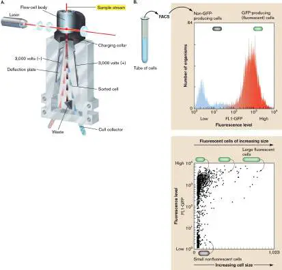
FIGURE 4.20 ■ Fluorescence-activated cell sorting. A. Schematic of a fluorescence-activated cell sorter (FACS) counting cells and conducting bidirectional sorting. B. Counting and separation of GFP-producing E. coli and non-GFP-producing E. coli. In the top panel, the low-level fluorescence (blue peak) produced by the cells on the left is baseline fluorescence (autofluorescence). Cells producing high-level fluorescence (red peak) are expressing the GFP protein. The scatterplot in the bottom panel displays the same FACS data, showing the size distribution of cells (x - axis) with respect to the level of fluorescence (y -axis). The larger cells may be cells that are about to divide.
Fig. 21.9 FIGURE 21.9 ■ Flow cytometry and FACS. A. In a fluorescence-activated cell sorter (FACS), a cell suspension is inoculated into flowing sheath fluid, which forms a stream of droplets. Each droplet carries one cell or none. Light from a
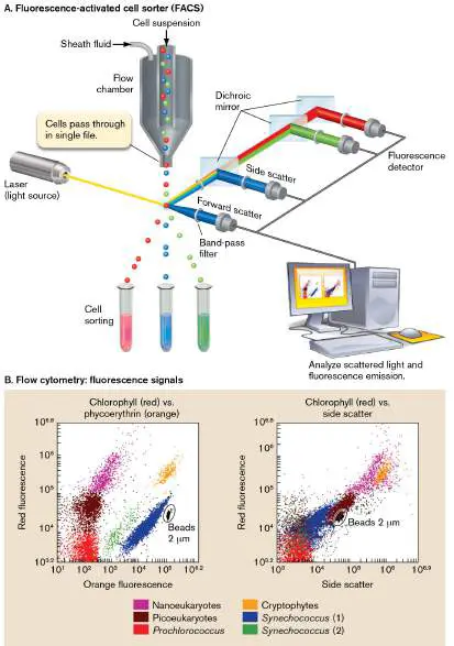
laser interacts with each cell, generating forward scatter, side scatter, and fluorescence modulated by various filters. The pattern of scatter and fluorescence is analyzed (flow cytometry). A computed “gate” determines a combination of scatter and fluorescence intensities that activates deflection of the droplet into a collection tube (cell sorting). B. Flow cytometry reveals subpopulations of microbes from a sample of Mediterranean seawater off the coast of Toulon, France. Each subpopulation is defined by the intensities of red autofluorescence (chlorophyll) and orange autofluorescence (phycoerythrin; left graph) or of red autofluorescence (chlorophyll) and side scatter (right graph).
Source: Modified from F. Delpy et al. 2018. Estuaries Coast 41 :2039, fig.
3a and 3b.
Fig. 24.29 FIGURE 24.29 ■ Live E. coli drives differentiation of CD4 pos -CXCR5 neg T cells into CD4 pos -CXCR5 pos T
FH
cells. Human APC monocytes were stimulated with control medium, live E. coli, or heat-killed E. coli. Stimulated APCs were then mixed with T cells and incubated for 5 days with staphylococcal enterotoxin B (superantigen). The x - and y - axes measure, respectively, the amount of surface CXCR5 marker and CD4 marker per cell. All T cells are plotted. Red boxes mark the graphical locales of CD4 pos -CXCR5 pos T
FH
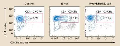
cells. Numbers in boxes indicate the percentage of total CD4 pos cells (T cells) that are also CXCR5 pos (T cells).
FH
Source: Modified from Ugolini et al. 2018. Nature Immunol. 19 :386–396,
fig. 1C.
Fig. 1.20B B.
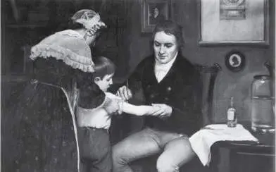
FIGURE 1.20 ■ Smallpox vaccination. B. Dr. Edward Jenner, depicted vaccinating 8-year-old James Phipps with cowpox matter from the hand of milkmaid Sarah Nelmes, who had caught the disease from a cow.
POPPERFOTO/GETTY IMAGES
24.2 Antibody Structure, Diversity, and Synthesisnot assigned
The broad overview of the immune response focused on how antibodies are made. But what are antibodies, and why are they important? Antibodies are proteins made by the body in response to foreign antigens, such as those from an invading pathogen. Antibodies circulate through blood, ready to bind a foreign antigen to remove it from the body. They are sometimes called immunoglobulins because they belong to the larger immunoglobulin superfamily of proteins. All members of this superfamily have a 110-amino-acid domain with an internal disulfide bond.
Note: The immunoglobulin superfamily of proteins includes many cell-surface-binding proteins, such as the major histocompatibility complex (MHC) proteins, various cytokine receptor proteins, and the non-antibody parts of the B-cell receptor (BCR). These cell-surface-binding proteins are not antibodies and are not called immunoglobulins. The term “immunoglobulins” is reserved for antibodies.
Like miniature “smart bombs,” antibodies individually circulate through blood, lymph and interstitial fluids to find and bind pathogens. An antibody will “ignore” all antigens except for the one they were selected to bind. When an antibody finds its antigenic match, it binds to the antigen and initiates several events that destroy the target. Antibodies are free-floating in blood, and they are also strategically situated on the surfaces of B cells as part of the B-cell receptor. A typical antibody consists of four polypeptide chains. There are two large heavy chains and two smaller light chains (Fig. 24.8). The four polypeptides combine to form a Y-shaped tetrameric structure held together by disulfide bonds. Two bonds connect the two identical heavy chains to each other. One light chain is then attached near its carboxyl end to the middle of each heavy chain by a single disulfide bond. The antigen-binding sites are formed at the amino-terminal ends of the light and heavy chains. One antibody molecule possesses two identical antigen-binding sites, one on each “arm” of the molecule. The two binding sites allow a single antibody to bind to two identical antigens. When enough antibodies bind to identical antigens on enough different molecules, a large cross-linked matrix will form and precipitate out of solution in a process called immunoprecipitation. Immunoprecipitation is the basis for a number of important molecular and clinical assays, such as rapid tests for strep throat or COVID-19 (see eAppendix 3).
FIGURE 24.8 ■ Basic antibody structure. A. The 3D structure of an antibody. (PDB code: 1R70) Every antibody contains two heavy chains (grey) and two smaller light chains (red) held together by disulfide bonds (shown in
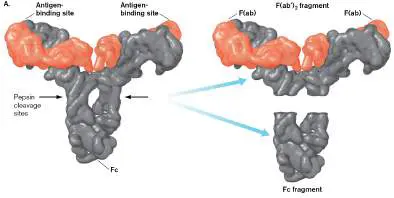
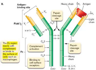
part B). The Y-shaped structure contains two antigen-binding sites, one at each arm [F(ab) region] of the molecule. These two sites are formed by the amino-terminal regions of the heavy-and light-chain pairs. The Fc portion points downward and is used to attach the antibody to different cell-surface molecules. Pepsin can be used to cleave a site in each heavy chain that releases the two F(ab) regions as one piece [F(ab′) 2] from the Fc region. B. Constant and variable regions in antibody structure. This schematic representation of IgG shows the various constant and variable regions of the heavy and light chains. Combined, the sequences V H and V L form antigen-binding sites at the amino termini of the heavy and light chains. An antigen is shown binding to one of the antigen-binding sites. Areas of the Fc region that can bind complement and cell-surface receptors are marked. Papain cleaves a site in each heavy chain that releases two F(ab) fragments, each one carrying one antigen-binding site.
Antibodies Have Constant and Variable Regions There are five classes of antibodies—defined by five different types, or isotypes, of heavy chains—called alpha (α), mu (μ), gamma (γ), delta (δ), and epsilon (ε). The heavy-chain classes are distinguished one from another by regions of highly conserved amino acid sequences, known as constant regions (denoted C H for the heavy chain; Fig. 24.8B ). Antibodies containing gamma heavy chains are called IgG; those with alpha, mu, delta, and epsilon heavy chains are called IgA, IgM, IgD, and IgE, respectively. Each antibody class serves a specific purpose in the immune system.
In contrast to heavy chains, there are only two classes of light chains— namely, kappa (κ) and lambda (λ)—which are defined by their own constant regions (C L; Fig. 24.8B ). A single antibody of any heavy-chain class (for example, IgG) may contain two kappa light chains or two lambda light chains, but never one of each. In humans, two-thirds of all antibody molecules carry kappa chains; the rest have lambda chains.
The antigen-binding part of an antibody is formed by highly variable amino acid sequences at the amino-terminal ends of the light and heavy chains. These variable regions are called the V L and V H regions, respectively (Fig. 24.8B ). The rest of each immunoglobulin chain is composed of highly conserved constant regions; C H 1, C H 2, and C H 3 in each heavy chain; and C L in each light chain. Shortly, we discuss the genes that code for these regions and how they assemble to form different antibody molecules.
In addition to the two “arms,” called F(ab) regions, that bind antigens, every antibody contains a “tail” called the Fc region (Fig. 24.8). The Fc region is not involved in antigen recognition but is important for anchoring antibodies to the surface of certain host cells (those with Fc receptors) and for binding components of the complement system.
Thought Question 24.4 Can an F(ab′) 2 antibody fragment prevent the binding of rhinovirus to the ICAM-1 receptor on host cells? And can an F(ab′) 2 antibody fragment facilitate phagocytosis of a microbe?
Isotypes, Allotypes, and Idiotypes We often discuss antibody diversity in terms of the amino acid sequence changes that distinguish antibodies between different mammalian species (isotype), between different individuals within a species (allotype), and between different antibodies within an individual (idiotype). Figure 24.9illustrates these differences. Understanding the differences between antibody isotype, allotype, and idiotype is important because these terms reflect different levels of antibody diversity.
FIGURE 24.9 ■ Isotype, idiotype, and allotype differences on antibodies. Colors indicate different amino acid sequences (or epitopes) within an individual (Sherrie) or between individuals (Sherrie and John). The Fc region is present in all of these antibodies but is marked only for IgE.
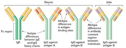
Put most simply, every single person on Earth has five antibody isotypes: IgG, IgA, IgD, IgE, and IgM. But each antibody isotype (IgG, for instance) in an individual (John) has certain allotypic sequence differences compared with the same isotype (IgG, again) in another individual (Sherrie). That is, all of John’s IgG molecules are of the same allotype, but John’s IgG allotype is different from that of Sherrie (an exception would be identical twins). Finally, the millions of molecules of the IgG allotype in a single person will contain millions of idiotypic differences that reflect the antigen specificity of each molecule. In Sherrie, for example, the antigen-binding site of an IgG molecule that binds a herpesvirus epitope possesses idiotypic amino acid differences from an IgG molecule in her that binds to a rhinovirus epitope. The synthesis and function of these different levels of antibody diversity are described in the next several sections. Thought Question 24.5 (refer to Fig. 24.9) Because antibodies are proteins, they are also antigens and can stimulate an immune response. What types of antibodies—anti-isotype, anti-allotype, or anti-idiotype—will IgG taken from Sherrie raise when injected into John?
Antibody Isotype Functions and “Super” Structures All antibody isotypes have the same basic structure. However, each isotype has a unique super structure (for example, monomer or dimer), and each is designed to carry out a different task. Some key properties of the five different immunoglobulin classes are listed in Table 24.2.
TABLE Properties of Human Immunoglobulins 24.2 Antibody isotype IgG Property IgM IgG1 IgG2 IgG3 IgG4 IgA IgD IgE Serum half-10 21 20 7 21 6 3 2 life (days)
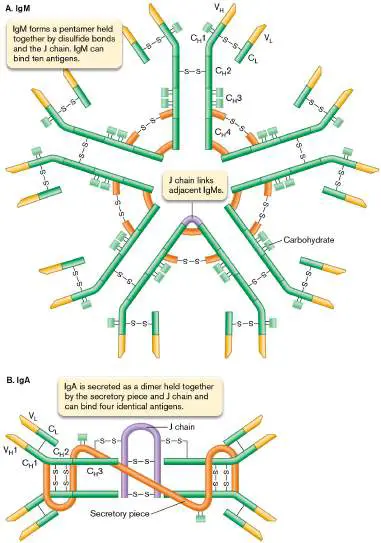
% Total 5%– 70% 15%– 0.2% 0.002% serum Ig 10 20 %% Antigen-2– 2 2–4 2 2 binding 10 sites Produced by Yes Poorly, if at all Poorly,? Poorly, fetus if at if at all all Transmitted No Yes No No No across placenta Binds Yes Yes No No No complem ent Opsonizing No Yes No No No Binds mast No No No No Yes cells IgM. IgM is the first antibody synthesized during an immune response. Circulating IgM is a multimeric, Ferris wheel–shaped molecule formed from five monomeric immunoglobulins tethered together by a protein called the J chain ( Fig. 24.10A). IgM can also be found in monomeric form attached by its Fc region to Fc receptors on the surface of B cells, where it forms part of the B-cell receptor. IgM is the first antibody isotype detected during the early stages of an immune response.
FIGURE 24.10 ■ Structures of IgM and IgA. The antibodies are made as multimers of immunoglobulin molecules: five in the case of IgM (A) ; two in the case of IgA (B) .
IgG. The simplest and most abundant antibody isotype in blood and tissue fluids is IgG (its superstructure is shown in Fig. 24.8B ). It is made as a monomer but has four subclasses (see Table 24.2). Each subclass varies in its amino acid composition and number of interchain cross-links. IgG molecules carry out several missions for the immune system. First, they bind and opsonize microbes; that is, they make microbes more susceptible to phagocytes. Opsonizing IgG antibodies use their antigen-binding sites to stick to microbes, thereby causing their Fc regions to point away from the microbe. Phagocytes possess surface Fc receptors that can attach to the Fc region of the antibody to gain a firmer “grip” on the microbe, facilitating phagocytosis (discussed in Section 23.5). IgG can also directly neutralize viruses by binding to virus attachment sites, and it is one of only two antibody types that can activate complement by the classical pathway (described in Section 24.4).
Another critical feature of IgG is that it can cross the placenta and enter a fetus. Other antibody isotypes cannot, because the antibody transport system in the placenta, called the neonatal Fc transporter, is specific for the Fc region of IgG.
IgA. IgA is secreted across mucosal surfaces (linings of the respiratory, gastrointestinal, urinary, and reproductive tracts) and is most commonly found as a dimer (Fig. 24.10B ). This conformation explains why IgA can bind four molecules of antigen (each monomer can bind two). The components of the IgA dimer are linked by disulfide bonds to a J chain protein, which joins two IgAs by their Fc regions. In addition to the eight molecules that comprise the two IgA monomers and the J chain, a tenth molecule—the secretory piece—is wrapped around the IgA dimer during the secretion process. The secreted molecule, now called sIgA (secretory IgA), is found in tears, breast milk, and saliva and on other mucosal surfaces. The molecule sIgA is important for mucosal immunity against pathogens that infect mucosal linings.
IgD. The last two antibody isotypes, IgD and IgE, are present at very low levels in the blood. IgD is a monomer that can neither bind complement nor cross the placenta. IgD molecules, however, are abundant on the surface of B cells. IgD, as with monomeric IgM, is attached to B-cell surfaces by its Fc region. IgD and IgM act as receptors that bind antigen and signal B cells to differentiate and make antibody.
IgE. While IgE is also present in only trace amounts in blood, it is more prominently found on the surfaces of mast cells and basophils. Mast cells and basophils contain granules loaded with inflammatory mediators. The primary role of IgE is to amplify the body’s response to invaders. Once secreted into serum by plasma cells, IgE attaches to Fc receptors on mast cells (Fig. 24.11Aand B )— again by way of its Fc region—and, like a Venus flytrap, waits until its matched antigen binds to its antigen-binding site. When two adjacent surface IgE molecules on a mast cell are cross-linked by antigen, a signal is sent internally that triggers degranulation (see Section 24.7). The release of histamine and other pharmacological mediators helps orchestrate an acute inflammation response early during a microbial infection (that is, while the antibody response is gearing up). The system can also cause severe allergic hypersensitivities (such as anaphylaxis) and milder forms like hay fever (Fig. 24.11C ).
FIGURE 24.11 ■ Mast cells are major players in the inflammation response. A. Mast cell (SEM). B. Granules (arrow) inside a mast cell (TEM). C. Hay fever is the result of degranulation of IgE-coated mast cells, which release histamine and other pharmacological mediators.
EYE OF SCIENCE/SCIENCE SOURCE
MICROSCAPE/SCIENCE SOURCE
ZURIJETA/SHUTTERSTOCK
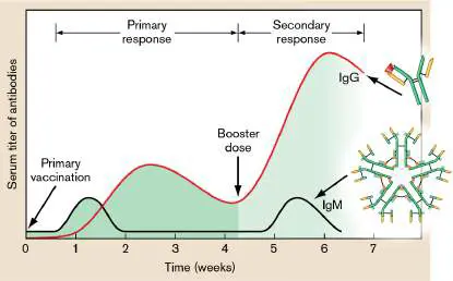
Primary and Secondary Antibody Responses Once you have been infected with a microorganism or have been given a vaccine, what happens? After a lag period of several days, antibodies begin to appear in the serum (the fluid that remains after the blood clots). During the lag period, called the primary antibody response (Fig. 24.12), a series of molecular and cellular events causes a distinct subset of B cells located in lymph nodes and the spleen to proliferate and differentiate into antibody-secreting plasma cells and memory B cells. Plasma cells are much larger than B cells because of an enormous increase in protein synthesis and secretion machineries. Remember that each B cell is genetically programmed to make antibodies to only one antigen or epitope.
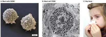
FIGURE 24.12 ■ Primary versus secondary antibody response.
Primary vaccination or infection leads to the early synthesis of IgM, followed by IgG. Either reinfection or a second (booster) dose of a vaccine results in a more rapid antibody response, consisting mainly of IgG, because of memory B cells formed during the primary response. Note that the time course and level of antibody made vary with the immunogen and the host.
A subsequent exposure to the antigen, which can take place months or years after the initial encounter, will trigger a rapid, almost instantaneous increase in the production of antibodies and is called the secondary antibody response (Fig. 24.12). This quick response occurs thanks to the memory B cells formed during the primary response. Memory B cells comprise approximately 40% of the B-cell population. Once restimulated, memory B cells rapidly differentiate into plasma cells and secrete antibody.
The net result of the primary antibody response is the early synthesis and secretion of pentameric IgM molecules specifically directed against the antigen (or immunogen). Later during the primary response, a process known as isotype switching (or class switching) occurs, and the predominant antibody type produced becomes IgG rather than IgM (discussed shortly). Antibodies made during the primary response, while specific for the immunogen, are actually not of the highest affinity. Later responses by memory B cells increase antibody affinity (see the next section).
As the immunogen is cleared from the body during the primary immune response, the levels of both IgG and IgM decline because the plasma cells that
produced them die. Plasma cells have an average life span of only 100 days. The antibodies themselves are slowly removed from serum by pinocytosis or renal clearance. The memory B cells, meanwhile, are maintained in lymph nodes or bone marrow and continue to divide, albeit slowly. If the immune system encounters the antigen again at a later date, the rapid secondary response (or “anamnestic response,” from the Greek anamnesis, meaning “remembrance”) ensues.
During the secondary response, memory B cells that have undergone isotype switching from IgM to IgG become plasma cells that secrete copious amounts of IgG antibody. These antibodies have a higher specificity for the antigen than do the antibodies produced during the primary response (see the paragraph “Making memory B cells” at the end of this section). The higher specificity—a result of hypermutation of the antigen-binding site—causes some plasma cells to produce antibodies that bind their antigens more strongly than primary-response antibodies do. Small amounts of IgM are also produced from the few memory cells that did not undergo isotype switching during the primary response. The secondary antibody response is why vaccinations work. Before vaccinations, the only way to be protected from an infection by a given pathogen was to have been infected earlier by that pathogen or a similar pathogen possessing similar immunogens. Memory cells made during the primary response will protect you against a second infection, but pathogens can do considerable harm during the primary-response lag phase. To avoid this harm, an innocuous version of a pathogen, or a harmless piece of it, can be injected into a person to trigger a primary response without producing disease (or, at worst, producing only a mild inflammatory response). Immunization thus primes the immune system to respond efficiently and without delay upon encountering the real pathogen. Table 24.1 lists a variety of viral and bacterial diseases for which immunizations are available.
Thought Question 24.6 The mother of a newborn was found to be infected with rubella, a viral disease. Infection of the fetus could lead to serious consequences for the newborn. How could you determine whether the newborn was infected in utero? How B Cells Differentiate into Plasma Cells As mentioned earlier, each B cell circulating throughout the body or nestled in a lymphoid organ is programmed to synthesize antibody that reacts with a single epitope (a small portion of a protein). Clonal selection is the process whereby a foreign antigen dictates which B-cell clone proliferates to large numbers and differentiates into antibody-producing plasma cells or memory B cells. The clonal expansion of the matching B cell enables large amounts of antibody specific for the antigen to be made. As illustrated in Figure 24.13, clonal selection begins when an antigen binds to an antigen-specific B-cell receptor on a matching B cell (see the discussion of B-cell receptors that follows here).
FIGURE 24.13 ■ Clonal selection. The B-cell population is composed of individuals, numbered here 1C to 4C, who have antibody specificity for different antigens (represented by differently colored antigen-binding sites on surface antibodies). When a B cell contacts its cognate antigen, an intracellular signal is generated, leading to proliferation and differentiation of that clone (clonal expansion). Plasma cells and memory B cells result.
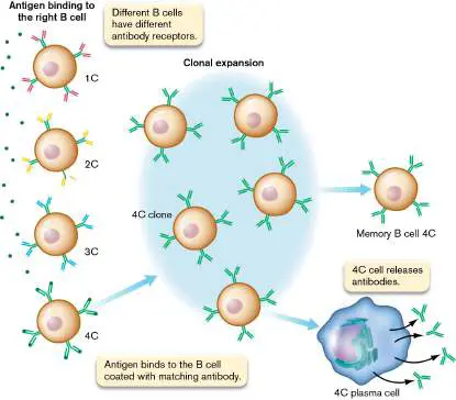
Mature but naive B cells (those that have not previously encountered antigen) can produce only IgM and IgD, which have identical antigen specificities. These two antibody classes are displayed like tiny satellite dishes on the B-cell surface, anchored by their Fc regions through hydrophobic transmembrane segments. These surface antibodies (B-cell receptors, or BCRs) are the keys to stimulating the proliferation and differentiation of B cells into antibody-secreting plasma cells or memory B cells. Upon binding to its corresponding antigen via these surface antibodies, the B cell is said to become activated, whereby it multiplies and differentiates into a plasma cell that ultimately synthesizes only one antibody isotype (for example, IgG1). Clonal selection has begun. In addition to antigen binding, most B cells require help from T cells to become plasma cells and memory B cells (discussed later).
Each BCR is composed of antibody and two other membrane proteins, called Igα and Igβ (Fig. 24.14). Igα and Igβ are not immunoglobulins, but they are designated “Ig” because they associate with the surface antibody. Each B cell may have upward of 50,000 identical B-cell receptors. A microbe generally has multiple copies of the same epitope on its surface (think about a virus capsid). Each capsid head protein has the same epitope. As such, the adjacent epitopes on a capsid head can bind adjacent B-cell receptors on one B cell.
FIGURE 24.14 ■ B-cell receptor. The B-cell receptor is formed as a complex consisting of a monomeric IgM plus Igα and Igβ in the membrane.
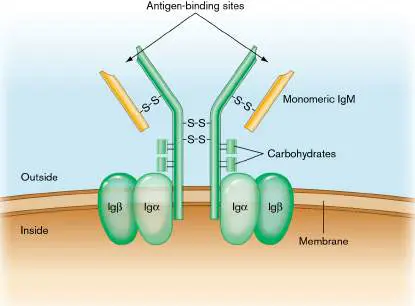
The Igαβ complex initiates the phosphorylation signal cascade (not shown). Once bound to adjacent epitopes, the surface B-cell receptors begin to cluster in a process called capping. Capping activates Igα and Igβ to initiate a phosphorylation signal cascade directed into the nucleus (Fig. 24.15). In a phosphorylation cascade, a phosphate group donated by ATP is passed from one protein to another, usually ending up on, and activating, a transcriptional regulator. The transcription factors at the end of the BCR cascade stimulate the transcription of genes that contribute to cell proliferation. Some of these now activated B cells can differentiate into plasma cells and secrete IgM antibody as part of the primary immune response.
FIGURE 24.15 ■ Capping and activation of the B cell. Two B-cell receptors can bind two identical epitopes on a pathogen. The resulting capping process initiates a signal cascade that activates differentiation and proliferation of the B cell independent of T-cell help. Antigens with repeating epitopes, such as polysaccharides, can directly cross-link B-cell receptors.
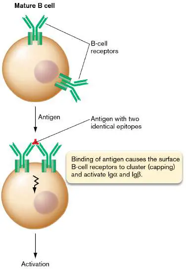
T cell–dependent and –independent antibody production. There are two routes by which antigens can stimulate B cells to differentiate into plasma cells. In one, called the T cell–independent route, antigens that possess multiple repeating epitopes (for example, polysaccharide antigens) can directly cross-link B-cell receptors (the capping process)—a step necessary for triggering differentiation (see Fig. 24.15). Proteins, however, which are the largest group of antigens, do not contain multiple repeating units. A single protein possesses many small, discrete, single epitopes, making the cross-linking of B-cell receptors difficult. B-cell responses to these types of antigens require help from specific T cells, and this constitutes the second, T cell–dependent, route to B-cell activation. Thus, B cells usually require multiple signals to initiate a primary response. How T cells help foster B-cell activation is discussed in Section 24.3. Note: A single protein with multiple nonidentical epitopes will not cross-link B-cell receptors. However, a microbe with many copies of that protein on its surface (think virus capsid) can cross-link B-cell receptors if the protein copies lie close enough together. To achieve cross-linking, the antigen-binding sites of a single B-cell receptor antibody must reach epitopes on adjacent proteins.
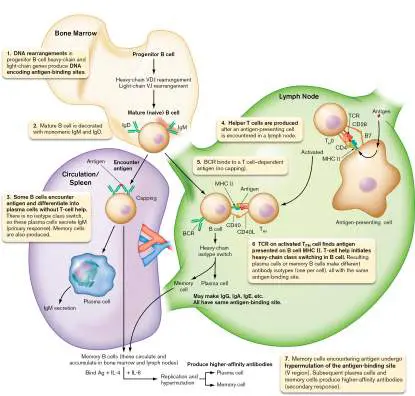
Figure 24.16summarizes the basic steps of antibody formation leading to the production of plasma cells and memory cells. More detailed descriptions of these events follow in the next section.
FIGURE 24.16 ■ Steps in antibody formation. Steps 1 and 2 (gene rearrangements) happen in bone marrow before the antibody encounters antigen. Step 3 (T cell–independent B-cell activation) occurs after antigen is encountered in the spleen or circulation. Steps 4–6 (T H 0-cell activation and T FH -dependent activation of B cells) can occur in the thymus, in the mucosa-associated lymphoid tissues (MALT), or in the lymph nodes. Step 7 (activation of memory B cells) can occur in bone marrow, spleen, or lymph nodes. To be activated, memory B-cell receptors must bind to antigen and come under the influence of cytokines IL-4 and IL-6 secreted by other cells.
Genetics of Antibody Production Before we discuss how T cells influence antibody production, let’s look at the process leading to antibody diversity. It is estimated that each human can
synthesize 10 11 different antibodies. Given that each B cell displays antibodies to only one antigenic determinant, it follows that there are 10 11 different B cells in the body. We have learned, however, that each person possesses only about a thousand genes or gene segments involved in antibody formation. How are 10 11 different antibodies made from only 10 3 genes?
Susumu Tonegawa was awarded the 1987 Nobel Prize in Physiology or Medicine for discovering that antibody genes can move and rearrange themselves within the genome of a differentiating cell. Three steps are involved: (1) the rearrangement of antibody gene segments (or cassettes), (2) the random introduction of somatic mutations, and (3) the generation of different codons during antibody gene (DNA) splicing. In humans, antibody diversity is generated continually over a lifetime.
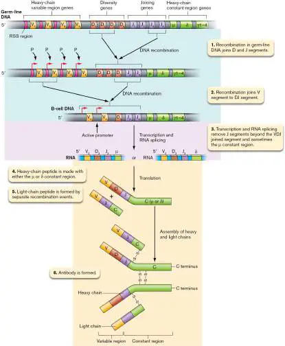
Making the antigen-binding site. The first step in making a specific antibody occurs in bone marrow when a progenitor stem cell (progenitor B cell) becomes a B cell before a foreign antigen is encountered (Fig. 24.16, step 1). This process happens throughout a person’s life. Immunoglobulin genes in a bone marrow progenitor B cell have many gene segments that can rearrange in many possible combinations. During differentiation into a mature B cell, DNA segments are deleted in a process called gene switching, which decreases the number of gene segments in the mature B-cell DNA. The process starts at the 5′ end of an immunoglobulin gene cluster corresponding to the eventual antigen-binding site ( Fig. 24.17). For now, we will focus on what happens in bone marrow, but will return later to the big picture summarized in Fig. 24.16.
FIGURE 24.17 ■ Formation of the VDJ regions of heavy chains.
Note that only a small subset of the V, D, and J genes listed in Table 24.3 is shown in this model. RSS = recombination signal sequence.
In both the heavy-and light-chain gene regions are a number of tandem gene cassettes encoding potential variable regions (antigen-binding sites), separated by recombination signal sequences (RSSs). RSSs allow recombination to bring two widely separated gene segments together. There are approximately 170
“variable”-region (V-region) gene segments for the heavy and light chains. The light-chain V-region gene cluster lies upstream of a cluster of J-region (“J” for “joint” or “joining”) genes that will eventually join a variable region to a light-chain constant region (C). Genes for the constant region reside farther downstream in the DNA. The arrangement of the heavy-chain genes is slightly more complex. In this case, the heavy-chain V cluster is followed by a D (“diversity”) cluster and then the J region.
Note: Do not confuse the J-region gene segments used to make heavy-and light-chain proteins with the J-chain protein that holds together IgM and IgA multimers. They are completely different and unrelated. The J-region gene segments do not encode the J chain.
A summary of the genetic processes leading to antibody formation is shown in Figure 24.17. Antibody formation begins with a recombination event between RSS sites at one heavy-chain D segment and one heavy-chain J segment, which deletes all the intervening D and J segments (Fig. 24.17, step 1). Next, the new DJ region joins to one of the heavy-chain V segments, deleting all of the intervening V and D segments (step 2). The result is a joined VDJ DNA sequence, which can then be transcribed (step 3).
Each V segment has its own promoter. However, if extra V segments remain upstream of the rearranged VDJ sequence on the DNA, then the only promoter that will fire is the one immediately upstream of the rearranged VDJ segment ( Fig. 24.17, step 3). The primary RNA transcript will then undergo RNA splicing to remove any J-segment RNA sequences that remain downstream of the VDJ RNA sequence (step 3). The result is a mature B cell that can synthesize a specific antibody (steps 4–6). Remember, all of the DNA recombination and RNA splicing happen before the B cell ever “sees” the antigen. Consequently, the mature B cell is called “naive” because it has not yet been stimulated by antigen. The sequence of events for light chains is similar, except that the product is VJ. Table 24.3 illustrates the amount of antibody diversity that can be achieved in humans simply by this combinatorial re-joining (a total of about 5 × 10 6 antigens can be recognized). However, that is only about 0.0001% of the total possible diversity.
Antibody Diversity Attributed to TABLE Combinatorial Joining in the Human 24.3 Germ Line Number of: Number of combinations Chain type V regions D regions J regions λ Light 30 0 4 30 × 4 = 120 chains κ Light 40 0 5 40 × 5 = 200 chains Heavy 100 27 6 100 × 27 × 6 chains = 16,200 Number of 16,200 heavy-chain combinations × 120 λ-chain possible combinations = 1.94 × 10 6 antibodie 16,200 heavy-chain combinations × 200 κ-chain s combinations = 3.24 × 10 6 (1.94 × 10 6) + (3.24 × 10 6) = 5.18 × 10 6 combinations Where does the rest of antigen-binding-site diversity arise? Additional diversity comes from the junctions of VJ and VDJ, where recombinational joining can occur between different nucleotides. Each genetic recombination event can generate additional codons by adding nucleotides, so the resulting peptides will differ by one or more amino acids. After recombination, the V regions of the germ lines are susceptible to high levels of somatic mutation (called hypermutation), resulting in the hypervariable regions. Hypermutation happens every time a memory B cell is exposed to the antigen; the memory B cells divide, and the hypervariable regions mutate. The interactions between the light-and heavy-chain hypervariable regions in an antibody form the antigen-binding sites. In sum, a combination of genetic recombination and random mutations provides the remarkable level of antigen-binding-site diversity that we all possess. As noted earlier, the human body is capable of responding to 10 11 antigens, yet there are only about 10 8 antigens in nature. This apparent overkill suggests that the immune system is well prepared to cope with any possible antigen it could encounter. Unfortunately for humans, enterprising microbes, such as the trypanosomes that cause sleeping sickness, can stay one step ahead of the immune system by changing the structure of key surface antigens. Changing the antigenic structure of a protein renders useless those antibodies made to the previous structure.
It is not hard to understand why multicellular organisms, like humans, need the capacity to make any one of billions of different antibodies quickly. Pathogens can undergo many generations of growth within the single life span of a human host. So, humans have to generate recombinant clones of cells quickly to overcome rapidly dividing pathogens.
The isotype class switch. We just described how a progenitor B cell becomes a mature (naive) B cell in bone marrow (see Fig. 24.16, steps 1 and 2). The mature but naive B cell (a B cell that has not yet “seen” the antigen but has already assembled its immunoglobulin VDJ binding site) produces both IgM and IgD B-cell receptors (so the naive B cell is referred to as IgM + IgD +). The primary immune response begins when a mature (naive) B-cell receptor finds its matched antigen (see Fig. 24.16, step 3, spleen). In the early stages of the response, plasma cells produced from these B cells secrete only IgM, but they eventually secrete IgM and IgD.
If the activated B cell reaches a lymph node the B cell will receive signals from a certain type of T cell known as a helper T cell (T H cell). Before a T H cell can send signals to a B cell, the T H cell must first be stimulated by an APC, shown in Fig. 24.16(step 4). This process will be described later (Section 24.3 ). The stimulated T H cell instructs the B cell to undergo additional immunoglobulin isotype switching (Fig. 24.16, lymph node, steps 5 and 6). The switched B cell may then make IgG, IgA, or IgE, each with the same antigen recognition domain (variable region) but different constant regions. The type of switch is influenced by cytokines secreted by helper T cells. How T cells meet B cells in a lymph node germinal center is discussed in eResearch Activity 24. What “flips” the isotype switch? Notice in Figures 24.17 (step 2) and 24.18 that the constant-region gene segments that encode different antibody heavy-chain classes are arranged in tandem after a VDJ region. The mechanism by which a B cell switches to make IgG (C regions gamma 1–4), IgE (C-epsilon), or IgA (C-alpha 1 and 2) is very similar to VDJ formation (Fig. 24.18). Each constant segment, except delta, contains a repeating DNA base sequence called a switch region. Recombination between these switch regions will delete the intervening DNA between the VDJ region and either the IgG, IgE, or IgA constant regions. Because the VDJ region is the same regardless of which C H gene is selected, the antibody produced will have the same antigenic specificity as the original IgM.
FIGURE 24.18 ■ Heavy-chain class switching. As a B cell becomes activated, a switch in antibody isotype will occur. The switch involves recombination between isotype cassettes that brings one heavy-chain constant region (Cα in the example, for IgA) in tandem with a VDJ sequence. However, the heavy-chain switch selection process is not random. The type of cytokine present at the time of the switch will influence which C H gene is selected. Consequently, cytokines control whether IgG is made for circulation or, instead, IgA is made for secretion. Note that any heavy-chain peptide (alpha,
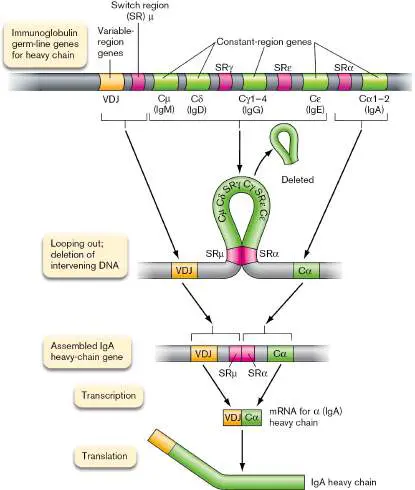
gamma, delta, and so on) can combine with any light chain (lambda or kappa). However, a single, mature B cell can make only one type of heavy chain and one type of light chain.
B-cell selection. We just described how the immune system can make antibodies against any antigen. But why aren’t antibodies made against our own antigens? Normally, antibodies against self are not made, because immature B cells in bone marrow undergo three forms of negative selection if their BCR binds to a self antigen. The immature B cell can undergo apoptosis (a type of programmed cell death), completely removing it from the B-cell repertoire. Alternatively, the immature B cell can perform receptor editing in which the B cell develops a new BCR that does not respond to self. Or it can induce a state of an ergy (an inability to respond) in which the mature B cell is released to the circulation but cannot react to self antigen because intracellular signaling pathways have been blocked.
Another reason self-reacting antibodies are not made involves the T-cell selection process that happens in the thymus (described in Section 24.3). As just described, many B cells require help from T cells to make antibody. To deliver that help, a T cell must recognize the same antigen as the B cell. As a failsafe measure, the T cell selection process kills T cells that can recognize self antigens. Thus, a B cell that could make self-reacting antibody will not, because the necessary self-reacting helper T cells have been deleted.
Thought Questions 24.7 B cells in early stages have both IgM and IgD surface antibodies, but the delta region has no switch region. Why does the delta region have no switch region?
24.8 Why do individuals with type A blood have anti-B and not anti-A antibodies?
Making memory B cells. An antigen-activated B cell will divide to make memory B cells as well as plasma cells (see Figs. 24.13 and 24.16 , step 3 [spleen] and step 6 [lymph node]). Memory B cells are B cells that have already undergone heavy-chain class switching (committed to making IgG, for instance) but, unlike other B cells, are very long-lived. Their long lives provide immunological memory.
Memory B cells also undergo a process known as affinity maturation. During their lifetimes, memory B cells will hypermutate the gene sequences that encode the antibody-binding sites (the VDJ and VJ regions) of the antibodies they make. Memory B cells whose mutations increase the affinity of the antibody toward an antigen are then clonally selected during a secondary antibody response (Fig. 24.16, step 7). As the infection clears and the antigen becomes more scarce, only B cells equipped with highest affinity B cell receptors will detect the antigen and remain active. Those cells have undergone affinity maturation. B cells with lower affinity BCRs are not maintained.
To Summarize Antibodies, or immunoglobulins, are Y-shaped molecules that contain two heavy chains and two light chains. There are five classes (isotypes) of antibodies defined by the structure of the heavy chains . IgM is composed of five antibody monomers. IgG is a monomer and the main antibody type in blood. IgA is mostly secreted as a dimer at mucosal areas, while IgD is mostly found bound to B cells. IgE is a circulating antibody that attaches to mast cells and mediates some allergic reactions. Antibodies are members of the immunoglobulin superfamily of proteins.
Each antibody molecule contains two antigen-binding sites. Each binding site is formed by the hypervariable ends of a heavy-and light-chain pair. The Fc portion (“tail”) of an antibody can bind to specific receptors on host cells. This binding is antigen independent.
Antibody “isotype,” “allotype,” and “idiotype” refer to amino acid sequence differences found at the different levels of antibody diversity. Antibody isotype is defined by sequences in a heavy chain that are unique to a species. Allotype is defined by isotype sequences that are consistent within one individual but are different from those of another individual of the same species. Idiotype is defined by sequence differences found within one person’s individual antibodies, usually at antigen-binding sites. Antibody diversity occurs within B-cell precursor cells via a complex series of splicing events between adjacent DNA cassettes, as well as mutational events in DNA sequences encoding the hypervariable regions of heavy and light chains. A mature (naive) B cell has randomly completed VDJ rearrangements in its DNA to make the specific antigen-binding site for antibodies. The naive B cell places IgM and IgD antibodies on its surface (part of the B-cell receptor).
A B-cell receptor consists of a membrane-embedded antibody in association with the Igα and Igβ proteins. BCRs on a single B cell are made of a single antibody isotype that specifically binds one epitope. Binding of antigen to the B-cell receptor triggers B-cell proliferation and differentiation called clonal selection , which occurs during the primary and secondary antibody responses.
The primary antibody response to an antigen begins when B cells differentiate into antibody-producing plasma cells and memory B cells. IgM antibodies are generally the first class of antibodies made during the primary response.
Isotype switching (or class switching) from IgM production to other antibody isotypes begins once the B-cell receptors (BCRs) on a naive B cell bind their target antigen.
The secondary antibody response occurs during subsequent exposures to an antigen. Memory B cells are activated, hypermutate the VDJ region, and then rapidly proliferate and differentiate into antibody-secreting plasma cells. IgG is the predominant antibody made.
Glossary
immunoglobulin A member of a family of proteins that contain a 110-amino-acid domain with an internal disulfide bond. Members include antibodies and major histocompatibility proteins.
heavy chain The larger of the two protein types that make up an antibody. Each antibody contains two heavy chains and two light chains.
light chain The smaller of the two protein types that make up an antibody. Each antibody contains two heavy chains and two light chains.
immunoprecipitation The antibody-mediated cross-linking of antigens to form large, insoluble complexes. It is used in research labs and is normally seen only in vitro. constant region The region of an antibody that defines the class of a heavy chain or a light chain.
variable region The amino-terminal portions of antibody light and heavy chains that confer specificity to antigen binding and define the antibody idiotype.
Fc region The region of an antibody that binds to specific receptors on host cells in an antigen-independent manner. It is found in the carboxy-terminal “tail” region of the antibody.
isotype An antibody class within a species that is defined by the structure of the antibody’s heavy chain. IgG, IgA, IgD, and IgE are examples of isotypes. An isotype from one species contains species-specific amino acid sequences that are present in the heavy chain of all members of that species.
allotype An amino acid difference in the antibody constant region that distinguishes different individuals within a species.
idiotype An amino acid difference in the antigen-binding site (N terminus of heavy or light chains) that distinguishes different antibodies within an individual. IgM The first antibody isotype detected during the early stages of an immune response. It contains the mu heavy chain and is found as a pentamer in serum.
IgG An antibody isotype that contains the gamma heavy chain. Produced by plasma cells, it is found in serum and is the predominant class of circulating antibodies for adaptive immunity.
opsonize To bind IgG antibodies to microbes in order to enhance microbial phagocytosis by host immune cells.
IgA An antibody isotype that contains the alpha heavy chain. It can be secreted and is found in tears, saliva, breast milk, and so on.
IgD An antibody isotype that contains the delta heavy chain. It is found on B-cell membranes.
IgE An antibody isotype that contains the epsilon heavy chain. It is involved in degranulation of mast cells.
serum The noncellular, liquid component of the blood.
primary antibody response The production of antibodies upon first exposure to a particular antigen. B cells become activated and differentiate into plasma cells and memory B cells.
plasma cell A short-lived antibody-producing cell.
memory B cell A long-lived type of lymphocyte preprogrammed to produce a specific antibody. After encountering their activating antigen, memory B cells differentiate into antibody-producing plasma cells.
secondary antibody response A memory B cell–mediated rapid increase in the production of antibodies in response to a repeat exposure to a particular antigen.
isotype switching Also called class switching. A change in the predominant antibody type produced by a cell.
class switching See isotype switching .
clonal selection The rapid proliferation of a subset of B cells during the primary or secondary antibody response.
B-cell receptor (BCR)
A B-cell membrane protein complex containing an antibody in association with the Igα and Igβ immunoglobulins.
capping The clustering of B-cell receptor molecules on the surface of B cells after binding antigens or epitopes.
gene switching Switching between two (out of five) different classes of immunoglobulin genes (e.g., from IgM to IgG) during B-cell development.
recombination signal sequence (RSS)
A DNA region downstream of antibody heavy-and light-chain genes that allows recombination between widely separated gene segments.
helper T cell (T H cell)
A T cell that expresses CD4 on its cell surface and secretes cytokines that modulate B-cell isotype, or class, switching.
cytokine A small, secreted host protein that binds to receptors on various endothelial and immune system cells, regulating the cells’ responses.
switch region A repeating DNA sequence interspersed between antibody constant-region genes that serves as a recombination site during isotype, or class, switching. affinity maturation The process by which the antigen-binding site of an antibody gains increased affinity for its target antigen or epitope.
24.3 T Cells Link Antibody and Cellular Immune Systemsnot assigned
Does each arm of the adaptive immune system (antibody-dependent or cell-mediated) know that the other exists? The systems must communicate with each other because different types of infections tilt the immune response one way or the other. T-cell lymphocytes, with integral roles in both antibody production and cell-mediated immunity, manage the balance.
Although derived from the same progenitor stem cell as B cells (see Fig. 23.18), T cells develop in the thymus rather than in the bone marrow where B cells develop, and they express surface protein markers (antigens) that differentiate them from B cells. Varieties of T cells are also marked by the presence of different cell differentiation (CD) surface proteins and by the types of cytokines they produce. Two critically important groups are helper T cells (T H cells, already mentioned) and cytotoxic T cells (T C cells). Helper T cells display the surface antigen CD4, while cytotoxic T cells display CD8. Cytotoxic T cells are the “enforcers” of the cell-mediated immune response. They destroy the membranes of host cells infected with viruses or bacteria.
Helper T cells come in several models, the first of which is the T H 0 cell. T H 0 cells are precursors to the other types, including: T FH (follicular helper T cells), a heterogeneous set of CD4 T cells that drives B-cell differentiation into antibody-secreting plasma cells. Different T FH cell subsets secrete different combinations of cytokines that trigger antibody isotype (class) switching.
T H 1 cells assist in the activation of cytotoxic T cells. T H 2 cells recruit eosinophils to combat parasitic infections and can inhibit T H 1 proliferation.
T H 17 cells stimulate inflammation by secreting interleukin 17 (IL-17), a pro-inflammatory cytokine. IL-17 helps trigger recruitment of neutrophils and macrophages to an infection site. Treg cells (or Tregs, regulatory T cells) dampen the inflammatory response and secrete the anti-inflammatory cytokine IL-10.
The ratios of these cells and the cytokines they produce drive the immune response toward being either more antibody-mediated or more cell-mediated, whichever is better for eliminating a specific infection. To be of any use, however, T cells first must be activated by an antigen.
T-Cell Activation Requires Antigen Presentation
Unlike B cells, T cells never bind free-floating antigen. T cells are activated only by antigen bound to another cell’s surface. These other cells are collectively called antigen-presenting cells (APCs) because they present antigens to T cells (as introduced in Fig. 24.3 ). The APC surface proteins that hold and present the antigen are known as major histocompatibility complex (MHC) proteins (Fig. 24.19A). MHC proteins differ between species and between individuals within a species. They help determine whether a given antigen is recognized as coming from the host (a self antigen) or from another source (a foreign antigen) in a phenomenon called histocompatibility (hence, the name “major histocompatibility complex”).
FIGURE 24.19 ■ Major histocompatibility complex proteins. A. Class I MHC molecules are composed of a 45-kDa chain and a small peptide called β 2 -microglobulin (12 kDa). Class II MHC molecules contain an alpha chain (30–34 kDa) and a beta chain (26–29 kDa). The peptide-binding regions of both classes show variability in amino acid sequence that yields different shapes and grooves. CD8 T cells recognize antigen peptides associated with class I molecules, while CD4 T cells recognize peptides bound to class II molecules. Peptide antigens nestle in grooves formed by these molecules and are held there awaiting interaction with T-cell receptors. B. Antigen binding to an MHC I molecule. C. Top view of antigen (red) nestled in the MHC peptide-binding site. (PDB code: 1BII)
The salient feature of all MHC molecules is that they bind antigen only after the antigen has entered the host cell. Once the antigen is bound to an internal MHC molecule, the complex moves to the cell surface, where the antigen is displayed. MHC molecules are critical to the immune system because T cells, to become activated, must recognize a foreign antigen-MHC complex on an APC surface. Two classes of MHC molecules are found on cell surfaces (Fig. 24.19A). Both classes belong to the immunoglobulin family of proteins, but they are not immunoglobulins (antibodies). Class I
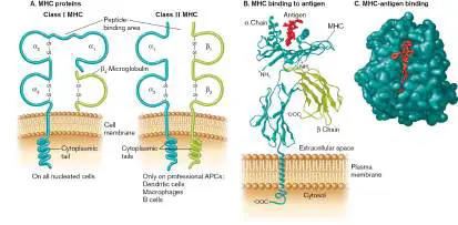
MHC molecules are found on all nucleated cells, whereas class II MHC molecules are found only on professional APCs—that is, on dendritic cells, B cells, and macrophages. Professional APCs display both class I and class II MHC molecules on their surface and specialize in presenting antigens to helper T cells and cytotoxic T cells (see Fig. 24.3). All other nucleated cells, called nonprofessional APCs, generally have only class I MHC molecules. MHC I is a heterodimer made of an alpha chain and a β 2 - microglobulin. The antigen-binding cleft is within the alpha chain. MHC II has alpha and beta subunits (different from MHC I), both of which contribute to the antigen-binding cleft.
The surface CD proteins CD8 and CD4, mentioned earlier, help T cells distinguish between MHC class I and class II molecules on antigen-presenting cells. The CD8 molecules on T C cells selectively bind MHC class I, while CD4 molecules on T H cells selectively bind MHC class II. Antigens presented on class I MHC molecules generally arise from intracellular pathogens, such as viruses and some bacteria that require cell-mediated immunity for resolution. In contrast, antigen peptides presented on class II MHC molecules originate from extracellular infections, which are best resolved by antibody.
APCs Receive, Process, and Present Antigens by Two Paths
How are foreign peptide antigens placed, or presented, on host cell surfaces? In the initial stages of an immune response, antigen-presenting cells, professional or not, internalize the pathogen, such as a virus or a bacterium. Inside the APCs, pathogen proteins are degraded into smaller peptides (epitopes). These epitopes are placed within the MHC-binding clefts of MHC I or II molecules and transported back to the cell surface.
Whether an antigen peptide binds to class I or class II MHC molecules generally depends on how the antigen initially entered the cell (Fig. 24.20). Endogenous antigens are synthesized by viruses and intracellular bacteria as they grow within the cytoplasm of an APC. These antigens will attach to class I MHC molecules on the endoplasmic reticulum and are moved to the cell surface (Fig. 24.20, left). In contrast, exogenous antigens, which are produced outside of the APC (as are most bacterial antigens), enter the cell via phagocytosis (Fig. 24.20, right). The antigen-containing phagosome then fuses with a vacuole whose interior is lined with MHC II molecules. There, compatible antigens bind to MHC II clefts and the MHC class II–peptide complex is carried to the cell surface. Once the antigen is presented on the surface of the APC, T cells can interact with the antigen-MHC complex via their T-cell receptors (discussed shortly). Professional APCs and naive T cells interact within the lymph nodes, the spleen, or Peyer’s patches in the gut. So, the professional APCs must make their way to those locations to generate an immune response.
FIGURE 24.20 ■ APC processing and presentation of peptide antigens on class I and class II MHC proteins.
Left: Microbial proteins made in the host cytoplasm are degraded, and peptides are placed on class I MHC molecules in the endoplasmic reticulum (ER). Right: Microbial proteins made outside the cell are phagocytosed (bottom), degraded in a phagosome or endosome, and placed on class II MHC molecules. LMP = low-molecular-mass polypeptide component of the proteasome; TAP = transporter of antigen peptides.
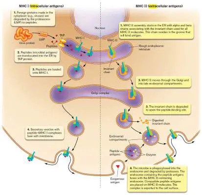
Note: MHC molecules present only peptide antigens.
Carbohydrate antigens cannot attach to the binding clefts of MHCs but will trigger antibody production independent of T cells. The antigen-binding clefts of MHC molecules are shown in Figure 24.19B and C . It is important to realize that the antigen-binding specificity of any single MHC molecule is very broad, unlike the highly antigen-specific targeting of an antibody molecule or T-cell receptor (described next). In contrast to the many billions of different antibody molecules made as a result of multiple gene rearrangements and somatic mutations, a person possesses genes that express only six MHC I molecules and six to eight MHC II molecules, with each parent donating half of the genes necessary (all of the MHC genes are on chromosome 6 and are codominantly expressed). MHC genes do not undergo rearrangement or somatic mutation. However, the promiscuous binding capability of each MHC binding site, plus the independent assortment of maternal and paternal alleles, means that this limited number of MHC molecules can bind most peptide antigens. Despite that diversity, not all possible antigens can be recognized by the MHCs in a single person. Among the human population, however, there are thousands of alleles for each MHC locus, each of which encodes a slightly different binding cleft that shifts specificity. This form of MHC diversity ensures that the population as a whole will not succumb to a new or mutated pathogen.
T-Cell Receptors
T-cell receptors (TCRs) are the antigen-binding molecules present on the surfaces of T cells (Fig. 24.21). Unlike BCRs, TCRs are not antibodies. As noted earlier, a TCR on CD4 + helper T cells will bind only to antigens attached to MHC II surface proteins on professional antigen-presenting cells. However, the TCRs of CD8 + cytotoxic T cells can bind viral antigens attached to MHC I surface proteins present on any virus-infected cell. Remember that all nucleated cells have surface MHC I molecules.
FIGURE 24.21 ■ The T-cell receptor (TCR) and CD3 complex. T-cell receptor proteins are associated with CD3 proteins at the cell surface. Antigen binds to the alpha and beta subunits. The positive and negative charges holding the complex together come from amino acids in the peptide sequences. Once
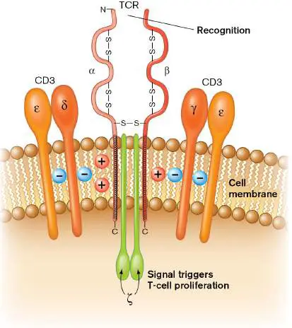
bound to antigen, the complex transduces a signal into the cell. This signal triggers T-cell proliferation.
The T-cell receptor is composed of several transmembrane proteins. Two protein chains—namely, alpha and beta—make up the part that recognizes antigen. The TCR alpha and beta proteins are found in a complex with four other peptides, which together form the CD3 complex (Fig. 24.21). When stimulated by binding to antigen, these ancillary CD3 complex proteins recruit and activate intracellular protein kinases and launch a phosphorylation cascade that triggers proliferation of the T cell and initiates key immunological events that we describe later in the chapter. How is diversity in antigen recognition generated in TCR molecules? Much like antibodies, the alpha and beta proteins of TCRs are formed from gene clusters that undergo gene rearrangements analogous to, but different from, those of the immunoglobulin genes. For instance, the antigen-binding site of an alpha chain is formed by VJ regions, while those of the beta chain are composed of VDJ regions. The alpha and beta V, D, and J genes are distinct from those of antibodies.
Why don’t your T cells see your body’s antigens? Are they tolerant somehow? Actually, T cells that bind self antigens are not made tolerant; they are killed in a process called T-cell education or T-cell selection.
T-Cell Education and Deletion
Greek mythology tells of Narcissus, a young man punished by the gods for scorning the women who fell in love with him. One day Narcissus saw a beautiful face in a pool of water. Not knowing it was his own reflection, he fell in love, tumbled into the pool, and drowned. His inability to recognize himself is analogous to the danger posed by an immune system. Immune systems must be able to distinguish what is self (meaning antigens present in the body’s own tissues) from what is not self. Otherwise, the immune system would constantly attack the body’s own cells.
Earlier we examined B-cell selection within bone marrow. Here we describe how T cells are selected in the thymus. The selection process basically has two steps: positive selection to retain T cells whose TCR can bind self MHC proteins, followed by negative selection to kill T cells whose TCR can bind other self antigens present on tissues. The goal is to educate T cells to distinguish self from nonself.
In the first step (positive selection), precursor T cells are passed by special thymus epithelial cells bearing MHC molecules. T cells with TCRs that bind these self MHC proteins are allowed to live, and they continue through the thymus. However, T cells that do not recognize self MHC peptides, or recognize them too strongly, are killed (deleted). Positive selection is important because T cells must recognize self MHC in order to bind antigen-MHC complexes on APCs.
Why kill T cells that strongly bind MHC molecules? If the immune system’s T-cell repertoire included cells that bound self MHC too tightly, then the T cells would constantly react to the body’s own MHC molecules, regardless of which antigen peptides were attached (self or nonself). Note, however, that some strongly self-reactive T cells are allowed to survive and are converted into regulatory T cells (Tregs). Regulatory T cells can block the activation of harmful, self-reactive (autoimmune) lymphocytes that escape deletion and enter the circulation. Note that this is the first way that Tregs can be made. The second way involves APC activation of T H 0 cells, discussed shortly.
The second stage of T-cell “education” is called negative selection. T cells positively selected for binding self MHC molecules move on to another section of the thymus, where their TCRs are further screened for an ability to bind self antigens, which is an undesirable property. But wait—doesn’t the thymus express only thymus antigens? How can this organ screen for T-cell recognition of antigens expressed on other host cells (for example, heart cells)? The answer is a special gene activator called AIRE in certain thymus epithelial cells. AIRE stimulates the synthesis of all human proteins in small amounts. A constellation of self proteins is then presented on the epithelial cells’ MHC I and MHC II molecules. T cells that originally survived positive selection are screened by these MHC–self antigen complexes to test for TCR recognition. If a TCR on one of the T cells binds to a self antigen, that T cell is instructed to kill itself (via apoptosis). Almost 95% of T cells entering the thymus die during these positive and negative selection processes. What remains are T cells that can recognize MHCs bound to foreign antigens. Having been “educated,” these T cells leave the thymus to seed secondary lymphoid organs such as the spleen.
What happens if someone has a thymectomy (a treatment for myasthenia gravis)? You might ask how someone whose thymus has been removed can live if the organ is critical for T-cell maturation and education. Actually, the thymus begins losing function shortly after birth, such that very little function remains in adults. Fortunately, a large amount of T-cell education occurs during fetal development. Once a T cell is educated, reserve pools of these T cells are maintained throughout life outside of the thymus. Some T cells, apparently, can also mature in secondary lymphoid tissues. Thus, adults without a thymus can live relatively normal lives. Babies born without a thymus, however, have a severe, life-threatening T-cell deficit.
APCs Activate T H 0 Helper T cells
Figure 24.22summarizes the steps that lead to T-cell activation in the lymph node and highlights how activated T cells influence the two types of adaptive immunity: humoral (antibody) and cellular. (Refer to Fig. 24.16for an overview of antibody production.) The different classes of T cells—T H and T C —require different but interrelated activation programs. Let’s start with T H cell activation. T H cells require three molecular signals to become active. First, T-cell receptors along with CD4 on precursor T H 0 cells specifically recognize and link to antigen-MHC II complexes on antigen-presenting cells (APCs; Fig. 24.22, step 1a). The second signal needed to activate a T H 0 cell is the binding of a CD28 molecule, present on the T H 0 cell surface, to a B7 protein (also known as CD80) on the APC cell surface (also step 1a). The third signal involves different cytokines produced by mast cells, macrophages, and epithelial cells during infection, which can convert activated T H 0 cells to T FH, T H 1, T H 2, T H 17, or Treg cells (step 2a). The activation of cytotoxic T cells (T C cells) is examined later. FIGURE 24.22 ■ Summary of the activation of humoral and cell-mediated pathways. Left: Extracellular pathogens tend to activate humoral immunity (B cells). Right: Intracellular pathogens generally activate cell-mediated immunity by
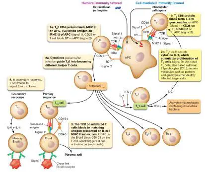
stimulating cytotoxic T cells (CD8 marker). The balance between cell-mediated and humoral immune responses to a given infection is regulated by the balance between the production of T H 1 (cell-mediated) versus other T H (antibody) helper T cells. This balance is influenced by whether the foreign antigen was made by intracellular pathogens (T H 1-favored) or extracellular pathogens (see Fig. 24.20). T H 1 cells will activate cytotoxic T cells (cell-mediated immunity; best for killing intracellular pathogens), while T FH cells will promote antibody production (humoral immunity; best for attacking extracellular pathogens). See text for more details.
T FH Cells Activate B Cells
During the early phase of a primary response, B-cell activation is T cell–independent (Fig. 24.16, step 3). One type of B cell in the lymph node or spleen can become activated after the BCR binds antigen if the B cell also binds to complement C3 on a bacterium (see Section 23.6) or if a microbe-associated molecular pattern (MAMP) binds to a Toll-like or NOD-like receptor on or in the B cell (see Section 23.5). This is an example of communication between the innate immune system (complement and MAMPs) and the adaptive system (B cells). Activation does not result in heavy-chain class switching; only IgM is secreted by the resulting plasma cell. This is partly why the primary response starts with IgM (see Fig. 24.12).
During the later stages of a primary response, however, activation of most B cells requires a helper T cell once the antigen binds to the B-cell receptor (Fig. 24.22, step 3). This assistance is called T-cell help. Interactions between B cells and T cells take place primarily in areas of lymph nodes called follicles, also called germinal centers (see eResearch Activity 24). The follicular helper T cell (T FH) is the primary T cell involved in driving B-cell activation. (Actually, any T H cell other than T H 0 and Treg can activate a B cell to make antibody, but to a lesser extent.)
T-cell help is specific to the antigen and triggers heavy-chain class switching. But how does a B cell gain specific T-cell help? The specific T FH cell must have a TCR able to bind the same antigen that the B cell binds. In this way the T FH cell “knows” which B cell to help. As part of the B-cell receptor capping mechanism (antigen cross-linking of BCR), some antigen bound by B-cell receptors becomes internalized and processed to be presented back on the B cell’s surface MHC II receptors (Fig. 24.22, step 3). For instance, a T FH cell that was activated by dendritic cells presenting a particular antigen (step 1a) can also use its T-cell receptor to bind that same antigen presented on a B-cell MHC II molecule (step 3). This contact allows CD40 on the B cell to bind CD154 on the T cell in an interaction that completes activation of the B cell. The B cell will now differentiate into a plasma cell (also step 3).
The isotype class switch from IgM to another antibody type is directed by cytokines secreted by the T FH cell. For instance, IL-4 and interferon-gamma (IFN-gamma or IFN-γ) trigger the switch from IgM to IgE and IgG, respectively.
Julie Blander (Fig. 24.23A) and her collaborators at Cornell University have shown that the innate immune system can tell the adaptive system when it will face a living pathogen rather than a dead one, better preparing T FH cells to trigger the B-cell isotype switch. The scientists demonstrated that one of the viability signals is bacterial RNA acting through Toll-like receptors (TLRs) to stimulate monocyte inflammosomes (see Section 23.5). The resulting increase in secreted interferon-β and IL-1β triggers T FH cell differentiation that, in turn, activates B cells in a germinal center. Figure 24.23shows the results of injecting mice with heat-killed E. coli (Fig. 24.23B ) or heat-killed E. coli plus bacterial RNA (Fig. 24.23C ). The addition of RNA led to greater numbers of germinal centers and activated B cells, as well as an increase in the number of B cells expressing anti-E.coli IgG antibodies on their surfaces. This research has implications for improving vaccine effectiveness by using innate immune mechanisms (cytokine production) to intensify B-cell activation.
FIGURE 24.23 ■ Pathogen viability signal molecules indirectly activate antibody production. A. Julie Blander of Cornell University studies connections between innate and adaptive immunity. B. and C. Mouse spleen germinal centers after vaccination with heat-killed E. coli (B. ) or with heat-killed E. coli plus E. coli RNA (C. ). Sections of spleen were stained with fluorescent antibodies to reveal B cells (blue), T cells (gray),
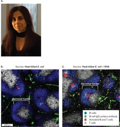
and activated B cells (red). The addition of RNA led to greater numbers of germinal centers and activated B cells. An increase was also observed in the number of B cells expressing anti-E.coli IgG antibodies on their surfaces (green).
COURTESY OF JULIE MAGARIAN BLANDER
MODIFIED FROM BARBET, G. ET AL. 2018. IMMUNITY 48 :584–598
MODIFIED FROM BARBET, G. ET AL. 2018. IMMUNITY 48 :584–598
During the secondary immune response, too, memory B cells need T-cell help to become plasma cells, but direct contact with helper T cells is not required. Memory B cells that have antigen bound to their B-cell receptors can respond to the soluble IL-4 and IL-6 cytokines secreted by activated helper T cells without having direct contact with the T H cell (Fig. 24.22, step 4). IL-4 stimulates B-cell proliferation, while IL-6 directs differentiation into antibody-secreting plasma cells.
Thought Questions
24.9 What would happen to someone lacking CD154 on T FH cells because of a gene mutation?
24.10 How can a stem cell be distinguished from a B cell at the level of DNA?
T H 1 Cells Activate Cytotoxic T Cells
Because they directly attack host cells, CD8 T C cells are the major “enforcers” of the cellular immune system, along with macrophages and natural killer (NK) cells. T C cells, however, are not actually cytotoxic until they are activated. As with helper T cells, activation of cytotoxic T cells takes place in the lymph node and requires three signals. The first signal is the binding of T-cell receptors (TCRs) to antigen–MHC class I complexes on an antigen-presenting cell (see Fig. 24.22, step 1b). CD8 on the T C cell recognizes an MHC I– antigen complex on the APC. Because class I MHC molecules are found on all nucleated cells, any infected cell can initiate activation of T C cells, converting them to cells that ultimately kill the infected cell. Figure 24.24Atakes a closer look at signal 1 interaction.
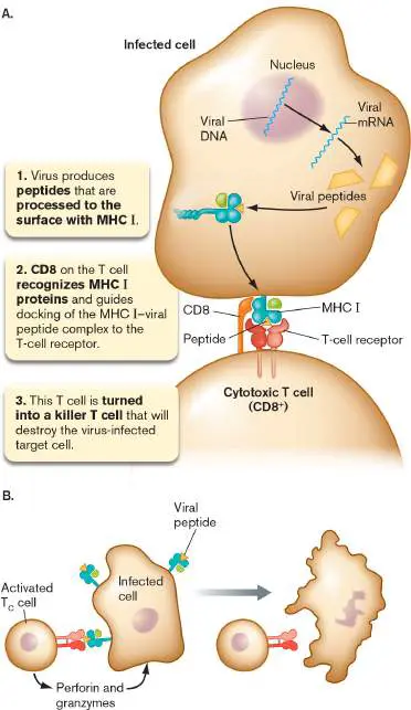
FIGURE 24.24 ■ Presentation of a viral antigen to a T cell and cytotoxic T-cell action. A. CD8 protein on a T C cell directs the interaction between a T-cell receptor and a viral antigen bound to a class I MHC protein on an antigen-presenting cell. Signal 2 for activation of cytotoxic T cells is an interaction between B7 (on an APC) and CD28 on a T C cell, described in the text (not shown here). B. The activated T cell (also called cytotoxic lymphocyte, or CTL) then leaves the lymph node and migrates to the site of infection, where it can recognize the viral peptide presented on the class I MHC receptor on an infected cell. This interaction authorizes the cytotoxic T cell to kill the infected cell.
The second signal needed to activate T C cells is a B7-to-CD28 interaction that further links the APC to the T C cell. This is the same second signal described earlier for helper T cells. Once activated, the T C cell gains cytotoxic activity and places receptors for the cytokine IL-2 on its surface. The third signal needed for T C cell activation is IL-2, secreted by the T H 1 class of helper T cells (activated earlier). IL-2 binds to the newly placed IL-2 receptors and stimulates proliferation of the cytotoxic T cell (see Fig. 24.22, step 2b). The resulting “platoon” of activated cytotoxic T cells (also called cytotoxic lymphocytes, or CTLs) moves from the lymph node to the site of infection, killing any cell bearing the same peptide–MHC class I complex (for example, cells infected with the same virus that triggered CTL production; Fig. 24.24B ).
Activated cytotoxic T cells kill infected target cells by releasing the contents of their granules, which contain the proteins granzyme and perforin. Perforin produces a pore in the target cell membrane through which granzymes can enter. In the cytoplasm, granzymes cleave and activate caspase proteases in the infected cell. The activated caspases then trigger apoptosis and cell death. The infected host cell is sacrificed for the good of the whole animal, human or otherwise. Another type of granzyme released into the target host cell can directly enter and kill intracellular bacteria residing within (see Section 23.3).
Why are cytotoxic T cells more effective than B cells and antibodies at clearing viral infections? A major reason is that intracellular pathogens (for example, viruses) hide inside host cells, where they are protected from antibody. Consequently, these pathogens are best killed when the harboring host cell is also sacrificed (via cellular immunity). Because all nucleated cells have class I MHC molecules, any infected cell can be recognized and killed by an appropriate T C cell for the good of the host.
T H 17 Cells Promote, and Treg Cells Limit, Inflammation
The immune system is full of checks and balances. Two sets of T cells critical to regulating the immune response are T H 17 cells that amplify inflammation and regulatory T cells (Tregs) that dampen inflammation. T H 17 cells are derived from T H 0 cells and are characterized by their secretion of pro-inflammatory cytokine IL-17. T H 17 cells are the first subset of T cells generated during an infection. Receptors for IL-17 are expressed on fibroblasts, epithelial cells, and keratinocytes.
Contact with IL-17 causes the production of several cytokines. These include IL-6 to initiate fever, chemokines like CXCL8 (formerly called IL-8) to recruit neutrophils and macrophages, and granulocyte-macrophage colony-stimulating factors (GM-CSFs) to enhance the synthesis of neutrophils and macrophages in bone marrow. IL-22, also produced by T H 17 cells, cooperates with IL-17 to induce the synthesis of antimicrobial peptides, such as beta-defensins, in epidermal keratinocytes. Antimicrobial peptides are discussed in Chapter 23. Altogether, T H 17 cells enhance the innate acute inflammatory response to infection. However, the problem with inflammation is that it also causes damage to local tissues. This is where Treg cells become crucial.
Treg cells can form during T-cell selection in the thymus (mentioned earlier) or differentiate from T H 0 cells in a lymph node. Treg cells help to shut down immune responses after an invading organism has successfully been eliminated, and they can prevent autoimmunity. Once activated by an antigen presented to its T-cell receptor, a Treg cell secretes the anti-inflammatory cytokine IL-10. IL-10 has several anti-inflammatory functions; it inhibits inflammatory cytokine secretion by macrophages [tumor necrosis factor alpha (TNF-alpha or TNF-α), IL-1, and IL-12], reduces MHC II expression in macrophages, blocks macrophage activation by IFN-gamma, and blocks cytokine IL-2 production from T H 1 cells, thereby limiting the production of cytotoxic T cells (see Fig. 24.22, step 2b).
Ratios of T H 17 to Treg cells vary depending on the severity of an infection, either exacerbating or quelling inflammation. The cytokines produced by APCs and other cells during an infection can tilt the balance one way or the other. In addition, Treg cells themselves can inhibit T H 17 cell functions.
CD4 Helper T Cells and Cytokines Balance the Immune Response
Antigens presented on class I MHC molecules elicit only cell-mediated immunity (via CD8 T cells). However, antigens presented on class II MHC molecules can stimulate both arms of the immune system by activating CD4 helper T cells. CD4 T H cells do not kill host cells directly but instead release various cytokines that incite other cells to do the killing. IL-8, for instance, attracts additional white blood cells to the area. Activated T H 1 cells secrete cytokines that stimulate cytotoxic T cells (see Fig. 24.22, step 2b), as well as macrophages and natural killer cells. However, sets of activated T FH cells secrete different combinations of cytokines that will trigger B cells to switch immunoglobulin classes. These B cells will then differentiate into plasma cells that secrete the new class of antibody (see Fig. 24.22, step 3). Table 24.4 lists a fraction of the many different cytokines produced by various cell types and describes their influence over the immune system.
TABLE Select Cytokines That Modulate
24.4 the Immune Response *
Cytokine Sample sources General functions IL-1 Many cell types, Pro-inflammatory; including endothelial affects differentiation cells, fibroblasts, and activity of cells neuronal cells, in inflammatory epithelial cells, response; acts as macrophages endogenous pyrogen in the central nervous system IL-2 T H 1 cells Stimulates T-cell and B-cell proliferation IL-3 T cells, mast cells, Stimulates production keratinocytes of macrophages, neutrophils, mast cells, others IL-4 T H 2 cells, mast cells Promotes differentiation of CD4 and T cells into T H 2 helper T cells; TABLE Select Cytokines That Modulate
24.4 the Immune Response *
Cytokine Sample sources General functions promotes proliferation of B cells, class switch to IgE IL-5 T H 2 cells Chemoattracts eosinophils; activates B cells and eosinophils IL-6 T H 2 cells, Stimulates T-cell and macrophages, B-cell growth; fibroblasts, stimulates endothelial cells, production of acute-hepatocytes, phase proteins neuronal cells IL-8 Monocytes, endothelial Chemoattracts PMNs; (CXCL8) cells, T cells, promotes migration keratinocytes, of PMNs through neutrophils endothelium IL-10 T H 2 cells, B cells, Inhibits production of macrophages, IFN-γ, IL-1, TNF-α, keratinocytes and IL-6 by macrophages TABLE Select Cytokines That Modulate
24.4 the Immune Response *
Cytokine Sample sources General functions IL-17 T H 17 cells (helper T Recruits macrophages cells unique from T H and neutrophils to 1 and T 2) sites of inflammation
H
IFN-α/β T cells, B cells, Promotes antiviral macrophages, activity fibroblasts IFN-γ T H 1 cells, cytotoxic T Activates T cells, NK cells, NK cells cells, macrophages, B-cell class switch to IgG TNF-α T cells, macrophages, Exerts wide variety of NK cells immunomodulatory effects TNF-β T cells, B cells Exerts wide variety of immunomodulatory effects Figure 24.25outlines the myriad effects that one cardinal cytokine, interferon-gamma (IFN-gamma or IFN-γ, the type II interferon), has on innate and adaptive immune cells. The cascade begins when an APC encounters microbes and releases IFN-alpha (see Chapter 23). IFN-alpha activates natural killer (NK) cells to secrete IFN-gamma, which interacts with key immune cell types to influence their functions. These functions include increasing antigen presentation and the secretion of inflammatory cytokines by macrophages, promoting CD4 T-cell differentiation into T H 1 cells, enhancing the cytotoxicity of CD8 CTL cells, stimulating isotype switching by plasma B cells, and promoting PMN adhesion to blood vessels before extravasation. IFN-gamma secreted from NK cells also stimulates additional IFN-gamma secretion from T H 1 cells, as well as CD8 T cells, amplifying the response. In addition, this cytokine will trigger apoptosis of infected cells (in collaboration with TLR3 interactions with viral dsRNA) and stimulate antiviral activities of uninfected bystander cells. The immunomodulatory reach of IFN-gamma is remarkable.
FIGURE 24.25 ■ Effects of interferon-gamma on the immune system. Interferon-alpha secreted by antigen-presenting cells (APCs) stimulates natural killer (NK) cells (the major producers of IFN-gamma) to secrete IFN-gamma. Binding of this cytokine to IFN-gamma receptors on a variety of cells can influence various aspects of innate and adaptive immunity, as well as trigger cell death of infected cells and activate antiviral
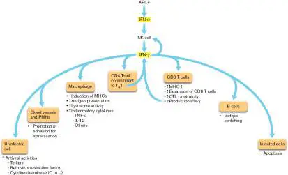
activities in uninfected cells. CTL = cytotoxic lymphocyte; PMN = polymorphonuclear leukocyte.
But what determines which class of T H cell predominates during an infection? Part of the answer is found in the blend of cytokines generated by macrophages or mast cells during different types of infection. Infections by viruses and intracellular bacteria, for instance, generate cytokines, such as IFN-gamma (as just discussed), that favor the production of T H 1 cells. T H 1 cells promote cell-mediated immunity. Infections by extracellular bacteria and parasites, however, generate IL-4, which inhibits T H 0 differentiation into T H 1 cells, so antibody-mediated immunity is favored (see Fig. 24.22, step 2a).
As a result, once the immune system starts to go down one pathway, the other pathway is held back. But this is not an all-or-none response. For instance, a viral infection will also result in T FH cells that stimulate antibody production. The tilt toward T H 1 or T FH predominance occurs because the body realizes which pathway— cell-mediated (T H 1) or humoral (T FH)—will more effectively clear a particular infection.
The importance of CD4 T cells in immunity is tragically illustrated by acquired immunodeficiency syndrome (AIDS, discussed in Chapter 11). The human immunodeficiency virus (HIV) binds to CD4 molecules to infect CD4 T cells. In the absence of antiviral treatment, the number of CD4 T cells declines below its normal level of about 1,000 per microliter. When the number of CD4 T cells drops below 400 per microliter, the ability of the patient to mount an immune response is jeopardized. The patient not only becomes hypersusceptible to infections by pathogens but also becomes susceptible to, and can die from, infections by commensal organisms. Today, because of highly effective antivirals, AIDS patients rarely contract these opportunistic infections.
Activated T H 1 Cells Also Activate Macrophages
Macrophages, too, must be activated to become highly effective killers of microbes. Activation of macrophages also requires two signals. One activation pathway involves interferon-gamma (IFN-γ) produced by nearby infected or damaged cells, followed by binding of the macrophage to lipopolysaccharide (LPS) or other microbial components via Toll-like or NOD-like receptors (TLRs or NLRs). Once activated, the macrophage becomes aggressive in terms of phagocytosis and increases its production of numerous antimicrobial reactive oxygen intermediates.
Some bacterial pathogens, such as mycobacteria, the causative agents of tuberculosis and leprosy, grow primarily in the phagolysosomes of macrophages. There they are shielded from antibodies and cytotoxic T cells. Intracellular pathogens live in the usually hostile environment of the phagocyte either by inhibiting the fusion of lysosomes to the phagosomes in which they grow or by preventing acidification of the vesicles needed to activate lysosomal proteases. However, a macrophage activated by a T H 1 cell can rid itself of such pathogens. Even unactivated macrophages are able to process some of the intracellular bacteria and place antigen from them on their class II MHC molecules. T H 1 cells then activate these macrophages by binding their TCR molecules to the macrophage MHC II–antigen complex and through secretion of IFN-gamma. The activated macrophages can then kill any bacteria that may be growing within them.
Given that activated macrophages are such effective assassins of microbial pathogens, why are macrophages not always kept in an active state? A major reason is that once activated, macrophages also damage nearby host tissue through the release of reactive oxygen radicals and proteases. Thus, effective killing of microbial pathogens comes at the expense of host tissue damage.
Superantigens Do Not Require Processing to Activate T Cells
Superantigens are a family of microbial toxins that induce an abnormally aggressive T-cell response. Normally, antigens require processing by antigen-presenting cells to stimulate T-cell responses. Each peptide epitope produced during antigen processing must be placed on an MHC molecule and presented to a cognate T cell. When an antigen is introduced into a host, only a few T cells have the proper TCR needed to recognize that antigen—in the range of 1– 100 cells per million. Proliferation of the T cells through antigen-dependent activation increases their number and increases the immune response to that antigen.
Superantigens, such as staphylococcal toxic shock syndrome toxin (TSST), bypass the normal route of antigen processing. In fact, antigen recognition is not even involved. As illustrated in Figure 24.26, superantigens can bind simultaneously to the outside of T-cell receptors on T cells and to the MHC molecules on APCs (for example, macrophages). This promiscuous joining of T cells and macrophages activates many more T cells than a typical immune reaction does, stimulating the release of massive amounts of inflammatory cytokines from both cell types.
FIGURE 24.26 ■ Superantigens. A. The difference between the presentation of antigen and superantigen. Antigen presentation requires the antigen to bind within the binding pockets of MHC and TCR molecules. Superantigens do not require processing. They can bind directly to the outer aspects of the TCR and MHC proteins, linking and activating the two cell types. B. Staphylococcal toxic shock syndrome toxin (TSST) is one example of a potent superantigen. Alpha helices are shown as red ribbons, while sections of the beta sheet are shown as blue ribbons. (PDB code: 2QIL)
The effect of superantigens can be devastating because cytokines, such as tumor necrosis factor (TNF), will overwhelm the
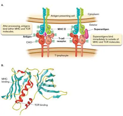
host immune system’s regulatory network and cause severe damage to tissues and organs. The result is disease and sometimes death. Jim Henson, the creator of Kermit the Frog and other Muppet characters, died in 1990 from complications of pneumonia caused by a potent superantigen produced by Streptococcus pyogenes. This example is but one of many ways that overreaction by the immune system causes morbidity (disease) and mortality (death)—effects more pronounced than the direct effects of the pathogen involved.
The Cytokine Storm
The term “cytokine storm” became a household catchphrase during the COVID-19 pandemic because of its role in causing severe outcomes (see Chapter 26). A cytokine storm is the exaggerated immune response to systemic infections such as bacterial sepsis or the toxic shock syndrome toxin (just discussed). However, the term also applies to the exaggerated, or hyperimmune, responses caused by viral infections such as influenza and SARS-CoV-2. “Cytokine storm” is an umbrella term characterized by constitutional symptoms such as fever, chills, and fatigue; systemic inflammation; and multiorgan dysfunction, leading to multiorgan failure and death. Laboratory findings that mark a cytokine storm are variable but always include elevated CRP levels (C-reactive protein, see Section 23.6) and usually include blood count abnormalities such as extremely high or low white blood cell numbers. The pathogenic mechanisms of these pathogens cause local T cells and antigen-presenting cells to synthesize large amounts of cytokines (IFN-gamma, TNF, interleukin-6, and others) and chemokines. Because of their sustained production, these molecules spill into the circulation and reach various organ systems where they trigger inflammation and organ damage, usually of the kidney, liver, and/or lung. In short, a cytokine storm involves an immune response that produces collateral damage beyond the immediate benefit of the immune response.
Microbial Evasion of Adaptive Immunity
As efficient as the immune system is, numerous viral and bacterial pathogens have developed effective means for avoiding the adaptive immune response. Many viruses produce proteins that down-regulate the production of class I MHC molecules on infected cell surfaces. On one hand, down-regulation of MHC I will limit antigen presentation, since MHC I is needed for that process. On the other hand, losing MHC I exposes the infected host cell to natural killer cells because NK cells attack peers that lack MHC I (discussed in Section 23.5). To surmount this obstacle, human cytomegalovirus, for example, places a decoy MHC I–like molecule on the surface of infected cells. These decoys are thought to bind inhibitory receptors on NK surfaces that block NK cell cytotoxicity. Like viruses, bacteria are masters of illusion when it comes to the immune system. A major cause of gastric ulcers, Helicobacter pylori, expresses proteins from a cluster of pathogenicity genes that trigger apoptosis (programmed cell death) of T cells. Other bacteria have evolved mechanisms that interfere with signal transduction pathways controlling the expression of cytokines. For example, YopP from Yersinia enterocolitica, one cause of gastroenteritis, inhibits a specific signal transduction pathway needed to produce TNF, IL-1, and IL-8. Thus, Yersinia avoids the detrimental effects of those pro-inflammatory cytokines.
Staphylococcus aureus is well known for repeatedly reinfecting a single individual, suggesting that natural immune defenses against this pathogen are ineffective. Actually, S. aureus plays an active role in thwarting both innate and adaptive immune mechanisms. For instance, secreted leukocidins produced by S. aureus (see Chapter 25) kill APCs, an essential link between innate and adaptive immunity. In another strategy, immunodominant but nonprotective S. aureus antigens will bind strongly to MHC molecules on APCs, impeding immune recognition of less dominant but potentially protective antigens that only weakly bind MHC molecules. These schemes limit the establishment of memory B cells and T cells that could respond to reinfection.
Another method of immune evasion is employed by various mycobacteria, some of which cause tuberculosis and leprosy, as noted earlier. These bacteria induce the production of anti-inflammatory cytokines, which dampen the immune response.
Mycobacterium -infected macrophages, for example, produce IL-10, which down-regulates the production of MHC II molecules needed to present antigens to helper T cells.
Despite the clever ways pathogens misdirect and try to overwhelm our immune system, the system usually catches on to their tricks. Our redundant humoral and cellular mechanisms can still “outwit” these infectious agents... so far.
Thought Questions
24.11 Transplant rejection is a major consideration in the transplantation of most tissues because host T C cells can recognize allotypic MHC on donor cells. Why, then, are corneas easily transplanted from a donor to just about any other person?
24.12 Why does attaching a hapten to a carrier protein enable antihapten antibodies to be produced?
To Summarize
The major histocompatibility complex (MHC) consists of membrane proteins with variable regions that can bind antigens. Class I MHC molecules are on all nucleated cells, while class II MHC molecules are found only on professional antigen-presenting cells.
Antigen-presenting cells (APCs) such as dendritic cells take antigens synthesized during an intracellular infection and present them on their surface class I MHC molecules, but they place antigens from engulfed microbes or allergens on their class II MHC molecules.
T-cell receptors (TCRs) on T-cell membranes bind to MHC-antigen complexes on the membranes of professional and nonprofessional antigen-presenting cells. TCR-MHC interactions trigger T-cell differentiation.
T-cell education in the thymus selects for T cells whose TCRs will not bind to self antigens but will bind to self MHC complexes (not too tightly).
Activation and differentiation of a T H 0 cell requires three signals. The first is TCR-CD4 binding to an MHC II– antigen complex on an antigen-presenting cell, and the second is B7-CD28 interaction. Subsequent T H 0 cell differentiation to T FH, T H 1, T H 2, T H 17, or Treg cells is influenced by different cytokine “cocktails” (the third signal) secreted by macrophages and NK cells during an infection. Activation of a B cell into an antibody-producing plasma cell usually requires two signals: a B-cell receptor binding to an antigen, and a B-cell MHC II-antigen complex binding to the TCR of a T FH cell activated by the same antigen.
Activation of cytotoxic T cells requires three signals. The first is TCR-CD8 recognition by T cells of MHC I–antigen complexes on APCs, and the second is a B7-CD28 interaction between the APC and the T C cell. The third signal involves IL-2 secreted from activated T H 1 cells, stimulating proliferation of cytotoxic T cells. The activated T C cells, in turn, destroy infected host cells.
Measures that balance immune responses include T H 17 cells and Treg cells that, respectively, promote and limit inflammation; and cytokine cocktails secreted by CD4 helper T cells that can tilt a response to either antibody-mediated or cell-mediated immunity.
Superantigens abnormally stimulate T cells by directly linking TCRs on T cells with MHCs on APCs without undergoing APC processing and surface presentation.
A cytokine storm occurs when cytokines and chemokines that are overproduced at the site of an infection spill into the circulation and cause inflammation in multiple organ systems. Pathogens have evolved ways to undermine and misdirect the immune system.
Glossary
cytotoxic T cell (T C cell)
A T cell that expresses CD8 on its cell surface and can secrete toxic proteins such as perforin and granzymes.
antigen-presenting cell (APC)
An immune cell that can process antigens into antigenic determinants and display those determinants on the cell surface for recognition by other immune cells.
major histocompatibility complex (MHC)
Transmembrane cell proteins important for recognizing self and for presenting foreign antigens to the adaptive immune system. class I MHC molecule A membrane surface protein on all nucleated cells of the human body (absent from red blood cells and platelets) that present intracellular foreign antigen epitopes to cytotoxic T cells. class II MHC molecule A membrane surface protein on antigen-presenting cells (dendritic cells, macrophages) and lymphocytes that present phagocytosed extracellular foreign antigen epitopes to helper T cells.
T-cell receptor (TCR)
A surface receptor on T cells that binds MHC-bound antigen on antigen-presenting cells.
positive selection In immunology, the survival of T cells bearing T-cell receptors that don’t recognize self MHC proteins displayed on thymus epithelial cells.
regulatory T cell (Treg)
A T cell that regulates the activity of another T cell, usually by suppressing its activity.
negative selection In immunology, the destruction of T cells bearing T-cell receptors that bind strongly to self MHC proteins displayed on thymus epithelial cells.
granzyme An enzyme, secreted by cytotoxic T cells, that damages target cells.
perforin A cytotoxic protein, secreted by T cells, that forms pores in target cell membranes.
T H 17 cell A class of helper T cell that secretes the inflammatory cytokine IL-17.
superantigen A molecule that directly stimulates T cells without undergoing antigen-presenting-cell processing and surface presentation. tumor necrosis factor (TNF)
A cytokine released by several cell types (e.g., macrophages) in response to cell damage.
cytokine storm An exaggerated inflammatory immune response produced during certain infections and autoimmune diseases that triggers a rapid, unbalanced release of cytokines.
Fig. 23.18 FIGURE 23.18 ■ Development of white blood cell components of the immune system. Pluripotent hematopoietic stem cells in bone marrow divide to form two lineages: myeloid cells and lymphoid cells. Myeloid stem cells develop into PMNs (the primary components of innate immunity), red blood cells, and platelets. Lymphoid stem cells differentiate into B cells, T cells, and natural killer cells. Final maturation into B cells and T cells (the principal cells involved in adaptive immunity) occurs in the bone marrow and thymus, respectively. Colors indicate a group of differentiated cells that arise from the same progenitor. Fig. 24.3
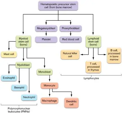
FIGURE 24.3 ■ Overview of the adaptive immune system.
Fig. 24.3 FIGURE 24.3 ■ Overview of the adaptive immune system.
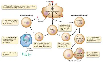
Fig. 24.16 FIGURE 24.16 ■ Steps in antibody formation. Steps 1 and 2 (gene rearrangements) happen in bone marrow before the antibody encounters antigen. Step 3 (T cell–independent B-cell activation) occurs after antigen is encountered in the spleen or circulation. Steps 4–6 (T H 0-cell activation and T FH -dependent activation of B cells) can occur in the thymus, in the mucosa-associated lymphoid tissues (MALT), or in the lymph nodes. Step 7 (activation of memory B cells) can occur in bone marrow, spleen, or lymph nodes. To be activated, memory B-cell receptors must bind to antigen and come under the influence of cytokines IL-4 and IL-6 secreted by other cells.
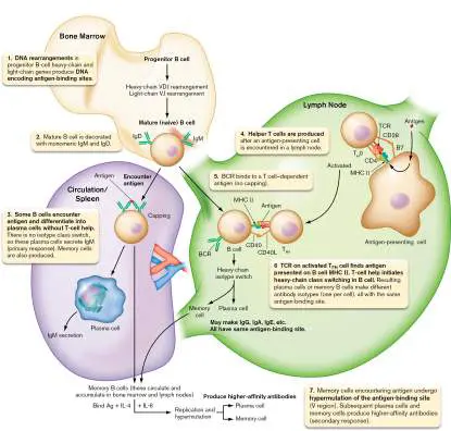
Fig. 24.12 FIGURE 24.12 ■ Primary versus secondary antibody response. Primary vaccination or infection leads to the early synthesis of IgM, followed by IgG. Either reinfection or a second (booster) dose of a vaccine results in a more rapid antibody response, consisting mainly of IgG, because of memory B cells formed during the primary response. Note that the time course and level of antibody made vary with the immunogen and the host.
Endnotes
1. Note *: IFN = interferon; IL = interleukin; NK = natural killer; PMN = polymorphonuclear leukocyte; TNF = tumor necrosis factor. Return to reference *
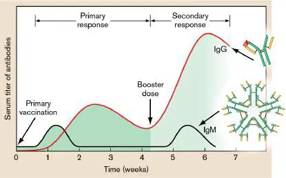
24.4 Complement as Part of Adaptive Immunitynot assigned
Chapter 23 describes how complement, in what is called the alternative pathway, can attack invading microbes before an adaptive immune response is launched (see Section 23.6). Factor C3b binds to lipopolysaccharides (LPS) and sets off a reaction cascade ending with a membrane attack complex (MAC) pore composed of C5b, C6, C7, C8, and C9 proteins (see Fig. 23.39). However, antibody made during the adaptive response to a pathogen offers another route to activate complement, called the classical complement pathway. Dubbed “classical” because it was the first complement pathway to be discovered, it requires a few additional proteins before reaching C3, the linchpin factor connecting the two pathways.
The classical cascade begins when a complement C1 protein complex binds to the Fc region of an antibody bound to a bacterial or viral pathogen (Fig. 24.27, step 1). The bound C1 complex then cleaves two other complement factors, C2 and C4, not used in the alternative pathway (step 2). Two fragments, one each from C2 and C4, combine to form another protease, called C3 convertase (different from C3 convertase in the alternative pathway), that cleaves C3 into C3a and C3b (step 3). C3b in the alternative pathway is stabilized by interacting with LPS. In the classical pathway, however, C3b combines with C3 convertase to make a C5 convertase (note that this C5 convertase is different from the C5 convertase formed in the alternative pathway; see Fig. 23.40). The subsequent steps leading to formation of a membrane attack complex (MAC) are the same as in the alternative pathway. As before, C5b binds to a target membrane and is joined by C6, C7, and C8. Multiple C9 proteins then assemble around the MAC and form a pore that compromises the integrity of the target cell, killing it.
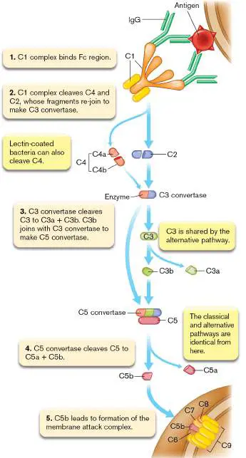
FIGURE 24.27 ■ Classical complement cascade. In Section 23.6 we noted the existence of two complement activation pathways that do not require antigen-antibody complexes. One of them—namely, the alternative complement pathway—was explained in detail, but discussion of the lectin activation pathway was deferred until now because it more closely resembles the classical pathway. The lectin activation pathway begins when lectins (produced by the liver) recognize and coat the sugar structures that decorate the surfaces of infectious organisms. The lectin-coated polysaccharides trigger cleavage of factor C4 to C4a and C4b (Fig. 24.27). C4b can cleave C2 and combine with one of the fragments to form C3 convertase. C3 convertase then continues through the classical activation pathway just described.
With all the other immune responses at the ready, why is complement also necessary? The need becomes evident in individuals with complement deficiencies. These patients are extremely susceptible to recurrent septicemic (blood) infections by organisms, such as Neisseria gonorrhoeae (the cause of gonorrhea), that normally do not survive forays into the bloodstream because they are killed by complement. Why, in a complement-deficient patient, won’t other defenses, such as antibodies and cytotoxic T cells, kill reinfecting Neisseria? The reason is that key antigens on the bacterial surface change shape over generations in a process called phase variation (see Section 10.5). The new antigens are “invisible” to antibodies made against earlier versions.
Regulating Complement Activation
How do normal body cells prevent self-destruction following complement activation? The sequential assembly of the membrane attack complex provides several places where regulatory factors can intervene. One such factor is the host cell-surface protein CD59. CD59 will bind any C5bC8 complex trying to form in the membrane and prevent C9 from polymerizing. Thus, no pore is formed and the host cell is spared. (Complement will not normally attack uninfected or infected host cells, since both contain CD59.)
Another regulatory mechanism hinges on a normal serum protein called factor H. Factor H prevents the inadvertent activation of complement in the absence of infection. If factor H is unavailable, then the uncontrolled activation of complement can damage host cells despite the presence of CD59. To short-circuit the cascade, factor H binds to C3bBb, displaces Bb from the complex, and acts as a cofactor for factor I protease, which then cleaves C3b. Without C3b, the complement cascade stops. Some bacteria, such as certain strains of Neisseria gonorrhoeae and Streptococcus pyogenes, have learned to protect themselves from complement by binding factor H. Factor H bound to the microbe provides a protective “force field” against local complement activation.
Microbiota can also assist host cells in resisting complement. The intestinal microbe Bacteroides thetaiotaomicron protects host cells from complement-mediated cytotoxicity by up-regulating a decay-accelerating factor present in host cell membranes. Decay-accelerating factor stimulates decay of complement factors and prevents their deposition at the cell surface.
Other Roles for Complement Fragments
The C3a, C4a, and C5a cleavage fragments generated by the complement cascade do not participate in MAC formation but are important for amplifying the immune reaction. These peptides act as chemoattractants to lure more inflammatory cells into the area. C3b, which does participate in MAC formation, also can act as an opsonin when it is bound to a bacterial cell. An opsonin is a protein factor that can facilitate phagocytosis. Because phagocytes contain C3b receptors on their surfaces, it is easier for them to grab and engulf cells coated with C3b. Additional roles for complement fragments were discussed in Section 23.6.
To Summarize
The classical pathway for complement activation begins with an interaction between the Fc portion of an antibody bound to an antigen and C1 factor in blood. (The alternative pathway does not need antibody but begins when C3b binds to LPS on a bacterium.)
The Fc-C1 complex reacts with C2 and C4, leading to production of C3b and a novel C5 convertase specific to the classical pathway.
After C5 convertase cleaves C5 , the classical pathway is the same as the alternative pathway, resulting in the formation of a membrane attack complex (MAC).
CD59 and factor H prevent inappropriate MAC formation in host cells.
Glossary
classical complement pathway An antibody-mediated pathway for complement activation. factor H A normal serum protein that prevents the inadvertent activation of complement in the absence of infection.
decay-accelerating factor A host cell membrane protein that stimulates the decay of complement factors and prevents their deposition at the cell surface.
Fig. 23.39 FIGURE 23.39 ■ Pore structure of the membrane attack complex (MAC). Multimer assembly of complement factor 9 forms a pore in a bacterial membrane that destroys membrane integrity. Image shows a model of the MAC complex derived from cyo-EM studies. Components are colored as follows: C5b (brown), C6 (light green), C7 (dark green), C8 subunits (orange, red, and dark blue), and finally C9 (light blue). Gray indicates the pore.
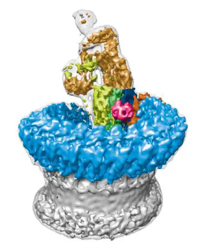
M. SERNA ET AL. 2016. NAT COMMUN. 7 :10587
Fig. 23.40 FIGURE 23.40 ■ The alternative complement pathway. Although called “alternative,” this complement cascade is part of the first-line innate defense system.
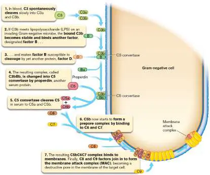
24.5 Gut Mucosal Immunity and the Microbiomenot assigned
The mucosal surface of the gastrointestinal tract is exposed to a vast number of food, chemical, and other ingested antigens. The gut also harbors an expansive microbiome whose members must be controlled and nurtured. Monitoring this large antigen load requires localized and highly integrated innate and adaptive immune systems and a system of gut-associated lymphoid tissues, or GALT (see Section 23.4). The innate immune mechanisms that keep gut microbes at bay were described in Chapter 23, but the intestine also has a dedicated adaptive immune system that monitors and shapes the microbiota, usually without causing excessive inflammation. In this section we describe how the innate and adaptive immune systems in the gut shape the microbiome while protecting the host.
The Gut Immune System
We discussed in Chapter 21 how the gut microbial community helps digest food and outcompetes pathogens (see Fig. 21.24). In Chapter 23 we described how gut microbiota are acquired and how dysbiosis can lead to intestinal inflammatory diseases, obesity and affect the gut-brain axis. Now in this chapter we explain how the microbiome trains the immune system and, in turn, how the immune system shapes the human microbiome. But first we must discuss the gut immune system, where the training and shaping take place. As described in Chapter 23, the intestinal epithelial barrier is one cell thick and composed mainly of columnar epithelial cells: intestinal epithelial cells (IECs) at site 1 in Figure 24.28, and goblet cells at site 2. Goblet cells secrete mucin and antimicrobial peptides. Epithelial cells express numerous pattern recognition receptors (PRRs), including TLRs and NLRs that can be activated by MAMPs shed from resident microbes (Fig. 24.28 blowup). Once activated, epithelial cells will synthesize and secrete a variety of cytokines that stimulate cells of the immune system—for instance, chemokines that attract neutrophils and lymphoid cells. The epithelial barrier is also punctuated by cells called intraepithelial lymphocytes (IELs), mostly T cells that have encountered antigens. IELs can secrete cytokines, including IFN-gamma, and maintain the mucosal barrier of the intestine through protective and inflammatory processes (Fig. 24.28, site 3).
FIGURE 24.28 ■ Mucosal immune system. Depicted is the region around one intestinal villus. Mucosal immunity functions independently of regional lymph nodes to control inflammation induced by microbiota or to stimulate inflammation when pathogens are present. See text for details. PGN = peptidoglycan; sIgA = secretory IgA.
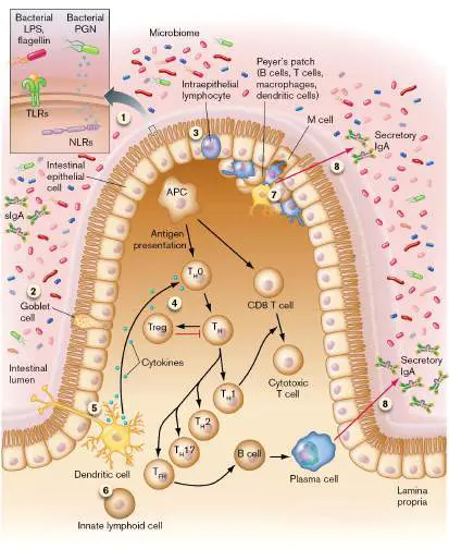
Beneath the epithelial layer is the lamina propria, rich in B cells and T cells. Epithelial cells can process antigens they encounter and place them onto MHC I and MHC II receptors for presentation to T cells residing in the lamina propria. (Although epithelial cells are not thought of as having MHC II, they will express the molecule if IFN-gamma is made during an infection.) After being presented with gut antigens, T cells can rapidly stimulate inflammatory responses via differentiation to T H 1, T H 2, and T H 17 cells (Fig. 24.28, site 4). Alternatively, the T cells can inhibit inflammatory responses by converting to Treg cells. In addition, dendritic cells in the lamina propria use their dendrite extensions to “reach” between epithelial cells and capture antigens in the intestinal lumen (site 5). One class of dendritic cells stimulates inflammation by producing cytokines TNF-alpha and IL-6. A separate set of dendritic cells, however, promotes the generation of regulatory T cells whose cytokine repertoire can dampen inappropriate inflammatory responses that might be triggered by microbiota.
Other important cells in the lamina propria include macrophages and innate lymphoid cells (ILCs). Macrophages were discussed earlier. ILCs are derived from lymphoid stem cells (see Fig. 23.18), but because they lack antigen-specific B-cell or T-cell receptors, they function as part of the innate immune system (Fig. 24.28, site 6). A diverse range of stimuli can activate ILCs, including neuropeptides, hormones, and cytokines produced by epithelial cells responding to microbiome MAMPs. ILCs include natural killer cells (described in Chapter 23). Other ILCs secrete the pro-inflammatory cytokine IL-17 to summon neutrophils and cytokine IL-22 to stimulate epithelial cells to secrete antimicrobial peptides. IL-22 also promotes tissue repair and regeneration in an inflamed intestine. Consequently, ILCs contribute to immunity, inflammation, tissue homeostasis, and microbiome control.
Peyer’s patches (Fig. 24.28, site 7), first described in Section 23.4, are lymphoid tissues made up of IgA + B-cell and T-cell centers. Peyer’s patches are ideal sites for adaptive immune responses along the intestinal tract. M cells in the epithelium above a Peyer’s patch will sample microbes and other antigens floating in the gut and offer them to underlying macrophages for engulfment and presentation to Peyer’s patch T cells.
T FH –B cell interactions taking place in sites 4 and 7 of Figure 24.28are influenced by local cytokines to trigger a B-cell class switch to IgA production and B-cell differentiation into IgA-secreting plasma cells. Secretion of dimeric sIgA (secretory IgA; site 8) into the intestinal lumen prevents microbes from penetrating through the epithelium and modulates the composition of the microbiome. Recent evidence also suggests that IgA coats many bacteria in the gut and fosters microbial colonization within specific intestinal niches. One such organism is Bacteroides fragilis, which produces a thick, fuzzy capsule, but only when growing in the gut. Gregory Donaldson (California Institute of Technology) and colleagues have shown that both the capsule and IgA are necessary for effective colonization. They propose that secretory IgA helps anchor beneficial gut microbiota to the intestine. Binding of the microbe-sIgA complex to the mucous lining is thought to be due to the secretory component of sIgA, not the Fc region.
Determining Friend or Foe
How might the gut immune system determine whether a microbe is indigenous microbiota or dangerous pathogen? The distinction, in large part, involves the strategic distribution of pattern recognition receptors (TLRs and NLRs) on and within epithelial cells (part of the innate immune system). TLRs are primarily on the cell surface, whereas NLRs are cytoplasmic. Recall that TLR and NLR interactions with MAMPs trigger the synthesis and release of various cytokines that impact innate and adaptive immune systems (see Chapter 23). Pathogenic bacteria possess virulence factors that enable them to attach to or invade host cells, thereby introducing MAMPs into the cytosol, where they are recognized by NLRs. However, bacteria of the indigenous microbiota are noninvasive and therefore less potent activators of NLRs. When the epithelium is intact, these “friendlies” interact only with the apical surface TLRs of epithelial cells, which are less responsive than TLRs on the host side (basolateral side) of epithelial cells.
However, an interaction with TLRs on the basolateral side of epithelial cells signals a barrier breach, which could be caused by either a pathogen or an indigenous microbe. The resultant cytokine cocktail will trigger inflammation. Nevertheless, members of the microbiome evolved to lessen inflammation by dampening epithelial cell TLR signaling. The repertoire of cytokines secreted by epithelium also supports microbiome tolerance by conditioning a subset of dendritic cells to become tolerogenic. Tolerogenic dendritic cells will present antigen to T cells but convert those T cells into regulatory T cells that suppress inflammatory responses. Treg cells are essential for maintaining tolerance to the microbiota. Without Treg cells, effector T-cell responses (for instance, those of cytotoxic T cells) go unopposed and produce inflammatory bowel diseases.
Compartmentalization of the Gut Immune System
It is important to note that adaptive immunity generated during breaches in the gut is typically limited to mucosal tissues. Systemic immunity is not generated. Compartmentalization is important because it allows the intestinal immune system to become tolerant of the microbiome without causing the systemic immune system to become tolerant too. Keeping systemic immunity relatively unaware of the “well-behaved” gut microbiome is important in the event a gut microbe escapes to the bloodstream. In this situation the host must be capable of mounting a vigorous systemic immune response to prevent infection.
Separation of the intestinal and systemic immune systems is possible, in part, because secreted IgA antibodies along the intestine will trap bacterial antigens on mucosal surfaces. The trapping decreases antigen movement into lymph nodes, which minimizes systemic antibody responses. In addition, adaptive immune cells produced in Peyer’s patches or in mesenteric lymph nodes lining the intestine are programmed to travel back to the mucosa. Programmed immune cells can circulate throughout the body, but they express homing receptors that bind molecules ( addressins) selectively present on vascular endothelial cells in different tissues (for example, in the mucosa). Interactions between homing receptors and addressins cause the immune cells to reside longer in a certain tissue.
Can intestinal microbiota influence immune responses at distal mucosal sites—in the lung, for instance? The mucociliary escalator of the upper airways naturally sweeps microparticles that enter the lung upward into the oral cavity, where the particles are ingested. Once in the intestine, the gut immune system can respond to those antigens and the activated immune cells generated will home to mucosal areas throughout the body, including lung. Gut microbiota, therefore, can influence immune responses in the lung. For instance, gut microbiota have been shown to influence the generation of influenza virus–specific CD4 and CD8 T cells and affect antibody responses to respiratory influenza virus infections.
The Microbiome Shapes Gut Immunity and Senses an Activated Immune Response
We have known for decades that germ-free animals devoid of intestinal microbiota exhibit major defects in the organization and activity of immune structures in the gut. Germ-free mice display an “underdeveloped” innate and adaptive immune system that includes reduced expression of antimicrobial peptides, reduced IgA production, fewer T-cell types, and increased susceptibility to microbial infections. Proper gut immunity can be restored, however, by simple microbial stimulation brought about by fecal transplant. Several gut microbiota with beneficial effects for human health were discussed in Chapters 13, 21, and 23.
Organisms such as Akkermansia muciniphila rejuvenate the mucin layer. Bacteroides thetaiotaomicron contributes greatly to carbohydrate utilization and to the production of short-chain fatty acids (SCFAs) that induce Treg cell production. Also contributing to Treg cell production are organisms such as Bacteroides fragilis, via its capsular p oly s accharide a ntigen (PSA). Faecalibacterium prausnitzii and a group of Clostridiales called segmented filamentous bacilli (SFBs) induce IgA-secreting cells in Peyer’s patches and stimulate the production of T H 17 cells. Bifidobacterium and Lactobacillus species also contribute to the shaping of the gut immune system.
As described earlier, the gut microbiome influences mucosal immunity through encounters with TLRs, NLRs, and other pattern recognition receptors on epithelial cells and a variety of immune cells. Influence is also provided through antigen interactions with dendritic cells and subsequent antigen presentation to T cells. The various blends of cytokines released can tilt immunity toward tolerance or inflammation and can even affect how strongly immune cells react to different antigens. For example, antibiotic-induced alterations in the microbiota will change not only the number of Treg cells but also the T-cell receptor (TCR) repertoire on the remaining Treg cells, suggesting that gut microbiome composition can influence the response of Treg cells to various antigens.
It has also become clear that the influence of the gut microbiome extends beyond the intestinal tract. Immune-related disorders such as inflammatory bowel disease (IBD), cancer, diabetes, allergies, and even obesity (see Chapter 23) may result from dysbiosis of the commensal microbial communities. Bacteria can produce neurotoxic metabolites such as D -lactic acid and ammonia. Even beneficial metabolites such as SCFAs may exert neurotoxicity. Gut microbes can also produce hormones and neurotransmitters that are identical to those produced by humans (discussed in Chapters 13 and 21). Consequently, gut bacteria can directly stimulate afferent neurons of the enteric nervous system to send signals to the brain via the vagus nerve. Through these varied mechanisms, gut microbes can shape the architecture of sleep and stress reactivity of the hypothalamic-pituitary-adrenal axis. Believe it or not, your gut microbes can influence your memory, mood, and cognition.
Can the microbiome also sense and react to an activated immune response? Simone Becattini from the University of Geneva and Eric Palmer from the University of Chicago have examined this question using antibiotic-treated mice that were artificially recolonized with four anaerobic members of the microbiome: two species each from the Bacteroidota and Bacillota phyla (formerly Bacteroidetes and Firmicutes). After the mice were recolonized, the researchers stimulated their immune systems by intraperitoneal injection of flagellin. Transcript levels of inflammatory cytokines increased in the mouse cecum, indicating inflammation. The four bacterial species responded by altering their mRNA profiles. Up-regulated genes included those encoding chaperones, reactive oxygen species (ROS) scavenging enzymes, and stress response mediators. Down-regulated genes were mostly involved in sugar catabolism and amino acid biosynthesis. The metabolic changes in the microbiome were also found to significantly alter the gut metabolome. The authors propose that these metabolic changes can impact host health and an unfolding immune response.
To Summarize
The gut immune system includes intestinal epithelial cells (IECs); goblet cells; innate lymphoid cells (ILCs); lamina propria, which contains B cells, T cells, and dendritic cells; and Peyer’s patches, which contain M cells, IgA + B cells, and T cells.
Secretion of sIgA helps control the composition and balance of the gut microbiome.
Intestinal epithelial cell TLRs and NLRs can distinguish normal microbiota from pathogens.
Microbiota composition influences the ratios of T FH, T H 1, T H 2, T H 17, and Treg cells to tilt the gut immune response between inflammatory and anti-inflammatory.
Mucosal immunity is an immune system compartment that is mostly separate from the rest of the immune system. Development of the mucosal immune system is guided by bacterial members of the gut microbiota.
Activation of the gut immune system causes transcriptional adaptations within the gut microbiome.
Glossary
intraepithelial lymphocyte (IEL)
A lymphocyte embedded among epithelial cells that line the intestine.
innate lymphoid cell (ILC)
A lymphocyte-like cell in the intestinal lamina propria that lacks B-or T-cell receptors but secretes pro-inflammatory cytokines. addressin A tissue-specific protein that is selectively present on vascular endothelial cells in different tissues.
mucosal immunity The portion of the innate and adaptive immune systems that protects the mucosa from microbial invasion.
Fig. 21.24 FIGURE 21.24 ■ Human gut microbiome digests our food and influences host development and function.
In a “restaurant” mixed-species biofilm, Bacteroides bacteria break down glycans into sugars that Escherichia coli catabolizes, consuming oxygen from the host blood supply. The sugars may also feed pathogens, which stimulate an immune response. Bacteria release peptides and neurotransmitters that influence brain function. SCFAs = short-chain fatty acids.
Fig. 23.18
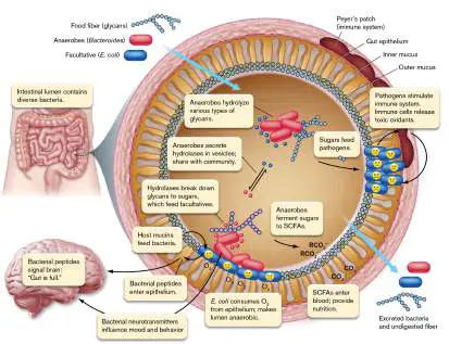
FIGURE 23.18 ■ Development of white blood cell components of the immune system. Pluripotent hematopoietic stem cells in bone marrow divide to form two lineages: myeloid cells and lymphoid cells. Myeloid stem cells develop into PMNs (the primary components of innate immunity), red blood cells, and platelets. Lymphoid stem cells differentiate into B cells, T cells, and natural killer cells. Final maturation into B cells and T cells (the principal cells involved in adaptive immunity) occurs in the bone marrow and thymus, respectively. Colors indicate a group of differentiated cells that arise from the same progenitor.
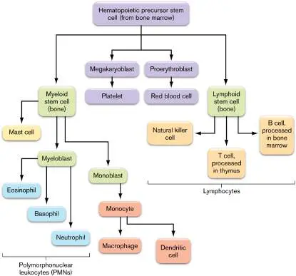
24.6 Immunizationnot assigned
Most of you have been vaccinated with many of the vaccines listed in Table 24.1, which include the
novel COVID-19 vaccines that are pulling us out of the recent pandemic. These vaccines work because
of our adaptive immune system. In this section we describe the various types of vaccines, note when
and how they are given, and discuss why some people refuse vaccinations.
Vaccines
Vaccines come in four basic types. As noted in Table 24.1, some vaccines utilize killed organisms
(examples are the hepatitis A vaccine and the Salk inactivated polio vaccine), while others contain live,
but attenuated, microbes (BCG for tuberculosis or the Sabin live polio vaccine). The third type of
vaccine consists of purified components (subunits) of an infectious agent (such as capsular antigens
from Streptococcus pneumoniae and Haemophilus influenzae type b, or virus capsids such as those
used in the Gardasil vaccine for human papillomavirus, a cause of genital warts and cancers of the
cervix and penis). The newest form of vaccine is generally called nucleic acid vaccines. One type
involves mRNA molecules encased within lipid nanoparticles. The mRNA molecules encode protective
antigens, such as a segment of the SARS-CoV-2 spike protein (see Chapter 16).
Most vaccines are administered during childhood, when the diseases can be the most devastating.
Some vaccines are injected in combination as polyvalent vaccines to control multiple diseases (for
example, MMR for measles, mumps, and rubella), while others are given individually but may change
from year to year (influenza viral envelope proteins).
Table 24.5 provides the current immunization schedule recommended for children and adolescents
by the Centers for Disease Control and Prevention (CDC). Notice that all of these vaccines are given in
multiple doses, called booster doses. The exception to this rule is the influenza vaccine, which is given
in a single dose but changes every year. The reason for multiple doses, as noted earlier, is that
secondary exposure to an antigen provides a more robust and long-lasting immunity. However, most
vaccines are not administered until 2 months of age, because maternal antibody crossing through the
placenta to the fetus (or to a newborn through breast milk) will persist for a short time in the newborn,
temporarily protecting the baby from disease and possibly dampening the response to a vaccine
antigen administered during that time.
TABLE 24.5 Recommended Immunization Schedule for Children and Ad
Age of administration
1 2 4 6 9 12 15–18 2
Vaccine Birth month months months months months months months mon
Hepatitis B b HepB HepB HepB
Rotavirus Rota Rota Rota
TABLE 24.5 Recommended Immunization Schedule for Children and Ad
Age of administration
1 2 4 6 9 12 15–18 2
Vaccine Birth month months months months months months months mon
Diphtheria, DTaP DTaP DTaP DTaP
tetanus,
acellular
pertussis c
Haemophilus Hib Hib Hib Hib
influenzae
type b d
Inactivated IPV IPV IPV
poliovirus
Measles, MMR
mumps,
rubella
Varicella Varicella
Meningococcal Administer to high-ri
(ACWY) e
Meningococcal
(B)
Pneumococcal PCV13 PCV13 PCV13 PCV13
f
TABLE 24.5 Recommended Immunization Schedule for Children and Ad
Age of administration
1 2 4 6 9 12 15–18 2
Vaccine Birth month months months months months months months mon
Influenza g Influenza (yearly)
Hepatitis A h HepA (2-dose series)
Human
papillomavirus
i
COVID-19 j
Source: Adapted from the Centers for Disease Control and Prevention website (http://www.cdc.gov).
Herd Immunity
You might wonder whether all members of a community must be vaccinated against a given microbe to
lower the risk of disease for every individual in that community. In fact, the risk of an infected person
spreading disease to an unvaccinated person can be lowered dramatically even when only about three-
fourths of the community is vaccinated. Vaccinating a large percentage of a community effectively
conveys community (or herd) immunity by interrupting transmission of the disease. If one individual
contracts the disease, the chance that they will come into contact with another unvaccinated person
and transmit the disease is much reduced. Thus, the risk of disease to any single unvaccinated person
is lessened as a result of community immunity. Measles vaccine is a good example of a vaccine that
can provide herd immunity. Note that the percentage of people in a community who must be
vaccinated to achieve herd immunity will vary depending on the disease.
Herd immunity works well for diseases such as diphtheria, whooping cough (pertussis), measles,
and mumps, all of which are infections spread by person-to-person contact. However, herd immunity
will not lower the risk that an unvaccinated person will contract tetanus, which is not spread by person-
to-person contact. Clostridium tetani, the agent whose toxin causes tetanus, is a ubiquitous soil
organism transmitted through punctured skin. The risk of tetanus for an unvaccinated person does not
change, even if every other person in the community is vaccinated against tetanus.
Vaccines and Immune System Compartments
What difference does it make which type of vaccine you take? Attenuated, killed, subunit, nucleic acid
—don’t they all generate immunity? Yes, but a live, attenuated vaccine is generally better than subunit
or killed vaccines. When a crippled but live microbe replicates at its normal body target site, the
immune response generated is most appropriate to that site; both the innate and adaptive responses
are engaged (discussed later). Take the case of the Salk (killed) and Sabin (attenuated) polio vaccines.
Poliovirus typically enters the body through ingestion, replicates in the mucosa, moves to the regional
lymph nodes, and produces a viremia. Eventually, the virus can attack the central nervous system and
cause paralysis.
The killed Salk vaccine (inactivated polio vaccine, or IPV) is injected and generates an antibody
response in the bloodstream. The antibody response prevents the viremia and paralytic consequences
of a natural infection, but it does not generate a mucosal immunity that would stop the natural virus, if
ingested, from replicating in the gut. Consequently, someone infected with live poliovirus after being
vaccinated with the Salk vaccine can still develop mild disease and shed poliovirus in their feces. This
shedding can transmit live poliovirus to unvaccinated people vulnerable to serious disease. This
situation was thought to initiate the 2022 outbreak of polio in New York State.
In contrast to the killed Salk vaccine, the live but attenuated virus in the Sabin vaccine, which is
administered orally (oral polio vaccine, or OPV), will replicate in the mucosal lymphatic system of the
gut, where secretory IgA antibodies are best generated (discussed earlier). Anti-polio IgA antibodies
secreted in these mucosal areas will prevent any wild-type poliovirus encountered later from
replicating in the intestinal mucosa, so the person vaccinated with OPV will be asymptomatic and
cannot shed virulent virus. However, OPV has not been used in the United States since the year 2000
for two reasons: First, polio in this country is rare, so fecal spread is unlikely, and second, the live
attenuated virus could produce polio in an immunocompromised, vaccinated person.
The situation described for the killed poliovirus vaccine appears to hold for the novel COVID-19
mRNA vaccines described in Chapter 16. The mRNA vaccines, which are injected, produce great
circulating antibody and T-cell immune responses, but they generate poor mucosal IgA immunity.
Without anti-SARS-CoV-2 IgA mucosal antibody, a fully vaccinated individual can still be infected and
contract mild COVID-19 (called a breakthrough infection). This person can still shed live virus through
respiratory secretions and infect unvaccinated or immunocompromised people, who can develop
serious disease.
Building better vaccines. Why are live vaccines better than dead, or component, vaccines at
producing effective antibody responses? Julie Blander’s group (see Fig. 24.23) discovered that
components of the innate immune system in mice can sense a “live” signal (bacterial RNA) during an
infection (or vaccination). Once engaged, the innate cells secrete a blend of cytokines (IFN-β and IL-
1β) that fosters the production of T FH cells. The T FH cells drive antibody class switching in B cells and
differentiation of those B cells into plasma cells.
Leif Sander (Charité–Berlin University of Medicine) made a similar discovery using human tissue
cultures. His group found that bacterial RNA from a live vaccine strain of E. coli binds to TLR8 in
monocytes. The activated monocytes secrete IL-12, which activates T H cells to become fully functional
human T FH cells. Figure 24.29 presents fluorescence-activated cell sorting (FACS) data supporting
the superiority of live E. coli over dead E. coli in this process. When a T H cell (CD4 pos) differentiates
into a T FH cell, the T FH cell will also express CXCR5, a surface receptor for a specific cytokine. APC
monocytes exposed to live E. coli were better at converting T H cells into T FH cells than were
monocytes exposed to dead E. coli (17% versus 1.4%, respectively, above the control value in Fig.
24.29 ).
FIGURE 24.29 ■ Live E. coli drives differentiation of CD4 pos -CXCR5 neg T cells into
CD4 pos -CXCR5 pos T FH cells. Human APC monocytes were stimulated with control medium,
live E. coli, or heat-killed E. coli. Stimulated APCs were then mixed with T cells and incubated for
5 days with staphylococcal enterotoxin B (superantigen). The x - and y -axes measure,
respectively, the amount of surface CXCR5 marker and CD4 marker per cell. All T cells are plotted.
Red boxes mark the graphical locales of CD4 pos -CXCR5 pos T FH cells. Numbers in boxes indicate
the percentage of total CD4 pos cells (T cells) that are also CXCR5 pos (T FH cells).
Source: Modified from Ugolini et al. 2018. Nature Immunol. 19 :386–396, fig. 1C.
The viability trigger that links innate and adaptive responses for mice and humans is the same
(bacterial RNA), but the differentiation pathways are not. The data suggest that adding viability
MAMPs, like bacterial RNA, to inactivated or component vaccines might greatly improve their efficacy.
Note that the phenomenon of RNA as a viability signal is one reason mRNA vaccines such as those for
COVID-19 are so effective.
Thought Question
24.13 Why do immunizations lose their effectiveness over time?
Are Vaccines Dangerous?
Successful vaccination programs carried out in the United States have come close to eradicating many
once-feared diseases, such as measles, polio, rubella, and diphtheria, and they have dramatically
lowered the morbidity and mortality of many others. The vast majority of people who receive vaccines
suffer no, or only mild, reactions, such as fever or soreness at the injection site. Very rarely do more
serious side effects occur, such as allergic reactions or disease. However, vaccinating individuals who
have a defect in their immune system—for example, severe combined immune deficiency (SCID) or
bare lymphocyte syndrome (mentioned at the start of the chapter)—can have severe consequences.
Immunocompromised people should never receive live, attenuated vaccines. Although innocuous to
someone with a healthy immune system, a live, attenuated vaccine strain will replicate unchecked in
someone lacking critical immune system components and can cause severe disease, even death.
Subunit and mRNA vaccines, in contrast, cannot replicate, and they remain safe for
immunocompromised individuals. Unfortunately, certain immunodeficiencies prevent subunit vaccines
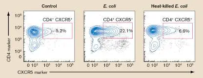
from generating an immune response. Infections in immunocompromised individuals must be treated
with antibiotics or intravenous administration of immunoglobulin (IVIG; pooled antibody collected from
a thousand or more random individuals).
Despite the unassailable proof that vaccines are extremely safe for people with healthy immune
systems, people known as anti-vaxxers circulate unsettling, erroneous information on the Internet
purporting a link between vaccinations and other diseases, such as diabetes or autism. No well-
controlled scientific study supports any of these claims. In fact, the fraudulent 1998 Lancet article
written by Andrew Wakefield that spawned this movement has been disavowed by all its coauthors
(except Wakefield) and was retracted by the Lancet.
The risk of disease and death from preventable infectious diseases far outweighs the minimal risk
associated with being vaccinated against them. As proof of this point, the undervaccination of children
in the United States starting in the 1980s led to an increase in cases of measles. Although measles was
declared eradicated from the United States in 2000, cases of the disease rose dramatically in recent
years. In 2002, there were only 50 cases, but the number of cases for 2019 was more than 1,282, a
level not seen in over 25 years. It is interesting to note that the number of measles cases then
dropped to only 13 in 2020. This decrease was due in large part to the public health measures, such as
mask wearing, social distancing, and hand hygiene, instituted to curtail the COVID-19 pandemic.
Likewise, vaccination efforts decreased the incidence of whooping cough from over 250,000 cases
per year in the 1930s and 1940s to 6,586 cases in 1993. In part because of decreased vaccination
rates, whooping cough cases slowly rose to 28,639 in 2013. Renewed vaccination efforts and
improvements in vaccine effectiveness have started to reverse this trend. There were 15,662 cases of
whooping cough in 2019.
Sometimes designing vaccines that stimulate adaptive immunity is very difficult, especially if the
infectious agent undergoes rapid antigenic shifts or drifts. A good example is the influenza virus, for
which yearly vaccines are required. Special Topic 24 discusses the heroic, ongoing search for
therapeutic antibodies to treat Ebola and describes an alternative to adaptive-immunity vaccines. The
technique is called passive immunity, in which agent-specific antibodies are injected directly into
patients. Protection is short-lived but can be essential if a vaccine is unavailable or a patient is
immunocompromised.
SPECIAL TOPIC 24 A Monoclonal Magic Bullet for Ebola?
Ebola virus, found primarily in Africa, causes a devastating, deadly hemorrhagic fever whose
symptoms include widespread damage to blood vessels and extensive internal and external
bleeding (see Chapter 26). Without treatment, mortality can reach as high as 90% of those
infected. Ebola virus comes in three clinically relevant but antigenically distinguishable strains:
EBOV (formerly Zaire), Bundibugyo (BDBV), and Sudan (SUDV). Fortunately, an Ebola vaccine
called Ervebo was approved in 2019, although it works only on EBOV. Efforts to develop a single
vaccine that could protect against all three strains have been stymied by their antigenic
differences.
Another way to cure or prevent Ebola disease would be to isolate broadly neutralizing anti-
Ebola antibodies from Ebola survivors. Broadly neutralizing antibodies would enable clinicians to
cure patients infected with any Ebola strain and to passively immunize people who have been in
contact with those patients. James Crowe (Fig. ST 24.1A ) from Vanderbilt University Medical
Center and Alexander Bukreyev (Fig. ST 24.1B ) from the University of Texas Medical Branch
have searched for these antibodies and found two promising candidates.
FIGURE ST 24.1 ■ Ebola disease and the scientists searching for a cure. A. James
Crowe (right) and Pavlo Gilchuk (left). B. From left to right: Alexander Bukreyev, Philipp
Ilinykh, and Kai Huang.
COURTESY OF VANDERBILT UNIVERSITY
THE UNIVERSITY OF TEXAS MEDICAL BRANCH
The researchers and their colleagues started their search by screening plasma from 17
survivors of the 2014 Ebola outbreak in West Africa, looking for patients whose antibodies would
cross-react with the surface glycoprotein (GP) of the three Ebola virus strains. GP mediates viral
attachment and entry into host cells. Antibodies that bind to GP can neutralize the virus by
preventing virus entry. Plasma from two of the survivors contained antibodies able to react
against GP from all three Ebola strains (Fig. ST 24.2 ). The scientists hoped that blood of these
two survivors had circulating memory B cells that produced broadly neutralizing, anti-Ebola
antibodies (bNAbs).
FIGURE ST 24.2 ■ Binding of Ebola survivor antibodies to modified GPs from the
three Ebola viruses. Enzyme-linked immunosorbent assay (ELISA), as described in Section
28.3 was used to determine binding. Dilutions of survivor plasma were added to microtiter
plate wells coated with different GP antigens whose transmembrane domains were deleted
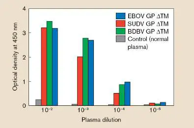
(ΔTM) to increase solubility. The level of absorbance at 450 nm is proportional to the amount
of anti-Ebola GP present in the survivor’s plasma.
Source: Modified from Gilchuk et al. 2018. Immunity 49 :363–374, fig. 1A.
Six hundred individual B-cell lines made from the two survivors produced antibodies that
reacted against EBOV GP. Six of those antibodies were able to bind to all three Ebola strains in
ELISA tests. These six short-lived B-cell lines were then fused to immortal myeloma cancer cells
to make six immortal B-cell hybridoma cell lines. Each B-cell hybridoma produced a single
antibody isotype (for instance, IgG) with binding specificity to a single epitope (or closely related
epitopes). Once made, a hybridoma can be stimulated to make large amounts of that antibody.
Because all of the antibodies made from a hybridoma are identical, they are called monoclonal
antibodies.
Two of the monoclonal antibodies generated from the study (EBOV-515 and -520) neutralized
all three Ebola strains, limiting their abilities to form plaques on tissue culture cells. The
antibodies also enhanced the survival of mice infected with mouse-adapted EBOV (Fig. ST 24.3
), wild-type SUDV in mice, and BDBV in ferrets (not shown).
FIGURE ST 24.3 ■ In vivo protection of bNAbs against EBOV-MA (mouse-
adapted EBOV). Mice (in groups of 5) were infected with EBOV-MA. One day after infection,
different groups were treated with the following antibodies: EBOV-515 (blue); EBOV-520
(red); EBOV-442 (green). The control mice (black) were treated with no antibodies.
Source: Modified from Gilchuk et al. 2018. Immunity 49 :363–374, fig. 2D.
Electron microscopy and other studies then revealed the binding sites (epitopes) for each
antibody [F(ab) regions] on the GP ΔTM (Fig. ST 24.4 ). Three F(ab) molecules of each antibody
are shown bound to the GP trimer, one at each monomer. The antibodies bind to quaternary-
structure epitopes formed by spatially aligned residues in the GP1 and GP2 subunits of a GP
monomer. These antibodies are broadly neutralizing because the residues critical for binding
EBOV-515 or -520 are identical among the EBOV, BDBV, and SUDV Ebola viruses.
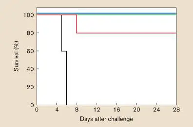
FIGURE ST 24.4 ■ 3D reconstructions of F(ab)–EBOV ΔTM complexes. Electron
density representation of F(ab) of EBOV-515 (blue) or -520 (orange) antibodies. GP1 and
GP2 subunits of each protomer of the GP1 trimer are not highlighted.
Source: Modified from Gilchuk et al. 2018. Immunity 49 :363–374, fig. 6A.
People living on the African continent are continually threatened by random outbreaks of Ebola
disease that can quickly spread to become epidemics. The authors propose that these broadly
neutralizing monoclonal antibodies, used either individually or as a cocktail, are promising
candidates for development as pan–Ebola virus therapeutic molecules. Support from several
funding agencies, including the National Institutes of Health, the Defense Threat Reduction
Agency, and the Bill and Melinda Gates Foundation, was instrumental for carrying out this
groundbreaking research.
RESEARCH QUESTION
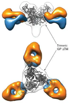
Viruses can evolve to evade the immune system by randomly changing one or more residues of
an epitope so that a neutralizing antibody can no longer bind. How might therapeutic strategies
be designed to minimize this risk?
Gilchuk, Pavlo, Natalia Kuzmina, Philipp A. Ilinykh, Kai Huang, Bronwyn M. Gunn, et al. 2018.
Multifunctional pan-ebolavirus antibody recognizes a site of broad vulnerability on the ebolavirus glycoprotein.
Immunity 49 :363–374.
Recent dramatic examples of passive immunity as a treatment regimen involve laboratory-made
monoclonal antibodies such as bebtelovimab, manufactured by Eli Lilly (see the chapter-opening
image) and developed to treat mild to moderate cases of COVID-19. These antibodies target the
SARS-CoV-2 attachment spike protein, blocking viral attachment and entry into host cells. Monoclonal
antibodies have proven very useful in preventing severe COVID-19 disease.
Thought Question
24.14 How might you design/construct a more effective vaccine for an antigen (for instance, the
Yersinia pestis F1 antigen or hepatitis A) that would harness the power of a Toll-like receptor (TLR)?
Hint: Look at TLR5 in Table 23.3.
To Summarize
Vaccines can be made from live, attenuated organisms; killed organisms; purified microbe
antigens; or mRNA-encoding protective antigens.
Herd immunity can help protect unimmunized persons from diseases transmitted person to
person.
Vaccine makeup and delivery influence whether the immune response is primarily humoral
or cellular in nature.
Serious side effects from immunizations are very rare.
Endnotes
1. Note a: This schedule indicates the recommended ages for routine administration of currently
licensed childhood vaccines, as of January 1, 2022. Range of recommended ages for each
vaccine dose. Catch-up immunization. Assessment at age 11–12 years. Return to
reference a
2. Note b: Hepatitis B vaccine (HepB). At birth: All newborns should receive monovalent HepB,
administered soon after birth and before hospital discharge. Return to reference b
3. Note c: Diphtheria, tetanus, and acellular pertussis (DTaP) vaccine. Tdap is a modified vaccine
with lower doses of diphtheria and tetanus toxoids. Return to reference c
4. Note d: Haemophilus influenzae type b (Hib) conjugate vaccine. Return to reference d
5. Note e: Meningococcal conjugate vaccine (MCV4). MCV4 should be administered to all children at
age 11–12 years, as well as to unvaccinated adolescents at high school entry (age 15 years). The
vaccine contains four types of capsules: A, C, W, and Y. Return to reference e
6. Note f: Pneumococcal vaccine. The 13-valent pneumococcal conjugate vaccine (PCV) is
recommended for all children aged 2–23 months and for certain children aged 24–59 months. The
final dose in the series should be administered at age ≥12 months. Pneumococcal polysaccharide
vaccine (PPSV) is a 23-valent vaccine recommended in addition to PCV for certain high-risk groups.
PPSV is also recommended for people over age 65. Return to reference f
7. Note g: Inactivated influenza vaccine should be administered annually starting at age 6 months.
Live, attenuated influenza vaccine should not be given until 2 years and not to
immunocompromised individuals. Return to reference g
8. Note h: Hepatitis A vaccine (HepA). HepA is recommended for all children at age 1 year (12–23
months). Return to reference h
9. Note i: Human papillomavirus (HPV) vaccine: HPV4 (Gardasil) and HPV2 (Cervarix). A 3-dose
series of HPV vaccine should be administered on a schedule of 0, 1–2, and 6 months to all
adolescents aged 11–12 years. Either HPV4 or HPV2 may be used for females; only HPV4 may be
used for males. Return to reference i
10. Note j: The CDC recommends that the COVID-19 Pfizer-BioNTech mRNA vaccine is suitable for
children age 6 months old and older. Return to reference j
Glossary
Fig. 24.23
FIGURE 24.23 ■ Pathogen viability signal molecules indirectly activate antibody
production. A. Julie Blander of Cornell University studies connections between innate and
adaptive immunity. B. and C. Mouse spleen germinal centers after vaccination with heat-
killed E. coli (B. ) or with heat-killed E. coli plus E. coli RNA (C. ). Sections of spleen were
stained with fluorescent antibodies to reveal B cells (blue), T cells (gray), and activated B
cells (red). The addition of RNA led to greater numbers of germinal centers and activated B
cells. An increase was also observed in the number of B cells expressing anti- E.coli IgG
antibodies on their surfaces (green).
COURTESY OF JULIE MAGARIAN BLANDER
MODIFIED FROM BARBET, G. ET AL. 2018. IMMUNITY 48 :584–598
MODIFIED FROM BARBET, G. ET AL. 2018. IMMUNITY 48 :584–598
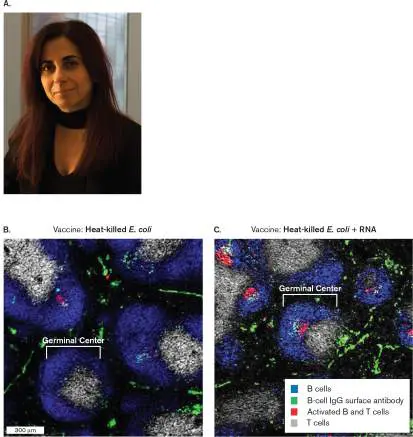
24.7 Hypersensitivity and Autoimmunitynot assigned
The immune system is not perfect. Sometimes the system overreacts to certain foreign antigens and causes more damage than the antigen (microbe) alone might cause. In addition, some foreign antigens can possess epitopes that look like host epitopes and can trick the immune system into reacting against self. These immune miscues are called allergic hypersensitivity reactions, and the antigen causing the reaction is called an allergen. There are four types of hypersensitivity reactions (see Table 24.6 ). Types I–III are antibody mediated; type IV is cell mediated.
TABLE Summary of Hypersensitivity Reactions 24.6
Type Description Time Mechanism a Manifestations of onset I IgE-mediated 2–30 Antigen induces Systemic hypersensitiv min cross-linking anaphylaxis, ity of IgE bound local to mast cells anaphylaxis, with release hay fever, of vasoactive asthma, eczema mediators.
II Antibody-5–8 h Antibody Blood transfusion mediated directed reactions, cytotoxic against cell-hemolytic hypersensitiv surface disease of the ity antigens newborn, mediates cell autoimmune destruction hemolytic via ADCC or anemia complement.
III Immune 2–8 h Antigen-Systemic complex– antibody reactions, mediated complexes disseminated hypersensitiv deposited at rash, arthritis, ity various sites glomerulonephri induce mast tis cell degranulation via Fc receptor; PMN degranulation damages tissue (localized reaction).
IV Cell-mediated 24–72 Memory T H 1 Contact hypersensitiv h cells release dermatitis, ity cytokines that tubercular recruit and lesions activate macrophages.
Case History: Type I Hypersensitivity
A bee stings a 9-year-old boy walking with his mother at the zoo. Within minutes, the boy begins sweating and itching. His chest then starts to tighten, and he has tremendous difficulty breathing. Terrified, he looks to his equally frightened mother for help.
This is a classic and severe example of type I (immediate) hypersensitivity, called anaphylaxis, in which smooth muscle contracts and capillaries dilate in response to the release of pharmacologically active substances. Type I hypersensitivity occurs within minutes of a second exposure to an allergen when the allergen reacts with IgE-coated mast cells. Recall that the primary role of IgE is to amplify the body’s response to invaders by causing mast cells to release inflammatory mediators (see Section 24.2). Allergic individuals with type I hypersensitivity produce an excessive amount of IgE to the allergen. On initial exposure, an allergen elicits the production of IgE antibodies specific to the allergen (bee venom in this example). The Fc portions of these antibodies bind to Fc receptors on the surfaces of mast cells, leaving the antigen-binding sites waving away from the cell, “looking” for antigen. IgE-coated mast cells are then said to be sensitized.
During a second exposure to the allergen, identical antigenic sites on the allergen bind to adjacent surface IgE molecules affixed to the mast cell. The result is a bridge between adjoining binding sites (Fig. 24.30). This cross-linked complex launches a complex signaling cascade that causes mast cell granules to quickly migrate to the cell surface and release their contents in a process called degranulation.
FIGURE 24.30 ■ Events leading to type I hypersensitivity reactions.
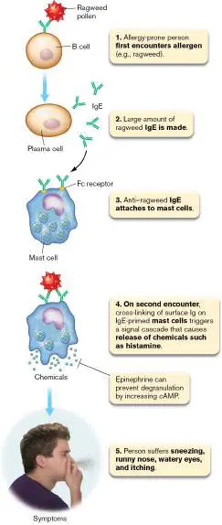
Mast cell degranulation releases chemicals with potent pharmacological activities. The most important of these is histamine, which binds to histamine receptors (H1 receptors) present on most body cells.
Antihistamines have a structure similar to histamine and work as an antagonist by preventing histamine from binding the H1 receptors. Histamine bound to H1 receptors on smooth muscle will trigger synthesis of a signaling molecule (inositol trisphosphate) that initiates smooth-muscle contraction to constrict small blood vessels. Histamine also weakens contacts between adhesion proteins (VE-cadherin) on vascular endothelial cells, causing gaps between cells through which blood fluids (not red blood cells) can seep. As a result, histamine-induced constriction of small blood vessels causes fluid to be forced from the circulation into the tissues. The immediate consequence is swelling (edema) in the joints and around the eyes and a rash (similar to hives), with burning and itching of the skin due to nerve involvement. In the case of the boy stung by a bee, the contraction of lung smooth muscles also led to breathing difficulties.
The bee sting in our example caused a severe type I allergic reaction, but type I hypersensitivity reactions do not usually involve the whole body. Most type I reactions are more localized and cause what is called atopic (“out of place”) disease. Hay fever, or allergic rhinitis, is a common manifestation of atopic disease that can be caused by the inhalation of dust mite feces (Fig. 24.31), animal skin or hair (dander), and certain types of grass or weed pollens. This disease affects the eyes, nose, and upper respiratory tract. Atopic asthma, another form of type I hypersensitivity resulting from inhaled allergens, affects the lower respiratory tract and is characterized by wheezing and difficulty breathing.
FIGURE 24.31 ■ Inhaled allergen. Dust mites (200–600 μm) and dust mite feces (colorized SEM).
ANDREW SYRED/SCIENCE SOURCE
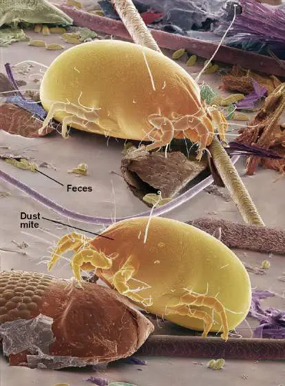
The administration of antihistamines is an effective treatment for allergic rhinitis, but the more important chemical mediators of asthma are the leukotrienes produced during what is called late-phase anaphylaxis. In late-phase anaphylaxis, mast cells release chemotactic factors that call in eosinophils. Eosinophils entering the affected area produce large amounts of leukotrienes that, like histamine, cause vasoconstriction and inflammation. At this point, antihistamines have little effect. Effective treatment includes inhaled steroids to minimize inflammation and bronchodilators (for example, albuterol) to widen bronchioles and facilitate breathing. One medication, montelukast (Singulair), works by blocking leukotriene receptors in the lung. During severe asthma attacks, injected epinephrine is critical. Epinephrine will open airways to ease breathing through direct hormonal action. People sensitized to allergens are not necessarily doomed to suffer with the allergy their entire life. A clinical treatment called desensitization can sometimes be used to prevent anaphylaxis. Desensitization involves injecting small doses of allergen over a period of months. This process is thought to produce IgG molecules that circulate, bind, and neutralize allergens before they contact sensitized mast cells. Desensitization has been useful in cases of asthma and bee stings. Desensitization by injection has not proved useful for food allergies; however, studies of oral desensitization are under way. Another treatment for allergic asthma, omalizumab (Xolair), can actually prevent sensitization. Omalizumab is a monoclonal antibody (an antibody preparation of one antibody type that binds only one epitope). Administered by injection, omalizumab selectively binds IgE. Given to allergic patients once a month, the monoclonal antibody prevents IgE from binding to Fc receptors on mast cells and thus prevents sensitization. A subsequent encounter with an allergen will not trigger an asthma attack.
Why don’t all antigens generate type I hypersensitivity? It turns out that most antigens do not elicit high levels of IgE antibodies. It is unclear what gives certain antigens this capability and what makes different individuals prone to different allergies. eResearch Activity 24 explores how a certain type of Treg cells can suppress the B cell IgE class switch to prevent hypersensitivity.
Case History: Type IV Hypersensitivity
The patient is an 8-year-old girl from Argentina. She received the BCG vaccine for tuberculosis about 4 months before coming to the United States. Upon entering school, she is required to take a skin test for tuberculosis. She tries to refuse but is told it is a requirement and allows the nurse to apply the test to her arm. Three days later, the test site has a large, red lesion and the skin is starting to slough (peel off). Does she have tuberculosis? Type IV hypersensitivity, also known as delayed-type hypersensitivity (DTH), is the only class of hypersensitivity triggered by antigen-specific T cells. Because T cells have to react and proliferate to cause a response, it generally takes 24–48 hours before a reaction is noticed. This delay distinguishes type IV hypersensitivity from the more rapid antibody-mediated allergic reactions (types I–III). The girl in the case study was initially sensitized by being vaccinated with BCG (an attenuated strain of Mycobacterium bovis, a close relative of M. tuberculosis). Because the organism is intracellular, the vaccination produced a cell-mediated immunity, complete with preactivated memory T cells (similar to memory B cells). When the girl was reinoculated by the skin test, the memory T cells activated and elicited a localized reaction at the site of injection. (Note: About 6 months after vaccination, the hypersensitivity usually diminishes and the tuberculin skin test becomes useful once again.)
Type IV hypersensitivity develops in two stages. In the first stage (sensitization), antigen is processed and presented on cutaneous dendritic cells (called Langerhans cells). These APCs travel to the lymph nodes, where T H 0 cells can react to them as described earlier (see Fig. 24.22), generating activated T cells and a subset of memory T cells. On second exposure, two routes leading to DTH are possible. In the first pathway (Fig. 24.32A), memory T H 1 cells bind antigen that is complexed to class II MHC receptors (this happens at the site of infection) and release IFN-gamma, TNF-beta, and IL-2. These cytokines recruit macrophages and PMNs to the site and activate macrophages and natural killer cells to release inflammatory mediators that damage innocent, uninfected bystander host cells. In the second pathway (Fig. 24.32B ), memory T C cells recognize antigen on class I MHC receptors, become activated, and directly kill the host cell presenting the antigen. A hallmark of type IV hypersensitivity is that white blood cells, not serum, can transfer the sensitivity to a naive animal. This is because T cells, not serum antibody, cause the reaction.
FIGURE 24.32 ■ Mechanisms of damage in delayed-type hypersensitivity (type IV). A. T H 1 cells react to antigen on dendritic cells. B. Antigen-sensitized cytotoxic T cells recognize allergen haptens
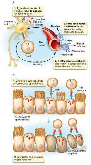
attached to proteins of other cells. This recognition triggers the release of granzymes and the production of cytokines, which cause apoptosis of the target cell.
Forms of DTH reactions include contact dermatitis (such as poison ivy rash) and allograft rejection. The antigens involved with contact dermatitis are usually small haptens that have to bind and modify normal host proteins to become antigenic (Fig. 24.33). In this case, the hapten-modified protein is processed, and fragments containing the bound hapten are presented on the surfaces of antigen-presenting cells. DTH reactions also play important roles in causing tissue damage during chronic infectious diseases, such as tuberculosis.
FIGURE 24.33 ■ Contact dermatitis. Pentadecylcatechols are chemicals present on the surface of poison ivy leaves. They are haptens
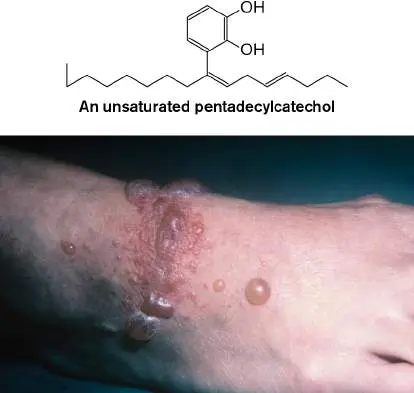
that bind to proteins in dermal cells, where they can activate T cells. The result of the ensuing delayed-type hypersensitivity is contact dermatitis.
DR. KEN GREER/VISUALS UNLIMITED
There are two other types of hypersensitivity, both of which involve antigen-antibody complexes. Type II hypersensitivity occurs when antibody binds to host cell-surface antigens. ABO blood group incompatibility is an example of type II hypersensitivity. Type III hypersensitivity happens when large complexes are formed between antibody and small, soluble antigens. The complexes can trigger complement activation that leads to rash formation.
Autoimmunity: An Inability to Recognize Self
The ability to distinguish between self antigens and foreign antigens is crucial to human survival; without this ability, the immune system would constantly attack us from within. Normally, the body develops tolerance to self; occasionally, however, an individual loses immune tolerance against some self antigens, and the body attacks its own tissues. The attack can involve antibodies or T cells and is called an autoimmune response. Autoimmune responses may or may not be associated with pathological changes (autoimmune disease). Almost 30% of humans will have an autoimmune antibody by age 65, but many will not exhibit disease. The mechanisms that lead to autoimmune disease are essentially hypersensitivity reactions.
Autoimmune disease results when an autoantibody or autoimmune lymphocyte (cytotoxic T cell) damages tissue components. Tissue damage develops when an autoantibody triggers antibody-dependent cell cytotoxicity (see Section 23.5). In this scenario, NK cell Fc receptors bind to autoantibodies affixed to a self antigen on host cells. The NK cell “thinks” the target cell is infected and kills it. In contrast, autoreactive cytotoxic T cells that escaped negative selection in the thymus can also directly kill host cells that express the cognate self antigen.
How might autoimmune antibodies be formed? One proposed mechanism starts with a self-reacting B cell occasionally escaping the negative selection process. A renegade autoreactive B cell is usually not a problem because the specific helper T cell needed to activate it was most likely deleted from the T-cell population. However, the B-cell receptor on that autoreactive B cell can take up and process a foreign antigen (from a pathogen, perhaps) that looks like a self antigen (Fig. 24.34, step 1). If the self-like antigen is processed and presented on a B cell–MHC II complex, nothing happens because the corresponding self-reactive helper T cell was deleted during negative selection in the thymus (step 2). However, the self-like foreign antigen taken into the self-reacting B cell (step 1) can also be linked to nonself epitopes. As a result, the nonself epitope can be processed and presented on one of the autoreactive B cell’s MHC molecules (step 3). Now, a helper T cell whose TCR recognizes and binds to the MHC– nonself epitope complex presented on the autoreactive B cell (step 4) will be tricked into activating the self-reacting B cell to become a plasma cell that secretes the autoantibody (step 5). Once made, the autoantibodies begin attacking whatever host tissues express the self antigen.
FIGURE 24.34 ■ Mimicry model for generating autoantibodies. A foreign antigen with multiple epitopes encounters an autoreactive B cell (step 1). An epitope that resembles a self-epitope can bind to the BCR with the other epitope(s) trailing. B cells are also antigen-presenting cells, so the BCR–foreign antigen complex enters the cell, and the various epitopes are processed and presented on surface MHC molecules. The self-like epitope–MHC complex cannot find T-cell help (step 2). The
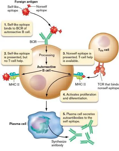
other foreign epitope–MHC complex can find T-cell help (step 3). This help activates proliferation and differentiation (step 4) of the autoreactive B cell, which becomes a plasma cell that secretes autoreactive antibody (step 5).
Rheumatic fever is a good example of an autoimmune disease caused by molecular mimicry. After a throat infection with some strains of Streptococcus pyogenes (“strep throat”), the patient’s heart may be attacked by autoantibodies to heart antigen. The cause appears to be an S. pyogenes surface protein called M protein. The M protein contains an epitope that resembles cardiac antigen, but the cardiac-like epitope is flanked by epitopes unrelated to the human host. The cardiac-like epitope can bind to the B-cell receptor of a cardiac self-reacting B cell and be taken up. The nonself epitope linked to the heart-like epitope will “piggyback” its way into the B cell. The nonself epitope is then processed and placed on a B-cell surface MHC II molecule. The TCR of T cells specific to the nonself epitope will bind to the MHC–nonself peptide complex and activate the B cell. But because the B cell is preprogrammed to make anti-cardiac antibody, the resultant plasma cells make autoantibodies to cardiac tissue. Cardiac tissue is damaged, and rheumatic fever results.
Other autoimmune diseases involve the production of autoantibodies that bind and block certain cell receptors. Graves’ disease (hyperthyroidism), for example, occurs when an autoantibody is made that binds to the thyroid-stimulating hormone (TSH) receptor. This receptor binding mimics actual TSH binding and continually stimulates the production of two hormones— namely, triiodothyronine (T3) and thyroxine (T4)—that increase metabolic rate. The thyroid is not destroyed but, in fact, enlarges to form a goiter. Examples of other important autoimmune diseases are listed in Table 24.7.
TABLE Examples of Autoimmune Diseases 24.7
Disease Autoantigen(s) Pathology Type II hypersensitivity—mediated by antibody to cell-surface antigens Acute Streptococcal M protein, Myocarditis, scarring of rheumatic cardiomyocytes heart valves fever Autoimmune Rh blood group Destruction of red blood hemolytic cells by complement, anemia phagocytosis Goodpasture’s Basement membrane Pulmonary hemorrhage, syndrome collagen glomerulonephritis Graves’ Thyroid-stimulating Antibody stimulation of disease hormone (TSH) T3, T4 production; receptor hyperthyroidism Myasthenia Acetylcholine receptor Interruption of electrical gravis transmission, progressive muscular weakness Type III hypersensitivity—mediated by antibody complexes with small soluble antigens (immune complex)
Systemic lupus DNA, histones, Arthritis, vasculitis, erythematos ribosomes glomerulonephritis us Type IV hypersensitivity—mediated by antigen-specific T cells Type 1 Pancreatic beta cell Beta cell destruction diabetes antigen Multiple Myelin protein Demyelination of axons sclerosis T-cell engineering: a way to fight autoimmune disease. Loss of self-tolerance leads to autoimmune disease. So, the ultimate goal of autoimmune therapy must be to restore self-tolerance. Because regulatory T cells are an important part of self-tolerance, scientists have been exploring ways to boost Treg numbers and alter their function. In one of the most promising approaches, called chimeric antigen receptor Treg cells (CAR-Treg), the patient undergoes adoptive transfer of Tregs whose TCR has been reengineered to direct the CAR-Treg cell to suppress specific aspects of innate and adaptive immunity. The basic strategy is to convert conventional T cells into Tregs by using retrovirus or lentivirus vectors to cotransfer the Treg-specific regulatory gene FoxP3 (needed to convert a T cell into a Treg) with a TCR gene that would confer antigen specificity on the Treg-like cell. TCR interaction with a cognate autoantigen would stimulate secretion of anti-inflammatory cytokines that could control T H 1-driven cytotoxic T cells, pro-inflammatory T H 17 cells, or T FH cells that promote autoantibody formation. Although these approaches have been shown to work only in mice, the prospect of producing tailor-made cellular therapies with disease-specific function could revolutionize the treatment of autoimmunity. Section 16.6 discusses more of the basics of CAR-T therapy.
This chapter strives to explain the complexity of the human immune system and its codependent relationship with the microbiome. That any one person’s immune system can recognize and respond to virtually any molecular structure and yet remain selectively “blind” to their own antigens is a stunning evolutionary feat crucial to human survival. Equally stunning is that we humans have evolved to depend on a diverse microbiome for good health. That’s not to say that some microbes, the pathogens, aren’t worrisome. In the next chapters we describe what defines a pathogen and the strategies that health care professionals use to identify, treat, and track these microscopic threats to our existence.
To Summarize
Allergens cause the host immune system to overrespond or react against self.
Type I hypersensitivity involves IgE antibodies bound to mast cells by the antibody Fc region. Binding of antigen to the mast cell– attached IgE causes mast cell degranulation. This type of hypersensitivity can occur within minutes of exposure.
Type IV hypersensitivity (delayed-type hypersensitivity) involves antigen-specific T cells. T H 1 cells release cytokines that activate macrophages and NK cells. T C cells can directly kill cells that present the antigen. Reaction is seen within a few days of exposure. Autoimmune disease is caused by the presence of lymphocytes that can react to self. These autoreactive lymphocytes escaped negative selection in the bone marrow or thymus.
Autoreactive B cells (B cells that make antibody directed against self epitopes) can be activated if their BCR takes up a self-mimic epitope linked to a nonself epitope and presents the nonself epitope on the surface MHC. T cells that recognize the nonself epitope can then activate the B cell, which secretes antibody against the self epitope. NK cells can target host cells decorated with those autoantibodies.
Cytotoxic T cells can produce autoimmune disease by killing host cells expressing the cognate self antigen.
Chimeric antigen receptor Treg cells can potentially control autoimmune diseases.
Glossary
allergen An antigen that causes an allergic hypersensitivity reaction. type I hypersensitivity Also called immediate hypersensitivity. An IgE-mediated allergic reaction that causes degranulation of mast cells within minutes of exposure to the antigen. The severe reaction known as anaphylaxis is triggered by type I hypersensitivity.
anaphylaxis A severe type I hypersensitivity reaction caused by chemically induced contraction of smooth muscles and dilation of capillaries.
degranulation The process whereby preformed chemical mediators in vesicles called granules are released to the outside of the cell.
edema Tissue swelling due to fluid accumulation.
late-phase anaphylaxis Anaphylaxis caused by leukotrienes that are released by eosinophils recruited by mast cells.
desensitization A clinical treatment to decrease allergic reactions by exposing patients to small doses of an allergen.
type IV hypersensitivity Also called delayed-type hypersensitivity (DTH). An immune response that develops 24–72 hours after exposure to an antigen that the immune system recognizes as foreign. The response is triggered by antigen-specific T cells. It is delayed because the T cells need time to proliferate after being activated by the allergen.
delayed-type hypersensitivity (DTH)
See type IV hypersensitivity .
type II hypersensitivity An immune response in which antibodies bind to the patient’s own cell-surface antigens or to foreign antigens adsorbed onto the patient’s cells. Antibody binding triggers cell-mediated cytotoxicity or activation of the complement cascade.
type III hypersensitivity Also called immune complex disease. An immune reaction triggered when IgG antibody binds to an excess of soluble foreign antigen in the blood. The immune complexes deposit in small blood vessels, where they interact with complement to initiate an inflammatory response. autoimmune response A pathology caused by lymphocytes that can react to self antigens. Fig. 24.22 FIGURE 24.22 ■ Summary of the activation of humoral and cell-mediated pathways. Left: Extracellular pathogens tend to activate humoral immunity (B cells). Right: Intracellular pathogens generally activate cell-mediated immunity by stimulating cytotoxic T cells (CD8 marker). The balance between cell-mediated and humoral immune responses to a given infection is regulated by the balance between the production of T H 1 (cell-mediated) versus other T H (antibody) helper T cells. This balance is influenced by whether the foreign antigen was made by intracellular pathogens (T H 1-favored) or extracellular pathogens (see Fig. 24.20). T H 1 cells will activate cytotoxic T cells (cell-mediated immunity; best for killing intracellular pathogens), while T FH cells will promote antibody production (humoral immunity; best for attacking extracellular pathogens). See text for more details.
Endnotes
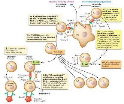
1. Note a: ADCC = antibody-dependent cell-mediated cytotoxicity; PMN = polymorphonuclear leukocyte. Return to reference a eResearch Activity 24
Can a Subset of Regulatory T Cells Prevent Allergies and Autoimmunity?
Follicular regulatory T cells (T FR) are a specific subclass of regulatory T cells found in secondary lymphoid tissues (lymph nodes, for instance). Among their functions, T FR cells somehow prevent excessive production of IgE antibodies associated with allergies, and they limit the emergence of autoantibodies that produce autoimmune diseases such as systemic lupus, rheumatoid arthritis, multiple sclerosis, type 1 diabetes, and others. Discovering how cells control these processes could suggest new ways to treat or prevent allergies and autoimmune disease. Carola G. Vinuesa (Fig. ERA 24.1A ), while at the Australian National University (she is currently at the Francis Crick Institute in London, England), and her colleagues in the United Kingdom and the United States recently identified one such mechanism. It involves a 143-amino-acid neuropeptide called neuritin.
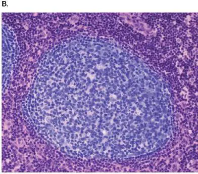
FIGURE ERA 24.1 ■ Lymph node germinal center. A. Carola G. Vinuesa, Francis Crick Institute in London, studies how our immune system produces long-lasting antibody responses. B. A lymph node germinal center where T cells and B cells interact to produce antibodies. C. Schematic model showing the cellular interactions at play in and around a germinal center. Dendritic cell (1) presents Ag to naive T cell. T cell differentiates into a T FH cell and enters germinal center. (2) The T FH cell helps GC cells (3) differentiate into plasma cells or memory B cells (4). T FR cells dampen GC B cell and T FH cell functions (5).
JAMIE KIDSTON
SCIENCE PHOTO LIBRARY/ALAMY STOCK PHOTO
Neuritin was originally discovered in an area of the brain called the hippocampus, a structure important for memory and learning (see eResearch Activity 23). This neuropeptide regulates synaptic plasticity (strength of a synaptic signal), controls axon and dendrite
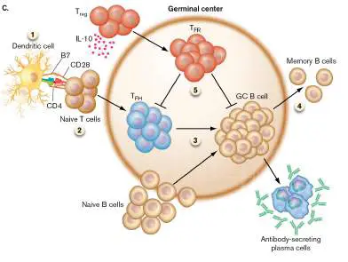
growth, and promotes the migration and survival of postmitotic neurons. Vinuesa and collaborators demonstrated that neuritin is not only produced in the brain but it is also made by T FR cells in lymph node germinal centers.
Germinal centers (GCs) are specialized structures inside lymph nodes that form where T follicular helper cells (T FH) and B cells interact to respond to foreign antigens (Fig. ERA 24.1B and C ). The result is the production of high-affinity antibodies against those antigens. However, GCs can also arise spontaneously in the absence of immunization or overt infection; such structures are called spontaneous GCs (Spt-GCs). In the absence of overt infection, it is possible that endogenous viruses and retroviruses in a host could elicit Spt-GC formation. In autoimmune-prone mice and in human patients with autoimmune disease, increased formation of Spt-GCs is thought to promote the development of pathogenic autoantibodies that cause autoimmune diseases and IgE antibodies that can lead to allergy.
The research team suspected that neuritin produced by T FR cells might be responsible for limiting Spt-GC formation. They focused on neuritin because the gene encoding this neuropeptide was highly expressed in T FR cells, more than in any other T-cell type in the mouse lymph node. Similar results were observed when cells isolated from human tonsil were stained for neuritin expression (Fig. ERA 24.2 ). All of the T FR -like cells and some Treg cells expressed neuritin protein, but barely any T FH cells did. To identify which cells in a lymphoid organ bind neuritin, suspensions of spleen cells from immunized mice and from the tonsils of human donors were stained with fluorescently labeled anti-neuritin antibody, and the stained cells were counted by flow cytometry (Fig. ERA 24.3A ). Germinal center B cells from mice (not shown) and humans (Fig. ERA 24.3B )
bound the highest amounts of neuritin, suggesting that GC B cells are the likely target. The authors also demonstrated that after entering B cells, neuritin phosphorylates a large suite of proteins.
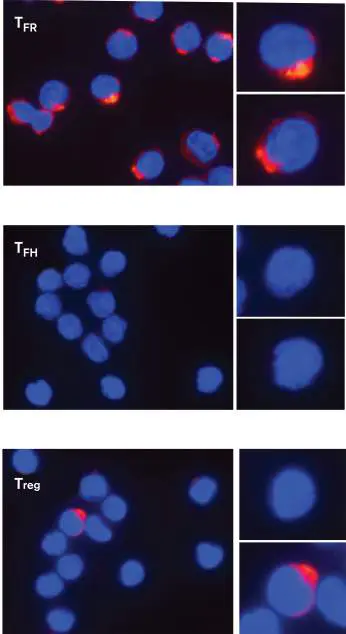
FIGURE ERA 24.2 ■ T FR cells highly express neuritin. T FR −like, T FH, and Treg cells were purified by FACS analysis from human tonsils (a secondary lymphoid tissue) and stained for DNA (blue) and with fluorescently labeled anti-neuritin antibody (red).
P. GONZALEZ-FIGUEROA ET AL. 2021. CELL 184 :1775–1789
FIGURE ERA 24.3 ■ Neuritin preferentially binds to B cells. A. Flow cytometry plots used to separate cells from the tonsils of human donors. Left plot shows the separation of B cells (CD19 pos CD4 neg) from T cells (CD19 neg CD4 pos). Right plot shows further FACS separation of the B cells into memory B cells (B mem; orange gate; CD27 pos CD38 neg), germinal center B cells (GCB; green gate; CD27 pos CD38 pos), and naive B cells (B naive; red gate; CD27 neg CD38 neg). Cell surface protein CD19 is a general surface marker for B cells; CD4 is a general marker for T cells; CD27 is a marker for B cells; CD38 is a marker for activated B cells. B. Quantitation (mean fluorescence intensity, gMFI) of fluorescently labeled neuritin binding to cells identified in panel A. Dotted lines represent results from the same human donor. FMO = Fluorescence minus one control. FMO controls determine the cutoff for background fluorescence.
P. GONZALEZ-FIGUEROA ET AL. 2021. CELL 184 :1775–1789
Next, the authors explored the effects of neuritin on human B cells, extracted from the tonsils of human donors. Naive B cells
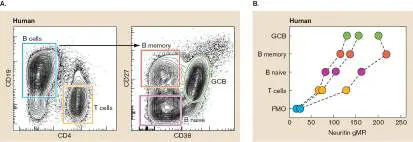
residing outside of a germinal center, in addition to germinal center B cells and memory B cells, were purified and cultured for 5 days with or without neuritin. The culturing was done in the presence of anti-CD40 and interleukin IL-21, which promote B-cell differentiation into plasma cells; antigen and T cells were not needed. Neuritin decreased plasma cell formation from GC B cells by 64% (Fig. ERA 24.4A ). The numbers of GC B cells did not change, and all cells remained viable during the experiment. Neuritin treatment also down-regulated the transcription factor BLIMP-1, required for plasma cell formation (Fig. ERA 24.4B ). These data suggest that neuritin can limit spontaneous germinal center formation.
FIGURE ERA 24.4 ■ Effect of neuritin on plasma cell formation and IgE production from GC B cells. A. GC B cells were cultured 5 days with (red) or without (purple) neuritin (2 mg/mL). Plasma cell formation was quantified by FACS (scored as having markers CD27 hi CD38 hi). B. Quantitation of BLIMP-1 by
immunofluorescence. C. Quantitation of secreted IgE and IgG from GC B cell cultured with and without neuritin. Nil = no neuritin. Asterisk indicates level of significance: * (0.05), ** (0.01), *** (0.001), **** (0.0001).
As noted earlier, a deficiency in T FR cells, the source of neuritin, leads to elevated IgE secretion and anaphylaxis. To test whether neuritin would specifically lower IgE secretion, the research team cultured human GC B cells under conditions known to promote IgE production, once again without added antigen. The addition of neuritin led to a dose-dependent reduction in IgE secretion but had no effect on IgG output (Fig. ERA 24.4C ). The effect on IgE production appeared to be the result of neuritin limiting B-cell class switching to the epsilon heavy chain.
Research then turned to potential neuritin effects on autoantibody production. A mouse strain was constructed whose T FR cells had the Nrn1 gene encoding neuritin removed. As shown in Figure ERA 24.5 (lower panels), serum from the neuritin-deficient mice contained spontaneous tissue-specific autoantibodies capable of binding to pancreas, stomach, and salivary gland tissue. These results and other data suggest that neuritin produced by T FR cells can prevent the emergence of self-reactive GC-derived PCs and autoantibodies. FIGURE ERA 24.5 ■ Neuritin limits autoantibody production. Tissues were removed from Rag −/− mice that lack functional B cells and T cells. Thin sections of each tissue were incubated with mouse plasma taken from control and neuritin-deficient mice, followed by fluorescently tagged donkey anti-mouse IgG antibody. Green fluorescence indicates tissues that were coated with IgG autoantibodies.
P. GONZALEZ-FIGUEROA ET AL. 2021. CELL 184 :1775–1789
The article concludes that neuritin produced by T FR cells is an immune system effector molecule that regulates B-cell responses by: (1) repressing germinal center B-cell differentiation into plasma cells, (2) preventing the appearance of self-reactive PCs in germinal centers, (3) limiting production of autoantibodies to tissue-specific antigens, and (4) restraining IgE switching to prevent IgE-mediated anaphylaxis. In sum, neuritin is an endogenous mediator of B-cell tolerance to self. It serves as a key repressor of autoantibodies against multiple self antigens and of allergenic IgE. This property, along with its ability to limit PC differentiation, makes neuritin an attractive therapeutic target for B cell–mediated autoimmune
diseases and IgE-mediated allergies. This remarkable study identifying a new immune modulator (neuritin) that prevents the appearance of self-reacting plasma cells would not have been possible without funding from the National Health and Medical Research Council of Australia.
Further Exploration
From the data in this paper, propose approaches you might use to control allergies or autoimmune disease in a patient with these diseases.
Source: Gonzalez-Figueroa, Paula, Jonathan A. Roco, Ilenia Papa, Lorena
Núnez Villacís, Maurice Stanley, et al. 2021. Follicular regulatory T cells
produce neuritin to regulate B cells. Cell 184 :1775–1789.
https://doi.org/10.1016/j.cell.2021.02.027.
CHAPTER REVIEW
Review Questions
1. Define “antigen,” “epitope,” “hapten,” and “antigenic determinant.”
2. What is the basic difference between humoral immunity and cellular immunity?
3. Why are proteins better immunogens than nucleic acids are?
4. What makes IgA antibody different from IgG?
5. Explain isotypic, allotypic, and idiotypic differences in antibodies.
6. How is IgE involved in allergic hypersensitivity? 7. Discuss differences in the primary and secondary antibody responses.
8. Outline the basic steps that turn a B cell into a plasma cell.
9. Describe isotype switching and how antibody diversity is achieved.
10. Differentiate between the signals needed to activate helper T cells and the signals needed to activate cytotoxic T cells.
11. Discuss the differences between transplant rejections of nucleated cells and the rejection of blood cells. 12. How do superantigens activate T cells?
13. Discuss the differences between the alternative and classical pathways of complement activation.
14. How does the host prevent membrane attack complexes from being formed in host cells?
15. Describe the development of type I and type IV hypersensitivity reactions.
16. How does a B cell programmed to make an antibody against self become activated in the absence of specific T-cell help?
17. Explain key features of the gut immune system. 18. Discuss the types of immune responses generated by different types of vaccines.
19. Why are live vaccines better than dead or subunit vaccines?
20. Describe how T-cell engineering might be used to treat autoimmune diseases.
Thought Questions
1. Cytotoxic T cells lyse the membranes of host cells carrying viruses or bacteria. Why doesn’t this lysis facilitate the spread of those organisms rather than help clear the infection?
2. Immunity raised by the poliovirus vaccine will prevent subsequent infection by poliovirus, but someone with a Staphylococcus aureus infection (and the attendant immune response) can be reinfected many times by S. aureus. Why does the immune system work so well against infection by some pathogens (for example, poliovirus) but not others (for example, S. aureus)? 3. The red blood cells of an individual are coated with blood group antigens: either A or B, both A and B (blood type AB), or neither A nor B (blood type O). People of blood type A have circulating anti-B antibodies. People of type B have circulating anti-A antibodies. People of type AB have neither anti-A nor anti-B antibodies. So, why are people of type O considered “universal donors, ” meaning that they can donate their red blood cells to type-O, type-A, type-B, or type-AB individuals? And which blood type corresponds to people who are considered universal recipients?
4. IgM is the first antibody produced during a primary immune response. Of the five different types of antibodies produced, what is the advantage of having IgM be secreted first?
5. Some pathogens, such as rubella virus, can cross the placental barrier from mother to fetus and cause a dangerous infection. Which antibody isotype against rubella virus would you look for in a newborn to diagnose congenital rubella syndrome: IgG or IgM?
6. Double positive CD4 + CD8 + T cells are present in urological cancers. Sketch and label a graph of flow cytometry data that shows where CD4 + T cells, CD8 + positive T cells, and CD4 + CD8 + double positive T cells would be located.
7. How might you design a chimeric antigen receptor (CAR-T) strategy involving cytotoxic T cells to treat a patient with Graves’ disease (hyperthyroidism)? Graves’ disease is caused by an autoantibody that continually activates thyroid-stimulating hormone receptor.
Key Terms
adaptive immunity (1000)
addressin (1034)
affinity maturation (1017)
allergen (1039)
allotype (1008)
anaphylaxis (1039)
antibody (1001)
antigen (1000)
antigen-presenting cell (APC) (1001, 1019) antigenic determinant (1000)
autoimmune response (1044)
B cell (1001)
B-cell receptor (BCR) (1012)
capping (1012)
cell-mediated (cellular) immunity (1001) class I MHC molecule (1019)
class II MHC molecule (1019)
class switching (1011)
classical complement pathway (1030) clonal selection (1011)
constant region (1008)
cytokine (1016)
cytokine storm (1028)
cytotoxic T cell (T C cell) (1001, 1018) decay-accelerating factor (1032) degranulation (1040)
delayed-type hypersensitivity (DTH) (1043) desensitization (1043)
edema (1042)
epitope (1000)
factor H (1031)
Fc region (1008)
gene switching (1013)
granzyme (1025)
hapten (1004)
heavy chain (1007)
helper T cell (T H cell) (1001, 1016) humoral immunity (antibody-dependent immunity) (1001) idiotype (1008)
IgA (1010)
IgD (1010)
IgE (1010)
IgG (1009)
IgM (1009)
immediate hypersensitivity (1039) immunogen (1000)
immunogenicity (antigenicity) (1004) immunoglobulin (1006)
immunological specificity (antigenic specificity) (1004) immunoprecipitation (1008)
innate lymphoid cell (ILC) (1033) intraepithelial lymphocyte (IEL) (1032) isotype (1008)
isotype switching (1011)
late-phase anaphylaxis (1042)
light chain (1007)
major histocompatibility complex (MHC) (1001, 1019) memory B cell (1011)
mucosal immunity (1034)
negative selection (1022)
opsonize (1009)
perforin (1025)
plasma cell (1001, 1011)
positive selection (1022)
primary antibody response (1010) recombination signal sequence (RSS) (1013) regulatory T cell (Treg) (1022) secondary antibody response (1011) serum (1010)
superantigen (1028)
switch region (1016)
T-cell receptor (TCR) (1021)
T H 17 cell (1025)
tumor necrosis factor (TNF) (1028) type I hypersensitivity (1039)
type II hypersensitivity (1043) type III hypersensitivity (1043) type IV hypersensitivity (1043) vaccination (1005)
variable region (1008)
Recommended Reading
Artis, David, and Hergen Spits. 2015. The biology of innate lymphoid cells. Nature 517 :293–301.
Attaf, Meriem, Mateusz Legut, David K. Cole, and Andrew K. Sewell. 2015. The T cell antigen receptor: The Swiss army knife of the immune system. Clinical and Experimental Immunology 181 :1–18.
Becattini, Simone, Matthew T. Sorbara, Sohn G. Kim, Eric L. Littmann, Qiwen Dong, et al . 2021. Rapid transcriptional and metabolic adaptation of intestinal microbes to host immune activation. Cell Host and Microbe 29 :378–393.
https://doi.org/10.1016/j.chom.2021.01.003.
Cyster, Jason G. 2010. B cell follicles and antigen encounters of the third kind. Nature Immunology 11 :989–996.
Donaldson, Gregory P., Mark S. Ladinsky, Kristie B. Yu, Jon G. Sanders, Bryan B. Yoo, et al. 2018. Gut microbiota utilize immunoglobulin A for mucosal colonization. Science 360 :795– 800.
Fajgenbaum, David C., and Carl H. June. 2020. Cytokine storm. New England Journal of Medicine 383 :2255–2273.
https://doi.org/10.1056/NEJMra2026131.
Garn, Holger, Daniel Piotr Potaczek, and Petra Ina Pfefferle. 2021. The hygiene hypothesis and new perspectives—current challenges meeting an old postulate. Frontiers in Immunology 12: 637087. https://doi.org/10.3389/fimmu.2021.637087. Hale, J. Scott, and Rafi Ahmed. 2015. Memory T follicular helper CD4 T cells. Frontiers in Immunology 6 :16.
Heesters, Balthasar A., Cees E. van der Poel, Abhishek Das, and Michael C. Carroll. 2016. Antigen presentation to B cells. Trends in Immunology 37 :844–854.
https://doi.org/10.1016/j.it.2016.10.003.
Hevia, Arancha, Susana Delgado, Borja Sanchez, and Abelardo Margolles. 2015. Molecular players involved in the interaction between beneficial bacteria and the immune system. Frontiers in Microbiology 6 :1285.
Iwasaki, Aikiko, and Rusian Medzhitov. 2015. Control of adaptive immunity by the innate immune system. Nature Immunology 16 :343–353.
Marciano, Beatriz E., and Steven M. Holland . 2017. Primary immunodeficiency diseases: Current and emerging therapeutics. Frontiers in Immunology 8 :937.
https://doi.org/10.3389/fimmu.2017.00937.
McCoy, Kathy D., Regula Burkhard, and Markus B. Geuking. 2019. The microbiome and immune memory formation.
Immunology and Cell Biology 97.
https://doi.org/10.1111/imcb.12273.
McGovern, Jenny L., Graham P. Wright, and Hans J. Stauss. 2017. Engineering specificity and function of therapeutic regulatory T cells. Frontiers in Immunology 8 :1517.
https://doi.org/10.3389/fimmu.2017.01517.
Rappuolia, Rino, Ennio De Gregorio, Giuseppe Del Giudice, Sanjay Phogat, Simone Pecetta, et al. 2021. Vaccinology in the post−COVID-19 era. Proceedings of the National Academy of Science 118 :e2020368118.
https://doi.org/10.1073/pnas.2020368118.
Starr, Tyler N., Nadine Czudnochowski, Zhuoming Liu, Fabrizia Zatta, Young-Jun Park, et al. 2021. SARS-CoV-2 RBD antibodies that maximize breadth and resistance to escape. Nature 597 :97–102. https://doi.org/10.1038/s41586-021-03807-6.
Teymournejad, Omid, and Christopher P. Montgomery .
2021. Evasion of immunological memory by S. aureus infection: Implications for vaccine design. Frontiers in Immunology 12 : 633672. https://doi.org/10.3389/fimmu.2021.633672.
Ugolini, Matteo, Jenny Gerhard, Sanne Burkert, Kristoffer Jarlov Jensen, Philipp Georg, et al. 2018. Recognition of microbial viability via TLR8 drives TFH cell differentiation and vaccine responses. Nature Immunology 19 :386–396.
Weisberg, Stuart P., Basak B. Ural, and Donna L. Farber . 2021. Tissue-specific immunity for a changing world. Cell 184:1517–1529.
Wieczorek, Marek, Esam T. Abualrous, Jana Sticht, Miguel Álvaro-Benito, Sebastian Stolzenberg, et al. 2017. Major histocompatibility complex (MHC) class I and MHC class II proteins: Conformational plasticity in antigen presentation. Frontiers in Immunology 8 :292.
https://doi.org/10.3389/fimmu.2017.00292.
Glossary
adaptive immunity Immune responses activated by a specific antigen and mediated by B cells and T cells.
addressin A tissue-specific protein that is selectively present on vascular endothelial cells in different tissues.
affinity maturation The process by which the antigen-binding site of an antibody gains increased affinity for its target antigen or epitope. allergen An antigen that causes an allergic hypersensitivity reaction. allotype An amino acid difference in the antibody constant region that distinguishes different individuals within a species. anaphylaxis A severe type I hypersensitivity reaction caused by chemically induced contraction of smooth muscles and dilation of capillaries.
antibody A host defense protein produced by B cells in response to a specific antigenic determinant. Antibodies, a type of immunoglobulin, bind to their corresponding antigenic determinants.
antigen A compound, recognized as foreign by the cell, that elicits an adaptive immune response. See also immunogen .
antigen-presenting cell (APC)
An immune cell that can process antigens into antigenic determinants and display those determinants on the cell surface for recognition by other immune cells.
antigenic determinant Also called epitope. A small segment of an antigen that is capable of eliciting an immune response. An antigen can have many different antigenic determinants.
autoimmune response A pathology caused by lymphocytes that can react to self antigens.
B cell An adaptive immune cell, developed in bone marrow tissue, that can give rise to antibody-producing cells.
B-cell receptor (BCR)
A B-cell membrane protein complex containing an antibody in association with the Igα and Igβ immunoglobulins.
capping The clustering of B-cell receptor molecules on the surface of B cells after binding antigens or epitopes.
cell-mediated (cellular) immunity A type of adaptive immunity employing mainly T-cell lymphocytes.
class I MHC molecule A membrane surface protein on all nucleated cells of the human body (absent from red blood cells and platelets) that present intracellular foreign antigen epitopes to cytotoxic T cells.
class II MHC molecule A membrane surface protein on antigen-presenting cells (dendritic cells, macrophages) and lymphocytes that present phagocytosed extracellular foreign antigen epitopes to helper T cells.
class switching See isotype switching .
classical complement pathway An antibody-mediated pathway for complement activation. clonal selection The rapid proliferation of a subset of B cells during the primary or secondary antibody response.
constant region The region of an antibody that defines the class of a heavy chain or a light chain.
cytokine A small, secreted host protein that binds to receptors on various endothelial and immune system cells, regulating the cells’ responses.
cytokine storm An exaggerated inflammatory immune response produced during certain infections and autoimmune diseases that triggers a rapid, unbalanced release of cytokines.
cytotoxic T cell (T C cell)
A T cell that expresses CD8 on its cell surface and can secrete toxic proteins such as perforin and granzymes.
decay-accelerating factor A host cell membrane protein that stimulates the decay of complement factors and prevents their deposition at the cell surface.
degranulation The process whereby preformed chemical mediators in vesicles called granules are released to the outside of the cell. delayed-type hypersensitivity (DTH)
See type IV hypersensitivity .
desensitization A clinical treatment to decrease allergic reactions by exposing patients to small doses of an allergen.
edema Tissue swelling due to fluid accumulation.
epitope See antigenic determinant .
factor H A normal serum protein that prevents the inadvertent activation of complement in the absence of infection. Fc region The region of an antibody that binds to specific receptors on host cells in an antigen-independent manner. It is found in the carboxy-terminal “tail” region of the antibody.
gene switching Switching between two (out of five) different classes of immunoglobulin genes (e.g., from IgM to IgG) during B-cell development.
granzyme An enzyme, secreted by cytotoxic T cells, that damages target cells.
hapten A small compound that must be conjugated to a larger carrier antigen in order to elicit production of an antibody that binds to it.
heavy chain The larger of the two protein types that make up an antibody. Each antibody contains two heavy chains and two light chains. helper T cell (T H cell)
A T cell that expresses CD4 on its cell surface and secretes cytokines that modulate B-cell isotype, or class, switching. humoral immunity (antibody-dependent immunity)
A type of adaptive immunity mediated by antibodies.
idiotype An amino acid difference in the antigen-binding site (N terminus of heavy or light chains) that distinguishes different antibodies within an individual.
IgA An antibody isotype that contains the alpha heavy chain. It can be secreted and is found in tears, saliva, breast milk, and so on.
IgD An antibody isotype that contains the delta heavy chain. It is found on B-cell membranes.
IgE An antibody isotype that contains the epsilon heavy chain. It is involved in degranulation of mast cells.
IgG An antibody isotype that contains the gamma heavy chain. Produced by plasma cells, it is found in serum and is the predominant class of circulating antibodies for adaptive immunity.
IgM The first antibody isotype detected during the early stages of an immune response. It contains the mu heavy chain and is found as a pentamer in serum.
immediate hypersensitivity See type I hypersensitivity .
immunogen An antigen that, by itself, can elicit antibody production. immunogenicity (antigenicity)
A measure of the effectiveness of an antigen in eliciting an immune response.
immunoglobulin A member of a family of proteins that contain a 110-amino-acid domain with an internal disulfide bond. Members include antibodies and major histocompatibility proteins.
immunological specificity (antigenic specificity)
The ability of antibodies produced in response to a particular epitope to bind that epitope almost exclusively. Antibodies made to one epitope bind only weakly, if at all, to other epitopes.
immunoprecipitation The antibody-mediated cross-linking of antigens to form large, insoluble complexes. It is used in research labs and is normally seen only in vitro.
innate lymphoid cell (ILC)
A lymphocyte-like cell in the intestinal lamina propria that lacks B-or T-cell receptors but secretes pro-inflammatory cytokines. intraepithelial lymphocyte (IEL)
A lymphocyte embedded among epithelial cells that line the intestine.
isotype An antibody class within a species that is defined by the structure of the antibody’s heavy chain. IgG, IgA, IgD, and IgE are examples of isotypes. An isotype from one species contains species-specific amino acid sequences that are present in the heavy chain of all members of that species.
isotype switching Also called class switching. A change in the predominant antibody type produced by a cell.
late-phase anaphylaxis Anaphylaxis caused by leukotrienes that are released by eosinophils recruited by mast cells.
light chain The smaller of the two protein types that make up an antibody. Each antibody contains two heavy chains and two light chains. major histocompatibility complex (MHC)
Transmembrane cell proteins important for recognizing self and for presenting foreign antigens to the adaptive immune system. memory B cell A long-lived type of lymphocyte preprogrammed to produce a specific antibody. After encountering their activating antigen, memory B cells differentiate into antibody-producing plasma cells.
mucosal immunity The portion of the innate and adaptive immune systems that protects the mucosa from microbial invasion.
negative selection In immunology, the destruction of T cells bearing T-cell receptors that bind strongly to self MHC proteins displayed on thymus epithelial cells.
opsonize To bind IgG antibodies to microbes in order to enhance microbial phagocytosis by host immune cells.
perforin A cytotoxic protein, secreted by T cells, that forms pores in target cell membranes.
plasma cell A short-lived antibody-producing cell.
positive selection In immunology, the survival of T cells bearing T-cell receptors that don’t recognize self MHC proteins displayed on thymus epithelial cells.
primary antibody response The production of antibodies upon first exposure to a particular antigen. B cells become activated and differentiate into plasma cells and memory B cells.
recombination signal sequence (RSS)
A DNA region downstream of antibody heavy-and light-chain genes that allows recombination between widely separated gene segments.
regulatory T cell (Treg)
A T cell that regulates the activity of another T cell, usually by suppressing its activity.
secondary antibody response A memory B cell–mediated rapid increase in the production of antibodies in response to a repeat exposure to a particular antigen.
serum The noncellular, liquid component of the blood.
superantigen A molecule that directly stimulates T cells without undergoing antigen-presenting-cell processing and surface presentation. switch region A repeating DNA sequence interspersed between antibody constant-region genes that serves as a recombination site during isotype, or class, switching.
T-cell receptor (TCR)
A surface receptor on T cells that binds MHC-bound antigen on antigen-presenting cells.
T H 17 cell A class of helper T cell that secretes the inflammatory cytokine IL-17.
tumor necrosis factor (TNF)
A cytokine released by several cell types (e.g., macrophages) in response to cell damage.
type I hypersensitivity Also called immediate hypersensitivity. An IgE-mediated allergic reaction that causes degranulation of mast cells within minutes of exposure to the antigen. The severe reaction known as anaphylaxis is triggered by type I hypersensitivity. type II hypersensitivity An immune response in which antibodies bind to the patient’s own cell-surface antigens or to foreign antigens adsorbed onto the patient’s cells. Antibody binding triggers cell-mediated cytotoxicity or activation of the complement cascade. type III hypersensitivity Also called immune complex disease. An immune reaction triggered when IgG antibody binds to an excess of soluble foreign antigen in the blood. The immune complexes deposit in small blood vessels, where they interact with complement to initiate an inflammatory response.
type IV hypersensitivity Also called delayed-type hypersensitivity (DTH). An immune response that develops 24–72 hours after exposure to an antigen that the immune system recognizes as foreign. The response is triggered by antigen-specific T cells. It is delayed because the T cells need time to proliferate after being activated by the allergen.
vaccination Exposure of an individual to a weakened version of a microbe or a microbial antigen to provoke immunity and prevent development of disease upon reexposure.
variable region The amino-terminal portions of antibody light and heavy chains that confer specificity to antigen binding and define the antibody idiotype.