Chapter introduction
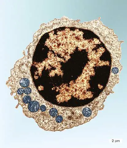
A eukaryotic cell. This eukaryotic cell, a lymphocyte white blood cell, shows numerous major organelles, such as a large nucleus (black) and multiple mitochondria (blue). GopalMurti/Visuals Unlimited
Appendix Sections
A2.1 The Cell Membrane A2.2 The Nucleus and Mitosis A2.3 Problems Faced by Large Cells A2.4 The Endomembrane System A2.5 The Cytoskeleton A2.6 Mitochondria and Chloroplasts Here in eAppendix 2 we present a review of cell biology principles that are generally covered in an introductory biology class. Cell biology encompasses fundamental principles of cell structure and function. All cells are enclosed by a cell membrane and need to regulate the transport of materials across the membrane. This appendix describes the cell structures that are found across all three domains of life and then focuses on those that are unique to the eukaryotic domain—specifically, organelles and the cytoskeleton. All eukaryotic cells contain a nucleus, an organelle that houses the DNA. We describe the structure of the nucleus and the processes of chromosome segregation through mitosis and meiosis. We then explore the endomembrane system of organelles that partition the cell into functional compartments, enabling eukaryotic cells to maintain larger sizes than most prokaryotic cells can. Eukaryotic cell structure includes the cytoskeleton, a collection of proteins that provide cell architecture and mediate cell movement. Finally, we discuss mitochondria and chloroplasts, the energy-producing organelles of eukaryotic cells.
A2.1 The Cell Membranenot assigned
The cell is the basic unit of life. All organisms consist of either a single cell or a collection of cells. Every cell can potentially perform a common set of tasks, including replication, catalysis, and regulation. The structures that enable these functions are found in all cells and include the cell membrane, DNA, and ribosomes (Fig. A2.1).
Although the detailed organization of these structures differs in cells from the three domains (Bacteria, Archaea, and Eukarya; see Table A2.1), the fundamental structure and function of the cell membrane, DNA, and ribosomes are the same in all cells.
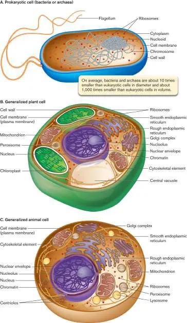
FIGURE A2.1 ■ The prokaryotic cell and the eukaryotic cell. A. The prokaryotic cell typically contains a single compartment, and its DNA is organized in the nucleoid region. B, C. Eukaryotic cells are typically much larger than prokaryotic cells and contain organelles.
TABLE Comparison of Cell Structures in the
A2.1 Three Domains
Feature Bacteria Archaea Eukarya Genome Usually Usually circular DNA Linear DNA circular Usually one Multiple DNA chromosome chromosom Usually one A few introns es, in pairs chromosom Most genes e have Usually lack introns introns Location of Nucleoid Nucleoid region in Contained DNA region in cytoplasm within cytoplasm membrane-enclosed nucleus Cell Straight-chain Branched-chain fatty Straight-chain membran fatty acids acids ether-linked fatty acids e ester-to glycerol ester-linked linked to to glycerol glycerol TABLE Comparison of Cell Structures in the
A2.1 Three Domains
Cell wall Usually If present, composed If present, present, of proteins or composed composed pseudopeptidoglyc of cellulose of an (algae) or peptidoglyc chitin an (fungi)
Internal May have Uncommon Extensive membran energy-membranou es transducing s organelles lamellae All cells are enclosed by a cell membrane (sometimes called the plasma membrane or cytoplasmic membrane), which maintains an internal environment distinct from the external environment. The aqueous fluid inside the membrane is called cytoplasm (or cytosol). Major functions of the cell membrane include the regulated transport of substances into and out of the cell and the reception of signals from the external environment. Membranes are also critical for energy production.
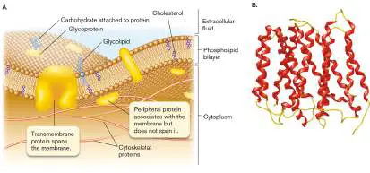
As shown in Figure A2.2, membranes consist mainly of lipids and proteins, but they also contain some carbohydrates found in hybrid structures such as glycolipids and glycoproteins (sugars joined to lipids or to proteins, respectively). Although the membrane contains a greater number of lipid molecules than protein molecules, proteins are large and contribute about half the mass of the membrane. The number and nature of proteins within a membrane depend on the membrane under consideration. For example, the inner membrane of the mitochondrion contains a rich array of proteins involved in energy production.
Membrane proteins can be classified by how they interact with membranes (Fig. A2.2A). Transmembrane proteins (integral proteins) span the bilayer. Transmembrane proteins are amphipathic, meaning they have both hydrophobic and hydrophilic portions; the hydrophilic portions face the cytoplasm and the extracellular environment, and the hydrophobic components span the membrane. The transmembrane domains are often alpha helices containing 15– 20 amino acid residues (Fig. A2.2B ). The portions of the protein that are intracellular, in the membrane, and extracellular depend on the protein and can be quite different for different proteins. Peripheral membrane proteins are associated with the cell membrane through noncovalent bonds but are not directly inserted into the bilayer. FIGURE A2.2 ■ The cell membrane. A. A cutaway view of the cell membrane. B. Ribbon diagram from X-ray-crystallographic data of the Escherichia coli EmrD protein, a multidrug transporter. Red portions are transmembrane alpha helices; yellow portions are intracellular and extracellular loops. (PDB code: 2GFP) Source: Y. Yin et al. 2006. Science 312 :741.
Membranes Are Composed of Lipids
The predominant lipids in membranes are phospholipids.
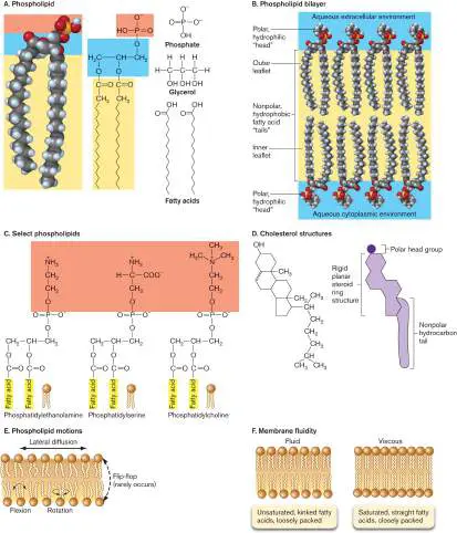
Phospholipids (Fig. A2.3A) consist of a core of glycerol, to which two fatty acids and a modified phosphate group are attached via ester linkages. Unlike eukaryotes and bacteria, archaea have phospholipids with ether linkages.
Phospholipids are amphipathic: the fatty acid hydrocarbon tails are hydrophobic, and the phosphate head group is hydrophilic.
Amphipathic lipids are most stable in water when the hydrophilic portions interact with water and the hydrophobic portions cluster together away from water. One way phospholipids can achieve stability is by forming a bilayer. The cell membrane is a phospholipid bilayer, two layers of phospholipids whose hydrocarbon fatty acid tails face the interior of the bilayer and whose charged phospholipid head groups face the aqueous cytoplasm and extracellular environment (Fig. A2.3B ). The phospholipid layer in contact with the cytoplasm is called the inner leaflet, and the layer in contact with the environment is called the outer leaflet. Cells contain a number of different phospholipids that vary in how the phosphate head group is modified (Fig. A2.3C ). Phospholipids also differ in the length and saturation of the fatty acid chains (discussed in eAppendix 1, Section A1.6).
In addition to phospholipids, eukaryotic membranes contain a variable amount of the steroid cholesterol (Fig. A2.3D ). Like phospholipids, cholesterol is amphipathic, with a hydrophilic hydroxyl group and hydrophobic hydrocarbon rings and tail. In membranes, cholesterol is oriented so that the hydrophilic hydroxyl group interacts with the phosphate head groups of phospholipids, while the hydrophobic rings and tail of cholesterol interact with the phospholipid hydrocarbon tails. The amount of cholesterol present in membranes varies among cells and also among organelles within a cell. Bacterial membranes do not contain cholesterol but do contain molecules of a similar form called hopanoids.
FIGURE A2.3 ■ Phospholipids, cholesterol, the lipid bilayer, and membrane fluidity. A. Saturated phospholipid. B. Orientation of phospholipids in the bilayer. C. Some phospholipids present in cell membranes. D. Structural formula and schematic drawing of cholesterol. E. Motions of phospholipids in membrane bilayers. F. The ratio of saturated and unsaturated fatty acids in the phospholipids affects membrane fluidity.
Movement of Membrane Lipids and Proteins
Membranes are not static structures; rather, many membrane lipids and proteins can move rapidly. The fluid mosaic model of membranes states that membrane components are free to diffuse in the plane of the membrane. Some membrane proteins are restricted to specific regions of the membrane by interactions with cytoskeletal proteins (discussed in Section A2.5).
Although many phospholipids and membrane proteins can move laterally within a leaflet, they do not “flip-flop” from one leaflet of the bilayer to the other (Fig. A2.3E ). Flip-flop of phospholipids is rare, owing to the highly unfavorable interactions of charged head groups moving through the hydrophobic interior of the membrane. Thus, the inner and outer leaflets of the membrane may be made up of different phospholipids. Such phospholipid asymmetry is important for the correct functioning of membrane proteins, which may work best when surrounded by particular phospholipids. Glycolipids and glycoproteins also contribute to membrane asymmetry, because the carbohydrate moieties always face the extracellular environment.
Membrane “fluidity”—the movement of membrane phospholipids within the plane of the membrane—is important for proper membrane function. For example, transport across the membrane is affected by membrane fluidity. Decreased fluidity is associated with decreased transport rates. Because a drop in temperature decreases fluidity, low temperatures may slow transport processes across the membrane. The composition of the membrane, especially the types of phospholipids present, can have a dramatic effect on membrane fluidity. For example, saturated fatty acids decrease membrane fluidity because the linear hydrocarbon tails pack together well. In contrast, unsaturated fatty acids have kinks in the hydrocarbon chains that limit packing and increase fluidity (Fig. A2.3F ). The length of the fatty acid chains also affects fluidity. Phospholipids with longer hydrocarbon chains have increased hydrophobic interactions with neighboring lipids, and thus decreased membrane fluidity. Organisms can alter membrane fluidity in response to temperature stress by changing the length and degree of saturation of fatty acids present in membrane phospholipids. For example, as environmental temperatures drop, both eukaryotes and prokaryotes maintain membrane fluidity by replacing long-chain fatty acids with shorter chains and increasing the percentage of unsaturated fatty acids in their membranes.
Cholesterol also influences membrane fluidity. The effects of cholesterol on membrane fluidity are complicated and depend on factors such as the ratio of saturated to unsaturated fatty acids in the membrane. Cholesterol may prevent packing of saturated fatty acids, thus increasing fluidity. In membranes with unsaturated fatty acids, cholesterol may fill in the spaces between adjacent phospholipids, stabilizing them and decreasing fluidity. In this case, cholesterol can decrease the permeability of the membrane to hydrophobic substances by packing between the hydrocarbon chains and preventing substances from slipping through.
Transport across Membranes
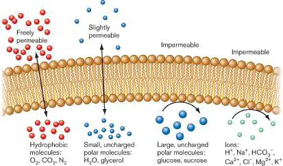
The major functions of membranes (such as containing cytoplasmic components, regulating which substances enter and leave cells and organelles, and producing energy) depend on the semipermeable nature of membranes. Semipermeable (also called selectively permeable) membranes are permeable to some substances but not to others. In general, the cell membrane is permeable to hydrophobic molecules and impermeable to charged molecules (Fig. A2.4). Diffusion across the membrane also depends on the size of the molecule. The membrane is freely permeable to small, nonpolar molecules such as O 2. Larger nonpolar molecules can also diffuse across the membrane, albeit more slowly. Molecules that are polar but small (such as ethanol and water) can also diffuse across the membrane. The membrane is impermeable to large, polar molecules such as glucose and to charged molecules, regardless of their size. The impermeability of the membrane to charged substances such as ions is important for energy production at membranes because the proton motive force depends on the ability of the membrane to separate compartments of different ion concentrations.
FIGURE A2.4 ■ Selective permeability of cell membranes. Ions and large polar molecules are moved across a membrane through specific transmembrane proteins such as channels and transporters. Channels can also increase the diffusion of molecules that, on their own, move across the membrane too slowly to supply the cell’s needs. For example, aquaporins can increase the rate of water movement across the membrane. There are many different types of transporters, each differing in its energy requirements and in the types of molecules that it transfers across the membrane. Diffusion is the net movement of molecules from an area of high concentration to one of low concentration. It is a spontaneous process because it is accompanied by an increase in entropy (positive Δ S), which results in a negative free energy change (negative Δ G). The process requires no energy input and is brought about by the random, thermal movement of molecules.
Factors that influence the diffusion of molecules across a membrane include: Temperature. Increased temperatures mean faster motion. The faster the molecules are moving, the faster they will arrive at the membrane and cross it.
Solubility of the molecules in the membrane. To cross the membrane, the molecules must penetrate it. Hydrophobic molecules will dissolve in the membrane and cross it; charged molecules will not.
Surface area of the membrane. To cross the membrane, molecules must first encounter it. The chances of such encounters are increased with a larger membrane surface area.
Concentration gradient of the dissolved molecules. A larger concentration gradient speeds up diffusion because the more molecules there are, the more they will encounter the membrane and cross it.
Thickness of the membrane. Diffusion rates are inversely proportional to the square of the distance the solute must travel across the membrane. The thinner the membrane, the faster the molecules can get across.
Mass of the molecule. Friction between a molecule and its medium is a source of resistance that slows down motion. Larger molecules with more mass experience more resistance and cross the membrane more slowly.
These factors can be expressed as follows: Conditions for the diffusion of gases and other substances across the cell membrane will be most favorable when the surface area of the membrane is large, the concentration gradient across the membrane is high, and the membrane is thin.
Transport of Water across the Cell Membrane
Osmosis is the diffusion of water across a selectively permeable membrane from regions of high water concentration (low solute) to regions of low water concentration (high solute). Diffusion of water can be demonstrated with an artificial membrane system as depicted in Figure A2.5A. In the compartment at left of the membrane, the lower solute concentration (and thus, higher water concentration) drives water across to the right-hand compartment, where the water level rises.
FIGURE A2.5 ■ Osmosis and water balance. A. Osmosis. B. Movement of water across the cell membrane, and shrinkage or expansion of the membrane in isotonic, hypertonic, and hypotonic environments. Black arrows indicate net water movement.
Cells must maintain osmotic balance with the surrounding environment. The direction of water movement depends on the concentration of dissolved solutes in the cell relative to the cell’s environment. When a cell is in an isotonic environment (equal concentrations of dissolved solutes inside the cell and out), there is osmotic balance, and water will enter and exit the cell at equal rates (that is, there is no net movement of water). In a hypertonic environment (higher concentration of solutes outside the cell), there
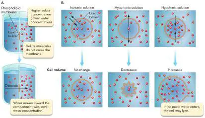
is a net loss of water from the cell. The cell shrinks, and the concentration of cell contents increases. In a hypotonic environment (lower concentration of solutes outside the cell), there is a net uptake of water by the cell; the cell swells, and the cell components are diluted (Fig. A2.5B ).
If enough water enters, the cell is destroyed by lysis, a rupturing of the cell membrane and dispersal of cell contents. Both hypertonic and hypotonic environments can cause other problems for cells. Proteins have specific salt requirements, and intracellular environments with salt concentrations that are either higher or lower than normal for a cell can cause denaturation of proteins, with potentially fatal results for the cell. Thus, transport of water across the cell membrane must be tightly controlled, and cells need mechanisms that allow them to live in environments that are not isotonic.
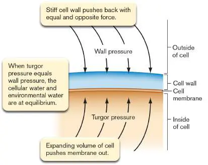
Most cells live in environments that are hypotonic. To deal with this challenge, most bacteria and many eukaryotes have a cell wall external to the cell membrane. As water enters by osmosis and pushes against the cell wall (“turgor pressure”), the wall resists the tension and pushes back with an equal but opposite force known as “wall pressure.” The wall pressure is an inward pressure exerted by the cell wall against the cell membrane (Fig. A2.6). When turgor pressure and wall pressure are equal in magnitude, the cell is at equilibrium with respect to water movement. Organisms that lack a cell wall employ other strategies to deal with hypotonic environments. For example, some freshwater protists that lack a cell wall expel excess water through a contractile vacuole.
FIGURE A2.6 ■ Turgor pressure and wall pressure. In contrast to freshwater microbes, ocean-dwelling organisms face the problem of water loss. Many of these organisms accumulate solutes known as osmolytes to ensure that they are isotonic to the external environment, thus preventing water loss.
Eukaryotes Transport Molecules by Endocytosis and Exocytosis
All cells use diffusion and transport proteins to move molecules into or out of the cell, but some eukaryotes can, in addition, use endocytosis and exocytosis to achieve this end.
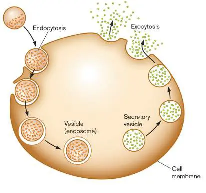
In endocytosis, parts of the cell membrane bud into the cytoplasm and eventually separate from it to form endosomes (Fig. A2.7). Endosomes are a type of vesicle, a small membranous sphere found
within a cell. The interior of these endosomes contains extracellular material. Phagocytosis (cell eating) is a form of endocytosis in which large extracellular particles are brought into the cell. Pinocytosis (cell drinking) is endocytosis of relatively small volumes of the extracellular fluid. Endocytosis is a controlled, energy-requiring process that relies on many proteins, including cytoskeletal proteins.
FIGURE A2.7 ■ Endocytosis and exocytosis. Exocytosis is the reverse of endocytosis (Fig. A2.7). In exocytosis, intracellular vesicles fuse with the cell membrane, and the contents of the vesicles are released to the extracellular environment. Cells can use exocytosis to release wastes.
Glossary
Bacteria One of the three domains of life, consisting of organisms with a last common ancestor not shared with members of Archaea or Eukarya. Organisms are prokaryotic (lacking nuclei, unlike eukaryotes) and possess primarily ester-linked phospholipid membranes (like eukaryotes, unlike archaea).
Archaea One of the three domains of life, consisting of organisms with a last common ancestor not shared with members of Bacteria or Eukarya. Organisms are prokaryotic (lacking nuclei, unlike eukaryotes) and possess ether-linked phospholipid membranes (unlike bacteria).
Eukarya One of the three domains of life, consisting of organisms with a last common ancestor not shared with members of Archaea or Bacteria. Cells possess nuclei, unlike cells of bacteria and archaea.
cell membrane Also called cytoplasmic membrane or plasma membrane. The phospholipid bilayer that encloses the cytoplasm.
plasma membrane Also called cell membrane or cytoplasmic membrane. The phospholipid bilayer that encloses the cytoplasm.
cytoplasmic membrane Also called cell membrane or plasma membrane. The phospholipid bilayer that encloses the cytoplasm.
cytoplasm Also called cytosol. The aqueous solution contained by the cell membrane in all cells and outside the nucleus (in eukaryotes). cytosol Also called cytoplasm. The aqueous solution contained by the cell membrane in all cells and outside the nucleus (in eukaryotes).
transmembrane protein Also called integral protein. A protein with a membrane-spanning region.
amphipathic Having both hydrophilic and hydrophobic portions.
peripheral membrane protein A protein that is associated with a membrane but does not span the phospholipid bilayer.
phospholipid The major component of membranes. A typical phospholipid is composed of a core of glycerol to which two fatty acids and a modified phosphate group are condensed.
phospholipid bilayer Two layers of phospholipids; the hydrocarbon fatty acid tails face the interior of the bilayer, and the charged phosphate groups face the cytoplasm and extracellular environment. The cell membrane is a phospholipid bilayer.
inner leaflet The layer of the cell membrane phospholipid bilayer that faces the cytoplasm.
outer leaflet The layer of the cell membrane phospholipid bilayer that faces away from the cytoplasm.
fluid mosaic model A model of the cell membrane in which proteins are free to diffuse laterally within the membrane.
diffusion The energy-independent net movement of a substance from a region of high concentration to a region of lower concentration. osmosis The diffusion of water from regions of high water concentration (low solute) to regions of low water concentration (high solute) across a semipermeable membrane.
isotonic Being in osmotic balance, having equal concentrations of solutes on both sides of a semipermeable membrane. A cell in an isotonic environment will neither gain nor lose water.
hypertonic Having more solutes than another environment separated by a semipermeable membrane. Water will tend to flow toward the hypertonic solution.
hypotonic Having fewer solutes than another environment separated by a semipermeable membrane. Water will tend to flow away from the hypotonic solution.
lysis Also called burst. The rupture of the cell by a break in the cell wall and membrane.
endocytosis The invagination of the cell membrane to form a vesicle that contains extracellular material.
endosome A vesicle formed from the pinching in of the cell membrane. vesicle A small, membrane-enclosed sphere found within a cell.
phagocytosis A form of endocytosis in which a large extracellular particle is brought into the cell.
pinocytosis A form of endocytosis in which only extracellular fluid and small molecules are brought into the cell.
exocytosis The fusion of vesicles with the cell membrane to release vesicle contents extracellularly.
A2.2 The Nucleus and Mitosisnot assigned
All cells need to synthesize proteins, and all cells contain DNA, the genetic material that encodes the information needed to specify protein primary structure. The central model of molecular biology holds that DNA is transcribed into messenger RNA (mRNA), and mRNA is translated into protein on ribosomes. Although this model holds for all cells, details in the structure of DNA and ribosomes vary among the three domains of life (Table A2.1). For example, ribosomes always function in protein synthesis, but ribosomes from organisms in different domains differ in their size and their sensitivity to various antibiotics. DNA always encodes the information needed for protein synthesis, but whereas bacterial chromosomes are usually circular, eukaryotic chromosomes are linear. Another difference between DNA in prokaryotes and DNA in eukaryotes is its location within the cell. In bacteria and archaea, DNA is found in an area of the cytoplasm known as the nucleoid, while eukaryotic DNA is contained within a nuclear membrane.
Eukaryotic DNA Is Housed in the Membrane- Enclosed Nucleus
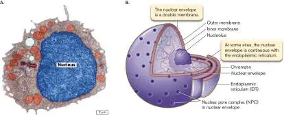
Eukaryotes derive their name (“eukaryote” means “true kernel”) from the fact that they possess a nucleus (plural, nuclei), and indeed, the nucleus is often the most prominent feature of eukaryotic cells viewed under a microscope (Fig. A2.8A). The nucleus is an organelle, an intracellular, membrane-enclosed compartment with a specific function. The nucleus contains chromatin, a complex of DNA and proteins. The nuclear membrane (envelope) consists of two concentric phospholipid membranes. The outer nuclear membrane is continuous with the membrane of the endoplasmic reticulum (ER), and the space between the two nuclear membranes is continuous with the lumen (inside) of the ER (Fig. A2.8B ). Nuclei contain a region called the nucleolus (plural, nucleoli), where ribosome assembly begins. At the nucleolus, multiple rRNA (ribosomal RNA) genes are transcribed, and the resulting rRNA combines with ribosomal proteins imported into the nucleus from the cytoplasm to form the ribosomal subunits. The ribosomal subunits then need to exit the nucleus.
FIGURE A2.8 ■ The nucleus. A. Electron micrograph of a eukaryotic yeast cell, showing the prominent nucleus. B. Diagram of a nucleus. NPC = nuclear pore complex.
GOPAL MURTI/VISUALS UNLIMITED
The nuclear membrane contains nuclear pore complexes (NPCs), which enable the transport of material into and out of the nucleus. Metabolites and small proteins can diffuse through the NPCs, but larger proteins and organelles cannot enter by diffusion. Large proteins that need to enter the nucleus are actively transported in through the NPCs. These selectively imported proteins contain a nuclear localization signal, a sequence of amino acids that acts like a zip code to direct them into the nucleus. In addition to their role in protein import, NPCs also function in exporting mRNAs out of the nucleus.
Eukaryotic Cells Replicate by Mitosis and Meiosis
Cells need to ensure the accurate replication and division of their DNA. Bacteria and archaea replicate by fission, as described in Chapter 3. Eukaryotic cells replicate their nuclear DNA and divide by a completely different process, called “mitosis.” For sexual reproduction, sex cells with a halved chromosome number are generated by the related process of “meiosis.” Mitosis and meiosis occur only in eukaryotes, never in bacteria and archaea.
Mitosis is a series of steps that segregates duplicated chromosomes and ensures that each daughter cell receives a copy of the genetic material. When the cell is not undergoing mitosis, it is in interphase (Fig. A2.9A). During interphase, the individual chromosomes are long and thin and not visible by a light microscope. Interphase can be divided into three phases: G 1, S, and G 2. Cells that are not committed to dividing are in G 1, the first gap phase. If cell division is to occur, then the chromosomes are replicated during S phase (S for “synthesis”). The duplicated chromosomes, called sister chromatids, remain attached to each other at the centromere (Fig. A2.9B ). Following chromosome replication, a second gap phase, G 2, occurs, after which mitosis can proceed.
FIGURE A2.9 ■ The cell cycle and mitosis. A. Stages of the eukaryotic cell cycle. G 1, S, and G 2 make up interphase (in blue). B. Duplication of the chromosomes during S phase. C. The phases of mitosis. See text for details.
Mitosis is divided into four steps (Fig. A2.9C ): Prophase. During prophase, the chromosomes condense and become visible by light microscopy. The nuclear membrane may break down. The mitotic spindle, which separates the sister chromatids to opposite poles of the cell, begins to form. The mitotic spindle is a network of microtubules (see Section A2.5) that originate from centrosomes. There are two centrosomes, and these migrate to opposite sides of the cell. Each centrosome contains two centrioles, and each centriole contains nine sets of microtubules in a radial formation (see Section A2.5). The free ends of the microtubules establish connections with the sister chromatids at a structure called the kinetochore.
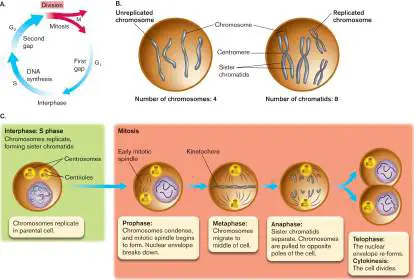
Metaphase. At metaphase, the spindle apparatus is complete and each sister chromatid is connected to a microtubule. The chromosomes are arranged along an imaginary plane in the middle of the cell.
Anaphase. In anaphase, the microtubules shorten and pull the sister chromatids apart, separating the replicated chromosomes. At the end of anaphase, each set of chromosomes is located on opposite sides of the cell.
Telophase. In this final step of mitosis, the nuclear membrane re-forms around each set of chromosomes and the chromosomes become long and thin again. Cytokinesis also occurs during telophase and partitions the original cell into two daughter cells by the formation of a cell membrane between them.
The reproduction of eukaryotic cells by mitosis is called asexual reproduction.
Some variations in the phases of mitosis are seen in microbial eukaryotes. In yeast, for example, the nuclear envelope does not break down. Despite these differences, the end result, the separation of duplicated chromosomes into two daughter cells, is the same.
Unlike asexual reproduction, sexual reproduction requires the reassortment of genetic material from different chromosomes. A sexual life cycle alternates between cells that are diploid (2 n, containing two copies of each chromosome) and sex cells that are haploid (1 n, containing a single copy of each chromosome). Two of the haploid 1 n sex cells (called gametes) can join each other by fertilization to regenerate a diploid cell (called a zygote). The diploid thus possesses two homologs of each chromosome; that is, two versions of the same chromosome from two different parents. The process of gamete formation requires a special modification of mitotic cell division called meiosis (Fig. A2.10). Meiosis includes two cell division series, called meiosis I and meiosis II. Like mitosis, meiosis I must be preceded by replication of all chromosomes during S phase (Fig. A2.10A). Thus, a diploid (2 n) cell becomes temporarily 4 n.
FIGURE A2.10 ■ Meiosis. The chromosomes of a diploid (2 n ) cell undergo replication. A. In meiosis I, homologous chromosomes exchange DNA, and all the pairs are separated between two daughter cells. B. In meiosis II, homologous pairs are separated to produce haploid (1 n) gametes.
Unlike the case in mitosis, in prophase I of meiosis I the replicated chromosomes do not separate; instead, each replicated pair lines up with its homolog, the same chromosome inherited from the other parent. Now the aligned homologs exchange portions of their DNA. This genetic exchange reassorts the traits and increases genetic diversity of the offspring.
After meiosis I, a short telophase occurs, in which each daughter cell is now diploid (2 n). Unlike what happens in the telophase of mitosis, in meiosis I no nuclear membrane forms, and no interphase occurs. Instead, the chromosome pairs immediately separate during meiosis II (Fig. A2.10B ). Meiosis II is completed by separation of the paired homologs through prophase II, metaphase II, anaphase II, and telophase II. The result is four haploid (1 n) gametes, as
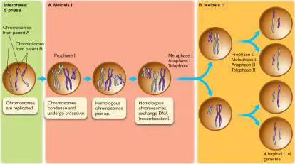
seen in Figure A2.10B . Depending on the species, these gametes may develop into specialized forms such as sperm and egg that reunite through fertilization, restoring the diploid form.
Glossary
nucleus pl. nuclei A eukaryotic organelle that contains DNA.
nucleus pl. nuclei A eukaryotic organelle that contains DNA.
organelle A membrane-enclosed compartment within eukaryotic cells that serves a specific function.
chromatin Chromosomal DNA complexed with proteins. Usually refers to a eukaryotic chromosome.
nucleolus pl. nucleoli A region inside the nucleus where ribosome assembly begins. nucleolus pl. nucleoli A region inside the nucleus where ribosome assembly begins. mitosis The orderly replication and segregation of eukaryotic chromosomes, usually prior to cell division.
asexual reproduction Reproduction of a cell by fission or by mitosis to form identical daughter cells.
sexual reproduction Reproduction involving the joining of gametes generated by meiosis.
homolog or homologous gene A gene derived from a common ancestral gene. Homologs may be orthologs or paralogs.
meiosis A form of cell division by which a diploid eukaryotic cell generates haploid sex cells that contain recombinant chromosomes.
A2.3 Problems Faced by Large Cellsnot assigned
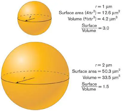
Most eukaryotic cells range in size from 10 to 100 micrometers (μm) in diameter, about ten times as large as the typical prokaryotic cell (1–10 μm in diameter). Cells face two major challenges as a result of increased cell size. The first problem is that as cells increase in size, their volume (the cytoplasm) increases faster than their surface area (the cell membrane) (Fig. A2.11). Cells are filled with metabolically active cytoplasm that requires nutrients and energy and produces wastes. Energy production, nutrient import, and waste disposal are events that take place at the cell membrane. As cells increase in size, the cell membrane area may not be able to keep up with the demands placed on it by a proportionally larger cytoplasm. The eukaryotic cell’s answer to this problem is the endomembrane system, an extensive network of internal membranes that effectively increases the membrane surface area without increasing cell volume.
FIGURE A2.11 ■ Cell volume increases faster than surface area. Because the surface area increases by the radius squared and the volume increases by the radius cubed, the volume increases faster than the surface area.
The second problem associated with an increase in size is related to diffusion. The amount of time it takes a molecule to diffuse a given distance is proportional to the distance squared. For example, if it takes a particular molecule 1 second to diffuse 1 μm, then it takes that same molecule 100 seconds to diffuse 10 μm. Many biochemical reactions depend on the partners in the reaction finding each other by diffusion (that is, the reaction rate is diffusion controlled), meaning that longer diffusion times result in slower reactions. Furthermore, signals received at the cell membrane need
to be communicated throughout the cell. In a large cell, the amount of time it takes for a molecule to traverse the cell by diffusion may be too slow for the cell to rapidly adjust to signals it receives from the environment or from other sites within the cell. To deal with this problem, eukaryotic cells possess a cytoskeleton, a group of proteins that, among other things, maintain cell shape and move molecules around the cell, relieving the cell from the need to rely on diffusion for transport.
As we will see in the next two sections, the endomembrane system and the cytoskeleton serve additional functions for the eukaryotic cell.
A2.4 The Endomembrane Systemnot assigned
The endomembrane system is a series of compartments found inside eukaryotic cells and separated by membranes that are distinct from the cell membrane. Organelles of the endomembrane system include the nuclear membranes, the endoplasmic reticulum (ER), the Golgi complex (also called the Golgi apparatus), lysosomes, and peroxisomes. Different organelles contain unique subsets of proteins that contribute to their function. To a large extent, the function of the ER and the Golgi complex is to direct to their proper cellular location proteins destined for lysosomes, for the cell membrane, or for secretion from the cell.
Membranous organelles serve many functions for the eukaryotic cell. In general: Endomembranes increase the membrane surface area without increasing cell volume.
Separating cellular contents in small, enclosed compartments increases the concentrations of enzymes and their substrates ( Fig. A2.12), allowing reactions to proceed faster.
Organelles can provide different environments that allow disparate reactions to occur simultaneously. For example, some proteins are being synthesized by ribosomes in the neutral-pH cytoplasm, while at the same time other proteins are being hydrolyzed within the acidic organelles known as lysosomes. Compartmentalization protects cytoplasmic components from harmful substances. For example, hydrogen peroxide (H 2 O 2), a product of cellular oxidation reactions, is produced and converted to water within peroxisomes. Localizing the reaction within the peroxisome keeps the toxic peroxide away from other cell components, such as proteins and DNA, that are sensitive to oxidative stress.
FIGURE A2.12 ■ Advantages of organelles. Solutes (green dots) are concentrated in organelles, reactive molecules are separated from the cytoplasm, and membrane surface area is increased without an increase in cell volume.
Lysosomes Digest Organic Matter
Lysosomes are membrane-enclosed organelles that help eukaryotic cells obtain nourishment from macromolecular nutrients. Lysosomes contain many hydrolytic enzymes (for example, proteases, nucleases, and lipases) and have an acidic pH of about 5. Lysosomes are formed when vesicles containing hydrolytic enzymes and proton pumps bud off from the Golgi complex (Fig. A2.13A). Lysosomes then fuse with vesicles containing the material to be digested. Often this material comes from outside the cell via phagocytosis. Phagocytosis and lysosomal digestion help the eukaryotic cell because they effectively increase the membrane surface area over which nutrients can be absorbed.
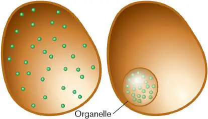
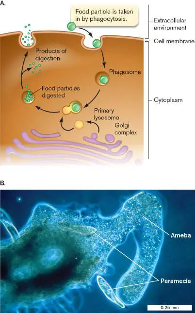
FIGURE A2.13 ■ Lysosomes. A. Lysosomes contain hydrolytic enzymes to digest material brought into the cell by phagocytosis. B. Amebas engulfing paramecia (light microscopy). The paramecia will be digested with the aid of lysosomes.
ERIC GRAVE/SCIENCE SOURCE
Bacteria lack phagocytosis; to obtain nutrition from large molecules in their environment, bacteria must secrete digestive enzymes. The extracellularly digested materials are subsequently transported across the bacterial cell membrane through specific transporters (see Section 4.2). In eukaryotes, by contrast, lysosomes allow for intracellular digestion, and digested material crosses the lysosomal membrane into the cytoplasm. Any waste products left in the lysosome can leave the cell via exocytosis.
The Volume within the ER Is Separate from the Cytoplasm
The endoplasmic reticulum is continuous with the outer nuclear membrane, and the lumen (interior) of the ER is continuous with the space between the two nuclear membranes (Fig. A2.14A). In addition, the lumen of the ER is spatially equivalent to the interior spaces of other endomembrane components and to the outside of the cell. This means that material in the ER does not need to cross a membrane to enter these other spaces, and mixing can occur via vesicle fusion. For example, material contained within the lumen of the ER can mix with the contents of the Golgi complex (Fig. A2.14B ) or with the extracellular milieu by fusion of vesicles from the ER with the Golgi membrane or the plasma membrane. These topologically equivalent areas, indicated by a common color in Figure A2.14A, are completely separated from the cytoplasm by endomembranes, so that the ER can be used to sequester substances that must be held at low concentrations in the cytoplasm; for example, calcium ions.
Smooth ER and Rough ER
There are two morphologically and functionally distinct types of endoplasmic reticulum: smooth ER and rough ER, as shown in Figure A2.14C . The smooth ER is the site of lipid synthesis and some detoxification of noxious compounds. The rough ER is the site where transmembrane proteins, secreted proteins, and resident proteins of the ER, Golgi, or lysosomes are translated. The rough ER appears rough because its cytoplasmic surface is studded with ribosomes. These ribosomes are located on the rough ER because the protein being synthesized by the ribosome has a hydrophobic signal sequence on its amino terminus (the first part of the protein translated from the mRNA).
FIGURE A2.14 ■ The endoplasmic reticulum and Golgi complex. A. The relationship of the ER to other cellular membranes and the flow of material through vesicles from the rough ER to the cell membrane. B. Electron micrograph of Golgi cisternae. C. Electron micrograph showing rough ER and smooth ER.
SPL/SCIENCE SOURCE
DON W. FAWCETT/SCIENCE SOURCE
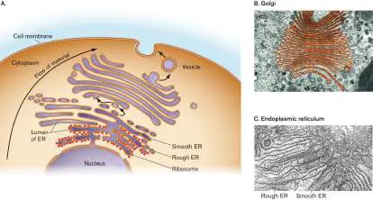
A signal sequence is a specific sequence of amino acids that directs proteins to a specific cellular location, such as a membrane. The signal sequence that directs proteins to the ER recognizes a receptor (the signal recognition particle) on the rough ER membranes (Fig. A2.15A), so the protein-ribosome complex is directed to the surface of the ER membrane. Note that the ribosomes attached to the rough ER are identical to cytoplasmic ribosomes and attach to the ER only transiently because of the types of proteins they are synthesizing—proteins that contain the correct signal sequence.
After the ribosome docks with the signal recognition particle, the growing polypeptide is threaded through the ER membrane as it is synthesized. Secreted proteins and proteins destined for the lumen of an organelle are threaded completely through the ER membrane and end up in the lumen of the ER (Fig. A2.15A). In contrast, transmembrane proteins are not threaded completely through, and part of the protein spans the membrane (Fig. A2.15B ). The membrane-spanning regions of transmembrane proteins usually contain a continuous stretch of about 20 hydrophobic amino acids that form an alpha helix with the hydrophobic side chains facing out toward the hydrophobic hydrocarbons of the membrane.
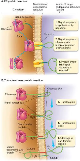
FIGURE A2.15 ■ Insertion of proteins into the ER. A. Proteins are targeted to the ER by a signal sequence on the growing polypeptide chain. Secreted proteins and proteins destined for the lumen of an organelle end up in the lumen of the ER. B. Transmembrane proteins are not completely inserted through the ER membrane, and a portion of the protein remains within the membrane.
As the nascent polypeptide chains are threaded across the ER membrane, the unfolded proteins are bound by chaperone proteins. Chaperones (some of which are heat-shock proteins) prevent partially folded proteins from clumping together and help proteins attain their correct tertiary structure. In the ER, proteins may be modified. The signal sequence that directed the proteins to the ER is usually cleaved off. The environment inside the ER allows disulfide bonds to form between cysteine residues of some proteins. Other ER proteins have oligosaccharide groups covalently attached—a posttranslational modification known as glycosylation. The enzymes that attach the sugars are found only in the ER lumen. In transmembrane proteins of the cell membrane, sugars always face the extracellular environment because the lumen of the ER forms vesicles that open out to the external medium (Fig. A2.14A). Resident ER proteins—those that will stay and function in the ER— are retained inside the ER because they contain a sequence of amino acids that acts as an ER retention signal.
The Golgi Complex Directs Protein Transport
Proteins not retained in the ER move on to the Golgi complex by way of vesicles. The Golgi complex consists of separate membrane stacks (cisternae) that each contain unique enzymes (Fig. A2.14B ). As proteins pass through the cisternae, the carbohydrates on them may be trimmed and modified. These modified carbohydrates can serve as address tags for targeting proteins to particular organelles. For example, proteins tagged with mannose 6-phosphate (mannose phosphorylated on its number 6 carbon) are selectively sent to lysosomes; that is, vesicles enriched in proteins containing mannose 6-phosphate bud off from the Golgi and are directed to lysosomes. Proteins not targeted to lysosomes and not marked for retention in the Golgi complex may be sent to the cell membrane. Vesicles leaving the Golgi complex may fuse with the cell membrane, releasing their contents to the extracellular environment (Fig. A2.14A). Transmembrane proteins in these vesicles can then become part of the cell membrane. Regions of transmembrane proteins that were inside vesicles will face the extracellular environment, and cytoplasmic portions will remain cytoplasmic.
Glossary
endomembrane system A series of membranous organelles that organize uptake, transport, digestion, and expulsion of particles through a eukaryotic cell. It includes endosomes, lysosomes, endoplasmic reticulum, and the Golgi complex.
peroxisome A eukaryotic organelle that converts hydrogen peroxide to water.
lysosome An acidic eukaryotic organelle that aids digestion of molecules. Not found in plant cells.
signal sequence A specific amino acid sequence on the amino terminus of proteins that directs them to the endoplasmic reticulum (of a eukaryote) or the cell membrane (of a prokaryote).
signal recognition particle (SRP)
A receptor that recognizes the signal sequence of peptides undergoing translation. The complex attaches to the cell membrane of prokaryotes (or the rough endoplasmic reticulum of eukaryotes), where it docks the protein-ribosome complex to the membrane for protein membrane insertion or secretion. Golgi complex or Golgi apparatus A series of membrane stacks that modifies proteins and helps sort them to the correct eukaryotic cell compartment.
A2.5 The Cytoskeletonnot assigned
Eukaryotic cells contain proteins called intermediate filaments, microfilaments, and microtubules, which are collectively termed the cytoskeleton. As the name implies, these proteins serve as a cell skeleton and impart specific shapes to eukaryotic cells. However, cytoskeletal proteins are multifunctional and are also involved in whole-cell movements and movements of substances within the cell.
Microfilaments and Intermediate Filaments
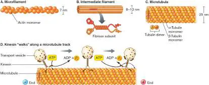
Microfilaments, also known as actin filaments, have a diameter of 7 nanometers (nm) (Fig. A2.16A). They are formed when individual actin monomers (globular actin, or G-actin) polymerize, in a process fueled by ATP hydrolysis, to form chains of filamentous actin (F-actin). Two F-actin chains twist around each other to form microfilaments that have a plus end and a minus end.
Microfilaments are dynamic structures, growing and shrinking in a controlled manner. New monomer units add to the plus end while old ones dissociate from the minus end. Whether actin will polymerize or depolymerize depends on a number of factors, including the concentration of G-actin. The critical concentration is a measure of the ability of actin to polymerize. At G-actin concentrations below the critical concentration, F-actin will depolymerize; and at concentrations greater than the critical concentration, G-actin will polymerize. The plus end of a microfilament has a critical concentration less than that of the minus end, so actin is preferentially added onto the plus end and removed from the minus end.
FIGURE A2.16 ■ Cytoskeletal proteins. A. Microfilaments consist of two strands of actin polymers twisted together. B. Intermediate filaments are ropelike assemblages of various proteins. C. Microtubules are polymers of tubulin dimers. D. Fueled by the hydrolysis of ATP, the motor protein kinesin can move vesicles or organelles toward the plus end of microtubules.
MATTEO OMIED/ALAMY STOCK PHOTO
Some microfilaments play a structural role in the cell to maintain cell shape. These structural microfilaments have protein caps at both ends to prevent changes in microfilament length. Other microfilaments have functions that require dynamic changes in length. For example, the pseudopod movement of an ameba depends on the polymerization of actin at the leading edge of growth. The plus end of the microfilament is located underneath the cell membrane of the extending pseudopod, and polymerization is enhanced by the actin-binding protein profilin. Microfilaments mediate cytoplasmic streaming, a mixing of the cytoplasm that aids diffusion. The protein myosin works with microfilaments to generate the forces needed for cell streaming, pseudopod formation, and cytokinesis, the separation of daughter cells after nuclear division. Intermediate filaments (Fig. A2.16B ) consist of various fibrous proteins that have a diameter of about 10 nm. Intermediate filaments often form a meshwork under the cell membrane and, in cells that lack a cell wall, help impart and maintain cell shape.
Intermediate filaments also strengthen the cell by resisting tension placed on the cell membrane. The proteins that make up intermediate filaments vary with cell type. Intermediate filaments are fairly stable and are not thought to undergo acute changes in length the way microfilaments and microtubules do.
Microtubules
Microtubules (Fig. A2.16C ) have a larger diameter (25 nm) than microfilaments and intermediate filaments. The hollow microtubule structure consists of 13 protofilaments made of tubulin dimers; one alpha-tubulin protein plus one beta-tubulin protein form one tubulin dimer. Like microfilaments, microtubules have plus (faster-growing) and minus (slower-growing) ends and are dynamic structures that can polymerize and depolymerize. Polymerization is an energy-requiring process, and the necessary energy is obtained by coupling polymerization to GTP (guanosine triphosphate) hydrolysis. Microtubules aid movement of substances within the cell and are also involved in powering whole-cell movement by eukaryotic cilia and flagella.
Traffic of proteins through the endomembrane system (see Section A2.4) relies on the controlled movement of vesicles from one cellular compartment to the next. Microtubules provide tracks that can move vesicles from one organelle to the next in an efficient, directed fashion. Working with microtubules to accomplish this task are motor proteins. Motor proteins such as kinesin and dynein can capture cargo (for example, vesicles or organelles) and walk them along microtubule tracks in an ATP-dependent process ( Fig. A2.16D ). Kinesin moves cargo toward the plus end of the microtubule, while dynein moves cargo toward the minus end. In addition to moving vesicles, microtubules segregate (pull apart) the duplicated chromosomes during mitosis.
Eukaryotic Cilia and Flagella
Cilia and flagella are tubular extensions of the cell membrane that can move in a whiplike fashion, driven by interactions between microtubules and the motor protein dynein. Flagella (singular, flagellum) are relatively long, and cells usually have only one or two of them; cilia (singular, cilium) are shorter and more numerous. Both flagella and cilia can move cells through space. Cilia may also aid in food capture; for example, by sweeping extracellular fluid into the gullet of a paramecium. Eukaryotic cilia and flagella differ from bacterial cilia and flagella; bacterial flagella are helical and use the proton motive force to rotate a motor that causes flagellar movement, whereas eukaryotic flagella are S-curved and use ATP hydrolysis by dynein to move the flagella in a whiplike fashion. The study of protists has played a key role in revealing the structure and function of eukaryotic flagella and cilia. Much information about flagellar structure has come from studies of a Chlamydomonas species, a unicellular alga that has two long flagella (Fig. A2.17A). Dynein was first discovered in the cilia of the unicellular eukaryote Tetrahymena (Fig. A2.17B ). Flagella and cilia have the same structure and mechanism of action, so for convenience we will restrict the following discussion to flagella. FIGURE A2.17 ■ Flagella and cilia. A. The protist Chlamydomonas has two long flagella (SEM). B. The protist Tetrahymena has numerous cilia (SEM). C. Structure of flagella
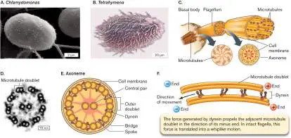
and cilia. D. Cross section of an axoneme (TEM). E. Cross section of an axoneme, indicating bridges and spokes formed by cross-linking proteins. F. Force generated by dynein on a microtubule doublet.
AARON J. BELL/SCIENCE SOURCE
AARON J. BELL/SCIENCE SOURCE
A cross section through a flagellum reveals a central bundle of microtubules called the axoneme (Fig. A2.17C ). The axoneme originates at a microtubule-organizing center called the basal body, which is similar to the centriole (see Section A2.2). The minus ends of the microtubules are at the basal body; the plus ends at the tip of the flagellum are capped to prevent changes in length.
As shown in Figure A2.17D , the axoneme has a characteristic arrangement of two central microtubules and nine microtubule doublets around the periphery. The microtubule doublets are connected to each other and to the central microtubules through protein cross-links called bridges and spokes (Fig. A2.17E ). The motor protein dynein connects adjacent microtubule doublets. If the cell membrane is removed from the surface of a flagellum and the protein cross-links (but not dynein) are dissolved, the microtubule doublets are observed to lengthen in the presence of ATP. This lengthening is due to the action of dynein, which slides the microtubule doublets past each other (Fig. A2.17F ). In intact flagella, the tension imparted as dynein tries to slide microtubules past each other is translated into a bending motion. Controlled cycles of dynein activation and inactivation on opposite sides of the axoneme lead to the whiplike motion of eukaryotic flagella.
Glossary
cytoskeleton A collection of filamentous proteins that impart structure to and aid movement of cells; in a eukaryote, these include microfilaments, intermediate filaments, and microtubules. microfilament Also called actin filament. A eukaryotic cytoskeletal protein composed of polymerized actin.
intermediate filament A eukaryotic cytoskeletal protein that is composed of various proteins depending on the cell type.
microtubule A eukaryotic cytoskeletal protein composed of polymerized tubulin.
flagellum pl. flagella A filamentous structure for motility. In prokaryotes, a helical protein filament attached to a rotary motor; in eukaryotes, an undulating membrane-enclosed complex of microtubules and ATP-driven motor proteins.
flagellum pl. flagella A filamentous structure for motility. In prokaryotes, a helical protein filament attached to a rotary motor; in eukaryotes, an undulating membrane-enclosed complex of microtubules and ATP-driven motor proteins.
cilium pl. cilia A short, hairlike structure of eukaryotes that is structurally similar to the prokaryotic flagellum. Cilia beat in waves to propel the cell.
cilium pl. cilia A short, hairlike structure of eukaryotes that is structurally similar to the prokaryotic flagellum. Cilia beat in waves to propel the cell.
A2.6 Mitochondria and Chloroplastsnot assigned
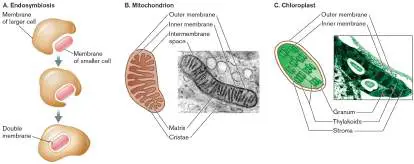
Mitochondria and chloroplasts are organelles involved in cellular energy production. Mitochondria (singular, mitochondrion) perform oxidative respiration and are found in nearly all eukaryotes. Chloroplasts perform photosynthesis and are found only in photosynthetic eukaryotes, such as green algae. Both organelles became part of eukaryotic cells through a process of endosymbiosis. The serial endosymbiosis theory states that mitochondria and chloroplasts were once free-living bacteria that became ingested, but not digested, by a larger (possibly eukaryotic) cell (Fig. A2.18A). A symbiotic relationship developed, with the larger eukaryotic cell providing protection to the intracellular bacterium and the bacterium providing energy to the eukaryote. As we will see, the structure of mitochondria and chloroplasts reflects their origin as internalized bacteria.
FIGURE A2.18 ■ Serial endosymbiosis, mitochondria, and chloroplasts. A. In endosymbiosis, organelles originate as a result of small bacterial cells being ingested by larger (possibly eukaryotic) cells. B. Structures in a mitochondrion. C. Chloroplast structure.
DON W. FAWCETT/SCIENCE SOURCE
GEORGE CHAPMAN/VISUALS UNLIMITED
Mitochondria Produce ATP by Oxidative Respiration
Mitochondria are the powerhouses of the eukaryotic cell. A cell may contain tens or hundreds of mitochondria, depending on its energy needs. Mitochondria have two membranes: an outer membrane and an inner membrane (Fig. A2.18B ). The inner membrane has numerous infoldings called cristae that increase its surface area. It is thought that as a result of the ancestral cell engulfing the bacterium, the inner mitochondrial membrane is derived from the bacterium and the outer membrane is derived from the larger ancestral host cell. Supporting this idea is the fact that the inner membrane has structural characteristics of a prokaryotic cell membrane, while the outer membrane is similar to the host eukaryotic membrane. For example, 20% of the phospholipids in the inner membrane are cardiolipin, a bacterial phospholipid largely absent from membranes of eukaryotic origin.
Mitochondria contain two distinct compartments: the intermembrane space between the two membranes, and the matrix enclosed by the inner membrane. Different stages of oxidative respiration occur in specific compartments. As predicted by the endosymbiosis theory, the topology of these processes is similar in bacteria and mitochondria (Table A2.2). For example, in bacteria and archaea, the tricarboxylic acid cycle (TCA cycle) occurs in the cytoplasm; in mitochondria, the TCA cycle takes place inside the matrix, the metabolic equivalent of the prokaryotic cytoplasm.
Location of Oxidative TABLE Respiration Components in A2.2 Bacteria and Mitochondria
Feature Bacteria Mitochondria Electron Cell membrane Inner membrane transport system (ETS)
ATP Cell membrane Inner membrane synthase TCA cycle Cytoplasm Matrix (compartment enclosed by the inner membrane)
Proton Protons diffuse from Protons diffuse from motive lower-pH lower-pH force extracellular intermembrane space environment into into the matrix across the cytoplasm the inner membrane across the cell membrane
DON W. FAWCETT/SCIENCE SOURCE
GEORGE CHAPMAN/VISUALS UNLIMITED
The fact that mitochondria contain their own DNA and ribosomes lends further support to the endosymbiosis model, as these cell features would be a necessary part of any free-living organism. Both the DNA and ribosomes of mitochondria show similarities with the DNA and ribosomes of bacteria. For example, like prokaryotic DNA, mitochondrial DNA is circular, and mitochondrial ribosomes are sensitive to antibiotics that disrupt prokaryotic ribosomes. Although nuclear DNA encodes some mitochondrial proteins, phylogenetic analysis has shown that these nuclear genes originated from bacteria, probably the engulfed ancestors of mitochondria. Furthermore, mitochondria replicate independently of cell division. The division of mitochondria within the cell is similar to the fission seen in prokaryotic cells.
Chloroplasts Perform Photosynthesis
Chloroplasts are organelles found only in photosynthetic eukaryotes. In the light reactions of photosynthesis, chloroplasts convert light energy from the Sun into ATP and reduced NADPH. In the subsequent carbon-fixation reactions, the ATP and NADPH are used to reduce CO 2 to sugar.
Like mitochondria, chloroplasts originated via endosymbiosis. Bacteria related to cyanobacteria were the prokaryotic partners that gave rise to chloroplasts. Like eukaryotic chloroplasts, cyanobacteria contain chlorophylls a and b and perform aerobic photosynthesis. In addition, cyanobacteria have extensive internal membranes, called thylakoids, that contain chlorophyll and participate in the light reactions of photosynthesis.
Chloroplasts have three membranes, whose topology can be understood in light of endosymbiosis (Fig. A2.18C ). The outer membrane is derived from the host eukaryotic cell, the inner membrane is equivalent to the bacterial cell membrane, and the thylakoid membrane is derived from the endosymbiotic bacterial thylakoid membranes. The region inside the inner membrane is called the stroma and is equivalent to the bacterial cytoplasm. The thylakoid membrane is packed with the chlorophyll pigments that give chloroplasts their green color. ATP and NADPH are produced in the stroma and used there in the reactions of CO 2 fixation. As would be expected as a result of endosymbiosis, chloroplasts, like mitochondria, contain their own circular DNA and their own ribosomes.
Glossary
mitochondrion pl. mitochondria An organelle of endosymbiotic origin that produces ATP through the use of an electron transport system to generate a proton potential. O 2 is the final electron acceptor to produce H 2 O. mitochondrion pl. mitochondria An organelle of endosymbiotic origin that produces ATP through the use of an electron transport system to generate a proton potential. O 2 is the final electron acceptor to produce H 2 O. chloroplast An organelle of endosymbiotic origin (sharing descent with cyanobacteria) that conducts oxygenic photosynthesis; found in algae and plant cells.
serial endosymbiosis theory The theory that mitochondria and chloroplasts were originally free-living prokaryotes that formed an internal symbiosis with early eukaryotes.