Chapter introduction
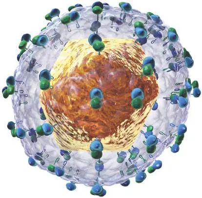
Hepatitis C virus. Illustration of the structure of the hepatitis C virion, based on data from electron microscopy. Source: Bruce Blaus, Blausen Medical 2014.
Courtesy Wikipedia Commons and Blausen Medical Communications, Inc. https://commons.wikimedia.org/wiki/File:HCV.png; https://creativecommons.org/licenses/by-sa/4.0/legalcode
Appendix Sections
A4.1 Phage T4: The Classic Molecular Model A4.2 The Filamentous Phage M13: Vaccines and Nanowires A4.3 Poliovirus: (+) Strand RNA Virus A4.4 Hepatitis C: (+) Strand RNA Virus Different viruses show very different replication cycles and molecular interactions with their host. Chapter 11 presented the molecular biology of several representative viruses: bacteriophage lambda, a double-stranded DNA virus infecting Escherichia coli; and the human-infecting viruses influenza A (IAV), human immunodeficiency virus (HIV), and herpes simplex virus (HSV). eAppendix 4 presents four additional viruses in molecular detail: Bacteriophage T4 of E. coli, a double-stranded DNA phage with a large genome and intricate assembly program.
Bacteriophage M13 of E. coli, a single-stranded DNA phage that replicates by slow release, neither lytic nor lysogenic. Human poliovirus, a plus-strand RNA enterovirus that infects the gut and may cause the paralytic disease poliomyelitis (polio).
Human hepatitis C virus (HCV), a plus-strand RNA virus that propagates in the liver and can cause cancer requiring treatment by liver transplant.
A4.1 Phage T4: The Classic Molecular Modelnot assigned
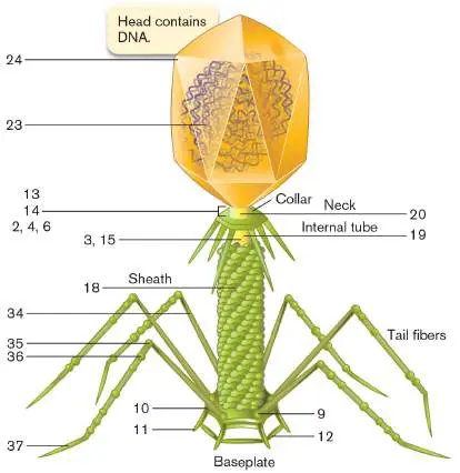
Phage T4 (Fig. A4.1) is among the trillions tailed phages that have evolved to make a home in the human gut, where they infect Gram-negative enteric bacteria. Other tailed phages permeate the soil and swarm in marine waters. Historically, phages provided the first living systems simple enough to dissect at the molecular level. Phages yielded fundamental discoveries in gene regulation, including the classic case of lambda lysogeny (discussed in Section 11.1). The first model for the genetic analysis of animal development was a phage system, the assembly of the “tailed phage” T4 (Myoviridae). The ATP-driven motor for DNA packaging of phage T4 provided a model for DNA packaging of herpesviruses. FIGURE A4.1 ■ Structure of a phage T4 capsid (simplified). Proteins are assigned to specific genes (numbered) that are mapped on the chromosome in Figure A4.2.
Phage T4 Structure
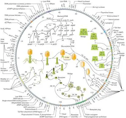
Phage T4 may be the most complicated noncellular reproductive unit that has been characterized. The T4 genome of 169 kilobase pairs (kb) specifies approximately 300 genes (Fig. A4.2), many of which encode enzymes that replace host cell functions such as
nucleic acid biosynthesis. The intricate structure of the virion requires 20 gene products just for assembly.
FIGURE A4.2 ■ Phage T4 genome, showing functions of mapped genes. The gene numbers correspond to proteins assembled in Figure A4.1.
Source: Based on C. K. Matthews et al. 1983. Bacteriophage T4. ASM Press, Washington, DC.
The phage T4 virion has a capsid, a device carrying the genome and composed of specialized protein subunits. The capsid is a polyhedral “head” that contains the phage genome, a linear double strand of DNA (Fig. A4.1). The head is attached to a delivery
device called the “tail”—hence the term tailed phage. The head is connected by a narrow neck tube to an internal tube that extends down within the sheath. The sheath plus the internal tube make up the tail. From the tail extend two sets of fibers—six connected at the neck, and six at the baseplate. The elegant mechanical structure and function of this molecular motor was an early inspiration for nanotechnology.
Adsorption to Host and DNA Injection
Viral infection requires a molecular “fit” between a viral component and one or more molecules of the host cell surface. Phage T4 infects strains of Escherichia coli, whose outer membrane possesses lipopolysaccharide (LPS) and the porin OmpC. The phage adsorbs (attaches) to the cell surface by contact between its tail fibers and the outer membrane (Fig. A4.3A). Once several of the tail fibers are anchored to the outer membrane by LPS, the phage baseplate makes contact. The contact between baseplate and cell outer membrane generates a conformational change in the tail. The outer tube of the tail (the sheath) is a helical tube of protein P18 subunits (Fig. A4.3B ). The sheath contracts by shortening and widening, enabling the internal tube to push through, like a molecular syringe. FIGURE A4.3 ■ Phage T4 adsorption and DNA injection. A. First the tail fibers contact the outer membrane; then the baseplate completes the connection. The sheath (composed of protein P18) contracts, the internal tube (composed of protein P19) penetrates the outer membrane, and then the peptidoglycan is digested. The tube extends to the inner membrane, and the head releases DNA under packing pressure, forcing it into the cytoplasm. B. Sheath contraction, based on crystallography of P18 and on cryo-electron microscopy (cryo-EM) of extended and contracted tails. This model was devised by Michael Rossmann and colleagues.
Source: Part B modified from Anastasia Aksyuk et al. 2009. EMBO J. 28 :821.
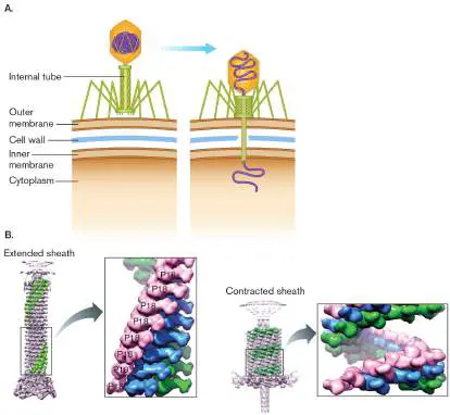
The dimensions of the sheath were modeled by combining X-ray crystallography of protein P18 with cryo-electron microscopy (cryo-EM) images of tails that were isolated in the extended or contracted position. This impressive model was built by Michael Rossmann (1930–2019) and colleagues at Purdue University. Rossmann’s lab developed techniques to visualize many challenging viral structures, including whole virions such as rhinovirus (cold virus), dengue fever flavivirus, and canine parvovirus. These viral structures may now be used to test new antiviral drugs.
The internal tube of the tail is capped by a needlelike “injector,” composed of proteins P27 and P5. As the sheath surrounding the internal tube contracts, the injector pokes through the host outer membrane and penetrates the cell wall. The peptidoglycan surrounding the injector is then digested by a lysozyme protein within the needle tip. The hole in the cell wall enables the entire tail to penetrate the cell wall and inner membrane. When the tip of the tail tube penetrates the inner membrane, the phage DNA is released and forced out of the head under pressure. Double-stranded DNA viruses are packed at a pressure of up to 50 atmospheres (atm), some of the highest pressures found in living organisms.
Virulent Replication
Phage T4 is fully virulent; that is, its only reproductive option is to assemble progeny virions while destroying a host cell (Fig. A4.4A ). By contrast, other tailed phages, such as phage lambda, can undergo lysogeny, the integration of their genome into that of their host cell. The integrated phage genome then replicates passively with the host. Lysogeny is described in Chapter 6, and the genetic regulation of phage lambda is presented in Section 11.1.
FIGURE A4.4 ■ Replication cycle of phage T4. A. The phage particle attaches to the surface of E. coli and injects its genome. The genome is reproduced and packaged into progeny virions, which are released upon lysis of the host cell. Blowup: Model of the T4 head with an ATP-driven motor packaging DNA.
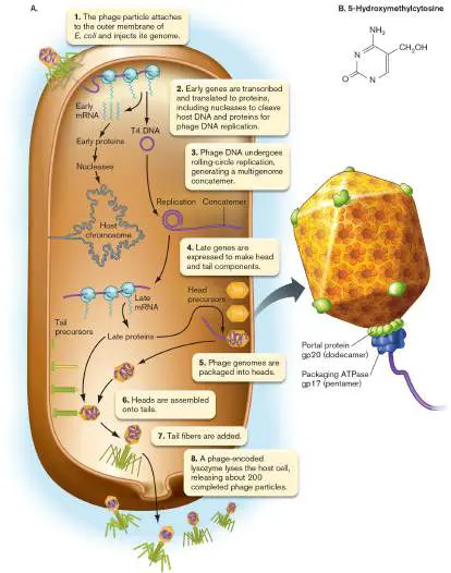
B. 5-Hydroxymethylcytosine replaces cytosine during DNA replication by T4-encoded DNA polymerase.
Source: Part A blowup modified from S. Hegde et al. 2012. J. Virol. 86 :4046. When phage T4 DNA enters the host cytoplasm, a set of phage genes is activated for transcription. Phage gene activation involves DNA-binding regulators like those of bacterial gene regulation (discussed in Chapters 8 and 10). The phage genes are transcribed by the host cell RNA polymerase and translated by the host ribosomes. These host components, however, are supplemented by phage-encoded components such as transfer RNAs (tRNAs).
Phage genes expressed early in the infection cycle are called early genes. The early-gene products include proteins needed to cleave host DNA, thus halting host macromolecular synthesis. (The phage’s own DNA avoids cleavage because its cytosine bases are hydroxymethylated.) In addition, several phage DNA synthesis enzymes replace key enzymes of the host. One T4 enzyme increases the rate of phage DNA replication tenfold. Another enzyme replaces cytosine with the modified base 5-hydroxymethylcytosine (Fig. A4.4B ), which substitutes for cytosine throughout the phage chromosome. This modification of phage DNA prevents cleavage by viral and host endonucleases. The endonucleases (enzymes that cleave DNA) fail to “fit” DNA containing the modified cytosine. Phage T4 DNA is synthesized within the host cell by rolling-circle replication. The advantage of rolling-circle replication is that many genome copies are made quickly. First the linear DNA duplex of phage T4 forms a circle by recombination between the terminally redundant ends. The circularized duplex then replicates by the rolling-circle method, generating a linear concatemer in which multiple genomes are joined end to end (Fig. A4.5). The concatemer serves as a template to synthesize the complementary strand. Out of the linear T4 DNA concatemer, the individual genomes are packaged into heads.
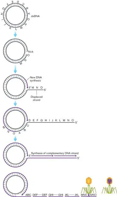
FIGURE A4.5 ■ Rolling-circle replication of phage T4. Initially, the linear genome circularizes; then it replicates as a concatemer. Out of the concatemer, the individual genomes are packaged into head coats and then cleaved. Each encapsidated DNA contains an end-duplicated 3% of its genome.
Each head actually contains sufficient volume to pack 3% more DNA than is contained in a phage genome. Thus, when the DNA duplexes extending from each head are cleaved, each phage DNA contains 3% extra DNA that repeats the same sequence from the other end. (This is called “terminal redundancy.”) Each cleavage of the concatemer must then occur another 3% farther along the genome and duplicate this next piece instead. Thus, every T4 DNA molecule ends up with a different 3% terminal repeat at some point throughout the genome.
Ultimately, the late genes are induced to produce the capsid and tail proteins that assemble at the membrane to make mature phage. During assembly, a key problem is how to get the entire DNA genome stuffed into the phage head. The process of DNA packaging actually requires energy spent by an ATP-driven “nanomotor.” The filled heads are attached to tails, and finally the various tail, collar, and capsid substructures are assembled (Fig. A4.4). One phage infection yields about 200 complete phage particles per cell. At last, an unknown signal triggers lysis. A late gene encodes a lysozyme that digests the cell wall, releasing the phages into their surroundings. If the lysozyme gene is defective, all the progeny phage particles remain trapped within the host cell. All viral infections need to coordinate the actions of the viral components with a much greater number of host components, such as those of the transcription and translation apparatus. In some cases, a virus actually evolves to “pick up” a host gene encoding a useful product—and then finds a novel use for that product. The phage T4 genome has acquired many host genes through recombination with scraps of host DNA. For example, the phage-expressed enzyme dihydrofolate reductase evolved from a host enzyme used to reduce the cofactor folic acid for use in biosynthesis. But the phage-encoded enzyme has a completely different function: assembly with folate as structural parts of the T4 injector baseplate.
Phage Particles Self-Assemble
The assembly of phage T4 particles within the host cytoplasm offers exceptional opportunities to visualize a molecular pathway. Each phage particle is assembled by convergence of three pathways involving the head coat, the tail, and the tail fibers (Fig. A4.6).
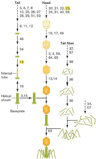
FIGURE A4.6 ■ Phage T4 assembly. The phage T4 capsid assembles automatically in a predetermined order from parts encoded by phage genes (numbers correspond to the gene numbers in Figure A4.2). Genes 19 and 23 (highlighted) are mutated in Figure A4.7.
All of these stages can be isolated and observed within infected cells by electron microscopy. But with all these many steps, how did we determine the order of assembly and identify the coding genes? The stages of phage assembly were discovered using strains that carry mutations in various genes that encode proteins essential for development (Fig. A4.7).
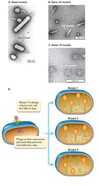
FIGURE A4.7 ■ Developmental mutants of phage T4. Analyzing mutants like these made it possible to decipher the genetic pathway governing assembly of the structure. A.
Mutations in developmental genes lead to variant capsid morphology such as a giant head. B. Gene 23 defect results in tails without heads. C. Gene 19 defect results in baseplates without tails. D. Design of experiment using temperature-dependent mutations to identify steps of T4 assembly.
H. DOERMAN ET AL. 1973. J. VIROL. 12 :374
Y. KIKUCHI AND J. KING 1975. J. MOL. BIOL. 99 :673
Y. KIKUCHI AND J. KING 1975. J. MOL. BIOL. 99 :673
In some cases, defective assembly leads to a bizarrely altered shape of the particle, such as a phage with a giant head (Fig. A4.7A). In other cases, a defective gene simply prevents progression in the pathway, halting assembly at an unfinished stage. For example, Figure A4.7B shows the phage particles found in a cell infected by a phage defective for gene 23. The unfinished particles consist of tail structures only. This defect occurs because gene 23 is required for assembly of the head, which must be completed before attachment to the tail. In Figure A4.7C , which shows phages defective for gene 19, we see only baseplates because the product of gene 19 is needed to build the tail on the baseplate. In this case, assembled heads are found near the cell membrane. Figure A4.7D summarizes how three different mutations lead to different defects in phage development. By analyzing the genetics and microscopy of hundreds of such mutants, we can map the assembly of the entire virion.
Glossary
capsid The protein shell that surrounds a virion’s nucleic acid. Within an enveloped virus, such as HIV, the capsid may be called a core particle.
tailed phage A phage such as T4 that contains a genome delivery device called the tail.
early gene A viral gene expressed early in the infection cycle.
late gene A viral gene expressed late in the replication cycle.
assembly 1. In a virus, the packaging of a viral genome into the capsid to form a complete virion. 2. In metagenomics, the piecing together of DNA sequence reads into contigs, and of contigs into a scaffold.
lysis Also called burst. The rupture of the cell by a break in the cell wall and membrane.
A4.2 The Filamentous Phage M13: Vaccines and Nanowiresnot assigned
Phage M13 has a filamentous structure, very different from the icosahedral capsids of phages T4 and lambda (Fig. A4.8).
Filamentous bacteriophages consist of an extended, flexible tube of variable length. The variable-length tube is able to package varying lengths of DNA—an advantage for genetic engineering. An experimental application of the filamentous phage M13 is to engineer vaccines. The vaccine is made by recombining the M13 coat protein gene with an antigenic gene of a pathogen such as hepatitis A virus. The recombinant M13 phage then displays the hepatitis A protein in its coat. The phage acts as an immunogenic carrier for the antigens—a technique known as “phage display.” Another impressive use of filamentous phages is to grow microscopic metal “nanowires” that conduct electricity. The wires are made using a mutant phage whose coat proteins bind metals such as gold and cobalt. Filaments form microscopic tubes of metal around the phage, and these can be used in the manufacture of tiny batteries.
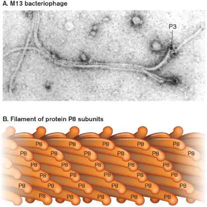
FIGURE A4.8 ■ A filamentous phage: phage M13. A. M13 filamentous phage particles (8 nm wide, 900 nm long) with the gene encoding tail protein P3 fused to a gene encoding anti-tetanus-toxoid antibody. The protein P3-antibody complex was stained with tetanus toxoid-colloidal gold, observed as a black sphere at the tip of the phage filament (TEM). B. Model of filamentous phage structure consisting of a helical tube of protein P8 subunits, based on X-ray crystallography.
CARLOS F. BARBAS III ET AL. 1991. PNAS 88 :7978, FIG. 2A
Filamentous Phage Structure
The M13 capsid consists of a flexible protein cylinder that is about 150 times as long as it is wide (Fig. A4.8A). The cylinder is assembled around a supercoiled, single-stranded circle of DNA. The advantage of the filamentous structure is that it requires a relatively small number of different protein subunits encoded in the genome. Similar flexible filamentous structures have evolved independently in animal and plant viruses; compare, for example, Ebola filovirus and tobacco mosaic virus (see Section 6.2). Filamentous animal viruses, however, show a virulent infection cycle completely different from the slow release of filamentous phages.
Filamentous phages, like the tailed phages, must construct progeny using the materials and metabolism of a host cell. Yet the means by which they achieve this goal differ greatly from those of T4. Where T4 features remarkable complexity in its capsid, the structure of M13 is a simple helical filament (Fig. A4.8). Instead of the 300 genes of the T4 genome, M13 manages just as successfully with 11, including some that overlap (Fig. A4.9). And instead of lysing its host, M13 allows the host to continue growing, although more slowly than uninfected cells do.
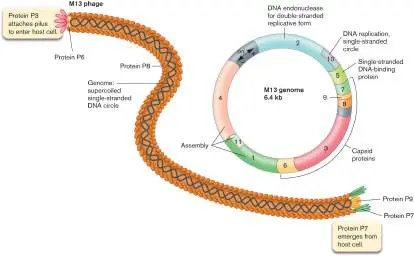
FIGURE A4.9 ■ The M13 phage structure and genome. Phage M13 structure (left) with proteins color-coded to match genes in the phage genome (right). Note that the actual phage filament is about 150 times as long as it is wide.
Each subunit of the M13 filament consists of a short, alpha-helical peptide encoded by gene 8. The protein P8 subunits have positively charged lysine residues at the internal end to bind the negatively charged DNA. The subunits extend outward at a shallow angle, looking like bunches of bananas stacked as a long tube (Fig. A4.8B ). The tube is tipped at one end by five copies of protein P3, a much larger and more flexible protein that binds the cell-surface receptor during phage adsorption (Fig. A4.9). At the opposite end of the tube are proteins P7 and P9, which enable a progeny phage particle to emerge from the cell.
The simple, flexible nature of the filamentous phage has proved useful for development of phage display technology. In phage display, a gene encoding a key portion of a protein domain (usually a short peptide) is cloned at the terminus of the sequence for one of the phage coat proteins, such as protein P8 or protein P3 (Fig. A4.9). The recombinant phage now displays the desired protein domain on its coat, where it can be recognized by antibodies or can selectively bind to a target molecule. This technique can be used to screen a library of recombinant phages for binding partners to an antibody or to a target regulatory protein, such as a cell cycle regulator involved in tumorigenesis.
Adsorption to Host and Delivery of DNA
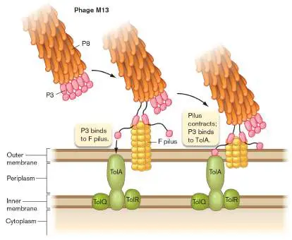
Phage M13 infects only F + strains of Escherichia coli, which possess F pili. The phage adsorbs by binding the tip protein P3 to an F pilus ( Fig. A4.10). The pilus contracts, bringing the phage into contact with a coreceptor, or secondary receptor protein, TolA. The TolA complex is an important structural anchor of the E. coli cell envelope. Coreceptors for host attachment are a common feature of viruses, including animal viruses such as influenza virus and HIV-1. FIGURE A4.10 ■ M13 adsorption to the pilus and TolA. Attachment to pilus of F + cell.
As the M13 filament contacts the cell, it transfers its DNA across the cell envelope by an unknown mechanism. Unlike phage T4, the empty M13 filament does not remain intact outside the cell. Instead, the filament subunits disassemble and insert into the inner membrane, where they eventually join the pool of newly synthesized subunits to form progeny phages. Because most M13 assembly steps involve the membrane, we know less about the details of M13 assembly than we do about the assembly of T4, whose components are all cytoplasmic and therefore easier to study.
M13 Reproduction Spares the Host
As the infecting capsid dissolves in the inner membrane, the phage DNA (single-stranded circle) enters the cytoplasm. Phage DNA is replicated by host enzymes while host cell growth continues. Replication of the single-stranded genome starts with expression of phage protein P2, which initiates synthesis of a double-stranded replicative form (Fig. A4.11A). The replicative form then undergoes rolling-circle replication of the (+) strand. The growing concatemer (multiple repeats of the (+) strand) is protected by subunits of protein P5, a single-strand binding protein. As each genome length is completed, it is nicked and circularized without overhang. The circular DNA is packaged with capsid proteins at the cell membrane.
FIGURE A4.11 ■ Assembly and export of M13 progeny phages. A. Replication of phage M13. Most assembly is conducted by phage proteins within the cell membrane. B.
Assembly for export through the pore made of P4 monomers. P4 makes a channel used to export filamentous phages without lysing the bacterial host. The single-stranded DNA of the filamentous virus is coated by P8 subunits as it passes through
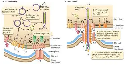
the membrane, and then is passed through the P4 channel to emerge as a mature, infectious phage.
Unlike phage T4, phage M13 replication is tightly controlled to limit the number of phage genomes and thus avoid destroying the host cell. Competing functions of phage proteins P2, P5, and P10 regulate levels of phage DNA replication and phage protein expression. The double-stranded DNA replication initiated by P2 is inhibited by protein P10. Replicated single-stranded DNA (ssDNA) genomes can be directed to the phage assembly machinery and packaged into progeny M13 particles—or, instead, the genomes can be sequestered by P5, a step that postpones progeny phage production. These molecular control steps limit the levels of phage DNA and proteins as the infected cell grows.
The transition of phage M13 from single-stranded to double-stranded was important historically because of its role in DNA sequencing. When DNA sequencing methods were first invented by Fred Sanger, they required a template that was single-stranded. But recombinant DNA techniques required double-stranded DNA for restriction endonuclease cleavage and ligation. The most effective way to obtain large quantities of a single-stranded template was to splice a gene sequence within a double-stranded M13 replicative form. The replicative form was then used to transform cells that subsequently produced M13 phages carrying single-stranded DNA. Today, a single-stranded template is no longer required for PCR-based sequencing, but M13-based vectors remain useful for special applications, such as phage display.
Filamentous Phages Self-Assemble at the Inner Membrane
The M13 genome expresses packaging proteins, including protein P8, as well as the specialized tip proteins P3, P7, and P9 (Fig. A4.9 ). All packaging proteins insert initially into the inner cell membrane. To mediate their assembly, a pore complex forms, composed of a ring of protein P4 monomers. The ring of P4 monomers extends through all layers of the envelope, where it acts as a pore. The positive charges on the N-terminal end of P4 help attract the negatively charged DNA into the pore. The pore guides the assembly of P8 monomers (also positively charged) and other packaging proteins around the DNA (Fig. A4.11B ). To avoid lysing the host cell, the pore is plugged by proteins P1 and P11 until phage assembly begins.
As the P5-coated chromosome enters the pore, the protein P5 subunits are replaced by the growing tube of P8 proteins. The final step of phage export involves capping with protein P3, which is needed for attachment to and infection of the next host cell. The pore complex is then re-capped with P1 and P11 to avoid lysis. The production of M13 phages slows growth of the host because of resource consumption, but it does not destroy the host cell; in fact, several of the phage’s 11 gene products are devoted to preventing host cell lysis. The evolved strategy of M13 is to maintain its current host at minimal cost, rather than to maximize its immediate number of progeny.
A4.3 Poliovirus: (+) Strand RNA Virusnot assigned
A medically important group of viruses is the picornaviruses (Table A4.1), a class of icosahedral RNA viruses whose life cycle has some interesting parallels to that of bacteriophage T4. The picornavirus poliovirus causes the paralytic disease poliomyelitis, or polio. The host range for infection by poliovirus is limited to humans and our closest relatives, such as chimpanzees. Closely related picornaviruses, such as the rhinoviruses and coxsackievirus, cause the common cold. A major veterinary concern is aphthovirus, the cause of foot-and-mouth disease in cattle and swine. In 2001, an outbreak of foot-and-mouth disease in the United Kingdom caused major economic and societal disruption.
TABLE Picornavirus Species (Examples) A4.1
Aphthovirus Foot-and-mouth disease Foot-and-Cattle, virus (FMDV) mouth swine disease Hepatovirus Hepatitis A virus (HAV) Hepatitis Humans Cardiovirus Encephalomyocardiovirus Encephalitis, Mice myocarditis Erbovirus Equine rhinotracheitis B Rhinotracheitis Horses virus (ERBV) (respiratory infection)
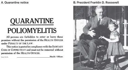
Poliovirus is famous historically for the outbreaks of poliomyelitis that swept the United States during the first half of the twentieth century (Fig. A4.12A). The virus enters the body through the mouth from fecal sources such as contaminated water. In most cases, poliovirus infection causes a mild gastrointestinal disease. But the virus can also invade motor neurons of the spinal cord and lower brain, resulting in paralysis. Tens of thousands of Americans were infected, including many young children. Many had few or no symptoms but could transmit the disease to others. About 1% became paralyzed, of whom about 15% died. Decades after infection, paralyzed individuals may suffer post-polio syndrome, a condition in which muscles that were previously affected by the polio infection experience atrophy and pain. Today, polio infection has nearly been wiped out worldwide, but outbreaks still occur where vaccination levels are low. For example, in 2022, a case of polio occurred in a New York community that refused vaccinations. FIGURE A4.12 ■ Polio in the United States. A. Board of Health quarantine notice, around 1900, aiming to exclude polio carriers. B. The most famous polio victim, President Franklin Delano Roosevelt, with his gardener’s granddaughter Ruthie Bie and his dog Fala. Roosevelt’s legs were paralyzed permanently, and he used a wheelchair throughout his presidency.
GRANGER
FRANKLIN D. ROOSEVELT LIBRARY
President Franklin D. Roosevelt (Fig. A4.12B ), himself affected by the disease, established a national foundation called the March of Dimes to develop a polio vaccine. Support from the March of Dimes led Jonas Salk and colleagues at the University of Pittsburgh to develop the first vaccine against polio in 1952. The spectacular success of the March of Dimes set the pattern for future public support for research on more challenging diseases, such as cancer and AIDS.
Despite the success of the polio vaccine, questions remain. The vaccine is not perfect; could better vaccines avoid the one-in-a-million deaths actually caused by the vaccine? What is the cause of the post-polio syndrome that affects some former polio victims 30– 40 years after their illness? Are there positive applications of
poliovirus, perhaps as a gene therapy vector or even as a treatment for brain tumors? To address these questions, we need to understand the virus and its host cell at the molecular level.
Poliovirus Structure
The poliovirus is so compact and symmetrical that it was crystallized early in the twentieth century. The 3D structure was modeled by X-ray crystallography and by digital reconstruction based on cryo-EM. Like many viruses, the capsid is fundamentally icosahedral (20-sided, as discussed in Chapter 6). Its form can be visualized as 60 triangular faces, each composed of proteins VP1, VP2, and VP3 (Fig. A4.13A). The three proteins arise from one long “polyprotein,” or polypeptide, synthesized as a unit and then cleaved into three pieces. The icosahedral form enables a small number of genes to encode a relatively large capsid. Because of this efficient coding, icosahedral capsids are common among animal and plant viruses, including, for example, the rhinoviruses, aphthovirus, herpesviruses, and rice yellow mottle virus.
FIGURE A4.13 ■ Poliovirus structure. A. Digital reconstruction of the poliovirus capsid based on TEM. The
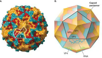
icosahedral structure is composed of external capsid proteins VP1 (orange), VP2 (blue), and VP3 (red). The three proteins form a structural unit, five of which form a pentamer. (PDB code: 1ASJ) B. Schematic of the capsid with RNA genome and VP4 proteins inside.
In the poliovirus capsid, each group of three triangular faces forms a pentamer. Beneath the pentamers of VP1, VP2, and VP3 lie subunits of a fourth protein, VP4 (Fig. A4.13B ). VP4 subunits coat the interior, strengthen the structure, and help package the RNA genome.
The RNA genome of poliovirus consists of a (+) strand (the strand that directly encodes protein sequences). As in eukaryotic messenger RNA (mRNA), its 3′ OH end has a poly-A tail but its 5′ OH end is capped with a special viral protein, VPg. The VPg serves as a primer for virally encoded RNA-dependent RNA synthesis. Unlike cellular polymerases (which use RNA primers exclusively), some viral polymerases are primed by proteins.
Virus Attachment and Genome Entry
The poliovirus attaches to the host at a species-specific receptor protein location. The virus can infect only those host cells that contain a poliovirus receptor (PVR), a cell membrane glycoprotein, also known as CD155, found on cells of the intestinal epithelium and on the surface of motor neurons. The normal function of CD155 is to help with adhesion of adjacent epithelial cells and to enhance the humoral immune response (antibody production). Unfortunately, poliovirus evolved to bind to this protein and gain entry into the cell. Poliovirus can bind only to the exact PVR sequence found in humans and chimpanzees. Thus, model animals such as mice cannot normally be infected. However, a transgenic mouse expressing a recombinant human gene for PVR can be infected by poliovirus. In 2003, the polio mouse became the first transgenic animal used to develop pharmaceuticals for a human disease. The mouse model was of limited use, however, because the mice develop only the intestinal symptoms but not the full neurological complications of poliomyelitis.
The PVR protein can bind the polio virion at any vertex of the capsid pentamer (a pentamer is shown in Fig. A4.13B ). PVR actually binds to adjacent VP2 and VP3 proteins surrounding the VP1 subunits, as shown in Figure A4.14. When poliovirus attaches to a cell, multiple receptor molecules diffuse near it and eventually bind to several faces of the capsid. As the poliovirus receptors bind, the membrane forms a cup around the capsid. Unlike other viruses, such as influenza virus, poliovirus requires only the earliest stage of endocytosis; the virion delivers its RNA to the cytoplasm immediately after internalization beneath the cell membrane. Thus, poliovirus infection is insensitive to drugs such as bafilomycin A1, which prevents infection by viruses that enter cells through endosomes. FIGURE A4.14 ■ Poliovirus attachment to the host cell. The poliovirus binds PVR at the VP2 and VP3 subunits. A
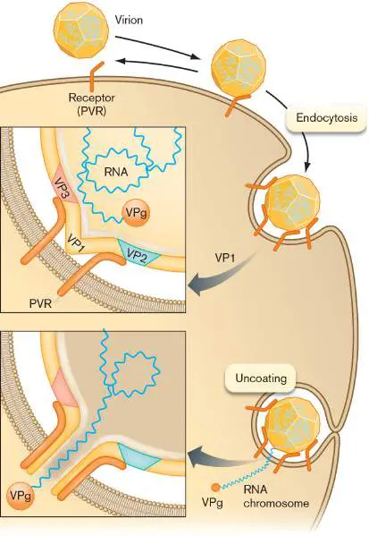
conformational change in the VP1 subunits allows insertion of the genome.
As the poliovirus interacts with the receptors, its pentamer structures shift so that the N-terminal ends of the five VP1 peptides are extruded (Fig. A4.14). The five VP1 molecules of the pentamer together poke through the cell membrane, generating a pore through which the chromosome passes into the cytoplasm. Transfer of the poliovirus chromosome into the cell parallels that of phage T4 (see Section A4.1) in that the nucleic acid is injected across the cell membrane through a viral device while the capsid proteins are left outside.
The viral chromosome, a (+) strand RNA with a protein VPg attached to the 5′ end, is said to be uncoated. The uncoated genome is now ready to direct the formation of progeny virions.
Gene Expression and Replication Cycle
The uncoated viral RNA has two distinct roles in producing progeny virions: (1) mRNA translation by host ribosomes and (2) template replication of progeny RNA genomes. These two processes occur in separate compartments within the host cells.
Translation to make peptides. In the cytoplasm, the RNA is translated to make a single large polyprotein (Fig. A4.15A). Polyprotein synthesis is a common feature of (+) strand RNA viruses (see, for example, the hepatitis C virus in Section A4.4). Within the polyprotein, domain 2A folds into conformation to act as a protease. The 2A protease cleaves the polyprotein between peptides 1 and 2– 3, and ultimately cleaves itself out of the polyprotein (autocatalysis). Overall, the three large precursor peptides (P1, P2, and P3) are cleaved by one of two viral proteases (products 2A and 3C) to generate a total of 11 different protein products. The P1 precursor forms the repeating unit of the icosahedral capsid; each unit is ultimately cleaved into four peptides. The P2 precursor is cleaved to form a two-subunit protease and an RNA-membrane interaction protein. The P3 subunit is cleaved to form the VPg primer for RNA synthesis (3B), another protease (3C), and RNA-dependent RNA polymerase (3D).
FIGURE A4.15 ■ Polio genome and capsid assembly. A. The (+) strand RNA genome encodes three precursor polypeptides: P1, P2, and P3. These precursors are cleaved by proteases to generate the virus capsid proteins (VP1, VP2, and VP3), as well as the RNA-dependent RNA polymerase, proteases, and other assembly proteins. B. The capsid proteins are assembled together into the 5S structural unit before cleavage by
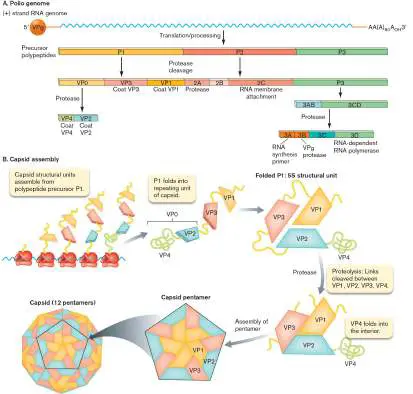
protease. Five 5S units are packed into a pentamer, 12 of which assemble to build the icosahedral capsid.
The capsid proteins ultimately arise from cleavage of P1, but not immediately (Fig. A4.15B ). First, the peptide domains VP4-VP2-VP3-VP1 fold into the tertiary structure of the capsid’s major repeating unit. The repeating unit is called the 5S structural unit, referring to its size in Svedberg units of sedimentation rate in a centrifuge. After folding together, each 5S unit then undergoes proteolysis at the junctions between the four subunits (VP1 through VP4). The 5S units then join together in groups of five to form pentamers, 12 of which ultimately assemble around a viral RNA chromosome.
RNA genome replication. In the overall cycle of replication (Fig. A4.16), the RNA chromosome serves as mRNA for peptide translation (step 1) and also as a template for RNA synthesis by RNA-dependent RNA polymerase (translated from the genomic RNA as part of precursor peptide P3). The RNA-dependent RNA polymerase synthesizes RNA within special vesicles formed out of the endoplasmic reticulum (ER), a process induced by the poliovirus (step 2). The virus-induced vesicles contain cytoplasm trapped within “inside-out” ER membrane. The vesicles contain no ribosomes, so only RNA synthesis occurs. Because these vesicles are the site of genome replication, they are known as “replication complexes” or “viral factories.”
FIGURE A4.16 ■ Replication cycle of poliovirus. Within the viral factories, the (+) strand RNA is tethered to the vesicle membrane by protein 2C (Fig. A4.16, step 3). The viral RNA-dependent RNA polymerase is tethered to the membrane by the membrane-embedded protein 3AB (step 4). Protein 3AB is cleaved by a protease, releasing the B portion, which is now called VPg. The VPg peptide provides a tyrosine-OH group, which primes RNA
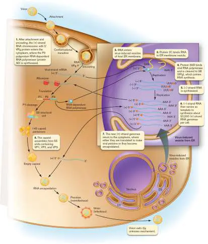
synthesis complementary to the (+) strand RNA template, generating (−) strand RNA (step 5). Protein primers are unique to viral genomes; by contrast, in uninfected host cells, all replication of DNA genomes is primed by the 3′ OH end of RNA. Protein primers, however, are common in many kinds of viruses.
The (−) strands then serve as templates to make new (+)
strands (Fig. A4.16, step 6). For each process of RNA synthesis, the template RNA is bound at the membrane by protein 2C. The daughter strand synthesis is again primed by a tyrosine-OH of VPg. The (+) strand RNAs now exit the ER and return to the cytoplasm (step 7), where some of them serve as mRNA for further translation to peptides, while others are packaged into capsids (step 8) to complete the poliovirus particles. Polioviral RNA synthesis is extremely efficient, generating about 50,000 copies per host cell.
Viral Exit and Dissemination
How mature polio virions exit the cell is poorly understood, but their release is rapid and destroys the cell. The cytopathic effects of release are compounded by the host’s immune response, which includes inflammation that further damages cells.
Animal viruses, unlike bacteriophages, require a means of transmission not only from cell to cell but also from host to host. To complete its infection cycle, the animal virus may need to infect several different kinds of cells in different tissues. Poliovirus initially infects epithelial cells of the gut lining (Fig. A4.17). Some of the progeny virions remain in the gut, sustaining infection cycles for weeks or months. In most cases, the infection remains confined to the gut, and infected people never realize that they carry a potentially deadly disease. But in some cases, virions move to the gut-associated lymphoid tissue, leading to dissemination (spread to different places) throughout the lymphatic system and associated organs. Large numbers of virus particles are released into the blood —a condition known as viremia. Viremia facilitates dissemination across the blood-brain barrier, leading to infection of neurons. The destroyed neurons cannot be regrown; thus, nerve loss results in paralysis.
FIGURE A4.17 ■ Poliovirus dissemination. Polio virions initially infect the gut epithelium, and then they are disseminated through the lymphatic system and blood. Some virions cross the blood-brain barrier to infect neurons, causing paralysis.
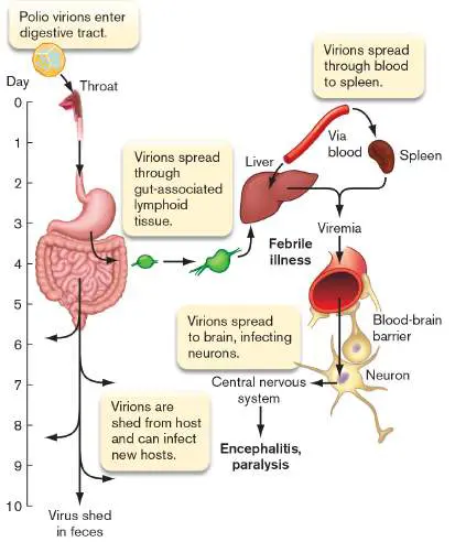
Throughout the course of infection, polio virions continue to be released from the host via the digestive tract. The process of viral release is known as virus shedding. Poliovirus can be shed from the gastrointestinal tract for several weeks after symptoms have ended, thus enhancing transmission to the next host. Even a person with only mild symptoms can transmit the disease to another individual, who may then develop paralysis. The implication for epidemiology is that any one case of poliomyelitis (paralytic polio) represents several hundred cases of mild or undetected infection, any of which could transmit to someone else and cause a case with paralysis. That is why widespread immunization is necessary to protect communities from paralytic polio.
Glossary
picornavirus A member of a medically important group of RNA viruses. An example is poliovirus.
poliovirus A human-specific RNA virus that is the causative agent of poliomyelitis.
poliomyelitis or polio A paralytic disease caused by the poliovirus.
poliomyelitis or polio A paralytic disease caused by the poliovirus.
poliovirus receptor (PVR)
A cell membrane glycoprotein, also known as CD155, found on cells of the intestinal epithelium and on the surface of motor neurons.
A4.4 Hepatitis C: (+) Strand RNA Virusnot assigned
Hepatitis C (HCV) is a blood-borne virus of the liver that infects 200 million people worldwide and now kills more people in the United States than AIDS does (at least 15,000 HCV deaths per year). In some countries, unscreened blood transfusions and reuse of medical supplies lead to high prevalence (proportion of individuals infected) of HCV. In the United States, an estimated 1% of the population carries HCV, acquired primarily through blood contact such as via transfusions and injection drug use, as well as by sexual transmission. However, most infected people have no recollection of risk factors and are unaware of their infection—which they could pass on to others. The potential consequences of HCV are particularly serious because they include cancer of the liver, now a leading cause of liver transplants. Since 2020, the Centers for Disease Control and Prevention (CDC) recommends HCV screening at least once in the lifetime of all patients age 18 or older, and periodic testing for those with risk factors.
About 15%–20% of infected individuals actually clear the virus, but in other patients HCV can persist for many decades without symptoms. We don’t know why 10%–15% of these patients go on to exhibit symptoms such as cirrhosis (the scarring of the liver leading to liver failure) and liver cancer. For those who develop symptoms, several new drugs have been developed that target various steps of the replication cycle (discussed later). New drugs are needed continually because the virus mutates into new resistant strains.
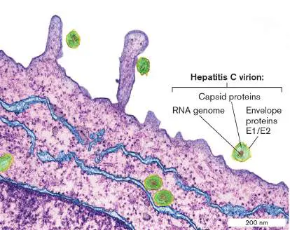
How do we discover new antiviral agents for HCV? The virus infects only humans and chimpanzees, so testing antiviral agents has been a challenge. While chimpanzees have yielded useful data, their study increasingly draws criticism from advocates for special treatment of these apes. A major challenge has been to grow the virus in cultured human cells. Between 2006 and 2008, several laboratories at last developed culture systems for HCV in hepatocarcinoma lines, as well as in normal hepatocyte tissue culture. Figure A4.18shows a TEM section through a cultured hepatocyte infected by HCV. Each virion contains an RNA genome packaged in an icosahedral capsid, surrounded by an envelope membrane containing glycoproteins. After binding to host cell-surface proteins, the virions become internalized by the cytoplasm as discussed shortly. Tissue culture enables us to characterize molecular targets for drugs—but culture does not replicate all aspects of infection of organs within an organism.
FIGURE A4.18 ■ Hepatitis C virus infection. Hepatitis C viruses infecting cultured liver cells (purple = cytoplasm; blue = ER membranes). Virions consist of a core of RNA (ribonucleic acid, brown) enclosed in a capsid (green), and surrounded by a glycoprotein envelope (yellow). Colorized TEM.
THOMAS DEERINCK, NCMIR/SCIENCE SOURCE
HCV Virion Structure and Genome
The HCV virion is small, appearing in electron micrographs to be 40– 70 nm in diameter. It consists of an icosahedral core particle surrounded by an envelope with spike proteins. Overall, the virion is composed of just three types of structural proteins: the protein C monomer, forming the icosahedral core that packages the RNA genomes; and envelope proteins E1 and E2, which form club-shaped dimers (Fig. A4.19). The genome encodes seven additional nonstructural (NS) proteins that function in the host cytoplasm during viral replication. The most crucial of these is NS5B, the RNA-dependent RNA polymerase, which synthesizes (−) strand template and (+) strand progeny genomes. Other nonstructural proteins include proteases (NS2, NS3, NS4A), RNA helicase (a second activity of protein NS3), interferon resistance protein (NS5A), and a protein in the replication complexes where virions assemble (NS4B).
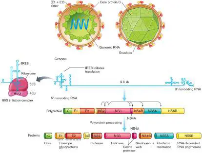
FIGURE A4.19 ■ Hepatitis C virus structure and genome. The (+) strand RNA genome is wound inside an icosahedral core composed of a single monomer type, protein C. The envelope, derived from host membrane lipids, contains paired glycoproteins E1 and E2. After the (+) strand RNA is uncoated, translation is initiated by the internal ribosome entry site (IRES; blowup), to synthesize one polyprotein. The polyprotein is processed (cleaved by proteases) to generate three structural proteins (C, E1, E2), plus seven nonstructural (NS) proteins that fill various functions during infection. But how is HCV viral RNA translated? The viral genomic RNA looks nothing like a host mRNA; it lacks the 5′ cap and 3′ poly-A “tail” signals that bind to a eukaryotic ribosome. This problem is solved differently by different kinds of viruses. Some RNA viruses, such as influenza virus, cleave the cap from a host mRNA to attach to their own (discussed in Section 11.2). But HCV uses a different mechanism. The HCV genome starts with a 5′ internal ribosome entry site (IRES). The IRES contains numerous stem loop structures in which the RNA doubles back and forms short regions of A-form duplex. These stems and loops twist around each other, forming a globular structure that has evolved to “fit” the ribosome in place of the cap used by standard host mRNA (Fig. A4.19). In the case of HCV, the IRES replaces most of the eukaryotic initiation factors as well; thus it initiates translation with high efficiency.
HCV Attachment and Host Cell Entry
What determines HCV host range and tropism, and how does the virion infect a cell? The host cell-surface receptors needed to bind the HCV virion play a major role in host range and tissue tropism ( Fig. A4.20A). The serum lipoproteins surrounding the HCV virion in the bloodstream bind to the low-density lipoprotein receptor (LDLR) on the luminal surface of the hepatic cell. Two other host proteins (CD81 and SR-B1) must bind E2 before infection can occur. Antibodies to these host proteins block HCV infection.
FIGURE A4.20 ■ HCV attachment and entry into liver cells. A. The virion, coated in lipoproteins, binds to the liver cell-surface proteins LDLR (low-density lipoprotein receptor), SR-B1 (scavenger receptor class B type 1), and CD81 (a tetraspanin).
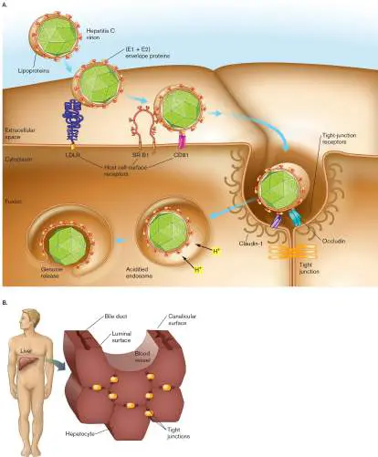
Virion entry also requires binding to the tight-junction proteins claudin-1 and occludin. The endocytic vesicle containing the virion fuses to a lysosome, whose acid induces viral envelope fusion with the vesicle membrane. The core particle undergoes uncoating, and the RNA genome enters the cytoplasm. B. Liver cells are connected by tight junctions, which is where HCV binds to infect.
Sources: Part A modified from D. Moradpour. 2007. Nat. Rev. Microbiol. 5:453; part B, from T. Pietschmann. 2009. Nature 4578 :797.
After initial binding to the proteins LDLR, CD81, and SR-B1, HCV then binds to additional proteins that form part of the “tight junction” between cells: claudin-1 and occludin. Tight junctions are structures that join adjacent cells within a tissue, preventing penetration of the luminal contents (Fig. A4.20B ). But surprisingly, viruses such as HCV can target the tight junction for host entry. It has also been proposed that progeny virions use the tight junction as a route for cell-to-cell transmission. Cell-to-cell transmission avoids exposing the virus to components of the host immune system.
In 2011, Alexander Ploss and colleagues used knowledge of the HCV host receptors to engineer a mouse that supports HCV infection. The mouse was “humanized” by transfection with adenoviral vectors expressing the human version of the genes that encode CD81 and occludin. These “human” proteins, in combination with the mouse versions of SR-B1 and claudin-1, enabled successful liver infection by HCV. The infection was visualized in the animal using a special HCV strain (HCV-CRE) modified to induce expression of the luciferase reporter, which generates bioluminescence. A drawback of the mouse system is that it supports only HCV uptake, but not HCV replication and virion release. Nevertheless, the achievement represents the first breakthrough in devising an immunocompetent model animal to test new anti-HCV drug therapies.
Replication Cycle of HCV
How can a virus expressing as few as ten genes commandeer the entire machinery of a host cell? As HCV infects its host, its replication cycle includes many processes in which viral components trick the host into cooperating (Fig. A4.21). This viral trickery is a recurring theme of all virus infections. However, each step of viral replication offers us an opportunity to devise new antiviral agents. FIGURE A4.21 ■ Replication cycle of HCV. The HCV virion binds to cell receptors and becomes endocytosed. The replication cycle includes lysosome fusion and uncoating, translation by host ribosomes, and replication by viral RNA-dependent RNA polymerase. The virions assemble and exit the host cell by exocytosis or by transfer through a tight junction into a neighboring cell (cell-to-cell transfer).
Source: Modified from Darius Moradpour. 2007. Nat. Rev. Microbiol. 5 :453.
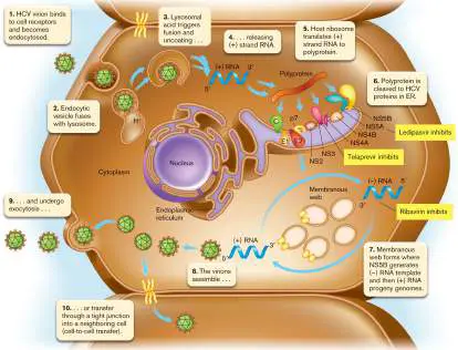
As the HCV virion binds host receptors at or near a tight junction (Fig. A4.21, step 1), the virion becomes endocytosed by the host cell membrane. The endocytic vesicle fuses with a lysosome (step 2). The acidity of the lysosome generally counteracts pathogenic microbes, but in this case it triggers the virion envelope to fuse with the endosome membrane. The membrane fusion generates an opening that releases the core particle into the cytoplasm (step 3). The core disassembles, releasing the (+) strand RNA genome (step 4).
In the cytoplasm, the IRES of the RNA genome binds to host ribosomes associated with the endoplasmic reticulum (ER). The RNA genome is translated to a polyprotein (Fig. A4.21, step 5). Polyprotein synthesis is a common mechanism found in RNA viruses. The polyprotein must be cleaved into separate proteins (step 6). First, a host enzyme, endoplasmic reticulum signal peptidase, cleaves the structural proteins (core subunit E, envelope proteins E1 and E2). (The normal function of the signal peptidase is to cleave the signal peptide from cell proteins after signal-directed transport to a membrane compartment.) The remainder of the HCV polyprotein gets cleaved by two virus-encoded proteases, composed, respectively, of subunits NS2 and NS3 (NS2-NS3) and of NS3 and NS4A (NS3-NS4A). The viral proteases are self-cleaving; that is, they actually fold into their active domains within the polyprotein, and then cleave themselves out.
The protease subunit NS3 is the target of important drugs such as telaprevir. Telaprevir binds the serine residue of the active site of NS3, preventing substrate binding. The drug has activity highly specific to HCV. Its cure rate is enhanced by combination with another antiviral that attacks a different step, such as ribavirin, a nucleoside analog that blocks the RNA-dependent RNA polymerase (protein NS5B). A combination of agents that blocks two different steps can prevent viral escape by mutation and greatly improve the cure rate.
Most of the virus-encoded proteins sit within the membranes of vesicles budding out of the ER. The formation of these viral vesicles is poorly understood, but it is thought that the vesicles detach from the ER and move into the cytoplasm, forming a “membranous web” containing replication complexes (Fig. A4.21, step 7). Figure A4.22shows a fluorescence micrograph in which the replication complexes are visualized by green fluorescent protein (GFP) fused to viral protein NS5A, which becomes part of the replication complex. NS5A is the target of ledipasvir, a member of yet another class of antivirals discovered through research on HCV replication.
FIGURE A4.22 ■ HCV replication complexes. Membranous replication complexes surround the nucleus of a transformed liver cell in tissue culture. The cultured cells have been engineered to
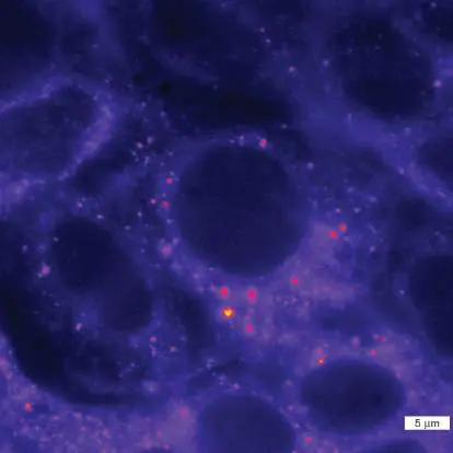
express viral protein NS5A fused to green fluorescent protein (GFP). When the cells are infected with HCV, the NS5A-GFP proteins are expressed and incorporated into the membranous HCV replication complexes.
BENNO WOLK ET AL. 2008. J. VIROL. 82 :10519
Within each replication complex, the (+) strand RNA serves as a template for the RNA-dependent RNA polymerase to make (−)
strand RNA (Fig. A4.21, step 7). The (−) strand RNA serves as a template to make (+) strand RNA to package into new core particles (step 8). The core particles then become coated by vesicle membranes containing E1 and E2. Vesicles carry the progeny virions to the cell membrane, where they fuse and the progeny virions are released (step 9). Alternatively, it has been proposed that some virions subvert the tight junctions for cell-to-cell transfer (step 10).
Mutation and Quasispecies Formation
Why does drug resistance arise so fast for HCV and for other RNA viruses? A trait found in all RNA viruses is an exceptionally high rate of mutation. This high mutation rate derives from the high error rates of RNA-dependent RNA polymerases (and of DNA-dependent reverse transcriptases such as that found in HIV). The error rate varies within the RNA genome and is typically higher at hypervariable regions within the envelope proteins exposed to the immune system. The high rate of mutation leads to production of exceptionally diverse progeny—so diverse that many progeny are actually defective in one or more aspects of viral propagation. For this reason, HCV propagated in tissue culture leads to virus that produces exceptionally high numbers of virions, but the virions show decreased infection rates within an organism.
Another source of genetic change in HCV is recombination. More than 10% of patients with HCV show evidence of genome recombination between different HCV strains. For recombination to occur, different virions must coinfect a common cell. Recombination then occurs by “template switching,” in which the RNA-dependent RNA polymerase complex detaches from the viral RNA template, still attached to its nascent RNA product, and then attaches to a new template at the same position and continues RNA synthesis. A recombinant progeny chromosome is formed, containing nucleic acid from two different parental virions.
Within a single infected patient, the high mutation rates generate multiple virus strains with differing properties of replication, tissue tropism, and resistance to antivirals. This dynamic population of diverse mutant strains is called a quasispecies. The term “quasispecies” acknowledges the limitations of characterizing genetic traits of all the individuals, as within the population individuals differ widely, and their differences change with time. Furthermore, some researchers argue that virions within a quasispecies interact cooperatively on a functional level, by serving complementary roles in the disease state, and thus collectively define the traits of the viral population. Clearer evidence for functional cooperation of a viral quasispecies is shown for HIV (discussed in Section 11.3).
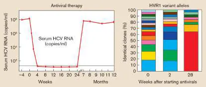
For hepatitis C, an example of a quasispecies evolving within a patient is illustrated in Figure A4.23. As HCV proliferates in the patient, mutations accumulate within hypervariable region 1 (HVR1) of envelope protein E2. Within 2 weeks of starting antiviral therapy, the quasispecies shows hugely different proportions of mutant strains (indicated by different-colored bars). Inevitably, some of the new strains turn out to be more resistant to the antiviral agents than the original parental strain was. Thus, after treatment stops, the quasispecies yet again shows vastly different strains—some of which lead to rapid resurgence of the virus. Nevertheless, as with HIV (see Section 11.3), effective new antiviral agents target various steps of the replication cycle. Current antiviral agents eliminate the virus from 90% of infected patients.
FIGURE A4.23 ■ Quasispecies formation in HCV. As HCV proliferates in the patient, the E2 hypervariable region 1 (HVR1) accumulates mutations leading to diverse alleles with different sensitivities to antiviral therapy. Colored bars represent variant alleles with mutant RNA sequences.
Source: Patrizia Farci. 2002. PNAS 99 :3081.
Glossary
internal ribosome entry site (IRES)
A site within an mRNA sequence where a ribosome can bind and initiate translation.
polyprotein A long peptide translated from one open reading frame but later cleaved into separate proteins with different functions. quasispecies A collection of isolates (usually viruses) from a common source of infection that have evolved into many different types within one host.