Chapter introduction
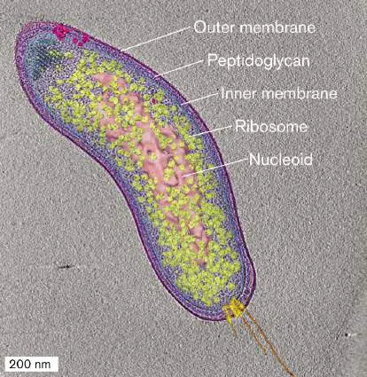
To grow and multiply, a microbial cell must obtain nutrients faster than its competitors do—or else share nutrients for mutual advantage. At the same time, each cell’s envelope must exclude toxicants such as your intestinal bile salts. To manage all these survival tasks, microbes have evolved an amazing set of molecular parts, from rotary flagellar motors to crystalline compartments that sequester CO 2. Cell extensions communicate with other cells and A bacterial cell contains surprising devices, such as a receptor array that acts like a “nose” to find food and motorized flagella to swim toward it. Unlike eukaryotes, a bacterium continually builds new parts while copying its DNA in time for cell division. This process requires an extraordinary ballet of proteins that form a “Z-ring” around the equator, while other proteins pulse back and forth from one pole to the other. These molecular dances appear in different colors under fluorescence microscopy, as we learned in For biotechnology, every part of a bacterial cell may be of use. Bacterial cell parts provide targets for antibiotics and vaccines. They offer tools for engineering, such as DNA polymerase and molecular motors. They provide models for design, such as thylakoids packed Chapter 3 explores the most common cell parts and functions of many species of bacteria and archaea. Further cell forms are explored in Chapter 18 (bacteria) and Chapter 19 (archaea). Microbial eukaryotes are presented in Chapter 20.
3.1 The Bacterial Cell: An Overviewnot assigned
All three domains of life—Bacteria, Archaea, and Eukarya—consist of one or more cells. Viruses are acellular, but they require a cell to infect. Certain features of a living cell are distinctive to one or more domains: Bacteria and Archaea are prokaryotes (cells that lack a nucleus). Bacteria have phospholipid bilayers similar to those of eukaryotes, but archaea have unique membrane and envelope structures such as ether membranes that enable survival in extreme environments. Nevertheless, many kinds of archaea live in moderate environments, such as soil, water, or human skin. Eukaryotic cells have extensive membranous organelles.
Organelles such as the endoplasmic reticulum and Golgi complex are reviewed in eAppendix 2. The mitochondria and chloroplasts of eukaryotic cells evolved by endosymbiosis with engulfed bacteria (see Chapter 17). Diverse microbial eukaryotes, such as fungi and protists, are explored in Chapter 20.
Chapter 3 focuses primarily on bacteria. Most bacteria share these traits: Thick, complex outer envelope. The envelope protects the cell from environmental stress and mediates exchange with the environment.
Compact genome. Prokaryotic genomes are compact, with relatively little noncoding DNA. Small genomes maximize the production of cells from limited resources.
Tightly coordinated functions. The cell’s parts work together in a highly coordinated mechanism, which may enable a high rate of reproduction.
Next, we embark on a tour of a typical Gram-negative bacterial cell. Along the way, we learn how our understanding of this model cell has emerged from microscopy, cell fractionation, and genetic analysis.
Note: This chapter assumes an understanding of introductory
biology and chemistry. For review, see eAppendix 1 (“Biological Molecules”) and eAppendix 2 (“Introductory Cell Biology”).
Model of a Bacterial Cell
A cell is more than a “soup” full of ribosomes and enzymes. In fact, the cell’s parts fit together in a structure that is ordered, though flexible. Cryo-electron microscopy (cryo-EM; see Section 2.6) reveals distinct parts of a cell (see the Chapter 3 opening image). On the basis of many such microscope images, we offer a model of a typical bacterium (Fig. 3.1). This model represents Escherichia coli, a common Gram-negative bacterium that resides on your gut epithelium. Its general features apply to many kinds of bacteria, particularly the Gram-negative inhabitants of your colon such as Proteobacteria and Bacteroidetes. Remember that we cannot literally “see” molecules within a cell, but microscopy and subcellular analysis provide the evidence for our model.
FIGURE 3.1 ■ Escherichia coli: a Gram-negative bacterium of the gut microbiome. The envelope includes the outer membrane, the cell wall and periplasm, and the inner (cell) membrane with an embedded chemoreceptor array. The cytoplasm contains enzymes, messenger RNA (mRNA) extending out of the nucleoid, and ribosomes. Ribosomes translate the mRNA to make proteins, which are folded by chaperones. The nucleoid contains the
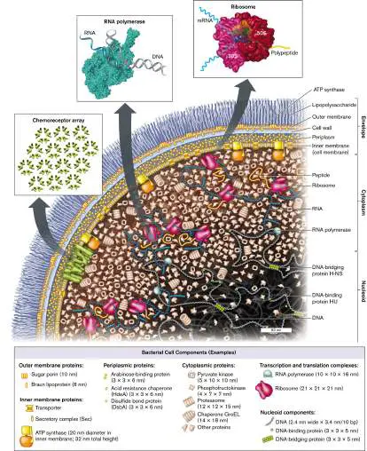
chromosomal DNA wrapped around binding proteins. (PDB codes: ribosome, 1GIX, 1GIY; RNA polymerase, 1MSW)
Within a cell, the cytoplasm consists of a gel-like network composed of proteins and other macromolecules. The cytoplasm is contained by a cell membrane, or plasma membrane. For E. coli, a Gram-negative bacterium, the plasma membrane is called the inner membrane, in order to distinguish it from the additional outer membrane. The inner membrane is composed of phospholipids, transporter proteins, and other molecules. This membrane prevents cytoplasmic proteins from leaking out and maintains gradients of ions and nutrients.
Between the inner and outer membranes lies the cell wall, a fortress-like structure composed of sugar chains linked covalently by peptides (peptidoglycan). The cell wall forms a single molecule that surrounds the cell. A Gram-positive species would have the cell wall outside its one plasma membrane. The wall material is flexible, but it limits expansion of the cytoplasm, keeping the cell membrane intact when water flows in. Like air inside a balloon, the resulting turgor pressure (expansion force) makes the cell rigid.
In a Gram-negative bacterium, such as our model E. coli (Fig. 3.1 ), the cell wall lies within the periplasm, a water-filled space containing nutrient-binding proteins and secretion machines. Outside the cell wall lies the outer membrane of phospholipids and lipopolysaccharides (LPS), a class of lipids attached to long polysaccharides (sugar chains). The LPS layer may be surrounded by a thick capsule. The capsule polysaccharides form a slippery layer that inhibits phagocytosis by amebas or white blood cells. The inner membrane, cell wall, and outer membrane constitute the Gram-negative cell envelope.
The bacterial envelope includes cell-surface proteins that enable the bacterium to interact with specific host organisms. For example, E. coli cell-surface proteins help the bacterium colonize the human intestinal epithelium. The nitrogen-fixing symbiont Sinorhizobium has cell-surface proteins that help the bacteria colonize legume plants for nitrogen fixation.
To sense the environment and to direct swimming, motile bacteria possess an array of chemoreceptors (receptor array), usually situated at one pole (rounded end of a rod-shaped cell). The receptors bind molecules from outside the cell or the periplasm and convert this binding information into signals within the cytoplasm. The signaling molecules direct the rotation of flagella (singular, flagellum), propeller-like organelles for swimming, described in Section 3.6. The amazing phenomenon of chemotaxis, how bacteria choose which direction to swim, is explored later in Chapter 12.
The Membrane Is a Two-Dimensional Fluid of Lipids and Proteins
The cell membrane (or inner membrane, for a Gram-negative cell) contains the cytoplasm within the external medium, and it can maintain aqueous compartments with very different properties. For example, the concentration of hydrogen ions (H +) can be a hundred times greater outside the cell than inside. The membrane consists of a phospholipid bilayer (double layer of phospholipids) including lipid-soluble proteins. The bilayer behaves as a two-dimensional fluid, within which proteins and lipids can diffuse (Fig. 3.2). The proteins form about half the mass of the membrane and provide specific functions, such as nutrient transport. We will examine this structure in detail in Section 3.2. FIGURE 3.2 ■ Bacterial cell membrane. The cell membrane consists of a phospholipid bilayer, in which the hydrophobic fatty acid chains are directed inward, away from water. The bilayer contains stiffening agents such as hopanoids. Half the membrane
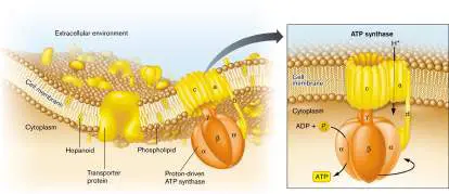
volume consists of proteins such as the ATP synthase protein complex.
Proteins embedded in the cell membrane often function together as a “complex.” The subunits of a complex fit together closely, like puzzle pieces. A famous protein complex is ATP synthase, an enzyme that couples the flow of H + ions across the membrane to the synthesis of ATP. The flow of H + ions through the ATP synthase is driven by the charge difference (more negative inside the cell) and the concentration difference (lower H + concentration inside). Thus, a surprisingly intricate “molecular machine” can sit in the membrane and synthesize ATP for the cell. Energy transduction by ion gradients is discussed further in Chapter 4 and Chapter 14.
Within the cell, the cell membrane and envelope provide an attachment point for one or more chromosomes. The chromosome is organized within the cytoplasm as a system of looped coils called the nucleoid. Unlike the round, compact nucleus of eukaryotic cells, the bacterial nucleoid is not enclosed by a membrane. Instead, loops of DNA extend throughout the cytoplasm. The DNA is transcribed by RNA polymerase to form messenger RNA (mRNA), as well as transfer RNA (tRNA) and ribosomal RNA (rRNA). As the mRNA transcripts extend, they bind ribosomes to start synthesizing polypeptide chains. As the polypeptides grow, they fold into their three-dimensional shape. This process may require help from protein complexes called chaperones. The concept of information flow from DNA to RNA to protein is presented in Chapters 7–10.
Biochemical Composition of Bacteria
The bacterial cell model in Figure 3.1represents the shape and size of cell parts but tells us little about the chemistry of the cell or about the cell’s environment. Chemistry explains, for example, why wiping a surface with ethanol kills microbes, whereas water has little effect. Water is a universal constituent of cytoplasm but is excluded by cell membranes. Ethanol, however, dissolves both polar and nonpolar substances; thus, ethanol disintegrates membranes and destroys the folded structure of proteins. For a review of elementary chemistry, see eAppendix 1.
All cells share common chemical components: Water , the fundamental solvent of life Essential ions , such as potassium, magnesium, and chloride ions Small organic molecules , such as lipids and sugars, that are incorporated into cell structures and that provide nutrition by catabolism Macromolecules , such as nucleic acids and proteins, that contain information, catalyze reactions, and mediate transport, among many other functions The details of bacterial chemistry emerged in the 1950s through pioneering studies by Fred Neidhardt at the University of Michigan and many colleagues. Cell composition varies with species, growth phase, and environmental conditions (as discussed in Chapters 4 and 5). Table 3.1summarizes the chemical components of an E. coli cell during exponential growth.
Molecules of a Bacterial Cell, TABLE Escherichia coli, during Balanced 3.1 a Exponential Growth
Number Percentage Approximate of of total number of different Component weight b molecules/cell kinds Water 70 20,000,000,000 1 Proteins 16 2,400,000 4,000 c RNA: rRNA, tRNA, and 6 250,000 200 other small RNA (sRNA) molecules mRNA 0.7 4,000 2,000 d Lipids: Phospholipids 3 25,000,000 50 (membrane)
Lipopolysaccharides 1 1,400,000 1 (outer membrane)
DNA 1 2 d 1 Metabolites and 1.3 50,000,000 1,000 biosynthetic precursors Peptidoglycan 0.8 1 1 (murein sacculus)
Inorganic ions 0.1 250,000,000 20 Polyamines (mainly 0.1 6,700,000 2 putrescine and spermidine)
Source: Modified from F. Neidhardt and H. E. Umbarger. 1996. Chemical composition
of Escherichia coli, p. 14. In F. C. Neidhardt (ed.), Escherichia coli and Salmonella:
Cellular and Molecular Biology, 2nd ed. ASM Press, Washington, DC.
Small molecules and ions. The E. coli cell consists of about 70% water, the essential solvent required to carry out fundamental metabolic reactions and to stabilize proteins. The water solution contains inorganic ions, predominantly potassium, magnesium, and phosphate. Inorganic ions store energy in the form of transmembrane gradients, and they serve essential roles in enzymes. For example, a magnesium ion is required at the active site of RNA polymerase to help catalyze the linking of ribonucleotides into RNA.
The cell also contains many kinds of small, charged organic molecules, such as phospholipids and enzyme cofactors. A major class of organic cations is the polyamines, molecules with multiple amine groups that are positively charged when the pH is near neutral. Polyamines balance the negative charges of the cell’s DNA and stabilize ribosomes during translation.
Macromolecules. Many cells have similar content of water and small molecules, but their specific character is defined by their macromolecules, especially their nucleic acids (DNA and RNA) and their proteins. DNA and RNA molecules can be isolated by size using agarose-gel electrophoresis, in which the negatively charged molecules migrate in an electrical field (see eAppendix 3). The total DNA plus RNA content of bacteria is relatively high, nearly 8% for E. coli —much higher than in multicellular eukaryotes. For microbes, the high nucleic acid content allows the cell to maximize reproduction of its chromosome while minimizing cell resources.
A high proportion of nucleic acids in food is a problem for us, because humans lack the enzymes to digest the uric acid waste product of nucleotides. That’s why we cannot eat most kinds of bacteria as a major part of our diet. Nevertheless, we do consume bacteria within vegetables, because all plants have bacteria growing within their transport tissues.
The cell’s genomic DNA directs expression of its proteins (discussed in Chapters 7–10). A given cell uses different genes to make different proteins, depending on environmental conditions such as temperature, nutrient levels, and entry into a host organism. Individual proteins are made in very different amounts, from 10 per cell to 10,000 per cell. The proteins expressed by a cell under given conditions are known collectively as a proteome. Other kinds of macromolecules are found in the cell wall and outer membrane. The bacterial cell wall consists of peptidoglycan, an organic polymer of peptide-linked sugars that constitutes nearly 1% of the cell mass, approximately the same mass as that of DNA. Peptidoglycan limits the volume of the enclosed cell, so water rushing in generates turgor pressure. This investment of biomass in the cell wall shows the importance (for most species) of maintaining turgor pressure in dilute environments, where water would otherwise enter by osmosis, causing osmotic shock (see eAppendix 2 and Section 3.2).
Thought Question
3.1 Which chemicals do we find in the greatest number in a bacterial cell? The smallest number? Why does a cell contain 100 times as many lipid molecules as strands of RNA?
Cell Fractionation
Cell fractionation (Fig. 3.3) is how we separate cell components such as membranes, ribosomes, and flagella. We can study these isolated parts in detail, though we lose information about interactions with other parts of the cell. Cell fractionation also provides purified proteins that act as antigens for candidate vaccines. For example, a vaccine against Neisseria meningitidis type B (meningococcus) contains a highly immunogenic outer membrane protein.
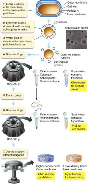
FIGURE 3.3 ■ Fractionation of Gram-negative cells. Cell periplasm fills with sucrose, and lysozyme breaks down the cell wall. Dilution in water causes osmotic shock to the outer membrane, and periplasmic proteins leak out. Subsequent centrifugation steps separate the proteins of the periplasm, cytoplasm, and inner and outer membranes. EDTA = ethylenediaminetetraacetic acid; OMP = outer membrane protein.
LARS D. RENNER AND DOUGLAS B. WEIBEL. PNAS 108 :6264
How can we disassemble a cell to isolate its parts? Early-twentieth-century microbiologists wondered how to separate cell parts without all the molecules mixing together. As we learned in Chapter 1, the answer was ultracentrifugation. Ultracentrifugation (that is, rotation at high speed) subjects a suspension of molecules to high g force by centripetal acceleration. The g force separates molecules by weight and density, as explained in eAppendix 3. Mary Jane Osborn (1927–2019) at the University of Connecticut Health Center discovered that the inner and outer membranes of Gram-negative bacteria have different densities, and thus can be separated by density gradient ultracentrifugation. Cell fractionation requires techniques that lyse (break open) the cell. The lysis method must generate enough force to separate the membrane lipids (held together by hydrophobic force) but not enough to disintegrate complexes of protein and RNA. For a Gram-negative cell, the method requires further specificity to separate “compartments” containing different sets of proteins: the inner and outer membranes, and the aqueous cytoplasm and periplasm.
Cell wall lysis and spheroplast formation. First we permeabilize the bacterial outer membrane by removal of Mg 2+ and Ca 2+ ions ( Fig. 3.3, step 1). The permeabilized membrane now allows sucrose to cross. Sucrose fills the periplasm, maintaining an osmotically stable solution.
Next, lysozyme cleaves peptidoglycan and thus breaks down the cell wall (step 2). Lacking the turgid cell wall, the cell swells into a sphere called a spheroplast. Spheroplasts can be seen by transmission electron microscopy (TEM).
To isolate the periplasmic contents, the spheroplasts are transferred to distilled water (Fig. 3.3, step 3). Water rushes in through the EDTA-weakened outer membrane, causing osmotic shock of the periplasmic compartment, while the inner membrane remains intact. The periplasm leaks out into the extracellular medium, where its proteins can be collected. The periplasm is a source of interesting proteins such as sugar transporters and chaperones (proteins that help other proteins fold under stress).
Note that for different kinds of cells, other means of cell disruption may work better. Mild-detergent lysis can dissolve membranes without denaturing proteins. Alternatively, sonication is a way to lyse a cell by intense ultrasonic vibrations that are above the range of human hearing. For especially tough cells, such as cyanobacteria, a “bead beater” with microscopic glass beads can tear the cells open. Ultracentrifugation. After osmotic shock, the spheroplasts undergo ultracentrifugation (Fig. 3.3, step 4) to separate the periplasmic contents from the other three types of cell compartments (cytoplasm, inner membrane, and outer membrane fragments, which form vesicles). The ultracentrifuge is a device in which tubes containing solutions of cell components are spun at very high speed. The high rotation rate generates centrifugal forces strong enough to separate subcellular particles (see eAppendix 3). The particles are collected in fractions of sample from the tube, and the fractions are observed by electron microscopy.
Spheroplast lysis. The pellet can now be further processed by a French press (Fig. 3.3, step 5), a device that squeezes cell contents through a narrow tube to break open the membranes. The broken membranes coalesce into tiny vesicles. A second step of ultracentrifugation now pellets the inner and outer membrane vesicles, while removing the cytoplasm in the supernatant (liquid above the pellet; step 6). The cytoplasm provides many types of proteins for study, such as FtsZ, as well as complexes (multiprotein structures) such as the ribosomes and DNA polymerases.
Finally, the membrane fraction contains a mixture of very different components of the inner membrane (such as electron transport proteins) and the outer membrane (such as cell-surface proteins that we could use to make vaccines). We can separate the inner and outer membranes by density gradient ultracentrifugation (Fig. 3.3, step 7). The gradient of solution density is generated by forming a gradient of sucrose concentration. In the gradient, the inner and outer membrane vesicles separate because of differences in their density, not their particle size. The lower-density fractions contain inner membrane vesicles, whereas the higher-density fractions contain outer membrane vesicles.
The membrane vesicle proteins are analyzed on electrophoretic gels (Fig. 3.4). We can identify the protein bands on the gel by enzyme digestion and mass spectrometry. The enzyme trypsin cleaves proteins only at aminoacyl residues lysine or arginine. The position of lysine or arginine residues is defined by the overall protein sequence; thus, the cleavage patterns generate peptides of defined composition.
FIGURE 3.4 ■ Protein analysis. A. Gel electrophoresis of total cell proteins compared to outer membrane proteins from cell fractionation. B. Outer membrane proteins are identified by tryptic digestion and mass spectrum analysis. The resulting peptide sequence is compared with those predicted from genomic data.
SDS-PAGE = SDS polyacrylamide gel electrophoresis.
M. THEIN ET AL. 2010. J. PROTEOME RES. 9 :6135
The mass of each defined peptide is then determined by mass spectrum analysis (mass spectrometry). Mass sizes are used to predict the peptide amino-acid sequence composition. The predictions are
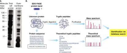
compared with a database of protein sequences that the organism’s genome predicts. Thus, a protein’s sequence data can be used to discover the gene that encodes it.
Thought Question
3.2 Suppose we wish to isolate multidrug efflux pumps, which are protein complexes that span the envelope from inner membrane to outer membrane. How might we modify the cell fractionation procedure to achieve such isolation?
A limitation of cell fractionation is that it provides little information about processes that require an intact cell, such as cell division. How can we remove or alter a part of a cell without breaking it open? An approach that is complementary to cell fractionation is genetic analysis. In genetic analysis, we can mutate a strain so as to lose or alter a gene; then we select mutant strains for loss of a given function. The phenotype of the mutant cell may yield clues about the function of the altered part, such as the bacterial cytoskeleton (see Section 3.3). Genetics and genetic analysis are discussed further in Chapters 7–12.
To Summarize
Bacterial cells are protected by a thick cell envelope. The envelope includes a cell membrane (plasma membrane) and a peptidoglycan cell wall.
A Gram-negative cell includes an outer membrane. The cell membrane of a Gram-negative cell is called the inner membrane. Between the outer membrane and inner membrane lies the periplasmic space, containing the cell wall. Bacteria are composed of nucleic acids, proteins, phospholipids, and other organic and inorganic chemicals. Proteins in the cell vary, depending on the species and environmental conditions.
The bacterial cytoplasm is highly structured. DNA replication, RNA transcription, and protein synthesis occur coordinately within the cytoplasm.
Cell fractionation isolates cell parts for structural and biochemical analysis. The compartments of a Gram-negative cell can be separated by cell lysis and ultracentrifugation. Cell compartments contain different types of proteins. The outer membrane and inner membrane, as well as the cytoplasm and periplasm, each contain distinct types of protein not found in the other compartments. Outer membrane proteins may be good candidate antigens for a vaccine.
Microscopy reveals cell structure. Transmission electron microscopy and cryo-electron microscopy show how cell parts fit within the cell as a whole.
Glossary
cell membrane Also called cytoplasmic membrane or plasma membrane. The phospholipid bilayer that encloses the cytoplasm.
plasma membrane See cell membrane .
inner membrane or inner cell membrane In Gram-negative bacteria, the membrane in contact with the cytoplasm, equivalent to the cell membrane.
outer membrane or inner cell membrane In Gram-negative bacteria, a membrane external to the cell wall. cell wall A rigid structure external to the cell membrane. The molecular composition depends on the organism; in bacteria, it is composed of peptidoglycan.
periplasm In Gram-negative bacteria, the gel-like solution between the outer and inner membrane; it contains the cell wall.
lipopolysaccharides (LPS)
Structurally unique phospholipids found in the outer leaflet of the outer membrane in Gram-negative bacteria. Many are endotoxins. envelope A structure external to the cell membrane, such as the cell wall or outer membrane of a bacterium. For a virus, the envelope is a membrane enclosing the capsid or core particle.
flagellum pl. flagella A filamentous structure for motility. In prokaryotes, a helical protein filament attached to a rotary motor; in eukaryotes, an undulating membrane-enclosed complex of microtubules and ATP-driven motor proteins.
flagellum pl. flagella A filamentous structure for motility. In prokaryotes, a helical protein filament attached to a rotary motor; in eukaryotes, an undulating membrane-enclosed complex of microtubules and ATP-driven motor proteins.
phospholipid bilayer Two layers of phospholipids; the hydrocarbon fatty acid tails face the interior of the bilayer, and the charged phosphate groups face the cytoplasm and extracellular environment. The cell membrane is a phospholipid bilayer.
nucleoid The looped coils of a bacterial chromosome.
electrophoresis A technique to separate charged proteins and nucleic acids that is based on how rapidly they migrate in an electrical field through a gel.
peptidoglycan Also called murein. A polymer of peptide-linked chains of amino sugars; a major component of the bacterial cell wall.
cell fractionation A procedure to separate cell components that often includes ultracentrifugation.
lyse Also called burst. The rupture of the cell by a break in the cell wall and membrane.
spheroplast A cell whose peptidoglycan is degraded by lysozyme; thus the cell loses its shape, forming a sphere.
ultracentrifuge A machine that subjects samples to high centrifugal forces and can be used to separate subcellular components.
Endnotes
1. Note a: Values shown are for a hypothetical “average” cell cultured with aeration in glucose medium with minimal salts at 37°C.
Return to reference a 2. Note b: The total weight of the cell (including water) is about 10⁻ 12 gram (g), or 1 picogram (pg). Return to reference b 3. Note c: The number of different kinds is difficult to estimate for proteins and mRNA because some genes are transcribed at extremely low levels and because proteins and RNA include kinds that are rapidly degraded. Return to reference c 4. Note d: In rapidly growing cells, cell fission typically lags approximately one generation behind DNA replication; hence, two identical DNA copies per cell. Return to reference d 5. Note d: In rapidly growing cells, cell fission typically lags approximately one generation behind DNA replication; hence, two identical DNA copies per cell. Return to reference d
3.2 Membrane Molecules and TransportUnit 3 · Structure
The form and function of a membrane depends upon its lipids and proteins (see Fig. 3.2). The most common type of membrane lipids are phospholipids (Fig. 3.5). Bacterial membranes consist of a bilayer of lipids that face each other tail to tail, keeping their hydrophobic side chains away from the water inside and outside the cell. The two layers of phospholipids in the bilayer are called leaflets. One leaflet of phospholipids faces the cell interior; the other faces the exterior. As a whole, the phospholipid bilayer imparts fluidity and gives the membrane a consistent thickness (about 8 nm).
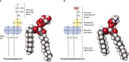
FIGURE 3.5 ■ Phospholipids. A. Phosphatidyl glycerol consists of glycerol with ester links to two fatty acids, and a phosphoryl group linked to a terminal glyceride. B. Phosphatidyl ethanolamine contains a glycerol linked to two fatty acids, and a phosphoryl group with a terminal ethanolamine. The ethanolamine carries a positive charge.
Membrane Lipids
A phospholipid possesses a charged phosphoryl “head” that contacts the water interface, as well as a hydrophobic “tail” of fatty acids packed within the bilayer. Lipid biosynthesis is a key process that is vulnerable to some antibiotics. For example, the bacterial enzyme enoyl reductase, which synthesizes fatty acids (discussed in Chapter 15), is the target of triclosan, a common antibacterial additive in detergents and cosmetics.
A typical phospholipid consists of glycerol with ester links to each of two fatty acids, and a phosphoryl polar head group, which at neutral pH is deprotonated (negatively charged; Fig. 3.5). This kind of phospholipid is called a phosphatidate. The negatively charged head group of the phosphatidate can contain various organic groups, such as glycerol to form phosphatidylglycerol (Fig. 3.5A). In other lipids, the polar head group has a side chain with positive charge. The positive charge commonly resides on an amine group, such as ethanolamine in phosphatidylethanolamine (Fig. 3.5B ). Phospholipids with positive charge or with mixed charges are concentrated in portions of the membrane that interact with DNA, which has negative charge.
Membranes require a uniform thickness and stability to maintain structural integrity and function. So why do individual membrane lipids differ in structure? Different environments favor different forms of membrane lipids. For example, lipid structure helps determine whether an organism can grow in a hot spring or whether it can colonize human lungs.
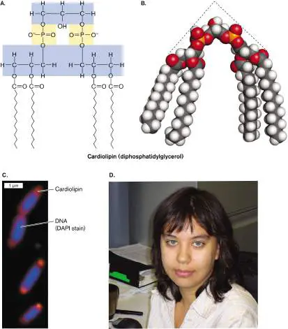
Environmental stress. Starvation stress increases bacterial production of lipids with an unusual type of phosphoryl head group. Cardiolipin, or diphosphatidylglycerol, is actually a double phospholipid linked by a glycerol (Fig. 3.6). Cardiolipin concentration increases in bacteria grown to starvation or stationary phase (discussed in Chapter 4). Tatyana Romantsov and Janet Wood, at the University of Guelph, showed how cardiolipin helps define the polar structure of a bacterial cell.
Within a rod-shaped cell, cardiolipin does not diffuse at random; it concentrates in patches called “domains” near the cell poles. The polar localization of cardiolipin was demonstrated by fluorescence microscopy, in which a cardiolipin-specific fluorophore localized to the poles of Escherichia coli. The “wedge” shape of cardiolipin (Fig. 3.6B ), with its narrow head group and wide fatty acid group, is thought to form concave domains of lipid that stabilize the curve of the polar membrane. Cardiolipin may enhance the formation of smaller cells during starvation. At the cell pole (Fig. 3.6C ), cardiolipin binds certain environmental stress proteins, such as a protein that transports osmoprotectants when the cell is under osmotic stress. Thus, a phospholipid can have specific functions associated with specific membrane proteins.
FIGURE 3.6 ■ Cardiolipin localizes to the poles. A. Cardiolipin is a double phospholipid joined by a third glycerol. B. A space-filling model of cardiolipin shows its triangular shape. C. Cardiolipin localizes to the bacterial cell poles, as shown by microscopy with a cardiolipin-specific fluorophore (orange). D. Tatyana Romantsov, at the University of Guelph, investigates how cardiolipin helps form the shape of a cell pole.
TATYANA ROMANTSOV ET AL. 2007. MOL. MICROBIOL. 64 :1455
TATYANA ROMANTSOV
The fatty acid component of phospholipids also varies. The most common bacterial fatty acids are hydrogenated chains of varying length, typically between 6 and 22 carbons. But some fatty acid chains are partly unsaturated (possess one or more carbon-carbon double bonds). Most unsaturated bonds in membranes are cis, meaning that both alkyl chains are on the same side of the bond, so the unsaturated chain has a “kink,” as in the cis form of oleic acid ( Fig. 3.7). Because the kinked chains do not pack as closely as the straight hydrocarbon chains do, the membrane is more “fluid.” This is why, at room temperature, unsaturated vegetable oils are fluid, whereas highly saturated butterfat is solid. The enhanced fluidity of a kinked phospholipid improves the function of the membrane at low temperature; hence, bacteria can respond to cold and heat by increasing or decreasing their synthesis of unsaturated phospholipids.
FIGURE 3.7 ■ Phospholipid side chains.
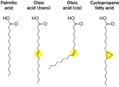
Another interesting structural variation is cyclization of part of the chain to form a stiff planar ring with decreased fluidity. The double bond of unsaturated fatty acids can incorporate a carbon from S -adenosyl-l-methionine to form a three-membered ring, generating a cyclopropane fatty acid (Fig. 3.7). Bacteria convert unsaturated fatty acids to cyclopropane during starvation and acid stress, conditions under which membranes require stiffening. Cyclopropane conversion is an important factor in the pathogenesis of Mycobacterium tuberculosis and in the acid resistance of food-borne toxigenic E. coli.
In addition to phospholipids, membranes include planar molecules that fill gaps between hydrocarbon chains. These stiff, planar molecules reinforce the membrane, much as steel rods reinforce concrete. In eukaryotic membranes, the reinforcing agents are sterols, such as cholesterol. In some bacteria, the same function is filled by pentacyclic (five-ring) hydrocarbon derivatives called hopanoids, or hopanes (Fig. 3.8). Like cholesterol, hopanoids fit between the fatty acid side chains of membranes and limit their motion, thus stiffening the membrane. Hopanoids provide biomarkers for petroleum exploration, as signs of potential petroleum formation in ancient rock.
FIGURE 3.8 ■ Hopanoids add strength to membranes. Hopanoids limit the motion of phospholipid tails, thus stiffening the membrane.
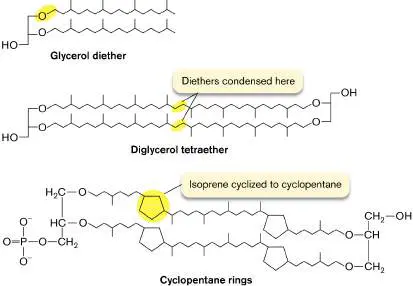
Archaea have unique membrane lipids. The membrane lipids of archaea differ fundamentally from those of bacteria and of eukaryotes. All archaeal phospholipids replace the ester link between glycerol and fatty acid with an ether link, C–O–C (Fig. 3.9 ). Ethers are much more stable than esters, which hydrolyze easily in water. This is one reason why some archaea can grow at higher temperatures than all other forms of life. Another modification is that archaeal hydrocarbon chains are branched terpenoids, polymeric structures derived from isoprene, in which every fourth carbon extends a methyl branch. The branches strengthen the membrane by limiting movement of the hydrocarbon chains.
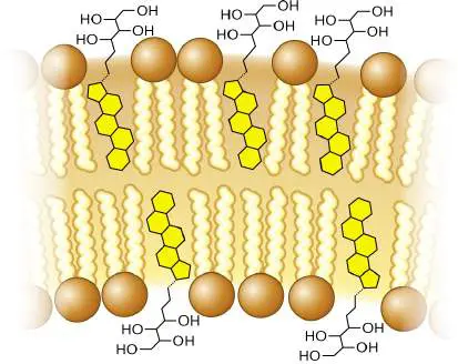
FIGURE 3.9 ■ Terpene-derived lipids of archaea. In archaea, the hydrocarbon chains are ether-linked to glycerol, and every fourth carbon has a methyl branch. In some archaea, the tails of the two facing lipids of the bilayer are fused, forming tetraethers; thus, the entire membrane consists of a monolayer. The most extreme hyperthermophiles, which live beneath the ocean at 110°C, have terpenoid chains linked at the tails, forming a tetraether monolayer. In some species, the terpenoids cyclize to form cyclopentane rings. These planar rings stiffen the membrane under stress to an even greater extent than do the cyclopropane chains of bacteria. (Thermophiles and other microbes in extreme environments are presented in Chapter 5. For more on archaea, see Chapter 19.)
Membrane Proteins
Membrane proteins are diverse and can serve different functions. Different membrane proteins provide structural support, communicate with the environment, secrete virulence factors, and mediate transport. Certain proteins have very specific roles; for example, the TetA protein expels the antibiotic tetracycline, making a pathogen resistant to tetracycline.
Structural support. Some membrane proteins anchor together different layers of the cell envelope (discussed in Section 3.3). Other proteins attach the membrane to the cytoskeleton or form the base of structures extending out from the cell, such as flagella.
Detection of environmental signals. In Vibrio cholerae, the causative agent of cholera, the membrane protein ToxR detects acidity and elevated temperature—signs that the bacterium is in the host’s digestive tract. The ToxR domain facing the cytoplasm then binds to a DNA sequence, activating expression of cholera toxin.
Secretion of virulence factors and communication signals. Membrane protein complexes export toxins and cell signals across the envelope. For example, symbiotic nitrogen-fixing rhizobia require membrane proteins NodI and NodJ to transport nodulation signals out to the host plant roots, inducing the plant to form root nodules containing the bacteria.
Ion transport and energy storage. Transport of ions across a membrane generates a transmembrane gradient that stores energy.
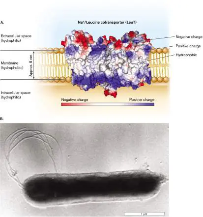
An example of a membrane protein is the leucine transporter LeuT (Fig. 3.10A). LeuT drives uptake of leucine, coupled to a gradient of sodium ions. The protein complex was purified for X-ray diffraction from Aquifex aeolicus, a thermophilic bacterium whose heat-stable proteins form durable crystals (Fig. 3.10B ).
Remarkably, LeuT is homologous (shares common ancestry) with a human neuron protein that transports neurotransmitters. Thus, this bacterial protein serves as a model for the study of neuron function. LeuT and other proteins embedded in a membrane require a portion of hydrophobic amino-acid side chains that are soluble amidst the hydrocarbon tails of the phospholipids. (For a review of protein structure, see eAppendix 1.) The hydrophobic portions of LeuT are shown in white in Figure 3.10A. Typically, several hydrophobic alpha helices thread back and forth through the membrane. Their middle surfaces, deeply embedded in membrane, have hydrophobic amino acid residues that interact favorably with the hydrocarbon chains of the phospholipids. Other peptide regions extend outside the membrane, containing charged and polar amino acids that interact favorably with the negatively charged head groups (such as phosphoryl groups) and with water.
Figure 3.10Aalso shows the LeuT charge distribution.
Hydrophobic amino acid residues (white) make the protein soluble in the membrane, while portions with negative charge (red) and positive charge (blue) interact with the phospholipid head groups and with water. The net result of these interactions is to lock the protein securely within the membrane.
FIGURE 3.10 ■ A cell membrane–embedded transport protein: the LeuT sodium/leucine cotransporter of Aquifex bacteria. A. The protein complex carries leucine across the cell membrane into the cytoplasm, coupled to sodium ion influx. (PDB code: 3F3E) B. Aquifex aeolicus grows at 96°C in hot springs.
KARL STETTER AND REINHARDT RACHEL, U. REGENSBURG, GERMANY
Molecules Cross the Cell Membrane
The cell membrane acts as a barrier to keep water-soluble proteins and other cell components within the cytoplasm. But how do nutrients from outside get into the cell—and how do secreted products such as toxins get out? Specific membrane proteins transport molecules across the membrane between the cytoplasm and the outside. Selective transport is essential for cell survival; it means the ability to acquire scarce nutrients, exclude waste, and transmit signals to neighbor cells.
Passive diffusion. Small, uncharged molecules, such as O 2, CO 2, and water, easily permeate the membrane. Some molecules, such as ethanol, also disrupt the membrane—an action that can make such molecules toxic to cells. By contrast, large, strongly polar molecules such as sugars, and charged molecules such as amino acids, generally cannot penetrate the hydrophobic interior of the membrane and thus require transport by specific proteins. Water molecules permeate the membrane, but their rate of passage is increased by protein channels called aquaporins (discussed in Chapter 4).
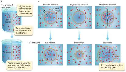
Osmosis. Most cells maintain a concentration of total solutes (molecules in solution) that is higher inside the cell than outside. As a result, the internal concentration of water is lower than the concentration outside the cell. Because water can cross the membrane but charged solutes cannot, water tends to diffuse across the membrane into the cell, causing the expansion of cell volume, in a process called osmosis. The resulting pressure on the cell membrane is called osmotic pressure, or turgor pressure (see Figure A2.5 in eAppendix 2). Osmotic pressure will cause a cell to burst, or lyse, in the absence of a countering pressure such as that provided by the cell wall. That is how penicillin kills bacteria—by disrupting cell wall synthesis.
Membrane-permeant weak acids and weak bases. A special case of movement across cell membranes is that of membrane-permeant weak acids and membrane-permeant weak bases (Fig. 3.11), which exist in equilibrium between charged and uncharged forms: Weak acid: HA ⇌ H + + A − Weak base: B + H O ⇌ BH + + OH −
2
FIGURE 3.11 ■ Common drugs are membrane-permeant weak acids and bases. A. The protonated form of the drug (HA) is hydrophobic and penetrates the cell membrane. The deprotonated charged form (A⁻) is soluble in the cytoplasm and in the bloodstream for drug delivery. B. The uncharged weak base (B) is hydrophobic and penetrates the membrane, whereas the protonated form (BH⁺) is soluble in the bloodstream.
Membrane-permeant weak acids and weak bases cross the membrane in their uncharged form: HA (weak acid) or B (weak
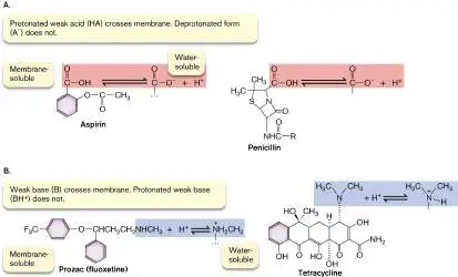
base). On the other side, entering the aqueous cytoplasm, the acid dissociates (HA to A − and H +) or the base reassociates with H + (B to BH +). In effect, membrane-permeant acids conduct acid (H + ) across the membrane, causing acid stress; similarly, membrane-permeant bases conduct OH − across the membrane, causing alkali stress. If the H + concentration (acidity) outside the cell is greater than inside, it will drive weak acids into the cell.
Many key substances in cell metabolism are membrane-permeant weak acids and weak bases, such as acetic acid. Most pharmaceutical drugs—therapeutic agents delivered to our tissues via the bloodstream—are weak acids or weak bases whose uncharged forms exist at sufficiently low concentration to cross the membrane without disrupting it. Examples of weak acids that deprotonate (acquiring negative charge) at neutral pH include aspirin (acetylsalicylic acid) and penicillin (Fig. 3.11A). Examples of weak bases that protonate (acquiring positive charge) at neutral pH include Prozac (fluoxetine) and tetracycline (Fig. 3.11B ).
Thought Question
3.3 Amino acids have acidic and basic groups that can dissociate. Why are they not membrane-permeant weak acids or weak bases? Why do they fail to cross the phospholipid bilayer?
Transmembrane ion gradients. Molecules that carry a fixed charge, such as hydrogen and sodium ions (H + and Na +), cannot cross the phospholipid bilayer. Such ions usually exist in very different concentrations inside and outside the cell. An ion gradient (ratio of ion concentrations) across the cell membrane can store energy for nutrition or to drive the transport of other molecules. Inorganic ions require transport through specific transport proteins, or transporters. So, too, do organic molecules that carry a charge at cytoplasmic pH, such as amino acids and vitamins.
Transport may be passive or active. In passive transport, molecules accumulate or dissipate along their concentration gradient. Active transport —that is, transport from lower to higher concentration—requires cells to spend energy. A transport protein obtains energy for active transport by cotransport of another substance down its gradient from higher to lower concentration or by coupling transport to a chemical reaction.
Note: Active transport by proteins is covered in detail in Chapter
4.
To Summarize
The cell membrane is a bilayer of phospholipids.
Microbes may change the composition of these lipids in response to their environment.
Archaeal membranes have ether-linked terpenoids , which confer increased stability at high temperature and extreme acidity. Some archaea have diglycerol tetraethers, which form a monolayer.
The membrane contains embedded proteins that have a hydrophobic surface. Membrane proteins serve diverse functions, including transport, cell defense, and cell communication.
Small uncharged molecules, such as oxygen, can penetrate the cell membrane by diffusion.
Membrane-permeant weak acids and weak bases exist partly in an uncharged form that can diffuse across the membrane and increase or decrease, respectively, the H + concentration within the cell.
Ion gradients generated by membrane pumps store energy.
Glossary
phospholipid The major component of membranes. A typical phospholipid is composed of a core of glycerol to which two fatty acids and a modified phosphate group are condensed.
leaflet One of the two lipid layers in a phospholipid bilayer. The inner leaflet of the cell membrane faces the cytoplasm.
cardiolipin Diphosphatidylglycerol, a double phospholipid linked by glycerol. cholesterol A sterol lipid found in eukaryotic cell membranes.
hopanoid or hopane A five-ringed hydrocarbon lipid found in bacterial cell membranes.
hopanoid or hopane A five-ringed hydrocarbon lipid found in bacterial cell membranes.
terpenoid A branched lipid derived from isoprene that is found in hydrocarbon chains of archaeal membranes.
osmotic pressure Also called turgor pressure. Pressure exerted by the osmotic flow of water through a semipermeable membrane.
turgor pressure Also called osmotic pressure. Pressure exerted by the osmotic flow of water through a semipermeable membrane.
membrane-permeant weak acid An acid that exists in equilibrium between negatively charged and uncharged forms, such as acetic acid. The uncharged form can penetrate the membrane.
membrane-permeant weak base A base that exists in equilibrium between positively charged and uncharged forms, such as methylamine. The uncharged form can penetrate the membrane.
ion gradient A difference in concentration of an ion across a membrane. transport protein or transporter A membrane protein that moves specific molecules across a membrane.
transport protein or transporter A membrane protein that moves specific molecules across a membrane.
passive transport Net movement of molecules across a membrane without energy expenditure by the cell.
active transport An energy-requiring process that moves molecules across a membrane against their electrochemical gradient.
Fig. 3.2: FIGURE 3.2 ■ Bacterial cell membrane. The cell membrane consists of a phospholipid bilayer, in which the hydrophobic fatty acid chains are directed inward, away from
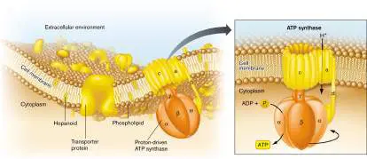
water. The bilayer contains stiffening agents such as hopanoids. Half the membrane volume consists of proteins such as the ATP synthase protein complex.
Figure A2.5: FIGURE A2.5 ■ Osmosis and water balance. A. Osmosis. B. Movement of water across the cell membrane, and shrinkage or expansion of the membrane in isotonic, hypertonic, and hypotonic environments. Black arrows indicate net water movement.
3.3 The Envelope and CytoskeletonUnit 3 · Structure
How do bacteria and archaea protect their cell membrane from a dangerous environment? For most species, the cell envelope includes at least one structural supporting layer, like an external skeleton, located outside the cell membrane. As seen in Figure 3.1, the most common structural support is the cell wall (Fig. 3.12). Many species possess additional coverings, such as an outer membrane or an S-layer. Nevertheless, a few prokaryotes, such as the mycoplasmas, have a cell membrane with no outer layers and depend on host fluids for osmotic balance. Some archaea with only a cell membrane grow in extreme acid (pH zero); for example, Ferroplasma, found in iron mines. How their cells survive is unknown.
FIGURE 3.12 ■ The cell wall: peptidoglycan sugar chains and cross-bridges. A. Isolated sacculus (entire cell wall) from Escherichia coli (TEM). B. A disaccharide unit of glycan has an attached peptide of four to six amino acids.
W. VOLLMER ET AL. 2008. FEMS MICROBIOL. REV. 32 :149, FIG. 3A
The Cell Wall Is a Single Molecule
The bacterial cell wall, also known as the sacculus, consists of a single interlinked molecule that envelops the cell. The sacculus has
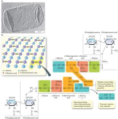
been isolated from Escherichia coli and visualized by transmission electron microscopy (TEM); in Figure 3.12A, the isolated sacculus appears flattened on the sample grid like a deflated balloon. Its geometrical structure encloses maximal volume with minimal surface area. The sacculus—unlike the membrane—is a single-molecule cage-like structure, highly porous to ions and organic molecules. The mesh grows by strand insertion and elongation in arcs around the cell. The cage-like form is not rigid; it is more like a flexible mesh bag with unbreakable joints. Turgor pressure within the enclosed cytoplasm fills out the cell’s shape, whether elongated rod, spherical coccus, or other.
Peptidoglycan structure. Most bacterial cell walls are composed of peptidoglycan, a polymer of peptide-linked chains of amino sugars. Peptidoglycan is synonymous with murein (“wall molecule”). The molecule consists of parallel polymers of disaccharides called glycan chains cross-linked with peptides of four to six amino acids ( Fig. 3.12B ). Peptidoglycan is unique to bacteria, although some archaea build analogous structures whose overall physical nature is similar. (Archaeal cell walls are presented in Chapter 19.) The long chains of peptidoglycan consist of repeating units of a disaccharide composed of N -acetylglucosamine (an amino sugar derivative) and N -acetylmuramic acid (glucosamine plus a lactic acid group; Fig. 3.12B ). The lactate group of muramic acid forms an amide link with the amino terminus of a short peptide containing four to six amino acid residues. The peptide extension can form cross-bridges connecting parallel strands of glycan.
The peptide contains two amino acids in the unusual D mirror form: D -glutamic acid and D -alanine. The third amino acid, m - diaminopimelic acid, has an extra amine group, which forms an amide link to a cross-bridged peptide. The amide link forms with the fourth amino acid of the adjacent peptide, D -alanine (Fig. 3.12B ). Removal of a second D -alanine at the end of the chain forms the cross-bridge. The cross-linked peptides of neighboring glycan strands form the cage of the sacculus.
Note: Amino acids have two forms that are mirror opposites, D
and L, of which only the L form is incorporated by ribosomes into protein. The D -form amino acids, however, are used by microbes for many nonprotein structural molecules.
The details of peptidoglycan structure vary among bacterial species. Some Gram-positive species, such as Staphylococcus aureus (a cause of hospital-acquired infections), have peptides linked by bridges of pentaglycine instead of the D -alanine link to m - diaminopimelic acid. In Gram-negative species, the m - diaminopimelic acid is linked to the outer membrane, as will be discussed shortly.
Peptidoglycan synthesis as a target for antibiotics. Synthesis of peptidoglycan requires many genes encoding enzymes to make the special sugars, build the peptides, and seal the cross-bridges (see Fig. 3.12B ). Many of these enzymes bind the antibiotic penicillin and are thus known as penicillin-binding proteins. Because peptidoglycan is unique to bacteria, enzymes of peptidoglycan biosynthesis make excellent targets for new antibiotics. For example, the transpeptidase that cross-links the peptides is the target of penicillin. Vancomycin, a major defense against Clostridioides difficile and drug-resistant staphylococci, prevents cross-bridge formation by binding the terminal D -Ala-D -Ala dipeptide. Vancomycin binding prevents release of the terminal D - alanine.
Unfortunately, the widespread use of such antibiotics selects for evolution of resistant strains. One of the most common agents of resistance is the enzyme beta-lactamase, which cleaves the lactam ring of penicillin, rendering it ineffective as an inhibitor of transpeptidase (also called penicillin-binding protein 2). In a different mechanism, strains resistant to vancomycin contain an altered enzyme that adds lactic acid to the end of the branch peptides in place of the terminal D -alanine. The altered peptide is no longer blocked by vancomycin. As new forms of drug resistance emerge, researchers continue to seek new antibiotics that target cell wall formation (discussed in Chapter 27).
How does peptidoglycan grow, overall, throughout the elongating cell? A peptidoglycan synthesis complex extends the chains of amino-sugars. So-called penicillin-binding proteins catalyze the formation of peptide cross-bridges (Fig. 3.13). These proteins were named for their property of binding the antibiotic penicillin, which disables cross-bridge formation (details in Chapter 27). The overall direction of cell wall extension is organized by a protein complex that includes MreB. MreB polymerizes in a helical direction along an arc beneath the plasma membrane (inner membrane, for a Gram-negative bacterium). The RodA and RodZ proteins are needed to shape the cell as a rod (bacillus).
FIGURE 3.13 ■ Peptidoglycan synthesis is organized by penicillin-binding proteins (PBP2, PBP1A) and by
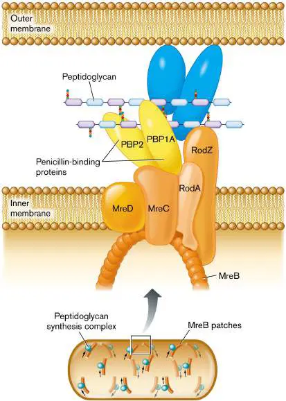
cytoskeletal proteins. Protein MreB guides the direction of synthesis in helical arcs around the cell.
Thought Question
3.4 What genetic experiments could you propose to test the model of envelope expansion shown in Figure 3.13?
Notably, different kinds of bacteria have evolved to organize their cell wall growth differently. Yves Brun and his student Erkin Kuru, at Indiana University, devised an ingenious way to reveal the growth pattern (Fig. 3.14). Kuru designed fluorophore-tagged D -amino acids that the growing cell wall incorporates (such as D -alanine; see Fig. 3.12). Because these are D -amino acids and not L -amino acids, ribosome-directed translation does not use them. Thus, the fluorophore-linked D -amino acids label cell wall only, not proteins. FIGURE 3.14 ■ Peptidoglycan growth in different species. A. Different species of bacteria synthesize new
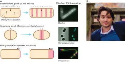
peptidoglycan in dispersed zones (top), at the septum only (middle), or at the poles (bottom). Fluorescent D -amino acids are added for short periods (pulse labeling) to reveal the growth zones. B. Erkin Kuru, in the laboratory of Yves Brun at Indiana University, devised the fluorescent D -amino acids to probe cell wall growth.
E. KURU ET AL. 2012. ANGEW. CHEM. INT. ED. ENGL. 51 :12519
© JEAN-FRANÇOIS GOUT
Kuru found that bacteria such as E. coli and Bacillus subtilis synthesize cell wall in zones dispersed throughout the cell. Gram-positive cocci, however, such as Streptococcus and Staphylococcus, synthesize cell wall only at the midpoint (or septum), where the cell is about to fission (discussed in Section 3.4). Still other bacteria, such as the actinomycete Streptomyces, form new cell wall only at the poles. Cell growth is discussed further in Section 3.4.
Thought Question
3.5 What other ways can you imagine that bacteria might mutate to become resistant to vancomycin?
Cell Envelope of Bacteria
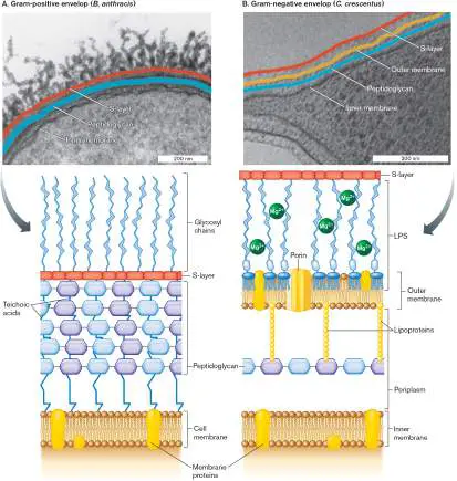
Most bacteria have additional envelope layers that provide structural support and protection from predators and host defenses (Fig. 3.15). Additional molecules are attached to the cell wall and cell membrane, and some thread through the layers. Here we present the envelope composition of three major kinds of bacteria, two of which (Firmicutes and Proteobacteria) are distinguished by the Gram stain. The third, Mycobacteria, is distinguished by the acid-fast stain. FIGURE 3.15 ■ Cell envelope: Gram-positive (Bacillus anthracis) and Gram-negative (Caulobacter crescentus ). A. Firmicutes (Gram-positive) cells have a thick cell wall with multiple layers of peptidoglycan, threaded by teichoic acids. The micrograph shows the Gram-positive envelope of Bacillus anthracis (TEM). B. Proteobacteria (Gram-negative) cells have a single layer of peptidoglycan covered by an outer membrane; the cell membrane is called the inner membrane. The outer membrane lipopolysaccharides (LPS) are cross-linked by positively charged magnesium ions (Mg 2⁺). The micrograph
shows the Gram-negative envelope of Caulobacter crescentus (TEM). LPS = lipopolysaccharides.
S. MESNAGE ET AL. 1998. J BACTERIOL. 180 :52–58
T. BHARAT ET AL. 2017. NAT MICROBIOL. 2 :17059
Firmicutes (Gram-positive) have a thick cell wall with 3–20 layers of peptidoglycan, interpenetrated by teichoic acids (Fig. 3.15A). The phylum Firmicutes consists of Gram-positive species such as Bacillus thuringiensis and Streptococcus pyogenes, the cause of strep throat.
Proteobacteria (Gram-negative) have a thin cell wall with one or two layers of peptidoglycan, enclosed by an outer membrane ( Fig. 3.15B ). Included among the phylum Proteobacteria are Gram-negative species such as Escherichia coli and nitrogen-fixing Sinorhizobium meliloti.
Mycobacteria of the phylum Actinomycetes have a complex, multilayered envelope that includes defensive structures such as mycolic acids. Examples include Mycobacterium tuberculosis (the cause of tuberculosis) and M. leprae (the cause of leprosy). Note that other important kinds of bacteria, such as cyanobacteria (see Fig. 18.8) and spirochetes (see Fig. 18.43), have very different envelope structures. These different envelopes may stain Gram-positive, Gram-negative, or variable (discussed in Chapter 18). Archaea have yet other diverse kinds of envelopes that cannot be distinguished by Gram stain (see Chapter 19).
Thought Question
3.6 Figure 3.15 highlights the similarities and differences between the cell envelopes of Gram-negative and Grampositive bacteria. What do you think are the advantages and limitations of a cell having one layer of peptidoglycan (Gramnegative) versus several layers (Gram-positive)?
Firmicute Cell Envelope—Gram-Positive
A section of a firmicute (Gram-positive) cell envelope is shown in Figure 3.15A. The multiple layers of peptidoglycan are reinforced by teichoic acids threaded through its multiple layers. Teichoic acids are chains of phosphodiester-linked glycerol or ribitol, with sugars or amino acids linked to the middle –OH groups (Fig. 3.16). The negatively charged cross-threads of teichoic acids, as well as the overall thickness of the Gram-positive cell wall, help retain the Gram stain.
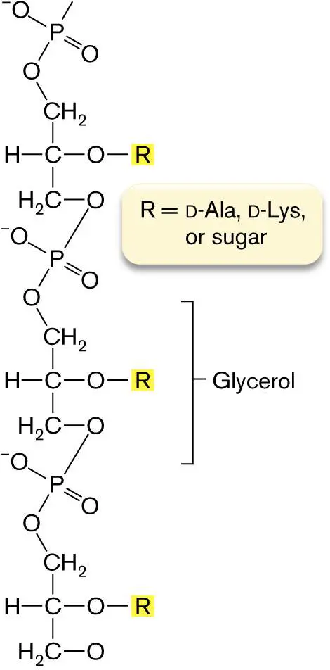
FIGURE 3.16 ■ Teichoic acids. Teichoic acids in the Gram-positive cell wall consist of glycerol or ribitol phosphodiester chains.
How does the cell wall attach extracellular structures? Gram-positive bacteria have a type of enzyme called sortase that forms a peptide bond from a cell wall cross-bridge to a protein extending from the cell. Proteins attached by sortases can help the cell acquire nutrients or help the cell adhere to a substrate.
S-layer. Many free-living species of bacteria and archaea possess a tough surface layer called the S-layer. An S-layer is composed of protein subunits that fit together like tiles. This tough layer may provide defense against phages or predators. The S-layer is rigid, but it also flexes and allows substances to pass through it in either direction.
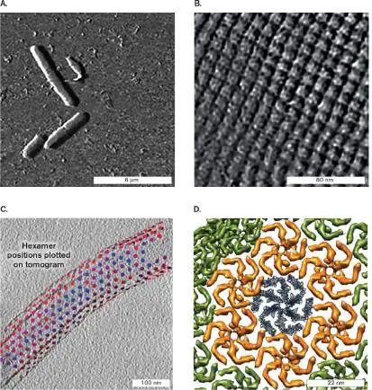
A Gram-positive example is Lysinibacillus sphaericus, a firmicute found on beets and carrots (Fig. 3.17A). Its S-layer is a crystalline sheet of thick subunits consisting of protein or glycoprotein (proteins with attached sugars) (Fig. 3.17B ). Each subunit contains a pore large enough to admit a wide range of molecules. As modeled by cryo-EM tomography (see Chapter 2), the S-layer proteins are arranged in a highly ordered tetragonal array. A Gram-negative example is Caulobacter crescentus, a stalked proteobacterium found in ponds and wastewater (Fig. 3.17C ). Its S-layer is packed in a hexagonal array (Fig. 3.17D ). S-layers help pathogens such as Bacillus anthracis bind and attack host cells. An S-layer contributes to biofilm formation (the periodontal bacterium Tannerella forsythia) and swimming (the aquatic cyanobacterium Synechococcus species).
FIGURE 3.17 ■ S-layer reconstructions from microscopy. A. Lysinibacillus sphaericus imaged by atomic force microscopy (AFM). B. S-layer surface of L. sphaericus imaged by AFM. C. Caulobacter crescentus imaged by cryo-EM; S-layer subunits are marked. D. C. crescentus S-layer unit; computational model based on cryo-EM.
TOBIAS GÜNTHER ET AL. 2014. RSC ADV. 4 (93)
TOBIAS GÜNTHER ET AL. 2014. RSC ADV. 4 (93)
T. BHARAT ET AL. 2017. NAT MICROBIOL. 2 :17059
T. BHARAT ET AL. 2017. NAT MICROBIOL. 2 :17059
The functions of the S-layer are hard to study in the laboratory because the S-layer is often lost by bacteria after repeated subculturing. Traits commonly disappear in the absence of selective pressure for genes encoding them—a process called reductive evolution (discussed in Chapter 17). For example, the mycoplasmas are close relatives of Gram-positive bacteria that have permanently lost their cell walls, as well as the S-layer. Mycoplasmas have no need for cell walls, because they are parasites living in host environments, such as the human lung, where they are protected from osmotic shock.
Capsule. Another common extracellular structure is the capsule, a slippery layer of loosely bound polysaccharides. The capsule of pathogens such as Staphylococcus aureus can prevent phagocytosis by white blood cells, thereby enabling the pathogen to persist in the blood.
Thought Question
3.7 Why would laboratory culture conditions select for evolution of cells lacking an S-layer?
Proteobacterial Cell Envelope—Gram-Negative
The cell envelope of the Proteobacteria (Gram-negative) includes one or two layers of peptidoglycan covered by an outer membrane ( Fig. 3.18). While free-living species such as C. crescentus have an S-layer, enteric bacteria such as E. coli often lack an S-layer. The Gram-negative outer membrane confers defensive abilities and toxigenic properties on many pathogens, such as Salmonella species and enterohemorrhagic E. coli (strains that cause hemorrhaging of the colon). Between the outer and inner (cell) membranes, the aqueous compartment (containing the cell wall) is called the periplasm.
FIGURE 3.18 ■ Gram-negative cell envelope (S-layer not shown). A. Murein lipoprotein has an N-terminal cysteine triglyceride inserted in the inward-facing leaflet of the outer membrane. The C-terminal lysine forms a peptide bond with the m -diaminopimelic acid (m -A 2 pm) of the peptidoglycan (murein) cell wall. Mg 2 ⁺ ions cross-link the LPS chains. B. Lack of murein lipoprotein in mutant Salmonella causes the outer membrane to balloon out (arrow) when the cell tries to divide (TEM).
JOAN FUNG ET AL. 1978. J. BACTERIOL. 133 :1467
Lipoprotein and lipopolysaccharide (LPS). In Gram-negative bacteria, the inward-facing leaflet of the outer membrane has a phospholipid composition similar to that of the inner membrane. The outer membrane’s inward-facing leaflet includes lipoproteins that connect the outer membrane to the peptide bridges of the cell
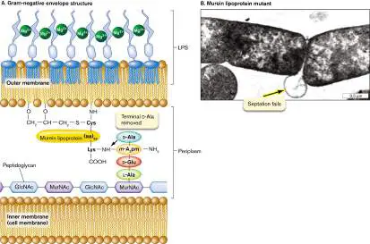
wall. The major lipoprotein is called murein lipoprotein, also known as Braun lipoprotein (Fig. 3.18A). Murein lipoprotein consists of a protein with an N-terminal cysteine attached to three fatty acid side chains. The side chains are inserted in the inward-facing leaflet of the outer membrane. The C-terminal lysine forms a peptide bond with the m -diaminopimelic acid of peptidoglycan (murein). What happens to a mutant cell that fails to make murein lipoprotein? As the cell grows and divides, it fails to attach its outer membrane to the growing cell wall, causing the outer membrane to balloon out in the region where the daughter cells separate (Fig. 3.18B ).
The outward-facing leaflet of the outer membrane has very different lipids from the inner leaflet. The main outward-facing phospholipids are called lipopolysaccharides (LPS; Fig. 3.19). LPS are of crucial medical importance because they act as endotoxins. Endotoxins are cell components that are harmless as long as the pathogen remains intact, but when released by a lysed cell, endotoxins overstimulate host defenses, inducing potentially lethal shock (discussed in Chapter 25). Thus, antibiotic treatment of an LPS-containing pathogen can kill the cells but can also lead to death of the patient.
FIGURE 3.19 ■ Lipopolysaccharide (LPS). A. Lipopolysaccharide (LPS) consists of core polysaccharide and O antigen linked to a lipid A. Lipid A consists of a dimer of phosphoglucosamine esterified or amidated to six fatty acids. B. Repeating polysaccharide units of O antigen extend from lipid A. The membrane-embedded anchor of LPS is lipid A, a molecule shaped like a six-legged giraffe (Fig. 3.19A). The lipid A moiety is the endotoxic part of LPS. The molecule’s six fatty acid “legs” have shorter chains than those of the fatty acids of the inner cell membrane, and two pairs are branched. The fatty acids have ester or amide links to the “body,” a dimer of glucosamine (an amino sugar also found in peptidoglycan). Analogous to the glycerol of glyceride phospholipids, each glucosamine has a phosphoryl group
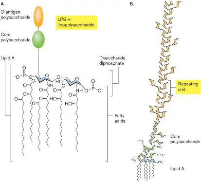
whose negative charge interacts with water. One glucosamine extends the long “neck” of the core polysaccharide, a sugar chain that reaches far outside the cell (Fig. 3.19B ).
The core polysaccharide consists of five to ten sugars with side chains such as phosphoethanolamine. It extends to an O antigen, or O polysaccharide , a chain of as many as 200 sugars. The O polysaccharide may be longer than the cell itself. These chains of sugars form a layer that helps bacteria resist phagocytosis by white blood cells. The combination of sugar units in the O antigen varies greatly; E. coli clinical isolates show hundreds of different O-antigen sugar chains, which are recognized by distinct antibodies. Thus, O-antigen diversity offers one means by which pathogens evade the host immune system.
The LPS sugar side chains have negative charges that bind positively charged magnesium ions (Mg 2+). Mg 2+ ions thus form ionic cross-links that bridge LPS chains. The Mg 2+ -linked LPS can provide structural support for the cell, with strength comparable to that of the peptidoglycan layer.
Outer membrane proteins. The outer membrane contains unique proteins not found in the inner membrane. Outer membranes contain a class of transporters called porins that permit the entry of nutrients such as sugars and peptides (Fig. 3.20). Outer membrane porins such as OmpF have a distinctive cylinder of beta sheet conformation (reviewed in eAppendix 1), also known as a beta barrel. A typical outer membrane porin exists as a trimer of beta barrels, each of which acts as a pore for nutrients.
FIGURE 3.20 ■ OmpF porin transports ampicillin. This model of the OmpF trimer is based on X-ray crystallography. (PDB code: 2OMF) Within each tubular porin monomer, charged amino acid residues contact ampicillin.
Source: Modified from Ekaterina M. Nestorovich et al. 2002. PNAS 99 :9789– 9794.
Outer membrane porins have limited specificity, allowing passive uptake of various molecules—including antibiotics such as ampicillin. Ampicillin is a form of penicillin, which must get through the outer membrane to access the cell wall in order to block the formation of peptide cross-bridges. Ampicillin contains two charged groups and is
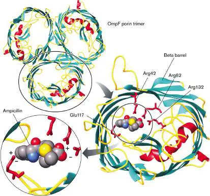
thus unlikely to diffuse through a lipid bilayer. But the molecule crosses the E. coli outer membrane by passing through OmpF, where its charged groups are attracted to charged amino acid residues extending inside the porin (Fig. 3.20). Ampicillin’s positively charged amine group is attracted to the carboxylate of glutamate-117, and its negatively charged carboxylate is attracted to the arginine amines.
If porins can admit dangerous molecules as well as nutrients, should a cell make porins or not? In fact, cells express different outer membrane porins under different environmental conditions. In a dilute environment, cells express porins of large pore size, maximizing the uptake of nutrients. In a rich environment—for example, within a host—cells down-regulate the expression of large porins and express porins of smaller pore size, selecting only smaller nutrients and avoiding the uptake of toxins. For example, the porin regulation system of Gram-negative bacteria enables them to grow in the colon, which contains bile salts—a hostile environment for Gram-positive bacteria, which lack an outer membrane.
Periplasm. The outer membrane is porous to most ions and many small organic molecules, but it prevents the passage of proteins and other macromolecules. Thus, the region between the inner and outer membranes of Gram-negative cells, including the cell wall, defines a separate membrane-enclosed compartment of the cell known as the periplasm (see Fig. 3.18). The periplasm contains specific enzymes and nutrient transporters not found within the cytoplasm, such as periplasmic transporters for sugars, amino acids, or other nutrients. Periplasmic proteins are subjected to fluctuations in pH and salt concentration because the outer membrane is porous to ions. Some periplasmic proteins help refold proteins unfolded by oxidizing agents or by acidification.
Overall, the outer membrane, periplasm, inner membrane, and cytoplasm define four different cell compartments within a Gram-negative cell: two membrane-soluble compartments (outer and inner membranes), and two aqueous compartments (periplasm and cytoplasm). Each type of protein is typically found in only one of these locations. For example, the proton-translocating ATP synthase is found only in the inner membrane fractions, whereas sugar-accepting porins are only in the outer membrane.
Capsule. Some Gram-negative bacteria also form a capsule of loose glycolipids exterior to the outer membrane. For example, a capsule is found in virulent strains of Haemophilus influenzae, which was the leading cause of childhood meningitis before development of the Hib vaccine (see Chapter 24).
Thought Question
3.8 Why would proteins be confined to specific cell locations? Why would a protein not be able to function everywhere in the cell?
Mycobacterial Cell Envelope
Exceptionally complex cell envelopes are found in Actinomycetes, a large and diverse phylum of soil bacteria that produce antibiotics and other industrially useful products (discussed in Chapter 18). The most complex envelopes known are those of actinomycete-related bacteria, the mycobacteria. Mycobacteria include the famous pathogens Mycobacterium tuberculosis (the cause of tuberculosis) and M. leprae (the cause of leprosy). Their complex envelope prevents use of the Gram stain. The mycobacterial envelope may be 1,000-fold less permeable to nutrients and toxins than the envelope of E. coli is. Thus, mycobacteria must grow slowly—but they effectively resist host defenses.
The mycobacterial envelope includes features of both Gram-positive and Gram-negative cells, as well as structures unique to mycobacteria (Fig. 3.21). In mycobacteria, the peptidoglycan is linked to chains of galactose, called galactans. The galactans are attached to arabinans, polymers of the five-carbon sugar arabinose. The arabinan-galactan polymers are known as arabinogalactans. Arabinogalactan biosynthesis is inhibited by two major classes of anti-tuberculosis drugs: ethambutol and the benzothiazinones.
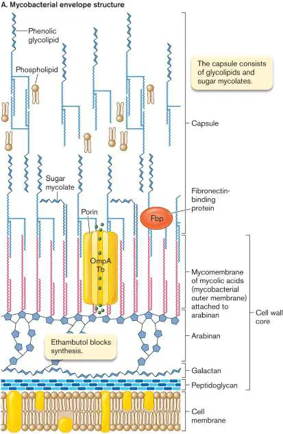
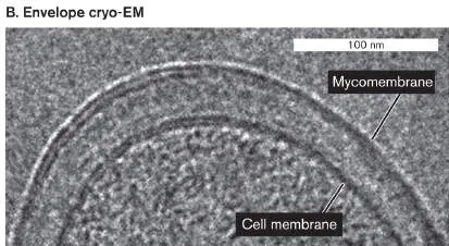
FIGURE 3.21 ■ Mycobacterial envelope structure. A. The peptidoglycan layer is linked to a chain of galactose polymer (galactan) and arabinose polymer (arabinan). Arabinan forms ester links to mycolic acids, which form an outer bilayer with phenolic glycolipids. B. Mycobacterial envelope (cryo-TEM).
B. CHRISTIAN HOFFMAN ET AL. 2008. PNAS 105 :3963
The ends of the arabinan chains form ester links to mycolic acids (uncharged mycolates). Mycolic acids provide the basis for acid-fast staining, in which cells retain the dye carbolfuchsin, an important diagnostic test for mycobacteria and actinomycetes (described in Chapter 28). Mycolic acids contain a hydroxy acid backbone with two hydrocarbon chains—one comparable in length to typical membrane lipids (about 20 carbons), the other about threefold longer. The long chain includes ketones, methoxyl groups, and cyclopropane rings. Hundreds of different forms are known. The mycolic acids form a bilayer interleaved with sugar mycolates—a kind of outer membrane, or “mycomembrane,”
analogous to the Gram-negative outer membrane. This mycomembrane even contains porins homologous to Gram-negative beta barrel porins such as OmpA. Other mycolate-embedded
proteins include virulence factors such as fibronectin-binding protein (Fbp). Fbp enhances the ability of M. tuberculosis to invade macrophages.
The outer ends of the sugar mycolates are interleaved with phenolic glycolipids, which include phenol groups linked to sugar chains. The extreme hydrophobicity of the phenol derivatives generates a waxy surface that prevents phagocytosis by macrophages. Overall, the thick, waxy envelope excludes many antibiotics and offers exceptional protection from host defenses, enabling the pathogens of tuberculosis and leprosy to colonize their hosts over long periods. However, the thick envelope also retards uptake of nutrients. As a result, M. tuberculosis and M. leprae grow extremely slowly and are a challenge to culture in the laboratory.
Bacterial Cytoskeleton
In eukaryotes, cell shape has long been known to be maintained by a cytoskeleton of protein microtubules and filaments (reviewed in eAppendix 2). But what determines the shape of bacteria? We saw earlier that bacterial shape is in part maintained by the cell wall and the resulting turgor pressure. But research over the past decade shows that bacteria also possess protein cytoskeletal components— and remarkably, some of them are homologous to eukaryotic cytoskeletal proteins. For example, the MreB tracker protein for peptidoglycan synthesis (Fig. 3.13) is a part of the bacterial cytoskeleton. MreB is a homolog of the eukaryotic microfilament protein actin.
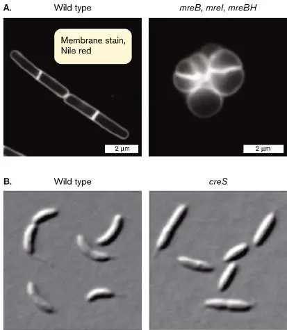
The bacterial cytoskeletal proteins are revealed by gene defects that drastically alter the cell shape. For example, Figure 3.22A compares wild-type cells of Bacillus subtilis with cells containing mutations in three mreB homologs (mreB, mreI, and mreBH). The wild-type cells have a defined rod shape, whereas the mutant shape is round and undefined. The mutant lacks the MreB complex that regulates peptidoglycan synthesis and thereby defines the rod-shaped cell. Another example of a shape-altering mutation affects the comma-shaped cell of Caulobacter crescentus (Fig. 3.22B ). A mutation in the gene creS results in cells that are straight instead of curved. The creS gene expresses the cytoskeletal protein CreS (crescentin).
FIGURE 3.22 ■ Cytoskeletal mutants. Wild-type cells compared with mutants in (A) Bacillus subtilis (fluorescence microscopy) and (B) Caulobacter crescentus (DIC).
Source: Part A from https://www.nature.com/articles/ncomms4442; https://creative commons.org/license/by/3.0/us/legalcode.
T. NILSEN ET AL. 2005. J. BACTERIOL. 187 :6187
M. CABEEN ET AL. 2010. J. BACTERIOL. 192 :3368
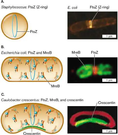
How do the various cytoskeletal proteins work together to generate the overall shape of a bacterial cell? The functions of cytoskeletal proteins are probed by fluorescent protein fusions (Fig. 3.23). In both spherical bacteria (cocci) and rod-shaped bacilli, cell division requires the protein FtsZ, a homolog of the eukaryotic protein tubulin (the subunit of eukaryotic microtubules). The bacterial protein FtsZ forms a ring-shaped complex around the middle of the cell, called the Z-ring. The Z-ring determines the cell diameter and manages the growth of the dividing partition, which is called the septum (plural, septa).
For a rod-shaped cell, elongation requires polymerization of MreB (Fig. 3.23B ). MreB travels in a helical arc beneath the cell membrane, guiding peptidoglycan elongation. If the rod shape is curved (forming a crescent shape), the third cytoskeletal protein, crescentin, polymerizes along the inner curve of the crescent (Fig. 3.23C ). The cell’s outer curve is visualized by a membrane-specific fluorophore. These cytoskeletal proteins, and their variants that have evolved in other species, work together within cells to generate the shapes of bacteria.
FIGURE 3.23 ■ Shape-determining proteins. A. Cell diameter is maintained by FtsZ polymerization to form the Z-ring. B. Elongation of a rod-shaped cell requires MreB proteins. MreB polymerizes around an E. coli cell (MreB-YFP fluorescence) along with a Z-ring of FtsZ (fluorescent anti-FtsZ antibody). C. Crescent-shaped cells possess a third shape-determining protein, CreS (crescentin), which polymerizes along the inner curve of the crescent. Crescentin protein fused to green fluorescent protein (CreS-GFP) localizes to the inner curve of Caulobacter crescentus
. Membrane-specific stain FM4-64 (red fluorescence) localizes to the membrane around the cell.
Q. SUN AND W. MARGOLIN. 1998. J. BACTERIOL. 180 :2050
PURVA VATS AND LAWRENCE ROTHFIELD. 2007. PNAS 104 :17795
NORA AUSMEES ET AL. 2003. CELL 115 :705
To Summarize
The cell wall maintains turgor pressure. The cell wall is porous, but its network of covalent bonds generates turgor pressure that protects the cell from osmotic shock. The Gram-positive cell envelope has multiple layers of peptidoglycan, threaded by teichoic acids.
The protein S-layer of Gram-negative and Gram-positive bacteria is highly porous but can prevent phagocytosis and protect cells in extreme environments. In many archaea, the S-layer serves the structural function of a cell wall.
The capsule , composed of polysaccharide and glycoprotein filaments, protects cells from phagocytosis. Both Gram-positive and Gram-negative cells may possess a capsule. The Gram-negative outer membrane regulates nutrient uptake and excludes toxins. The outer membrane contains LPS and protein porins of varying selectivity.
The mycobacterial cell wall includes features of both Gram-positive and Gram-negative cells. The arabinogalactan layer adds thickness to the cell wall. The mycolate outer membrane and phenolic glycolipids limit uptake of nutrients and antibiotics.
The bacterial cytoskeleton includes proteins that regulate cell size, play a role in determining the rod shape of bacilli, and generate curvature in crescent-shaped cells.
Glossary
cell wall A rigid structure external to the cell membrane. The molecular composition depends on the organism; in bacteria, it is composed of peptidoglycan.
sacculus pl. sacculi The bacterial cell wall, consisting of a single covalent molecule. murein Also called peptidoglycan. A polymer of peptide-linked chains of amino sugars; a major component of the bacterial cell wall. glycan A polysaccharide chain composed of oxygen-linked (O-linked) monosaccharides.
cross-bridge An attachment that links parallel molecules, such as the peptide link between glycan chains in peptidoglycan.
penicillin-binding protein (PBP)
A bacterial protein, involved in cell wall synthesis, that is the target of the antibiotic penicillin.
teichoic acid A chain of phosphodiester-linked glycerol or ribitol that threads through and reinforces the cell wall in Gram-positive bacteria. S-layer A surface layer of crystalline protein subunits, either replacing the cell wall or external to it in many species of archaea and bacteria.
capsule A slippery outer layer composed of polysaccharides that surrounds the cell envelope of some bacteria.
inner membrane or inner cell membrane In Gram-negative bacteria, the membrane in contact with the cytoplasm, equivalent to the cell membrane.
murein lipoprotein Also called Braun lipoprotein. The major lipoprotein that connects the outer membrane of Gram-negative bacteria to the peptidoglycan cell wall.
lipopolysaccharides (LPS)
Structurally unique phospholipids found in the outer leaflet of the outer membrane in Gram-negative bacteria. Many are endotoxins.
endotoxin A lipopolysaccharide in the outer membrane of Gram-negative bacteria that becomes toxic to the host after the bacterial cell has lysed.
lipid A The anchor lipid of lipopolysaccharide (LPS), composed of glucosamine plus six lipid chains.
O antigen or O polysaccharide A sugar chain that connects to the core polysaccharide of lipopolysaccharides.
porin A transmembrane protein complex that allows movement of specific molecules across the cell membrane or the outer membrane.
septum pl. septa A plate of cell wall and envelope that forms to separate two daughter cells.
septum pl. septa A plate of cell wall and envelope that forms to separate two daughter cells.
Figure 3.1: FIGURE 3.1 ■ Escherichia coli: a Gram-negative bacterium of the gut microbiome. The envelope includes the outer membrane, the cell wall and periplasm, and the inner (cell) membrane with an embedded chemoreceptor array. The cytoplasm contains enzymes, messenger RNA (mRNA) extending out of the nucleoid, and ribosomes. Ribosomes translate the mRNA to make proteins, which are folded by chaperones. The nucleoid contains the
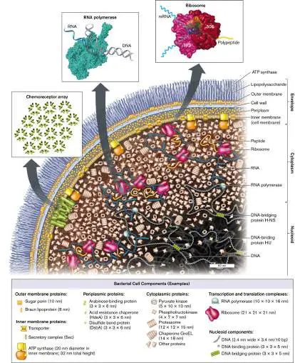
chromosomal DNA wrapped around binding proteins. (PDB codes: ribosome, 1GIX, 1GIY; RNA polymerase, 1MSW)
Fig. 3.18: FIGURE 3.18 ■ Gram-negative cell envelope (S-layer not shown). A. Murein lipoprotein has an N-terminal cysteine triglyceride inserted in the inward-facing leaflet of the outer membrane. The C-terminal lysine forms a peptide bond with the m -diaminopimelic acid (m -A 2 pm) of the peptidoglycan (murein) cell wall. Mg 2 ⁺ ions cross-link the LPS chains. B. Lack of murein lipoprotein in mutant Salmonella causes the outer membrane to balloon out (arrow) when the cell tries to divide (TEM).
JOAN FUNG ET AL. 1978. J. BACTERIOL. 133 :1467
Fig. 3.13:
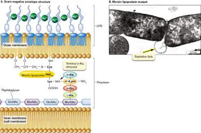
FIGURE 3.13 ■ Peptidoglycan synthesis is organized by penicillin-binding proteins (PBP2, PBP1A) and by cytoskeletal proteins. Protein MreB guides the direction of synthesis in helical arcs around the cell.
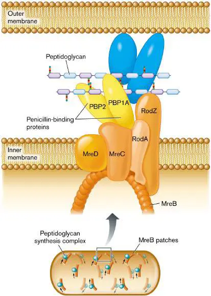
Fig. 18.8: FIGURE 18.8 ■ Colonial cyanobacteria. A. Gloeocapsa is surrounded by mucus. Cells grow as single cells, doublets, or quartets. B. Merismopedia forms extended quartets, octets, and so on. C. Unicellular cyanobacterium Chroococcidiopsis sp. with baocytes (arrows) formed by successive multiple divisions.
MICHAEL ABBEY/SCIENCE SOURCE
MICHAEL ABBEY/SCIENCE SOURCE
BURKHARD BÜDEL
Fig. 18.43: FIGURE 18.43 ■ Spirochete structure. A. Spirochete cell structure, showing the arrangement of periplasmic flagella. B. Cross section through a human gingival spirochete, showing outer envelope and flagella (axial fibrils; TEM).
M. A. LISTGARTEN AND S. S. SOCRANSKY. 1964. J. BACTERIOL. 88 :1087
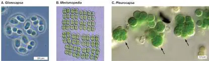
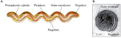
3.4 Bacterial Cell DivisionUnit 3 · Structure
How does a growing bacterial cell divide, or fission, into daughter cells? Bacterial cell fission requires highly coordinated growth and formation of all the cell’s parts. Unlike eukaryotes, prokaryotes synthesize RNA and proteins continually while the cell’s DNA undergoes replication. Bacterial DNA replication is coordinated with the expansion of the cell wall and the separation of the cell into two daughter cells. Bacterial DNA replication is outlined here as it relates to cell division; the genetic aspects of DNA replication are discussed in Chapter 7.
Note: Bacteria do not undergo mitosis or meiosis. These
eukaryotic processes are reviewed in eAppendix 2.
Cell Division by Septation
In rod-shaped cells, the envelope elongates to a consistent length by extension of peptidoglycan chains in tracks around the cell, as we saw in Figure 3.13. But as DNA synthesis terminates, the cell divides by a process called septation, the formation of the septum, the partition that divides the envelope. How does the septum actually form, completing two entire cell envelope layers back-to-back? The laboratories of Ethan Garner at Harvard University and of Yves Brun at Indiana University revealed the progression and timing of septum growth (Fig. 3.24).
The septum grows inward from all around the cell equator. As it grows inward, its inner hole constricts and seals off the two daughter cells. The inward growth of peptidoglycan can be seen in Figure 3.24A, where a cell of Bacillus subtilis incorporates D - alanine fluorophores. D -Alanine is an amino acid that gets incorporated into peptidoglycan cross-bridges (Fig. 3.12) but not into proteins, which use only L -form amino acids. Thus, the D - alanine fluorophore labels only peptidoglycan.
FIGURE 3.24 ■ The Bacillus subtilis septum grows from the outer ring inward. A. Cells were pulse-labeled with different-colored fluorescent D -alanine molecules that are incorporated into peptidoglycan, catalyzed by penicillin-binding proteins. The D -alanine fluorophores are: HADA, blue (first 60 minutes); BADA, green (next 5 minutes); TADA, red (final 30 seconds). B. Yves Brun.
Source: Alexandre Bisson-Filho et al. 2017. Science 355 :739, fig. 1A.
ALEXANDRE BISSON-FILHO ET AL. 2017 SCIENCE 355 :739, FIG. 1A
PHOTO BY SANDEE MILHOUSE
In the experiment, the D -alanine fluorophores emit one of three different colors, in successive periods of pulse labeling. The initial, longest period (60 minutes) is required to grow the outermost ring of cell wall pinching in; then shorter pulses (5 minutes, 30 seconds) rapidly complete the septum. Garner’s team showed that FtsZ subunit assembly circles around the septum in a “treadmilling” pattern, stepwise around the cell, that directs septal growth. Septation requires rapid biosynthesis of all envelope components, including membranes and cell wall (Fig. 3.25).
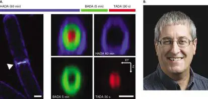
Envelope expansion must coordinate the extension of all layers—and regulate the placement and timing of the septum. As we saw earlier (Fig. 3.13), the enzymes of cell wall biosynthesis must coordinate the formation of new links as closely as possible with subunit insertion. The site of septation is uniquely vulnerable because two new large enclosures must form simultaneously. Thus, the enzymes of septum biosynthesis are of great interest as antibiotic targets. FIGURE 3.25 ■ Divisome enables cell division. A. E. coli cells divide normally. B. Cells that lack FtsN (divisome component) balloon out at the septum. C. The divisome complex coordinates the extension of all envelope layers while the septum constricts.
Source: Part C modified from C. Typas et al. 2012. Nat. Rev. Microbiol. 10:123, fig. 2.
B. LIU ET AL. 2015. MOL. MICROBIOL. 95 :945
B. LIU ET AL. 2015. MOL. MICROBIOL. 95 :945
The overall process of septation is managed by a protein complex called the divisome (Fig. 3.25C ). The divisome manages assembly of the septum with its two envelopes back-to-back. One
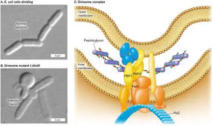
component of the divisome is FtsZ, which polymerizes to form the Z-ring, as seen previously, in Figure 3.23. Mutations in a gene such as ftsZ cause Escherichia coli to form long filaments instead of dividing normally. Another divisome component, FtsN, helps regulate the timing of constriction of the septum. A cell was constructed that requires an inducer molecule to express FtsN (Fig. 3.25C ). Cells that lack FtsN fail to constrict their septum, and their membranes balloon out at cell division. Such divisome components could be targets for new antibiotics, just as penicillin-binding proteins are targets for penicillin.
The bacterial process of cell fission must solve a key problem: how to coordinate septation with the replication of DNA. Indeed, mutations exist that lead to septum formation across DNA with replication incomplete. The result is to “guillotine” the cell. To avoid this disastrous situation, septation is coordinated with DNA replication.
DNA Is Organized in the Nucleoid
The genetic functions of microbial DNA are discussed in detail in Chapters 7–12. Here we focus on the physical organization of DNA within the nucleoid of bacterial and archaeal cells.
Bacteria organize their DNA very differently from eukaryotes. For example, Figure 3.26shows enteropathogenic E. coli cells growing on a cultured human cell. Enteropathogenic E. coli (EPEC) are diarrheal pathogens that attach to the host cell membrane and inject toxins (discussed in Chapter 25). In this thin-section transmission electron micrograph, each bacterium contains a filamentous nucleoid region that extends through the cytoplasm. In contrast, the nucleus of the eukaryotic cell (not shown) is many times larger than the entire bacterial cell, and the chromosomes it contains are separated from the cytoplasm by the nuclear membrane.
FIGURE 3.26 ■ Bacteria invading a human cell. Enteropathogenic Escherichia coli bacteria attached to the surface of a tissue-cultured human cell (TEM). The bacterial nucleoid appears as a lighter region with few ribosomes.
ISABEL C. A. SCALETSKY ET AL. 1996. INFECT. IMMUN. 64 :4876–81
Note: In bacteria and archaea, the genome typically consists of a
single circular chromosome, but some species have a linear chromosome or multiple chromosomes. In this chapter we focus on the simple case of a single circular chromosome.
In a bacterial cell, the DNA is organized in loops called domains, which extend throughout the cytoplasm. The midpoint on the DNA is the origin of replication, which is attached to the cell envelope at a
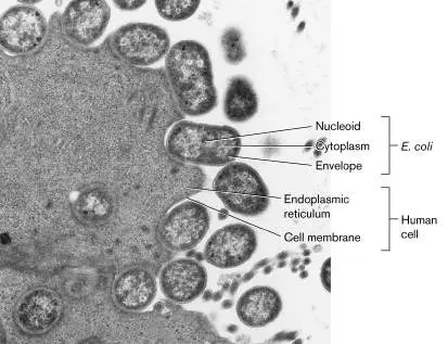
point on the cell’s equator, halfway between the two poles (Fig. 3.27). To initiate DNA replication, the DNA double helix at the origin is opened by binding proteins, and then DNA polymerase synthesizes new strands in both directions (bidirectionally). The origin and other aspects of DNA replication are covered in detail in Chapter 7.
FIGURE 3.27 ■ DNA transcription and RNA translation to peptides. The nucleoid forms chromosome loops called domains, which loop out from the origin of attachment to the cell envelope. Bacterial transcription of DNA to RNA is coordinated with translation of RNA to make proteins. Growing peptide chains destined for the membrane bind the signal recognition particle (SRP) for membrane insertion.
How does all of the cell’s DNA fit neatly into the nucleoid? In some bacteria, the domains loop back to the center of the cell, near
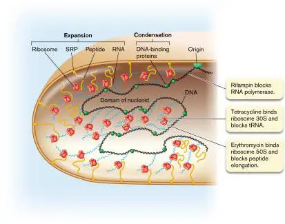
the origin of replication. Within the domains, the DNA is compacted by supercoils. Supercoils (or superhelical turns) are extra twists in the chromosome, beyond those inherent in the structure of the DNA double helix (discussed in Chapter 7). In most bacteria and in eukaryotes, the extra twists actually go against the twist direction of the DNA helix; thus they tend to unwind DNA slightly.
The supercoiling causes portions of DNA to double back and twist upon themselves, resulting in compaction of the chromosome. Supercoils are generated by enzymes such as gyrase, which are a major target for antibiotics such as quinolones. DNA is also compacted by DNA-binding proteins (green spheres in Fig. 3.27). Binding proteins can respond to the state of the cell; for example, under starvation conditions, when most RNA synthesis ceases, the binding protein Dps is used to organize the DNA into a protected crystalline structure. Such “biocrystallization” by Dps and related proteins may be a key to the extraordinary ability of microbes to remain viable for long periods in stationary phase or as endospores.
Note: In biology, the word “domain” is used in several different
ways, each referring to a defined portion of a larger entity. DNA domains of the nucleoid are distinct loops of DNA that extend from the origin.
Protein domains are distinct functional or structural regions of a protein.
Lipid domains are patches of membrane that are enriched for certain lipids.
Taxonomic domains are genetically distinct classes of organisms, such as Bacteria, Archaea, and Eukarya.
Transcription and translation are coupled (discussed in Chapter 8 ). The information encoded in DNA is “read” by the processes of transcription and translation to yield gene products. In bacteria and archaea, some translation is tightly coupled to transcription; the ribosomes bind to mRNA and begin translation even before the mRNA strand is complete. Thus, a growing bacterial cell is full of mRNA strands dotted with ribosomes (Fig. 3.27). Some of the mRNA strands with their growing peptide chains extend to the membrane for protein insertion and secretion.
In rapidly growing bacteria, the DNA is transcribed and the messenger RNA is translated to proteins while the DNA itself is being replicated. This remarkable coordination of replication, transcription, and translation explains why some bacterial cells can divide in as little as 10 minutes. An example is the hot-spring bacterium Geobacillus stearothermophilus cultured at 60°C. DNA synthesis, transcription to RNA, and translation to proteins are discussed further in Chapters 7 and 8.
Some of the newly translated proteins are destined for the cell membrane or for secretion outside. Proteins destined for the membrane are synthesized in association with the membrane, directed there by signal recognition particles (Fig. 3.27). This coupling of transcription and translation to membrane insertion has the effect of expanding the nucleoid into distal parts of the cell, partly counteracting the condensation of DNA by DNA-binding proteins. Membrane protein maturation and secretion are discussed in Chapter 8.
DNA Replication Regulates Cell Division
The process of synthesizing daughter cells begins at the origin of replication, a unique DNA sequence in the chromosome. In bacteria, the origin is attached to a site on the envelope—most commonly, at a point on the cell’s equator (Fig. 3.28). At the origin sequence, the DNA double helix begins to unzip, forming two replication forks. At each replication fork, DNA is synthesized by DNA polymerase. The complex of DNA polymerase with its accessory components is called a replisome. The replisome actually has a double complex of DNA polymerase that simultaneously replicates the “leading strand” and the “lagging strand” of the helix. The lag time is short compared with the overall time of replication; thus, as the replisome travels along the DNA, it converts one helix into two progeny helices almost simultaneously (details presented in Chapter 7).
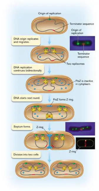
FIGURE 3.28 ■ Replisome movement within a dividing cell. The DNA origin-of-replication sites (green) move apart in the expanding cell as the two replisomes (yellow) stay near the middle, where they replicate around the entire chromosome, completing the terminator sequence last (red). As the terminator sequence nears completion, FtsZ proteins assemble the Z-ring organizing septum formation.
Source: Top 2 insets: Ivy Lau et al. 2003. Mol. Microbiol. 49 :731. Bottom inset: Jackson Buss et al. 2015. PLoS Genet. 11 (4).
I. LAU ET AL. 2003. MOL. MICROBIOL. 49 :731, FIG. 2A
I. LAU ET AL. 2003. MOL. MICROBIOL. 49 :731, FIG. 2A
J. BUSS ET AL. 2015. PLOS GENET. 11 (4):E1005128, FIG. 1G
Within the cell, two replisomes proceed outward in opposite directions around the genome. Thus, bidirectional replication requires a replisome for each replicating fork. Fluorescent probes show that two replisomes are located near the middle of the growing cell (Fig. 3.28). The two copies of the DNA origin of replication (green in the figure), attached to the cell envelope, move apart as the cell expands. The termination site (red) remains in the middle of the cell, where the two replisomes continue replication at both forks. Finally, as the two replisomes approach each other, the termination site replicates. The two replisomes then separate from the DNA.
In a fast-growing cell, however, two new origin sites have already formed. At each new origin, two pairs of new replisomes have formed. The new origin sites begin a second round of replication, even before termination of the previous round. Slow-growing cells may instead have a pause before the next round of replication begins.
Completion of replication triggers Z-ring formation. For the cell to divide, DNA replication must be complete. During the process of replication, the cytoplasm contains several kinds of proteins that will determine septation, among them FtsZ (Fig. 3.28). Other septation-related proteins are bound to the cell membrane or to DNA. Replication of the DNA termination site triggers several proteins to form the divisome. For simplicity, only FtsZ is shown, as the subunits assemble to form the Z-ring.
Ultimately, septation completes cell division, and the two envelope ends come apart. In some species, such as filamentous cyanobacteria, the actual separation of cells may occur long after septation, forming extended filaments of individual cells. Others, such as Bacillus megaterium, commonly separate two or three generations after septation. Nutrient conditions may affect cell separation and cell size.
Thought Question
3.9 Suppose a cell has a defect in its ftsZ gene. What might happen to the cell during growth? How could such a mutant strain be maintained in the laboratory?
Septation of spherical cells. In spherical cells (cocci), such as Staphylococcus aureus, the process of septation generates most of the new cell envelope to enclose the expanding cytoplasm (Fig. 3.29A–C ). The cell envelope pushes in, forming a ring that encircles the cell equator, as the completed envelope layers of the septum peel apart. Unlike rod-shaped cells (which elongate between divisions), the facing halves of each spherical cell form out of septum envelope as the two halves peel apart.
FIGURE 3.29 ■ Septation without cell elongation. A. Staphylococcus aureus fissions in one plane. Arrows mark furrows in the cell envelope that constrict the cell, all around the equator, where the completed septum comes apart (TEM). B. Two new envelope partitions are complete. C. The two daughter cells peel apart. The facing halves of each cell contain entirely new cell wall. D. In Micrococcus tetragenus, septation in two planes forms a tetrad (TEM with negative stain).
AHMED TOUHAMI ET AL. 2004. J. BACTERIOL. 186 :3286
AHMED TOUHAMI ET AL. 2004. J. BACTERIOL. 186 :3286
AHMED TOUHAMI ET AL. 2004. J. BACTERIOL. 186 :3286
KWANGSHIN KIM/SCIENCE SOURCE
The spatial orientation of septation has a key role in determining the shape and arrangement of cocci. If the cell always septates in parallel planes, as in Streptococcus species, cells form chains. If, however, the cell septates in random orientations or if cells
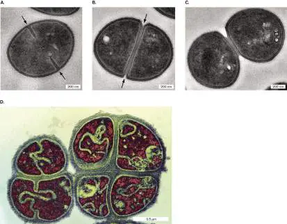
reassociate loosely after septation, they form compact hexagonal arrays similar to the grape clusters portrayed in classical paintings— hence the Greek-derived term staphylococci (staphyle means “bunch of grapes”). Such clusters are found in colonies of Staphylococcus aureus. If subsequent septation occurs at right angles to the previous division, the cells may form tetrads and even cubical octads called “sarcinae” (singular, sarcina). Tetrads are formed by Micrococcus tetragenus, a cause of pulmonary infections (Fig. 3.29D ).
Bacterial cell size. How do cells “know” how large to grow? This question is hard to answer, as we are still discovering ever-smaller forms of life. An investigation of river bacteria, from Jillian Banfield’s lab at UC Berkeley, revealed tiny cells that pass through a 0.2-μm filter. These cells represent new kinds of life accounting for 15% of all taxa known at the time (discussed in Chapter 18). At the other end of the size range, the marine sulfur-oxidizing bacterium Thiomargarita namibiensis grows as a bubble of cytoplasm 200 μm across (presented in Chapter 1).
For a given species, cell size depends on genetic regulators and environmental constraints. When a bacterial population is first diluted into fresh medium, with abundant nutrients, cells elongate faster and reach larger sizes before septation and division. As nutrients become scarce, cell growth slows, and early division produces smaller cells. Thus, cell size is one factor in the phases of growth of bacterial populations (discussed in Chapter 4). Yet, repeated cycles of growth and starvation in minimal glucose medium lead E. coli populations to undergo selection for cells that are larger. Experimental evolution (discussed in Chapter 17) thus enables us to test models of cell size development.
To Summarize
Bacterial cell division includes elongation and septation.
DNA is organized in the nucleoid. In most bacterial species, the DNA is attached to the envelope at the origin of replication, on the cell’s equator. Loops of DNA called domains are supercoiled and bound to DNA-binding proteins.
While DNA undergoes transcription, the growing RNA chain already binds ribosomes for translation. Newly transcribed RNA can be immediately translated, generating new proteins quickly.
DNA is replicated bidirectionally by the replisome.
During bacterial DNA replication, genes continue transcription and translation.
Completion of DNA replication triggers Z-ring formation and septation. Septation may occur in one plane (forming a chain of cells) or at right angles to the previous septation (forming a tetrad).
Bacterial cell size varies widely among taxa. Within a population, environmental parameters such as nutrient availability may determine cell size.
Glossary
septation The formation of a septum, a new section of cell wall and envelope to separate two daughter cells.
divisome A protein complex that manages the overall process of septation.
domain 1. In taxonomy, one of three major subdivisions of life: Archaea, Bacteria, and Eukarya. 2. In protein structure, a portion of a protein that possesses a defined function, such as binding DNA. 3. In membranes, a region of membrane consisting of certain types of phospholipids that are distinct from surrounding lipids. DNA-binding protein A protein that binds to DNA and modulates its function. replisome A complex of DNA polymerase and other accessory molecules that performs DNA replication.
staphylococcus A hexagonal arrangement of cells formed by septation in random orientations.
Figure 3.13: FIGURE 3.13 ■ Peptidoglycan synthesis is organized by penicillin-binding proteins (PBP2, PBP1A) and by cytoskeletal proteins. Protein MreB guides the direction of synthesis in helical arcs around the cell.
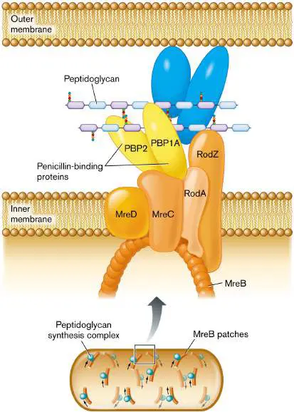
Fig. 3.12: FIGURE 3.12 ■ The cell wall: peptidoglycan sugar chains and cross-bridges. A. Isolated sacculus (entire cell wall) from Escherichia coli (TEM). B. A disaccharide unit of glycan has an attached peptide of four to six amino acids.
W. VOLLMER ET AL. 2008. FEMS MICROBIOL. REV. 32 :149, FIG. 3A
Fig. 3.13:
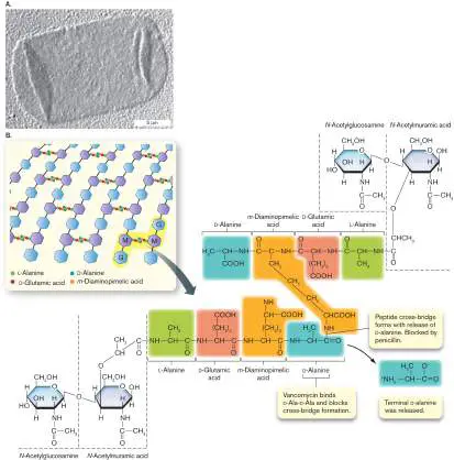
FIGURE 3.13 ■ Peptidoglycan synthesis is organized by penicillin-binding proteins (PBP2, PBP1A) and by cytoskeletal proteins. Protein MreB guides the direction of synthesis in helical arcs around the cell.
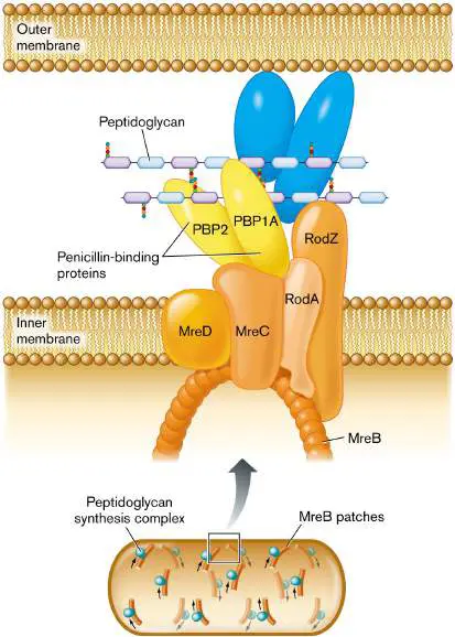
Figure 3.23: FIGURE 3.23 ■ Shape-determining proteins. A. Cell diameter is maintained by FtsZ polymerization to form the Z-ring. B. Elongation of a rod-shaped cell requires MreB proteins. MreB polymerizes around an E. coli cell (MreB-YFP fluorescence) along with a Z-ring of FtsZ (fluorescent anti-FtsZ antibody). C. Crescent-shaped cells possess a third shape-determining protein, CreS (crescentin), which polymerizes along the inner curve of the crescent. Crescentin protein fused to green fluorescent protein (CreS-GFP)
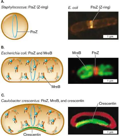
localizes to the inner curve of Caulobacter crescentus. Membrane-specific stain FM4-64 (red fluorescence) localizes to the membrane around the cell.
Q. SUN AND W. MARGOLIN. 1998. J. BACTERIOL. 180 :2050
PURVA VATS AND LAWRENCE ROTHFIELD. 2007. PNAS 104 :17795
NORA AUSMEES ET AL. 2003. CELL 115 :705
3.5 Cell Asymmetry, Membrane Vesicles, and ExtensionsUnit 3 · Structure
Do dividing bacteria produce symmetrical offspring? Even superficially symmetrical bacilli such as Escherichia coli show underlying chemical and physical asymmetry, such as possession of a chemoreceptor array at the “forward” pole. Other species, such as Caulobacter crescentus, develop different structures at either pole, and their cell division generates two different cell types. And many kinds of bacteria extend their cytoplasm in surprising ways, by forming extracellular membrane vesicles and extensions that may interact with other cells. Such cell extensions complicate the very definition of an individual cell.
Bacterial Cell Differentiation
Bacteria whose poles have different structures generate two different forms of progeny. The wetland bacterium Caulobacter crescentus has one plain pole and one pole with either a flagellum or a cytoplasmic extension called a stalk (Fig. 3.30). A flagellated cell (also known as a swarmer cell) swims freely in an aqueous habitat, such as a pond or a sewage bed. After swimming for about half an hour, if the bacterium finds a place with enough nutrients, the cell sheds its flagellum and replaces it with a stalk. The stalked cell attaches to sediment and then immediately starts to replicate its DNA and divides, producing a flagellated daughter cell, as well as a daughter cell containing the original stalk.
FIGURE 3.30 ■ Asymmetrical cell division: a model for development. A swarmer cell of Caulobacter crescentus loses its flagellum and grows a stalk. The stalked cell divides to produce a swarmer cell (TEM).
YVES BRUN
How does C. crescentus organize itself to produce two different cell types, each with a different organelle at one pole? The process is a rudimentary form of cell differentiation, comparable to the differentiation processes that animal cells undergo in the embryo. The C. crescentus life cycle is governed by regulator proteins such as TipN, studied by students of Christine Jacobs-Wagner at Yale University. Mutants lacking TipN make serious mistakes in development. Instead of making a single flagellum at the correct cell pole, the cell makes multiple flagella at various locations, even on the stalk (Fig. 3.31A). Jacobs-Wagner proposed that TipN is a landmark protein that correctly marks the site of a new cell pole and directs the polar placement of flagella.
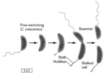
Figure 3.31B shows cells expressing TipN fused to the fluorescent protein GFP, which is then detected by fluorescence microscopy. (A gene fusion expresses fused proteins that fluoresce but may still perform the original protein’s function, as shown in Figure 2.29.) The fluorescent fusion protein TipN-GFP localizes to the cell pole opposite the stalk. As the cell prepares to divide, TipN leaves the pole, delocalizing around the cell. Eventually, the TipN protein relocalizes at the septum, where the new poles appear. FIGURE 3.31 ■ A landmark protein for the cell pole. A. Caulobacter mutants lacking TipN protein make mistakes: flagella grow out of stalks (left) or at the stalked pole (right; fluorescence microscopy). B. The protein TipN appears at the pole of a Caulobacter stalk cell, visualized as TipN-GFP [differential interference contrast microscopy, or DIC (top row); and fluorescence microscopy (bottom row)]. As the cell grows, TipN delocalizes and then localizes again at the septum.
Septation yields two daughter cells with TipN at the pole of each.
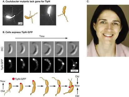
C. Christine Jacobs-Wagner, 2011 winner of the American Society for Microbiology’s Eli Lilly Award for her studies of Caulobacter development.
HUBERT LAM ET AL. 2006. CELL 124 :1011
© JASON VARNEY/VARNEYPHOTO.COM
Cell development involves many such proteins working together. Figure 3.32shows how TipN interacts with two other polar proteins: the flagellar marker PodJ, and the stalk marker DivJ. Each young cell (swarmer cell at top of cycle) has a new pole containing TipN. To prepare for cell division, the swarmer loses its flagellum. The flagellar pole then grows a stalk, with a holdfast for the cell to attach in a favorable environment. PodJ now migrates from the stalk pole to the opposite pole with TipN.
FIGURE 3.32 ■ Cell cycle of Caulobacter. A swarmer cell loses its flagellum and grows a stalk. PodJ protein (purple) is at the flagellar pole, while DivJ protein (red) is at the stalk. TipN
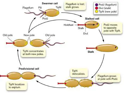
(yellow) is found at “new” poles (newly septated). TipN delocalizes and then localizes at the cell equator, midway between poles. The pole with PodJ grows a flagellum. The cell septates, forming two new poles, each containing TipN. The stalked cell still has DivJ at the stalk, and the new swarmer cell has PodJ at the flagellum.
Source: Modified from Melanie Lawler and Yves Brun. 2006. Cell 124 :891. The stalk marker DivJ is now produced at the stalk pole, where PodJ was previously. As the stalked cell grows, TipN proteins delocalize around the cell. Then TipN localizes again at the middle, where the cell septates and divides. Once division is complete, TipN is concentrated at both new poles. The pole containing PodJ now grows a flagellum. The new flagellated cell is ready to find a new favorable environment to form a stalk and settle. Overall, throughout the cycle, a series of polar proteins localize and delocalize to define the polar functions.
Thought Question
3.10 Figure 3.31 presents data from an experiment that allows the function of the TipN protein of Caulobacter to be visualized by microscopy. Can you propose an experiment with mutant strains of Caulobacter to test the hypothesis that one of the proteins shown in Figure 3.32is required for one of the cell changes shown?
Growth Asymmetry and Polar Aging
Does an apparently symmetrical cell such as E. coli actually possess two different polar forms? In fact, cell division generates daughter cells with chemically different poles (Fig. 3.33). Each cell starts out with one “old” pole (red in the figure) and one “new” pole (blue) where the parental cell septated. As the next cell divides, two daughter cells form, each with another “new” pole. But meanwhile, the “old” poles continue to age. With each generation, the polar cell wall material degrades slightly, increasing the chance of cell lysis. In a population of E. coli under environmental stress, at each cell division some members of the population die—of polar old age. FIGURE 3.33 ■ Bacterial cell division generates cells with an old pole and a new pole. Succeeding generations
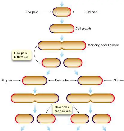
have cells with diverse combinations of new poles (blue), old poles (red), and very old poles (two or more generations, also red).
Source: Modified from Eric Stewart et al. 2005. PLoS Biol. 3 :e45 The cause of polar aging in stressed E. coli is the preferential accumulation of protein aggregates, which are nonfunctional and cannot be unfolded or degraded. For unknown reasons, proteins aggregate more frequently under a stressful condition, such as low pH or the presence of an antibiotic. Proteins damaged by a stressful condition are packed away in the cell’s older pole, allowing the new-pole cells to remain intact and grow faster. This asymmetrical cell provisioning may represent a form of “altruism” in which the older half cell promotes faster growth of the younger half cell.
Yet other kinds of cells grow by extending one pole only. The actinomycete Corynebacterium glutamicum, a soil bacterium useful for industrial production, positions its replisome at one cell pole. As DNA replication begins, a second replisome moves to the opposite pole, while new cell wall forms at the poles. In the next generation, the opposite pole possesses the replisome and undergoes extension. Unequal or unipolar cell extension is common among actinomycetes and mycobacteria, such as Mycobacterium tuberculosis.
Why does polar aging matter? One consequence of polar aging is that cells of different polar ages may differ in their resistance to antibiotics. This phenomenon could cause problems for antibiotic therapy. In M. tuberculosis, alternate polar aging generates variable resistance to antibiotics. The result may give tuberculosis bacteria the opportunity to “try out” resistance to various antibiotics applied in chemotherapy.
An extreme form of asymmetrical growth is endospore formation by Firmicutes such as Bacillus and Clostridium species. An endospore is an inert but viable cell form, having no active metabolism but capable of germination under the right conditions. Under starvation, desiccation, or other stress conditions, a bacterium can undergo an asymmetrical cell division to develop an endospore at one end. Endospore formation requires an extreme form of cellular altruism, in which the mother cell sacrifices itself for the spore-forming cell. The process generates an endospore capable of remaining dormant but viable for thousands of years.
Note: Endospore formation is covered in detail in Section 4.6. Membrane Vesicles
Our concept of the cell assumes a defined boundary of membrane that encloses the cell’s contents and separates them from the external space. The cell’s cytoplasm is a precious limited resource. Yet surprisingly, isolated microbial cells continually export bits of cytoplasm in membrane vesicles. Some kinds of microbes share their materials with other cells—even cells of other species—via intercellular nanotubes. How does this cytoplasmic sharing serve the cell?
Membrane vesicles carry proteins and nucleic acids. An example of cytoplasmic export via membrane vesicles in the marine cyanobacterium Prochlorococcus was documented by Sallie Chisholm at the Massachusetts Institute of Technology. Prochlorococcus is one of the smallest yet most abundant phototrophs in Earth’s oceans, having a global population estimated at 3 × 10 27 cells, which perform 20% of all photosynthesis in our oceans. Because Prochlorococcus cells are so small and their nutrients so scarce, it is remarkable that these tiny cells release their cytoplasm by pinching off vesicles (Fig. 3.34). Chisholm and her postdoctoral fellow analyzed these vesicles by ultracentrifugation (discussed in Section 3.1) and biochemical assays of their contents. The vesicles were found to contain diverse proteins, RNA molecules, and even fragments of DNA.
FIGURE 3.34 ■ Marine bacteria release membrane vesicles. A. Prochlorococcus cyanobacteria release vesicles (arrows) of cell membrane into the open ocean (SEM). B.
Vesicles collected from Prochlorococcus (TEM). C. Sallie Chisholm, professor of environmental studies, pioneered the study of Prochlorococcus and its significance for marine ecology.
S. J. BILLER ET AL. 2014. SCIENCE 343 (6167):183–186, FIG. 1A
S. J. BILLER ET AL. 2014. SCIENCE 343 (6167):183–186, FIG. 1B
RICHARD HOWARD
What functions are served by vesicle production that outweigh the loss of precious resources? Chisholm finds evidence for several possibilities:
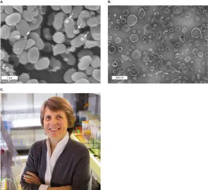
Attraction of partner heterotrophs. Heterotrophic bacteria attracted by released carbon sources consume the excess oxygen and reactive oxygen species (ROS) produced by cyanobacterial photosynthesis. Cyanobacteria require heterotrophic partners for optimal growth.
Phage decoys. Bacteriophages readily infect Prochlorococcus and deplete its populations. But the bacterial membrane vesicles possess envelope receptors for phages, which can trap the phages and prevent them from infecting cells.
DNA transfer. The DNA released by Prochlorococcus may provide useful genetic traits for other members of the population as a form of horizontal gene transfer.
Another system in which membrane vesicles are shared is that of human gut bacteria such as Bacteroides and related anaerobes. One way the gut environment differs from the open ocean is in the abundance of nutrients available. In the gut, many anaerobes release vesicles of partly digested complex polysaccharides for further catabolism by other species. Often, both community members benefit as a result. Gut microbial interactions are discussed further in Chapter 21.
Membrane Extensions and Nanotubes
From the observations of the early microscopists of the nineteenth century, most bacteria were thought to have relatively simple shapes such as rods or spheres. But cryo-electron microscopy revealed surprisingly delicate extensions of the cell or outer membranes, such as filaments and “pearling” chains of vesicles. What is the function of these cell extensions? In some cases, cell extensions expand the cell’s reach for scarce nutrients (see Chapters 4 and 18). The purpose of other membrane extensions remains unclear, but exciting discovery continues.
Some bacteria and archaea can form membrane extensions that merge directly with the membranes of neighboring cells. Sigal Ben-Yehuda and students at the Hebrew University of Jerusalem revealed such extensions, called intercellular nanotubes, between cells of Bacillus subtilis (Fig. 3.35). B. subtilis is a Gram-positive bacterium common in soil, a highly complex environment full of diverse nutrients and antimicrobial toxins (discussed in Chapter 21). The nanotubes enable bacteria to directly share proteins and messenger RNA that encodes products useful under hostile conditions, such as exposure to antibiotics (Fig. 3.35B ). Ben-Yehuda showed that two Bacillus cells encoding resistance to two different antibiotics—chloramphenicol (Cat protein) and lincomycin (Erm protein)—could share their resistance proteins and messenger RNA via nanotubes. The connected bacteria resist both antibiotics. FIGURE 3.35 ■ Intercellular nanotubes. A. Bacillus subtilis bacteria connected by intercellular nanotubes, which pass material from one cell to the next. B. Nanotubes connect bacteria with two different genes encoding proteins that confer resistance to an antibiotic: chloramphenicol (Cat) or lincomycin (Erm). The connected bacteria share mRNA and resistance proteins for both chloramphenicol and lincomycin resistance. Cm R = chloramphenicol resistance; Lin R = lincomycin resistance.
G. P. DUBEY AND S. BEN-YEHUDA. 2011. CELL 144 :590
A similar experiment showed that even bacteria of different species can share beneficial components of cytoplasm. Christian Kost and students at Max Planck Institute for Chemical Ecology in
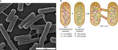
Jena, Germany, used fluorescence microscopy to show that Escherichia coli bacteria can form nanotubes with the Gram-negative bacterium Acinetobacter baylyi. The nanotubes facilitate exchange of different amino acids between these two species. Remarkably, the nanotubes form only when the two types of cells each produce an amino acid lacking in the other. Thus, nanotubes facilitate metabolic cross-feeding.
Archaea show various kinds of intercellular nanotubes that are essential parts of the cell. Examples are found in Pyrodictium and Thermococcus species, as well as the Asgard archaeon, Prometheoarchaeum synthrophicum (see Chapter 1 opening image). For more examples, see Chapter 19.
Note: Bacterial nanotubes between cells remain a subject of
controversy, because some laboratories fail to replicate their discovery, even using the same bacterial strains. In Archaea, intercellular nanotubes are more clearly documented.
Thought Question
3.11 Could two bacteria share protein complexes via nanotubes? What about hydrogen molecules (H 2) as electron donors?
SPECIAL TOPIC 3 Bacteria Reach Out with Pearling Tubes and Nanopods
What if you found an intricate membrane structure like a string of pearls and had no idea what it does? Mohammed Kaplan, along with colleagues at the California Institute of Technology and several other universities, hunted for novel membrane extensions in cryo-electron tomograms (tomography slices) from 90 species of bacteria. The resolution and clarity of cryo-EM and the absence of heavy-metal stains revealed fine structures that were missed by earlier electron microscopy. Thirteen of the bacteria showed novel structures never before reported—and whose functions remain unclear (Fig. ST 3.1 ). FIGURE ST 3.1 ■ Mysterious membrane extensions. A. Pearling tubes of Helicobacter hepaticus (cryo-EM). B. Mohammed Kaplan, graduate student in the lab of Grant Jensen, California Institute of Technology.
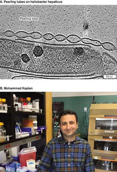
Source: Mohammed Kaplan et al. 2021. Elife 10 :e73099.
M. KAPLAN ET AL. 2021. ELIFE. 10:E73099
MOHAMMED KAPLAN
Helicobacter hepaticus is a spiral-shaped Gram-negative proteobacterium that infects the livers of mice and is a relative of the ulcer-causing human pathogen Helicobacter pylori. The tomograms of H. hepaticus showed long strings of interconnected vesicles of outer membrane, each with a round shape like pearls on a string (Fig. ST 3.1A ). The function of these vesicles is not known, but similar “pearling tubes” are found in aquatic Shewanella bacteria, where they may carry electricity. Bacterial electricity and its use for fuel cells are described in Chapter 14.
A different kind of outer membrane extension was found in Myxococcus xanthus, a predatory bacterium that collects in swarms to form a fruiting body and spores. M. xanthus cells extend multiple long tubes of outer membrane (Fig. ST 3.2A ). The function of these tubes is unknown, although in other microbes lengthy extensions help the cell obtain nutrients from nutrient-poor environments. Similar membrane extensions are found in Caulobacter crescentus, but its tubes are filled with S-layer subunits. The subunits are wrapped in membrane, like a sandwich in plastic wrap. These S-layer tubes are called nanopods (Fig. ST 3.2B ). Because S-layer proteins generally protect a cell surface, it is puzzling to find them contained by a membrane. We do not know what substances are contained by pearling tubes, long extensions, or nanopods. We only know that the more we investigate microbial cells, the more amazing and unexpected components we find.
FIGURE ST 3.2 ■ Membrane extensions and nanopods. A. Myxococcus xanthus membrane extensions
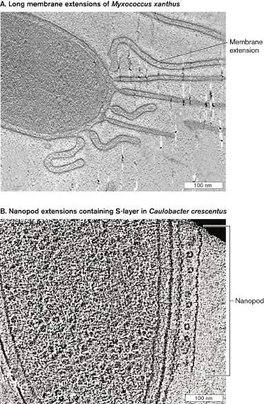
(cryo-EM). B. Caulobacter crescentus nanopod (cryo-EM). Source: Mohammed Kaplan et al. 2021. Elife 10 :e73099.
M. KAPLAN ET AL. 2021. ELIFE. 10 :E73099
M. KAPLAN ET AL. 2021. ELIFE. 10 :E73099
RESEARCH QUESTION
What experiments might you perform to reveal the function of a mysterious bacterial structure?
Kaplan, Mohammed, Georges Chreifi, Lauren A. Metskas, Janine Liedtke, Cecily R. Wood, et al. 2021. In situ imaging of bacterial membrane projections and associated protein complexes using electron cryo-tomography. Elife 10 :e73099.
To Summarize
The poles of a bacterial cell may differ in form and function. Caulobacter crescentus has one plain pole and one pole that has either a flagellum or a stalk. A stalked cell fissions to produce one stalked cell and one flagellar cell. The two bacterial poles differ in age. One pole arises from the septum of the parental cell, whereas the other pole arises from a parental pole. In E. coli, successive cell divisions yield progeny with a mixture of polar ages. Cells with a very old pole may cease replication and die.
Polar aging is increased by stress. Environmental stress, such as an antibiotic or low pH, causes protein aggregates to collect at the cell’s older pole. Actinobacterial cells extend at alternating poles.
Membrane vesicles transmit cytoplasmic contents.
Vesicles share proteins, nucleic acids, and other cytoplasmic contents with the exterior environment and other cells. Intercellular nanotubes directly share cytoplasmic contents. Nanotubes between individual bacteria share drug resistance, cross-feed nutrients, and mediate electron transfer.
Glossary
nanotube A tube of plasma membrane that connects the cytoplasm of two cells, forming a conduit through which intracellular materials or pathogens may pass.
Figure 2.29: FIGURE 2.29 ■ The fluorophore green fluorescent protein (GFP). A. Green fluorescent protein (GFP) is expressed endogenously by the cell. Blowup: Three GFP amino acid residues (serine, tyrosine, and glycine) condense to form the fluorophore. B. The gene encoding GFP can be fused to a target gene (Target′- gfp). The fused gene then expresses a fused protein in which the GFP portion
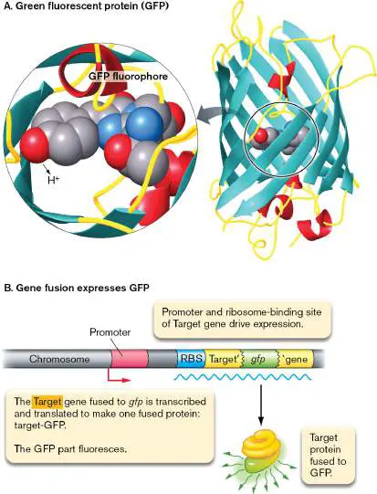
fluoresces. The fluorescent protein is expressed under control of the target gene promoter and ribosome-binding site (RBS).
3.6 Specialized StructuresUnit 3 · Structure
We have introduced the major structures that cells need to contain and organize their contents, maintain their DNA, and synthesize new parts. Besides these fundamental structures, different species have evolved specialized devices adapted to diverse metabolic strategies and environments. And microscopy continually reveals new microbial structures whose functions remain a mystery (Special Topic 3).
Thylakoids, Carboxysomes, and Storage Granules
Cyanobacteria are phototrophs that produce food and oxygen for marine and freshwater ecosystems; their diversity is explored in Section 18.2. In the water, cyanobacteria must absorb sufficient amounts of light to drive photosynthesis (see Section 14.6). To maximize the collecting area of their photosynthetic membranes, cyanobacteria have evolved specialized systems of extensively folded intracellular membrane called thylakoids (Fig. 3.36A). Thylakoids consist of layers of folded sheets (lamellae) or tubes of membranes packed with chlorophylls and electron carriers.
Cyanobacteria containing thylakoids structurally resemble eukaryotic chloroplasts, which evolved from a common ancestor of modern cyanobacteria.
The thylakoids conduct only the “light reactions” of photon absorption and energy storage. The energy obtained is rapidly spent to fix carbon dioxide—a process that occurs within carboxysomes ( Fig. 3.36A; see also Fig. 15.7). Carboxysomes are polyhedral, protein-covered bodies packed with the enzyme Rubisco for CO 2 fixation (see Section 15.2).
How do phototrophs keep themselves at the top of the water column? Some bacteria and archaea form gas vesicles to increase buoyancy and keep the cell afloat. Figure 3.36B shows a cross section of Microcystis, a cyanobacterium that forms toxic algal blooms in lakes polluted by agricultural runoff. Microcystis shows typical gas vesicles, which are hollow protein structures that collect gases. The gases are hydrogen or carbon dioxide produced by the cell’s metabolism. Each vesicle consists of a tube of protein with two conical ends. The tubes pack in hexagonal arrays.
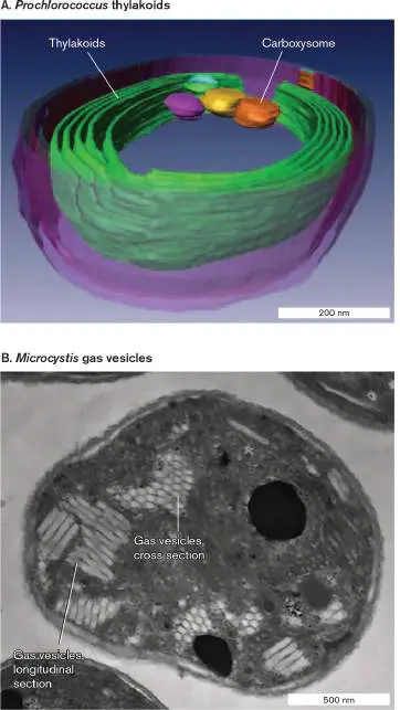
FIGURE 3.36 ■ Organelles of phototrophs. A. The marine phototroph Prochlorococcus contains photosynthetic double membranes called thylakoids. Carboxysomes are polyhedral, protein-covered bodies packed with the Rubisco enzyme for CO 2 fixation (cryo-EM). B. Microcystis gas vesicles (protein) in hexagonal arrays. Gas vesicles provide buoyancy, enabling the phototroph to remain at the surface of the water, exposed to light (TEM). Source: Part A from Clare Ting. 2007. J. Bacteriol. 189 :4485.
C. S. TING ET AL. 2007. J BACTERIOL. 189 :4485–4493
ALYSSA MLOUKA ET AL. 2004. J. BACTERIOL. 186 :2355
When light is scarce, cyanobacteria may digest their thylakoids for energy and as a source of nitrogen. Alternatively, the cell may digest energy-rich materials from storage granules composed of glycogen or other polymers, such as polyhydroxybutyrate (PHB) and poly-3-hydroxyalkanoate (PHA). PHB and PHA polymers are of interest as biodegradable plastics, and bacteria have been engineered to produce them industrially. Similar storage granules are also produced by nonphototrophic soil bacteria.
Another type of storage device is sulfur—granules of elemental sulfur produced by purple and green phototrophs through photolysis of hydrogen sulfide (H 2 S). Instead of disposing of the sulfur, the bacteria store it in granules, such as those of the giant sulfur-oxidizing bacterium Thiomargarita namibiensis (Fig. 3.37). Sulfur-reducing bacteria also make sulfur globules; for example, by reducing sulfate (SO 2−) to sulfur. The sulfur granules may later be
4
used as an oxidant when reduced substrates are available (see Chapter 14). And the presence of potentially toxic sulfur granules may help cells avoid predation.
FIGURE 3.37 ■ Intracellular sulfur globules. Sulfur globules dot the cytoplasm of Thiomargarita namibiensis, an anaerobic, thermophilic bacterium that gains energy by oxidizing hydrogen sulfide (H S) to elemental sulfur (S 0).
2
Source: Heide N. Schulz and Horst D. Schulz. 2005. Science 307 :416–418.
H. N. SCHULZ ET AL. 2005. SCIENCE. 307 :416–418
Pili and Stalks
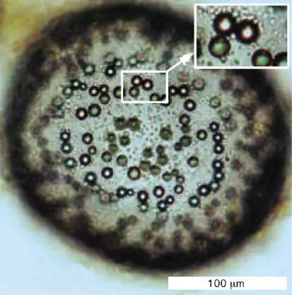
In a favorable habitat, such as a running stream full of fresh nutrients or the epithelial surface of a host, it is advantageous for a cell to adhere to a substrate. Adherence, the ability to attach to a substrate, requires specific structures. A common adherence structure is the pilus (plural, pili), which is constructed of straight filaments of protein monomers called pilin. Short attachment pili are also called fimbriae. For example, the sexually transmitted pathogen Neisseria gonorrhoeae uses pili to attach to the mucous membranes of the reproductive tract (Fig. 3.38). Pili can also provide a form of motility called “twitching,” in which the pili act as limbs to “walk” the bacterium across a substrate (presented in Chapter 4). Bacteria such as Pseudomonas aeruginosa use twitching motility to begin biofilm formation (discussed in Section 4.5).
FIGURE 3.38 ■ Pili are protein filaments for attachment. Neisseria gonorrhoeae, cause of the sexually transmitted infection gonorrhea, use pili to attach to the host mucous membrane (SEM).
L. CRAIG ET AL. 2006. MOL. CELL 23 :651–662
In Gram-negative enteric bacteria, pili of a different kind, also called the sex pili, attach a donor cell to a recipient cell for transfer of DNA. This process of DNA transfer is called conjugation. The genetic consequences of conjugation are discussed in Chapter 9.
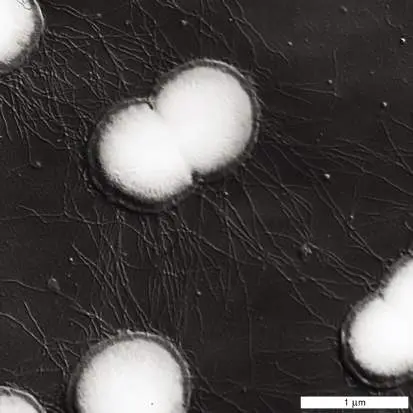
A different kind of attachment organelle is an extension of the envelope and cytoplasm called a stalk, seen earlier in the stalked cell of Caulobacter (Fig. 3.30). The tip of the stalk secretes adhesion factors that form a “holdfast,” which firmly attaches the bacterium in an environment that has proved favorable. A stalk and holdfast enable iron-oxidizing bacteria to form large biofilms in streams contaminated by iron drainage. The biofilms become coated by orange iron hydroxides, tinting the stream orange.
Rotary Flagella
What happens when the cell’s environment runs out of nutrients or becomes filled with waste? In rapidly changing environments, cell survival requires motility, the ability to move and relocate. Many bacteria and archaea can swim by means of rotary flagella (singular, flagellum). Flagellar motility benefits the cell by causing the population to disperse, decreasing competition. Motility also enables cells to swim toward a favorable habitat (by chemotaxis, discussed shortly).
Flagellar motility. Flagella are helical propellers that drive the cell forward like the motor of a boat. Howard Berg (1934–2021) at the California Institute of Technology originally described the bacterial flagellar motor, which was the first rotary device to be discovered in a living organism. Different bacterial species have different numbers and arrangements of flagella. Peritrichous cells, such as Escherichia coli and Salmonella species, have flagella randomly distributed around the cell (Fig. 3.39A). The flagella rotate together in a bundle behind the swimming cell (Fig. 3.39B ). Lophotrichous cells, such as Rhodospirillum rubrum, have flagella attached at one or both ends. In monotrichous (polar) species, such as the Caulobacter swarmer cell (see Fig. 3.30), the cell has a single flagellum at one end.
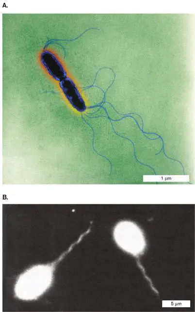
FIGURE 3.39 ■ Flagellated Salmonella bacteria. A. Salmonella enterica has multiple flagella (colorized TEM). B. The flagella collect in a bundle behind a swimming cell. Under dark-field microscopy, the cell body appears overexposed, about five times as large as the actual cell.
KWANGSHIN KIM/SCIENCE SOURCE
ROBERT MACNAB. 1976.
J. CLIN. MICROBIOL. 4 :258
How does a rotary flagellum work? Each flagellum has a spiral filament of protein monomers called flagellin (protein FliC). The filament actually rotates by means of a motor driven by the cell’s transmembrane proton current—the same proton potential that drives the membrane-embedded ATP synthase (presented in Chapter 14). The flagellar motor is embedded in the layers of the cell envelope (Fig. 3.40). The motor possesses an axle and rotary parts, all composed of specific proteins. For example, protein MotB forms part of the ion channel whose flux of hydrogen ions powers rotation. Another protein, FliG, forms part of the device that generates torque (rotary force). Much of the motor’s structure and function was elucidated by Scottish microbiologist Robert Macnab (1940–2003) at Yale University.
FIGURE 3.40 ■ The flagellar motor. A. The basal body, or motor, of the bacterial flagellum (TEM). This image is based on digital reconstruction, in which electron micrographs of purified basal bodies were rotationally averaged. B. H⁺ flow through the MotA-MotB complex drives rotation of the flagellar motor.
N. R. FRANCIS ET AL. 1994. J. MOL. BIOL. 235 :1261.
What kinds of experiments reveal the motor components?
Results from an experiment dissecting the flagellar motor are shown in Figure 3.41. Japanese microbiologist Tohru Minamino (Osaka University) and colleagues constructed strains of Salmonella enterica in which the gene that encodes fluorescent GFP is fused to a gene encoding a flagellar protein, MotB or FliG, each of which is proposed to be a part of the motor. (Gene fusion is explained in Figure 2.29.) For each flagellar construct strain, fluorescence microscopy reveals the GFP fluorescence at one or two positions within the cell (green dots). A second fluorophore, Alexa, is conjugated to an anti-flagellin antibody. The Alexa fluorescence (red) reveals the flagellar filament. When the green and red fluorescence images are merged, the red flagellar filaments appear to extend from the motor positions that
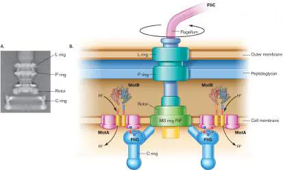
contain either MotB or FliG. Further experiments dissect the roles of key amino acid residues in the function of these proteins.
FIGURE 3.41 ■ Flagellar motor proteins localized by fluorescence microscopy. A. Cells of Salmonella enterica express a GFP fused to flagellar motor protein MotB or FliG. Bright green dots (noted by white leader lines) indicate MotB-GFP or FliG-GFP complexed with a flagellar motor. The flagellar filament is visualized via the red fluorophore Alexa, conjugated to an anti-flagellin antibody. The merged image shows how each flagellar filament (red) extends from a motor containing either MotB or FliG (green, protein fused to GFP). B. Tohru Minamino investigates the structure and function of the flagellar motor.
YUSUKE MORIMOTO ET AL. 2010. MOL. MICROBIOL. 78 :1117
COURTESY OF PROTONIC NANOMACHINE GROUP, OSAKA UNIVERSITY
Note: Bacterial flagella differ completely from the whiplike
flagella and cilia of eukaryotes and evolved separately. Eukaryotic flagella are much larger structures containing multiple microtubules enclosed by a membrane (shown in Chapter 20). They move with a whiplike motion, powered by ATP hydrolysis all along the flagellum.
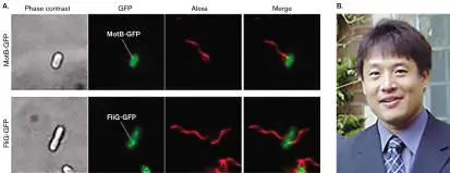
How do cells decide where to swim? Most flagellated cells have an elaborate sensory system for taxis, the ability to swim toward favorable environments (attractant signals, such as nutrients) and away from inferior environments (repellent signals, such as waste products). Taxis to specific chemicals is called chemotaxis. Chemotaxis requires receptors (chemoreceptors) that act like a “nose,” telling the bacterium when it is swimming toward a source of attractant such as a sugar or an amino acid. The attractant and repellent molecules are detected by arrays of chemoreceptors that are located near a cell pole (Fig. 3.42). This remarkably precise, ordered structure was first visualized in Gram-negative bacteria by Ariane Briegel and colleagues, at the California Institute of Technology and the University of Leiden. Figure 3.42shows a TEM section of Vibrio cholerae, the cause of cholera. Briegel used cryo-electron tomography to identify the arrays at the cell pole, and she then used computation to define the repeated form of the array’s subunits.
FIGURE 3.42 ■ Receptor array detects chemotactic signals for motility. A. Cryo-electron tomography section through Vibrio cholerae was used for digital reconstruction of chemotaxis receptor arrays. Inset: Cross section through an array. B. Ariane Briegel’s cryo-electron tomography team first imaged the Gram-negative receptor array.
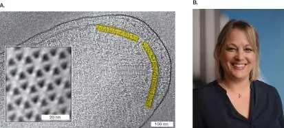
Source: Ariane Briegel et al. 2016. PNAS 113 :10412.
A. BRIEGEL ET AL. 2016. PROC NATL ACAD SCI USA. 113: 10412–7
INSTITUTE OF BIOLOGY LEIDEN
Note: Chemotaxis control of motility, including the biased random
walk, is covered in Section 12.1. Related topics of internalized flagella of spirochetes are shown in Section 18.5, and phototaxis (taxis toward light) for haloarchaea is presented in Section 19.5. Besides motility and chemotaxis, surprisingly, flagella have also evolved an alternate function: adherence of cells to a substrate to begin forming a biofilm (discussed in Chapter 4). Thus, an organism can evolve a structure that serves one function but later evolves to serve another function.
In addition to flagellar rotation, other forms of bacterial motility are just beginning to be understood, such as pili-dependent twitching motility (discussed in Section 4.5). Another kind of motility, called “gliding,” is observed in cyanobacteria and in myxobacteria.
Thought Question
3.12 Most laboratory strains of E. coli and Salmonella commonly used for genetic research lack flagella. Why and how do bacterial strains evolve to lose flagella? How can a researcher maintain a motile strain?
To Summarize
Cyanobacteria possess thylakoid membrane organelles packed with photosynthetic apparatus and carboxysomes for carbon dioxide fixation. Gas vesicles provide buoyancy in the water column.
Storage granules store elemental sulfur or organic carbon polymers for energy.
Adherence structures enable prokaryotes to remain in an environment with favorable environmental factors. Major adherence structures include pili or fimbriae (protein filaments) and the holdfast (a cell extension).
Flagellar motility involves rotary motion of helical flagella. Flagellar rotation is driven by the transmembrane proton motive force.
Chemoreceptors provide information that directs flagellar motility.
Glossary
thylakoid An intracellular chlorophyll-containing membrane folded within a phototrophic bacterium or a chloroplast.
carboxysome A protein-enclosed compartment containing Rubisco to fix CO 2. gas vesicle An organelle that traps gases to increase the buoyancy of aquatic microbes.
pilus pl. pili Also called fimbria. A straight protein filament composed of a tube of protein monomers that extend from the bacterial cell envelope.
pilus pl. pili Also called fimbria. A straight protein filament composed of a tube of protein monomers that extend from the bacterial cell envelope.
stalk An extension of the cytoplasm and envelope that attaches a microbe to a substrate.
motility The ability of a microbe to direct its own movement.
flagellum pl. flagella A filamentous structure for motility. In prokaryotes, a helical protein filament attached to a rotary motor; in eukaryotes, an undulating membrane-enclosed complex of microtubules and ATP-driven motor proteins.
flagellum pl. flagella A filamentous structure for motility. In prokaryotes, a helical protein filament attached to a rotary motor; in eukaryotes, an undulating membrane-enclosed complex of microtubules and ATP-driven motor proteins.
chemotaxis The ability of organisms to move toward or away from specific chemicals.
Fig. 15.7 FIGURE 15.7 ■ Carboxysomes. A. Thin section of Halothiobacillus neapolitanus, a sulfur-oxidizing lithoautotroph (TEM), showing polyhedral carboxysomes (arrows). B. Carboxysome isolated from Synechococcus cyanobacterium is packed with Rubisco complexes (cryo-electron tomography). C. A 3D model of Rubisco complexes packed inside a carboxysome.
Source: Part A modified from Y. Tsai et al. 2007. PLoS Biol. 5 :E144.
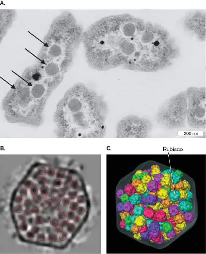
COURTESY OF SABINE HEINHORST AND DR. GORDON C. CANNON
C. V. IAN ET AL. 2007. J MOL BIOL. 372 :764–773
C. V. IAN ET AL. 2007. J MOL BIOL. 372 :764–773
Figure 2.29
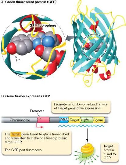
FIGURE 2.29 ■ The fluorophore green fluorescent protein (GFP). A. Green fluorescent protein (GFP) is expressed endogenously by the cell. Blowup: Three GFP amino acid residues (serine, tyrosine, and glycine) condense to form the fluorophore. B. The gene encoding GFP can be fused to a target gene (Target′- gfp). The fused gene then expresses a fused protein in which the GFP portion fluoresces. The fluorescent protein is expressed under control of the target gene promoter and ribosome-binding site (RBS).
Fig. 3.30 FIGURE 3.30 ■ Asymmetrical cell division: a model for development. A swarmer cell of Caulobacter crescentus loses its flagellum and grows a stalk. The stalked cell divides to produce a swarmer cell (TEM).
YVES BRUN
Fig. 3.30
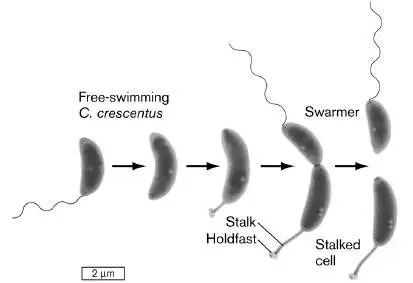
FIGURE 3.30 ■ Asymmetrical cell division: a model for development. A swarmer cell of Caulobacter crescentus loses its flagellum and grows a stalk. The stalked cell divides to produce a swarmer cell (TEM).
YVES BRUN
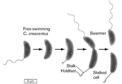
eResearch Activity 3
How Do a Hundred Cells Divide at Once?
Dividing the cell is a complicated business, even for a relatively simple bacterium such as Escherichia coli. Forming two functional daughter cells requires intricate coordination of the DNA replication and cell growth, culminating in septation at the Z-ring (see Figures 3.23 and 3.25 ). Now suppose you have to form a hundred cells at once. How would that ever work?
The actinomycete Streptomyces venezuelae grows as a filament (or hypha), copying its DNA many times and extending without cell division. In a sporogenic (spore-forming) hypha, a molecular regulator promotes simultaneous septation at dozens of Z-rings. When FtsZ is labeled with a fluorophore, the fluorescence generates a dramatic ladder-like pattern called a “Z-ladder” (Fig. ERA 3.1A ). The Z-ladder enables a filament to divide and develop into numerous spores, which are carried off on air currents to colonize a new location. Streptomyces species are important to understand because they give us many antibiotics, such as streptomycin for tuberculosis and tetracycline for acne and respiratory bacterial infections. A. S. venezuelae B. S. venezuelae ΔsepH
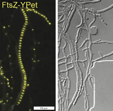
FIGURE ERA 3.1 ■ Multiple Z-rings in Streptomyces venezuelae. A. Normal sporogenic hyphae of S. venezuelae form multiple Z-rings and undergo coordinated septation. FtsZ protein is visualized by fluorescence microscopy of an FtsZ protein fused to the protein fluorophore YPet. B. A strain deleted for the sepH gene (Δ sepH) shows abberant septation (arrowheads).
Fluorescence microscopy and differential interference contrast microscopy (DIC). Source: F. Ramos-León et al. 2021. Elife 10:e63387. (Panel A:; Panel B:)
F. RAMOS-LEÓN ET AL. 2021. ELIFE. 10:E63387
F. RAMOS-LEÓN ET AL. 2021. ELIFE. 10:E63387
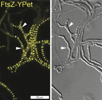
How do Streptomyces hyphae manage to coordinate a hundred cell divisions? Susan Schlimpert, at the John Innes Centre in Norwich, England, investigates the mechanisms of cell division in Streptomyces venezuelae, a model actinomycete (Fig. ERA 3.2A ). A key cell division regulator was revealed by her postdoctoral fellow Félix Ramos-León and her research assistant Matthew Bush (Fig. ERA 3.2B ).
FIGURE ERA 3.2 ■ Researchers investigate cell division in Streptomyces venezuelae. A. Susan Schlimpert, group leader, John Innes Centre. B. Félix Ramos-León, postdoctoral fellow, John Innes Centre now at NIH, Bethesda (left) and co-first author Matthew Busch (right), Research Assistant, John Innes Centre.
SUSAN SCHLIMPERT
Bush identified sepH as a gene of unknown function in the Streptomyces genome and showed that its expression is controlled by a major sporulation regulator. First, Ramos-León and colleagues investigated the SepH gene product by constructing a gene fusion to the gene encoding the fluorescent protein YPet. (Gene fusions for fluorescent proteins are discussed in Chapter 2.) The resulting fusion gene, sepH-ypet, encodes a protein that functions as SepH but also fluoresces as YPet by absorption of green light with emission of yellow light. In microscopy of S. venezuelae carrying the sepH-ypet gene, we see yellow fluorescence at the position of Z-rings. Thus,
SepH has an appropriate location for a protein that assists FtsZ function.
Next, the researchers observed a strain of S. venezuelae that expresses FtsZ fused to YPet, so that the ladder of Z-rings fluoresces. The FtsZ-YPet fluorescence was observed in two different strains, either expressing sepH (Fig. ERA 3.1A ) or deleted for sepH (Δ sepH) (Fig. ERA 3.1B ). For each experiment, a movie was made showing the coordinated development of the Z-ladder and sporulation. In the Δ sepH strain (a strain lacking sepH), sporulation becomes disorganized, causing cells to bulge into distorted forms and even to form filament branches. While some sporulation occurs, the process is greatly disrupted. Thus sepH was confirmed as a gene whose protein product organizes septation and sporulation in S. venezuelae.
Ramos-León further revealed that SepH stimulates the GTPase activity of FtsZ, a common mechanism of molecular regulation. A SepH homolog was found in the genus Mycobacterium, a group of Actinobacteria that includes the famous pathogen Mycobacterium tuberculosis. In pathogenic mycobacteria, SepH assists cell division without sporulation. Thus, discovery of this cell division protein advances our understanding of both an important drug producer and a major pathogen whose mechanisms may lead to novel antibiotic therapies.
Further Exploration
What experiments might you perform to show whether SepH interacts directly with the Z-ladder by binding to FtsZ? What other kinds of proteins might be needed?
Ramos-León, Félix, Matthew J. Bush, Joseph W. Sallmen, Govind
Chandra, Jake Richardson, et al. 2021. A conserved cell division protein directly
regulates FtsZ dynamics in filamentous and unicellular actinobacteria. Elife 10
:e63387.
Glossary
Figure 3.23 FIGURE 3.23 ■ Shape-determining proteins. A. Cell diameter is maintained by FtsZ polymerization to form the Z-ring. B. Elongation of a rod-shaped cell requires MreB proteins. MreB polymerizes around an E. coli cell (MreB-YFP fluorescence) along with a Z-ring of FtsZ (fluorescent anti-FtsZ antibody). C. Crescent-shaped cells possess a third shape-
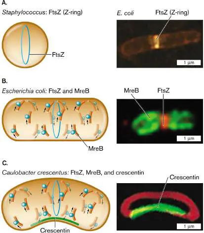
determining protein, CreS (crescentin), which polymerizes along the inner curve of the crescent. Crescentin protein fused to green fluorescent protein (CreS-GFP) localizes to the inner curve of Caulobacter crescentus. Membrane-specific stain FM4-64 (red fluorescence) localizes to the membrane around the cell.
Q. SUN AND W. MARGOLIN. 1998. J. BACTERIOL. 180 :2050
PURVA VATS AND LAWRENCE ROTHFIELD. 2007. PNAS 104 :17795
NORA AUSMEES ET AL. 2003. CELL 115 :705
Figure 3.25 FIGURE 3.25 ■ Divisome enables cell division. A. E. coli cells divide normally. B. Cells that lack FtsN (divisome component) balloon out at the septum. C. The divisome complex coordinates the extension of all envelope layers while the septum constricts.
Source: Part C modified from C. Typas et al. 2012. Nat. Rev. Microbiol. 10
:123, fig. 2.
B. LIU ET AL. 2015. MOL. MICROBIOL. 95 :945
B. LIU ET AL. 2015. MOL. MICROBIOL. 95 :945
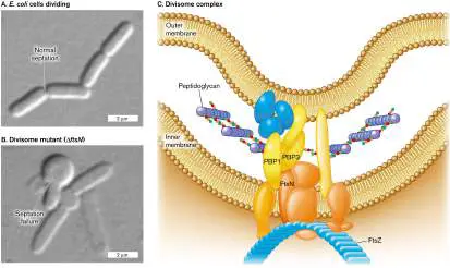
CHAPTER REVIEW
Review Questions
1. What are the major features of a bacterial cell, and how do they fit together for cell function as a whole?
2. What fundamental traits do most prokaryotes share with eukaryotic microbes? What traits are different?
3. Explain how cell fractionation enables us to separate proteins of the Gram-negative outer membrane, periplasm, inner membrane, and cytoplasm.
4. Outline the structure of the peptidoglycan sacculus, and explain how it expands during growth. Cite two different kinds of experimental data that support our current views of the sacculus.
5. Compare and contrast the structure of Gram-positive and Gram-negative cell envelopes. Explain the strengths and weaknesses of each kind of envelope.
6. Explain how the process of DNA replication is coordinated with cell wall septation.
7. Explain how the asymmetry of a bacterial cell generates daughter cells with different structure and function. Explain the significance for environmental adaptation and for antibiotic therapy.
8. What kinds of subcellular structures are found in certain cells with different functions, such as photosynthesis or chemotaxis?
9. Compare and contrast bacterial structures for attachment and motility.
Thought Questions
1. The aquatic bacterium Caulobacter crescentus alternates between two cell forms: a cell with a flagellum that swims, and a stalked cell that adheres to particulate matter. The flagellar cell can discard its flagellum to grow a stalk and adhere, and then the stalked cell divides to give one stalked cell and one flagellated cell. What would be the adaptive advantage of this alternating morphology?
2. Suppose that one cell out of a million has a mutant gene blocking S-layer synthesis, and suppose that the mutant strain can grow twice as fast as the S-layered parent. How many generations would it take for the mutant strain to constitute 90% of the population?
3. Explain two different ways that an aquatic phototroph might use its subcellular structures to maximize its access to light. Explain how an aerobe (an organism requiring molecular oxygen for growth) might remain close to the surface, with access to air.
4. How do pathogenic bacteria avoid being engulfed by phagocytes of the human bloodstream? How do you think various aspects of the cell structure can prevent phagocytosis?
Key Terms
active transport (92) capsule (98)
carboxysome (115)
cardiolipin (88)
cell fractionation (84) cell membrane (80)
cell wall (82, 93)
chemotaxis (118)
cholesterol (89)
cross-bridge (94)
divisome (104)
DNA-binding protein (106) domain (of proteins) (105) electrophoresis (84) endotoxin (99)
envelope (82)
flagellum (82, 117)
gas vesicle (115)
glycan (93)
hopanoid (hopane) (89) inner membrane (80, 98) ion gradient (91)
leaflet (87)
lipid A (99)
lipopolysaccharide (LPS) (82, 99) lysis (lyse) (84)
membrane-permeant weak acid (91) membrane-permeant weak base (91) motility (117)
murein (93)
murein lipoprotein (98) nanotube (112)
nucleoid (83)
O antigen (99)
osmotic pressure (91) outer membrane (80)
passive transport (92) penicillin-binding protein (94) peptidoglycan (84)
periplasm (82)
phospholipid (87)
phospholipid bilayer (82) pilus (116)
plasma membrane (80) porin (99)
replisome (106)
S-layer (97)
sacculus (93)
septation (103)
septum (102)
spheroplast (84)
stalk (117)
staphylococcus (107) teichoic acid (97)
terpenoid (89)
thylakoid (114)
transport protein (transporter) (92) turgor pressure (91) ultracentrifuge (85)
Recommended Reading
Abe, Keigo, Toshiki Kuribayashi, Kyosuke Takabe, and Shuichi Nakamura. 2020. Implications of back-and-forth motion and powerful propulsion for spirochetal invasion. Scientific Reports 10 :1–7.
Aldridge, Bree B., Marta Fernandez-Suarez, Danielle Heller, Vijay Ambravaneswaran, Daniel Irimia, et al. 2012.
Asymmetry and aging of mycobacterial cells lead to variable growth and antibiotic susceptibility. Science 335 :100–103. Belin, Brittany J., Nicolas Busset, Eric Giraud, Antonio Molinaro, Alba Silipo, et al. 2018. Hopanoid lipids: From membranes to plant–bacteria interactions. Nature Reviews. Microbiology 16 :304–315.
Bisson-Filho, Alexandre W., Yen-Pang Hsu, Georgia R. Squyres, Erkin Kuru, Fabai Wu, et al. 2017. Treadmilling by FtsZ filaments drives peptidoglycan synthesis and bacterial cell division. Science 355 :739–743.
Cesar, Spencer, and Kerwyn C. Huang. 2017. Thinking big: The tunability of bacterial cell size. FEMS Microbiology Reviews 41:672–678.
Dubey, Gyanendra P., and Sigal Ben-Yehuda. 2011.
Intercellular nanotubes mediate bacterial communication. Cell 144 :590–600.
Kaplan, Mohammed, Georges Chreifi, Lauren A. Metskas, Janine Liedtke, Cecily R. Wood, et al. 2021. In situ imaging of bacterial membrane projections and associated protein complexes using electron cryo-tomography. Elife 10 :e73099. Moore, Jeremy P., Haofan Li, Morgan L. Engmann, Katrina M. Bischof, Karina S. Kunka, et al. 2019. Inverted regulation of multidrug efflux pumps, acid resistance and porins in benzoate-evolved Escherichia coli K–12. Applied and Environmental Microbiology 85 :e00966.
Oikonomou, Catherine M., Yi-Wei Chang, and Grant J. Jensen. 2016. A new view into prokaryotic cell biology from electron cryotomography. Nature Reviews. Microbiology 14:205–221.
Pande, Samay, Shraddha Shitut, Lisa Freund, Martin Westermann, Felix Bertels, et al. 2015. Metabolic cross-feeding via intercellular nanotubes among bacteria. Nature Communications 6 :6238.
Renner, Lars D., and Douglas B. Weibel. 2011. Cardiolipin microdomains localize to negatively curved regions of Escherichia coli membranes. Proceedings of the National Academy of Sciences USA 108 :6264–6269.
Rojas, Enrique R., Gabriel Billings, Pascal D. Odermatt, George K. Auer, Lillian Zhu, et al. 2018. The outer membrane is an essential load-bearing element in Gram-negative bacteria. Nature 559 :617–621.
Romantsov, Tatyana, Karen Gonzalez, Naheda Sahtout, Doreen E. Culham, Chelsea Coumoundouros, et al. 2018. Cardiolipin synthase A colocalizes with cardiolipin and osmosensing transporter ProP at the poles of Escherichia coli cells. Molecular Microbiology 107 :623–638.
Schwechheimer, Carmen, and Meta J. Kuehn. 2015. Outer-membrane vesicles from Gram-negative bacteria: Biogenesis and functions. Nature Reviews. Microbiology 13 :605–619. Sun, Jiawei, Steven T. Rutherford, Thomas J. Silhavy, and Kerwyn Casey Huang. 2022. Physical properties of the bacterial outer membrane. Nature Reviews. Microbiology 20:236–248.
Wagstaff, James, and Jan Löwe. 2018. Prokaryotic cytoskeletons: Protein filaments organizing small cells. Nature Reviews. Microbiology 16 :187–201.
Glossary
cell membrane Also called cytoplasmic membrane or plasma membrane. The phospholipid bilayer that encloses the cytoplasm.
plasma membrane Also called cell membrane or cytoplasmic membrane. The phospholipid bilayer that encloses the cytoplasm.
inner membrane or inner cell membrane In Gram-negative bacteria, the membrane in contact with the cytoplasm, equivalent to the cell membrane.
cell wall A rigid structure external to the cell membrane. The molecular composition depends on the organism; in bacteria, it is composed of peptidoglycan.
periplasm In Gram-negative bacteria, the gel-like solution between the outer and inner membrane; it contains the cell wall.
lipopolysaccharides (LPS)
Structurally unique phospholipids found in the outer leaflet of the outer membrane in Gram-negative bacteria. Many are endotoxins.
envelope A structure external to the cell membrane, such as the cell wall or outer membrane of a bacterium. For a virus, the envelope is a membrane enclosing the capsid or core particle.
flagellum pl. flagella A filamentous structure for motility. In prokaryotes, a helical protein filament attached to a rotary motor; in eukaryotes, an undulating membrane-enclosed complex of microtubules and ATP-driven motor proteins.
phospholipid bilayer Two layers of phospholipids; the hydrocarbon fatty acid tails face the interior of the bilayer, and the charged phosphate groups face the cytoplasm and extracellular environment. The cell membrane is a phospholipid bilayer.
nucleoid The looped coils of a bacterial chromosome.
electrophoresis A technique to separate charged proteins and nucleic acids that is based on how rapidly they migrate in an electrical field through a gel.
peptidoglycan Also called murein. A polymer of peptide-linked chains of amino sugars; a major component of the bacterial cell wall. cell fractionation A procedure to separate cell components that often includes ultracentrifugation.
spheroplast A cell whose peptidoglycan is degraded by lysozyme; thus the cell loses its shape, forming a sphere.
ultracentrifuge A machine that subjects samples to high centrifugal forces and can be used to separate subcellular components.
phospholipid The major component of membranes. A typical phospholipid is composed of a core of glycerol to which two fatty acids and a modified phosphate group are condensed.
leaflet One of the two lipid layers in a phospholipid bilayer. The inner leaflet of the cell membrane faces the cytoplasm.
cardiolipin Diphosphatidylglycerol, a double phospholipid linked by glycerol.
cholesterol A sterol lipid found in eukaryotic cell membranes.
hopanoid or hopane A five-ringed hydrocarbon lipid found in bacterial cell membranes.
terpenoid A branched lipid derived from isoprene that is found in hydrocarbon chains of archaeal membranes.
osmotic pressure Also called turgor pressure. Pressure exerted by the osmotic flow of water through a semipermeable membrane.
turgor pressure Also called osmotic pressure. Pressure exerted by the osmotic flow of water through a semipermeable membrane.
membrane-permeant weak acid An acid that exists in equilibrium between negatively charged and uncharged forms, such as acetic acid. The uncharged form can penetrate the membrane.
membrane-permeant weak base A base that exists in equilibrium between positively charged and uncharged forms, such as methylamine. The uncharged form can penetrate the membrane.
ion gradient A difference in concentration of an ion across a membrane. transport protein or transporter A membrane protein that moves specific molecules across a membrane.
passive transport Net movement of molecules across a membrane without energy expenditure by the cell.
active transport An energy-requiring process that moves molecules across a membrane against their electrochemical gradient.
sacculus pl. sacculi The bacterial cell wall, consisting of a single covalent molecule. murein Also called peptidoglycan. A polymer of peptide-linked chains of amino sugars; a major component of the bacterial cell wall. glycan A polysaccharide chain composed of oxygen-linked (O-linked) monosaccharides.
cross-bridge An attachment that links parallel molecules, such as the peptide link between glycan chains in peptidoglycan.
penicillin-binding protein (PBP)
A bacterial protein, involved in cell wall synthesis, that is the target of the antibiotic penicillin.
teichoic acid A chain of phosphodiester-linked glycerol or ribitol that threads through and reinforces the cell wall in Gram-positive bacteria. S-layer A surface layer of crystalline protein subunits, either replacing the cell wall or external to it in many species of archaea and bacteria.
capsule A slippery outer layer composed of polysaccharides that surrounds the cell envelope of some bacteria.
murein lipoprotein Also called Braun lipoprotein. The major lipoprotein that connects the outer membrane of Gram-negative bacteria to the peptidoglycan cell wall.
endotoxin A lipopolysaccharide in the outer membrane of Gram-negative bacteria that becomes toxic to the host after the bacterial cell has lysed.
lipid A The anchor lipid of lipopolysaccharide (LPS), composed of glucosamine plus six lipid chains.
O antigen or O polysaccharide A sugar chain that connects to the core polysaccharide of lipopolysaccharides.
porin A transmembrane protein complex that allows movement of specific molecules across the cell membrane or the outer membrane.
septum pl. septa A plate of cell wall and envelope that forms to separate two daughter cells.
septation The formation of a septum, a new section of cell wall and envelope to separate two daughter cells.
divisome A protein complex that manages the overall process of septation.
domain 1. In taxonomy, one of three major subdivisions of life: Archaea, Bacteria, and Eukarya. 2. In protein structure, a portion of a protein that possesses a defined function, such as binding DNA. 3. In membranes, a region of membrane consisting of certain types of phospholipids that are distinct from surrounding lipids.
DNA-binding protein A protein that binds to DNA and modulates its function. replisome A complex of DNA polymerase and other accessory molecules that performs DNA replication.
staphylococcus A hexagonal arrangement of cells formed by septation in random orientations.
nanotube A tube of plasma membrane that connects the cytoplasm of two cells, forming a conduit through which intracellular materials or pathogens may pass.
thylakoid An intracellular chlorophyll-containing membrane folded within a phototrophic bacterium or a chloroplast.
carboxysome A protein-enclosed compartment containing Rubisco to fix CO 2.
gas vesicle An organelle that traps gases to increase the buoyancy of aquatic microbes.
pilus pl. pili Also called fimbria. A straight protein filament composed of a tube of protein monomers that extend from the bacterial cell envelope.
stalk An extension of the cytoplasm and envelope that attaches a microbe to a substrate.
motility The ability of a microbe to direct its own movement.
chemotaxis The ability of organisms to move toward or away from specific chemicals.
outer membrane In Gram-negative bacteria, a membrane external to the cell wall.
lysis Also called burst. The rupture of the cell by a break in the cell wall and membrane.