Chapter introduction
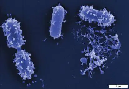
Under attack. Bacterial viruses (bacteriophages) prey on bacteria to maintain population balance in nature. These bacteriophages (light blue) are attacking Acinetobacter baumannii, a soil and water bacterium that is also an emerging pathogen. One of the victims (lower right) has been lysed. Viruses such as these are being considered as substitutes for treating antibiotic-resistant pathogens.
Microbes have both the fastest and the slowest growth rates of any known organisms. Some hot-springs bacteria can double their population in as little as 10 minutes, whereas deep-sea-sediment microbes may take as long as 100 million years. What determines these differences in growth rate? Nutrition is one factor, but niche-A microbe’s physiology typically operates within a very narrow range of these parameters. Yet in nature, an environment surrounding a microbe can change quickly and dramatically. The dangers faced include extremes in temperature, pH, pressure, and osmolarity. Many marine microbes, for instance, move within seconds from deep-sea cold to the searing heat of a thermal vent. How do organisms survive these stresses? Most species have stopgap measures, called stress responses, which temporarily protect the organism from brief forays into potentially lethal environments. But some organisms evolve to thrive, not just survive, in extreme environments. Even in the absence of environmental threats, microbes in nature can wage wars among themselves. Such confrontations include predators such as bacteriophage and ameba attacking bacteria, bacteria retaliating against the predators, and bacteria and fungi secreting antibacterial We begin Chapter 5 by describing how physical and chemical changes in the environment affect the growth of different groups of microbes. We also explore how microorganisms adapt to different environments in ways both transient (involving temporary expression of genes) and permanent (modifications of the gene pool). The permanent genetic changes have led to biological diversity. Finally, we examine the different ways humans use physical, chemical, and biological means to limit microbial growth and protect plants, animals, and ourselves from disease.
5.1 Extremophiles and the Constraints of Temperature and PressureUnit 5 · Regulation
What is a normal environment? With our human frame of reference, we tend to think
that “normal” growth conditions are those found near sea level with a temperature
between 20°C and 40°C, a near-neutral pH, a salt concentration of 0.9%, and ample
nutrients. Any habitat outside this window is labeled “extreme,” and the organisms
(bacteria, archaea, and some eukaryotes) populating them are called extremophiles.
One group of extremophiles can grow at temperatures above the boiling point of
water (100°C), while another group requires a strongly acidic (pH 2) environment to
grow. Actually, a single environment can simultaneously encompass multiple
extremes. In Yellowstone National Park, an acid pool can be found next to an alkali
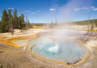
pool, both at extremely high temperatures (Fig. 5.1 ). Teus, extremophiles typically
evolve to survive multiple extreme environments. These organisms are called
polyextremophiles.
FIGURE 5.1 ■ Hot spring in Yellowstone National Park.
ROBERT HARDING/ALAMY STOCK PHOTO
When did extremophiles first appear on Earth? The conditions on Earth when life
began were certainly extreme (hot and acidic, with high osmolarity, low oxygen, and
high carbon dioxide; see Chapter 17), so the earliest microbes likely grew in these
extreme environments. As Earth’s environment slowly changed to present-day
conditions, some of the ancient “extremophiles” adapted to the change and became
what could be called today’s “normalophiles.” Species that did not adapt were
constrained to live in environments that remained extreme.
Some scientists study extremophiles to gain insight into the physiology of
extraterrestrial microbes that we may one day encounter. NASA biochemist Robert
MacElroy first coined the term “extremophile” in his quest for life forms that might
inhabit other planets. Astrophysicists predict that some extreme environments on
Earth could be similar to those of known planets in our galaxy. For instance, the
hypersaline Gypsum Hill spring system on Axel Heiberg Island in Canada is an analog
for the putatively habitable, subsurface briny aquifers on Mars. The Gypsum Hill spring
system harbors a rich yet unique microbial ecosystem, suggesting that Mars’s cold
aquifers may do the same.
Our experiences with extremophiles should also caution us when handling
extraterrestrial samples brought back to Earth. For instance, we should not assume
that irradiation will sterilize samples from future planetary or interstellar missions.
Such treatments do not even kill Deinococcus radiodurans, an extremophile found on
Earth. Scientists have also taken extreme steps not to contaminate other planets with
Earth’s microbes. For example, select components of the Mars rovers were heated to
over 110°C (230°F) for up to 144 hours.
Back on Earth, how do we even begin to study organisms that grow in boiling
water or sulfuric acid solutions or that we cannot culture in the laboratory? New DNA
sequencing technologies, such as metagenomics, have been instrumental in this task.
Metagenomic analysis includes the random screening for, and sequencing of, genes
that encode ribosomal RNA from a mixed environmental sample. Ribosomal RNA
genes from all known life forms have conserved sequences, which permit us to find
and selectively amplify these genes from a complex mixture of DNA. These genes also
contain unique DNA sequences (you can think of them as “fingerprints”) that are
specific to different genera. Metagenomics, then, allows scientists to inventory all the
culturable and uncultured organisms in any given niche, or suitable environment for
growth.
Bioinformatic analysis uses the DNA sequence of a gene to identify the probable
function of its protein product. Using bioinformatics, scientists can make predictions
about the biology of extremophiles and even uncultured species (see Chapter 9). In
addition to bioinformatics, techniques that can identify and quantify RNA transcripts
(transcriptomics) and expressed proteins (proteomics) can reveal all of the genes
expressed and proteins made by bacteria as they adjust to environmental changes
such as shifts in temperature or pH.
What enables different microbes to grow in such physically diverse environments?
Species find their niche, in part, because every protein and macromolecular structure
within a cell is affected by changes in environmental conditions. For example, every
enzyme works best under a unique set of temperature, pH, and salt conditions
because those conditions favor folding of an enzyme into its optimal shape, or
conformation. Deviations from these optimal conditions cause the protein to fold a
little differently and become less active. While not all enzymes within a given cell
boast the same physical optima, these optima must at least be similar and matched
to the organism’s environment for the organism to function effectively.
As the preceding discussion suggests, microbes are commonly classified by their
environmental niche. Table 5.1 summarizes these environmental classes.
Basic Environmental Classification of TABLE 5.1 Microorganisms
Environmental Classification (optimal growth
parameter condition)
Temperature Hyperthermophile Thermophile Mesophile Psychrophile
* (above 80°C) * (between (between * (below
50°C and 15°C and 15°C)
80°C) 45°C)
pH Alkaliphile * Neutralophile Acidophile *
(above pH 9) (between pH (below pH 3)
5 and 8)
Osmolarity Halophile * (high Halotolerant
salt, >2 M NaCl) (high salt not
required, but
able to grow
at up to 2 M
NaCl)
Oxygen Strict aerobe Facultative Microaerophile Strict
(only in O 2) microbe (only in small anaerobe
(with or amounts of O (only
without O 2) 2) without O 2
)
Basic Environmental Classification of TABLE 5.1 Microorganisms
Pressure Barophile * (high Barotolerant
pressure, greater (between 10
than 380 atm) and 500 atm)
Temperature Effects on Physiology
How do microbes react to hot and cold? Unlike humans (and mammals in general),
microbes cannot control their temperature; thus, bacterial cell temperature matches
that of the immediate environment. Because temperature affects the average rate of
molecular motion, changes in temperature affect every aspect of microbial physiology,
including membrane fluidity, nutrient transport, DNA stability, RNA stability, and
enzyme structure and function. Every organism has a temperature optimum at which
it grows most quickly, as well as minimum and maximum temperatures that define
the limits of growth.
Growth temperature limits are imposed, in part, by the thousands of proteins in a
cell, all of which must function within the same temperature range. A species grows
most quickly at temperatures where all of the cell’s proteins work efficiently as a
group to produce energy and synthesize cell components. Growth stops when rising
temperatures cause critical enzymes or cell structures (such as the cell membrane) to
fail. At cold temperatures, growth
ceases because enzymatic processes become too sluggish and the cell membrane
becomes too rigid. The membrane needs to remain fluid so that it can expand as cells
grow larger and so that proteins needed for solute transport can be inserted into the
membrane.
The different branches of life reflect narrowing growth tolerance to heat. Different
archaeal species, for example, can grow in extremely hot or extremely cold
temperatures, and some can grow in the middle range. Bacteria, for the most part,
tolerate temperatures between the archaeal extremes. Eukaryotes are the least
tolerant, with individual species capable of growth between 10°C and 65°C, although
some unicellular eukaryotes can grow as low as −20°C.
Growth Rate and Temperature
Increasing the temperature of incubation will, to a point, help microbes grow more
quickly (Fig. 5.2A ). For any one species the relationship between growth
temperature and the growth rate constant k (the number of generations per hour; see
Section 4.4) obeys the Arrhenius equation, which describes the effect of temperature
on reaction rate for simple chemical reactions. The general result of the Arrhenius
equation with respect to bacterial growth is that growth rate roughly doubles for
every 10°C rise in temperature (Fig. 5.2A ). This same relationship is observed for
most chemical reactions with activation energies of about 50 kilojoules per mole
(kJ/mol).
At the upper and lower limits of the growth range, however, the Arrhenius effect
breaks down. Critical proteins denature at high temperatures, whereas membrane
fluidity stiffens at lower temperatures. Both situations limit the conformational
mobility of enzymes and lower their activity. As a result, growth stops at temperature
extremes. The typical temperature growth range for most bacteria spans the
organism’s optimal growth temperature by 30°C–40°C, but some organisms have a
much narrower tolerance. Even within a species, we can find mutants that are more
sensitive to one extreme or the other (heat sensitive or cold sensitive). The mutant
proteins that result can function at one end of the organism’s temperature range (the
cell grows) but fail at the other end (the cell dies). Consequently, temperature-
sensitive mutations have been used to define essential molecular components of cell
physiology.
A great diversity exists among microbes with respect to growth temperature
range. Different groups have evolved to grow within very different thermal ranges
because the group’s proteins have evolved to tolerate and function within that range.
Outside of that range, the proteins denature or function too slowly for growth. The
upper limit for protists (eukaryotic microbes) is about 50°C, while some fungi can
grow at temperatures as high as 60°C. Prokaryotes (Bacteria and Archaea), however,
have been found to grow at temperatures ranging from below 0°C to above 100°C.
Temperatures over 100°C are usually found near thermal vents deep in the ocean.
Vent water temperature can rise to 400°C (750°F), but the pressure at that depth is
sufficiently high to keep the water in a liquid state. Thermal vent microbes, often
chemolithoautotrophs, supply the base of the food web for the numerous animals
associated with these ecosystems.
FIGURE 5.2 ■ Relationship between temperature and growth rate. A.
The growth rate constant (k) of the enteric organism Escherichia coli is plotted
here against the inverse of the growth temperature on the Kelvin scale. Units of
1,000/ T are used to give a convenient scale on the x -axis, where T is growth
temperature on the Kelvin scale (37°C = 310 K). As temperature in Celsius rises
above or falls below the optimal range, growth rate decreases faster than is
predicted by the Arrhenius equation. B. The relationship between temperature
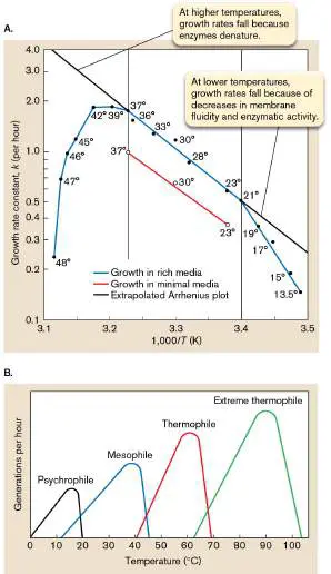
and growth rate for different groups of microbes. Note that the peak growth rate
increases linearly with temperature and obeys the Arrhenius equation.
Source: Part A from Sherrie L. Herendeen et al. 1979. J. Bacteriol. 139 :185.
Thought Question
5.1 Why haven’t cells evolved so that all their enzymes have the same temperature
optimum? If they did, wouldn’t they grow even more rapidly?
Microbial Classification by Growth Temperature
Microorganisms can be classified as mesophiles, psychrophiles, or thermophiles
according to their temperature ranges for optimal growth (Fig. 5.2B ).
Mesophiles include organisms such as Escherichia coli and Bacillus subtilis, two
time-honored model bacteria used in laboratories. Their growth optima range
between 20°C and 40°C, with a minimum of 15°C and a maximum of 45°C. Because
they are easy to grow and because most human pathogens are mesophiles, much of
what we know about protein, membrane, and DNA structure came from studying this
group of organisms. However, detailed 3D views of protein structures are frequently
based on studies of two other classes of organisms whose optimal growth
temperature ranges flank that of the mesophiles; namely, psychrophiles (on the low-
temperature side) and thermophiles (on the high-temperature side). For instance,
proteins from thermophiles are more stable than proteins from mesophiles, so they
are easier to crystallize. These protein crystals are essential for X-ray crystallography
studies of protein structure (see Section 2.6).
An underappreciated fact is that Earth’s biosphere is predominantly cold and
permanently exposed to temperatures below 5°C. Psychrophiles are microbes that
can grow in extreme cold temperatures, as low as −10°C (or even −20°C in the brine
between polar sea ice crystals). Consequently, psychrophiles are prominent members
of microbial communities beneath icebergs in the Arctic and in Antarctic soil and lakes
(Fig. 5.3 ). Despite their abilities to grow in extreme cold, the optimal growth
temperature of psychrophiles is usually about 15°C. In addition to true psychrophiles,
there are cold-resistant mesophiles (psychrotolerant bacteria and archaea, also called
psychrotrophs) that can grow below 7°C but have optima between 20°C and 35°C.
Both psychrophiles and psychrotrophs can be isolated from Antarctic lakes, beneath
several meters of ice (Fig. 5.3C ). Entire microscopic ecosystems flourish there,
capable of surviving (not growing) at −40°C all winter, and then growing at near 0°C
during the sunlit summer. Polar microorganisms are being screened for novel
compounds with anticancer and antimicrobial potential. Closer to home, in human
environments, psychrotolerant bacteria cause milk to spoil in the refrigerator. Even
some pathogens, such as Listeria monocytogenes (one cause of food poisoning and
septic abortions), can grow at refrigeration temperatures.
FIGURE 5.3 ■ Psychrophilic environments and microbes. A. The
continent of Antarctica is an extreme environment populated by many species of
psychrophilic microorganisms, most of them unknown. In addition to being
brutally cold, this extreme environment is nutrient-poor and subject to high levels
of solar UV irradiation. Inset: Asim Bej, University of Alabama at Birmingham,
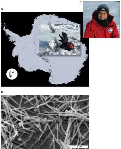
collects samples from the ecosystem at Schirmacher Oasis (location as marked).
Genome sequences from the captured microbes, like those shown in (C), reveal
the composition and metabolic capabilities of the South Pole microbiome. B. Asim
Bej. C. Psychrotolerant Flavobacterium (grows between 0°C and 22°C) from a
South Pole lake made from glacial meltwater in summer (high temperature =
0.9°C; SEM). Novel compounds made by members of the polar microbiome are
screened for anticancer and antimicrobial potential.
COURTESY OF ASIM BEJ
COURTESY OF ASIM BEJ
COURTESY OF DR. ALFONSO DAVIL
Why do these organisms grow so well in the cold? One reason is the proteins of
psychrophiles are more flexible than those of mesophiles and require less energy
(heat) to function. Of course, the downside to the increased flexibility of psychrophilic
proteins is that they require less heat to be denatured than do their more
thermotolerant mesophilic counterparts. As a result, psychrophiles grow poorly, if at
all, when temperatures rise above 20°C. This can be considered an evolutionary
trade-off. Another reason psychrophiles favor cold is their membranes are more fluid
at low temperatures (because they contain a high proportion of unsaturated fatty
acids such as oleic acid; see Fig. 3.7 ); at higher temperatures their membranes are
too flexible and fail to maintain cell integrity. Finally, bacteria and archaea that grow
at 0°C in glaciers also contain antifreeze proteins and other cryoprotectants (such as
trehalose) that can lower the freezing point by 2°C. So, these organisms can grow in
ice but will not freeze. Notably, some psychrophilic and psychrotolerant bacteria
actually stimulate ice formation in clouds and are used to make snow on ski slopes.
Psychrophilic enzymes are of commercial interest because their ability to carry out
reactions at low temperature is useful in food processing and bioremediation.
Enzymes help brew beer more quickly, break down lactose in milk, and can remove
cholesterol from various foods. Producing foods at lower temperatures is beneficial,
too, because the colder processing temperatures minimize the growth of typical
mesophiles that degrade and spoil food. Genetically engineered psychrophilic
organisms can safely degrade toxic organic contaminants (for example, petroleum) in
the cold. Arctic environments are particularly sensitive to pollution because
contaminants are slow to degrade in the freezing temperatures. Consequently, the
ability to seed Arctic oil spills with psychrophilic organisms armed with petroleum-
degrading enzymes could more rapidly restore contaminated environments.
Thermophiles (Fig. 5.4 ) are species adapted to growth at high temperatures
(typically 50°C and higher). Hyperthermophiles (also called extreme thermophiles),
which grow at temperatures as high as 121°C (autoclave temperature), are found
near thermal vents that penetrate Earth’s crust on the ocean floor and on land (for
example, hot springs). The thermophile Thermus aquaticus was the first source of a
high-temperature DNA polymerase used for polymerase chain reaction (PCR)
amplification of DNA. T. aquaticus was discovered in a hot spring at Yellowstone
National Park by microbiologist Thomas Brock (1926–2021), a pioneer in the study of
thermophilic organisms. Its application to the polymerase chain reaction, which
requires periods of high temperature, revolutionized molecular biology (discussed in
eAppendix 3).
FIGURE 5.4 ■ Thermophilic environments and thermophiles. A.
Thermus aquaticus, a hyperthermophile first isolated at Yellowstone by Thomas
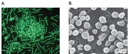
Brock. Cell length varies from 3 to 10 μm. B. Thermophile Methanocaldococcus
jannaschii, grown at 80°C and 114 psi.
P. BRUMM ET AL. 2015. PLOS ONE 10 (10):E0138674
C. B. PARK AND D. S. CLARK. 2002. APPL. ENVIRON. MICROBIOL. 68 (3):1458–63.
Extreme thermophiles often have specially adapted membranes and protein
sequences. The thermal limits of these structures determine the specific high-
temperature ranges in which various species can grow. Because enzymes in
thermophiles (thermozymes) do not unfold as easily as mesophilic enzymes do, they
more easily hold their shape at higher temperatures. Thermophilic enzymes are
stable, in part, because they contain relatively low amounts of glycine, a small amino
acid that contributes to an enzyme’s flexibility. Glycines do not contain side chains, so
they cannot form stabilizing intramolecular bonds. In addition, the amino termini of
proteins in these organisms often are “tied down” by hydrogen bonding to other parts
of the protein, making them harder to denature.
Like all microbes, thermophiles have chaperone proteins that help refold other cell
proteins that may undergo thermal denaturation. Thermophile genomes are also
packed with numerous DNA-binding proteins that stabilize DNA. Special DNA coiling
enzymes (DNA gyrases) in these organisms will tightly coil DNA in a way that makes
it more thermostable and less likely to denature (think of a helically coiled phone cord
that has twisted and bunched up on itself).
Special membranes also aid stability at high temperatures. Unlike the typical lipid
bilayers of mesophiles, the membranes of thermophiles manage to “glue” together
parts of the two hydrocarbon layers that point toward each other, making them more
stable. They do this by incorporating more saturated linear lipids into their
membranes. Saturated lipids form straight hydrocarbon tails that align well with
neighboring lipids and form a highly organized structure that is stable in heat. The
membranes of mesophiles are composed mostly of unsaturated lipids that bend
against each other and align poorly. Consequently, the membranes of mesophiles are
more fluid at lower temperatures.
The membranes of hyperthermophilic archaea impart an amazing level of heat
resilience by being lipid monolayers, not bilayers (see Fig. 3.9 and eAppendix 2).
Lipid bilayers peel apart under withering heat. Monolayers, built for extremophile
living, do not. Monolayer membranes are heat stable because long hydrocarbon
chains directly tether glycerophosphates on opposite sides of the membrane. The
chains (40 carbons long) do not contain fatty acids, but are made of isoprene units
bonded by ether linkages to glycerol phosphate. More on thermophiles can be found
in Chapter 19.
How might the study of thermophiles enhance human existence? The polymerase
chain reaction using thermophilic DNA polymerases revolutionized biology. PCR is
used to amplify DNA directly from a natural environment—a feat that helps identify
the unique 16S ribosomal RNA (rRNA) sequences of uncultured extremophiles,
including those of new thermophiles and hyperthermophiles. In addition, random PCR
amplification and sequencing of DNA fragments from an environment enables
scientists to see the entire repertoire of genes, regardless of species, present in that
environment. The ability to peek at the genomes of hyperthermophiles and predict
what the various genes do led Robert Kelly and colleagues at North Carolina State
University to actually mix genes, and thus enzymes, from different species to invent
new metabolic pathways (Fig. 5.5 ). Their quest is to use thermophilic
microorganisms as metabolic platforms to engineer a new organism able to convert
abundantly available CO 2 and H 2 directly into high-energy liquid fuels. Success in this
endeavor would further decrease dependence on dwindling fossil fuels. Thermophiles
are desirable because they can be used in high-temperature fermentations, which
occur at temperatures close to those employed in chemical refineries.
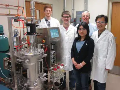
FIGURE 5.5 ■ Bioreactor used to grow thermophilic microorganisms.
Robert Kelly and students stand next to a 20-liter bioreactor that they use for the
engineering analysis of biofuel-producing microbes. Left to right: Aaron Hawkins,
Andrew Loder, Hong Lian, Kelly, and Yejun Han.
COURTESY OF ROBERT KELLY
The Heat-Shock Response
What happens to a bacterium that accidentally encounters a temperature above its
comfort zone? Most microorganisms possess quick-response genetic programs that
remodel their physiology into one that can temporarily survive this inhospitable
condition, which is called heat shock. Rapid temperature changes experienced during
growth activate batches of stress response genes, resulting in the heat-shock
response (discussed in Chapter 10). The protein products of these heat-activated
genes include chaperones that maintain the shapes of proteins and enzymes that
change membrane lipid composition. The heat-shock response, first identified in E.
coli by Tetsuo Yamamori and Takashi Yura in 1982, has since been documented in
almost all living organisms examined thus far. Antarctic marine organisms that live
under extremely stable temperatures (−1.9°C) are an exception.
Thought Question
5.2 If microbes lack a nervous system, how can they sense a temperature change?
Adaptation to Pressure
Living creatures at Earth’s surface (sea level) are subjected to a pressure of 1
atmosphere (atm), which is equal to 0.101 megapascal (MPa) or 14.7 pounds per
square inch (psi). At the bottom of the ocean, however—thousands of meters deep—
hydrostatic pressure averages a crushing 400 atm and can reach as high as 1,000 atm
(101 MPa, or 14,700 psi) or more in ocean trenches (Fig. 5.6 ). Organisms adapted
to grow at these overwhelmingly high pressures are called barophiles or piezophiles.
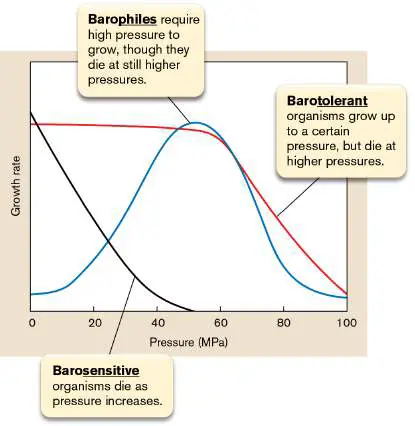
From the curves in Figure 5.7 , notice that barophiles actually require elevated
pressure to grow, while barotolerant organisms grow well in the range of 1–50 MPa,
but their growth falls off thereafter.
FIGURE 5.6 ■ Barophilic environments and piezophiles. A. The deepest
part of the ocean is at the bottom of the Mariana Trench, a depression in the floor
of the western Pacific Ocean, just east of the Mariana Islands. The Mariana
Trench is 2,500 km (1,554 miles) long and 70 km (44 miles) wide. Near its
southwestern extremity, about 340 km (210 miles) southwest of Guam, lies the
deepest point on Earth. This point, referred to as the Challenger Deep, plunges to
a depth of 11,035 meters (nearly 7 miles). The pressure there (110 MPa) is over
1,000 times higher than what we experience on land (0.1 MPa). B. A Gram-
negative barophile in the genus Shewanella, isolated from sea sediment located
6.8 miles below sea level.
C. KATO ET AL. 1998. APPL. ENVIRON. MICROBIOL. 64 :1510–13.
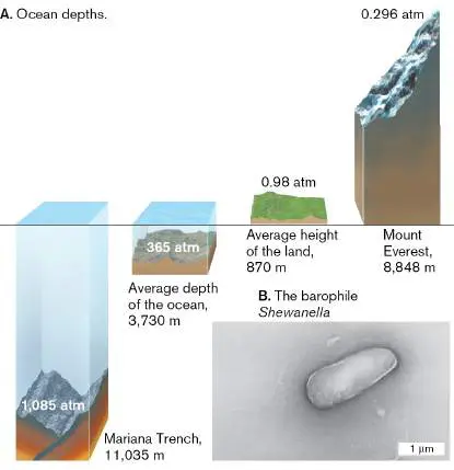
FIGURE 5.7 ■ Relationship between growth rate and pressure.
Many barophiles are also psychrophilic, because the average temperature at the
ocean’s floor is 2°C. However, barophilic hyperthermophiles form the basis of thermal
vent communities that support symbiotic worms and giant clams (see Chapter 21).
For instance, a thermophilic piezophile, Thermococcus piezophilus, was isolated in
2016 from a hydrothermal vent 4,969 meters below sea level. This archaeon has the
broadest known pressure range for growth ever described for a microorganism,
extending from 1 to 120 MPa. Its optimal growth pressure is 50 MPa, consistent with
the hydrostatic pressure in its natural habitat.
How bacteria survive pressures of 80–120 MPa (11,600–17,400 psi) is still a
mystery. It is known, though, that increased hydrostatic pressure and cold
temperatures decrease membrane fluidity. Because fluidity of the cell membrane is
critical to survival, the phospholipids of deep-sea bacteria commonly have high levels
of polyunsaturated fatty acids to increase membrane fluidity. It is thought that in
addition to these membrane changes, internal structures must be pressure adapted.
For example, ribosomes in the barosensitive organism E. coli (maximum growth
pressure 50 MPa) dissociate at pressures above 60 MPa, so barophiles must contain
uniquely designed ribosome structures that can withstand even higher pressures.
A study using the obligate piezophile Pyrococcus yayanosii CH1 revealed that
either above or below optimal growth pressure (50 MPa), this archaeon will alter the
expression of many ribosomal genes, some of which increase while others decrease.
What effect these changes have on ribosome function is unclear. One protein whose
levels increased under high and low pressures has a predicted role in ribosome
recycling. Another report found that the extent to which a barophilic species can resist
pressure correlates to the helical length of stems in certain 16S rRNA stem-loop
structures; both findings support the idea that ribosomal structure and function are
especially sensitive to pressure.
Thought Question
5.3 Predict how hyperthermophilic microorganisms colonize a newly formed
hydrothermal vent (black smoker), which is sterile at its formation. How do the
microbes get there through ice-cold water (0°C–3°C)?
To Summarize
Different species exhibit different optimal growth values of temperature, pH,
and osmolarity.
Extremophiles inhabit fringe environments with conditions that do not
support human life.
The environmental habitat (for example, the temperature or pressure
condition) of a particular species is defined by the tolerance of that organism’s
proteins and other macromolecular structures to the physical conditions within
that niche.
Global approaches used to study gene expression help us view how
organisms respond to changes in their environment.
The Arrhenius equation applies to the growth of microorganisms: within a
specific growth temperature range, the growth rate doubles for every 10°C rise
in temperature.
Mesophiles, psychrophiles, and thermophiles are groups of organisms
that grow at moderate, low, and high temperatures, respectively.
Membrane fluidity varies with the composition of lipids in a membrane,
which in turn dictates the temperature and pressure at which an organism can
grow.
The heat-shock response produces a series of protective proteins in
organisms exposed to temperatures near the upper edge of their growth range.
Barophiles (piezophiles) can grow at pressures up to 1,000 atm or more but
fail to grow at low pressures. Growth at high pressure requires specially
designed membranes and protein structures.
Glossary
extremophile
An organism that grows only in an extreme environment; that is, an environment
including one or more conditions that are “extreme” relative to the conditions for
human life.
niche
An organism’s environmental requirements for existence and its relations with
other members of the ecosystem.
mesophile
An organism with optimal growth between 20°C and 40°C.
psychrophile
An organism with optimal growth at temperatures below 20°C.
psychrotroph
A cold-resistant organism that can grow at temperatures between 0°C and 7°C
but shows optimal growth between 20°C and 35°C.
thermophile
An organism adapted for optimal growth at high temperatures, usually 50°C or
higher.
hyperthermophile
Also called extreme thermophile. An organism adapted for optimal growth at
extremely high temperatures, generally above 80°C, and as high as 121°C.
heat-shock response
A coordinated response of cells to higher-than-normal temperatures. It includes
changes in the membrane and expression of heat-shock genes.
barophile
Also called piezophile. An organism that requires high pressure to grow.
piezophile
See barophile .
Fig. 3.7
FIGURE 3.7 ■ Phospholipid side chains.
Fig. 3.9
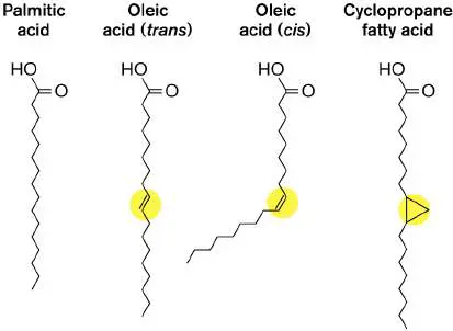
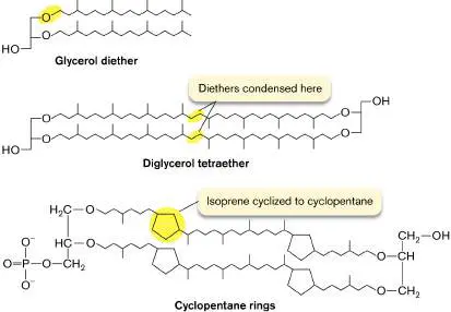
FIGURE 3.9 ■ Terpene-derived lipids of archaea. In archaea, the
hydrocarbon chains are ether-linked to glycerol, and every fourth carbon has
a methyl branch. In some archaea, the tails of the two facing lipids of the
bilayer are fused, forming tetraethers; thus, the entire membrane consists of
a monolayer.
Endnotes
1. Note *: Considered extremophiles. Return to reference *
2. Note *: Considered extremophiles. Return to reference *
3. Note *: Considered extremophiles. Return to reference *
4. Note *: Considered extremophiles. Return to reference *
5. Note *: Considered extremophiles. Return to reference *
6. Note *: Considered extremophiles. Return to reference *
7. Note *: Considered extremophiles. Return to reference *
5.2 Osmolarity and HalophilesUnit 5 · Regulation
Water is critical to life, but environments differ in the amount of water actually available to growing organisms. Water availability in any solution is measured as water activity (a w), a quantity affected by solute concentration. Interactions between water and solutes in solution will lower water activity. So, the more solutes there are in a solution, the less water there is available for microbes to use for growth. Water activity is typically measured as the ratio of the solution’s vapor pressure in a sealed chamber relative to that of pure water. If the air above the sample is 97% saturated relative to the moisture present over pure water, the relative humidity is 97% and the water activity is 0.97.
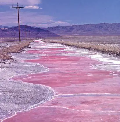
Most bacteria growing on land or in freshwater habitats require water activity to be greater than 0.95, the approximate water activity of seawater, which contains 3.5% NaCl (0.6 M). Other archaea and bacteria, classified as halophiles, have evolved to require high salt (NaCl) concentrations that can result in a water activity as low as 0.75 (equivalent to the solubility limit of NaCl, about 30% wt/vol, or 5 M). The high tolerance of halophiles to salt enables them to grow on Earth’s salt flats—salt-encrusted areas resulting from the evaporation of rising ground water (Fig. 5.8). In general, extreme halophiles require NaCl concentrations between 15% and 30% to grow, whereas moderate halophiles prefer 6%– 15% NaCl. Halotolerant bacteria can grow at elevated salt concentrations but do not require high salt to grow.
FIGURE 5.8 ■ Halophilic salt flats. The halophilic salt flats along Highway 50 east of Fallon, Nevada, are colored pinkish red by astronomical numbers of halophilic bacteria.
WAYNE P. ARMSTRONG, PALOMAR COLLEGE
Osmotic Stress
Osmolarity is a measure of the number of solute molecules in a solution and is inversely related to a w. The more particles there are in a solution, the greater the osmolarity and the lower the water
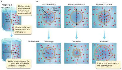
activity. Osmolarity is also important for a cell because of the cell’s semipermeable cytoplasmic membrane. This membrane allows the osmolarity inside the cell to differ from the osmolarity outside. The principles of physical chemistry dictate that a solute present at different concentrations in two chambers separated by a semipermeable membrane will tend to equilibrate. But if the semipermeable membrane does not allow solutes to move through the membrane, water will move through the membrane. Water leaves the chamber with the lower solute concentration and moves into the chamber with the higher solute concentration (see eAppendix 2, Fig. A2.5).
Water does not move across cell membranes primarily by simple diffusion. Instead, special membrane water channels formed by proteins called aquaporins enable water to traverse the membrane much faster than by unmediated diffusion (Fig. 5.9). Rapid movement of water helps protect cells against osmotic stress. However, too much water moving in or out of a cell is detrimental. Cells may ultimately explode or implode, depending on the direction the water moves. Even bacteria with a rigid cell wall suffer. They may not explode like a human cell, but the forces placed on the cell membrane are great. Membrane transport systems, for example, can be inactivated.
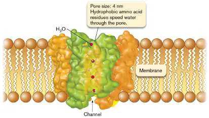
FIGURE 5.9 ■ Aquaporin. Transverse view of the channel through which water molecules move. (PDB code: 1J4N)
Protection against Osmotic Stress
In addition to moving water, microbes have at least two other mechanisms to minimize osmotic stress across membranes. When stranded in a hypertonic medium (higher osmolarity than the cell), bacteria try to protect their internal water from leaving the cell by synthesizing or importing compatible solutes that increase intracellular osmolarity. Compatible solutes are small molecules that do not disrupt normal cell metabolism even at high intracellular concentrations. Increasing the intracellular levels of these compounds (such as proline, glutamic acid, potassium, or betaine) elevates cytoplasmic osmolarity without any detrimental effects, making it unnecessary for water to leave the cell. In contrast, ions such as Na + are not compatible solutes and will disturb metabolism at high intracellular concentrations.
Cells also contain pressure-sensitive (mechanosensitive)
channels that can be used to leak solutes out of the cell. Internal pressure rises in cells immersed in a hypotonic medium (having lower osmolarity than the cell has). The mechanosensitive channels open in response to increased internal pressure. The opened channels allow small solutes to escape, thereby lowering internal osmolarity and preventing too much water from entering the cell. Outside a certain range of external osmolarity, the aquaporin and compatible solute strategies become ineffective at controlling internal osmolarity. To adapt, microbes launch a larger global response in which cell physiology is transformed to tolerate brief encounters with potentially lethal salt (or other solute)
concentrations. Some changes are similar to those provoked by heat shock, such as the increased synthesis of chaperone proteins that protect critical cell proteins from denaturation. Other changes include alterations in outer membrane pore composition (for Gram-negative organisms).
Thought Question
5.4 How might the concept of water availability be used by the food industry to control spoilage?
Halophiles Require High Salt
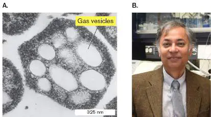
As mentioned earlier, halophiles can grow at an a w of 0.94–0.75, associated with NaCl concentrations of 2–5 M, or 10%–30% NaCl ( Fig. 5.10). In contrast, most bacteria can grow only in salt concentrations ranging from 0.05 to 1 M (0.2%–5% NaCl). All cells, even halophiles, nevertheless prefer to keep a relatively low intracellular Na + concentration so that some solutes can be moved into the cell by symport with Na +. To achieve a low internal Na + concentration, halophilic microbes use special ion pumps to excrete sodium and replace it with other cations, such as potassium, which is a compatible solute. Halophilic bacteria rely mainly, but not exclusively, on organic compatible solutes (such as proline, betaine, or glutamic acid) to counter extracellular extremes in osmolarity, whereas extreme halophilic archaea rely mostly on the accumulation of inorganic ions such as potassium. Halophilic archaea can actually accumulate molar quantities of potassium. In fact, the proteins and cell components (for example, ribosomes) of halophilic archaea have evolved to require remarkably high intracellular potassium levels to maintain their structure, which is partly why halophilic archaea cannot grow at low salt concentrations. The proteins of halophiles also have large numbers of acidic side chains, around which water molecules form a cage to protect proteins from dehydration by high salt. Chapter 19 covers halophilic archaea in greater detail. FIGURE 5.10 ■ Halophilic bacteria. A. Cross section of the archaeon Halobacterium species (TEM). Gas vesicles allow the organism to float in liquid and acquire more oxygen. B.
Shiladitya DasSarma and colleagues at the University of Maryland sequenced the genome of Halobacterium species NRC-1 and are developing gas-vesicle nanoparticles as a novel vaccine delivery system.
COURTESY OF S. DASSARMA, UNIVERSITY OF MARYLAND SCHOOL OF MEDICINE
COURTESY OF SHILADITYA DASSARMA. UNIVERSITY OF MARYLAND SCHOOL OF
MEDICINE
To Summarize
Water activity (a w) is a measure of how much water in a solution is available for a microbe to use.
Osmolarity is a measure of the number of solute molecules in a solution and is inversely related to a w.
Aquaporins are membrane channel proteins that allow water to move quickly across membranes to equalize internal and external pressures.
Compatible solutes are used to minimize pressure differences across the cell membrane.
Mechanosensitive channels can leak solutes out of the cell when internal pressure rises.
Halophilic organisms require high salt concentrations to grow.
Glossary
water activity A measure of the water that is not bound to solutes and is available for use by organisms.
halophile An organism that requires a high extracellular sodium chloride concentration for optimal growth.
osmolarity A measure of the concentration of solute molecules in solution. compatible solute A small molecule that does not disrupt normal cell metabolism even at high intracellular concentrations.
Fig. A2.5 FIGURE A2.5 ■ Osmosis and water balance. A. Osmosis. B. Movement of water across the cell membrane, and shrinkage or expansion of the membrane in isotonic, hypertonic, and hypotonic environments. Black arrows indicate net water movement.
5.3 Hydronium (pH) and Hydroxide Ion ConcentrationsUnit 5 · Regulation
As with salt and temperature, the concentration of hydrogen ions (H +)—actually, hydronium ions (H O +)—also has a direct effect on
3
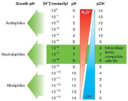
the cell’s macromolecular structures. (For a brief review of pH, refer to eAppendix 1.) Extreme concentrations of either hydronium or hydroxide ions (OH −) in a solution will limit growth and kill cells. But despite this sensitivity to pH extremes, living cells can tolerate a greater range of hydronium and hydroxide ion concentration than of virtually any other chemical substance. Escherichia coli, for example, tolerates a pH range from 2 to 10, a 100-million-fold concentration difference (but grows only between pH 4.5 and 9). Groups of microbes have evolved to inhabit diverse niches, for which pH values can range from 0 to 11.5 (Fig. 5.11).
FIGURE 5.11 ■ Classification of organisms according to their optimal growth pH. pOH is the log 10 of the reciprocal of the hydroxide ion (OH −) concentration; that is, pOH = −log[OH −].
Note: Recall that pH = −log of H + concentration in moles per
10
liter. Thus, a solution containing 1 × 10 −6 mole/liter of H + ions has a pH of 6. Likewise, pOH = −log of OH − concentration. The
10
thermodynamic properties of water dictate that pH + pOH always equals 14.
It is important to note that pH can have a dramatic effect on the availability of some nutrients. The classic example is iron. Iron
hydroxide [Fe(OH) 3], the predominant form of iron in nature, is very insoluble above pH 7. Organisms that thrive under moderate to severe alkaline conditions have evolved highly efficient iron transport systems that can scavenge what little soluble Fe(OH) 3 remains in those environments. Iron transport is discussed in Section 4.2.
pH Optima, Minima, and Maxima
The charges on various amino or carboxyl groups within a protein help forge the intramolecular bonds that dictate protein shape and thus protein activity. Because shifting H + concentration, [H +], will affect the protonation of these ionizable groups, changing the pH will alter protein structure and activity. The result is that all enzyme activities exhibit pH optima, minima, and maxima. However, species differences in optimal growth pH are not dictated by the pH limits at which critical cell proteins function.
Generally speaking, the majority of enzymes, regardless of the pH at which their source organism thrives, tend to operate best between pH 5 and 8.5 (which, if you think about it, is still a 3,000-fold range in hydrogen ion concentration). Yet many microbes, described shortly, grow in even more acidic or more alkaline environments.
Unlike its temperature, the intracellular pH of a microbe, as well as its osmolarity, is not necessarily the same as that of its environment. Biological membranes are relatively impermeable to protons—a fact that allows the cell to maintain an internal pH compatible with protein function when growing in extremely acidic or alkaline environments. When the difference between the intracellular and extracellular pH (ΔpH) is very high, protons can leak through proteins that thread the membrane. Excessive influx or efflux of protons can cause problems by altering internal pH. Membrane-permeant organic acids, also called weak acids (discussed in Chapter 3), can accelerate the leakage of protons into a cell. Unlike H +, the uncharged form of an organic acid (HA) can freely permeate cell membranes and dissociate intracellularly, releasing a proton that then acidifies the internal pH. Lactic acid produced and secreted by lactobacilli during the formation of yogurt is an example of self-imposed organic acid stress. The buildup of lactic acid (via fermentation) limits bacterial growth, leaving yogurt with plenty of food value. The food industry has taken advantage of this phenomenon by preemptively adding citric acid or sorbic acid to certain foods. This practice controls microbial growth under pH conditions that do not destroy the flavor or quality of the food. Food microbiology is discussed further in Chapter 16.
Neutralophiles, Acidophiles, and Alkaliphiles Grow in Different pH Ranges
Defined groups of cells have evolved to live under different pH conditions. They do not do this by drastically changing the pH optima of their enzymes. Instead they use novel pH homeostasis strategies that maintain intracellular pH between pH 5 and 8, even when the cell is immersed in pH environments well above or below that range.
Three classes of organisms are differentiated by the pH range at which they grow: neutralophiles, acidophiles, and alkaliphiles (see Fig. 5.11).
Neutralophiles, which generally grow between pH 5 and 8, include most human pathogens. Many neutralophiles, including E. coli and Salmonella enterica, adjust their metabolism to maintain an internal pH slightly above neutrality, which is where their enzymes work best. They maintain this pH even in the presence of moderately acidic or basic external environments (Fig. 5.12A). Other neutralophiles allow their internal pH to fluctuate with external pH but usually maintain a pH difference (ΔpH) of about 0.5 pH unit across the membrane at the upper and lower limits of growth pH. The ΔpH value is an important component of the transmembrane proton potential, a source of energy for the cell (see Chapter 14).
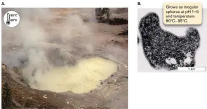
Acidophiles are bacteria and archaea that live in extreme acidic environments. They are often lithotrophs (chemolithoautotrophs) that oxidize reduced metals and generate strong acids, such as sulfuric acid. Consequently, they grow between pH 0 and 5. Acidophiles generally maintain an internal (cytoplasmic) pH that is considerably more acidic than that of neutralophiles but still less acidic than their growth environment (Fig. 5.12B ). The ability to grow at this pH is due partly to altered membrane lipid profiles (high levels of tetraether lipids) that decrease proton permeability, and partly to ill-defined proton extrusion mechanisms. Acidophiles also have a net positive electrical potential charge internally, which can repel the high H + concentrations surrounding them. Often, an organism that is an extremophile with respect to one environmental factor is an extremophile with respect to others as well. Sulfolobus acidocaldarius, for example, is a thermophile and an acidophile ( Fig. 5.13). It grows in acidic hot springs rich in sulfur, and it oxidizes reduced sulfur as an energy source.
FIGURE 5.12 ■ Maintaining internal pH (pH homeostasis) over a wide range of external pH. A. Internal pH (pH int) of the neutralophile Escherichia coli measured after the addition of acid to change external pH (pH ext) and the
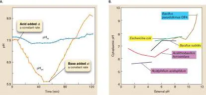
subsequent addition of base. Internal pH was determined using nuclear magnetic resonance to measure changes in methyl phosphate. The two phosphate species titrate over different pH ranges. B. Cytoplasmic pH as a function of the external pH among acidophiles (purple), neutralophiles (yellow), and alkaliphiles (blue).
Source: Part A from Joan L. Slonczewski et al. 1981. PNAS 78 :6271. Part B modified from Joan L. Slonczewski et al. 2009. Adv. Microb. Physiol. 55 :1–79. FIGURE 5.13 ■ Sulfur Caldron acid spring and Sulfolobus acidocaldarius. A. Sulfur Caldron, in the Mud Volcano area of Yellowstone National Park, is one of the most acidic springs in the park. It is rich in sulfur and in Sulfolobus, an archaeon that thrives in hot, acidic waters with temperatures from 60°C to 95°C and a pH of 1–5. B. Thin-section electron micrograph of S. acidocaldarius.
JENNIFER COULTER/ALAMY STOCK PHOTO
E. JAYASHANTHA. 2015. AUSTRALIAN JOURNAL OF PUBLIC ADMINISTRATION
Alkaliphiles occupy the opposite end of the pH spectrum, growing best at values ranging from pH 9 to 11. They are commonly found in
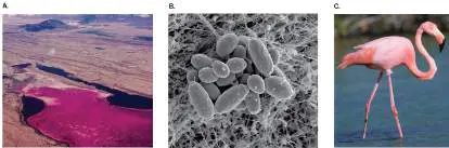
saline soda lakes, which have high salt concentrations (so, alkaliphiles are often halophiles) and pH values as high as pH 11. Soda lakes, like Lake Magadi in Kenya’s Great Rift Valley (Fig. 5.14A), are steeped in carbonates, which explains their extraordinarily alkaline pH. An alkaliphilic organism first identified in Lake Magadi is the halophilic archaeon Natronobacterium gregoryi ( Fig. 5.14B ).
The cyanobacterium Spirulina is another alkaliphile that grows in soda lakes. Its high concentration of carotene gives the organism a distinctive pink color (note the color of the lake in Fig. 5.14A). Spirulina is also a major food for the famous pink flamingos indigenous to these African lakes and is, in fact, the reason pink flamingos are pink. After the birds ingest Spirulina, or other organisms such as brine shrimp that eat Spirulina, digestive processes release the carotene pigment to the circulation, which then deposits it in the birds’ feathers, turning them pink (Fig. 5.14C ). Humans who consume Spirulina as a health food supplement do not turn pink, however, because the cyanobacteria are only a small component of their diet.
FIGURE 5.14 ■ A soda lake ecosystem. A. Lake Magadi in Kenya. Its pink color is due to the cyanobacterium Spirulina. B. Alkaliphile Natronobacterium gregoryi. Cell size, approx. 1 μm × 3 μm. C. Pink flamingos turn pink because they ingest large quantities of Spirulina.
BIRUTE VIJEIKIENE/SHUTTERSTOCK
REPUBLISHED WITH PERMISSION OF JOHN WILEY & SONS, INC., FROM
NATRONOBACTERIUM BY A. OREN, WILEY BOOKS, 2018
WILD HORIZONS/UIG VIA GETTY IMAGES
The internal enzymes of alkaliphiles, like those of acidophiles, exhibit rather ordinary pH optima (around pH 8). The key to the survival of alkaliphiles is the cell-surface barrier that sequesters fragile cytoplasmic enzymes away from harsh extracellular pH. Key structural features of the cell wall, such as the presence of acidic polymers and an excess of hexosamines in the peptidoglycan, appear to be essential. The reason is unclear. In their membranes, some alkaliphiles also possess a high level of diether lipids (more stable than ester-linked phospholipids), which prevent protons from leaking out of the cell (see Section 3.2).
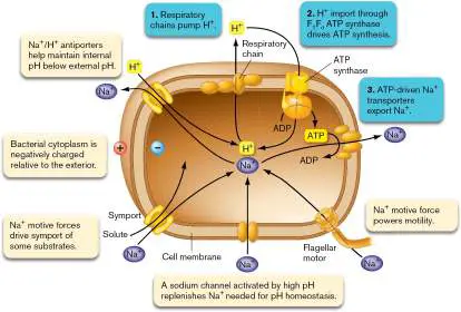
Because external protons are in such short supply at alkaline pH, most alkaliphiles use a sodium motive force in addition to a proton motive force to do much of the work of the cell (see Section 14.1). They also rely heavily on Na + /H + antiporters (see Section 4.2) to bring protons into the cell. This H + influx keeps the internal pH well below the extremely alkaline external pH. The Na + /H + antiporters partly explain why many alkaliphiles are resistant to high salt (NaCl) concentrations: Sodium ions are expelled while protons are sucked in. Some important aspects of sodium circulation in alkaliphiles are depicted in Figure 5.15.
FIGURE 5.15 ■ Na + circulation in alkaliphiles. Cells of alkaliphiles use Na + in place of H + to do some of the work of the cell. They require an inwardly directed sodium gradient to rotate flagella and transport nutrient solutes. The numbered steps in the blue bubbles outline how proton motive force is converted to sodium motive force. The tan bubbles indicate the role of sodium motive force in these bacteria.
In contrast to proteins within the cytoplasm, enzymes secreted from alkaliphiles are able to work in very alkaline environments. The inclusion of base-resistant enzymes such as proteases, lipases, and cellulases in laundry detergents, typically used at a pH of 10, helps get our “whites whiter and our brights brighter.” See Chapter 16 for more on industrial microbiology.
Thought Question
5.5 Recall from Section 4.2 that an antiporter couples movement of one ion down its concentration gradient with movement of another molecule uphill, against its gradient. For Na + /H + symporters, that means more sodium inside than outside and more protons outside than inside. If this is true, how could a Na + /H + antiporter work to bring protons into a haloalkaliphile growing in high salt at pH 10? Hint: In this situation (high-salt and high-pH media), there will be more sodium outside than there is inside and more H + inside than outside—the opposite of what you’d think the cell would need. Both ions would have to move AGAINST their concentration gradients: Sodium moves out, protons move in.
pH Homeostasis and Acid Resistance
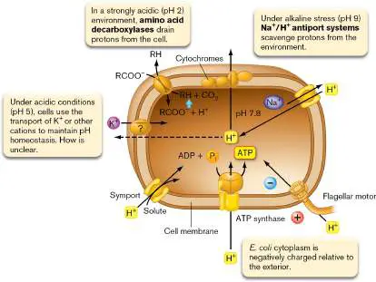
When cells are placed in pH conditions below their optimum, protons can enter the cell and lower internal pH to lethal levels. Microbes can prevent the unwanted influx of protons in a variety of ways (Fig. 5.16). E. coli, for example, can counter proton influx by transporting a variety of cations, such as K + or Na +. How cation transport accomplishes H + efflux is unclear. Some evidence suggests a link to the role of K + in osmoprotection. At the other extreme, under extremely alkaline conditions, the cells can use the Na + /H + antiporters mentioned previously (and in Section 4.2) to recruit protons into the cell in exchange for expelling Na +. FIGURE 5.16 ■ Proton circulation and pH homeostasis. A typical E. coli cell uses various proton transport strategies to maintain an internal pH near pH 7.8 in the face of different external pH stresses. Proton pumping through cytochromes also establishes a proton gradient, which drives flagellar rotation and solute transport.
Some organisms can also change the pH of the medium by using various amino acid decarboxylases and deaminases. For instance, E. coli consumes organic acids when growing at low pH, but it produces these acids while trying to grow under alkaline conditions. Helicobacter pylori, the causative agent of gastric ulcers, employs an exquisitely potent urease to generate massive amounts of ammonia, which neutralizes the acid pH environment. These acid stress and alkali stress protection systems are usually not made or at least do not become active until the cell encounters an extreme pH.
Many, if not all, microbes also possess an emergency global response system referred to as acid tolerance or acid resistance. In a process analogous to the heat-shock response, bacterial physiology undergoes a major molecular reprogramming in response to hydrogen ion stress. The levels of a large number of proteins increase, while the levels of others decrease. Many of the genes and proteins involved in the acid stress response overlap with other stress response systems, including the heat-shock response. These physiological responses include modifications in membrane lipid composition, enhanced pH homeostasis, and numerous other changes with unclear purpose. Some pathogens, such as Salmonella, sense a change in external pH as part of the signal indicating that the bacterium has entered a host cell environment.
To Summarize
Hydrogen ion concentration affects protein structure and function. Thus, enzymes have pH optima, minima, and maxima.
Microbes use pH homeostasis mechanisms to keep their internal pH near neutral when in acidic or alkaline media. Adding weak acids to certain foods undermines bacterial pH homeostasis mechanisms, thereby preventing food spoilage and killing potential pathogens.
Neutralophiles, acidophiles, and alkaliphiles prefer growth under neutral, low, and high pH conditions, respectively.
Acid and alkali stress responses result when a given species is placed under pH conditions that slow its growth. The cell increases the levels of proteins designed to mediate pH homeostasis and protect cell constituents.
Glossary
neutralophile An organism with an optimal growth range in environments between pH 5 and 8.
acidophile An organism that grows fastest in acid (generally defined as below pH 5).
alkaliphile An organism that grows fastest in alkali (generally defined as above pH 9).
5.4 OxygenUnit 5 · Regulation
Imagine having the ability to swim underwater without breathing oxygen. This sounds like science fiction for humans, but many microorganisms can grow in the absence of molecular oxygen (O 2). Environments that lack oxygen, such as sediments at the bottom of the ocean, hot springs, and vertebrate intestines, are described as anaerobic (or, more properly, anoxic). Microbes that grow in those environments are called anaerobes. One class of anaerobes, designated as facultative anaerobes, can grow with or without molecular oxygen. Aerobes, however, grow only in the presence of oxygen. In this section we describe the aerobes’ need for oxygen, why anaerobes cannot grow in oxygen, and how facultative anaerobes grow with or without oxygen.
Oxygen Has Benefits and Risks
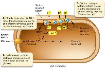
The key to understanding the relationships between microbes and oxygen is to know how organisms gain energy. Some microbes gain energy (ATP) only through fermenting carbohydrates, an oxygen-free process. Others require a series of membrane proteins and lipids known as an electron transport system (ETS) to extract intrinsic energy from electrons pulled from an energy source (Fig. 5.17). Note that the term “cytochrome system” is synonymous with “ETS.” The energy released from electrons moving down an ETS is then used to pump protons (H +) out of the cell. The resulting unequal distribution of H + across the membrane produces a transmembrane electrochemical gradient, a kind of “biobattery” called the proton motive force (details are discussed in Section 14.1). The movement of electrons along an ETS, coupled with the generation of proton motive force, is called respiration if the electron donor is from an organic source or lithotrophy if the source is inorganic (discussed in Chapters 13 and 14).
FIGURE 5.17 ■ The role of oxygen as a terminal electron acceptor in respiration. The pumping of protons out of the cell by electron transport systems produces more positive charges outside the cell than inside, resulting in an electrochemical gradient (also called proton motive force). At the end of the ETS, the electron must be passed to a final (terminal) electron acceptor (for example, O 2), thus clearing the path for the next electron. This net process is called respiration. Glucose 6-P = glucose 6-phosphate.
Once the ETS has drained as much energy as possible from an electron, that electron must be passed to a final (terminal) electron acceptor molecule, such as oxygen gas (O 2), which, in its reduced form (H 2 O), diffuses away in the medium. This clears the way for another electron to be passed down the chain (Fig. 5.17). Aerobes use O 2 as the terminal electron acceptor for their ETS in a process called aerobic respiration (see Chapter 14). Strict aerobes use only aerobic respiration to gain energy. Anaerobes, as we will see later, have options.
We just described how O 2 benefits some organisms as a terminal electron acceptor. But why is oxygen also a risk to living systems? Regardless of whether a microbe utilizes oxygen as a terminal electron acceptor, oxygen’s breakdown products, called reactive oxygen species (ROS), can
severely damage cells. As a result, different bacteria have evolved to either tolerate or avoid oxygen altogether.
How are reactive oxygen species made? Any organism, including bacteria, that possesses NADH dehydrogenase 2—aerobe or anaerobe—will, in the presence of oxygen, inadvertently autooxidize the flavin adenine dinucleotide (FAD) cofactor within the enzyme and produce dangerous amounts of superoxide radicals (•O −; Fig. 5.18). Superoxide will be
2
reduced to hydrogen peroxide (H 2 O 2), another reactive molecule. Iron, present as a cofactor in several enzymes, can then catalyze a reaction with hydrogen peroxide to produce the highly toxic hydroxyl radical (•OH). All of these reactive oxygen species seriously damage DNA, RNA, proteins, and lipids by stripping them of electrons.
FIGURE 5.18 ■ Generation and destruction of reactive oxygen species (ROS). ROS are marked yellow. The autooxidation of flavin adenine dinucleotide (FAD) and the Fenton reaction occur spontaneously to produce superoxide and hydroxyl radicals, respectively. The ferrous (Fe 2+) form of iron needed for the Fenton reaction comes from intracellular sources such as cytochromes. The other reactions require enzymes. FAD is a cofactor for a number of enzymes (for example, NADH dehydrogenase 2). Catalase and peroxidase detoxify hydrogen peroxide.
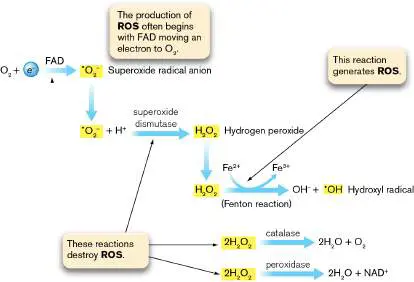
Consequently, oxygenated environments are toxic, and organisms that live in them require special talents to survive. Aerobes, for instance, destroy reactive oxygen species with an ample supply of enzymes such as superoxide dismutase (to remove superoxide) and peroxidase and catalase (to remove hydrogen peroxide). Aerobes also have resourceful enzyme systems that detect and repair macromolecules damaged by oxidation.
Aerobes versus Anaerobes
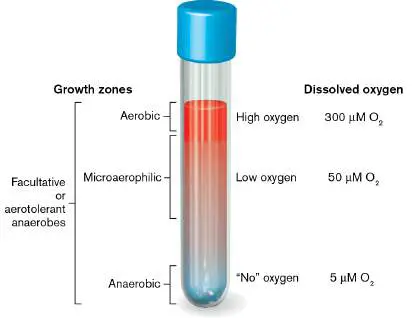
Table 5.2gives examples of microbes that grow at different levels of oxygen. Consider where these different classes of microbes would grow in a standing test tube containing growth medium (Fig. 5.19). The top of the tube, closest to air, is oxygenated; the bottom of the tube has almost no oxygen (5 μM or less dissolved O 2). In between is a gradient of decreasing dissolved oxygen. Some microbes grow only at the top of the tube (aerobes), while others grow only at the bottom (anaerobes) and die (or stop growing) if they enter higher levels in the tube. Facultative anaerobes, however, can grow from top to bottom.
TABLE 5.2 Examples of Aerobes and Anaerobes Aerobic Facultative Microaerophilic Anaerobic microbes microbes microbes microbes Azotobacter Bacillus Campylobacter Actinomyces spp. anthracis spp. spp.
Soil Cause of anthrax One cause of Soil microorganisms; gastroenteritis microorganisms; fix atmospheric synthesize nitrogen antibiotics Neisseria spp. Escherichia coli Helicobacter Azoarcus pylori tolulyticus TABLE 5.2 Examples of Aerobes and Anaerobes Causative Normal gut biota; Cause of gastric Degrades organisms of additional ulcers toluene meningitis, pathogenic gonorrhea strains Pseudomonas Saccharomyces Lactobacillus Bacteroides fluorescens cerevisiae spp. spp.
Found in soil; Yeast; used in Ferment milk to Normal gut degrades 2, 4, baking form yogurt biota 6-trinitrotoluene (TNT) and aromatic hydrocarbons Rhizobium Staphylococcus Treponema Clostridium spp. spp. pallidum spp.
Soil Found on skin; Cause of syphilis Soil microorganisms; cause boils microorganisms; plant symbionts causative agents of tetanus and botulism Vibrio cholerae Desulfovibrio spp.
Cause of cholera Reduce sulfate FIGURE 5.19 ■ Oxygen-related growth zones in a standing test tube containing culture medium.
Why do anaerobes perish in oxygen? Some anaerobes die in oxygen because they lack the enzymes needed to destroy ROS molecules produced by their own metabolism. Other anaerobes have enzymes that can protect them from ROS but will not allow them to grow. The dissolved oxygen these anaerobes encounter raises the redox potential to a point that interferes with the use of alternative (non-oxygen) electron acceptors that the organism needs to make energy. Oxygen also disrupts the metabolism of anaerobes by directly oxidizing metal cofactors, thereby inactivating key enzymes. Consequently, strict anaerobes grow only at the bottom of the tube shown in Figure 5.19. As we will discuss later, some bacteria previously considered strict anaerobes are not so strict.
Classes of anaerobes. Anaerobic microbes fall into several categories. Some anaerobes actually do respire by means of electron transport systems, but instead of using oxygen, they rely on alternative terminal electron acceptors such as nitrate (NO −) to conduct anaerobic respiration and
3
produce energy. Anaerobes of another ilk do not possess cytochromes (members of various ETSs), thus cannot respire, and so must rely on carbohydrate fermentation for energy; that is, they conduct fermentative metabolism. In fermentation, ATP energy is produced through substrate-level phosphorylation (for example, phosphoenolpyruvate + ADP → pyruvate + ATP). In either case, tolerance for ROS is low.
Facultative anaerobes (such as E. coli) possess enzymes that destroy toxic oxygen by-products, but they have both fermentative and respiratory potential. Whether a member of this group uses aerobic respiration, anaerobic respiration, or fermentation depends on the availability of oxygen or other terminal electron acceptor and the amount of carbohydrate present. Aerotolerant anaerobes (Streptococcus pneumoniae, for instance) use only fermentation to provide energy but contain superoxide dismutase and peroxidase (and sometimes small amounts of catalase) to protect them from ROS. These enzymes enable aerotolerant anaerobes to grow in air (containing oxygen) while retaining a fermentation-based (anaerobic) metabolism. Aerotolerant anaerobes will grow throughout the tube in Figure 5.19. Microorganisms that possess decreased levels of superoxide dismutase and/or catalase will be microaerophilic, meaning they will grow only at low oxygen concentrations.
Some organisms previously considered anaerobes (for example, Bacteroides fragilis) are really transiently aerotolerant. These “anaerobes” tolerate oxygen because they possess low levels of ROS-protective enzymes and can even use very low levels of oxygen as terminal electron acceptors. The fundamental composition of all cells reflects their evolutionary origin as anaerobes. Lipids, nucleic acids, and amino acids are all highly reduced— which is why our bodies are combustible (not spontaneously, however). We never would have evolved that way if molecular oxygen had been present from the beginning. Even today, most microbes are anaerobic, growing buried in the soil, within our anaerobic digestive tract, or within biofilms on our teeth.
Thought Questions
5.6 If anaerobes cannot live in oxygen, how do they incorporate oxygen into their cell components?
5.7 How can anaerobes grow in the human mouth, where there is so much oxygen?
Culturing Anaerobes in the Laboratory
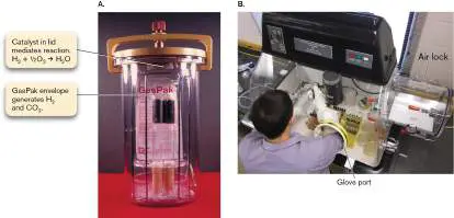
Many anaerobic bacteria cause horrific human diseases, such as tetanus, botulism, and gangrene. Some of these organisms or their secreted toxins are even potential weapons of terror (for example, Clostridium botulinum). Because of their ability to wreak havoc on humans, culturing these microorganisms was an early goal of microbiologists. Despite the difficulties involved, laboratory conditions were eventually contrived in which all, or at least most, of the oxygen could be removed from a culture environment. Three oxygen-removing techniques are used today. Special reducing agents (for example, thioglycolate) or enzyme systems (such as Oxyrase) that eliminate dissolved oxygen can be added to ordinary liquid media. Anaerobes can then grow beneath the culture surface. A second, very popular way to culture anaerobes, especially on agar plates, is to use an anaerobe jar (Fig. 5.20A). Agar plates streaked with the organism are placed into a sealed jar with a foil packet that releases H 2 and CO 2 gases. A palladium packet hanging from the jar lid catalyzes a reaction between the H 2 and O 2 in the jar to form H 2 O and effectively removes O 2 from the chamber. The CO 2 released is required by some reactions to produce key metabolic intermediates. Some microaerophilic microbes, such as the pathogens Helicobacter pylori (the major cause of stomach ulcers) and Campylobacter jejuni (a major cause of diarrhea), require low levels of O 2 but elevated amounts of CO 2. Another type of gas-generating packet can produce these conditions.
Because strict anaerobes are exquisitely sensitive to oxygen, even more heroic efforts are required to establish an oxygen-free environment. A special anaerobic glove box must be used in which the atmosphere is removed by vacuum and replaced with a precise mixture of N 2 and CO 2 gases (Fig. 5.20B ).
FIGURE 5.20 ■ Anaerobic growth technology. A. An anaerobe jar. B. Student researcher using an anaerobic chamber with glove ports.
JACK BOSTRACK/VISUALS UNLIMITED
JOAN SLONCZEWSKI
The implementation of mass spectrometry (MS) in clinical laboratories is becoming a relatively fast and easy way to identify anaerobes from clinical samples such as blood. This method could eventually eliminate the need for culturing in clinical settings. The process starts by separating bacterial pathogens from blood and lysing the bacteria with laser irradiation to release proteins. Mass spectrometry techniques then determine each protein’s identity by measuring the exact mass of that protein. The method then catalogs all the proteins released, and that information is used to name the pathogen (see Chapter 28 for more).
Thought Question
5.8 What evidence led people to think about looking for anaerobes? Hint: Look up “Spallanzani,” “Pasteur,” and “spontaneous generation” on the Internet.
To Summarize
Oxygen is a benefit to aerobes , organisms that can use it as a terminal electron acceptor to extract energy from nutrients. Oxygen is toxic to all cells—for example, anaerobes—that may lack enzymes able to destroy reactive oxygen species (ROS) or that may have oxygen-sensitive metallic cofactors for their enzymes. Anaerobic metabolism can be either fermentative or respiratory. Anaerobic respiration requires the organism to possess cytochromes that can transfer electrons to terminal electron acceptors other than oxygen. Fermentative metabolism uses substrate-level phosphorylation to generate ATP in a process that does not involve cytochromes.
Aerotolerant anaerobes grow in either the presence or the absence of oxygen, but they use fermentation as their primary, if not only, means of gathering energy. These microbes also have enzymes that destroy ROS, allowing them to grow in oxygen.
Facultative anaerobes grow with or without oxygen and have enzymes that destroy ROS. Some utilize only fermentative metabolism, while others can ferment and respire via anaerobic and/or aerobic means. Those that aerobically respire use oxygen as a terminal electron acceptor.
Glossary
anaerobic Lacking oxygen.
anaerobe An organism that grows in an environment lacking oxygen.
facultative anaerobe An organism that can grow in either the presence or absence of oxygen. aerobe An organism that grows only in the presence of oxygen.
electron transport system (ETS) or electron transport chain (ETC) Also called cytochrome system. A series of membrane-embedded proteins that converts the energy of redox reactions into a proton potential.
aerobic respiration The use of oxygen as the terminal electron acceptor in an electron transport chain. A proton gradient is generated and used to drive ATP synthesis.
strict aerobe An organism that performs aerobic respiration and can grow only in the presence of oxygen.
strict anaerobe An organism that cannot grow in the presence of oxygen.
anaerobic respiration The use of a molecule other than oxygen as the final electron acceptor of an electron transport chain.
fermentation Also called fermentative metabolism. 1. The production of ATP via substrate-level phosphorylation, using organic compounds as both electron donors and electron acceptors. 2. Industrial fermentation is the production of microbial products that are made by microbes grown in fermentation vessels; it may include respiratory metabolism to maximize microbial growth.
fermentative metabolism See fermentation .
facultative anaerobe An organism that can grow in either the presence or absence of oxygen. aerotolerant anaerobe An organism that does not use oxygen for metabolism but can grow in the presence of oxygen.
microaerophilic Requiring oxygen at a concentration lower than that of the atmosphere, but unable to grow in high-oxygen environments.
5.5 Starvation and EutrophicationUnit 5 · Regulation
Obviously, limiting the availability of a carbon source or other essential nutrient will limit growth. Less obvious are the dramatic molecular events that cascade through a starving cell. Optimizing growth rate when nutrient levels are suboptimal is an important aim of free-living bacteria, given that intestinal, soil, and marine environments rarely offer excess nutrients. A dramatic example of how some bacteria cope with starvation is seen during growth on nutrient-limited agar plates. These bacteria form colonies with intricate geometrical shapes that help the population cope, in some unknown way, with nutrient stress (Fig. 5.21).
FIGURE 5.21 ■ Effects of starvation on colony morphology. A. Starving E. coli colony after 7 days (diameter, 6 cm). Compare with Figure 4.13B, which shows smooth, dome-shaped, nonstarving colonies of a closely related species, Salmonella enterica (diameter, 0.2 cm). Both E. coli and S. enterica produce identical colonies after one day of growth. B. Paenibacillus dendritiformis grown under starvation conditions. The colony consists of branches with chiral twists, all with the same handedness.
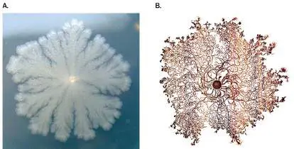
JOHN FOSTER, U. OF SOUTH ALABAMA
® LAST REFUGE/ARDEA.COM/AGE FOTOSTOCK
Starvation Activates Survival Genes
Numerous gene systems are affected when nutrients decline (see Chapter 10). Growth rate slows, and daughter cells become smaller and begin to experience what is called a “starvation” response, in which the microbe senses a dire situation developing but still strives to find new nourishment. The resulting metabolic slowdown generates increased concentrations of critically important small signaling molecules, such as cyclic adenosine monophosphate (cyclic AMP, or cAMP) and guanosine tetraphosphate (ppGpp), which globally transform gene expression. The highly soluble nature of these small molecules means they can quickly diffuse throughout the cell, promoting a fast response. During this metabolic retooling, transport systems for potential nutrients are produced even if the matching substrates are unavailable. Cells begin to make emergency internal energy stores such as glycogen, in case no other nutrient is found.
As nutrient conditions worsen, the organism prepares for famine by activating many different stress survival genes. The products of these genes afford protection against stressors such as reactive oxygen radicals or temperature and pH extremes. No cell can predict the precise stresses it might encounter while incapacitated, so it is advantageous to be prepared for as many as possible. As described in Section 4.6, some species undergo elaborate developmental processes that ultimately produce dormant spores. When severely stressed by starvation, some members of a bacterial population appear to sacrifice themselves to save others by undergoing what is termed programmed cell death. The dying cells release nutrients that neighboring cells use to survive. One of the mechanisms for programmed cell death involves so-called toxin-antitoxin (TA) systems. For each TA pair, the toxin protein will stop growth or kill the cell, but the antitoxin (sometimes a protein, sometimes a small RNA molecule) can inactivate the toxin.
An important toxin-antitoxin system in Escherichia coli is the MazE (antitoxin)–MazF (toxin) module (Fig. 5.22A). Because toxin and antitoxin are simultaneously made, healthy cells live. However, MazE antitoxin is unstable (degraded by the ClpAP protease) and must continually be replenished by synthesis to inactivate MazF toxin. If cells are starved, they stop making MazE and MazF. MazE antitoxin is degraded, leaving the more stable MazF toxin free to cleave many cellular mRNA molecules. As a result, the cell first enters stasis (dormancy, see Chapter 4), from which it can recover if more MazE is made. But if MazE is not forthcoming, the cell dies and releases nutrients. The dying cell will also signal nearby cells to undergo programmed cell death. A peptide cleaved from glucose 6-phosphate dehydrogenase is released from the dying cell and enters nearby cells. The peptide binds to MazE antitoxin and prevents it from neutralizing MazF. The MazF toxin, now active, will eventually kill the bystander cell. Combined, enough nutrients are released to rescue a subset of the population. Nancy Woychik at Rutgers University studies how toxin-antitoxin systems, including MazEF, contribute to latency of the pathogen Mycobacterium tuberculosis ( Fig. 5.22B ).
FIGURE 5.22 ■ Programmed cell death in response to starvation. A. The E. coli MazEF toxin-antitoxin system is thought to play an important role in bacterial survival during stress. MazE antitoxin is continually degraded by ClpAP and must be replenished to neutralize MazF toxin. Other toxin-antitoxin systems are found in other bacterial species. Stresses such as starvation, oxidative stress, or antibiotics can activate this system. G6PD = glucose 6-phosphate dehydrogenase. B. Nancy Woychik and graduate student Valdir Barth study homologs of MazEF toxin-antitoxin systems in Mycobacterium tuberculosis, the cause of tuberculosis. Her lab explores how these toxin-antitoxin systems contribute to the latency of this pathogen.
JOHN EMERSON/RUTGERS UNIVERSITY
When all else fails, a small subset of cells in a starving bacterial culture or biofilm will enter a dormant state (different from
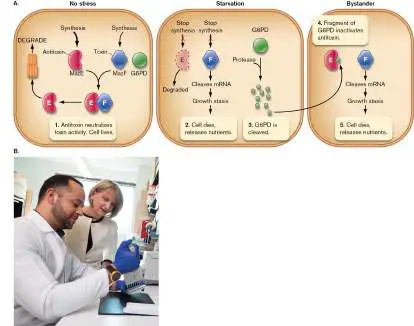
sporulation) in which cells remain viable but do not grow (described in Section 4.4). As growth slows during starvation, ribosome numbers initially decrease as protein synthesis demand slows. To become truly dormant, cells must stabilize the few remaining ribosomes in a state of hibernation that can be reversed. One protein critical to this process in many species is called hibernation promoting factor (HPF). The binding of HPF to ribosomes halts protein synthesis and stabilizes ribosome pairs as inactive 100S dimers (Fig. 5.23A). This state of ribosome hibernation prevents ribosome turnover during dormancy and provides a reservoir of ribosomes that can reactivate when conditions improve. How ribosome dimers dissociate when conditions improve is unclear but appears to require a “wake-up” GTPase protein called HflX, as discovered by Mee-Ngan F. Yap and colleagues from Saint Louis University (Fig. 5.23B ).
FIGURE 5.23 ■ Ribosome dimerization protects ribosomes and stops protein synthesis in dormant cells.
A. Structure of 100S ribosome dimer from Staphylococcus aureus. Two 70S ribosomes (each marked by a box) are linked at their 30S subunits (pink), in a process promoted by HPF (not shown). Dimerization prevents protein synthesis in dormant bacteria but stores functional ribosomes that can reemerge when stress is
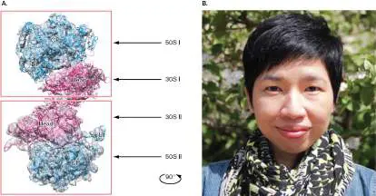
relieved. B. M.-N. Frances Yap discovered that dissociation of ribosome dimers requires the GTPase HflX.
COURTESY OF ADA YONATH. NATURE COMMUNICATIONS.
HTTP://CREATIVECOMMONS.ORG/LICENSES/BY/4.0/
COURTESY OF M.-N. FRANCES YAP
Microbes Encounter Multiple Stresses in Real Life
Bacterial stress responses have traditionally been studied by exposing organisms to individual stresses such as starvation, high or low osmolarity, or extreme pH in controlled laboratory situations. In the world outside of the laboratory, however, environmental situations can be quite complex, involving multiple, not just single, stressors. An organism could simultaneously undergo carbon starvation in a high-salt, low-pH environment. Thus, caution is advised when trying to use controlled laboratory studies that alter a single parameter to predict cellular responses to real-world situations.
Environmental stresses can, of course, drive evolution. Long-term exposure to multiple stresses provides the organism with broad evolutionary opportunities. For example, a variety of stresses can develop within a single colony of bacteria as that colony ages. Starvation, reactive oxygen species, toxic end products of metabolism, and changes in pH can all build to a point that threatens survival of the entire colony. Stress response systems alone cannot save them. Yet some cells in an aging colony develop mutations that will facilitate survival. The altered physiology of some of these mutants also enables them to outgrow their parental strain when both are mixed on fresh agar medium. Thus, multiple stresses encountered when bacteria age can select for mutations that maintain cell viability but also provide a competitive growth advantage.
Humans Influence Microbial Ecosystems
Human activities have striking effects on microbial ecosystems. A major example of such effects is eutrophication. Natural ecosystems are typically low in nutrients (oligotrophic; see eResearch Activity 5 ) but teem with diversity, so numerous species compete for the same limiting nutrients. Maximum diversity in a given ecosystem is maintained, in part, by the different nutrient-gathering profiles of competing microbes. However, the sudden infusion of large quantities of a formerly limiting nutrient, a process called eutrophication, can lead to a “bloom” of microbes, typically autotrophic cyanobacteria (formerly called blue-green algae; see Section 18.2). One species initially held in check by the limiting nutrient now exhibits unrestricted growth, consuming other nutrients as it grows to a degree that threatens the existence of competing species.
Humans cause eutrophication in several ways. Fertilizer runoff from agricultural fields, urban lawns, and golf courses is one source. Untreated or partially treated domestic sewage is another. Spilling large amounts of phosphates or nitrogen into lakes—Lake Erie, for example (Fig. 5.24)—powerfully stimulates cyanobacterial growth. The resulting bacterial blooms (wrongly called “algal blooms”) can deplete the oxygen in the water and lead to fish kills. Native fish species can disappear, to be replaced by species more tolerant of the new conditions. Some cyanobacteria, such as Microcystis aeruginosa, produce liver toxins such as microcystin. A Microcystis bloom in 2014 shut down the entire water supply of Toledo, a city on the shores of Lake Erie. The concept of limiting nutrients in ecosystems is covered further in Chapter 21.
FIGURE 5.24 ■ Eutrophication in Lake Erie. Cyanobacterial bloom (bright blue-green color) in Lake Erie caused by excessive phosphorous eutrophication.
NASA
Eutrophication is not the only human activity affecting Earth’s microbiota. Acid mine drainage resulting from abandoned coal and mineral mines is another. After a mine is abandoned, groundwater is no longer pumped out and the mine floods. Acid mine drainage develops from the oxidation of pyrite (FeS 2) unearthed by the mining operations. The exposed pyrite oxidizes in air to form sulfuric acid that, along with soluble Fe 2+, can drain from the mine and destroy natural ecosystems. Acidophiles such as Acidithiobacillus ferrooxidans are key contributors to pyrite oxidation.
Climate change caused by human activity is one other process that will gradually alter microbial ecosystems. Put simply, the spewing of heat-trapping CO 2 into the atmosphere by burning hydrocarbons is speeding Earth’s warming. Since 1980, the average surface temperature has risen 0.98°C (1.76°F). A 10-year study published in 2014 by Konstantinos Konstantinidis from the Georgia Institute of Technology and Jizhong Zhou from the University of Oklahoma found that a mere 2°C difference in soil

temperature will alter a community of soil microorganisms. Using an infrared light, the team warmed a patch of Oklahoma prairie soil 2°C above that of an adjacent control patch of soil. They then used DNA-based techniques to catalog the microbial genera present (Fig. 5.25). Some taxonomic groups of organisms became more dominant (Actinobacteria), while others became less abundant (Proteobacteria and Acidobacteria). What future effects these kinds of changes will have on the carbon and nitrogen cycles, as well as on farm productivity, remain to be seen.
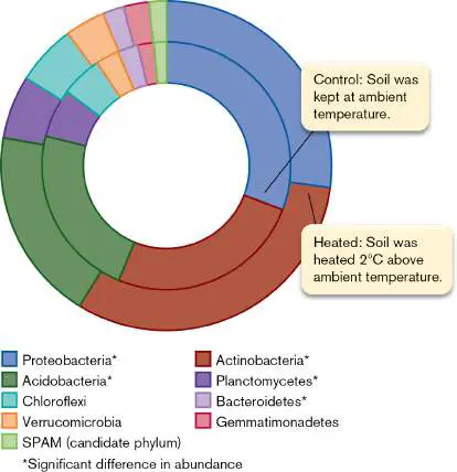
FIGURE 5.25 ■ Effect of warming on the soil microbiome. Two patches of Oklahoma soil were monitored for microbial taxa over 10 years. The two rings illustrate the average abundances of various taxa. SPAM = candidate division, microbes discovered in sp ring in a lpine and m eadow soils.
Thought Question
5.9 Given a mixture of two microbes, A and B—where organism A can utilize limiting phosphate more efficiently than organism B, but organism B can utilize limiting nitrogen better than organism A— what would happen to the relative growth of the two organisms placed in a limiting nitrogen and phosphate medium if excess nitrogen were added to the mixed culture? What about adding both excess phosphate and excess nitrogen?
To Summarize
Starvation is a stress that can elicit a molecular response in many microbes. Enzymes are produced to increase the efficiency of nutrient gathering and to protect cell macromolecules from damage.
The starvation response is usually triggered by the accumulation of small signaling molecules, such as cyclic AMP or guanosine tetraphosphate.
Human activities can cause eutrophication , which damages delicately balanced ecosystems by introducing nutrients that can allow one member of the ecosystem to flourish at the expense of other species.
Glossary
programmed cell death Cell death mediated by a regulated intracellular process. eutrophication A sudden increase of a formerly limiting nutrient in an aquatic environment, leading to overgrowth of algae and grazing bacteria and subsequent oxygen depletion.
5.6 Physical, Chemical, and Biological Control of MicrobesUnit 5 · Regulation
We have seen how adept microbes are at surviving environmental stresses; now, how can we kill them? A primary goal of our health care system is to control or kill microbes that can potentially harm us. Within the recent past, infectious disease was an imminent and constant threat to most of the human population. For instance, during the civil war, life expectancy fell from 40 years to 35 years due, in large part, to lethal infections suffered by wounded soldiers and civilians. The average family in the United States prior to 1900 had four or five children, but parents could expect half of them to succumb to deadly infectious diseases. Improvements in sanitation procedures and antiseptics and the advent of antibiotics have, to a large degree, curtailed the incidence and lethal effects of many infectious diseases. These advancements have played a major role in extending life expectancy (Fig. 5.26).
FIGURE 5.26 ■ Life expectancy in the United States. Although medical advances, sanitation, and nutrition played major roles in extending life expectancy, so, too, did phenolic disinfectants and antibiotics introduced in 1880 and 1945, respectively. Notice the dips in life expectancy associated with the influenza pandemic in 1918 and the COVID-19 pandemic in 2020.
A variety of terms are used to describe antimicrobial control measures. The terms convey subtle, yet vitally important, differences in control strategies and outcomes: Sterilization is the process by which all living cells, spores, and viruses are destroyed on an object.
Disinfection is the killing, or removal, of disease-producing organisms from inanimate surfaces; it does not necessarily result in sterilization.
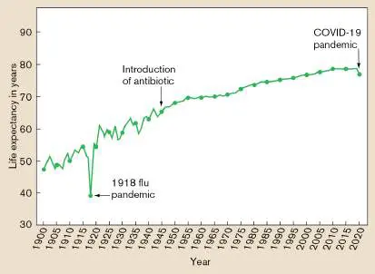
Antisepsis is similar to disinfection, but it applies to removing pathogens from the surface of living tissues, such as the skin. Antiseptic chemicals are usually not as toxic as disinfectants, which frequently damage living tissues.
Sanitation is closely related to disinfection. It consists of reducing the microbial population to safe levels and usually involves both cleaning and disinfecting an object.
Antimicrobials can also be classified on the basis of the specific groups of microbes destroyed, leading to the terms “microbicide,” “bactericide,” “algicide,” “fungicide,” and “virucide.” These agents can be classified further as either “-static” (inhibiting growth) or “-cidal” (killing cells). For example, antibacterial agents may be bacteriostatic or bactericidal. Chemical substances are germicidal if they kill pathogens (and many nonpathogens), but germicidal agents do not necessarily kill spores.
Although these descriptions emphasize the killing of pathogens, it is important to note that antimicrobial agents can also kill or prevent the growth of nonpathogens. Many public health standards are based on total numbers of microorganisms on an object, regardless of pathogenic potential. For example, to gain public health certification, the restaurants we frequent must demonstrate low numbers of bacteria (pathogenic or not) wherever food is prepared.
Thought Question
5.10 Bacteriostatic antibiotics do not kill bacteria; they only inhibit their growth. Why are they nevertheless effective at treating bacterial infections? Hint: Is the human body a quiet bystander during an infection?
Antimicrobials Kill at a Logarithmic Rate
Exposing microbes in a solution or on a surface to lethal chemicals or conditions will not instantly kill all the microorganisms. Microbes die according to a negative exponential curve, where cell numbers decrease in equal fractions at constant intervals. The efficacy of a given lethal agent or condition is measured as decimal reduction time (D-value), which is the length of time it takes that agent (or condition) to kill 90% of the population (a drop of one log unit, or a drop to 10% of the original value). Figure 5.27illustrates the exponential death profile of a bacterial culture heated to 100°C. The D-value (called D 100 in this instance) is a little over 1 minute. The food industry uses D-value and several other parameters to evaluate the efficiency of killing (see Section 16.4).
FIGURE 5.27 ■ The death curve and the determination of D-values. Bacteria were exposed to a temperature of 100°C, and survivors were measured by viable count. The D-value (D 100 ) is the time required for 100°C to kill 90% of cells (that is, the
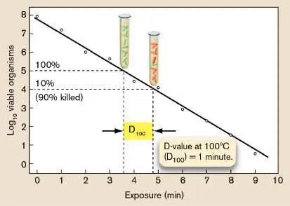
time it takes for the viable cell count to drop by one log 10 unit). The bacteria in the test tubes are colored green if they are viable and red if they are nonviable (dead). Realize that the dead bacteria remain targets for the agent, which is why the live cells in the 90%-killed tube do not die faster than those in the 100%- living tube.
Thought Question
5.11 If a disinfectant is added to a culture containing 1 × 10 6 colony-forming units (CFUs) per milliliter and the D-value of the disinfectant is 2 minutes, how many viable cells will be left after 4 minutes of exposure?
Several factors influence the ability of an antimicrobial agent to kill microbes. These include the initial population size (the larger the population, the longer it takes to reduce it to a specific number), the population composition (are spores involved?), the concentration of the antimicrobial agent, and the duration of exposure. Although the effect of concentration seems intuitively obvious, an increase in concentration is matched by an increase in death rate only over a narrow concentration range. Increases above a certain level might not accelerate killing at all. For example, 70% ethanol is actually better than pure ethanol at killing organisms, because some water is needed to help ethanol penetrate cells. The ethanol then dehydrates cell proteins.
Why, then, is death an exponential function? Why don’t all cells in a population die instantly when treated with lethal heat or chemicals? The reason is based, in part, on the random probability that an agent will cause a lethal “hit” to a cell component, a protein, or a gene in a given cell. Cells contain thousands of different proteins and thousands of molecules of each one. Not all proteins and not all genes in a chromosome are damaged by an agent at the same time. Damage accumulates. Only when enough molecules of an essential protein or a gene encoding that protein are damaged will the cell die. Cells that die first are those that accumulate lethal hits early. Members of the population that die later have, by random chance, absorbed more hits on nonessential proteins or genes, temporarily sparing the essential ones.
Why, if 90% of a population is killed in 1 minute, isn’t the remaining 10% killed in the next minute? It seems logical that all should have perished. Yet after the second minute, 1% of the original population remains alive. This phenomenon can also be explained by the random-hit concept. Although fewer viable cells remain after 1 minute, each has the same random chance of having a lethal hit as when the treatment began. Thus, death rate is an exponential function, much like radioactive decay is an exponential function.
A final consideration is the overall fitness of individual cells. It is a mistake to assume that all cells in a population are identical. At any given time, for instance, one cell may express a protein that another cell has just stopped expressing (for example, superoxide dismutase). In that instant, the first cell might contain a bit more of that protein. If the protein is essential or confers a level of stress protection (such as against superoxide), the cell with more of that protein can absorb more punishment before it dies. The presence of lucky individuals expressing the right repertoire of proteins might also explain why death curves commonly level off after a certain point.
Physical Agents That Kill Microbes
Physical agents are often used to kill microbes or control their growth. Commonly used physical control measures include temperature extremes, pressure (usually combined with temperature), filtration, and irradiation.
High temperature and pressure. Even though microbes were discovered nearly 350 years ago, thermal treatment of foods to render them safe has been practiced for over 5,000 years. Moist heat is a much more effective killer than dry heat, thanks to the ability of water to penetrate cells. Many bacteria, for instance, easily withstand 100°C dry heat but not 100°C boiling water. We humans are not so different, finding it easier to endure a temperature of 32°C (90°F) in dry Arizona than in humid Louisiana.
While boiling water (100°C) can kill most vegetative (actively growing) organisms, spores are built to withstand this abuse, and thermophiles prefer it. Killing spores and thermophiles usually requires combining high pressure and temperature. At high pressure, the boiling point of water rises to a temperature rarely experienced by microbes living at sea level. Even endospores quickly die under these conditions. This combination of pressure and temperature is the principle behind sterilization using the steam autoclave (Fig. 5.28). Standard conditions for steam sterilization are 121°C (250°F) at 15 psi for 20 minutes—a set of conditions that experience has taught us will kill all spores except those of some thermophiles. (Thermophiles, however, do not affect food or human health.) Standard sterilization conditions are also produced in pressure cookers used for home canning of vegetables and fruits.
FIGURE 5.28 ■ Steam autoclave. Failure to adhere to sterilization heat and pressure parameters can have deadly consequences, even in your own home. For instance, Clostridium botulinum is a spore-forming soil microbe that commonly contaminates fruits and vegetables used in home canning. The improper use of a pressure cooker while canning these goods will allow spores of this pathogen to survive. Once the can or jar is cool, the spores will germinate and begin producing their deadly toxin. All of this happens while the canned goods sit on a
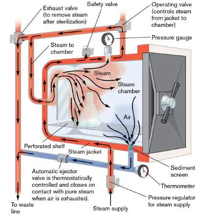
shelf waiting to be opened and consumed. Once ingested, the toxin makes its way to the nervous system and paralyzes the victim. Several incidents of this disease, called botulism, occur each year in the United States. (For more on food poisoning, see Chapter 16.)
Thought Question
5.12 How would you test the killing efficacy of an autoclave? Pasteurization. Originally devised by Louis Pasteur to save products of the French wine industry from devastating bacterial spoilage, pasteurization today involves heating a particular food (such as milk) to a specific temperature long enough to kill Coxiella burnetii, the causative agent of Q fever, the most heat-resistant non-spore-forming pathogen known. In the process, pasteurization also kills other disease-causing microbes.
Three U.S. government–approved time and temperature combinations can be used for pasteurization of milk: LTLT (low temperature, long time). In this method, the milk is brought to a temperature of 63°C (145°F) for 30 minutes. HTST (high temperature, short time). This method (also called “flash pasteurization”) brings the milk to a temperature of 72°C (161°F) for only 15 seconds.
UHT (ultra-high temperature). In this method, the milk is brought to a temperature of 138°C (280°F) for 1–2 seconds.
Both the LTLT and HTST methods accomplish the same thing—the destruction of C. burnetii and other bacteria—but they do not sterilize milk. UHT pasteurization decreases bacterial content even more than the LTLT and HTST methods, producing nearly sterile milk with an unrefrigerated shelf life of up to 6 months. This is important, especially in developing countries, where refrigeration is not always available.
Cold. Low temperatures have two basic purposes in microbiology: to slow growth and to preserve strains. Bacteria not only grow more slowly in cold but also die more slowly. Refrigeration temperatures (4°C–8°C, or 39°F–46°F) are used for food preservation because most pathogens are mesophilic and grow poorly, if at all, at those temperatures. One exception is the Gram-positive bacillus Listeria monocytogenes, which can grow reasonably well in the cold and causes disease when ingested.
Long-term storage of bacteria usually requires placing solutions in glycerol at very low temperatures (−80°C). Glycerol prevents the production of razor-sharp ice crystals that can pierce cells from without or within. This deep-freezing suspends growth altogether and keeps cells from dying. Another technique, called lyophilization, freeze-dries microbial cultures for long-term storage. In this technique, cultures are quickly frozen at very low temperatures to limit ice crystal formation and placed under vacuum, where the resulting sublimation removes all water from the media and cells, leaving just the cells in the form of a powder. These freeze-dried organisms remain viable for years. Finally, viruses and eukaryotic cells must be kept at extremely low temperatures (−196°C), submerged in liquid nitrogen. Liquid nitrogen freezes cells so quickly that ice crystals do not have time to form.
Filtration. Filtration through micropore filters with pore sizes of 0.2 μm or less can remove protozoans, fungi, and most species of bacteria from solutions. Viruses, being much smaller, are not trapped. Samples from 1 milliliter to several liters can be drawn through a membrane filter by vacuum or can be forced through it with a syringe (Fig. 5.29). Filter “sterilization” avoids the use of heat, which can damage certain materials. Strictly speaking, though, the solutions are not really sterile, because these filters do not trap viruses. Bottled drinking water labeled as purified undergo additional, more stringent processes such as ozonation, distillation, or UV sterilization.
FIGURE 5.29 ■ Membrane filtration devices. Air can also be sterilized by filtration. This process forms the basis of several personal protective devices. A surgical mask is a crude example, while laminar flow biological safety cabinets are more elaborate (and more effective). These cabinets force air through high-efficiency particulate air (HEPA) filters and remove over 99.9% of airborne particulate material 0.3 μm in size or larger. Although the 0.3 μm pore size might seem too large to trap viruses, viruses in air generally travel on larger particles such as saliva. Biosafety cabinets are critical to protect individuals working with highly pathogenic material (Fig. 5.30Aand B ). Newer technologies have been developed that embed antimicrobial agents or enzymes directly into the fibers of the filter (Fig. 5.30C ).
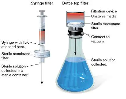
Organisms entangled in these fibers are not just trapped; they are attacked by the antimicrobials and lysed. Note that HEPA filters are also used in some home air filtration systems to minimize pollen and dust allergens.
FIGURE 5.30 ■ Biological safety cabinet. A. A scientist examines a sample under the hood. B. Schematic of the safety cabinet. Air from the room enters the cabinet through the cabinet opening (1) or is pumped in (2) through a HEPA filter (3). It then passes behind the negative-pressure exhaust plenum (4) and is passed from the cabinet through another HEPA filter (5). C. Antibacterial activity of silver-silica coated particles on an air filtration unit. The primary function of this filter is to kill airborne microorganisms caught on the surface of the filter, thus protecting against secondary contamination by microorganisms in air filtration systems. The photo shows that Staphylococcus epidermidis cells attached to the smaller silver-silica beads on the filter fiber have an altered morphology.
WILL & DENI MCINTYRE/SCIENCE SOURCE
REPUBLISHED WITH PERMISSION OF ROYAL SOCIETY OF CHEMISTRY. Y. KO ET AL.
2014. J. MATER. CHEM. B, NO. 39
Irradiation. Public health authorities worldwide are constantly worried about food being contaminated with pathogenic
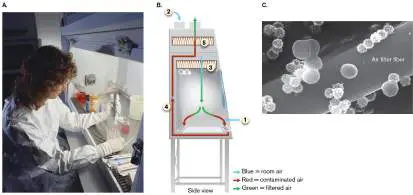
microorganisms such as Salmonella species, Escherichia coli O157:H7, Listeria monocytogenes, and Yersinia enterocolitica. Irradiation, the bombardment of foods with high-energy electromagnetic radiation, has long been a potent strategy for sterilizing food after harvesting. The food consumed by NASA astronauts, for example, has for some time been sterilized by irradiation as a safeguard against food-borne illness in space. Early public concerns that irradiation might produce dangerous toxic by-products have largely disappeared. Numerous studies have proved that foods do not become radioactive when irradiated, nor are long-lived reactive molecules produced that are dangerous to humans. Aside from ultraviolet light, which, owing to its poor penetrating ability, is useful only for surface sterilization, there are three other sources of irradiation: gamma rays, electron beams, and X-rays. Radiation dosage is usually measured in a unit called the gray (Gy), which is the amount of energy transferred to the food, microbe, or other substance being irradiated. A single chest X-ray delivers roughly half a milligray (1 mGy = 0.001 Gy). To kill Salmonella, freshly slaughtered chicken can be irradiated at up to 4.5 kilograys (kGy)—about 7 million times the energy of a single chest X-ray. The U.S. Food and Drug Administration (FDA) has also approved the use of irradiation (4 kGy) on beef, pork, fruits, vegetables, oysters, seeds, shell eggs, and spices.
How does radiation kill bacteria? When microbes present in food are irradiated, water and other intracellular molecules absorb the energy and form very short-lived reactive chemicals, typically reactive oxygen species, that damage DNA and proteins. Unless this damage is prevented or repaired, the organism will die. Microbes differ greatly in their sensitivity to irradiation, depending on the size of their genome, the rate at which they can repair damaged DNA, and other factors. Whether the food to be irradiated is frozen or fresh also matters, as it takes a higher dose of radiation to kill microbes in frozen foods.
The size of the DNA “target” is a major factor in radiation efficacy. Parasites and insect pests, which have large amounts of DNA, are rapidly killed by extremely low doses of radiation, typically with D-values of less than 0.1 kGy (in this instance, the D-value is the dose of radiation needed to kill 90% of the organisms). It takes more radiation to kill bacteria (D-values in the range of 0.3–0.7 kGy) because they have less DNA per cell unit (less target per cell). It takes even more radiation to kill bacterial spores (D-values of the order 2.8 kGy) because they contain little water, the source of most ionizing damage to DNA.
Viral pathogens have the smallest amount of nucleic acid, making them resistant to irradiation doses approved for foods (viruses have D-values of 10 kGy or higher). Infectious agents that do not contain nucleic acids are an even bigger problem. Prions, for example, are misfolded brain proteins that “self-replicate” and cause neurodegenerative diseases (see Section 26.6). Because prions do not contain nucleic acids, the agent can be inactivated by irradiation only at extremely high doses. Thus, irradiation of food is effective in eliminating parasites and bacteria but is woefully inadequate for eliminating viruses or prions.
Note: Electromagnetic radiation emitted by microwave ovens
does not directly kill bacteria. However, the heat generated when electromagnetic radiation excites water molecules in an organism will kill the organism if the temperature attained is high enough. Resistance to ionizing radiation. Deinococcus radiodurans could be branded the “Iron Man of Microbes” and the mascot of extremophiles (Fig. 5.31A). D. radiodurans was discovered in 1956 in a can of meat that had spoiled despite having been sterilized by radiation. The microbe, which is also a moderate thermophile, has the greatest ability of any known organism to survive radiation. It could probably even survive an atomic blast. The bacterium’s ability to withstand radiation may have evolved as a side effect of developing resistance to extreme drought, as desiccation and radiation produce similar types of DNA damage. FIGURE 5.31 ■ Deinococcus radiodurans. A. The amount of radiation that Deinococcus radiodurans can survive is equivalent to that of an atomic blast. The nature of the dark inclusion bodies in three of the four cells in this quartet is currently not known. B. Lydia Contreras at the University of Texas at Austin explores how D. radiodurans regulates its exceptional ability to repair radiation-damaged DNA.
JOHN R. BATTISTA, LOUISIANA STATE UNIVERSITY
REBECCA WUNDERLICH
What properties account for this microbe’s amazing resistance to radiation? One is that D. radiodurans possesses an unusual capacity for repairing damaged DNA. Many mechanisms are involved, including a highly efficient double-strand-break DNA repair system requiring homologous recombination (see Section 9.2). Each cell of this organism contains four to six copies of its two chromosomes and two plasmids. So even when its DNA is irradiated and broken into thousands of fragments, overlapping intact fragments can be found and spliced together in proper order.
Many of the DNA repair genes needed to survive radiation are part of a gene set, collectively called the radiation and desiccation response regulon, whose transcriptional expression increases during
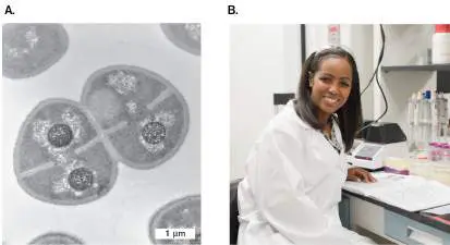
radiation or desiccation conditions. Lydia Contreras at the University of Texas at Austin has been a major contributor to research in this field (Fig. 5.31B ). Her team has identified eight noncoding small RNA (sRNA) molecules that are expressed by D. radiodurans during recovery from ionizing radiation. One of these molecules, PprM, stabilizes an important protein regulator of the radiation and desiccation response. As proof of its importance, Contreras’s group demonstrated that D. radiodurans pprM mutants are extremely sensitive to ionizing radiation.
A second property that contributes to radiation resistance is the ability of D. radiodurans to aggressively protect its proteins, which are even more susceptible to radiation than is DNA. This mechanism, discovered by Michael Daly at the Uniformed Services University of the Health Sciences in Maryland, involves the intracellular accumulation of large amounts of manganese-metabolite complexes that can nonenzymatically remove highly damaging free radicals generated by radiation. This mechanism limits damage to macromolecules (proteins, DNA, RNA, lipids), including DNA repair proteins needed to fix macromolecules that are damaged.
Because of its incredible level of radiation resistance, D. radiodurans was genetically engineered to treat radioactive mercury–contaminated waste from nuclear reactors in a process called bioremediation (discussed in Chapter 22). The genes for mercury conversion were spliced from a strain of E. coli that is resistant to particularly toxic forms of mercury and inserted into D. radiodurans. The genetically altered superbug was able to withstand the ionizing radiation and convert toxic waste into forms that could be removed safely. Fortunately, there is little need to worry about D. radiodurans becoming a superpathogen, because the organism does not cause disease and is susceptible to antibiotics.
Chemical Agents
Disinfection by physical agents is very effective, but in numerous situations their use is impractical (kitchen countertops) or plainly impossible (skin). In these instances, chemical agents are the best approach. A number of factors influence the efficacy of a given chemical agent. These include: The presence of organic matter. A chemical placed on a dirty surface will bind to any inert organic material present, lowering the agent’s effectiveness against microbes. It is not always possible to clean a surface prior to disinfection (as in a blood spill), but the presence of organic material must be factored into estimates of how long to disinfect a surface or object.
The kinds of organisms present. Ideally, the agent should be effective against a broad range of pathogens.
Corrosiveness. The disinfectant should not corrode the surface or, in the case of an antiseptic, damage skin.
Stability, odor, and surface tension. The chemical should be stable during storage, possess a neutral or pleasant odor, and have a low surface tension so that it can penetrate cracks and crevices.
The phenol coefficient. Phenol, first introduced by Joseph Lister in 1867 to reduce the incidence of surgical infections, is no longer used as a disinfectant because of its toxicity, but its potency makes it the benchmark against which other disinfectants are measured. The phenol coefficient is determined by comparing the highest dilution of a test agent capable of killing pathogens to that of phenol. Derivatives of phenol, such as cresols and orthophenylphenol, are still in use: The household product Lysol (liquid), for instance, contains o -benzyl-p -chlorophenol. Phenolics are useful disinfectants because they denature proteins, are effective in the presence of organic material, and remain active on surfaces long after application.
Commercial Disinfectants
Ethanol, iodine, chlorine, and surfactants (for example, detergents) are all used to decrease or eliminate microbial content from commercial products (chemical structures are shown in Fig. 5.32). The first three are compounds that damage proteins, lipids, and DNA. Highly reactive iodine complexed with an organic carrier forms an iodophor, a compound that is water-soluble, stable, nonstaining, and capable of releasing iodine slowly to avoid skin irritation. Wescodyne and Betadine (trade names) are iodophors used, respectively, for the surgical preparation of skin and for wounds. Chlorine is another highly reactive disinfectant with universal application. It is recommended for general laboratory and hospital disinfection. Sodium hypochlorite (liquid bleach) is a common disinfectant in bathroom cleaners such as Tilex.
FIGURE 5.32 ■ Structures of some common disinfectants and antiseptics.
Detergents can also be antimicrobial agents. The hydrophobic and hydrophilic ends of detergent molecules (the coexistence of which makes the molecules amphipathic) will emulsify fat into water. Cationic (positively charged) detergents are useful as disinfectants because the positive charges can gain access to the negatively charged bacterial cell and disrupt membranes. Anionic (negatively charged) detergents are not antimicrobial but do help remove bacteria from surfaces. Quaternary ammonium compounds such as benzalkonium chloride or alkyl dimethyl benzyl ammonium
saccharinate (the agent in Lysol disinfectant spray used during the COVID-19 pandemic) are useful cationic disinfectants. These compounds target membranes at high concentrations but can also inflict damage to internal cell components at lower concentrations. For example, a study with Acinetobacter baumannii (a Gram-negative bacillus that causes hospital-derived infections) found that low concentrations of benzalkonium chloride, similar to what might ultimately penetrate a biofilm, could cause cell death by increasing oxidative stress and by interfering with protein homeostasis mechanisms. The result is the intracellular accumulation of protein aggregates that are dangerous to the microbe.
Low-molecular-weight aldehydes such as formaldehyde (HCHO)
are very potent disinfectants. The aldehydes are highly reactive, combining with and inactivating proteins and nucleic acids. However, the noxious odor of aldehydes and the classification of formaldehyde as a suspected carcinogen limit their use in sterilizing medical equipment.
Disposable plasticware (including Petri dishes, syringes, sutures, and catheters) cannot undergo heat sterilization or liquid disinfection because the items will melt or dissolve. These materials are best sterilized using antimicrobial gases. Ethylene oxide gas (EtO) is a very effective sterilizing agent; it destroys cell proteins, is microbicidal and sporicidal, and rapidly penetrates packing materials, including plastic wraps. Deployed in an instrument resembling an autoclave, EtO at 700 milligrams per liter (mg/l) will sterilize an object after 8 hours at 38°C or 4 hours at 54°C if the relative humidity is kept at 50%. Unfortunately, EtO is explosive. A less hazardous gas sterilant is betapropiolactone. It does not penetrate as well as EtO, but it decomposes after a few hours, which makes it easier to dispose of than EtO.
A relatively new procedure, known as gas discharge plasma sterilization, may replace EtO because it is less harmful to operators. Gas discharge plasma is made by passing certain gases through an electrical field to produce highly reactive chemical species that can damage membranes, DNA, and protein. A form of this process called low-temperature plasma sterilization, involving hydrogen peroxide vapor and plasma, is commercially available.
Antimicrobial touch surfaces. Despite widespread use of disinfectants and antibiotics by medical personnel, hospital-acquired infections remain a major concern. A promising antimicrobial technology that can help reduce infections in hospitals and elsewhere involves embedding antimicrobial compounds such as copper (Cu) in the surfaces that people touch. For example, a bed rail made with copper can kill pathogens deposited by one person before the organism can be transmitted to a second person touching the same surface. Upon contact with bacteria, metallic copper releases toxic Cu + ions that trigger the lysis of bacterial membranes within minutes, although the mechanism involved is unclear (neither DNA damage nor reactive oxygen species are involved). Companies are incorporating metallic copper into objects such as handrails, door releases, and hospital bed rails.
In a newer development, an antimicrobial coating composed of silver plus rubidium (trade name AGXX) was found to prevent pathogenic methicillin-resistant Staphylococcus aureus (MRSA) from forming biofilms on metal surfaces. The coating limited the expression of S. aureus attachment proteins (adhesins), toxins, and other virulence factors. AGXX may be useful in hospital environments, but it is also being developed for air-conditioning cooling towers, which can harbor pathogens such as Legionella pneumophila, washing machines, dishwashers, and even drinking-water purifiers.
Bacteria Can Develop Resistance to Disinfectants
We know that bacteria can develop resistance to antibiotics used to treat infections, but can bacteria also develop resistance to disinfectants that prevent infections? The answer is yes—and no. It is difficult for a bacterium to develop resistance to chemical agents that have multiple targets and can easily diffuse into a cell. Iodine, for example, has both of these characteristics. However, disinfectants that have multiple targets at high concentrations may have only a single target at lower concentrations—a situation that can foster the development of resistance. For instance, triclosan (a halogenated bisphenol compound used in many soaps, deodorants, and toothpastes) targets several cell constituents, making it reliably bactericidal at high concentrations. However, at low concentrations triclosan only inhibits fatty acid synthesis and is merely bacteriostatic. Organisms have developed resistance to triclosan at low concentrations by altering the fatty acid synthesis protein normally targeted by triclosan. Consequently, in 2016 the FDA banned triclosan in consumer antiseptic washes.
Bacteria can use membrane-spanning, multidrug efflux pumps (described in Section 4.2) to achieve low-level resistance to disinfectants. For instance, the MexCD-OprJ efflux system of Pseudomonas aeruginosa, a Gram-negative bacterium that causes infections in burn and cystic fibrosis patients, can pump several different biocides, detergents, and organic solvents out of the cell, thereby reducing their efficacy. This finding and other reports of Pseudomonas gaining resistance to disinfectants have led many clinicians to advocate caution in the widespread use of certain chemical disinfectants.
Biofilm formation is another ingenious way that bacteria survive exposures to disinfectants. A biofilm is a 3D community of bacterial cells attached to a solid surface (see Section 4.5). A biofilm can protect cells in several ways. For example, the extracellular matrix proteins and polysaccharides that hold biofilms together also bind disinfectants, slowing their penetration into the deeper recesses of the structure. Slower penetration means that cells deep in the biofilm have time to activate protective stress response systems before destructive levels of disinfectant reach them. Biofilms also exhibit stratified growth patterns based in part on nutrient access (see Fig. 4.32). Cells at the periphery have ample access to oxygen and nutrients, while cells within do not. Nutrient starvation worsens the farther a cell is from the surface. Because nutrient starvation activates stress response systems, each biofilm has stratified layers of increasingly stress-resistant cells that can better tolerate chemical insults.
Finally, biofilms with multiple species can develop protective, interspecies collaborations. Protective enzymes from one species could protect another species from chemical insult, much as a big brother protects a little brother from a bully. For example, Figure 5.33shows a biofilm of a Bacillus subtilis strain (green), isolated from an endoscope washer-disinfector, mixed with pathogenic Staphylococcus aureus (red). Romain Briandet and colleagues from the French National Institute of Agricultural Research observed that B. subtilis in this biofilm protected S. aureus from the disinfectant peracetic acid. Multispecies biofilms can also be more massive than monospecies biofilms. The food pathogen Escherichia coli O157:H7 forms a biofilm with 400 times more volume when grown with Acinetobacter calcoaceticus, an organism found in meatpacking plants. Increased volume alone will slow the penetration of the disinfectant and protect the collective.
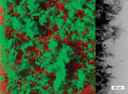
FIGURE 5.33 ■ Mixed biofilm. Three-dimensional projection of a mixed 24-hour biofilm of Bacillus subtilis expressing green fluorescent protein (GFP; green) and Staphylococcus aureus expressing mCherry fluorescent protein (red). The presence of B. subtilis protects S. aureus from peracetic acid treatment. The right side of the image is a black-and-white photo of the biofilm’s edge.
ROMAIN BRIANDET. FROM L. KHALIFA ET AL. 2015. APPL. ENVIRON. MICROBIOL.
81 :2696
Antibiotics Selectively Control Bacterial Growth
Antibiotics as made in nature are chemical compounds synthesized by one microbe to either kill (bactericidal) or stop the growth of (bacteriostatic) other competing microbial species. Chapter 27 describes the various antibiotic modes of action. When purified and administered to patients suffering from an infectious disease, antibiotics can produce seemingly miraculous recoveries.
As we saw in Chapter 1, penicillin, produced by Penicillium notatum, was discovered serendipitously in 1929 by Alexander Fleming. Because the penicillin structure mimics a part of the cell wall, this drug binds to biosynthetic proteins involved in peptidoglycan synthesis and prevents cell wall formation. The drug is bactericidal, in part, because without the support of a cell wall, actively growing cells will lyse (Fig. 5.34). Other antibiotics target protein synthesis, DNA replication, cell membranes, and various enzyme reactions. Antimicrobials that affect macromolecular synthesis are described throughout Parts 1–4 of this book. FIGURE 5.34 ■ Effect of cephalexin (a penicillin-like antibiotic) on E. coli. Time-lapse light microscopy follows the antibiotic’s effect over 73 minutes of growth. Arrows indicate directions of cell and bulge expansions. Cephalexin weakens the cell wall of growing cells and causes a bulge to form. The bulge enlarges until the cell lyses.
Z. YAO ET AL. 2012. MOL. CELL 48 :705–12
So how do antibiotic-producing microbes avoid “suicide”? In some instances, the producing organism lacks the target molecule.
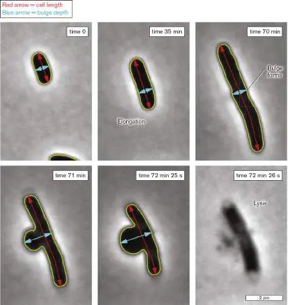
Penicillium mold, for instance, lacks peptidoglycan and is immune to penicillin by default. Some bacteria produce antimicrobial compounds that target other members of the same species. In this case, the producing organism can modify its own receptors to no longer recognize the compound (as with some bacterial colicins). Another strategy is to modify the antibiotic if it reenters the cell. This is the case with streptomycin produced by Streptomyces griseus. Streptomycin inhibits bacterial protein synthesis and does not discriminate between the protein synthesis machinery of Streptomyces and that of others. However, while making enzymes that synthesize and secrete streptomycin, S. griseus simultaneously makes the enzyme streptomycin 6-kinase, which remains locked in the cell. If any secreted streptomycin reenters the cell, this enzyme renders the drug inactive by attaching a phosphate group to it. Because many microorganisms have become resistant to commonly used antibiotics, pharmaceutical companies continually search for new antibiotics and use a variety of approaches. Traditional procedures include scouring soil and ocean samples collected from all over the world for new antibiotic-producing organisms, and chemically redesigning existing antibiotics so that they can bypass microbial resistance strategies. Newer techniques allow scientists to “mine the genomes” of microbes for potential drug targets and use computer-based methods to predict the structure and function of potential new antibiotics.
Microbial Antagonism: Biological Control of Microbes
One of the hallmarks of a healthy ecosystem is a diversity of species. This is true not only for tropical rain forests and coral reefs but also for the human intestinal tract. Microbial diversity is maintained, in part, by what could be described as “tribal warfare” or antagonism between competing species of bacteria, fungi, and viruses. We have already discussed how some species of fungi and bacteria produce antibiotics that can kill or stunt the growth of competing bacteria, but there are many other examples. The pathogenic soil fungus Phytophthora cinnamomi causes root rot in plants but is biologically controlled by Myrothecium fungi that produce toxins and cellulolytic enzymes. Another illustration is the human intestine, populated by 500 or more microbial species that compete with each other for food and for real estate along the gut mucosa. Permeant weak acids produced by fermenting members of the normal intestinal microbiome not only limit the growth of competitors but also thwart infection by numerous pathogens, a prominent example of the latter being Clostridioides difficile. Bacteriophages and viruses present in all environments contribute to diversity by preventing microbial overgrowth. There are even examples of tiny bacteria such as Bdellovibrio or Halobacteriovorax literally chasing down larger bacteria to devour them (Fig. 5.35). FIGURE 5.35 ■ Bdellovibrio bacteriovorus infecting Escherichia coli. Bdellovibrio cells are shown attaching to the larger host bacterium. One cell enters, replicates, and causes the host cell to lyse and release progeny B. bacteriovorus bacteria (not shown). Helium-ion microscopy.
N. SAID ET AL. 2019. ADV BIOSYST. 3:E1800250
Microbial competition has been widely exploited for agricultural purposes and to improve human health. One way is through the intake of probiotics, first suggested by the Russian biologist Ilya Mechnikov (1845–1916), winner of the 1908 Nobel Prize in
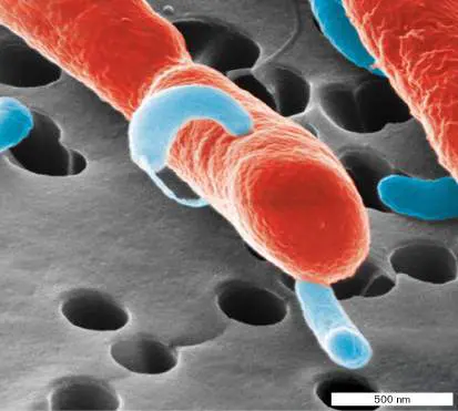
Physiology or Medicine. A probiotic is a food or supplement that contains live microorganisms and improves intestinal microbial balance. Newborn baby chicks, for instance, are fed a microbial cocktail of normal gut microbes that quickly colonize the intestinal tract and prevent colonization by Salmonella, a frequent contaminant of factory-farmed chicken. In another example, Lactobacillus and Bifidobacterium have been used to prevent and treat diarrhea in children. Yogurt is a probiotic that contains Lactobacillus acidophilus and a number of other lactobacilli. It is often recommended as a way to restore a normal balance to gut microbiota after antibiotic treatment, and it appears useful in the treatment of some mild forms of inflammatory bowel syndrome (discussed further in Chapter 23).
Phage therapy, another biocontrol method, was first described in 1907 by Félix d’Herelle (1873–1949) at France’s Pasteur Institute, long before antibiotics were discovered. Bacteriophages are viruses that prey on bacteria (discussed in Section 6.1). Because a phage infection often causes bacterial lysis, it was considered feasible to treat infectious diseases with a phage targeted to the pathogen. At one time, doctors used phages as medical treatment for illnesses ranging from cholera to typhoid fever. Sometimes the treatments worked; sometimes they did not.
SPECIAL TOPIC 5 Phage Plus Antibiotics: A One-Two Punch against Bacterial Infections?
For decades, humans have successfully treated infectious diseases with a weapon that microbes commonly deploy against each other—antibiotics. Unfortunately, pathogens are increasingly becoming resistant to antibiotics and may soon become impervious to them. A possible solution to this problem is to use bacteriophage to treat infectious disease.
Unfortunately, bacteria can also become resistant to phage attack. But what if we use phage and antibiotics together? The probability that a pathogen will become resistant to both is very, very small. One team of scientists headed by Bruce Levin at Emory University in Atlanta (Fig. ST 5.1 ) is exploring this possibility by treating cultures of Staphylococcus aureus with a staphylococcal bacteriophage known as PYO Sa (Fig. ST 5.1 Inset ) in combination with a variety of antibiotics. They discovered a problem but also a clever solution.

FIGURE ST 5.1 ■ Ingrid C. McCall, Bruce Levin, and Brandon Berryhill from Dr. Levin’s lab. Inset: Transmission electron micrograph of Staphylococcus aureus bacteriophage PYO Sa, a K-type Myoviridae.
PHOTO BY RODRIGO EMILIO GARCIA GONZALEZ
DICKEY ET AL. 2019. PLOS ONE. 14: E0209390
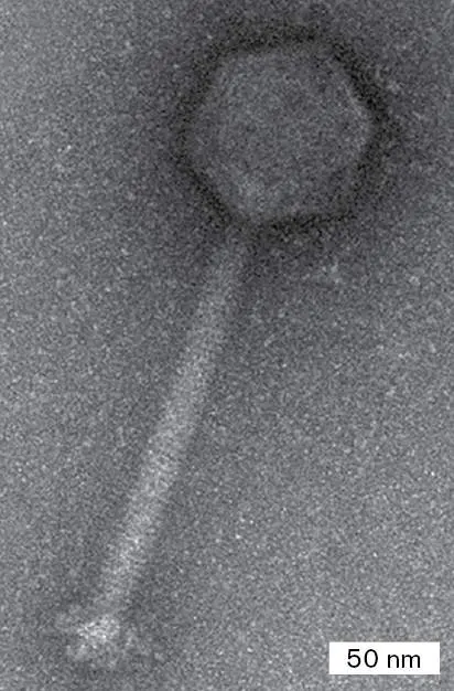
PYO Sa has many properties desireable for a clinically useful phage. For instance, target bacteria do not easily become resistant to PYO Sa because the bacterial receptor for this phage (an N -acetylglucosamine in the cell wall–teichoic acid backbone) is critical to viability of S. aureus. Also, S. aureus does not possess any known DNA restriction enzymes or CRISPR systems that can interfere with PYO Sa replication. In addition, a PYO Sa -like phage has already been used therapeutically in humans.
Levin’s group wanted to be sure that PYO Sa could attack a broad range of S. aureus strains and rigorously tested whether the development of phage resistance was a problem. They found that 71 clinical isolates of S. aureus, many of which were methicillin resistant (MRSA), were susceptible to PYO Sa. Twelve strains examined more closely also failed to generate mutants resistant to PYO Sa attachment. This form of resistance is tested by mixing, in broth, a single colony of S. aureus with 10 6 phage particles and incubating the culture for 24 hours. If the medium remains clear and samples plated onto agar do not produce any colonies, the cells did not develop classic phage resistance. This was the case for the 12 strains.
However, when Levin’s group serially passaged a broth culture containing phage plus bacteria into fresh medium for 5 consecutive days, the cells did multiply in the broth. Plating the culture after 5 serial passages revealed two types of colonies: a large, normal-sized colony and a small-colony variant (Fig. ST 5.2A ). This result signaled a phage resistance problem that could arise when trying to use PYO Sa therapeutically for extended periods of time.
What happened? Figure ST 5.2B shows what the scientists think takes place when a broth culture of PYO Sa mixed with cells is repeatedly allowed to grow from low cell density to high cell density for 5 days. Over the first 2 days most wild-type cells die, but some cells develop a random mutation in a gene ( femA) that affects cell wall assembly. The mutation (femA) confers phage resistance but also impairs cell growth, producing the small-colony variant. These small-colony variant cells at first grow slowly in liquid culture, but they eventually develop a second mutation in one of a variety of genes that can somehow counteract the growth defect (these are the large, so-called evolved, or wild-type, colonies in Fig. ST 5.2A ). The secondary mutations also partially restore phage sensitivity, but these “evolved” cells are not uniformly killed. Thus, the evolved cells are phenotypically different from wild type.
Does this mean that PYO Sa phage cannot be used to treat infections? Levin’s group suspected that combining phage with antibiotics might prevent phage resistance by blocking the establishment of small-colony variants. However, the scientists realized that simultaneous addition of phage and antibiotic would not work, because the antibiotics would keep cell numbers low and limit phage replication. They decided instead to add phage first, let the phage destroy cell density for 24 hours, and only then add an antibiotic to prevent bacterial population recovery and eliminate or prevent the selection of small-colony variants.
Figure ST 5.2C and D show the results of this scheme for two antibiotics: ciprofloxacin and vancomycin. Panel C shows the results of antibiotic only (no phage), whereas panel D illustrates the effect of adding phage at day 0 followed by antibiotics at day 1. Compare the panel D results to those shown in Figure ST 5.2B using phage only. The antibiotics in panel D prevented population recovery after the PYO Sa phage decimated bacterial cell numbers. By killing bacteria that survived the initial phage treatment, the antibiotics eliminated the selective pressure needed to evolve phage-resistant small-colony variants. The results show that PYO Sa can be effective for treating S. aureus infections, but its effectiveness is greatest if bactericidal antibiotics are added after phage PYO Sa does its thing.
FIGURE ST 5.2 ■ Small-colony variants and population dynamics of a PYO Sa and Staphylococcus aureus mixture during serial transfers. A. Small-colony variants and wild type–sized (“evolved”) colonies. B.
Densities of Staphylococcus aureus cells and PYO Sa phage after daily 1:100 dilutions into fresh medium. Day 0 CFUs and plaque-forming units (PFUs) mark when cells and phage were initially mixed. The experiment was done in triplicate, but only a single series is shown for clarity. The control was cells only, no phage. C. Densities of S. aureus cells mixed with antibiotic after daily 1:100 dilutions into fresh medium
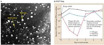
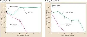
containing antibiotic. D. Densities of S. aureus cells mixed with phage at day 0 and antibiotics at day 1 during daily dilutions (1:100) into fresh medium
B. BERRYHILL ET AL. 2021. PROC NATL ACD SCI USA. 118: E2008007118
RESEARCH QUESTION
Argue the potential limitations of this combined therapy in treating actual infections. What kinds of infections can S. aureus cause, and which ones would be amenable to this combined therapeutic approach?
Berryhill, Brandon A., Douglas L. Huseby, Ingrid C. McCall, Diarmaid Hughes, and Bruce R. Levin. 2021. Evaluating the potential efficacy and limitations of a phage for joint antibiotic and phage therapy of Staphylococcus aureus infections. Proceedings of the National Academy of Sciences USA 118 : e2008007118.
When antibiotics came into the mainstream, phage therapy largely faded. Now that strains of antibiotic-resistant bacteria are on the rise, the idea of phage therapy has enjoyed renewed interest from the medical community. The treatment is being considered for many infections, including respiratory, gastrointestinal, and pregnancy-related infections, and it is even being tested for potential dental applications.
A dramatic example of the power of phage therapy played out in 2016 (as recounted in the TedX talk, “How Sewage Saved My Husband’s Life from a Superbug”). Tom Patterson, a professor at UC San Diego, had been in a coma for months, the result of an infection by the drug-resistant Gram-negative bacterium Acinetobacter baumannii. Granted emergency approval by the FDA, an experimental phage cocktail targeting A. baumannii was injected intravenously. Almost immediately Dr. Patterson began to improve and soon emerged from his coma. Based in part on this success, the FDA, in 2019, approved a clinical trial to study the safety and efficacy of a different experimental phage therapy for patients with ventricular assist devices who have developed Staphylococcus aureus infections.
Commercial phage products are currently available to target the food-borne pathogens E. coli O157:H7, Salmonella enterica, and Listeria monocytogenes. E. coli O157:H7, for instance, is a pathogen that can contaminate hamburger and cause bloody diarrhea. The phage product contains several different phages and is sprayed onto the hides of cattle 1–4 hours before slaughter. Cattle carcasses are steam-pasteurized and acid-washed to diminish bacterial contamination. E. coli cells that manage to survive these treatments will be infected and killed by the phage, reducing the consumer’s risk of disease. Using multiple phages in each preparation nearly eliminates the risk that phage resistance will develop in the pathogen.
Justin Meyer at the University of California San Diego and collaborators have even learned how to “train” phage to better withstand the evolutionary countermeasures that phage-targeted bacteria might take to prevent phage infection. In their experiments, phage and target microbes were coincubated for 28 days, which gave the phage time to confront and genetically adapt to whatever strategies bacteria used to prevent infection. After purification, the trained phage were able to suppress bacterial growth 1,000 times more effectively than untrained phage. Other groups of scientists are combining bacteriophage therapy with another weapon of warring microbes—antibiotics—as a way to prevent the development of resistance in one organism to both forms of bactericide (Special Topic 5 ).
To Summarize
Antisepsis is the removal of potential pathogens from the surfaces of living tissues, while disinfection kills pathogens on inanimate objects. Sterilization kills all living organisms. Antimicrobial compounds can be bacteriostatic or bactericidal .
The D-value is the time (or dose, for irradiation) needed to decrease the number of viable cells to 10% of the original number.
An autoclave uses high pressure to achieve temperatures that will sterilize objects.
Food can be preserved by pasteurization, refrigeration, filtration, and irradiation.
The phenol coefficient is used to compare one disinfectant to another.
Physical and chemical agents kill microbes by denaturing proteins or DNA or by disrupting lipid bilayers. Biocontrol is the use of one microbe to control the growth of another. Antibiotics are compounds produced by one living microorganism that kill other microorganisms.
Probiotics contain certain microbes that, when ingested, aim to restore balance to the intestinal microbiome.
Phage therapy offers a possible alternative to antibiotics in the face of rising antibiotic resistance.
Glossary
sterilization The destruction of all cells, spores, and viruses on an object. disinfection The removal of pathogenic organisms from inanimate surfaces. antisepsis The removal of pathogens from living tissues.
sanitation The safe disposal of wastes hazardous to humans.
bacteriostatic Having the ability to inhibit the growth of bacterial cells. bactericidal Having the ability to kill bacterial cells.
germicidal Able to kill cells but not spores.
decimal reduction time (D-value)
The length of time it takes for a treatment to kill 90% of a microbial population, and hence a measure of the efficacy of the treatment.
pasteurization The heating of food at a temperature and time combination that will kill spore-like structures of Coxiella burnetii. lyophilization Also called freeze-drying . The removal of water from food, by freezing under vacuum, to limit microbial growth.
laminar flow biological safety cabinet An air filtration appliance that removes pathogenic microbes from within the cabinet.
bioremediation The use of microbes to detoxify environmental contaminants. antibiotic A molecule that can kill or inhibit the growth of selected microorganisms.
probiotic A food or nutritional supplement that contains live microorganisms and aims to improve health by promoting beneficial bacteria.
biocontrol Use of a beneficial organism to control a harmful organism. Fig. 4.32 FIGURE 4.32 ■ Two-layer differentiation in Escherichia coli biofilms. A. Side view of a cross section through a ridge of a macrocolony (inset) grown on salt-free Luria Bertani (LB) agar plates (a complex medium) for 5 days (SEM). Areas false-colored in red and blue represent zones of cells exhibiting stationary-phase and post-exponential-phase physiologies, respectively. The narrow purple area represents the physiological transition zone between the lower and upper layers. B. SEM images showing stationary-phase cells on the macrocolony surface covered with secreted cellulose (top), transition zone cells covered with pili and cellulose next to “naked” cells (middle), and mesh-entangled flagella of post-exponential-growth cells (bottom).
DIEGO O. SERRA AND REGINE HENGGE. 2014. ENVIRON. MICROBIOL. 16
:1455
DIEGO O. SERRA AND REGINE HENGGE. 2014. ENVIRON. MICROBIOL. 16
:1455
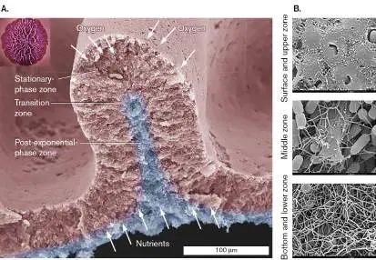
eResearch Activity 5
Can Microbes Survive Trapped for 100 Million Years beneath a Seabed?
How tenacious is microbial life? We know that some resilient species of bacteria and archaea survive and grow in punishing environments situated at the extremes of pH, osmolarity, or temperature. But can microbial life exist deep underneath the seabed in places where nutrients and oxygen exist at vanishingly low levels?
To find out, Yuki Morono (Fig. ERA 5.1 ) and Fumio Inagaki, both of the Japan Agency for Marine-Earth Science and Technology, and Steven D’Hondt of the University of Rhode Island (Fig. ERA 5.1 ) organized a drilling expedition to the South Pacific Gyre, a system of rotating ocean currents east of Australia that is almost devoid of nutrients. It is considered the deadest part of Earth’s oceans. Funded by the Japan Society for the Promotion of Science and the U.S. National Science Foundation, the research team drilled about 80 meters below the seabed (which itself is 2.5 miles below the sea surface) to reach a sediment layer estimated to have formed about 101.5 million years ago. Because the layers of sediment above the collection site were nearly impermeable with very tiny pore sizes (0.02 μm), any bacteria found within the collected sediment were almost certainly trapped shortly after deposition—that is, 100 million years ago. The region is an extremely oligotrophic environment, severely deficient in carbon and energy sources. Despite those exceedingly poor conditions, Morono and colleagues found that microbial life did exist there, albeit at very low numbers (about 200 cells per milliliter) compared to what is found at equivalent sediment depths near the ocean margins (10 7 to 10 9 cells per milliliter). But were those organisms alive, dormant, or dead?
FIGURE ERA 5.1 ■ Yuki Morono (second from left), Steven D’Hondt (second from right), and other expedition scientists huddle over a core sample of sediment taken during the expedition.
FUMITO SHIRAISHI/CREDIT: INTERNATIONAL OCEAN DISCOVERY PROGRAM/JOIDES
RESOLUTION SCIENCE OPERATOR (IODP JRSO); CC-BY OPEN USE
While still aboard ship, the crew used a SYBR Green I stain to identify 6,986 individual microbial cells containing double-stranded DNA, suggesting that the cells were at least alive (Fig. ERA 5.2A and E ). They then asked whether or not those presumably ancient cells were physiologically active—and if so, would they grow? To start, they examined single-cell anabolic activity in each cell by injecting vials with tracer substrates labeled with heavy isotopes of carbon and nitrogen, such as 13 C-acetate, 13 C-glucose, 13 C-bicarbonate, and 15 N-ammonia, and they incubated the mix under microaerobic conditions similar to the sediment environment. (The normal, lighter isotopes for carbon and nitrogen are 12 C and 14 N.) An individual cell that can metabolize those substrates will incorporate the heavy isotopes into macromolecules, and the
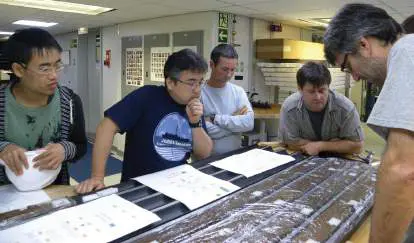
masses of those molecules will increase. Incubations were carried out for 68 days.
FIGURE ERA 5.2 ■ Microscopic analysis of DNA-containing cells and ratio imaging of isotope incorporation. Cells from core samples after 68 days of incubation with 13 C-bicarbonate and 15 N-ammonium (A–D )
and a 13 C, 15 N-amino acid mix (E–H ). Panels A and E show the sampled cells stained with SYBR Green I. Viable cells are green. Color-scale ranges of the 13 C/ 12 C and 15 N/ 14 N ratios are shown at the top and bottom of the color bars. Images in panels D and H are of the same regions in A and E obtained with single cell–targeted ion imaging analysis (NanoSIMS microscopy). Scale bars = 5 μm.
Y. MORONO ET AL. 2020. NAT COMMUN. 11 :3626
The technology subsequently used to detect single-cell incorporation of those isotopes is called nanoscale secondary ion mass spectrometry (NanoSIMS; see Section 2.5 in the printed book). NanoSIMS can measure element and isotope (12 C, 13 C, 14 N, or 15 N) distributions across a tiny (10–50 nm) target area, well within the size of a single bacterial cell.
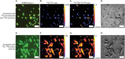
The results shown in Figure ERA 5.2B , C , F , and G illustrate that many of the cells incorporated the added heavy isotopes, indicating that those cells regained metabolic potential. The data also suggested that these ancient cells were potentially capable of growth if given adequate energy sources. The best sources were amino acid mixes, which achieved the highest ratios of 13 C and 15 N incorporation (Fig. ERA 5.2F and G ). The revivable heterotrophic population within the 101.5-million-year-old sediment was 39.6%– 99.1% of the original community.
Figure ERA 5.3 shows the growth of those same organisms over the time of incubation. Initial cell numbers in the sediment were extremely low (100–200 cells per milliliter), but the population responded rapidly to substrate injection by increasing to 1 million cells per milliliter over a period of 68 days. Of the labeled substrates tested, organic (heterotrophic) substrates were generally best at supporting biomass growth.
A curious finding from these studies came from comparing biomass-based growth rates, as determined by the increase in cell numbers, to the generation rates derived from substrate incorporation, which were calculated from ratios of heavy isotope incorporated before and after incubation. The biomass-based specific growth rates were generally higher (faster) than the substrate-based biomass generation rates. The difference can be explained by native organic carbon present in the sediment. Those carbon sources, although minuscule, still appear to be bioavailable; however, unless stimulated by the addition of new substrate, the ancient bacteria won’t use them.
Another curious finding was observed in the ammonia-only experiments. Ammonia assimilation into amino acids requires energy. Nevertheless, ammonia was incorporated into cells even without an added carbon source (data not shown), and the cells multiplied (Fig. ERA 5.3 ). The high ammonia incorporation and biomass increase during those incubations suggests at least a minor contribution of chemolithotrophic ammonia oxidizers in the sediment.
FIGURE ERA 5.3 ■ Growth response to the addition of carbon and nitrogen substrates. Using the 101.5-million-year-old sediment sample, this graph plots cell numbers during incubation. Time 0 indicates cell numbers in the sediment samples before initiating incubation with added substrates. For the samples incubated with carbon substrates (bicarbonate, acetate, glucose, and pyruvate), ammonia was added as the nitrogen source. The incubation labeled “Ammonia” received ammonia as the nitrogen source with no additional carbon. AAmix = amino acid mix.
The scientists then used 16S rRNA gene sequencing to reveal the taxonomy of microbial cells in the sediment over time (Fig. ERA 5.4 ). The community was dominated by bacteria, which included members of Actinobacteria, Bacteroidetes, Firmicutes, Alphaproteobacteria, Betaproteobacteria, Gammaproteobacteria,
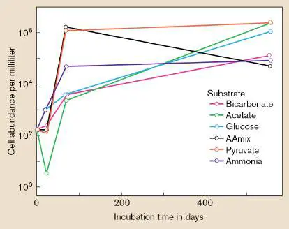
and Deltaproteobacteria phyla. Very few spore-forming genera were found. The researchers also observed that any changes in community composition during the heavy-isotope incubations were unrelated to the substrate added. This finding agrees with the NanoSIMS results suggesting that the microbial community in the sediment can utilize native carbon and nitrogen compounds, but only if the quiescent, or dormant, bacteria become activated by the addition of new substrates.
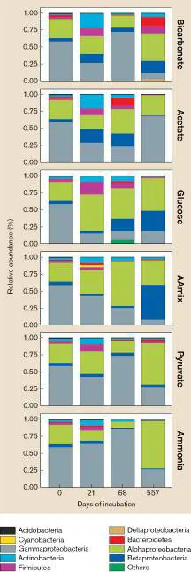
FIGURE ERA 5.4 ■ Community makeup over time with the addition of substrates. The community compositions on incubation day 0 show the compositions before incubation with substrates.
However, the scientists did find that changes in community composition correlated to duration of incubation. Different taxa became activated at different times during incubation, regardless of which substrate was added.
So, what have these microbes been doing for all these millions of years? Are they ancient bacteria that do not replicate but merely repair damaged macromolecules to stay alive? Or have they been dividing extremely slowly, making them distant relatives of ancestors millions of years old? Whatever the answer, these microbes have found a way to persevere during eons of exceedingly harsh periods of food and energy deprivation—another striking example of microbial tenacity.
Further Exploration
How do data from the following papers confirm or refute the idea that these “ancient” organisms are metabolically active and can multiply in the sediment?
Nature Geoscience 8 :299–304 (2015).
https://doi.org/10.1038/ngeo2387.
Science Advances 6 :eaba0697 (2020).
https://doi.org/10.1126/sciadv.aba0697.
Morono, Yuki, Motoo Ito, Tatsuhiko Hoshino, Takeshi Terada, Tomoyuki Hori, et al. 2020. Aerobic microbial life persists in oxic marine sediment as old as 101.5 million years. Nature Communications 11 :3626.
https://doi.org/10.1038/s41467-020-17330-1.
CHAPTER REVIEW
Review Questions
1. Explain the nature of extremophiles, and discuss why these organisms are important.
2. What parameters define any growth environment? 3. List and define the classifications used to describe microbes that grow in different physical growth conditions.
4. What do thermophiles have to do with PCR technology? 5. Why is water activity important to microbial growth? What changes water activity?
6. How do cells protect themselves from osmotic stress? 7. Why do changes in H + concentration affect cell growth? 8. How do acidophiles and alkaliphiles manage to grow at the extremes of pH?
9. If an organism can live in an oxygenated environment, does that mean the organism uses oxygen to grow? If an organism can live in an anaerobic environment, does that mean it cannot use oxygen as an electron acceptor? Why or why not?
10. What happens when a cell exhausts its available nutrients?
11. List and briefly explain the various means by which humans control microbial growth. What is a D-value? 12. How do microbes prevent the growth of other microbes?
Thought Questions
1. Given a natural lake environment with 100 species of bacteria, why does the species with the fastest generation time not overwhelm the others? Or does it? 2. Escherichia coli is a facultative species, able to grow with or without oxygen. What would it take to make this organism a strict anaerobe?
3. Two spore formers— Geobacillus thermophilus and Bacillus coagulans —have D-values at 121°C of 5 minutes and 0.07 minute, respectively. How could the spores from these organisms have such different D-values? Hint: Find the organisms’ optimal growth temperatures.
4. Phage therapy is touted by some as a solution to antibiotic resistance. Discuss three possible problems we could encounter with phage therapy and how we could overcome them.
5. With respect to bacteria, is the physiological effect of hydrochloric acid (HCl) at pH 4 the same as that of an organic acid at pH 4?
Key Terms
acidophile (176)
aerobe (179)
aerobic respiration (180) aerotolerant anaerobe (181) alkaliphile (176)
anaerobe (179)
anaerobic (179)
anaerobic respiration (181) antibiotic (194)
antisepsis (187)
bactericidal (187)
bacteriostatic (187)
barophile (172)
biocontrol (195)
bioremediation (192)
compatible solute (174)
decimal reduction time (D-value) (187) disinfection (187)
electron transport system (ETS) (179) eutrophication (185)
extremophile (166)
facultative anaerobe (179, 181) fermentation (fermentative metabolism) (181) germicidal (187)
halophile (173)
heat-shock response (171) hyperthermophile (170)
laminar flow biological safety cabinet (189) lyophilization (189)
mesophile (169)
microaerophilic (181)
neutralophile (176)
niche (167)
osmolarity (173)
pasteurization (189)
piezophile (172)
probiotic (195)
programmed cell death (183) psychrophile (169)
psychrotroph (169)
sanitation (187)
sterilization (187)
strict aerobe (180)
strict anaerobe (181)
thermophile (170)
water activity (173)
Recommended Reading
Ahn, Do-Hwan, Ki-Young Lee, Sang Jae Lee, Sung Jean Park, Hye-Jin Yoon, et al. 2017. Structural analyses of the MazEF4 toxin-antitoxin pair in Mycobacterium tuberculosis provide evidence for a unique extracellular death factor. Journal of Biological Chemistry 292 : 18832–18847.
https://doi.org/10.1074/jbc.M117.807974.
Berryhill, Brandon A., Douglas L. Huseby, Ingrid C. McCall, Diarnaid Hughes, and Bruce R. Levin. 2021. Evaluating the potential potency and limitations of a phage for joint antibiotic and phage therapy of Staphylococcus aureus infections. Proceedings of the National Academy of Sciences USA 118:e2008007118. https://doi.org/10.73/pnas.2008007118.
Borin, Joshua M., Sarit Avrani, Jeffery Barrick, Katherine Petrie, and Justin Meyer. 2021. Coevolutionary phage training leads to greater bacterial suppression and delays evolution of phage resistance. Proceedings of the National Academy of Sciences USA 118 :e2104592118.
Cavallo, Francis M., Lorea Jordana, Alexander W. Friedrich, Corinna Glasner, and Jan Maarten van Dijl. 2021.
Bdellovibrio bacteriovorus: A potential ‘living antibiotic’ to control bacterial pathogens. Critical Reviews in Microbiology 47:630–646. https://doi.org/10.1080/1040841X.2021.1908956. Coker, James A. 2019. Recent advances in understanding extremophiles. F1000Research 2019, 8 (F1000 Faculty Rev):1917. https://doi.org/10.12688/f1000research.20765.1. Furfaro, Lucy L., Barbara J. Chang, and Matthew S. Payne. 2018. Applications for bacteriophage therapy during pregnancy and the perinatal period. Frontiers in Microbiology 8 :2660. https://doi.org/10.3389/fmicb.2017.02660.
Gohara, David W., and Mee-Ngan F. Yap. 2017. Survival of the drowsiest: The hibernating 100S ribosome in bacterial stress management. Current Genetics 64 : 753–760.
https://doi.org/10.1007/s00294-017-0796-2.
Hori, Hiroyuki. 2019. Regulatory factors for tRNA modifications in extreme-thermophilic bacterium Thermus thermophiles.
Frontiers in Genetics 10 : article 204.
https://doi.org/10.3389/fgene.2019.00204.
Khademian, Maryam, and James A. Imlay. 2021. How microbes evolved to tolerate oxygen. Trends in Microbiology 29:428–440. https://doi.org/10.1016/j.tim.2020.10.001.
Luo, Chengwei, Luis M. Rodriguez-R, Eric R. Johnston, Liyou Wu, Lei Cheng, et al. 2014. Soil microbial community responses to a decade of warming as revealed by comparative metagenomics. Applied and Environmental Microbiology 80:1777–1786.
Poli, Annarita, Ilaria Finore, Ida Romano, Alessia Gioiello, Licia Lama, et al. 2017. Microbial diversity in extreme marine habitats and their biomolecules. Microorganisms 5 :25. https://doi.org/10.3390/microorganisms5020025.
Sapers, Haley M., Jennifer Ronholm, Isabelle Raymond-Bouchard, Raven Comrey, Gordon R. Osinski, et al. 2017. Biological characterization of microenvironments in a hypersaline cold spring Mars analog. Frontiers in Microbiology 8 :2527. https://doi.org/10.3389/fmicb.2017.02527.
Shuryak, Igor, Vera Y. Matrosova, Elena K. Gaidamakov, Rok Tkav, Olga Grichenko, et al. 2017. Microbial cells can cooperate to resist high level chronic ionizing radiation. PLoS One 12 :e0189261.
Vaishampayan, Ankita, Anne de Jong, Darren J. Wight, Jan Kok, and Elisabeth Grohmann. 2017. A novel antibacterial coating represses biofilm and virulence-related genes in methicillin-resistant Staphylococcus aureus. Frontiers in Microbiology 9 :221. https://doi.org/10.3389/fmicb.2018.00221.
Glossary
extremophile An organism that grows only in an extreme environment; that is, an environment including one or more conditions that are “extreme” relative to the conditions for human life. niche An organism’s environmental requirements for existence and its relations with other members of the ecosystem.
mesophile An organism with optimal growth between 20°C and 40°C. psychrophile An organism with optimal growth at temperatures below 20°C. psychrotroph A cold-resistant organism that can grow at temperatures between 0°C and 7°C but shows optimal growth between 20°C and 35°C.
thermophile An organism adapted for optimal growth at high temperatures, usually 50°C or higher.
hyperthermophile Also called extreme thermophile. An organism adapted for optimal growth at extremely high temperatures, generally above 80°C, and as high as 121°C.
heat-shock response A coordinated response of cells to higher-than-normal temperatures. It includes changes in the membrane and expression of heat-shock genes.
barophile Also called piezophile. An organism that requires high pressure to grow.
piezophile Also called barophile. An organism that requires high pressure to grow.
water activity A measure of the water that is not bound to solutes and is available for use by organisms.
halophile An organism that requires a high extracellular sodium chloride concentration for optimal growth.
osmolarity A measure of the concentration of solute molecules in solution. compatible solute A small molecule that does not disrupt normal cell metabolism even at high intracellular concentrations.
neutralophile An organism with an optimal growth range in environments between pH 5 and 8.
acidophile An organism that grows fastest in acid (generally defined as below pH 5).
alkaliphile An organism that grows fastest in alkali (generally defined as above pH 9).
anaerobic Lacking oxygen.
anaerobe An organism that grows in an environment lacking oxygen. aerobe An organism that grows only in the presence of oxygen. electron transport system (ETS) or electron transport chain (ETC) Also called cytochrome system. A series of membrane-embedded proteins that converts the energy of redox reactions into a proton potential.
aerobic respiration The use of oxygen as the terminal electron acceptor in an electron transport chain. A proton gradient is generated and used to drive ATP synthesis.
strict aerobe An organism that performs aerobic respiration and can grow only in the presence of oxygen.
strict anaerobe An organism that cannot grow in the presence of oxygen. anaerobic respiration The use of a molecule other than oxygen as the final electron acceptor of an electron transport chain.
fermentation (fermentative metabolism)
Also called fermentative metabolism. 1. The production of ATP via substrate-level phosphorylation, using organic compounds as both electron donors and electron acceptors. 2. Industrial fermentation is the production of microbial products that are made by microbes grown in fermentation vessels; it may include respiratory metabolism to maximize microbial growth. facultative anaerobe An organism that can grow in either the presence or absence of oxygen.
aerotolerant anaerobe An organism that does not use oxygen for metabolism but can grow in the presence of oxygen.
microaerophilic Requiring oxygen at a concentration lower than that of the atmosphere, but unable to grow in high-oxygen environments. programmed cell death Cell death mediated by a regulated intracellular process. eutrophication A sudden increase of a formerly limiting nutrient in an aquatic environment, leading to overgrowth of algae and grazing bacteria and subsequent oxygen depletion.
sterilization The destruction of all cells, spores, and viruses on an object. disinfection The removal of pathogenic organisms from inanimate surfaces. antisepsis The removal of pathogens from living tissues.
sanitation The safe disposal of wastes hazardous to humans.
bacteriostatic Having the ability to inhibit the growth of bacterial cells. bactericidal Having the ability to kill bacterial cells.
germicidal Able to kill cells but not spores.
decimal reduction time (D-value)
The length of time it takes for a treatment to kill 90% of a microbial population, and hence a measure of the efficacy of the treatment.
pasteurization The heating of food at a temperature and time combination that will kill spore-like structures of Coxiella burnetii. lyophilization Also called freeze-drying. The removal of water from food, by freezing under vacuum, to limit microbial growth.
laminar flow biological safety cabinet An air filtration appliance that removes pathogenic microbes from within the cabinet.
bioremediation The use of microbes to detoxify environmental contaminants. antibiotic A molecule that can kill or inhibit the growth of selected microorganisms.
probiotic A food or nutritional supplement that contains live microorganisms and aims to improve health by promoting beneficial bacteria.
biocontrol Use of a beneficial organism to control a harmful organism.