Chapter introduction
Food products that have undergone controlled microbial growth are called fermented foods.
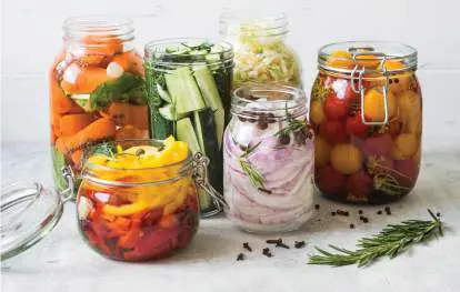
Fermented food products such as yogurt and kimchi accumulate lactic and acetic acids, which limit microbial catabolism and preserve protein content.
SENTELIA/SHUTTERSTOCK
Microbes have nourished humans for centuries, generating cheese, bread, wine and beer, tempeh, and soy sauce. Yet from the moment of harvest, microbes on the food’s surface or from the air colonize the food. The need for food preservation has led to drying, salting, smoking, and adding spices, all of which retard microbial growth. The principles of food microbiology extend to a growing field, industrial microbiology. Industrial microbiology includes the development of microbial antibiotics and enzymes that are produced by engineered microbes in giant fermentors. For example, the first major new drug for COVID-19, called molnupiravir, was synthesized in 2021 using enzymes engineered from microbes by Novozymes, a global biotechnology company. Microbial enzymes represent a growing source of industrial products made by cultivating In Chapter 16 we discuss how the many kinds of microbial biochemistry presented in Chapters 13–15 give rise to the characteristics of food that are so familiar—from the taste of cheese and chocolate to the rising of bread dough and the physiological effects of alcoholic beverages. Industrial research continues to improve food through food biochemistry, discovering the molecular basis for the flavor and texture of microbial foods; food preservation, eliminating undesirable microbial decay; and food engineering, improving the quality, shelf life, and taste of microbial foods. The lessons learned from food engineering and other biotechnologies now lead to extraordinary medical products such as the coronavirus mRNA vaccines and lentiviral cures for cancer.
16.1 Microbial Foodsnot assigned
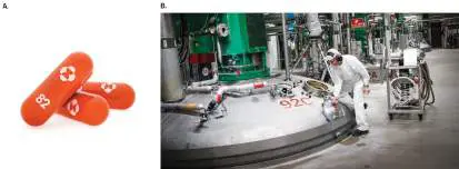
For thousands of years, long before microbes were discovered as such, their metabolism was a hidden essential part of human life and commerce. Microbial fermentation generates foods such as cheeses, pickles, and kimchi, as well as ethanolic beverages such as wine and beer. Today, the fermentation industry refers to the culturing of microbes to make products for market, including highly engineered products such as enzymes. Commercial products are made by microbes grown in giant fermentation vessels (Fig. 16.1; discussed in Section 16.5). Industrial “fermentation” generally uses respiratory metabolism as well as fermentation to maximize microbial growth. Companies such as Novozymes now earn hundreds of millions of dollars in annual revenues from microbial products ranging from contact lens cleansing agents to agricultural chemicals. FIGURE 16.1 ■ An experimental drug for COVID-19 is synthesized using commercial microbial enzymes. A.
Capsules of the antiviral agent molnupiravir, manufactured by Merck. Synthesis of the compound requires enzymes produced by microbial cultures. B. An industrial fermentation apparatus for growing microbes to produce enzyme products at the world’s largest enzyme manufacturing plant, Novozymes, in Kalundborg, Denmark.
QUALITY STOCK/ALAMY STOCK PHOTO
©NOVOZYMES, 2019
The fermentation industry originated from long-standing microbial associations with human food. Until the relatively recent invention of steam-pressure sterilization, all foods contained live microbes. Microbial metabolism could spoil food—or it could improve the food by adding flavor, preserving valuable nutrients, and preventing growth of pathogens. As we described in Chapter 1, one of the first great microbiologists, Louis Pasteur (1822–1895), began his career as a chemist investigating fermentation in winemaking. Certain kinds of microbial bodies are eaten as food, especially mushrooms. Agaricus bisporus is the mushroom variety most commonly sold in the United States as button mushrooms and portobellos (Fig. 16.2). Other edible microbes include various types of algae, such as spirulina. These foods can provide important sources of protein, vitamins, and minerals. Still other microbes, such as yeasts and lactic acid bacteria, ferment food substrates to form valuable food products, such as bread, cheese, and alcoholic beverages.
FIGURE 16.2 ■ Commercial mushroom production. Farming of Agaricus bisporus mushrooms on horse manure
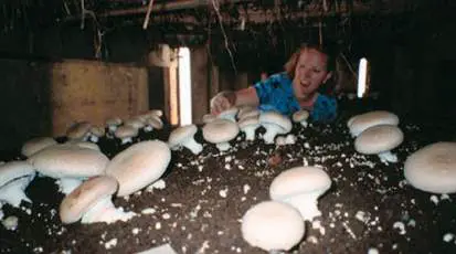
compost by Penn State graduate student Kelly Ivors.
KELLY IVORS
Edible Fungi
Fungal fruiting bodies, multicellular reproductive structures that generate spores, are commonly known as mushrooms (see Chapter 20). Mushrooms offer a flavorful source of protein and minerals. The protein content of edible mushrooms can be as high as 25% dry weight, comparable to that of whole milk, and includes all essential dietary amino acids.
In the children’s classic Homer Price, by Robert McCloskey, settlers on the Ohio frontier save themselves from starvation when they discover “forty-two pounds of edible fungus, in the wilderness a-growin’.” Mushrooms aided the survival of preindustrial humans but killed those unlucky enough to consume varieties that were poisonous. Less than 1% of mushrooms are poisonous, but those few are deadly, such as the amanita, or “destroying angel,” which produces toxins including the RNA polymerase inhibitor alpha-amanitin. People who ingest the amanita typically die of liver failure. Other kinds of mushrooms actually invite consumption in order to disperse their spores. For example, the underground truffles prized in European cooking produce odorant molecules that attract animals to dig them up. Truffle odorants include sex pheromones such as androstenol, found in human perspiration. Many varieties of edible mushrooms are farmed and marketed for human fare. Button mushrooms are harvested at an early stage, whereas portobellos (the same species) are harvested later, when the gills are fully exposed and some of the moisture has evaporated. The decrease in moisture in portobellos concentrates the flavor and gives them a dense, meaty texture; the mushrooms are often served in gourmet sandwiches as a vegetarian alternative to hamburger. Agaricus culture was first developed around 1700 in France, where the mushrooms were grown in underground caves. In modern mushroom farms, Agaricus mushrooms are cultured on composted horse manure or chicken manure in chambers controlled for temperature and humidity (Fig. 16.2).
Other mushroom varieties, from China and Japan, are grown on logs or wooden blocks. Wood-grown mushrooms include the strong-flavored Lentinula edodes (black forest mushrooms, or shiitake), Pleurotus (oyster mushrooms), and Flammulina velutipes (enoki mushrooms), with long, thin, white stalks and delicate flavor. Yeasts (single-celled fungi) can provide a supplemental source of protein and vitamins, most importantly vitamin B 12. Historically, yeasts such as Saccharomyces species have been grown to high concentration in fermented milks and grain beverages. Traditional beers contained only a low percentage of alcohol with a thick suspension of nutrient-rich yeasts.
Edible Algae and Cyanobacteria
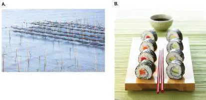
Several kinds of seaweed (marine algae) are cultivated, most notably in Japan. The red alga Porphyra, a eukaryotic “true alga” (discussed in Chapter 20), forms large, multicellular fronds cultured for nori (Fig. 16.3). Nori is best known for its use in wrapping rice, fish, and vegetables to form sushi. For nori production, the red algae are grown as “seeds,” or starter cultures, in enclosed tanks. The starter cultures are then distributed on nets in a protected coastal area, usually an estuary. The cultures grow until they hang heavy from the nets, when they are harvested for processing into sheets. The sheets are toasted, turning dark green. Other edible seaweeds include wakame and kombu, forms of kelp.
FIGURE 16.3 ■ Nori production for sushi. A. Nori grows from nets in seawater. B. Toasted sheets of nori are used to wrap rice, vegetables, and fish to make sushi.
XVISION/GETTY IMAGES
STOCKFOOD LTD./ALAMY STOCK PHOTO
Few bacteria are edible as isolated organisms, mainly because their small cells contain a relatively high proportion of DNA and RNA. The high nucleic acid content is a problem because nucleic acids contain purines, which the human digestive system converts to uric acid. Because we humans lack the enzyme urate oxidase, the uric acid cannot be metabolized. Consumption of more than 2 grams per day of nucleic acids causes uric acid to precipitate, resulting in painful conditions such as gout and kidney stones. For this reason, most bacteria can be consumed only as a minor component of food mass, as in fermented foods.
An exception is the cyanobacterium Spirulina, whose purine content is low enough to include as a modest part of the human diet. Spirulina consists of spiral-shaped cells that grow photosynthetically in freshwater. Spirulina is sold as a food additive rich in protein, vitamin B 12, and minerals. It also contains antioxidant substances that may prevent cancer. Spirulina is grown with illumination in special ponds lined for food production. The final
product is collected and vacuum-dried to form a dark green powder of flour-like consistency.
In the food industry, Spirulina is classified as a form of single-celled protein, a term for edible microbes of high food value. Other kinds of single-celled protein foods include eukaryotes such as yeast and algae.
Fermented Foods: An Overview
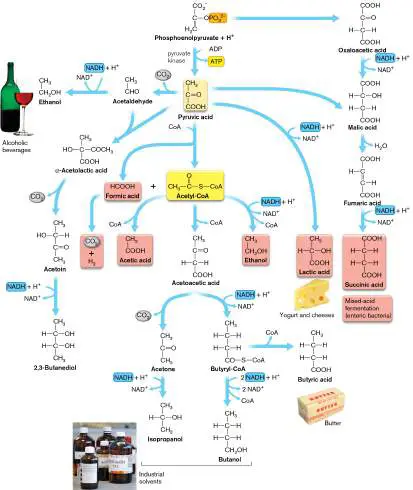
Virtually all human cultures have developed varieties of fermented foods, food products that are modified biochemically by microbial growth. In most cases, the modification involves true fermentation, a process of metabolism in which the electrons transferred from an electron donor are returned to the organic substrates, generating molecules that are rearranged. Recall from Chapter 13 how glucose catabolism leads to pyruvate, yielding energy in the form of ATP (see Fig. 13.15). Electrons are transferred to NADH, but fermentation returns the electrons from NADH to pyruvate, generating various organic products (see Fig. 13.20). The absence of a strong electron acceptor (O 2) limits the breakdown of substrate and thus preserves food value.
The purposes of food fermentation include the following: Preserve food. Certain microbes, particularly the lactobacilli, metabolize only a narrow range of nutrients before their waste products build up and inhibit further growth. Typically, the waste fermentation products that limit growth are carboxylic acids, ammonia (alkaline), or alcohol. Buildup of these substances renders the product stable for much longer than the original food substrate; for example, sauerkraut from cabbage.
Improve digestibility. Microbial action breaks down fibrous macromolecules and makes the food easier for humans to digest. Meat and vegetable products are tenderized by fermentation.
Add nutrients and flavors. Microbial metabolism generates vitamins, particularly vitamin B 12 and riboflavin (vitamin B 2). Microbial action also generates flavor molecules that lead to the diverse tastes of foods such as cheeses and chocolate (discussed in Section 16.2).
Different societies have devised thousands of different kinds of fermented foods. Examples are given in Table 16.1. Fermented foods that are produced commercially include dairy products such as cheese and yogurt, soy products such as miso (from Japan) and tempeh (from Indonesia), vegetable products such as sauerkraut and kimchi, and various forms of cured meats and sausages. Alcoholic beverages are made from grapes and other fruits (wine), grains (beer and liquor), and cacti (tequila). Other kinds of foods require microbial treatment for special purposes, such as leavening by yeast (for bread) or cocoa bean fermentation (for chocolate). Besides commercial production, numerous fermented products are homemade by traditional methods thousands of years old. Such products are known as “traditional fermented foods.” Occasionally, a traditional fermented food enters commercial production and becomes widespread. For example, soy sauce, a traditional Japanese product, was marketed by the Kikkoman company and achieved global distribution in the twentieth century.
TABLE Fermented Foods and Beverages 16.1
Product Description Microbial genera (origin)
Acid fermentation of dairy products, meat, and fish Buttermilk Bovine milk; lactic Lactococcus (Asia, fermented Europe)
Yogurt Bovine milk; lactic Lactobacillus, (Asia, fermented and Streptococcus Europe) coagulated Kefir Bovine or sheep’s milk; Lactobacillus, (Russia) mixed fermentation, Streptococcus, acid with some yeasts, others alcoholic Sour cream Bovine cream; lactic Lactococcus (Asia, fermented Europe)
Cheese Milk (bovine, sheep, or Acid fermentation: (Asia, goat); lactic fermented, Lactobacillus, Europe) coagulated, and Streptococcus, pressed; in some cases Propionibacterium cooked; mold ripened Mold ripening: (spiked or coated) Penicillium Sausage Ground beef and/or pork Lactobacillus, (Asia, encased with starter Pediococcus, Europe) culture; lactic Staphylococcus, fermented, then dried others or smoked Fermented Many kinds of fish; mixed Halotolerant fish fermentation, acid and bacteria and (Africa, amines produced haloarchaea Asia)
Acid fermentation of vegetables Tempeh Soybean cakes; fungal Rhizopus (Indones fermentation oligosporus ia)
Miso Soy and rice paste; fungal Aspergillus (Japan) fermentation Soy sauce Extract of soy and wheat; Aspergillus, (China) fungal fermentation, followed by brined, bacterial halotolerant fermentation bacteria and haloarchaea Kimchi Cabbage, peppers, and Leuconostoc, other (Korea) other vegetables, with bacteria fish paste, brined; container is buried Sauerkraut Cabbage; fermented, Leuconostoc, (Europe) making lactic and Pediococcus, acetic acids, ethanol, Lactobacillus and CO 2 Pickled Cucumbers, carrots, fish; Leuconostoc, foods brined, then fermented Pediococcus, (Asia) Lactobacillus Kenkey Maize; fermented, Unknown (western wrapped in banana Africa) leaves and cooked Chocolate Cocoa beans; soaked and Lactobacillus, (South fermented before Bacillus, America) processing to chocolate Saccharomyces Alkaline fermentation Pidan Duck eggs; coated in lime Bacillus (China, (CaO), aged, producing Japan) ammonia and sulfur odorants Natto Whole soybeans; Bacillus natto (China, fermented Japan)
Dawadawa Locust beans; fermented Bacillus (Africa)
Ogiri Melon seed paste; Bacillus (Africa) fermented Leavened bread dough Yeast Ground grain; dough Saccharomyces breads leavened by yeast (Asia, Europe)
Sourdough Ground grain; dough Saccharomyces, (Egypt) leavened by starter Torulopsis, culture from previous Candida dough Injera Ground teff grain; dough Candida (Ethiopia leavened and ) fermented 3 days by organisms from the grain Alcoholic fermentation Wine (Asia, Grape juice; yeast Saccharomyces, Europe) fermented, followed by Oenococcus malolactic fermentation Beer (Asia, Barley and hops; yeast Saccharomyces Europe, fermented Africa)
Sake Rice extract; yeast Saccharomyces (Japan) fermented Tequila Blue agave; yeast Saccharomyces (Mexico) fermented and distilled Whiskey Barley or other grains or Saccharomyces (United potatoes; fermented Kingdom and distilled )
The nature of fermented foods depends on the quality of the fermented substrate, as well as on the microbial species and the type of biochemistry performed. Traditional fermented foods usually depend on indigenous microbiota (that is, microbes found naturally in association with the food substrate) or on starter cultures derived from a previous fermentation, as in yogurt or sourdough fermentation. Commercial food-fermenting operations use highly engineered microbial strains to inoculate their cultures, although in some cases indigenous microbiota still participate. For example, wines and cheeses aged in the same caves over centuries often include fermenting organisms that persist in the air and the containers used.
Major classes of fermentation reactions are summarized in Figure 16.4(see also Fig. 13.20). The most common conversions involve anaerobic fermentation of glucose. Recall from Chapter 13 how glucose is fermented to lactic acid (lactic acid fermentation). This fermentation occurs in cheeses and sausages, primarily by lactic acid bacteria such as Lactobacillus.
FIGURE 16.4 ■ Major chemical conversions in fermented foods.
PICTURE PARTNERS/ALAMY STOCK PHOTO
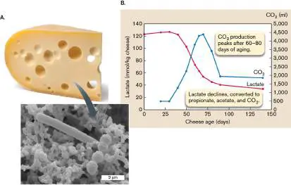
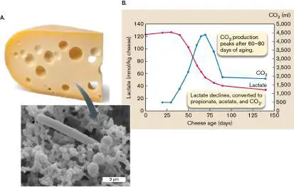
In Swiss cheese, a second stage of lactic acid fermentation produces propionic acid (propionic acid fermentation; Fig. 16.5). This fermentation, by Propionibacterium freudenreichii, generates the distinctive flavor of Swiss and related cheeses. As the cheese ages, first the lactic acid bacteria, such as Lactobacillus helveticus, convert sugars to lactic acid (Fig. 16.5B ). Then P. freudenreichii starts to break down lactate further to propionate, acetate, and CO 2 —a reaction that yields ATP. The CO 2 gas forms the bubbles, or “eyes,” that are characteristic of Swiss cheese (Fig. 16.5A).
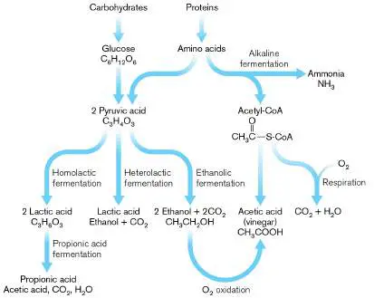
FIGURE 16.5 ■ Propionic acid fermentation within Swiss cheese. A. Emmentaler (Swiss) cheese with “eyes” is made by a bacterial community (inset). Bacterial species shown here include Lactobacillus helveticus (rods, 2.0–4.0 μm in length) and Streptococcus thermophilus (cocci). B. As Swiss cheese ages, lactate is converted to propionate, acetate, and CO 2 (the gas that forms the eyes).
SVETLANA FOOTE/ALAMY STOCK PHOTO
SCIMAT/SCIENCE SOURCE
Some kinds of vegetable fermentation, as in sauerkraut, involve production of lactic acid and CO 2, as well as small amounts of acetic acid and ethanol. This heterolactic fermentation is conducted primarily by Leuconostoc. Fermentation to ethanol plus carbon dioxide without lactic acid (ethanolic fermentation, also called alcoholic fermentation) is conducted by yeast during bread leavening and production of alcoholic beverages.
In some food products, particularly those fermented by Bacillus species, proteolysis and amino acid catabolism generate ammonia in amounts that raise pH (alkaline fermentation). For example,
alkaline fermentation forms the soybean product natto. Other products require the growth of mold, such as the mold-spiked Roquefort cheese and the soy product tempeh. Mold growth requires some oxygen for aerobic respiration. Respiration must be limited, however, to avoid excessive decomposition of food substrate and loss of food value.
Note that the conversions cited here include only the major reactions in achieving the food product. In addition, thousands of minor or secondary reactions occur, some of which produce tiny amounts of potent odorants and flavors. While these flavor molecules have less nutritional consequence than the main fermentation products have, they provide the complex, “sophisticated” taste for which fine cheeses, wines, and soy products are known.
Thought Questions
16.1 Why do the lipid components of food experience relatively little breakdown during anaerobic fermentation?
16.2 Why does oxygen allow excessive breakdown of food, compared with anaerobic processes?
To Summarize
Edible fungi are protein-rich foods, including yeasts, mushrooms, and truffles (fruiting bodies).
Edible algae include nori (toasted red algae, used to wrap sushi), as well as wakame and kombu.
Spirulina is an edible cyanobacterium, a source of single-celled protein. Most bacteria, however, are inedible in isolation because of their high concentration of nucleic acids. Anaerobic fermentation of food enhances preservation, digestibility, nutrient content, and flavor. Breakdown of lipids and peptides is limited under anaerobic conditions of fermentation.
Acid fermentation of food generates organic acid fermentation products that lower pH, such as lactate and propionate.
Alkaline fermentation of food produces ammonia, which increases the food pH.
Ethanolic fermentation produces ethanol and carbon dioxide.
Glossary
fermentation industry Commercial production of microbial products that are made by microbes grown in fermentation vessels; it may include respiratory metabolism to maximize microbial growth. nori A Japanese food obtained from the red algae Porphyra species. single-celled protein An edible microbe of high food value, such as Spirulina or some yeasts.
fermented food Food products that are biochemically modified by microbial growth.
indigenous microbiota Microbes found naturally in a particular location, often in association with a food substrate.
lactic acid fermentation A fermentation reaction that generates lactic acid from reduction of pyruvic acid.
propionic acid fermentation The fermentation of lactic acid to propionic acid by Propionibacterium species; used in the production of Swiss cheese.
heterolactic fermentation A fermentation reaction in which the products are lactic acid, ethanol, and CO 2.
ethanolic fermentation Also called alcoholic fermentation. A fermentation reaction yielding 2 ethanol and 2CO 2 as products.
alkaline fermentation Bacterial fermentation in conjunction with proteolysis and amino acid catabolism that generates ammonia in amounts that raise pH.
Fig. 13.15 FIGURE 13.15 ■ From glucose to pyruvate: three pathways. The Embden-Meyerhof-Parnas (EMP) pathway of glycolysis, the Entner-Doudoroff (ED) pathway, and the pentose phosphate pathway (PPP) catabolize carbohydrates by related but different routes.
Fig. 13.20
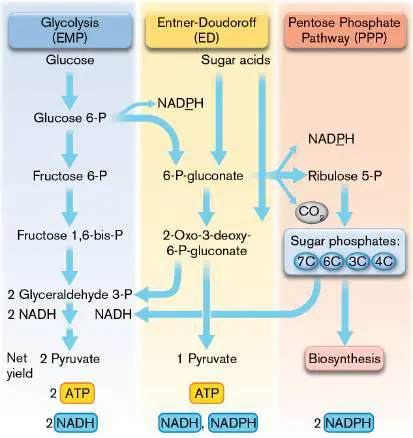
FIGURE 13.20 ■ Fermentation pathways. Alternative pathways from pyruvate and phosphoenolpyruvate to end products, many of which we use for food or industry. Different species conduct different portions of the pathways shown.
FOOD COLLECTION/SUPERSTOCK
AGE FOTOSTOCK/ALAMY STOCK PHOTO
LEE HACKER/ALAMY
D. HURST/ALAMY
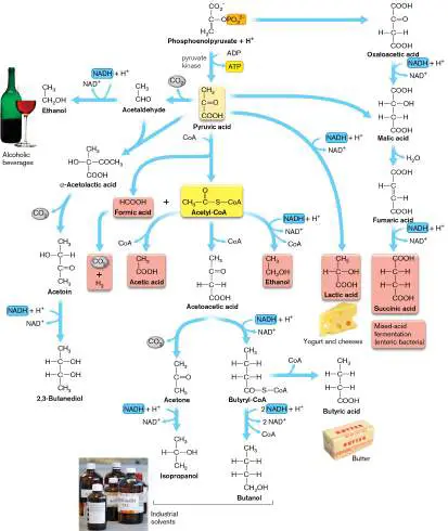
Fig. 13.20 FIGURE 13.20 ■ Fermentation pathways. Alternative pathways from pyruvate and phosphoenolpyruvate to end products, many of which we use for food or industry. Different species conduct different portions of the pathways shown.
FOOD COLLECTION/SUPERSTOCK
AGE FOTOSTOCK/ALAMY STOCK PHOTO
LEE HACKER/ALAMY
D. HURST/ALAMY
16.2 Acid-Fermented and Alkali- Fermented Foodsnot assigned
Many food fermentations produce acids or bases. An acid or base serves as an effective preservative because the pH change is unlikely to be reversed, and because animal or plant bodies grown at near-neutral pH are unlikely to support growth of acidophiles or alkaliphiles, which grow at extreme pH conditions.
Acid Fermentation of Dairy Products
The major organic components of cow’s milk are butterfat (about 4% unless skimmed), protein (3.3%), and the sugar lactose (4.7%). The conversion of milk to solid or semisolid fermented products dates far back in human civilization. The practice of milk fermentation arose among herders who collected the milk of their pack animals but had no way to prevent the rapid growth of bacteria. The milk had to be stored in a portable container such as the stomach of a slaughtered animal. After hours of travel, the combined action of lactic acid– producing bacteria and stomach enzymes caused the coagulation of milk proteins into curd. The curd naturally separates from the liquid portion, called whey. Both curds and whey can be eaten, as in the nursery rhyme “Little Miss Muffet.” The curds, however, are particularly valuable for their concentrated protein content. Curd formation. A cheese is any milk product from a mammal (usually cow, sheep, or goat) in which the milk protein coagulates to form a semisolid curd. Curd formation results from acidification, usually as a result of the microbial production of lactic acid; see Figure 16.4, and also Chapter 13, Figure 13.20.
Milk starts out at about pH 6.6, very slightly acidic. At this pH, the milk proteins are completely soluble in water; otherwise, they would clog the animal’s udder as the milk came out. Fermentation generally begins with bacteria such as Lactobacillus and Streptococcus (Fig. 16.5A, inset). As bacteria ferment lactose to lactic acid, the pH starts to decline. Lactic acid has a dissociation constant (p K a) of 3.9, which means that half of the molecules will still be protonated at pH 3.9. The low p K a of lactic acid allows greater acidification than with other fermentation products, such as acetate (p K a = 4.8). Thus, lactic acid rapidly acidifies the milk product to levels that halt further growth of bacteria. Halting bacterial growth minimizes the oxidation of amino acids, thereby maintaining food quality.
Milk contains micelles (suspended droplets) of hydrophobic proteins called caseins. As the pH of milk declines below pH 5, the acidic amino acid residues of caseins become protonated, eventually destabilizing the tertiary structure. As the casein molecules unfold (or “denature”), they expose hydrophobic residues that regain stability by interacting with other hydrophobic molecules. The intermolecular interaction of caseins generates a gel-like network throughout the milk, trapping other substances, such as droplets of butterfat. This protein network generates the semisolid texture of yogurt, a simple product of milk acidified by lactic acid bacteria.
In most kinds of cheese formation, an additional step of casein coagulation is accomplished by proteases such as rennet. Rennet derives from the fourth stomach of a calf, although modern versions are made by genetically engineered bacteria. Calf rennet includes two proteolytic enzymes: chymosin and pepsin. Chymosin specifically cleaves casein into two parts, one of which is charged and water-soluble, the other hydrophobic. The hydrophobic portion forms a curd that is firmer than intact casein and results in the harder texture of solid cheeses. The water-soluble portion, about one-third of the total casein, enters the whey and is lost from the curd. Processing of some cheese varieties includes exposure to high temperature, which denatures even the whey protein, so it is retained in the curd. Varieties of cheese. An extraordinary number of cheese varieties have been devised (Fig. 16.6). These fall into several categories on the basis of particular steps in their production.
FIGURE 16.6 ■ Cheese varieties. A. Cottage cheese, an unripened perishable cheese. B. Emmentaler Swiss cheese, with eyes produced by carbon dioxide fermentation. C. Feta cheese, a soft cheese from goat’s milk, preserved in brine. D. Roquefort, a medium-hard cheese ripened by spiking with Penicillium roqueforti.
SVETLANA FOOTE/ALAMY STOCK PHOTO
DORLING KINDERSLEY LTD/ALAMY STOCK PHOTO
MAXIMILIAN STOCK LTD./JUPITER IMAGES
BARMALINI/SHUTTERSTOCK
Soft, unripened cheeses , such as cottage cheese and ricotta, are coagulated by bacterial action, without rennet. The curd is cooked slightly, and the whey is partly drained, but their water content is 55% or greater. These cheeses spoil easily; there are no steps of aging, or ripening.
Semihard, ripened cheeses , such as Muenster and Roquefort, include rennet for firmer coagulation, and the curd is cooked down to a water content of 45%–55%. The cheese is aged for several months.
Hard cheeses , such as Swiss cheese and cheddar, are concentrated to even lower water content. Extra-hard varieties, such as Parmesan and Romano, have a water content as low as 20%. These cheeses are aged for many months, even several years.
Brined cheeses , such as feta, are permeated with brine (concentrated salt), which limits further bacterial growth and develops flavor. Harder cheeses, such as Gouda, may be brined at the surface.
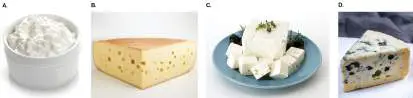
Mold-ripened cheeses are inoculated with mold spores that germinate and grow during the ripening, or aging, process to contribute texture and flavor. The mold may be inoculated on the surface, to form a crust (as in Brie and Camembert), or it may be spiked deep into the cheese (as in blue cheese or Roquefort). Cheese production. Commercial production of cheese involves a standard series of steps (Fig. 16.7). At each of these steps, choices of treatment lead to very different varieties. Key steps are illustrated in Figure 16.8.
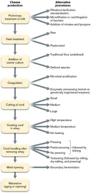
FIGURE 16.7 ■ Flowchart for cheese production. The alternative procedures shown on the right produce different varieties.
FIGURE 16.8 ■ Cheese production. A. Milk is poured into a fermentation tub with a bacterial starter culture and rennet. B. The milk curd is cut, or “cheddared,” a process that drains out the whey. C. The curds are shaped in round molds to solidify. D. The
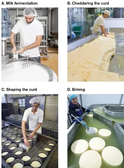
solidified curds are floated in brine. The cheese then dries and ripens on the shelf.
ROSSHELEN EDITORIAL/ALAMY STOCK PHOTO
JUICE IMAGES/ALAMY STOCK PHOTO
PHOTO BY OLIVER RING/IMAGE BROKER/SHUTTERSTOCK
ITAR-TASS NEWS AGENCY/ALAMY STOCK PHOTO
In the first step, the milk is filtered to remove particulate objects, such as straw, and microfiltered or centrifuged to remove potentially pathogenic bacteria and spores. Most modern production includes flash pasteurization (brief heating to 72°C; discussed in Chapter 5), although some traditional cheeses continue to be made from unpasteurized milk. Unpasteurized milk in cheese has been linked to illness, particularly from Listeria, bacteria that grow at typical refrigeration temperatures.
The fermenting microbes are added as a starter culture (Fig. 16.8A). The starter was traditionally derived from a sample of the previous fermented product, in which case the flora is undefined. Traditional cheeses are defined by the precise location where they are made; for example, Emmentaler cheese is made only in the Emmen Valley of Switzerland. On a larger scale, commercial cheese production uses defined strains of bacteria, subject to government regulations on flavor, acid, and odor.
In all but the soft cheeses, bacterial coagulation and curd formation are supplemented by rennet or by genetically engineered proteases. The solid curd is then cut, or cheddared (hence the name “cheddar” cheese; Fig. 16.8B ). The finer the pieces, the more whey that can be pressed out and the harder the cheese produced. Curd is then heat-treated, with or without the whey; if whey is included, more protein is retained. Brining at this stage leads to a salty cheese, such as feta.
The pressed curd is then shaped in a mold (Fig. 16.8C ), which determines the ultimate shape of the cheese. Before ripening (or aging), the cheese may be floated in brine to generate a rind (Fig. 16.8D ) or it may be coated or spiked with a Penicillium mold. The ripening period then allows flavor to develop. Texture also changes; for example, where fermentation has produced CO 2, the trapped gas forms “eyes,” or holes.
Thought Questions
16.3 In an outbreak of listeriosis from unpasteurized cheese, only the refrigerated cheeses were found to cause disease. Why would this be the case?
16.4 Cow’s milk contains 4% lipid (butterfat). What happens to the lipid during cheese production?
Flavor generation in cheese. In all fermented foods, microbial metabolism generates by-products that confer a characteristic aroma and flavor. In some cases, particular species confer distinctive flavors; for example, Propionibacterium ferments lactate or pyruvate to propionate, a flavor component of Swiss cheese. All bacteria generate a surprising range of side reactions, forming trace products that confer distinctive flavors. For example, while Lactobacillus converts most of the lactose to lactic acid, a small fraction of the pyruvate is converted to acetoin, acetaldehyde, or acetic acid, which contributes flavor. Most amino acids are retained intact, but traces are converted to flavorful alcohols, esters, and sulfur compounds (Fig. 16.9). For example, methanethiol (CH 3 SH) contributes to the desirable flavor of cheddar cheese. Lipids are not significantly metabolized by Lactobacillus, but in mold-ripened cheeses such as Camembert, Penicillium oxidizes a small amount of the lipids to flavorful methylketones, alcohols, and lactones.
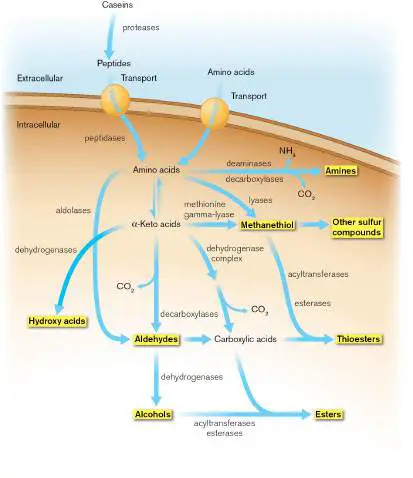
FIGURE 16.9 ■ Flavor generation from amino acid catabolism. Casein catabolism generates flavor molecules (highlighted). Extracellular enzymes break down casein into peptides and amino acids, which are taken into the bacterial cell by membrane transporters. The amino acids are fermented to volatile alcohols and esters. In some cases, they combine with sulfur to form methanethiol and other sulfur-containing odorants characteristic of cheese.
Acid Fermentation of Vegetables
Many kinds of vegetable products are based on microbial fermentation. Commercial products marketed globally include pickles, soy sauce, and sauerkraut. Other products provide staple foods for particular nations or regions, such as Indonesian tempeh and Korean kimchi.
Soy fermentation. Soybeans offer one of the best sources of vegetable protein and are indispensable for the diets of millions of people, particularly in Southeast Asia. In North America, soy products are important for vegetarian diets and as a milk substitute, as well as for animal feed. But soybeans also contain substances that decrease their nutritive value. Phytate, or inositol hexaphosphate, chelates minerals such as iron, inhibiting their absorption by the intestine. Lectins are proteins that bind to cell-surface glycoproteins within the human body. At high concentration, soybean lectins may upset digestion and induce autoimmune diseases. Soy protease inhibitors interfere with digestive enzymes chymotrypsin and trypsin, thus decreasing the amount of protein that can be obtained from soy-based food.
All of these drawbacks of soybeans are diminished by microbial fermentation, while the protein content remains comparable to that of the unfermented bean (40%). A variety of fermented soy foods have been developed. Most soy fermentation involves mold growth, supplemented by bacteria that contribute vitamins, including vitamin B 12.
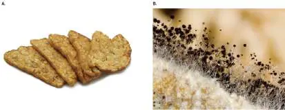
A major fermented soy product is tempeh (Fig. 16.10A), a staple food of Indonesia, the world’s fourth-most-populous country, as well as of other countries in Southeast Asia. Tempeh consists of soybeans fermented by Rhizopus oligosporus, a common bread mold. Besides decreasing the negative factors of soy, the mold growth breaks down proteins into more digestible peptides and amino acids. During World War II, tempeh was fed to American prisoners of war held by the Japanese. The tempeh was later credited with saving the lives of prisoners whose dysentery and malnutrition had impaired their ability to absorb intact proteins.
FIGURE 16.10 ■ Tempeh, a mold-fermented soy product. A. Fried tempeh. B. Rhizopus oligosporus mold, used to make tempeh.
CRISTY LIEM/SHUTTERSTOCK
GREGORY G. DIMIJIAN/SCIENCE SOURCE
Tempeh is commonly produced in home-based factories in Indonesia. The soybeans are soaked in water overnight, allowing initial fermentation by naturally present lactic acid bacteria; in some cases, a crude “starter” may be introduced from the water of soybeans soaked previously. This pre-fermentation allows bacterial generation of vitamins and produces mild acid that promotes growth of mold. The soaked beans are then hulled, cooked, and cooled to room temperature for inoculation with R. oligosporus spores or with a previous tempeh culture. The inoculated beans are wrapped in banana leaves or in perforated plastic bags and then allowed to incubate for 2 days. The mold grows as a white mycelium that permeates the beans (Fig. 16.10B ), joining them into a solid cake. The final product has a mushroom-like taste and is served fried or grilled like a hamburger.
Other soy products undergo acid fermentation by the mold Aspergillus oryzae. The Japanese condiment miso is made from ground soy and rice, salted and fermented for 2 months by A. oryzae
. Soy sauce is made from jiang, a Chinese condiment similar to miso in which the rice starter culture is replaced by wheat. The fermentation generates glutamic acid, a flavor-enhancing compound known popularly in the form of its salt: monosodium glutamate, or MSG.
Fermentation of cabbage and other vegetables. Various leaf vegetables are fermented by traditional societies, originally as a means of storage over the winter months. In Europe and North America, the best-known fermented products include sauerkraut and pickles. Sauerkraut production involves heterolactic fermentation by Leuconostoc mesenteroides. In heterolactic fermentation, each fermented sugar molecule yields lactic acid, as well as ethanol and carbon dioxide. The culture is used to inoculate shredded cabbage, which is layered in alternation with salt. The salt helps limit the number of species and the extent of microbial growth. A similar brine-enhanced fermentation process is used to pickle cucumbers, olives, and other vegetables.
Brine-fermented cabbage forms the basis of Korean kimchi (Fig. 16.11). Kimchi is traditionally prepared by an annual process known as gimjang, in which groups of people work together, chopping large quantities of ingredients. Kimchi is prepared from Chinese cabbage, salted and layered with radishes, peppers, onions, and other vegetables. The vegetables are layered with fish, rice, and chili peppers. Pickled seafoods such as shrimp or oysters may be included. The entire mixture is stored in a pot, either refrigerated or buried underground for several months over the winter. The main fermentation organism is Leuconostoc mesenteroides, although Streptococcus and Lactobacillus species participate.
FIGURE 16.11 ■ Kimchi preparation. Cabbage leaves are layered alternating with chili paste containing salted fish and vegetables. The salted layers are packed for fermentation at cold temperature.
XINHUA/ALAMY STOCK PHOTO
Chocolate from Cocoa Bean Fermentation
Chocolate, the product of the cocoa bean, Theobroma cacao (or “food of the gods”), requires one of the most complex fermentations of any food. The multimicrobial process has been characterized by Rosane Freitas Schwan and colleagues at Universidade Federal de Lavras, Brazil (Fig. 16.12). While Brazil and other equatorial countries produce cocoa beans, the beans cannot be exported and fermented later; the fermentation must occur immediately where they are harvested. The beans are heaped in mounds upon plantain leaves for fermentation by indigenous microorganisms, essentially the same way cocoa has been processed for thousands of years. For all the commercialization of chocolate production, totaling 2.5 billion kilos per annum worldwide, no “starter culture” has yet been standardized to ferment the cocoa bean.
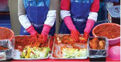
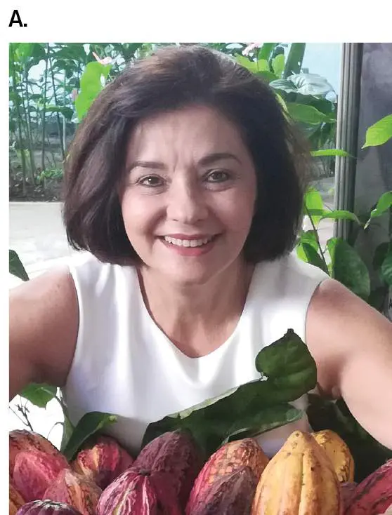
FIGURE 16.12 ■ Microbial succession during cocoa pulp fermentation. A. Rosane Freitas Schwan, at Universidade Federal de Lavras, Brazil, studies cocoa bean fermentation. B. Cocoa pods with beans.
COURTESY OF ROSANE FREITAS SCHWAN
FREEDOMNARUK/SHUTTERSTOCK
The microbial fermentation actually takes place outside the cocoa bean, within the pulp that clings to the beans after they are removed from the cocoa fruit. The pulp contains approximately 15% sugars and pectin (a branched polysaccharide), 2% citric acid, plus a rich supply of amino acids and minerals. As shown by Schwan, these nutrients support growth of many kinds of microbes, including three stages of succession: yeasts, lactic acid bacteria, and acetic acid bacteria (Fig. 16.13A).
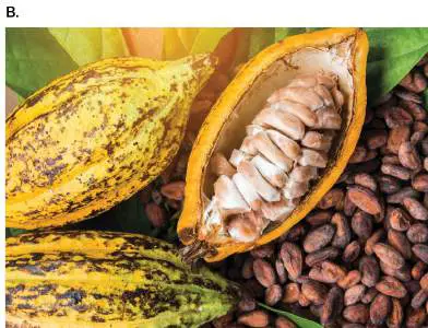
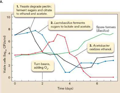
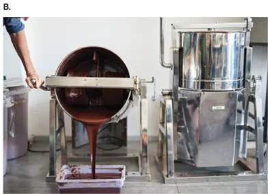
FIGURE 16.13 ■ Cocoa fermentation and chocolate manufacture. A. Yeasts ferment citrate to alcohol; then, lactic acid bacteria convert sugars to lactate. With aeration, acetic acid bacteria oxidize ethanol to CO 2. CFUs = colony-forming units. B. Cocoa mass contains cocoa butter and cocoa liquor that are extracted from the cocoa beans and mixed with sugar and other ingredients. Source: Data from R. Schwan.
MARIUSZ SZCZAWINSKI/ALAMY STOCK PHOTO
Yeasts (anaerobic). Citric acid acidifies the pulp (pH 3.6). The acidity favors growth of yeasts, including Candida, Kloeckera, and Saccharomyces. The yeasts consume sugars and citric acid, increasing pH. They also degrade pectin into glucose and fructose, allowing the pulp to liquefy. As the liquefied pulp drains, yeast ferments the remaining sugars to ethanol, CO 2, and mixed acids, such as acetate. The reactions release heat, increasing the temperature and accelerating metabolism. But the acetate, a permeant acid (discussed in Chapter 13), crosses the yeast cell membrane, where it lowers cell pH and inhibits growth. The ethanol and acetate also penetrate the bean embryo, killing the cells and releasing enzymes that generate key flavor molecules of chocolate. Lactic acid bacteria (anaerobic). The consumption of citric acid by yeast increases the bean pH to pH 4.2, encouraging growth of lactic acid bacteria, such as Lactobacillus plantarum. Lactobacillus species convert sugars to lactic acid, as well as acetate and CO 2. As fermentable substrates disappear, however, lactic acid bacteria are inhibited.
Acetic acid bacteria (aerobic). After 2 days, the beans are turned over and mixed periodically to permit access to oxygen. Aerobic acetic acid bacteria such as Acetobacter now oxidize the ethanol and acids to CO 2. The consumption of acids neutralizes undesirable acidity. Heat is released, increasing the temperature to as high as 50°C. Oxygen penetrates the bean, oxidizing key components such as polyphenols. Polyphenol oxidation generates the brown color of cocoa and contributes flavor.
After fermentation and pulp drainage, the beans are dried and roasted. The roasting process completes the transformation of cocoa substances that contribute flavor. Cocoa liquor and cocoa butter are extracted from the beans and then recombined with sugar and other components to make “cocoa mass” (Fig. 16.13B ). The cocoa mass is stirred for several days to achieve a smooth texture; then it is molded into the decorative forms known as chocolate. But without the preceding fermentation process, no flavor would develop.
Alkaline Fermentation: Natto and Pidan
In Western countries, food-associated fermentation is almost synonymous with acidification. In Africa and Southeast Asia, however, many food conversions involve increased pH. Such fermentations typically release small amounts of ammonia, which raises the pH to about pH 8, retarding growth of all but alkali-tolerant bacteria. The fermenting bacteria are usually Bacillus, aerobic species tolerant of moderate alkali and capable of extensive proteolysis and amino acid decomposition. The fermentation needs to be controlled to limit the loss of protein content, but the end result is a highly stable food product.
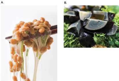
Natto. The Japanese soybean product natto is prepared by a process similar to that for tempeh. Soybeans are washed, pre-fermented, and cooked briefly before incubation with the starter organism, Bacillus natto. The fermenting beans are incubated in a shallow, ventilated container at a slightly raised temperature (40°C). B. natto secretes numerous extracellular enzymes, including proteases, amylases, and phytases. These enzymes decrease the undesirable components of soy, such as phytates and lectins, while liberating more easily digestible peptides and amino acids. Some of the amino acids are deaminated, generating ammonia; a well-ventilated natto chamber allows most of this gas to escape. In addition, B. natto synthesizes extracellular polymers such as polyglutamate (a peptide chain consisting exclusively of glutamic acid residues). Polyglutamate generates long, elastic strings that bind the beans together. The stretching of these strings from chopsticks is considered a sign of a good natto (Fig. 16.14A).
FIGURE 16.14 ■ Alkali-fermented foods. A. Natto consists of soybeans fermented by Bacillus natto. The fermentation generates long strings of polyglutamate. B. Pidan, or “century egg,” consists of duck eggs coagulated by sodium hydroxide and fermented by Bacillus species. Eggs are cut open, revealing the transformed yolk, which develops a greenish color.
AFLO CO. LTD/ALAMY STOCK PHOTO
AGE FOTOSTOCK/ALAMY STOCK PHOTO
Alkali-fermented vegetables. In Africa, numerous vegetable products are based on alkaline fermentation, predominantly by Bacillus species. An example is dawadawa, a paste of fermented locust beans common in western Africa. The locust beans are washed and supplemented with potash (potassium hydroxide) originally
obtained from wood ashes. This addition of alkali retards growth of bacteria other than Bacillus species, which predominate at higher pH. The beans are fermented by indigenous bacteria (bacteria already present in the beans). The fermented beans are sun-dried, releasing most of the ammonia, and pounded into cakes for storage. Similar alkali-fermented vegetables include ogiri (from melon seeds) and ugba (from oil beans).
Pidan. An ancient means of preserving eggs led to the famous Chinese delicacy pidan, or “century egg,” now a favorite at dim sum restaurants (Fig. 16.14B ). To make pidan, duck eggs are covered with a mixture of brewed tea, lime (CaO), and sodium carbonate (Na 2 CO 3). The lime and sodium carbonate react to form sodium hydroxide (NaOH), which penetrates the eggshells, raising pH and coagulating the egg white proteins. The eggs are buried in mud for several months, during which time the combined action of alkali and Bacillus fermentation generates dark colors and interesting flavors.
Thought Question
16.5 In traditional fermented foods, without a pure starter culture, how could someone control the kind of fermentation that occurs?
To Summarize
Milk curd forms by lactic acid fermentation and rennet proteolysis, rendering casein insoluble. The cleaved peptides coagulate to form a semisolid curd.
Cheese varieties include unripened cheeses, semihard and hard cheeses that are cooked down and ripened, brined cheeses, and mold-ripened cheeses. Flavors are generated by minor side products of fermentation.
Soy fermentation to tempeh by Rhizopus oligosporus improves digestibility and decreases undesirable soy components such as phytates and lectins.
Vegetables are fermented and brined to make sauerkraut, pickles, and kimchi.
Cocoa fermentation for chocolate requires complex fermentation of cocoa beans within the fruit pulp, including anaerobic fermentation by yeast and lactic acid bacteria, and aerobic respiration by Acetobacter species.
Alkali-fermented vegetables include the soy product natto, the egg product pidan, and the locust bean product dawadawa. The main fermenting organisms are Bacillus species.
Glossary
curd Coagulated milk proteins produced by the combined action of lactic acid–producing bacteria and stomach enzymes of certain mammals, such as cattle.
whey The liquid portion of milk after proteins have precipitated out of solution, usually during cheese production.
cheese A solid or semisolid food product prepared by coagulating milk proteins, forming curd. Its production commonly involves microbial fermentation.
yogurt A semisolid food produced through acidification of milk by lactic acid–producing bacteria.
ripening Ripening or aging is a process by which cheese undergoes drying and slow microbial conversion over a period of weeks or months. starter culture A mixture of fermenting microbes added to a food substrate to generate a fermented product.
cheddared curd Curd that has been cut and piled in order to remove the liquid whey.
tempeh A mold-fermented soy product, popular as a food in parts of Asia. miso A Japanese condiment, made from ground soy and rice, salted and fermented by the mold Aspergillus oryzae.
kimchi A popular Korean food based on brine-fermented cabbage. natto A soybean product, similar to tempeh, produced by alkaline fermentation.
pidan Pidan, or “century egg,” is a food made by alkali treatment and microbial fermentation of eggs over several months.
Figure 16.4 FIGURE 16.4 ■ Major chemical conversions in fermented foods.
PICTURE PARTNERS/ALAMY STOCK PHOTO
Figure 13.20
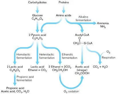
FIGURE 13.20 ■ Fermentation pathways. Alternative pathways from pyruvate and phosphoenolpyruvate to end products, many of which we use for food or industry. Different species conduct different portions of the pathways shown.
FOOD COLLECTION/SUPERSTOCK
AGE FOTOSTOCK/ALAMY STOCK PHOTO
LEE HACKER/ALAMY
D. HURST/ALAMY
Fig. 16.5A
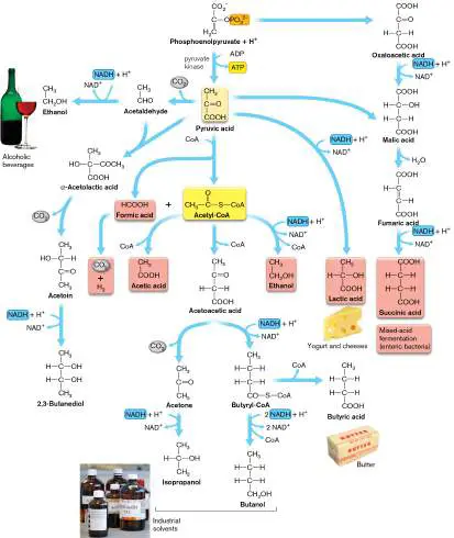
FIGURE 16.5 ■ Propionic acid fermentation within Swiss cheese. A. Emmentaler (Swiss) cheese with “eyes” is made by a bacterial community (inset). Bacterial species shown here include Lactobacillus helveticus (rods, 2.0–4.0 μm in length) and Streptococcus thermophilus (cocci). B. As Swiss cheese ages, lactate is converted to propionate, acetate, and CO 2 (the gas that forms the eyes).
SVETLANA FOOTE/ALAMY STOCK PHOTO
SCIMAT/SCIENCE SOURCE
16.3 Ethanolic Fermentation: Bread and Winenot assigned
Some of our most nutritionally significant and culturally important foods, including most bread and alcoholic beverages, require ethanolic fermentation by yeast fungi. Ethanolic fermentation converts pyruvic acid to ethanol and carbon dioxide: C 3 H 4 O 3 → CH 3 CH 2 OH + CO 2 The most prominent yeast used is Saccharomyces cerevisiae, known as baker’s yeast or brewer’s yeast (Fig. 16.15). A hardy organism, S. cerevisiae easily survives on a grocery shelf for home use and is genetically tractable for fundamental research. The yeast has been studied since the time of Pasteur, who used it to prove the biological basis of fermentation (presented in Chapter 1). S. cerevisiae today is a major model system of cell biology, yielding the molecular secrets of human cancer and other diseases.
FIGURE 16.15 ■ Baker’s yeast, the “champion” fermenter. A. Saccharomyces cerevisiae cells budding; some show bud scars (SEM). B. S. cerevisiae is used to study the
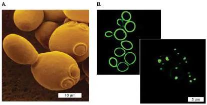
function of human proteins such as alpha-synuclein (green fluorescence), which plays a role in Parkinson’s disease. Yeast cells engineered to express one copy of the gene (left panel) show the protein normally within their cell membrane. Two gene copies (right panel) cause the protein to clump and kill the cells.
DAVID SCHARF/SCIENCE SOURCE
T. F. OUTEIRO AND S. LINDQUIST. 2003. SCIENCE 302 :1772. REPRINTED WITH
PERMISSION FROM AAAS
Bread making depends on carbon dioxide to form air spaces; the carbon dioxide itself is generated by yeast or other microbes that leaven the dough. The air spaces make the baked bread easier to chew and digest. The small amount of ethanol produced by yeast is eliminated during baking. For alcoholic beverages, however, ethanol is the key product, accompanied by carbon dioxide bubbles for “fizz,” known as carbonation.
Bread Dough Is Leavened by Microbes That Produce CO 2
Bread is made in many different forms (Fig. 16.16) and from diverse kinds of flour, or ground grain. The earliest breads probably arose from grain mush naturally contaminated by yeast. Later, bread makers learned to include yeast left over from wine or beer production as a starter culture.
FIGURE 16.16 ■ Yeast bread. Many varieties of bread are made.
JOHN FOXX/GETTY IMAGES
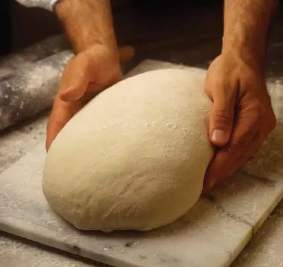
Yeast bread production. The preparation of all forms of yeast bread requires the same fundamental steps. A starter culture of yeast is included in the dough. The yeast can be commercial baker’s yeast or it can be sourdough starter, an undefined microbial population derived from a previous batch of dough. Analysis of sourdough shows mainly yeasts and lactobacilli, which release acids that favor the growth of the yeasts. The dough is kneaded to develop a fine network of air pockets and allowed to rise, expanding with production of the carbon dioxide gas (Fig. 16.17). The finest-textured breads are made from wheat flour that contains gluten. Gluten is a protein complex that forms a fine molecular network supporting the rising dough. In some individuals, gluten can trigger an autoimmune condition leading to gluten intolerance.
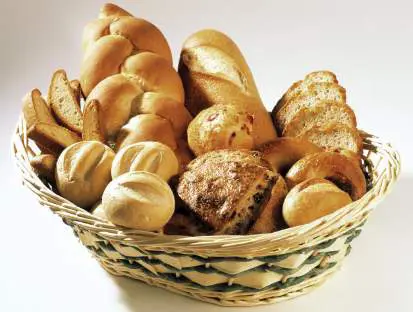
FIGURE 16.17 ■ Making bread. As yeast fermentation generates carbon dioxide gas, the dough rises.
SUSAN C. BOURGOIN/FOODPIX/JUPITERIMAGES
Thought Question
16.6 Compare and contrast the role of fermenting organisms in the production of cheese and bread.
Injera: extended fermentation. Most kinds of bread involve only a short fermentation period, just long enough to produce enough gas for leavening. A prolonged fermentation, with more extensive microbial activity, unfolds in the dough for an Ethiopian bread called injera (Fig. 16.18). The high microbial content provides a substantial source of vitamins not found in quick-rising breads.
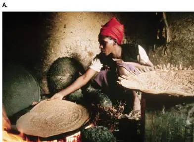
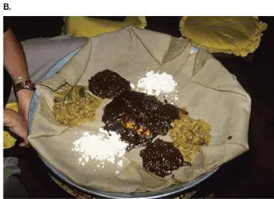
FIGURE 16.18 ■ Injera. A. After 3 days of fermentation, injera dough is baked in a ceramic pan upon a “Mirte” charcoal stove. B. Injera forms an edible tablecloth for a variety of Ethiopian foods.
JIM SUGAR/GETTY IMAGES
BERT DE RUITER ALAMY STOCK PHOTO
Injera is made from teff (Eragrostis tef), a grain with small, round kernels that have high protein and lack gluten. Teff grows in arid regions and is now being cultivated in the United States for gluten-free products. Because teff lacks gluten, it cannot rise as much as wheat flour, but it makes a kind of flatbread. The dough is spread into a wide pancake, and the organisms present in the grain and air are allowed to ferment it for 3 days. The fermentation includes a succession of species, usually dominated by the yeast Candida. The extended fermentation generates a complex range of by-products that confer exceptional flavors in the baked product. In Ethiopia, injera forms the basis of an entire meal, served with other food items placed upon it as an edible tablecloth. Diners wrap samples of each food in a fold of injera and consume them together.
Alcoholic Beverages: Beer and Wine
Ethanolic fermentation of grain or fruit was important to early civilizations because it provided a drink free of waterborne pathogens. Traditional forms of beer also provided essential vitamins in the unfiltered yeasts.
Ethanol is unique among fermentation products in that it provides a significant source of caloric intake, but it is also a toxin that impairs mental function. A modest level of ethanol enters the human circulation naturally from intestinal flora, equivalent to a fraction of a drink per day. The human liver produces the enzyme alcohol dehydrogenase, which detoxifies ethanol. This enzyme in a healthy liver can metabolize small amounts of alcohol without harm. However, excess alcohol consumption can overload the liver’s capacity for detoxification and permanently damage the liver and brain.
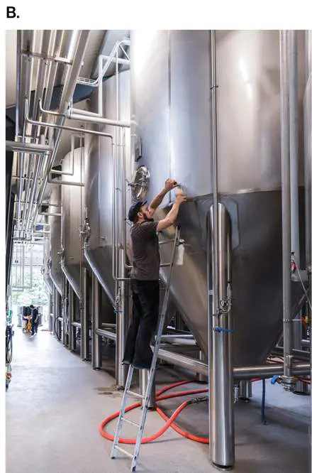
Beer: alcoholic fermentation of grain. Beer production is one of the most ancient fermentation practices and is depicted in the statuary of ancient Egyptian tombs dated to 5,000 years ago (Fig. 16.19A). The earliest Sumerian beers were made from bread soaked in water and fermented. Today, most beer is produced commercially by fermenting barley using giant vats (Fig. 16.19B ). Production of high-quality beer involves complex processing with many steps, including germination of barley grains, mashing in water and cooking, and introduction of hops for flavor.
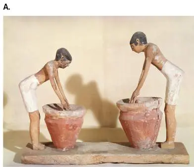
FIGURE 16.19 ■ Beer production: ancient and modern. A. Making beer in ancient Egypt, circa 3000 BCE. The mash was stirred in earthen jars. B. Fermentors in a modern brewery.
BORROMEO/ART RESOURCE, NY
LIOR+LONE/STOCKSY
First the barley grains must germinate; that is, the seed embryos must start to grow. The germinating embryo makes enzymes needed
to break down the barley starch to maltose (disaccharide) and glucose. Most of the sugars are fermented by yeast to ethanol and carbon dioxide. However, minor side products contribute flavors—or unpleasant off-flavors if present in too great an amount (Fig. 16.20 ). For example, off-flavors may result from the presence of small amounts of oxygen that oxidize some ethanol to acetaldehyde. FIGURE 16.20 ■ Alcoholic fermentation in beer and wine. Yeast fermentation generates ethanol in substantial quantities. The biosynthesis of amino acids generates by-products that contribute both desirable flavors (long-chain alcohols and esters) and off-flavors (acetaldehyde and diacetyl).
The yeast ferment most sugars to ethanol, but a small fraction is drawn off to make amino acids via tricarboxylic acid (TCA) cycle intermediates, as discussed in Chapter 15. The 2-oxo acids of the TCA cycle are analogous to pyruvate, with the methyl group replaced
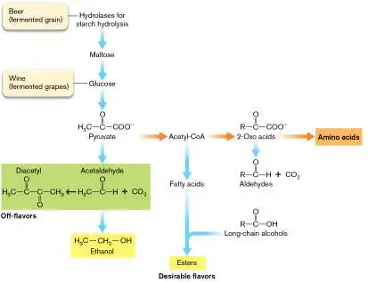
by extended carbon chains (R group). A tiny amount of the 2-oxo acids is converted to long-chain alcohols, which add desirable flavor to beer.
Thought Question
16.7 Compare and contrast the role of low-concentration by-products in the production of cheese and beer.
Wine: alcoholic fermentation of fruit. The fermentation of fruit gives rise to wine, another class of alcoholic products of enormous historical and cultural significance. Grapes produce the best-known wines, but wines and distilled liquors are also made from apples, plums, and other fruits. The key difference between fermentation of fruits and fermentation of grains is the exceptionally high monosaccharide content in fruits. Grape juice, for example, can contain concentrations of glucose and fructose as high as 15%. The availability of simple sugars allows yeast to begin fermenting immediately, with no need for preliminary breakdown of long-chain carbohydrates, as in the malting and mashing of beer.
Most modern wine production uses strains of the grape Vitis vinifera. The grapes are crushed to release juices, usually in the presence of antioxidants such as sulfur dioxide (Fig. 16.21). For white wine, the skins are removed before juice is fermented. For red wine, the skins are included in early fermentation to extract the red and purple anthocyanin pigments, as well as phenolic flavor compounds. The first few days of fermentation are dominated by indigenous species of yeast naturally present on the grapes, such as Kloeckera and Hanseniaspora species. Commercial producers usually inoculate with standard Saccharomyces cerevisiae, whose population dominates the late stage of fermentation (6–20 days). Yeast growth ends once the ethanol level reaches about 15%; to achieve higher alcohol content, distillation is required.
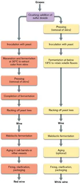
FIGURE 16.21 ■ Production of red and white wines. Left: For red wine, the grapes are fermented with the skins at a temperature that increases extraction of color and tannins. Right: For white wine, the skins are removed before addition of yeast starter, and the temperature is kept low to retain volatile flavors. Both kinds of wine usually undergo malolactic fermentation by Oenococcus oeni, a species of lactic acid bacteria that consume malic acid.
After fermentation, the wine is drained, or “racked,” from the sediment of grape and yeast material, the lees. The liquid may be further clarified by centrifugation. Then it is stored for 2–3 weeks in tanks or barrels. During storage, a second stage of fermentation may be performed, called malolactic fermentation. Malolactic fermentation is needed to decrease the acidity from malic acid (found in grapes). Malic acid is converted to lactic acid, with a higher dissociation constant (a weaker acid). The L -malate is decarboxylated to L - or D -lactate: − HOOC—CH —CHOH—COOH − → CH —CHOH—COOH + CO
2 3 2
The wine is seeded with Oenococcus oeni bacteria, which ferment L - malate (deprotonated L -malic acid).
As in beer production, yeast fermentation of wine produces numerous minor products contributing flavor, such as long-chain alcohols and esters. At the same time, overgrowth of yeast or the growth of undesired species can produce excess amounts of these compounds, such as sulfides and phenolics, giving rise to off-flavors. Some undesired species require oxygen exposure, whereas others can grow during storage and bottling. The balance of microbial populations is challenging to control and has a major role in determining the quality of a given wine vintage.
To Summarize
Bread is leavened by yeasts conducting limited ethanolic fermentation, producing enough carbon dioxide gas to expand the dough.
Injera bread dough undergoes more extensive fermentation by indigenous organisms that, as a result, generates multiple flavors.
Beer requires alcoholic fermentation of grain. Barley grains are germinated, allowing enzymes to break down the starch to maltose for yeast fermentation.
Secondary products of grain fermentation , such as long-chain alcohols and esters, generate the special flavors of beer. Wine derives from alcoholic fermentation of fruit, most commonly grapes. The grape sugar (glucose) is fermented by yeast to alcohol. A secondary product, malate, undergoes malolactic fermentation by Oenococcus oeni bacteria.
Glossary
leaven For bread dough, to cause to rise by generating air spaces, usually through carbon dioxide production by microbial fermentation.
sourdough An undefined yeast population, derived from a previous batch of dough, that is used in bread production.
injera A highly fermented Ethiopian flatbread made from the grain teff. malolactic fermentation Fermentation of L -malate (a side product of glucose fermentation) by Oenococcus oeni bacteria; an important process in winemaking.
16.4 Food Spoilage and Preservationnot assigned
We humans have always competed with microbes for our food. When early humans killed an animal, microbes commenced immediately to consume its flesh. Because meat perished so fast, it made economic sense to share the kill immediately and consume it all as soon as possible. Vegetables might last longer, but eventually they succumbed to mold and rot. Later societies developed preservation methods, such as drying, smoking, and canning, that enabled humans to survive winters and dry seasons on stored food. Here we focus on microbial contamination and food preservation from the perspective of the food industry.
Food Spoilage and Food Contamination
After food is harvested, several kinds of chemical changes occur. Some begin instantly, whereas others take several days to develop. Some changes, such as meat tenderizing, may be considered desirable; others, such as putrefaction, render food unfit for consumption. Much research focuses on the development of technologies to prevent undesirable changes such as oxidation (Fig. 16.22).
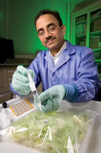
FIGURE 16.22 ■ Research to prevent oxidation of produce. At the U.S. Department of Agriculture, microbiologist Arvind Bhagwat withdraws a gas sample from bagged lettuce, to test an anaerobic packaging method.
U.S. DEPARTMENT OF AGRICULTURE
The major classes of food change include: Enzymatic processes. Following the death of an animal, its flesh undergoes proteolysis by its own enzymes. Limited proteolysis tenderizes meat. Plants after harvest undergo other changes; for example, in harvested corn the sugar rapidly converts to starch. That is why vegetables taste sweetest immediately after harvest.
Chemical reactions with the environment. The most common abiotic chemical reactions involve oxidation by air; for example, lipid autooxidation, which generates rancid odors. To prevent oxidation, produce may be packaged under an anaerobic atmosphere, wrapped in a film that prevents oxygen transmission. This process is called modified atmospheric processing.
Microbiological processes. Microbes from the surface of the food begin to consume it—some immediately, others later in succession—generating a wide range of chemical products. In meat, internal organs of the digestive tract are an important source of microbial decay.
Microbial activity can aid food production, but it can also have various undesirable effects. Two different classes of microbial effects are distinguished: food spoilage and food contamination with pathogens.
Food spoilage refers to microbial changes that render a product obviously unfit or unpalatable for consumption. For example, rancid milk and putrefied meats are unpalatable and contain metabolic products that may be deleterious to human health, such as oxidized fatty acids or organic amines. Even in these cases, however, the definition of spoilage depends partly on cultural practice. What is sour milk to one person may be buttermilk to another; what one society considers spoiled meat, another may consider merely aged. Different pathways of microbial metabolism lead to different kinds of spoilage. Sour flavors result from acid-fermentation products, as in sour milk. Alkaline-fermentation products generate bitter flavor. Oxidation, particularly of fats, causes rancidity, whereas general decomposition of proteins and amino acids leads to putrefaction. The particularly noxious odors of putrefaction derive from amino acid decarboxylation generating amines that often have apt names, such as the amines cadaverine and putrescine and the aromatic product skatole.
“Food contamination,” or food poisoning, refers to the presence of microbial pathogens that cause human disease; for example, rotaviruses that cause gastrointestinal illness. Other pathogens, such as Clostridium botulinum, produce toxins with deadly effects (discussed in Chapter 25). Pathogens usually go unnoticed as food is consumed, because their numbers are very low, and they may not even grow in the food. Even the freshest-appearing food may cause serious illness if it has been contaminated with a small number of pathogens.
As our intestines are full of beneficial bacteria, what makes a pathogen? Enteric pathogens are often closely related to members of our gut microbiome, which normally outcompete invaders (discussed in Chapters 21 and 23). Nevertheless, a pathogen may overcome our defenses, either by using its virulence factors (see Chapter 25) or by taking advantage of host weakness due to immunosuppression or to antibiotics depleting the normal microbiome.
How Food Spoils
Different foods spoil in different ways, depending on their nutrient content, the microbial species, and environmental factors such as temperature. Table 16.2summarizes common forms of spoilage. TABLE 16.2 Food Spoilage (Examples) Signs of Microbial cause Food product spoilage Dairy products Milk Sour flavor Lactic acid bacteria produce lactic and acetic acids.
Coagulation Lactic acid bacteria produce proteases that destabilize casein and lower pH, causing coagulation.
Bitter flavor Psychrophilic bacteria degrade proteins and amino acids.
Cheese Open texture, Lactic acid bacteria fissures produce carbon dioxide.
Discoloration and Molds such as colonies Penicillium and Aspergillus grow on the cheese.
Meat, poultry, and eggs Meat and poultry Rancid flavor Psychrotrophic bacteria produce fatty acids that become oxidized.
Putrefaction Pseudomonas and other aerobes degrade amino acids, producing amines and sulfides.
Discolored Molds such as Mucor patches and Penicillium grow on the surface.
Eggs Pink or greenish Pseudomonas and egg white related bacteria grow on albumin, producing water-soluble pigments.
Sulfurous odor Bacterial growth on albumin releases hydrogen sulfide.
Seafood Fish Fishy smell Anaerobic psychrophiles such as Photobacterium convert trimethylamine oxide to trimethylamine.
Odor of Pseudomonas and putrefaction other Gram-negative species degrade amino acids, producing amines and sulfides.
Shellfish Odor of Vibrio and other marine putrefaction bacteria decompose the protein.
Fruits, vegetables, and grains Plants before Rotting or wilting Plant pathogens, most harvest commonly fungi such as Alternaria, Aspergillus, and Penicillium.
Stored plant Rotting or wilting Molds or bacteria foods produce degradative enzymes, such as pectinases and cellulases.
Apples, pears, Geosmin off-Penicillium mold.
cherries flavor Peeled oranges Discoloration and Enterobacter and off-flavor Pseudomonas spp.
Pasteurized fruit Medicine-like Acid-and heat-tolerant juices phenolic off-spore former, flavor Alicyclobacillus sp., produces 2-methoxyphenol (guaiacol).
Bread Ropiness Bacillus spp. grow, forming long filaments.
Red discoloration Serratia marcescens.
Dairy products. Milk and other dairy products contain carbon sources, such as lactose, protein, and fat. In fresh milk, the nutrient most available for microbial catabolism is lactose, which commonly supports anaerobic fermentation to sour milk. Fermentation by the right mix of microbes, however, leads to yogurt and cheese production, as previously described.
Under certain conditions, bitter off-flavors may be produced by bacterial degradation of proteins. The release of amines causes a rise in pH. Protein degradation is most commonly caused by psychrophiles, species that grow well at cold temperatures, such as those of refrigeration.
Cheeses are less susceptible than milk to general spoilage, because of their solid structure and lowered water activity (partial vapor pressure; water available to microbial cells). However, cheeses can grow mold on their surface. Historically, the surface growth of Penicillium strains led to the invention of new kinds of cheeses. But other kinds of mold, such as Aspergillus, produce toxins and undesirable flavors.
Meat and poultry. Meat in the slaughterhouse is easily contaminated with bacteria from hide, hooves, and intestinal contents. Muscle tissue offers high water content, which supports microbial growth, as well as rich nutrients, including glycogen, peptides, and amino acids. The breakdown of peptides and amino acids produces the undesirable odorants that define spoilage (for example, cadaverine and putrescine).
Meat also contains fat, or adipose tissue, but the lipids are largely unavailable to microbial action because they consist of insoluble fat (triacylglycerides). Instead, meat lipids commonly spoil abiotically by autooxidation (reaction with oxygen) of unsaturated fatty acids, independent of microbial activity. Thus, when meats are exposed to air during storage, they turn rancid—particularly meats such as pork, which contains highly unsaturated lipids. Autooxidation can be prevented by anaerobic storage, such as vacuum packing, which also prevents growth of aerobic microorganisms. The absence of the suppressed organisms, however, favors growth of lactic acid bacteria and facultative anaerobes such as Brochothrix thermosphacta. These organisms generate short-chain fatty acids, which taste sour. In industrialized societies, the most significant factor determining microbial populations in meat spoilage is the practice of refrigeration. Refrigeration prolongs the shelf life of meat because contaminating microbes are predominantly mesophilic (grow at moderate temperatures, as discussed in Chapter 5). But ultimately, the few psychrotrophs initially present do grow; typically, these are Pseudomonas species. The pseudomonads are also favored by the low pH of meat (pH 5.5–7.0), which results from the accumulation of lactic acid in the muscle.
Seafood. Fish and other seafood contain substantial amounts of proteins and lipids, as well as amines such as trimethylamine oxide. Fish spoils more rapidly than meat and poultry for several reasons. First, fish do not thermoregulate, and they inhabit relatively low-temperature environments. Because fish grow in low-temperature environments, their surface microorganisms tend to be psychrotrophic and thus grow well under refrigeration. In addition, marine fish contain high levels of the osmoprotectant trimethylamine oxide, which bacteria reduce to trimethylamine, a volatile amine that gives seafood its “fishy” smell. Finally, the rapid microbial breakdown of proteins and amino acids leads to foul-smelling amines and sulfur compounds, such as hydrogen sulfide and dimethyl sulfide.
Thought Question
16.8 Why would bacteria convert trimethylamine oxide (TMAO) to trimethylamine? Would this kind of spoilage be prevented by exclusion of oxygen?
Plant foods. Fruits, vegetables, and grains spoil differently than animal foods because of their high carbohydrate content and their relatively low water content. The low water content of plant foods usually translates into considerably longer shelf life than animal-based foods have. Carbohydrates favor microbial fermentation to acids or alcohols that limit further decomposition, and this microbial action can be managed to produce fermented foods, as described in Section 16.2.
Plant pathogens rarely infect humans but may destroy the plant before harvest. Most plant pathogens are fungi, although some are bacteria, such as Erwinia species. Historically, plant pathogens have caused major agricultural catastrophes, such as the Irish potato famine, caused by a fungus-like pathogen. Plant pathogens continue to devastate local economies and cause shortages worldwide; for example, the witches’-broom fungus Crinipellis perniciosa causes a fungal disease of cocoa trees that has drastically cut Latin American cocoa production.
After harvest, various molds and bacteria can soften and wilt plant foods by producing enzymes that degrade the pectins and celluloses that give plants their structure. In general, the more processed the food, the greater the opportunities for spoilage. For example, citrus fruits generally last for several weeks, but peeled oranges are susceptible to spoilage by Gram-negative bacteria.
Baked bread usually resists spoilage, except for surface molds. In rare cases, however, improperly baked bread can show contamination. The appearance of red bread, caused by the red bacterium Serratia marcescens, is believed to have been the source of the “blood” observed in communion bread during a Catholic mass in the Italian town of Bolsena in 1263—an event that became known as the miracle of Bolsena.
Pathogens Contaminate Food
Intestinal pathogens spread readily because microbes can be transmitted through food without any outward sign that the food is spoiled. The U.S. Centers for Disease Control and Prevention (CDC) estimates that there are 48 million cases of food-borne gastrointestinal illness a year in the United States. Thus, one in six Americans gets sick from food in a given year. In 2013, the U.S. Food and Drug Administration (FDA) implemented new controls and rules as part of the 2011 Food Safety Modernization Act, the largest reform of U.S. food safety laws in 70 years. The new laws require, for example, that crop irrigation water be free of pathogens and that agricultural workers have access to bathroom facilities.
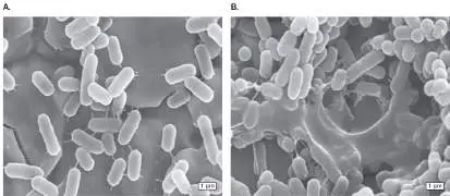
A pathogen that contaminates a wide range of products is Salmonella species. During the year 2021, for example, in the United States, Salmonella epidemics came from contamination of produce such as lettuce and onions, poultry and eggs, seafood and crabmeat, and processed foods such as tahini, salami sticks, raw flour, and cake mixes. The means of contamination could involve any stage of processing, such as water rinsing. Salmonella bacteria can persist on a dry surface for years. Figure 16.23compares the morphology of Salmonella bacteria exposed on a metal surface for 1 hour versus 2 months. The bacteria that persisted retained viability. FIGURE 16.23 ■ Salmonella bacteria persist on dry surface. Salmonella Agona exposed on a steel surface. A. One hour of exposure at 35% relative humidity. B. Two months of exposure (SEM).
O. HABIMANA ET AL. 2014. LETT. APPL. MICROBIOL. 59 :464–470, FIG. 3A
O. HABIMANA ET AL. 2014. LETT. APPL. MICROBIOL. 59 :464–470, FIG. 3B
An example of a food contamination epidemic is the 2008 outbreak of Salmonella enterica from peanut products. Peanuts contaminated at one processing plant led to an epidemic that sickened 700 people across the United States (Fig. 16.24A). The first cases of Salmonella infection were reported to the CDC on
September 1, 2008. Most infected individuals developed diarrhea, fever, and abdominal cramps 12–72 hours after infection, and symptoms lasted 4–7 days. Over the next 6 months, cases were reported from nearly all U.S. states. The curve of the outbreak (cases rising and then falling) followed the profile of a single-source epidemic, in which all infections are ultimately traced back to one source. (Epidemics are discussed in Chapter 28.)
FIGURE 16.24 ■ Salmonella enterica outbreak from contaminated peanut butter. A. Number of infected individuals reported to the CDC from September 1, 2008, through April 20, 2009. B. Electrophoretic separation of restriction-digest DNA fragments from bacterial strains isolated from humans with illness. (1) Peanut butter containing the same strain; (2) peanut butter containing a different strain of S. enterica.
Source: http://www.cdc.gov.
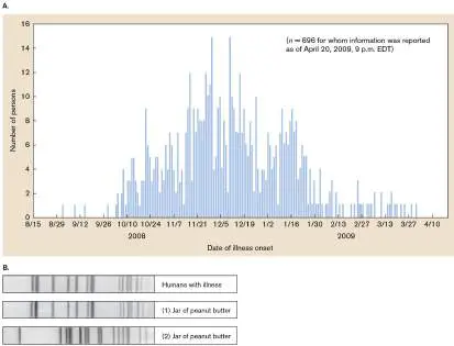
What was the original source of the widespread outbreak? The CDC researchers compared the food intake histories of ill persons against matched controls. They found a statistical association between illness and intake of peanut butter, eventually narrowed to a specific brand of peanut butter sold to institutions. As the epidemic grew, cases emerged in which the contaminated food product was crackers filled with peanut butter cream. Ultimately, the peanut butter and cream were traced back to peanuts from a single factory in Georgia. At the food plant, the source of Salmonella contamination could not be identified, but the plant records showed that product samples had tested positive for Salmonella. Instead of discarding the product, the plant had retested the samples until they “tested negative.” Numerous health violations were cited, including gaps in the walls and dirt buildup throughout the plant.
Did all of the cases of illness result from a common strain? The CDC used DNA analysis to show that all patients carried a common strain of S. enterica serovar Typhimurium. (A serovar is a strain whose surface proteins elicit a distinctive immune response.) The strain was identified by analysis of its genomic DNA cleaved by restriction endonucleases (see Section 12.3). Each restriction endonuclease cleaves DNA at sequence-specific positions. Strains that differ at key restriction sites generate cleavage fragments of differing length, which are separated by pulsed-field electrophoresis ( Fig. 16.24B ).
In electrophoresis, applied voltage causes DNA fragments to migrate different distances according to size; the pulsed field optimizes separation of the largest sizes. The distance each fragment moves is visualized as a band in the gel. The band pattern, or “fingerprint,” of Salmonella DNA from infected patients showed the same fragment lengths as Salmonella DNA from one peanut butter sample—the sample labeled “(1)” in Figure 16.24B . The band pattern from this peanut butter sample differed from that of another contaminated peanut butter sample, labeled “(2)”; the bacterium in the second sample proved unrelated to the Salmonella outbreak. The result of this simple test can be confirmed by whole-genome sequencing and comparison with known pathogenic strains.
This case illustrates several features of food contamination in modern society. It shows the consequence of a food production plant’s failure to follow regulations and the failure of health inspection to enforce them. The contaminated product shipped out to a diverse array of institutions such as schools and to secondary producers such as cookie manufacturers, which incorporated the peanut butter cream ingredient. The bacteria then remained viable in contaminated food products for many months, sickening people long after the contamination event occurred.
Food-Borne Pathogens Emerge from the Environment and Agriculture
Food-borne pathogens can arise from a surprising variety of sources. Consider, for example, the transmission routes of Listeria monocytogenes, a psychrotrophic pathogen that invades the cells of the intestinal epithelium, causing listeriosis (Fig. 16.25). Psychrotrophic organisms grow optimally at moderate temperatures but also grow slowly at lower temperatures, typically 0°C–30°C. Listeria can be transferred from soil and feed to cattle, whose manure then cycles it back to soil. From cattle, the pathogen contaminates milk and meat, where it can eventually infect human consumers. Because Listeria is a psychrotroph, it outcompetes other food-borne bacteria under refrigeration.
FIGURE 16.25 ■ Transmission of Listeria monocytogenes. Listeria is transmitted through various routes, including passage through food products. Colored arrows indicate transmission of disease.
The U.S. Public Health Service judges the importance of food-borne pathogens by their incidence and/or the severity of the diseases they cause (Table 16.3). For example, the noroviruses infect about 20 million Americans per year. Norovirus outbreaks are hard to control, especially in close quarters such as a cruise ship, where an outbreak can easily infect a large proportion of passengers. The course of illness is usually short, but it can lead to complications from dehydration. By contrast, the spore-forming pathogen
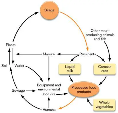
Clostridium botulinum causes only about 100 cases of botulism per year. The incidence is relatively low, but if untreated, the fatality rate is 50%. In this case, the low incidence of botulism actually enhances its danger because the condition is likely to go undiagnosed.
Food-Borne Pathogens in the
TABLE 16.3 a
United States
Incidence and Pathogen transmission Course of illness Norovirus Most common Disease lasts 1 or 2 (Norwalk and cause of days. Includes Norwalk-like diarrhea; also vomiting, diarrhea, viruses) called “stomach and abdominal flu” (no pain; headache connection with and low-grade influenza). fever may occur.
Twenty million cases per year are estimated.
Transmitted mainly by virus-contaminated food and water.
Infection rates are highest under conditions of crowding in close quarters, such as inside a ship or a nursing home.
Food-Borne Pathogens in the
TABLE 16.3 a
United States
Incidence and Pathogen transmission Course of illness Salmonella Most common Gastrointestinal food-borne disease that cause of death; includes diarrhea, more than 1 fever, and million cases per abdominal cramps year; estimated lasting 4–7 days. 600 deaths per Fatal cases are year. most common in Transmission immunocompromis nearly always ed patients.
through food— raw, undercooked, or recontaminated after cooking, especially eggs, poultry, and meat; also contaminates dairy products, seafood, fruits, and vegetables.
Campylobacter More than 1 million In humans, usually cases of severe bloody campylobacterio diarrhea, fever, sis per year; and abdominal estimated 100 cramps lasting 7
Food-Borne Pathogens in the
TABLE 16.3 a
United States
Incidence and Pathogen transmission Course of illness deaths per year. days. Fatal cases Grows in poultry are most common without causing in symptoms. immunocompromis Transmission is ed patients.
mainly through raw and undercooked poultry; contaminates half of poultry sold. Occurs less often in dairy products or in foods contaminated after cooking.
Escherichia coli An emerging In humans, usually O157:H7 pathogen, first severe bloody recognized in a diarrhea and hamburger abdominal cramps outbreak in lasting 5–10 days. 1982; now About 5% of known to infect patients, especially 73,000 people children and yearly, including elderly, develop 60 deaths per hemolytic uremic
Food-Borne Pathogens in the
TABLE 16.3 a
United States
Incidence and Pathogen transmission Course of illness year. Grows in syndrome, in which cattle without the red blood cells causing are destroyed and symptoms. the kidneys fail.
Transmitted through ground beef; also through unpasteurized cider and from plant produce, where it grows as an endophyte.
Clostridium Causes about 100 Botulinum toxin from botulinum cases per year of growing bacteria botulism, with a causes progressive 50% fatality rate paralysis, with if untreated. blurred vision, Grows in drooping eyelids, improperly slurred speech, home-canned difficulty foods, more swallowing, and rarely in muscle weakness.
commercially Infant botulism canned low-acid causes lethargy foods and and impaired
Food-Borne Pathogens in the
TABLE 16.3 a
United States
Incidence and Pathogen transmission Course of illness improperly muscle tone, stored leftovers leading to such as baked paralysis.
potatoes. Spores occur in honey, endangering infants under 2 years of age.
Listeria Listeria bacteria Listeriosis involves monocytogenes grow in animals fever, muscle without causing aches, and symptoms. sometimes Animal feces gastrointestinal may symptoms. In contaminate pregnant women, water, which is symptoms may be then used to mild but lead to wash serious vegetables. complications for Transmission the unborn child. occurs mainly through vegetables washed in contaminated water and through soft
Food-Borne Pathogens in the
TABLE 16.3 a
United States
Incidence and Pathogen transmission Course of illness cheeses. Listeria is psychrotrophic, growing at refrigeration temperatures.
Shigella Infects about Shigellosis involves 18,000 people a gastrointestinal year in the symptoms such as United States; in diarrhea, fever, developing and stomach countries, cramps, usually Shigella lasting 7–10 days.
infections are Complications are endemic in most rare.
communities.
Transmission occurs through fecal-oral contact or from foods washed in contaminated water.
Staphylococcus Best known as the S. aureus causes aureus cause of skin toxic shock infections syndrome. Can transmitted also cause food
Food-Borne Pathogens in the
TABLE 16.3 a
United States
Incidence and Pathogen transmission Course of illness through open poisoning via wounds. preformed toxins.
However, can also be transmitted through high-protein foods such as ham, dairy products, and cream pastries.
Toxoplasma A parasite believed Toxoplasmosis gondii to infect 60,000 causes mild flu-like people annually, symptoms, but in most with no pregnant women, symptoms. In a its transmission to few cases, the unborn child serious disease can lead to severe results. neurological Transmitted defects, including through contact death.
with feces of Neurological infected animals, complications also particularly cats, occur in or through immunocompromis contaminated ed patients.
Food-Borne Pathogens in the
TABLE 16.3 a
United States
Incidence and Pathogen transmission Course of illness foods such as pork.
Vibrio vulnificus A free-living V. vulnificus can marine organism infect the that bloodstream, contaminates causing septic seafood or open shock. Threatens wounds. About mainly people with 200 cases per preexisting year are conditions such as reported. liver disease.
What distinguishes a pathogen from a spoilage organism?
Pathogens possess highly specific mechanisms for host colonization, as discussed in Chapter 25. Figure 16.26shows intestinal crypt cells covered with Escherichia coli O157:H7 bacteria, an emergent pathogen first recognized in 1982 in fast-food hamburgers. Since then, E. coli O157:H7 has also been found to contaminate spinach and other vegetables. The bacteria can actually grow as endophytes (plant endosymbionts) within the plant transport vessels. By 2010, six lesser-known strains of E. coli had sickened people through contaminated lettuce or beef.
FIGURE 16.26 ■ Intestinal crypt cells with adherent bacteria, Escherichia coli strain O157:H7. A gnotobiotic (germ-free) piglet was infected with the bacteria (arrows; TEM).
REPUBLISHED WITH PERMISSION OF MICROBIOLOGY SOCIETY. A. D. WALES ET AL.
2002. J. MED. MICROBIOL. 51
Bacterial factors that contribute to disease are often encoded together in the genome in a region known as a pathogenicity island. A pathogenicity island consists of a set of genes and operons that function coordinately (as discussed in Section 9.5). The colocalization of the genes enables transfer of virulence capability to other species as pathogens evolve. Figure 16.27shows an example of a pathogenicity island in Salmonella. Four of its operons contribute to the type III secretion complex, which secretes toxins and host
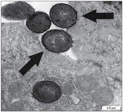
colonization factors (discussed in Chapter 25). Other genes encode outer membrane proteins that counteract host defenses, as well as regulators of virulence gene expression.
FIGURE 16.27 ■ Virulence genes of Salmonella pathogenicity island SPI2. Thirty-three virulence genes contained in nine contiguous operons (ssa, sse, and so on) help Salmonella bacteria grow within macrophages.
Source: Modified from M. P. Doyle (ed.). 2001. Salmonella species. Chapter 8 in Food Microbiology, ASM Press.
The most dangerous consequence of infection by food-borne pathogens is the production of a potentially fatal toxin. For example, E. coli O157:H7 infection of the intestines can be overcome, but the bacteria produce Shiga toxin, which can destroy the kidneys. In cases of adult botulism, Clostridium botulinum does not usually grow within the patient; the botulinum toxin comes from bacteria that grew previously in improperly sterilized food. The botulinum toxin has a highly specific effect, inhibiting synaptic vesicle fusion in the terminals of peripheral motor neurons (Fig. 16.28). Synaptic inhibition prevents activation of muscle cells, causing flaccid paralysis. Microbial toxins are discussed further in Chapter 25.
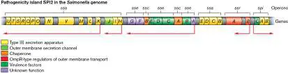
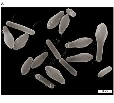
FIGURE 16.28 ■ Clostridium botulinum produces botulinum toxin. A. Club-shaped morphology of Clostridium botulinum cells containing endospores. B. Botulinum toxin inhibits synaptic vesicle fusion in the terminal of a peripheral motor neuron, preventing activation of the muscle cell.
DENNIS KUNKEL MICROSCOPY/SCIENCE SOURCE
Food Preservation
Cultural practices and cuisines have long evolved to limit food spoilage. Such practices include cooking (heat treatment), addition of spices (chemical preservation), and fermentation (partial microbial digestion). In modern commercial food production, spoilage and contamination are prevented by numerous methods that are based on fundamental principles of physics and biochemistry that limit microbial growth (discussed in Chapter 5). Processes that preserve
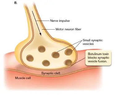
food are based on physical factors such as temperature and pressure, as well as preservative chemicals that retard microbial growth. The aim of a food preservative practice is to increase the exponential death rate of microbes present in the food (discussed in Chapter 5). For example, María Elena Sosa-Morales, at the University of Guanajuato, Mexico, conducts experiments on food disinfection procedures, such as ultraviolet irradiation of dried nuts (Fig. 16.29 ). In the experiment shown, roasted almonds and unshelled peanuts were surface sterilized with ethanol and then inoculated with Salmonella enterica serovar Typhimurium. The nuts were exposed to UV-C, ultraviolet light at wavelengths in the range of 200–280 nm. Over time, the viable colony counts of Salmonella declined as a negative exponential function. This result is consistent with the exponential decay model of microbial death described in Chapter 5. Thus, UV-C can be an effective disinfectant for dried foods, although it is limited by the need to expose food particles directly to the incident rays without shadowing.
FIGURE 16.29 ■ UV-C treatment disinfects food. Ultraviolet (UV-C) inactivation curve of Salmonella Typhimurium on peanuts and almonds.
Source: K. Ruiz-Hernández et al. 2021. Food Eng. Rev. 13 :706–712.
JOANNE LAWTON/ WASHINGTON BUSINESS JOURNAL
Dehydration and freeze-drying. Removal of water prevents microbial growth. Water is removed either by application of heat or by freezing under vacuum (known as freeze-drying, or lyophilization ). Drying is especially effective for vegetables and pasta. The disadvantage of drying is that some nutrients are broken down. Refrigeration and freezing. Refrigeration temperature (typically 4°C–8°C) slows microbial growth, as shown in an experiment comparing bacterial growth in ground beef at different temperatures (
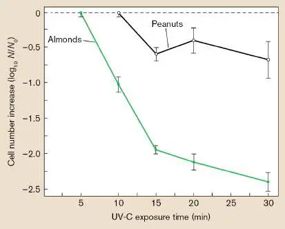
Fig. 16.30A). Nevertheless, refrigeration also selects for psychrotrophs, such as Listeria. Freezing halts the growth of most microbes, but preexisting contaminant strains often survive to grow again when the food is thawed. This is why deep-frozen turkeys, for example, can still cause Salmonella poisoning, especially if the interior is not fully thawed before roasting.
FIGURE 16.30 ■ Bacterial growth as a function of temperature and pH. A. The growth rate of total aerobic bacteria in ground beef declines at lower storage temperatures. B. Survival curves of E. coli O157:H7 in eggplant salad stored at 5°C at pH 4.0, pH 4.5, and pH 5.0. CFUs = colony-forming units. Source: Part A modified from M. P. Doyle (ed.). 2001. Meat, poultry, and seafood. Chapter 5 in Food Microbiology, ASM Press. Part B modified from Panagiotis N. Skandamis and George-John E. Nychas. 2000. Appl. Environ. Microbiol. 66 :1646.
Acid treatment. The pH of food may be decreased by microbial processes such as fermentation or by the addition of acids such as citric acid or lemon juice. Figure 16.30B shows the effect of pH on the survival of Escherichia coli O157:H7 in refrigerated Greek eggplant salad. Note the critical threshold pH required to decrease bacterial counts. At pH 4.0, about the pH of lemon juice, the bacteria show a steep exponential death curve, whereas at pH 4.5, the bacteria remain viable for many days.
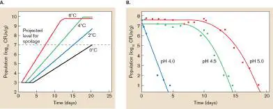
Controlled or modified atmosphere. Food can be packed under vacuum or stored under atmospheres with decreased oxygen or increased CO 2. Controlled atmospheres limit abiotic oxidation and microbial growth. For example, CO 2 storage is particularly effective for extending the shelf life of apples.
Pasteurization. Invented by Louis Pasteur, pasteurization is a short-term heat treatment designed to decrease microbial contamination with minimal effect on food value and texture (discussed in Chapter 5). For example, milk is commonly pasteurized at 63°C for 30 minutes, followed by quick cooling to 4°C.
Pasteurization is most effective for extending the shelf life of liquid foods with consistent, well-understood microbial flora, such as milk and fruit juices.
Chemical additives. Many kinds of chemicals are used to preserve foods. Major classes of chemical preservatives include organic acids and esters, as well as inorganic food preservatives such as phosphates, nitrites, and sulfites. Nitrites and sulfites inhibit aerobic respiration of bacteria, and their effectiveness is enhanced at low pH. These substances, however, may have harmful effects on humans; nitrites can be converted to toxic nitrosamines, and sulfites cause allergic reactions in some people.
Sterilization
Some practices aim for complete elimination of microbes, a practice known as sterilization (discussed in Chapter 5). How does the food industry know how long to treat food for sterilization? The exponential death function poses a challenge, as a few microbes may persist—and restart growth—long after several “half-lives” of population decay.
For heat treatment, several measures are used to define the efficiency of killing. The D-value (decimal reduction time) was defined in Chapter 5 as the time of treatment needed to decrease a microbial population by 90% (that is, by one log 10 unit). A more stringent measure is 12D, the amount of time required to kill 10 12 spores (or to decrease a population by 12 log 10 units). A different kind of measure is the z-value, the increase in degrees Celsius needed to lower the D-value to 1/10 of the time. If, for example, D 100 (the D-value at 100°C) and D 110 (the D-value at 110°C) for a given organism are 20 minutes and 2 minutes, respectively, then 12D 100 equals 240 minutes (that is, 20 minutes × 12), and the z-value is 10°C (because a 10°C increase in temperature reduced the D-value to 1/10, from 20 minutes to 2 minutes). These measurements are determined empirically for each organism. The values are extremely important to the canning industry, which must ensure that canned goods do not contain spores of Clostridium botulinum, the anaerobic soil microbe that causes the paralyzing food-borne disease botulism ( Fig. 16.28).
Because the tastes of certain foods suffer if they are overheated, z-values and 12D-values are used to adjust heating times and temperatures to achieve the same sterilizing result. Consider an example in which D 121 is 10 minutes and 12D 121 is 120 minutes. Sterilizing at 121°C for 120 minutes might result in food with a repulsive taste, whereas decreasing the temperature and extending the heating time might yield a more palatable product. The D-values and z-values are used to adjust conditions for sterilization at a lower temperature. If D 121 is 15 minutes (the time needed to kill 90% of cells) and the z-value is known to be 10°C (the temperature change needed to change the D-value tenfold), then decreasing temperature by 10°C, to 111°C, will mean D 111 is 150 minutes (10 × D 121). Therefore, the value of 12D (required to decrease a population by 12 logs) is 12D 111 = 1,800 minutes. Sterilization may take longer, but food quality is likely to remain high, because the sterilizing temperature is lower.
Canning. In canning, the most widespread and effective means of long-term food storage, food is cooked under pressure to attain a temperature high enough to destroy endospores (typically 121°C). Commercial canning effectively eliminates microbial contaminants, except in very rare cases. The main drawback of canning is that it incurs some loss of food value, particularly that of labile biochemicals such as vitamins, as well as loss of desirable food texture and taste. Ineffective canning can allow bacterial growth, causing toxin and gas production.
Ionizing radiation. Exposure to ionizing radiation, known as food irradiation, effectively sterilizes many kinds of food for long-term storage. The main concerns about food irradiation are its potential for unknown effects on food chemistry and the hazards of the irradiation process itself for personnel involved in food processing. Nevertheless, irradiation has proved highly effective at eliminating pathogens that would otherwise cause serious illness.
Thought Question
16.9 Is it possible for physical or chemical preservation methods to completely eliminate microbes from food? Explain.
To Summarize
Food spoilage refers to chemical changes that render food unfit for consumption. Food spoils through degradation by enzymes within the food, through spontaneous chemical reactions, and through microbial metabolism.
Food contamination, or food poisoning , refers to the presence of microbial pathogens that cause human disease or of toxins produced by microbial growth. Food harvesting, food processing, and shared consumption are all activities that spread pathogens.
Dairy products can be soured by excessive fermentation or made bitter by bacterial proteolysis.
Meat and poultry are putrefied by decarboxylating bacteria, which produce amines with noxious odors.
Fish and other seafood spoil rapidly because their unsaturated fatty acids rapidly oxidize, they harbor psychrotrophic bacteria that grow under refrigeration, and their trimethylamine oxide is reduced by bacteria to the fishy-smelling trimethylamine.
Vegetables spoil by excess growth of bacteria and molds. Plant pathogens destroy food crops before harvest.
Food preservation includes physical treatments, such as freezing and drying, as well as the addition of chemical preservatives such as benzoates and nitrites.
Food sterilization aims to kill all, or nearly all, microbes in a food product. Examples include canning and irradiation (exposure to ionizing radiation).
Glossary
food spoilage Microbial changes that render a food unfit or unpalatable for consumption.
rancidity Food spoilage due to the oxidation of fats; it may or may not involve microbial activity.
putrefaction Food spoilage due to the decomposition of proteins and amino acids.
food poisoning Food contamination, the presence of human-disease-causing microbial pathogens or toxins in food.
endophyte An endosymbiont of vascular plants.
pathogenicity island A type of genomic island in which the stretch of DNA contains virulence factors and may have been transferred from another genome.
freeze-drying Also called lyophilization. The removal of water from food, by freezing under vacuum, to limit microbial growth.
lyophilization See freeze-drying .
Endnotes
1. Note a: Ten major food-borne pathogens highlighted by the U.S. Public Health Service (USPHS). Return to reference a
16.5 Industrial Microbiologynot assigned
The production and preservation of food is only one field of industrial microbiology, the
commercial exploitation of microbes. Microbes may be cultured as targets for pharmaceutical
agents, generally antibiotics or antimicrobial agents for pathogens. Alternatively, microbes can
produce complex molecules, such as vitamin B 12, more cheaply and easily than by abiotic
chemistry (see Special Topic 16). The product may be generated by the original organism
whose genome possesses the pathway, but more commonly the genes encoding the pathway
may be cloned into a heterologous host (different or distantly related species). A heterologous
host may be optimized by genetic engineering for industrial control. Finally, the product may
be the microbe itself or its genome, such as vaccines and microbial vectors (viruses and
plasmids) used for genetic modification of animals and plants.
SPECIAL TOPIC 16 Microbial Vitamins for Sale
If you read the fine print of your vitamin supplement, did you ever wonder where all
those molecules come from? Many of them were made by microbes. Vitamin B 12 is an
essential vitamin in our diet, required for red blood cell formation and brain function. This
vitamin is one of the more complex molecules made by a living organism (Fig. ST 16.1
). Its synthesis requires more than 25 unique enzymes, with distinctive remodeling of a
tetrapyrrole (uroporphyrinogen III) into the asymmetrical corrin ring. An unusual feature
is the placement of the metal cobalt at the center of the heteroaromatic ring, held in
place by a metal-carbon bond to a deoxyadenosyl group.
FIGURE ST 16.1 ■ Vitamin B 12 structure. Vitamin B 12 is a complex molecule
that includes cobalt at the center of a corrin ring system.
STEPHEN BARNES/MEDICAL/ALAMY STOCK PHOTO
Since prehistoric times, vitamin B 12 has been available to the human diet via
consumption of animal products or of microbes such as yeasts. Today the vitamin is one
of many sold in purified form, often added as a supplement to processed foods. Its
industrial production requires a microbial host, most commonly a Gram-negative
bacterium, either Propionibacterium shermanii or Pseudomonas denitrificans. The
producer microbe must possess the entire biosynthetic pathway in its genome, starting
from simple catabolic substrates such as sugars.
For industrial use, however, these native vitamin producers have drawbacks. Their
fermentation takes longer than that of well-domesticated species such as Escherichia coli
. Their growth requires substrates that add expense. And the genetic tools available for
engineering them are limited. A more economical alternative would be to transfer the
biosynthetic genes into a heterologous strain that is optimized for commercial use.
Commercial strains grow well in large fermentors and catabolize inexpensive carbon
sources.
Dawei Zhang at the Tianjin Institute of Industrial Biotechnology, China, investigates
biosynthetic pathways that offer potential success in commercial production of vitamins
and amino acids. His lab is interested in developing a heterologous host strain for
economical production of vitamin B 12 (Fig. ST 16.2 ).
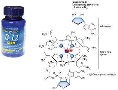
FIGURE ST 16.2 ■ Developing a heterologous producer strain for vitamin
B 12.
To construct a heterologous producer requires cloning all genes (more than 25) into
the producer host. Genes must be mined from native producer organisms (Fig. ST 16.2 ,
step 2). In the case of E. coli, the host already possesses genes for synthesis of the
pyrrole precursor, delta-aminolevulinate. Genes encoding enzymes farther down in the
pathway were transferred from the closely related species Salmonella enterica.
The biosynthetic pathway must be constructed for optimal overproduction, which
differs from the native host’s optimal needs for moderate production (Fig. ST 16.2 ,
step 3). Most important, all the native regulatory elements, such as promoters,
activators, and ribosome-binding sites (RBS), must be deleted. For example, the B 12
riboswitch (see Chapter 10) detects the vitamin molecule at a low concentration and
terminates transcription and translation. This feedback regulation makes sense for the
native host but is undesirable for industrial production, which aims for as much product as
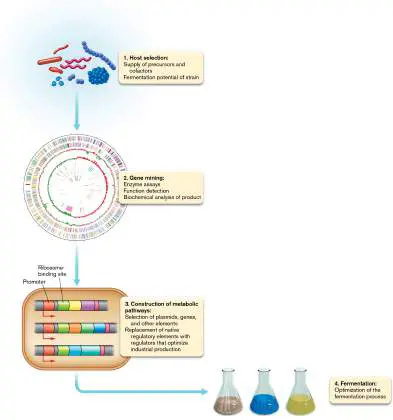
possible. The native regulatory elements are then replaced by regulators that avoid
bottlenecks (imbalance of substrate and product at intermediate steps) and optimize
production per unit substrate. Finally, environmental variables such as media components
and temperature are adjusted to optimize the rate of fermentation (step 4). Ultimately,
the aim is to produce more of the vitamin at lower cost to consumers, while increasing
profits for the company shareholders.
RESEARCH QUESTION
What kind of DNA regulatory elements might you select to replace the native ones? What
regulatory behavior would you aim for in the producer strain?
Fang, Huan, Jie Kang, and Dawei Zhang. 2017. Microbial production of vitamin B 12: A review and
future perspectives. Microbial Cell Factories 16 :15.
And researchers are still finding new ways to use microbes. For example, in 2021, a new
drug for COVID-19 called molnupiravir was developed by the companies Merck and Ridgeback
Biotherapeutics. The drug, which halved the rate of hospitalizations and deaths in a clinical
trial, is produced by a “biocatalytic cascade”; that is, a route to chemical synthesis that
includes five microbial enzymes. Each enzyme was originally isolated from a microbe; for
example, Novozym 435, a lipase that originated from the Antarctic yeast Candida antarctica.
Now companies such as Novozymes engineer microbes to produce industrial enzymes that
vastly extend our pharmaceutical capabilities (see eResearch Activity 16). Other fields of
industrial microbiology include wastewater treatment, bioremediation, and environmental
management, which are covered in Chapters 21 and 22.
Case Example: A Microbiologist Founds a Company
Carol Nacy (Fig. 16.31A ) studied tropical infectious diseases for 17 years at the Walter Reed
Army Institute of Research. She saw the need to fight tuberculosis (TB), a disease that infects
one-quarter of the world’s population and kills over a million people annually. The United
States spends $1 billion yearly to treat 9,000 incident cases, yet the standard antibiotics for
tuberculosis were developed before 1970, and the main diagnostic test available (the
tuberculin skin test) dates to 1880, the time of Robert Koch. The best available vaccine—the
Bacille Calmette-Guérin (BCG) live, attenuated vaccine—is only 50% effective.
FIGURE 16.31 ■ Sequella develops antibiotics to fight tuberculosis. A. Carol
Nacy founded the Sequella company. B. A new antibiotic for tuberculosis is obtained by
screening ethambutol analogs. A combinatorial library of compounds containing the
ethambutol diamine core (yellow) was screened for antibacterial effect against
Mycobacterium tuberculosis. The most promising agent screened was SQ109, which has
an unusual carbon-cage side group (pink).
Nacy obtained business experience by working for several years as a chief scientific officer
for the pharmaceutical company EntreMed, Inc. She then founded her own company, Sequella,
to develop innovative drugs and treatment devices. Sequella targets innovative ideas that
have high risk but also high potential to improve performance, rapidity, and safety of
diagnosing and treating TB infections. Nacy and her cofounders scanned the academic
community for novel ideas that had succeeded against TB in “proof of principle” animal
models. The most promising ideas were developed for improved antibiotics, rapid and less
invasive tests for TB exposure, and devices to measure the extent of pulmonary infection.
Because any one idea had a high risk of failure, researchers pursued multiple prospects in each
category. The company also took on multiple target pathogens, including methicillin-resistant
Staphylococcus aureus (MRSA) and vancomycin-resistant Enterococcus (VRE).
Sequella’s most promising antibiotic, SQ109, was discovered via high-throughput screening
of a chemical library, in collaboration with Clifton Barry at the National Institutes of Health.
The chemical library consisted of over 60,000 analogs of a known TB antibiotic, ethambutol (
Fig. 16.31B ). Ethambutol is part of the current standard treatment for TB, whose 6-month
time course has a poor compliance rate. It is hoped that improved drugs will shorten the time
course and improve compliance, thereby decreasing the appearance of drug-resistant strains.
The analog molecules were selected for their common diamine core but different combinations
of side chains. The 60,000 compounds in the library were subjected to combinatorial
screening, a mathematically intensive analysis that is based on numerous tests. Of the
compounds tested, 2,796 showed activity against Mycobacterium tuberculosis in the test tube.
The 69 best compounds were tested for cytotoxicity in tissue culture, activity in TB-infected
macrophages, and activity in infected animals. The compound with the greatest efficacy and
fewest side effects was SQ109, a molecule with an unusual cage-like side group of three fused
rings (Fig. 16.31B ).
In 2018, SQ109 passed a phase IIb/III human clinical trial. The aim of the trial was to test
the efficacy and safety of the molecule in human patients. The drug was tested in seven
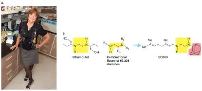
clinical centers in Russia, where the tuberculosis infection rate is high. A significantly greater
number of patients treated with SQ109 showed clearing of tuberculosis bacteria in their
sputum, compared with control patients on placebo. The drug also shows promise for
treatment of Helicobacter pylori, as well as the parasites Leishmania and Trypanosoma cruzi.
Industrial Microbiology Aims for Commercial Success
The practical application of a microbial product or device may arise out of an industrial
laboratory or it may be conceived by a research scientist such as Nacy with the aim of meeting
a compelling need in society. In all cases, however, the key goal is to succeed in the
marketplace; that is, to generate a product that customers adopt over alternative
technologies. The product’s sales must cover the costs of raw materials and production and (in
a for-profit company) generate a profit for the shareholders. Success requires:
Identifying a useful product. Possible products include small molecules such as
antibiotics; human proteins from cloned genes; or microbial proteins that have useful
properties, such as thermostability or increased catalytic activity for a chemical process.
Isolating a microbe to produce the product. A novel product, such as an antibiotic, is
generally developed from a naturally occurring microbe. The genes encoding product
biosynthesis may then be cloned into an industrial vector.
Scaling up production in quantity. The producer microbial strain must be grown on an
industrial scale, and the product must be isolated and purified.
Developing a business plan. The scientist-entrepreneur must obtain partners skilled in
industrial management, finance, and marketing. Patents must be filed to protect
intellectual property rights.
Safety and efficacy testing. Human consumption, environmental introduction, or
consumer use requires many levels of testing prior to commercialization, including, in
some cases, approval by government agencies.
Effective marketing. The benefits of the new product must be communicated effectively
to convince customers of its superiority over current products or processes.
Failure at any of these tasks spells doom for the product. Thus, a prudent business plan
includes having multiple alternative products in development. Although the failure rate of new
products is high, all the products we use had to overcome these risks.
Microbial Products
An important source of microbial products is cloned human genes that encode a protein of
valuable function in the human body. For example, Therabron Therapeutics produces the
recombinant human protein CC10, a lung development protein that is often deficient in the
lungs of premature infants. The recombinant protein, produced and purified from a
recombinant bacterium, can be used to reduce lung inflammation in premature infants, as well
as in patients with chronic obstructive pulmonary disease (COPD). Cloning of gene products in
recombinant organisms was discussed in Chapter 7.
Increasingly, industry mines natural sources for novel microbes that produce useful products
(Table 16.4 ). Each product requires a gene or operon of genes encoding either the product
itself or the enzymes for the product’s biosynthesis. For example, the Danish company
Novozymes markets over 700 microbial enzymes for products ranging from laundry detergents
to agricultural inoculants. Novozymes also produces numerous enzymes as biocatalysts for
green chemistry. “Green chemistry” refers to environmentally friendly procedures for reactions
in organic chemistry, typically using water solution in place of petroleum-derived organic
solvents. Microbial enzymes often replace earlier chemical processes that were more costly,
generated greater waste, and had more negative effects on environmental quality and global
climate.
TABLE Consumer Products Made with Industrial 16.4 Microbiology
Old New industrial
manufacturing microbial Climate Consumer
Product process process benefits benefit
Bread Potassium Genetically Reduction of CO High-quality
bromate, a enhanced 2 emissions in bread; longer
suspected microorganisms grain shelf life;
cancer- produce baking production, eliminates
causing agent, enzymes to milling and suspected
added as a enhance rising, baking, and carcinogen
preservative strengthen transportation potassium
and a dough- dough, and bromate
strengthening prolong
agent freshness.
Vitamin B 2 Toxic chemicals, Lactobacillus Up to 33% Greatly reduces
(Riboflavin) such as developed for reduction in hazardous
aniline, used one-step energy use; waste
in a nine-step fermentation 25%–33% generation
chemosynthes process uses reduction in and disposal
is process vegetable oil as CO 2
(hazardous feedstock and emissions
waste sugar as
generated) nutrient.
Personal Chemicals such Genetically 20% reduction High purity;
care as propylene enhanced of environmenta
glycol and microbe greenhouse lly
butylene produces 1,3- gas emissions sustainable
glycol from propanediol compared to and
petroleum from renewable 1,3- renewable
used as feedstocks, propanediol process;
solvents to which can from nonirritating
mix function as a petroleum for sensitive
ingredients solvent, skin;
humectant, enhanced
emollient, or clarity
hand-feel
modifier.
TABLE Consumer Products Made with Industrial 16.4 Microbiology
Old New industrial
manufacturing microbial Climate Consumer
Product process process benefits benefit
Detergent Phosphates Microbes or fungi Elimination of Brighter,
added as a produce water cleaner
brightening enzymes, which pollution due clothes with
and cleaning are added as to phosphates lower wash
agent brightening and temperature;
cleaning agents. energy
Protease savings
enzymes remove
protein stains;
lipases remove
grease;
amylases
remove starch.
Textiles New cotton Microbial cellulase 25% decrease New fabrics
textiles enzymes in greenhouse have lower
prepared with produce gases; 25% impact on the
chlorine or peroxides, decrease in environment,
chemical allowing nonrenewable better dyeing
peroxide bleaching of energy use results, and a
bleach textiles at low permanent
temperature soft and
(65°C) and in a bulky handle
neutral pH
range.
Paper Wood chips are Wood-bleaching Reduction in Cost savings
boiled in a enzymes are use of from lower
harsh produced by chlorine energy and
chemical genetically bleach, and in chemical use
solution to enhanced dioxins in the
yield pulp for microbes to environment
papermaking selectively
degrade lignin
and break down
wood cell walls
during pulping.
Diapers Woven fabric Bacillus ferments 50%–70% Biodegradable;
coverings corn sugar to decrease in disposal
made from lactic acid, which CO 2 options
petroleum- is heated to emissions include
TABLE Consumer Products Made with Industrial 16.4 Microbiology
Old New industrial
manufacturing microbial Climate Consumer
Product process process benefits benefit
based generate a composting
polyesters biodegradable rather than
polymer for landfills
woven fabrics.
Polyesters Polyester, a Bacillus ferments 75% decrease Polylactic acid
synthetic corn sugar to of CO 2, (PLA)
polymer fiber, lactic acid, which compared to polyester is
produced is heated to PET; 90% biodegradabl
chemically generate a reduction of e, does not
from biodegradable CO 2 harbor body
petroleum polymer (such equivalent odors, and
feedstock as NatureWorks’ compared to does not give
Ingeo). nylon 6 off toxic
smoke if
burned
Stonewashed Open-pit mining Fabric is washed Less mining; Softer fabric;
jeans of pumice; with microbial decreased lower cost
fabric washed enzymes energy
with crushed (cellulases) to consumption
pumice stone fade and soften
and/or acid jeans or khakis.
Enzymes Chemical Alpha-amylase— Decreased Lower cost
processes Bacillus subtilis energy
using Amyloglucosidase consumption;
materials — Aspergillus decreased
generally niger (fungus) pollution of
derived from Lactase (beta- environment
petroleum galactosidase)—
Kluyveromyces
lactis (fungus)
Lipases— Candida
cylindraceae
(yeast)
Alkaline protease
— Aspergillus
oryzae (fungus)
TABLE Consumer Products Made with Industrial 16.4 Microbiology
Old New industrial
manufacturing microbial Climate Consumer
Product process process benefits benefit
Food Derived from l-Lysine— Decreased Lower cost
supplements petroleum Brevibacterium energy
lactofermentum consumption;
l-Tryptophan— decreased
Klebsiella pollution of
aerogenes environment
Monosodium
glutamate
(MSG)—
Corynebacterium
ammoniagenes
Vitamin B 12 —
Pseudomonas
denitrificans
Vitamin C
(ascorbic acid)—
Acetobacter
suboxidans
Sources: Biotechnology Innovation Organization, 2018 (http://www.bio.org); and M. J. Waites. 2001.
Industrial Microbiology. Blackwell Science.
To identify new microbial products, companies screen thousands of microbial strains from
diverse ecosystems. The search for organisms with potential commercial applications is called
bioprospecting. Bioprospecting can be done anywhere, from one’s backyard to Yellowstone
National Park. Unique ecosystems are the most promising sources of previously unknown
microbial strains that have valuable properties. Extreme environments such as the hot springs
of Yellowstone are particularly promising because their products may tolerate higher
temperatures that are required for industrial use. Psychrophiles from extremely cold
environments such as Antarctica are a useful source of enzymes that are active in a wide
range of temperatures, such as enzymes to clean clothing.
An important aspect of bioprospecting is “mining the genome.” Once a promising source
strain is obtained, its genome is sequenced to investigate the gene sequences that encode the
useful product and regulate its expression. The operons encoding the product (or enzymes for
its production) can then be cloned for optimal production. The cloned genes are transferred
into an industrial strain, a strain whose growth characteristics are well studied and optimized
for industrial production. An industrial strain must possess the following attributes:
Genetic stability and manipulation. The industrial strain must reproduce reliably,
without major DNA rearrangements. It must also have an efficient gene transfer system
by which vectors can introduce genes of interest into its genome.
Inexpensive growth requirements. Industrial strains must grow on low-cost carbon
sources and with minimal special needs, such as vitamins, and must grow at easily
maintained conditions of temperature and gases.
Safety. Industrial strains must be nonpathogenic and must not produce toxic by-products.
High level of product expression. The strain or recombinant vector must possess an
efficient gene expression system to generate the desired product as a high proportion of
its cell mass.
Ready harvesting of product. Either the product must be secreted by the cell or, if the
product is intracellular, the cells must be easily breakable to liberate the product.
Common species for industrial strains include the bacteria Escherichia coli and Bacillus subtilis,
the yeast Candida utilis, and the filamentous fungus Aspergillus niger. Each of these species
is safe; grows to high density on inexpensive carbon sources, such as molasses; and expresses
desired products at high concentration.
Thought Question
16.10 Why would different industrial strains or species be used to express different kinds of
cloned products?
Fermentation Systems
Commercial success requires optimizing every detail of the fermentation system.
“Fermentation” in industrial terms refers not just to anaerobic metabolism but to all means of
growth of microbes on an industrial scale. In an industrial fermentor, the growth vessel and all
its environmental supports, such as temperature control and oxygenation, must be scaled up
to thousands of liters (Fig. 16.32 ). This increase in scale generates many problems of
quality control, such as maintaining uniform temperature, pH, and oxygenation throughout the
vessel and minimizing foaming of the culture liquid. A small change in any of the growth
factors can affect production costs and profit margin. Another major concern is to avoid
contamination by other organisms.
FIGURE 16.32 ■ Industrial fermentation. A. Industrial production of microbial
products requires scaled-up culture of the production microorganism. B. An industrial
fermentor.
MAXIMILIAN STOCK LTD./SCIENCE SOURCE
The fermentor is the core of the first half of industrial production, known as upstream
processing (Fig. 16.33 , top). Upstream processing is the culturing of the industrial microbe
to produce large quantities of product or cell mass. All aspects of the process must be
controlled to maximize the final concentration of product, which in most cases peaks at a
specific time in the microbial growth cycle. After microbial growth, the culture must be
harvested and the product purified. These processes constitute downstream processing (Fig.
16.33 , bottom). The first step of downstream processing is to separate the microbial cells
from the culture fluid by centrifugation or by filtration. Next, the primary recovery of product
follows one of two different pathways, depending on whether the product is maintained within
the cells or secreted into the culture fluid. Many kinds of subsequent purification and finishing
steps are necessary before the product has acceptable quality for its desired use. Again, failure
of any detail can render the entire product unusable.
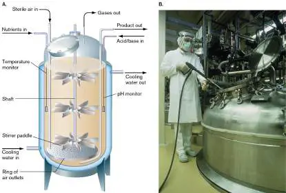
FIGURE 16.33 ■ Details of upstream and downstream processing. Top:
Upstream processing consists of engineering the microbial strain and large-scale growth
to generate the product. Bottom: Downstream processing consists of product
concentration and purification.
Products designed for human consumption or use in the environment face formidable
hurdles in clinical testing, toxicological studies, and, finally, approval by the appropriate
regulatory agency, such as the FDA. After millions of dollars are invested in process
development, the product may still fail one of these late-stage hurdles and never come to
market. Not surprisingly, a company must research thousands of potential products before
achieving one that makes a profit. The consumer cost inevitably includes the development
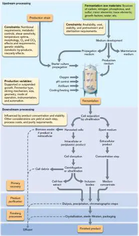
costs not only of the one successful product, such as recombinant insulin, but also of all the
products that failed.
Microbe as Product
Microbes as biological agents possess enormous potential for environmental use. The best-
known example is the bacterial insecticide Bacillus thuringiensis. Such microbes, derived from
natural sources, may improve crop production and enhance agricultural sustainability. Most
methods used by farmers to improve crop yields require large amounts of fertilizer and pest
management, including herbicides and fungicides. To alleviate the financial and environmental
effects of large-scale farming, scientists are working to develop microbial solutions that can
supplement chemical additives and pesticides, resulting in higher yields to farmers.
Scientists screen thousands of microbes for positive effects on agriculture. One
example is JumpStart, which consists of the mold Penicillium bilaiae (Fig. 16.34).
The mold solubilizes phosphate from the soil and makes it available to the roots of
plants, whose exudates feed the mold. This phosphate-solubilizing inoculant improves growth
for a variety of crops. Its enhancement of plant nutrition is comparable to the relationships
between many kinds of fungi and plant roots found in natural ecosystems (discussed in
Chapter 21).
FIGURE 16.34 ■ A bacterial supplement for agriculture. Penicillium bilaiae
solubilizes phosphates from the soil, making it available for plant growth. Plate culture.
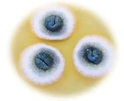
Source: Nelly S. Raymond et al. 2018. Process Biochem. 69 :169.
N. S. RAYMOND ET AL. 2018. SCIENCE DIRECT 69 :169–177. WITH PERMISSION FROM ELSEVIER
Another microbial product is Actinovate, a biological fungicide used for the suppression of
root rot and damping-off fungi, as well as the suppression or control of leaf fungal pathogens.
The active ingredient of Actinovate is the actinomycete Streptomyces lydicus. S. lydicus grows
as a mutualist associated with the roots of crop plants. The bacteria feed off plant exudates
while secreting antimicrobial substances that suppress pathogens. These examples are the
start of a growing field of microbial applications for improving agriculture in an environmentally
sustainable manner.
To Summarize
Industrial microbiology includes the production of vaccines and clinical devices,
industrial solvents and pharmaceuticals, and genetically modified organisms.
Bioprospecting is the mass screening of new microbial strains for promising traits and
potential use in a product. Thermophiles and psychrophiles are particularly important
sources of new strains that potentially have interesting new properties.
Microbial products must be competitive with alternative technologies. Developing
a competitive new molecular product requires identifying a useful molecule, isolating
and developing a fermentation technique to produce it, scaling up production in
quantity, developing a business plan, and testing for safety.
Industrial strains , commonly Escherichia coli or Bacillus subtilis, are often used to
incorporate the newly discovered genes into an industrially useful microbe.
Upstream processing is the culturing of the industrial microbe to produce large
quantities of product. Downstream processing involves product recovery and
purification.
Glossary
industrial microbiology
The commercial exploitation of microbes.
heterologous host
An organism engineered for expression of a recombinant product from genes obtained
from a species that expresses the product in a natural environment.
bioprospecting
The search for organisms with potential commercial applications.
industrial strain
A microbial strain whose characteristics are optimized for industrial use.
industrial fermentor
The equipment used to grow microbes on an industrial scale.
upstream processing
The culturing of industrial microbes to produce large quantities of a desired product.
downstream processing
The recovery and purification of a commercial product produced by industrial microbes.
primary recovery
The initial isolation of commercial product from industrial microbes.
16.6 Vaccines and Gene Therapy Vectorsnot assigned
One of the most important microbial products—one we often take for granted—is vaccines. Vaccines consist of a pathogenic cell or cell component that is used to induce immunity to disease (discussed in Chapter 24). The production of vaccines, such as the new COVID-19 messenger RNA (mRNA) vaccines, presents immense challenges for efficacy and safety. Another industry that requires manipulation of potential pathogens is that of virus-delivered gene therapy. Viruses provide the vectors (carriers) for transferring genes into multicellular organisms. Later in this section we discuss the example of lentiviral vectors for human gene therapy. These therapeutic vectors, remarkably, are derived from the deadly retrovirus HIV (discussed in Chapter 11).
Producing a Messenger RNA Vaccine
The Pfizer-BioNTech and Moderna vaccines for SARS-CoV-2 were the first in history to use mRNA to stimulate immunity. For context, recall that the first vaccines discovered were made from entire virus particles, such as the anthrax and rabies vaccines discovered by Louis Pasteur (1822–1895), as presented in Chapter 1. Most early vaccines were live, attenuated vaccines, in which a live virus was weakened by procedures such as heat treatment or by mutation as in the polio vaccine devised by Albert Sabin (1906–1993). The drawback of live vaccines is the potential for rare cases of disease. Other vaccines were made from inactivated (killed) virus, such as the polio vaccine devised by Jonas Salk (1914–1995). Inactivated viruses are incapable of starting infection. By the twenty-first century, vaccine manufacturers were replacing entire-virus formulations with vaccines derived from one or more components, such as capsid subunit proteins produced by engineered industrial bacteria. These cloned products are called subunit vaccines. The most well-known subunit vaccine is Gardasil, for human papilloma virus (HPV), developed by Merck and first approved by the FDA in 2006. The mechanisms of immune response are discussed in Chapter 24.
A messenger RNA (mRNA) vaccine is made from a cloned gene that expresses mRNA encoding an immunogenic viral protein, such as the spike protein of a coronavirus. In recent years, researchers discovered that a cloned mRNA molecule induces a stronger immune response than do cloned proteins. This is because the mRNA is taken up by host cells, which then express the viral protein on the cell surface, stimulating cellular T-cell immunity in addition to a B-cell immune response (discussed in Chapter 24).
As you can imagine, the commercial production of an mRNA vaccine poses extraordinary challenges: How to stabilize the fragile mRNA molecule? The mRNA can be stabilized within a coating of lipids, forming lipid nanoparticles. Some of the lipids possess amine groups with a stable positive charge. Positive charge helps the lipids interact with the negatively charged mRNA to form the nanoparticle. How can the mRNA enter host cells? The hydrophobic lipid part of the nanoparticles dissolves in the host membrane, carrying the mRNA into the cell.
How is quality controlled throughout the manufacturing process? At numerous points, electrophoretic gels and other analysis methods must test samples of the DNA, mRNA, and lipid components to ensure correct structure and function and the absence of contaminants.
The steps of manufacturing a coronavirus mRNA vaccine are outlined in Figure 16.35.
FIGURE 16.35 ■ Manufacturing a messenger RNA vaccine. The gene encoding SARS-CoV-2 spike protein is cloned in a plasmid and synthesized by Escherichia coli in an industrial fermentor. The plasmid is cut to release the spike gene, which is transcribed to mRNA. The mRNA is mixed with positively charged lipids to form nanoparticles. The nanoparticles are stabilized in solution and packed as vaccine.
COLIN CUTHBERT/SCIENCE SOURCE
The manufacture of the coronavirus mRNA vaccine is based on a foundation of much research demonstrating the potent antigenicity of the coronavirus spike protein (presented in Chapter 6). The gene encoding SARS-CoV-2 spike protein is cloned in a plasmid and transformed into an industrial strain of Escherichia coli (transformation is discussed in Chapter 9). The industrial strain is engineered for growth in a large industrial fermentor, where it achieves a high biomass. From the bacteria, the plasmid is cut by
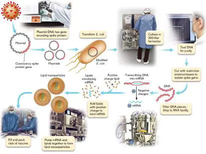
restriction enzymes to release the spike gene. The DNA gene undergoes testing by gel electrophoresis, to ensure that the correct sequence is taken further.
The cloned spike genes are then sent to a different facility, which undertakes in vitro transcription to make mRNA. Synthesis of mRNA requires a recombinant RNA polymerase enzyme, as well as enzymes adding the 7-methylguanosine “cap” and the 5′ poly(A) “tail.” [The poly(A) tail, alternatively, may be included in the cloned DNA gene.] The mRNA requires these modifications for eventual recognition by ribosomes within the cells of the immunized patient. The manufactured mRNA must be isolated away from the DNA template and subjected to further quality control testing. At the same time, several kinds of lipids are synthesized and tested, including lipids with positive charges to bind the negatively charged phosphoryl groups of RNA. Finally, solutions containing the lipids and the mRNA are pumped together in a highly controlled method to form nanoparticles. The nanoparticles are stabilized in solution and packed as vaccine.
All aspects of the manufacturing process require years to optimize and ensure consistent product. Then the completed vaccine package requires cold storage in order to maintain the mRNA intact within the nanoparticles. Cold storage and time limits for use play key roles in all public health campaigns to vaccinate the public.
Thought Question
16.11 What might be the relative advantages and limitations of a live, attenuated virus vaccine, as compared with a messenger RNA vaccine?
Lentiviral Vectors for Human Gene Therapy
Viruses that infect humans and integrate in our DNA cause some of the world’s most devastating diseases, such as AIDS. Yet the very properties that make these viruses such effective pathogens can provide the greatest promise of effective therapy for incurable genetic conditions such as inherited blood disorders and brain defects, as well as therapy for blood cancers. The most promising vectors are derived from HIV, owing to this virus’s ability to integrate into the genome of nondividing cells—and to avoid activating oncogenes.
The viral basis of gene therapy was discussed in Section 11.4. Here we present the industrial perspective: Once viral vectors are approved for therapy, how are they produced and quality assured? We focus on one class of viral vector: the lentiviral vector, or lentivector. Amazingly, a lentiviral vector is now approved by the FDA as a standard cure for certain B-cell leukemias (see Section 11.4). The challenges of lentivector production are addressed by Bruce Levine, at the Center for Cellular Immunotherapies in Philadelphia. Levine has focused on the use of lentivectors in chimeric antigen receptor T-cell therapy (CAR-T therapy) for B-cell leukemia. (For a discussion of B cells and T cells, see Chapter 24.) In 2018, CAR-T therapy for leukemia was the first lentiviral therapy approved by the FDA. In 2022, another remarkable form of CAR-T treated patients with lupus erithematosus, a disease in which the immune system generates antibodies that react with one's own organs.
CAR-T therapy. For CAR-T therapy, the patient’s own T cells are removed for modification by the lentivector (Fig. 16.36). The T cells are obtained by leukapheresis, a process in which the patient’s blood goes through a machine that separates out certain classes of cells, in this case T lymphocytes. The lymphocytes are separated by size and other properties; their identity can be tested by a fluorescence-activated cell sorter (FACS; see Chapter 24). After lentivector treatment, the cells are returned to the patient. FIGURE 16.36 ■ Patients’ T-cells are obtained for CAR-T therapy. A patient undergoes leukapheresis (A) to provide T cells for CAR-T therapy (B) .
Source: Modified from B. L. Levine et al. 2017. Mol. Ther. Methods Clin. Dev. 4:92–101, fig. 1.
COLIN CUTHBERT/SCIENCE SOURCE
The CAR-T lentivector integrates a gene into the T-cell DNA to express a chimeric antigen receptor (T-cell receptor, or TCR) that recognizes and attacks malignant B cells. The antigen receptor is then expressed by the helper CD4 T cells and cytotoxic CD8 T cells (discussed in Chapter 24). The chimeric TCR enables those T cells to recognize a B cell–specific antigen (CD19) found on malignant B cells and attack them. Normal B cells are also destroyed, but further immunotherapies can compensate.
The chimeric (fused gene) antigen receptor enables T cells to recognize the B cells growing out of control. To transfer the CAR gene into recipient T cells, lentivectors have several advantages. They express the HIV capsid, which enables the vector particle to enter the nuclear pore complex of human cell nuclei. Lentivectors
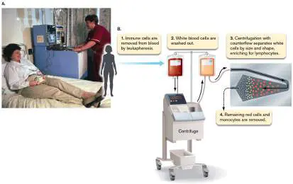
are capable of integration at many different places in the human genome, but for unknown reasons they avoid integrating at sites that will start overexpression of oncogenes. The lentiviral cell envelope can undergo pseudotyping, the replacement of envelope proteins by those of another virus such as vesicular stomatitis virus (envelope protein VSV-G). Pseudotyping can enable lentivectors to infect and integrate within a wide range of cell types.
Lentivector design and production. CAR-T therapy requires a dependable large supply of the lentivector. How do we generate large quantities of the lentivector in a process that avoids any chance of viral replication in humans? For safe production, the lentiviral genome is divided into four plasmids: a vector plasmid, two packaging plasmids, and an envelope plasmid (Fig. 16.37; see also Fig. 11.33). Only the vector plasmid has the long terminal repeats (LTRs) that enable its RNA transcript to be packaged in virions (lentivector particles).
FIGURE 16.37 ■ The lentiviral vector is made by a host cell with four plasmids. The vector plasmid encodes a gene of interest, flanked by lentiviral long terminal repeat (LTR) sequences that enable integration into the host cell genome. These sequences have been adjusted to be self-inactivating (SIN); that is, no further recombination occurs after genome
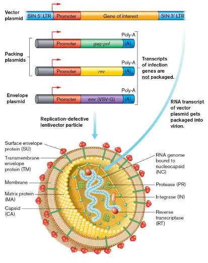
integration. Packaging plasmids encode the Gag capsid protein and the Pol reverse transcriptase protein, which are needed to form infective virus particles. The envelope plasmid encodes VSV-G envelope protein from vesicular stomatitis virus.
Source: Modified from M. C. Milone and U. O’Doherty. 2018. Leukemia 32:1529–1541, fig. 2.
When the lentiviral vector infects a host cell from the patient, only the RNA genome carrying the gene of interest (chimeric antigen receptor, CAR) integrates into the host cell genome. The plasmids from the vector-producing cells had expressed proteins needed to accomplish integration of the desired gene but are not replicated, so they disappear without any chance of future virus propagation. In the vector production culture, the vector plasmid encodes the CAR gene flanked by lentiviral long terminal repeat (LTR) sequences that enable integration into the host cell genome, as discussed in Section 11.4. The LTR sequences have been modified by deletion of a promoter sequence. This promoter deletion, called SIN (for “self-inactivating”), prevents future transcription of the full-length retrovirus from its position integrated in the host genome. Only the CAR gene is expressed from its own promoter. The packaging plasmids encode the Gag capsid protein and the Pol reverse transcriptase and integrase, which are needed to form infective virus particles. The envelope plasmid encodes VSV-G envelope protein from vesicular stomatitis virus.
To produce the replication-defective vector particles, the four plasmids are obtained from frozen storage (Fig. 16.38) and transfected into a tissue culture line such as HEK293T cells. Unlike the patient’s cells, these culture cells were derived from an epithelial cell line but now contain many modifications that enable rapid protein expression to form virions. Sterility and clean room conditions are important because the CAR-T vector particles cannot undergo complete sterilization. Over several days, the cell medium is exchanged from the transfected cells, and the progeny vector particles are harvested. Filtration removes cell debris, and benzonase enzyme removes traces of DNA from the cell culture; only the RNA viral particles remain. The final lentivector is stored at −70°C.
FIGURE 16.38 ■ Commercial production of a CAR-T lentivector. The HEK293T host cells undergo cell expansion (culture growth) for 10 days. The cultured cells are transfected with the four plasmids of CAR-T lentivector (see Fig. 16.38). Media exchange enables harvest of large quantities of lentivector. The lentivector suspension is filtered to remove cell debris. The product is stored at −70°C.
Source: Modified from B. L. Levine et al. 2017. Mol. Ther. Methods Clin. Dev. 4:92–101, fig. 3.
CAR-T production requires extreme quality control at all stages of production and use of the lentivector. The original cell culture must
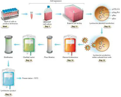
be kept free of contaminating viruses and cells such as mycoplasmas, a problem for all cell lines. During production of the lentiviral particles, filtration can remove some contaminants, but complete purity is challenging to obtain. At every stage of production, good manufacturing practice (GMP) must be maintained. GMP regulations mandate maintaining clean room conditions, avoiding open processing, and testing at every stage. The final stage of lentivector production must avoid the presence of any packaging cells (host cells) from the transfected culture.
Thought Question
16.12 Why do you think the SIN mutation is important in the lentivector, even though the integrated viral sequence lacks all the genes for virus replication (provided on the original plasmids)?
To Summarize
An mRNA vaccine consists of a cloned viral spike mRNA. The mRNA is expressed in vitro from a gene cloned on a bacterial plasmid.
The vaccine mRNA is packaged in lipid nanoparticles. Manufacture requires DNA amplification, transcription to mRNA, and packaging in lipids with positive charge. Lentiviral vectors, or lentivectors, are derived from HIV. The HIV virus can integrate in the genome of nondividing cells, and it avoids activating oncogenes. CAR-T therapy uses a lentivector to modify the T cells of a cancer patient. The CAR (chimeric antigen receptor) gene enables T cells to recognize malignant B cells and destroy them.
Production of lentivector particles starts with transfection of cultured host cells by plasmids that express genes needed for virus infection and genome integration. After genome integration, the plasmids are left behind, and the vector genome is trapped in the host genome. SIN promoter deletions ensure that the entire viral genome cannot be transcribed from the host.
CAR-T virus particles are produced in large quantities from tissue cultures. Extremely clean rooms, ultrafiltration, and product testing are required at all stages.
Glossary
messenger RNA (mRNA) vaccine An mRNA vaccine is made from a cloned gene that expresses mRNA encoding an immunogenic viral protein. The mRNA is encased in liposomes for delivery to the body.
nanoparticle A particle of matter that ranges in size from approximately 1 to 100 nanometers.
lentivector or lentiviral vector A gene transfer vector derived from a lentivirus such as HIV; designed to integrate genes into a host chromosome.
chimeric antigen receptor T-cell therapy (CAR-T therapy) CAR-T therapy uses a lentivector to modify the T cells of a cancer patient. The CAR (chimeric antigen receptor) gene enables T cells to recognize malignant B cells and destroy them. Fig. 16.37 FIGURE 16.37 ■ The lentiviral vector is made by a host cell with four plasmids. The vector plasmid encodes a gene of interest, flanked by lentiviral long terminal repeat (LTR) sequences that enable integration into the host cell genome. These sequences have been adjusted to be self-inactivating (SIN); that is, no further recombination occurs after genome integration. Packaging plasmids encode the Gag capsid protein and the Pol reverse transcriptase protein,
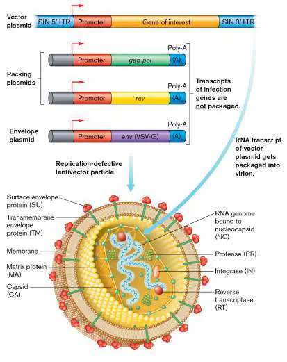
which are needed to form infective virus particles. The envelope plasmid encodes VSV-G envelope protein from vesicular stomatitis virus.
Source: Modified from M. C. Milone and U. O’Doherty. 2018. Leukemia 32:1529–1541, fig. 2.
Fig. 11.33 FIGURE 11.33 ■ Lentivector with helper plasmids. The lentivector genome consists of an RNA sequence containing HIV signal elements required for genomic integration (dark blue), promoter and regulator elements (red) derived from various other viruses, and the therapeutic human transgene (orange). In order to produce the virions in cell culture, essential HIV genes are provided on DNA helper plasmids. Their expression is driven by regulatory elements from other viruses (red).
Source: Modified from A. Blesch. 2004. Methods 33 :164.
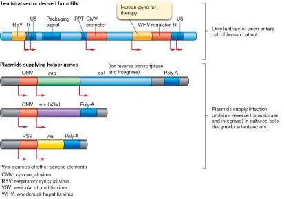
eResearch Activity 16
How Did Microbial Enzymes Make the First COVID-19 Drug?
In 2021, the Merck company announced that molnupiravir was the first oral medication to receive government approval for treatment of severe COVID-19 (Fig. ERA 16.1 ). Everyone’s first question was: How can we get this drug? And the next question was: How much will it cost? The original route of organic synthesis required ten steps of chemical conversion, losing more than 90% of the product. But Merck scientists developed an ingenious synthesis that cut the process down to three steps with 69% yield. What was their secret? Five microbial enzymes saved the day—enzymes evolved by diverse naturally occurring microbes and produced commercially by industrial culture at Novozymes.
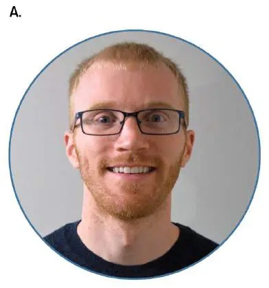
FIGURE ERA 16.1 ■ Discovering a new antiviral agent, molnupiravir. A. Patrick Fier, at Merck, devised an industrial synthesis pathway for molnupiravir. B. The molnupiravir structure is a nucleoside analog, ribose condensed to N -hydroxycytidine. The 5′ isobutyric acid group is hydrolyzed within the patient’s body, allowing viral incorporation into RNA.
PATRICK FIER
The structure of molnupiravir consists of a ribose condensed with a modified cytosine (N -hydroxycytidine). A 5′ isobutyryl group acts as a leaving group inside the patient’s body; its slow hydrolysis gradually converts the “prodrug” into the active form. The active form is recognized as cytidine nucleotide by the coronavirus RNA– dependent RNA polymerase, which incorporates it into genomic RNA. But N -hydroxycytidine tautomerizes to base-pair like uridine, leading future rounds of synthesis to replace G with A. The result leads to C/U transition mutations at such a high rate that the progeny viruses lose infectivity, an effect called “error catastrophe.”
At Merck, senior scientist Patrick Fier devised a simplified synthesis from three commodity chemicals; that is, chemicals already produced in bulk and available from multiple sources (Fig. ERA 16.2 ). In step 1, the core substrate ribose is combined with isobutyric anhydride as donor of the isobutyryl group, forming 5-isobutyrylribose. The reaction is catalyzed by the enzyme Novozym
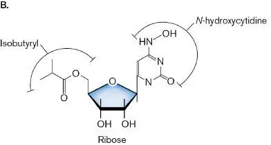
435. This commercial enzyme originated as a lipase (lipid hydrolysis) from Candida antarctica, a psychrotrophic yeast from Antarctica. Psychrotrophs often make enzymes that function well at moderate temperatures. The Novozymes company devised a process to stabilize the enzyme for industrial synthesis.
The second step of synthesis devised by Fier (Fig. ERA 16.2 , step 2) incorporates a uridine molecule by use of four enzymes in one pot: Pyruvate oxidase from Aerococcus viridans , a marine environmental bacterium and occasional opportunistic pathogen. Pyruvate oxidase catalyzes formation of acetyl phosphate, a high-energy donor of a phosphoryl group.
Acetate kinase from Bacillus stearothermophilus or Escherichia coli to catalyze regeneration of ATP from acetyl phosphate.
S -methyl-5-thioribose (MTR) kinase from Klebsiella sp.
The original Klebsiella MTR kinase underwent “directed evolution” to achieve an enzyme that could phosphorylate uridine instead of MTR. In directed evolution, many mutant forms of a gene are cloned and expressed in a laboratory strain, and the expressed enzymes are then screened for activity on a substrate. The evolved MTR kinase is used to phosphorylate 5-isobutyrylribose at the 1′ carbon, the position where uridine will be added.
Uridine phosphorylase from E. coli. This enzyme condenses uridine with isobutyrylribose, releasing inorganic phosphate. The third and final reaction step (Fig. ERA 16.2 , step 3)
consists of chemical reaction of hydroxylamine (NH 2 OH) with the uridine ketone (R−C=O) to an oxime (R−C=N−OH). The reaction includes dehydration (H 2 O removal) from NH 2 OH by use of the reactant hexamethyldisilazane (HMDS). Now the final drug molnupiravir is complete, ready to release its cytidine/uridine analog for viral RNA synthesis with error catastrophe. This simplified synthesis devised by Fier’s team⎯with microbial helpers—enables Merck to offer a global world discount on the drug for economically disadvantaged countries.
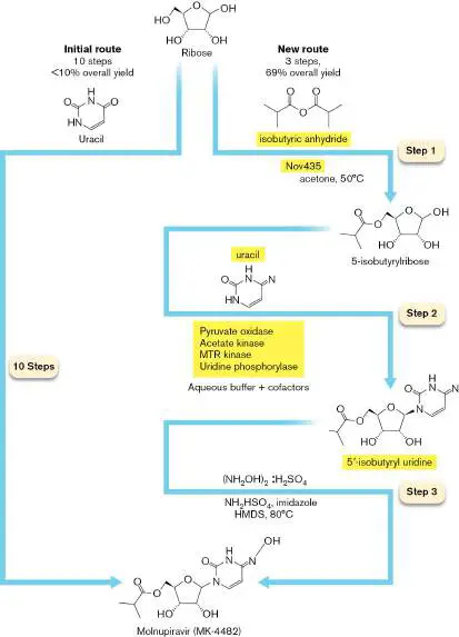
FIGURE ERA 16.2 ■ Biocatalytic synthesis of molnupiravir. The initial route of molnupiravir synthesis required ten steps, providing less than 10% overall yield. The new synthesis devised by Fier’s team at Merck requires three steps, providing 69% yield. These steps require five industrially produced microbial enzymes: Novozym 435 (Nov435), pyruvate oxidase, acetate kinase, MTR kinase, and uridine phosphorylase. Source: John McIntosh et al. 2021. ACS Central Science 7 :1980–1985.
Further Exploration
Besides RNA-dependent RNA polymerase, which other viral enzymes and processes could be blocked by a molecular analog? How could a drug company go about testing the viral processes to discover new antiviral agents? How could we test an antiviral agent to determine whether the virus evolves resistance?
McIntosh, John A., Tamas Benkovics, Steven M. Silverman, Mark A.
Huffman, Jongrock Kong, et al. 2021. Engineered ribosyl-1-kinase enables
concise synthesis of molnupiravir, an antiviral for COVID-19. ACS Central Science 7
:1980–1985.
CHAPTER REVIEW
Review Questions
1. What kinds of microbes are consumed as food? Why are most bacteria inedible for humans?
2. What are the advantages of fermentation for a food product?
3. What are the differences between traditional fermented foods and commercial fermented foods?
4. How do acid fermentations contribute to the formation of different kinds of cheeses? What is the role of different kinds of metabolism performed by different microbial species?
5. Compare and contrast acid and alkaline fermentation processes. What different kinds of foods are produced? 6. Compare and contrast the role of ethanolic fermentation in bread making and winemaking.
7. How do relatively minor fermentation reactions contribute to the flavor of food?
8. Explain the differences between food spoilage and food poisoning.
9. What are the most important food-borne pathogens, based on infection rates? Based on mortality rates? 10. What are the major means of preserving food? Compare and contrast their strengths and limitations.
11. What tasks must be accomplished to develop a microbial product for commercial marketing?
12. What are the sources of potential new microbial products? What is the difference between a source strain and an industrial strain?
13. Explain upstream processing and downstream processing. 14. Explain why genes encoding industrially useful products may be transferred into plant or animal systems for production. What are the roles of microbes in these systems?
15. Outline the process of producing an mRNA vaccine. How does an mRNA vaccine differ from a subunit vaccine? An inactivated vaccine?
16. Explain the construction and use of lentiviral vectors. Explain how these vectors can be safe and effective.
Thought Questions
1. In cheese production, how do different kinds of fermenting microbes generate different flavors?
2. If you were a food safety regulator, which pathogen on the list in Table 16.3would you consider your top priority? Defend your answer by citing factors such as numerical incidence of infection, severity of disease, and economic losses due to illness.
3. Suppose you undertake industrial production of a recombinant glycoprotein for human therapy. Would you synthesize your product in bacteria, in yeast, or in human tissue culture? Explain the advantages and limitations of your choice.
Key Terms
alkaline fermentation (634) bioprospecting (661)
cheddared (636)
cheese (635)
chimeric antigen receptor T-cell therapy (CAR-T therapy) (667) curd (635)
downstream processing (662) endophyte (651)
ethanolic fermentation (634) fermentation industry (630) fermented food (632)
food poisoning (647)
food spoilage (647)
freeze-drying (653)
heterolactic fermentation (634) heterologous host (656)
indigenous microbiota (632) industrial fermentor (662) industrial microbiology (656) industrial strain (661)
injera (643)
kimchi (639)
lactic acid fermentation (634) leaven (642)
lentiviral vector (lentivector) (666) lyophilization (653)
malolactic fermentation (645) messenger RNA (mRNA) vaccine (665) miso (639)
nanoparticle (665)
natto (641)
nori (631)
pathogenicity island (651) pidan (641)
primary recovery (664)
propionic acid fermentation (634) putrefaction (647)
rancidity (647)
ripening (636)
single-celled protein (632) sourdough (642)
starter culture (636)
tempeh (638)
upstream processing (662) whey (635)
yogurt (635)
Recommended Reading
Acevedo-Rocha, Carlos G., Luisa S. Gronenberg, Matthias Mack, Fabian M. Commichau, and Hans J. Genee. 2019.
Microbial cell factories for the sustainable manufacturing of B vitamins. Current Opinion in Biotechnology 56 :18–29.
Centers for Disease Control and Prevention. 2008. Outbreak of Listeria monocytogenes infections associated with pasteurized milk from a local dairy—Massachusetts, 2007. Morbidity and Mortality Weekly Report 57 :1097–1100.
Centers for Disease Control and Prevention. 2009. Multistate outbreak of Salmonella infections associated with peanut butter and peanut butter–containing products—United States, 2008– 2009. Morbidity and Mortality Weekly Report 58 :1–6.
Chang, Shu-Ting, and Philip G. Miles. 2004. Mushrooms: Cultivation, Nutritional Value, Medicinal Effect, and Environmental Impact. 2nd ed. CRC Press, New York.
Habimana, O., L. L. Nesse, T. Møretrø, K. Berg, E. Heir, et al. 2014. The persistence of Salmonella following desiccation under feed processing environmental conditions: A subject of relevance. Letters in Applied Microbiology 59 :464–470.
Leggett, Mary, Nathaniel K. Newlands, David Greenshields, Lee West, S. Inman, et al. 2014. Maize yield response to a phosphorus-solubilizing microbial inoculant in field trials. Journal of Agricultural Science 153 :1464–1478.
Levine, Bruce L., James Miskin, Keith Wonnacott, and Christopher Keir. 2017. Global manufacturing of CAR-T cell therapy. Molecular Therapy: Methods & Clinical Development 4:92–101.
Mackensen, Andreas, Fabian Müller, Dimitrios Mougiakakos, Sebastian Böltz, Artur Wilhelm, et al. 2022. Anti-CD19 CAR T cell therapy for refractory systemic lupus erythematosus. Nature Medicine in press.
Marilley, L., and M. G. Casey. 2004. Flavours of cheese products: Metabolic pathways, analytical tools and identification of producing strains. International Journal of Food Microbiology 90:139–159.
Mills, David A., Helen Rawsthorne, C. Parker, D. Tamir, and K. Makarova. 2005. Genomic analysis of Oenococcus oeni PSU-1 and its relevance to winemaking. FEMS Microbiological Reviews 29 :465–475.
Milone, Michael C., and Una O’Doherty. 2018. Clinical use of lentiviral vectors. Leukemia 32 :1529–1541.
Polack, Fernando P., Stephen J. Thomas, Nicholas Kitchin, Judith Absalon, Alejandra Gurtman, et al. 2020. Safety and efficacy of the BNT162b2 mRNA Covid-19 vaccine. New England Journal of Medicine 383 :2603–2615.
Schwan, Rosane F., and Alan E. Wheals. 2004. The microbiology of cocoa fermentation and its role in chocolate quality. Critical Reviews in Food Science and Nutrition 44 :205– 221.
Glossary
fermentation industry Commercial production of microbial products that are made by microbes grown in fermentation vessels; it may include respiratory metabolism to maximize microbial growth. nori A Japanese food obtained from the red algae Porphyra species. single-celled protein An edible microbe of high food value, such as Spirulina or some yeasts.
fermented food Food products that are biochemically modified by microbial growth.
indigenous microbiota Microbes found naturally in a particular location, often in association with a food substrate.
lactic acid fermentation A fermentation reaction that generates lactic acid from reduction of pyruvic acid.
propionic acid fermentation The fermentation of lactic acid to propionic acid by Propionibacterium species; used in the production of Swiss cheese.
heterolactic fermentation A fermentation reaction in which the products are lactic acid, ethanol, and CO 2.
ethanolic fermentation Also called alcoholic fermentation. A fermentation reaction yielding 2 ethanol and 2CO 2 as products.
alkaline fermentation Bacterial fermentation in conjunction with proteolysis and amino acid catabolism that generates ammonia in amounts that raise pH.
curd Coagulated milk proteins produced by the combined action of lactic acid–producing bacteria and stomach enzymes of certain mammals, such as cattle.
whey The liquid portion of milk after proteins have precipitated out of solution, usually during cheese production.
cheese A solid or semisolid food product prepared by coagulating milk proteins, forming curd. Its production commonly involves microbial fermentation.
yogurt A semisolid food produced through acidification of milk by lactic acid–producing bacteria.
ripening Ripening or aging is a process by which cheese undergoes drying and slow microbial conversion over a period of weeks or months.
starter culture A mixture of fermenting microbes added to a food substrate to generate a fermented product.
cheddared curd Curd that has been cut and piled in order to remove the liquid whey.
tempeh A mold-fermented soy product, popular as a food in parts of Asia.
miso A Japanese condiment, made from ground soy and rice, salted and fermented by the mold Aspergillus oryzae.
kimchi A popular Korean food based on brine-fermented cabbage. natto A soybean product, similar to tempeh, produced by alkaline fermentation.
pidan Pidan, or “century egg,” is a food made by alkali treatment and microbial fermentation of eggs over several months.
leaven For bread dough, to cause to rise by generating air spaces, usually through carbon dioxide production by microbial fermentation.
sourdough An undefined yeast population, derived from a previous batch of dough, that is used in bread production.
injera A highly fermented Ethiopian flatbread made from the grain teff.
malolactic fermentation Fermentation of L -malate (a side product of glucose fermentation) by Oenococcus oeni bacteria; an important process in winemaking.
food spoilage Microbial changes that render a food unfit or unpalatable for consumption.
rancidity Food spoilage due to the oxidation of fats; it may or may not involve microbial activity.
putrefaction Food spoilage due to the decomposition of proteins and amino acids.
food poisoning Food contamination, the presence of human-disease-causing microbial pathogens or toxins in food.
endophyte An endosymbiont of vascular plants.
pathogenicity island A type of genomic island in which the stretch of DNA contains virulence factors and may have been transferred from another genome.
freeze-drying Also called lyophilization. The removal of water from food, by freezing under vacuum, to limit microbial growth.
lyophilization See freeze-drying .
industrial microbiology The commercial exploitation of microbes.
heterologous host An organism engineered for expression of a recombinant product from genes obtained from a species that expresses the product in a natural environment.
bioprospecting The search for organisms with potential commercial applications.
industrial strain A microbial strain whose characteristics are optimized for industrial use.
industrial fermentor The equipment used to grow microbes on an industrial scale. upstream processing The culturing of industrial microbes to produce large quantities of a desired product.
downstream processing The recovery and purification of a commercial product produced by industrial microbes.
primary recovery The initial isolation of commercial product from industrial microbes.
messenger RNA (mRNA) vaccine An mRNA vaccine is made from a cloned gene that expresses mRNA encoding an immunogenic viral protein. The mRNA is encased in liposomes for delivery to the body.
nanoparticle A particle of matter that ranges in size from approximately 1 to 100 nanometers.
lentivector or lentiviral vector A gene transfer vector derived from a lentivirus such as HIV; designed to integrate genes into a host chromosome.
chimeric antigen receptor T-cell therapy (CAR-T therapy) CAR-T therapy uses a lentivector to modify the T cells of a cancer patient. The CAR (chimeric antigen receptor) gene enables T cells to recognize malignant B cells and destroy them.