Chapter introduction
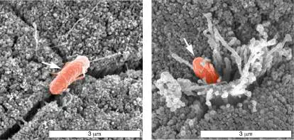
Pathogens that practice breaking and entering. Some bacterial pathogens use secreted effector proteins to engineer entry into host cells. Salmonella enterica serovar Typhimurium (S. Tm), for example, is a gastrointestinal pathogen that invades host cells to live and to hide from the immune system. These SEM micrographs show S. Tm entering epithelial cells that line a mouse gut. The left image shows the bacterium and cells just before invasion when effector proteins are injected. The right image shows invasion at cell-cell junctions and the required ultrastructural changes in microvilli caused by some of the effector proteins. Video shows S. enterica entering nonpolarized tissue culture epithelial cell (MDCK cell) with membrane ruffle.
How long have humans suffered with infections? Millions of years, it turns out. For instance, paleopathologists found evidence of the infectious disease brucellosis in a skeleton from an Australopithecus africanus male, a predecessor of Homo sapiens that lived over 2 million years ago. The vertebrae of this individual exhibited damage that is characteristic of disease caused by a pathogenic species of Brucella. Even though we have long been plagued by infectious diseases, the idea that these diseases can be caused by tiny living organisms invading our bodies became apparent only 150 years ago, when Robert Koch discovered Bacillus anthracis, the microbial cause of anthrax.
Pathogens such as Brucella and Bacillus anthracis use molecular tools to avoid or subvert the immune system, as well as to exploit host cell functions. In Chapter 25 we explore the diverse strategies that pathogenic bacteria and viruses employ to infect hosts, subvert immune responses, and cause disease. We will see how the resulting degree of harm depends on the virulence mechanisms that the pathogen wields and the immune response to the pathogen’s presence. We start by examining a case that begins simply but progresses quickly into a life-threatening infection.
25.1 Host-Pathogen Interactionsnot assigned
A 45-year-old construction worker from Idaho accidentally hit his bare right knee against a cement wall and sustained a seemingly minor abrasion (Fig. 25.1A). The next day he started complaining of knee pain. Overnight the pain had intensified, causing him to visit the emergency department, where he received intravenous benzylpenicillin and flucloxacillin antibiotics (see Chapter 27) and was referred to the orthopedic unit. On arrival, now 70 hours postinjury, he was in considerable pain, even though the wound appeared superficial. He was afebrile (had no fever), but the wound area was reddened, swollen, and warmer than normal. The patient’s white blood cell count was 18,000 per microliter (normal is 4,000– 11,000), and his C-reactive protein was 63.1 mg/l (normal is <3 mg/l). These were sure signs of a serious infection.
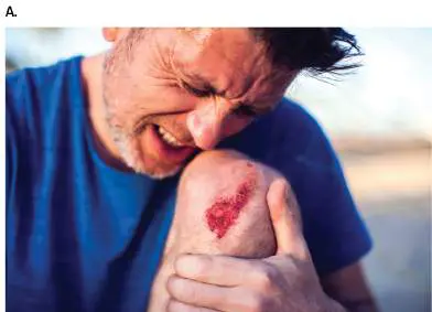
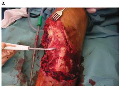
FIGURE 25.1 ■ Knee abrasion develops into serious necrotizing fasciitis infection. A. Infection starts with a minor abrasion. B. Attempt by clinician to remove dead tissue (debridement) of this subcutaneous infection of the fascia.
ALEKS333/SHUTTERSTOCK
P. S. CORONA ET AL. 2018. INJURY 47 :S66–S71
Despite continued antibiotic therapy, the man’s pain and tenderness expanded up his thigh. Clinicians now suspected necrotizing fasciitis, a dangerous soft-tissue infection that can lead to amputation or even death. Emergency surgery revealed necrotic fasciae and underlying tissue. Fasciae (singular, fascia) are sheets of connective tissue lying below the skin that enclose muscles and organs. Surgeons carefully debrided (removed) the necrotic tissue ( Fig. 25.1B ), and new antibiotics were added to the treatment regimen. Fortunately, the patient slowly recovered without needing amputation. The causative agent was found to be Streptococcus
pyogenes (also called group A streptococci, or GAS), the same organism that causes strep throat.
S. pyogenes is a Gram-positive coccus that is visually indistinguishable from many other nonpathogenic microbes, such as S. salivarius, a normal member of the oral microbiome. Why does one Gram-positive coccus cause life-threatening necrotizing fasciitis, while the other is a normally harmless host symbiont? In the case of S. pyogenes, the difference between friend and foe lies with numerous secreted toxins (absent from S. salivarius) that enter host cells and hijack host cell functions. Such factors contribute to pathogenesis, the process by which microbes cause disease.
The Language of Pathogenesis
Before discussing the microbial mechanisms of infectious disease, we should establish their vocabulary. In its broadest sense, the term parasite —defined as an organism that receives benefits at the expense of a host—includes bacteria, viruses, fungi, protozoa, and worms that colonize and harm their hosts. In practice, however, “parasite” is usually reserved to describe disease-causing protozoa and worms. Bacterial, viral, and fungal agents of disease are referred to as pathogens. Parasitic protozoans can also be called pathogens. Pathogens and parasites infect their animal and plant hosts in a variety of ways and enter into a variety of host-pathogen relationships depending on which part of the host they colonize. For example, organisms that live on the surface of a host are called ectoparasites (here, “parasite” is used in its broadest sense). The fungus Trichophyton rubrum, one cause of athlete’s foot, is an ectoparasite (Fig. 25.2A). Wuchereria bancrofti, the worm parasite that causes elephantiasis, is an endoparasite because it lives inside the body (Fig. 25.2B ).
FIGURE 25.2 ■ An ectoparasite and endoparasite. A. Athlete’s foot, an ectoparasitic disease, can be caused by the fungus Trichophyton rubrum. Blowup shows the branching conidia of T. rubrum. Conidia are asexual spores that grow on stalks called conidiophores (see Chapter 20). B. The disease filariasis, commonly known as “elephantiasis” for obvious reasons, is caused
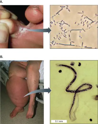
by the endoparasitic worm Wuchereria bancrofti, which enters the lymphatics and blocks lymphatic circulation. Adult worms are threadlike and measure 4–10 cm in length. The young microfilariae (blowup) are approximately 0.5 mm in length.
Though not a problem in the United States, W. bancrofti and elephantiasis are found throughout Asia and middle Africa.
JANE SHEMILT/SCIENCE SOURCE
CDC/LIBERO AJELLO, PH.D.
JOHN GREIM/SCIENCE SOURCE
CDC
An infection occurs when a pathogen or parasite enters or begins to grow on a host. But the term infection does not necessarily imply overt disease. Any potential pathogen growing in or on a host is said to cause an infection, but that infection may be only transient if immune defenses kill the pathogen before noticeable disease results. Indeed, most infections go unnoticed. For example, every time you have your teeth cleaned by a dentist, your gums bleed and your resident oral microbes transiently enter the bloodstream, but you rarely suffer any consequences.
Primary pathogens are disease-causing microbes that can breach the defenses of a healthy host. Shigella flexneri, the cause of bacillary dysentery, is a primary pathogen of humans and non-human primates but no other animals (see eResearch Activity 25). When ingested, Shigella can survive the natural barrier of an acidic (pH 2) stomach, enter the intestine, and begin to replicate. Opportunistic pathogens, on the other hand, cause disease only in a compromised host. Pneumocystis jirovecii (formerly P. carinii) is an opportunistic yeast pathogen that causes life-threatening lung infections in AIDS patients whose immune systems have been eroded by HIV (Fig. 25.3A). Another opportunist is Pseudomonas aeruginosa, a bacterial pathogen that commonly infects burn victims who have compromised skin barriers. Some microbes even enter into a latent state during infection, in which the organism cannot be found by culture. Herpesvirus, for instance, can enter the peripheral nerves, remain dormant for years, and then suddenly emerge to cause cold sores (Fig. 25.3B ). The bacterium Rickettsia prowazekii causes epidemic typhus, but it can also enter a latent phase and then, months or years later, cause a disease relapse called recrudescent typhus.
FIGURE 25.3 ■ Opportunistic and latent infections. A. Pneumocystis jirovecii cysts in bronchoalveolar material. Notice that the fungi look like crushed Ping-Pong balls. B. Cold sore produced by a reactivated herpesvirus hiding latent in nerve cells.
CDC/LOIS NORMAN
CDC/HERMANN
Pathogenicity is an organism’s ability to cause disease. It is defined in terms of how easily an organism causes disease (infectivity) and how severe that disease is (virulence). Pathogenicity, overall, is shaped by the genetic makeup of the pathogen. In other words, an organism is more—or less—pathogenic, depending on the tools at its disposal (such as toxins) and their effectiveness. Virulence is a measure of the degree, or severity, of disease. For instance, Ebola virus has a case fatality rate near 50%, so the virus is highly virulent (Fig. 25.4). By contrast, rhinovirus, the cause of the common cold, is very effective at causing disease but almost never kills its victims, so it is highly infective but has low virulence. Both organisms are pathogenic, but one lets you live while the other may kill you.
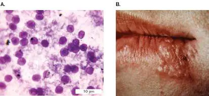
FIGURE 25.4 ■ Highly virulent viruses. A. Ebola virus (approx. 1 μm long; TEM). B. A health care worker tends to an Ebola patient in Kenema, Sierra Leone. Ebola causes hemorrhagic infections in which patients bleed from the mouth, nose, eyes, and other orifices. The mortality rate for this disease is approximately 50%.
CDC/FREDERICK A. MURPHY
TOMMY TRENCHARD/ALAMY STOCK PHOTO
One way to measure virulence is to determine how many bacteria or virions are required to kill 50% of an experimental group of animal hosts. This value is called the lethal dose 50% (LD 50). A pathogen with a low LD 50, in which very few organisms (or viruses) are required to kill 50% of the hosts, is more virulent than one with a high LD 50 (Fig. 25.5). For organisms that colonize but do not kill the host, the infectious dose needed to colonize 50% of the experimental hosts—that is, the infectious dose 50% (ID 50) —can be measured. ID 50 is the dose required to cause disease symptoms in half of an experimental group of hosts.
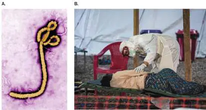
FIGURE 25.5 ■ Measurement of virulence. Each LD 50 measurement requires infecting small groups of animals with increasing numbers of the infectious agent and observing how many animals die. The number of microbes that kill half the animals is called the LD 50 dose. In this example, agent 1 is more virulent than agent 2.
Although it might be possible to measure the infectious dose rather than the lethal dose for a lethal pathogen, it is not typically done. LD 50 gives a clear end point, so it is much easier to use when trying to determine the effectiveness of a given treatment (an antibiotic, for example) or to quantify the role of a given gene in pathogenesis.
Thought Question
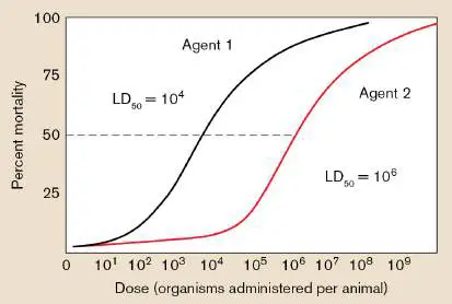
25.1 Figure 25.5 presents the association between LD 50 and virulence. What does this figure tell you about infectivity?
Infection Cycles
Pathogens must pass from one person or animal to another, if a disease is to spread. The route of transmission an organism takes is called its infection cycle. The two main forms of transmission are horizontal transmission, in which an infectious agent is transferred from one person or animal to the next (Fig. 25.6Aand B ), and vertical transmission, whereby the agent is transferred from an infected parent to offspring (Fig. 25.6C ). Vertical transmission in humans can occur directly via transplacental transmission from mother to fetus (for example, Treponema pallidum) or during birth (parturition; for example, Neisseria gonorrhoeae).
FIGURE 25.6 ■ Infection cycles. Infectious agents can be transmitted by a variety of means. A. Horizontal transmission most commonly involves direct contact between two people via touch or between a person and an animal reservoir, such as cats harboring Bartonella hensalae (cat scratch disease). Airborne person-to-person transmission of respiratory pathogens begins when an infected person sneezes and expels tiny aerosol particles or larger droplets of contaminated secretions. The droplets float in air and are inhaled by a nearby person. B. Horizontal transmission can also involve indirect contact via inanimate objects (fomites) or the ingestion of food or water. C. In vertical transmission, a pathogen passes directly from parent to offspring. In humans, vertical transmission takes place when the organism passes through the placenta to the fetus or from mother to newborn
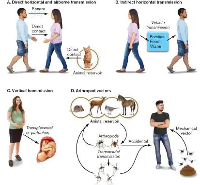
during birth. D. Indirect transmission can also occur through arthropod vectors (insect or tick) feeding on animal reservoir hosts. Some insects also practice vertical transmission wherein the egg itself is infected during formation. Accidental transmission happens when a host that is not part of the normal infection cycle unintentionally encounters an animal host-insect vector cycle. In mechanical transmission, a pathogen is transported on the body surface of a vector (for instance, a fly after landing on fecal matter) to a susceptible host.
There are at least five forms of horizontal transmission: direct contact, airborne, indirect contact, vehicles, and vectors. Pathogens that spread horizontally from person to person are often transferred by direct contact (handshaking or other forms of touching, including sexual contact; Fig. 25.6A). Handshaking is an efficient way to horizontally transmit pathogens. A hand contaminated with a viral or bacterial pathogen can pass the pathogen to several people by handshaking. Newly contaminated people can then infect themselves by touching their hands to their nose or mouth. During the COVID-19 pandemic, handshaking became socially taboo.
Respiratory pathogens such as rhinovirus and other common cold viruses can be passed from person to person through the air by sneezing (airborne transmission). A sneeze will spray massive numbers of microbe-laden secretion particles into the air that people nearby can inhale (Fig. 25.6A). Some particles are large (droplets), and others are small (aerosols). Aerosols can remain suspended in air for long periods of time and travel 20–25 feet before falling to the ground. For this reason, aerosols are worrisome during any epidemic involving a respiratory disease; they were another major concern during the COVID-19 pandemic.
Indirect contact is a broad term that covers all types of transmission that are not directly passed from person to person. Respiratory viruses, for instance, can be transferred via indirect contact by touching inanimate objects (fomites) onto which pathogen-laden aerosols or droplets have fallen. Fomites can include contaminated utensils (such as a fork or pen), towels, cloth handkerchiefs, countertops, and doorknobs. Many gastrointestinal infectious agents can be indirectly transmitted between people or animals when food, water, or fomites become contaminated with fecal material containing the pathogen. This is why handwashing after using the bathroom is an important control measure. Indirect contact via fomites, food, or water is also called vehicle transmission (Fig. 25.6B ).
More complex infection cycles often involve vectors, usually insects or ticks (arthropods), and reservoirs, animals or environments that harbor an infectious agent (Fig. 25.6D ). The housefly is a simple mechanical vector that can transmit disease by landing on a contaminated material (feces, for example) and carrying any pathogens present to a living host or to food. In contrast, mosquito and tick vectors transmit infectious agents by biting an infected reservoir host. They ingest the pathogen and then pass it to a new host during a subsequent blood meal. A zoonotic disease is an infectious disease that is primarily seen in animals but can be transmitted to humans either by vector or other means. West Nile virus and Lyme disease are zoonotic diseases.
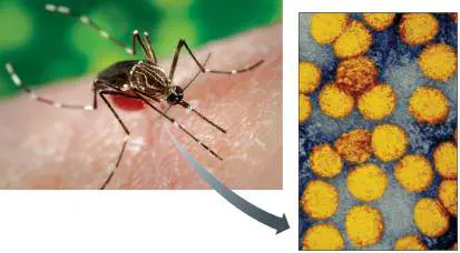
Sometimes a vector also serves as the reservoir for an infectious disease. The mosquito vector Aedes, for example, can horizontally transfer many viral pathogens, including yellow fever virus, from infected to uninfected individuals (Fig. 25.7). But besides horizontally transmitting the virus between humans, the mosquito can also bequeath this particular virus to its offspring by infecting its eggs in a form of vertical transmission called transovarial transmission (Fig. 25.6D) . Although yellow fever is not a problem in the United States today, a flavivirus closely related to yellow fever virus, called West Nile virus (WNV), claims several victims each year in the United States. West Nile virus is also transmitted to humans by a mosquito vector (Culex) that transovarially passes the virus though its eggs.
FIGURE 25.7 ■ Insect vector and yellow fever. The mosquito Aedes aegypti can carry the yellow fever virus (inset; colorized TEM). The virus, which varies in size from 50 to 90 nm, causes a disease that includes jaundiced skin (hence the name “yellow fever”) and can progress to bleeding, shock, organ failure, and death. Yellow fever remains endemic in the northern part of South America and in central Africa.
CDC
CDC/SCIENCE SOURCE
Because insects and ticks can transmit many pathogens, strategies to repel or kill arthropods, or to prevent them from laying eggs, are effective ways to halt the spread of disease. Interventions include spraying insecticide in a community during egg-hatching season or using other microbes as “assassins” trained to kill the vector. For example, the insect virus baculovirus has been developed to kill the Culex mosquito that carries West Nile virus. The advantage of these vector-targeting microbes is that, unlike many chemical insecticides, they do not kill other insects or animals.
Reservoirs of Infection
As mentioned earlier, a reservoir of infection is an environment (water or soil) or animal—often a mammal, bird, or arthropod (insect or tick)—that normally harbors the pathogen. Monkeys, for example, serve as the animal reservoir for yellow fever. In contrast, the virus causing eastern equine encephalitis (EEE, a potentially lethal brain infection seen in some parts of the United States) uses birds as a reservoir. The EEE virus is normally transmitted from bird to bird via a mosquito vector. However, the virus does not persist in the insect. Transmission of EEE virus to new avian hosts by the mosquito vector is what keeps the virus alive. Humans or horses entering geographic areas that harbor the disease (called endemic areas) can also be bitten by the infected mosquito. When this happens, they become accidental hosts and contract this zoonotic disease. But EEE virus does not replicate to high titers in mammals, which means that horses and humans are poor reservoirs for the virus and are called “dead-end hosts.” EEE virus, however, does replicate to high numbers in the avian reservoir host. Reservoirs, then, are crucial for the survival of a pathogen and as a source of infection. If the EEE virus had to rely on humans to survive, it would cease to exist because of limited replication potential and limited access to mosquitoes. Remember, too, that the reservoir of a given pathogen might not exhibit disease.
A special type of reservoir is the asymptomatic carrier —that is, a person who harbors a potential disease agent but has no symptoms of disease. This is how Neisseria meningitidis, an important cause of meningitis, remains in a population. The bacterium has no animal reservoir other than humans, so where does it go between outbreaks? It colonizes the nasopharynx (the area behind the nose down to the throat) of unsuspecting human hosts whose immune systems keep the bacterium from entering the bloodstream. A simple sneeze from the carrier, however, can aerosolize the pathogen and transfer it to a susceptible host who then contracts the disease. Asymptomatic carriers were yet another major concern during the COVID-19 pandemic.
Portals of Entry
How do infectious agents enter the body? Each organism is adapted to enter the body in specific ways. Portals of entry may be respiratory, oral, ocular, urogenital, parenteral (injection by mosquito bite or contaminated needle), or via a wound. Food-borne pathogens such as Salmonella, E. coli, Shigella, and rotavirus are ingested by mouth and ultimately colonize the intestine. They have an oral portal of entry. These gastrointestinal microbes are also described as having a fecal-oral route of transmission, in which pathogens or parasites excreted in the fecal matter of an infected person are indirectly ingested by an uninfected person. For example, fecal organisms are often found on hands or surfaces and can be transferred through touch or contaminated foods to someone else. That newly contaminated person can then unknowingly complete the cycle by ingesting the virus, bacterium, or parasite.
Airborne organisms, in contrast, infect through the respiratory tract (for example, rhinovirus or Mycobacterium tuberculosis). Other microbes enter through the conjunctiva of the eye or through the mucosal surfaces of the genital and urinary tracts. An important factor that dictates the organism’s preferred portal of entry is attachment capability: Does the pathogen contain surface receptors capable of binding host surface proteins present at a given portal? To infect, the organism must colonize its host, and to colonize it must attach.
Agents that are transmitted only by mosquitoes or ticks enter their human hosts via the parenteral route, meaning injection into the bloodstream. Wounds and needle punctures can also serve as portals of entry for many microbes. Thus, shared needle use between drug users has been an important factor in the spread of HIV, hepatitis B, and hepatitis C viruses.
Immunopathogenesis
Although we focus in this chapter on the mechanisms that microbes use to cause disease (toxins, for example), it is often “friendly fire” by our immune system reacting to a pathogen that causes major tissue and organ damage. The immune response to any infection involves activating a complex network of cell types and soluble factors (discussed in Chapters 23 and 24) that may inadvertently damage the host to such a degree that it causes illness and even death. This collateral damage, or “immunopathology,” is a calculated risk taken by the host in its haste to eradicate the pathogen. The term immunopathogenesis applies when the immune response to a pathogen is a contributing cause of pathology and disease.
The disease dengue hemorrhagic fever is a case in point. Caused by the dengue virus and transmitted by the Aedes mosquito, dengue fever manifests as a severe headache, muscle and joint pain, fever, and rash. The symptoms can also include abdominal pain, nausea, and vomiting. However, these symptoms are more a consequence of immunopathogenesis than they are a direct result of viral replication. Replication of the virus in host cells will produce a massive activation of T cells (CD4 + and CD8 +). Activation triggers a cytokine cascade (some call it a “storm”) that loosens tight junctions between vascular endothelial cells to produce an endothelial “sieve” that leaks fluid and protein from the bloodstream into surrounding tissues (known as edema). Cytokines such as TNF-alpha, IL-1, and IFN-gamma (discussed in Chapters 23 and 24), along with many others, contribute to inflammation by attracting and activating neutrophils and macrophages. The disease symptoms mentioned earlier can result.
So, to fully understand any infectious disease, not only must we be aware of the pathogenic mechanisms wielded by the pathogen, we must also realize that many disease symptoms are due to immunopathogenesis. The immunopathogenic features of various diseases are described in the next chapter.
Effect of Infections on the Microbiome
A pathogen’s growth and the resulting immune response will also affect the host’s microbiome. For example, diarrhea can reduce overall numbers of gut microbiota by causing more forceful and frequent evacuations. Intestinal pathogens can change microbiome diversity by occupying limited host binding sites or by altering available nutrients. Infection-induced inflammation may kill microbiota more effectively than the pathogen, depending on the pathogen’s virulence mechanisms. Aspects of inflammation can even generate nutrients that feed the pathogen but not normal microbiota (for example, tetrathionate for Salmonella, discussed later). All of these mechanisms affect competition between microbial species and influence species diversity. Finally, as victims recover from a disease, their gut microbiota may not always achieve preinfection balance (for instance, following cholera; see Fig. 26.17. The resulting dysbiosis, as discussed in Chapters 23 and 24, can have negative health effects beyond those of the infection itself.
Virulence Factors and How to Find Them
To cause disease, all pathogens must enter a host; find their unique niche; avoid, circumvent, or subvert normal host defenses; multiply; and eventually be transmitted to a new susceptible host. Pathogens employ virulence factors, encoded by virulence genes, to accomplish these goals. Virulence factors include toxins, attachment proteins, capsules, and other devices used by the pathogen to avoid host innate and adaptive immune systems. The next section describes some of these factors. But, first, how are virulence genes identified? Molecular Koch’s postulates. Identifying genes for a metabolic or biosynthetic pathway is relatively easy because it requires merely observing a clear phenotype on an agar plate. If a gene involved with an amino acid biosynthetic pathway is defective, then the amino acid is not made and must be added to the medium or else the mutant will not grow. In contrast, there is no straightforward way to identify a virulence phenotype using an agar plate. Finding a virulence gene is hard because the screen involves growth in a host. True virulence genes can be recognized only if mutants defective in the gene fail to sicken test animals or fail to survive in them.
Numerous clever techniques have been developed over past decades to identify potential virulence genes. Older methods include looking for bacterial genes that are expressed only during infection (called in vivo expression) and screening for genes critical to in vivo growth of a pathogen but irrelevant to growth on agar plates (signature-tagged mutagenesis). Section 25.6 describes how large-scale RNA sequencing protocols can simultaneously identify host and pathogen genes that respond to an infectious process (dual RNAseq). A more recent example uses large-scale cloning of potential virulence factors, expressing them one by one in host cells to observe their effects (see eResearch Activity 25).
Regardless of how the suspected virulence gene is found, it can be confirmed as having a role in virulence or pathogenicity only if it fulfills a set of “molecular Koch’s postulates” originally formulated by Stanley Falkow (1934–2018), a preeminent infectious disease scientist. The molecular postulates are as follows: 1. The phenotype under study should be associated with pathogenic strains of a species.
2. Specific inactivation of the suspected virulence gene(s) should lead to a measurable loss in virulence or pathogenicity. The gene(s) should be isolated by molecular methods.
3. Reversion or replacement of the mutated gene should restore pathogenicity.
A variety of other molecular questions can suggest the role of a virulence protein in pathogenesis. For example: Does moving the virulence gene into an avirulent strain impart a pathogenicity trait on the avirulent strain? For instance, does moving a gene for attachment from a pathogen to a nonpathogen allow the nonpathogen to attach to host cells?
Does the suspected virulence protein bind to important host proteins? Binding to a host protein could indicate the target of the microbial virulence protein.
Does the microbial protein resemble the sequence or structure of an important host protein? Such resemblance might indicate that the microbial protein mimics the function of the orthologous host protein.
Does introducing the suspected virulence protein (or its encoding gene) into a host cell alter the host cell’s physiology, or does a version of the virulence protein tagged with green fluorescent protein (GFP) localize to a specific host cell compartment or organelle? A positive finding in either case could reveal a host target for the suspected virulence protein.
In addition to the experimental approaches just described, there are bioinformatic ways to identify potential pathogenicity genes.
Pathogenicity Islands
Extensive sequencing efforts have enabled us to compare the genomes of many pathogens and expose some “footprints” of their evolution. For example, in bacterial pathogens, most chromosomes are dotted with clusters of pathogenicity genes that encode virulence functions. These gene clusters, called pathogenicity islands, can be considered the toolboxes of pathogens (originally discussed in Section 9.5). Many, but not all, virulence genes reside in pathogenicity islands. Some virulence genes reside on plasmids (for example, the genes for the diarrhea-producing labile toxin of certain E. coli strains) or in phage genomes (such as the genes encoding the diphtheria toxin of Corynebacterium diphtheriae).
Most pathogenicity islands appear to have been horizontally transmitted via conjugation or transduction (discussed in Section 9.5 ) from long-extinct organisms into the ancestors of today’s pathogens. Horizontal gene transfers move whole blocks of DNA (more than 10 kb) from one organism to another, placing the blocks directly into the chromosome in what is called a genomic island (see Chapter 9). If the island increases the “fitness” (virulence) of a microorganism (pathogen) that interacts with a host, it is called a pathogenicity island. Genomic islands generally reveal themselves by several anomalies that they possess with respect to the rest of the host genome: A GC content strikingly different from the rest of the genome. For example, a plot of GC (guanine + cytosine)
content (as opposed to adenine + thymine) along the length of a chromosome may reveal that most of the genome has a 50% GC content. But somewhere in the middle, a 50-kb region sticks out on the graph, showing a GC content of 40% (Fig. 25.8A). This deviation probably reflects the GC content of the microbe that long ago donated the island.
FIGURE 25.8 ■ Model pathogenicity island. A. The guanine + cytosine (G + C) content of the island is different from that of the core genome. B. Schematic model of a pathogenicity island. The horizontally transferred DNA island is linked to a tRNA gene and flanked by direct repeats (DRs) that may be “footprints” of a transposon or viral-mediated transfer. The integrase gene (int) and insertion sequences (ISs) may also be remnants of transposition.
Linkage to a tRNA gene. The reason for this linkage, however, is not clear. One hypothesis is that the conserved secondary
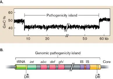
structure of tRNA facilitates integration by an integrase.
Association with genes homologous to phage or plasmid genes. Typically, genomic islands are flanked by genes that show homology to phage or plasmid genes (Fig. 25.8B ). This arrangement is thought to reflect the transfer vector used to move the island from one organism to another.
Figure 25.9provides examples of pathogenicity islands from different pathogens.
FIGURE 25.9 ■ Examples of bacterial pathogenicity islands. A. The cag island of Helicobacter pylori (stomach ulcers) harbors genes for a type IV secretion system that can translocate the toxin CagA into human gastric cells, causing an inflammatory response. B. The v Saα island of a particularly virulent strain of Staphylococcus aureus (MRSA) encodes a remarkably high number of enterotoxins. C. The SPI-1 island of Salmonella enterica (enteritis disease) encodes a type III secretion system (gray),
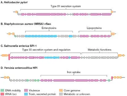
secreted effector proteins (dark gray), and regulatory proteins. The island includes genes for metabolic proteins unrelated to virulence. D. The high-pathogenicity island (HPI) of Yersinia enterocolitica (enteritis) carries genes for a high-affinity iron uptake system (dark gray) needed for extracellular growth during host colonization.
But what do the pathogenicity gene products actually do? Some genes encode molecular “grappling hooks,” such as pili that attach to host cells. Once attached, microbes can secrete toxins that injure the host cell. Other bacteria wall themselves off to prevent damage by host inflammatory responses. Some bacterial pathogens are even capable of what could be called “host cell reprogramming.” These organisms inject proteins directly into the host cell to disrupt normal signaling pathways. The reprogrammed target cell can be made to do one of several things: engulf the bacterium; “commit suicide” (undergo apoptosis); engineer a tighter, more intimate pathogen-host attachment platform at the cell surface; or alter the amounts or types of cytokines that the affected cell produces. Detailed functions of various pathogenicity genes are described in Section 25.5.
Caught in the Act: Examples of Pathogen Evolution by Horizontal Gene Transfer
Pathogens and hosts continually coevolve, with each trying to gain the upper hand. The process is typically slow, but scientists sometimes catch a pathogen in the act. Examples include Escherichia coli and Streptococcus pyogenes.
Escherichia coli. E. coli is a member of the normal gut microbiota but includes many pathovars that evolved through horizontal gene transfers. A pathovar represents one strain of an organism that causes disease in a specific organ system. Different E. coli pathovars cause diseases ranging from urinary tract infections to diarrhea, sepsis, and meningitis (see Chapter 26). A relatively recent example of E. coli evolving via horizontal gene transfer involves the enteroaggregative hemorrhagic strain O104:H4, which caused the frightening 2011 outbreak of diarrhea and hemolytic uremic syndrome that began in Germany and spread through much of Europe. This new pathovar originated from a well-known enteroaggregative E. coli (EAEC) but included genes for a powerful Shiga toxin (described later) donated as part of a prophage released from an enterohemorrhagic E. coli, plus numerous antibiotic resistance genes carried on plasmids, and new virulence traits missing from its closest relative. Horizontal gene transfers were instrumental in gaining all of these traits.
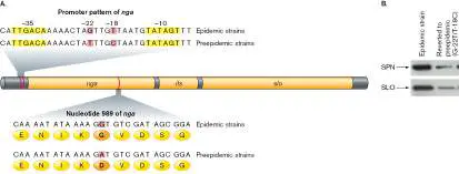
Streptococcus pyogenes. Another dangerous pathogen caught in the act of evolving is Streptococcus pyogenes, otherwise known as group A streptococci (GAS), a strict human pathogen that can cause sore throat, scarlet fever, and necrotizing fasciitis (as we saw at the beginning of this section; see also Section 26.1). For a century, GAS strains have been assigned serological types by antigenic differences in a cell-surface molecule called M protein. James Musser from the Houston Methodist Research Institute and his colleagues determined, through large-scale genome sequencing, that new epidemics of streptococcal disease happen after horizontal gene transfers yield new strains of S. pyogenes and not by the simple reemergence of older strains. For instance, the origin of the most recent GAS M1 global pandemic strain was traced to about 1983, when a horizontal gene transfer event introduced a 36-kb chromosomal region encoding several proteins. Two of those proteins, NAD glycohydrolase (or SPN; the gene is nga) and streptolysin O (SLO; gene slo), are potent toxins (Fig. 25.10A).
FIGURE 25.10 ■ Emergence of a pandemic clone of group A streptococci. A. Sequence comparison in the nga-slo operons of preepidemic and epidemic strains of Streptococcus pyogenes revealed three consistent single nucleotide polymorphisms (SNPs; red lines in DNA). The nga and slo genes encode NAD glycohydrolase (SPN) and streptolysin O (SLO), respectively. The ifs gene encodes a regulator of SPN. B. Western blot of secreted proteins, showing that changing the SNPs in the promoter of an epidemic strain back to those found in a preepidemic strain reduced secretion of SPN and SLO.
Source: Western blots in part B from Luchang Zhu et al. 2015. J. Clin. Invest.
125 (9):3545–3559, fig. 3C. ©2015, American Society for Clinical Investigation.
Compared to preepidemic strains, all of the new epidemic strains contained the same three single-nucleotide changes, or polymorphisms (SNPs), within the 36-kb region. Two SNPs were located in the upstream promoter region of the SPN operon. These two promoter mutations increased expression of the SPN and SLO proteins (Fig. 25.10B ) and dramatically increased virulence. The third change was a missense mutation in SPN that changed an aspartate to glycine and restored NAD glycohydrolase enzymatic activity. The restored activity of NAD glycohydrolase was required to maximize virulence of the epidemic GAS strain. Up-regulating SPN and SLO in the pandemic strain enhanced tissue destruction, heightened resistance to killing by polymorphonuclear leukocytes, and helped propagate GAS infections around the world.
Sections 25.2 through 25.5 describe some of the specific tools that pathogens have evolved to undermine the integrity of the body. The infection process is like a chess match, with each side, human and microbe, trying to outmaneuver the other.
To Summarize
Infection does not equal disease. The immunocompetence of the host and the virulence potential of the pathogen influence whether an infection causes disease.
Primary pathogens have mechanisms that help the organism circumvent host defenses in a healthy host, whereas opportunistic pathogens cause disease only in a compromised host.
Pathogenicity refers to the mechanisms a pathogen uses to produce disease and how efficient the organism is at causing disease, whereas virulence is a measure of disease severity. Diseases can be spread by direct or indirect contact between infected and uninfected persons/animals or by insect vectors.
Pathogens use portals of entry best suited to their mechanisms of pathogenesis.
Immunopathogenesis is damage to host tissues caused by the immune system’s response to an infection.
Virulence genes encode products that enhance the disease-causing ability of the organism. Many virulence genes can be found within pathogenicity islands, but some are located outside of an obvious genomic island or reside in plasmids. Fulfilling the molecular Koch’s postulates validates the identity of a virulence gene.
Pathogenicity islands are DNA sequences within a species that are acquired by horizontal gene transfer from a different species. They contain distinct features, such as GC content and the remnants of phages or plasmids, that mark them as being different from the rest of the genome.
Horizontal gene transfer mechanisms move virulence factor genes and pathogenicity islands among bacterial strains and species.
Glossary
pathogenesis The processes through which microbes cause disease in a host. parasite Any bacterium, virus, fungus, or protozoan (protist) that colonizes and harms its host; the term commonly refers to protozoa and to invertebrates.
pathogen A bacterial, viral, or fungal agent of disease.
infection The growth of a pathogen or parasite in or on a host.
primary pathogen A disease-causing microbe that can breach the defenses of a healthy host.
opportunistic pathogen A microbe that normally is not pathogenic but can cause infection or disease in an immunocompromised host organism.
latent state A period of the infection process during which a pathogenic agent is dormant in the host and cannot be cultured.
pathogenicity The ability of a microorganism to cause disease.
virulence A measure of the severity of a disease caused by a pathogenic agent.
lethal dose 50% (LD 50)
A measure of virulence; the number of bacteria or virions required to kill 50% of an experimental group of hosts. infectious dose 50% (ID 50)
The number of bacteria or virions required to cause disease symptoms in 50% of an experimental group of hosts.
infection cycle The route a pathogen takes as it moves from one host into another.
horizontal transmission In disease, the transfer of a pathogen from one organism into another, nonprogeny organism.
vertical transmission In disease, the transfer of a pathogen from parent to offspring. See also transovarial transmission .
transplacental transmission The process by which certain pathogens in maternal blood can pass through the placenta to infect the fetus.
direct contact The process by which a disease-causing microbe is transmitted from an infected person to an uninfected person by direct physical contact with skin, blood, or body fluids.
airborne transmission In disease, the transfer of a pathogen via dust particles or on respiratory droplets produced when an infected person sneezes or coughs.
indirect contact The process by which a disease-causing microbe is transferred from an infected person to an inanimate object (fomite), food, or water and then to an uninfected person that touched or ingested the contaminated material.
fomite An inanimate object on which pathogens can be transmitted from one host to another.
vehicle transmission In disease, the transfer of a pathogen when an infected person deposits it on a surface or in food or drink that another person touches or consumes.
vector 1. An organism (e.g., insect) that can carry infectious agents from one animal to another. 2. In molecular biology, a molecule of DNA into which exogenous DNA can be inserted to be cloned; or an engineered virus that can clone a gene in its DNA or RNA genome.
reservoir 1. The major part of the biosphere that contains a significant amount of an element needed for life. 2. An organism that maintains a virus or bacterial pathogen in an area by serving as a high-titer host.
mechanical vector A vector that conveys pathogens to a susceptible individual or food without the pathogen needing to replicate in the vector; a housefly, for example.
zoonotic disease An infection that normally affects animals but can be transmitted to humans.
transovarial transmission The transfer of a pathogen from parent to offspring by infection of the egg cell. Typically seen in insects.
asymptomatic carrier Person or animal that lacks symptoms of a disease despite being infected and can unknowingly transmit that pathogen to others. portals of entry Openings in the body, either natural (e.g., gastrointestinal and respiratory tracts) or caused by trauma (e.g., wound or injection), through which pathogens can gain entrance and cause disease.
fecal-oral route of transmission A method by which pathogens or parasites excreted in the fecal matter of an infected person are then indirectly ingested by an uninfected person.
parenteral route A method by which an infectious agent enters the body via injection into the bloodstream, often by a mosquito or other insect.
immunopathogenesis The process by which an immune response or the products of an immune response cause disease.
virulence factor A trait of a pathogen that enhances the pathogen’s disease-producing capability.
pathogenicity island A type of genomic island in which the stretch of DNA contains virulence factors and may have been transferred from another genome.
genomic island A region of DNA sequence whose properties indicate that it has been transferred from another genome. Genomic islands usually comprise a set of genes with shared function, such as pathogenicity or symbiosis support.
Fig. 26.17 FIGURE 26.17 ■ Successive repopulation of the gut microbiome following Vibrio cholerae infection. A.
Lawrence David (pictured) and Peter Turnbaugh (not shown) unravel the complexities of the human microbiome. B. Fecal samples were taken at 1, 7, and 30 days past presentation (dpp) of diarrhea. Patients, identified by letters, were given a single dose of azithromycin on the day of presentation, which nearly eliminated V. cholerae by 1 dpp. Healthy contacts of each patient are identified by numbers and are shown on the left. For example, A-1, A-2, A-3, and A-4 are the healthy contacts of patient A. Colored boxes reflect groups of different genera that are prominent during the infection and afterward, during early, mid, and late stages of repopulation. The x -axis values reflect relative abundances of genera. “All swab” microbes were present only in rectal swab samples.
Source: Part B modified from Harvard Press Office figure.
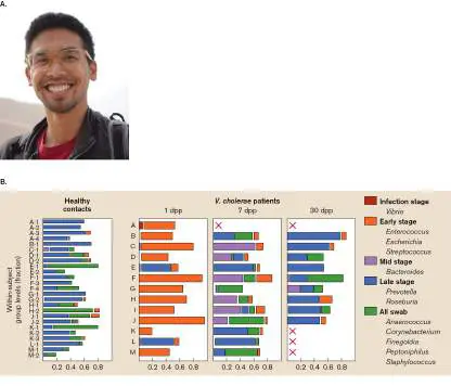
COURTESY OF LAWRENCE DAVID
25.2 Microbial Attachment: First Contactnot assigned
Regardless of the disease, pathogens must reach a colonization site either through their own
motility or by hitchhiking with a vector. Once at the site, the pathogen needs attachment
mechanisms to stay there.
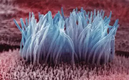
The human body has many ways to exclude pathogens. The lungs use a mucociliary escalator
(see Fig. 23.5) to rid themselves of foreign bodies, the intestine uses peristaltic action to ensure
that its contents are constantly flowing, and the bladder uses contraction to propel urine through
the urethra with tremendous force. How do bacteria ever manage to stick around long enough to
cause disease? Like a person grasping a telephone pole during a hurricane, successful pathogens
moving through the body manage to grab and tenaciously hold on to host cells. Thus, the first step
toward infection is attachment, also called adhesion. Any microbial factor that promotes
attachment is called an adhesin.
Viruses attach to the host through their capsid or envelope proteins, which bind to the specific
host cell receptors discussed in Chapters 6 and 11. The spike protein on the SARS-CoV-2
membrane, for instance, binds to the ACE2 membrane receptor on host cells. Bacteria use a variety
of similar strategies to attach to host cells. They can use hairlike appendages called pili (also called
fimbriae; composed of a string of protein monomers), whose tips contain receptors for mammalian
cell-surface structures. (The terms pili and fimbriae can be used interchangeably, but some
scientists restrict the term pili to the structures used in conjugation). Besides pili, bacteria can use
a variety of adherence proteins or other cell-surface molecules that are not part of a pilus.
Sometimes they use both. Table 25.1 provides examples of bacterial attachment strategies. Note
that different pili in the same species can impart different tissue specificity for attachment (for
example, uropathogenic versus diarrheagenic E. coli).
TABLE Examples of Bacterial Adhesins 25.1
Bacterium Adhesin Host receptor Attachment Disease
site
Streptococcus Protein F Amino terminus of Pharyngeal Sore throat
pyogenes fibronectin epithelium
Streptococcus Glucan Salivary Pellicle of tooth Dental caries
mutans glycoprotein
Staphylococcus Clumping factors A Fibronectin Mucosal Various
aureus and B epithelium
Neisseria N - Glucosamine Urethral/cervical Gonorrhea
gonorrhoeae methylphenylalanine galactose epithelium
pili carbohydrate
TABLE Examples of Bacterial Adhesins 25.1
Bacterium Adhesin Host receptor Attachment Disease
site
Uropathogenic Type I fimbriae (pili) Complex Urethral Urethritis
E. coli carbohydrate epithelium
P pili (pyelonephritis- P blood group Upper urinary Pyelonephritis
associated pili) tract
Bordetella Pili (“filamentous Galactose on Respiratory Whooping
pertussis hemagglutinin”) sulfated epithelium cough
glycolipids
Chlamydia Lipooligosaccharide, Sulfonated Conjunctival or Conjunctivitis
OmcB surface glycosaminoglyca urethral or urethritis
protein ns epithelium
Pili
Pili types are defined by protein sequence homologies. In this chapter we consider two groups,
called type I and type IV pili. Note that pili and other adhesins can be virulence factors on some
organisms but nonpathogenic attachment structures on others.
How bacteria assemble pili on their cell surfaces is an engineering marvel. The shafts of pili are
cylindrical structures composed of identical pilin protein subunits (described in Chapter 3). Several
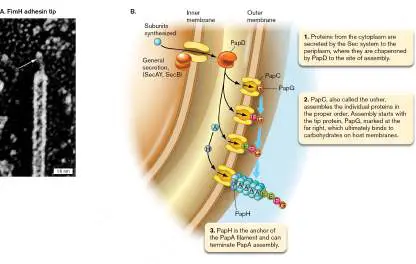
different proteins adorn the tip, including one at the very apex, such as FimH, that binds to host
receptors (Fig. 25.11A ). Much like Velcro, the tip proteins of type I pili manage to “hold on” in
the face of tremendous shear forces (such as those exerted during urination) by tightening their
grip on the host receptor. In addition to the structural components of pili, numerous other proteins
collaborate to assemble the structure. Genes encoding a given pilin protein and the associated
assembly apparatus are typically arranged on the chromosome as an operon.
FIGURE 25.11 ■ Attachment pilus and pilus assembly. A. High-resolution micrograph
showing a type I pilus (TEM). The FimH adhesin at the tip (arrow) is the protein that binds to
the cell receptor (cell-surface mannose residues). B. The pyelonephritis-associated pilus (Pap)
is assembled in a stepwise fashion. The protein subunits fit together like pieces of a jigsaw
puzzle. New protein subunits are added to the bottom of the growing structure.
C. H. JONES ET AL. 1995. PNAS 92 :2081–2085, FIG. 3
Assembly of type I pili. Pyelonephritis-associated pili (Pap) of the uropathogenic E. coli are type
I pili that bind to a digalactoside present on host urinary tract surfaces called the P-blood-group
antigen. “Pyelonephritis” is the medical term for kidney infection. Pap pili are essential for
uropathogenic E. coli to cause this disease. Figure 25.11B illustrates how the type I pilus from
uropathogenic E. coli (Pap) is assembled. The mechanism is representative of other type I pili; only
the names of the proteins will differ for each system. Protein components synthesized in the
cytoplasm are secreted into the periplasm by the SecA-dependent general secretory system
(discussed in Section 8.5). Once in the periplasm, the subunits are chaperoned one at a time by
PapD to the membrane site of assembly, which is marked by the presence of the usher protein
PapC. Like an usher in a theater, PapC directs the subunits to their proper places.
Assembly of pili at the usher site starts with the tip protein, PapG, which will ultimately bind to
carbohydrates on host membranes after the pilus is complete. After PapG, the ushers add PapF and
PapE, forcing PapG farther away from the surface. Then identical PapA pilin subunits are strung
together in a series to form the shaft. PapA subunits assemble sequentially by sharing a domain
with one another, linking together like pieces of a jigsaw puzzle. Once assembled, type I pili are
static. They simply stick to the host receptor. Type IV pili, however, are more dynamic, continually
extending and contracting.
Type IV pili. Another group of pili with important roles in pathogenesis is the type IV pili. After
assembly, these pili repeatedly extend and contract and are found in a broad spectrum of Gram-
negative pathogens (Pseudomonas and Neisseria, for instance). What makes these pili amazing is
their ability to continually assemble and disassemble—a feat that produces a remarkable type of
cell movement called twitching motility (see Section 12.2 and described later).
The assembly machinery for type IV pili involves at least a dozen proteins (Fig. 25.12 ). A
major difference between type IV pilus assembly and that of type I pili is that type IV pilus proteins
are never free in the periplasm; instead, they are inserted and assembled at the cytoplasmic
membrane, after which the assembled pilus is “pushed” outside the cell through a channel in the
outer membrane (Fig. 25.12A ). Over the course of thousands of years, if not longer, the genes
for type IV pili duplicated, and one set of those genes evolved to encode a protein secretion
mechanism called type II secretion that exports virulence proteins unrelated to pili (discussed in
Section 25.4).
FIGURE 25.12 ■ Type IV pili. A. Model of pilus assembly and disassembly. In this
example, PilA is the pilin protein, and PilC1 and Y1 form the attachment tip. Filament is
approx. 6 nm in diameter. Assembly and disassembly require the hydrolysis of nucleoside
triphosphate (NTP) and take place at the inner membrane, not in the periplasm. B.
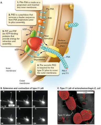
Photographic evidence of type IV pilus retraction in cells of Pseudomonas aeruginosa.
Filament c attaches briefly at its distal tip (note straightening at 24 seconds) and then begins
to retract. Fluorescent microscopy. t = time, in seconds. C. Type IV pili (green) are essential
for enterohemorrhagic E. coli to attach to epithelial cells. SEM.
Source: Part A modified from Bardy et al. 2003. Microbiology 149: 295–304.
SKERKER, J., ET AL. 2001. PNAS 98 : 6901.
J. XICOHTENCATL-CORTES ET AL. 2007. J CLIN INVEST. 117 :3519–29
How does the type IV pilus assembly mechanism make bacteria move by twitching motility? The
assembly process involves the reiterative elongation and retraction of the pili. The pilus elongates,
attaches to a surface (many type IV pili can attach to a variety of surfaces), and then
depolymerizes from the base, which shortens the pilus and pulls the cell forward (Fig. 25.12B ).
This mechanism is akin to using a grappling hook to scale a building. Similarly, the slime mold
Myxococcus xanthus uses type IV pili to mediate gliding motility. The type IV pili of Neisseria
meningitidis are essential for crossing the blood-brain barrier to cause bacterial meningitis.
As noted for N. meningitidis, type IV pili are also important to the pathogenesis of some
organisms. The retraction of type IV pili in diarrhea-causing strains of E. coli, for example, helps
disrupt the tight junctions connecting adjacent host cells that line the intestine (Fig. 25.12C ).
Tight junctions normally form a permeability barrier between the intestinal lumen and the intestine,
through which nutrients, ions, and water are absorbed. Disrupting tight junctions prevents the
absorption of water and electrolytes, which accumulate in the intestine and contribute to the
diarrhea.
Nonpilus Adhesins
Bacteria also sport proteins that bind host tissues but are not pili (Fig. 25.13 ). Some examples
include Bordetella pertactin that binds to host cell integrin, and Streptococcus pyogenes M protein
that binds to fibronectin. Many Gram-positive bacteria have other surface-exposed proteins with
serine-rich repeats able to bind host sialic acid or keratin.
FIGURE 25.13 ■ Nonpilus adhesins. A. M-protein surface fibrils on Streptococcus
pyogenes (TEM). Cells 0.5–1 μm in diameter. B. Colonization of tracheal epithelial cells by
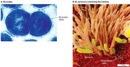
Bordetella pertussis (colorized SEM). This organism uses a surface protein called pertactin, as
well as a pilus called filamentous hemagglutinin (FHA) to bind bronchial cells.
MARIO FAZIO AND VINCENT A. FISCHETTI, PH.D. WITH PERMISSION. THE LABORATORY OF BACTERIAL PATHOGENS AND
IMMUNOLOGY, ROCKEFELLER UNIVERSITY.
NIBSC/SCIENCE PHOTO LIBRARY/SCIENCE SOURCE
Initial binding between bacterium and host commonly involves pili, after which a more intimate
attachment is formed by a nonpilus attachment protein. In the case of Neisseria gonorrhoeae,
once the type IV pilus has attached to the surface of the mucosal epithelial cell, the filamentous
pilus contracts, pulling the bacterium down onto the host cell membrane. Tight secondary
interactions are then mediated by the neisserial Opa membrane proteins—another example of
nonpilus adhesins. (Opa gets its name from the opacity it adds to colony appearance.)
An interesting nonpilus adhesin widely distributed among Gram-negative bacterial pathogens is
an outer-membrane protein called multivalent adhesion molecule 7 (MAM7) that binds
phospholipids and fibronectin in host cell membranes. Because of its wide distribution among
pathogens, it is considered to be an attractive target for antimicrobial development. Kim Orth
(University of Texas Southwestern Medical Center) and Anne Marie Krachler (University of Texas
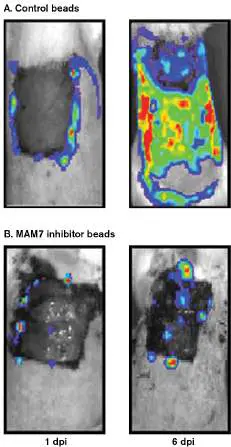
McGovern Medical School, Houston) developed microbeads coated with fragments of MAM7 that can
bind host tissues and competitively inhibit binding by multidrug-resistant Pseudomonas aeruginosa
. When used in a burn model of infection, topical applications of the inhibitor-coated microbeads
prevented the spread of infection into adjacent tissues (Fig. 25.14 ). More recently, the MAM7
microbeads successfully prevented P. aeruginosa and Staphylococcus aureus from causing surgical
wound infections in a rat model. Although S. aureus does not have MAM7, it does use another
adhesion that targets the same fibronectin binding site as MAM7.
FIGURE 25.14 ■ Treatment of burn wounds with inhibitor MAM7 beads. Shaved
areas of heavily anesthetized rats were burned with 100°C water for 12 seconds. Two days
later, dead skin over the burn was excised and 5 × 10 6 colony-forming units (CFUs) of
bioluminescent Pseudomonas aeruginosa were applied to the exposed area. A. Control rats
were treated with a daily application of control microbeads over a period of 6 days
postinfection (dpi). B. Test rats were treated with a daily application of inhibitor microbeads.
Infections were monitored by fluorescence using an in vivo imaging system. Green and orange
sections over the burn area indicate the presence of P. aeruginosa.
HUEBINGER ET AL. 2016. SCI. REP. 6 :39341, FIG. 2B.
HUEBINGER ET AL. 2016. SCI. REP. 6 :39341, FIG. 2B.
Host receptors dictate susceptibility to pathogens. Why are some people susceptible to
certain infections while others are not? Part of the reason is individual differences in immune
competence, but another part is receptor availability. Pathogens rely on key host surface structures
such as gangliosides to recognize and attach to the correct host cell by the mechanisms just
described. But a host species can become resistant to infection when the gene encoding the
receptor (or receptor synthesis) mutates. The mutation could completely eliminate the protein or
change its shape to prevent recognition or alter its function. An example is the T-cell surface
protein CCR5, which acts as a coreceptor for HIV (see Section 11.3). Individuals with a genetic
defect that eliminates CCR5 resist HIV infection, so even without methods for preventing and curing
HIV infection, humans could eventually evolve a level of resistance to HIV. Differences in
attachment receptors also explain, at least in part, why some pathogens have broad host specificity
while others more narrowly target their host. It can also explain aspects of tissue specificity.
Biofilms and Infections
As first discussed in Section 4.5, bacteria in most environments form organized, high-density
communities of cells called biofilms that are embedded in self-produced exopolymer matrices (see
Fig. 4.30). Biofilm development is an ancient prokaryotic adaptation that enables microorganisms
to adhere to any surface, living or nonliving, and facilitates survival in hostile environments. Within
a single biofilm you can find localized differences in the expression of surface molecules, antibiotic
resistance, nutrient utilization, and virulence factors. Bacteria in biofilms also coordinate their
behavior through cell-cell communication using secreted chemical signals.
Biofilms, once they form, tend to cause chronic infections that may linger for months, years, or
even a lifetime. The lingering presence of the pathogen in a chronic infection continually stimulates
innate immune mechanisms through interactions with Toll-like receptors. The result is chronic
inflammation. However, the reason chronic infections persist is that biofilms can stunt the
effectiveness of the inflammatory response they provoke. For example, Special Topic 25 describes
how Staphylococcus aureus growing as a biofilm will trick invading neutrophils into killing
themselves through NETosis.
Biofilms are important features in chronic infections found on oral, lung, and urogenital
(bladder) tissues. Pseudomonas aeruginosa causes a life-threatening, chronic lung infection in
individuals with cystic fibrosis (CF). This microbe has been found growing as aggregates enclosed in
a matrix within mucus from CF patients. It is thought that insufficient mucociliary clearance
contributes to P. aeruginosa biofilm formation. Biofilms are also important in periodontitis (gum
disease), indwelling catheter infections, infections of artificial heart valves, chronic urinary tract
infections, recurrent tonsillitis, rhinosinusitis, chronic otitis media (middle ear infection), chronic
wound infections, and osteomyelitis (bone infection). Figure 25.15A shows a scanning EM of a
Campylobacter jejuni biofilm formed on intestinal mucosa. The confocal fluorescent image in
Figure 25.15B shows top and side views of a biofilm on adenoid tissue, with live cells stained
green and dead cells stained red. The side view illustrates biofilm depth.
FIGURE 25.15 ■ A bacterial biofilm infection. A. Scanning EM showing a biofilm of
Campylobacter jejuni cells adhering to human intestinal mucosa. B. Confocal micrograph
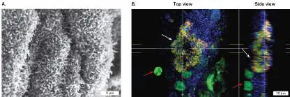
(described in Chapter 2) showing top and side views of biofilm clusters (white arrows)
consisting of rods and cocci on the mucosa of a pediatric adenoid. Removal of the adenoid is a
routine treatment for recurrent otitis media (middle ear infection). Specimens were treated
with nucleic acid stains using the LIVE/DEAD Bac Light Bacterial Viability Kit, in which live
bacteria stain green and dead bacteria stain red. Host inflammatory cells (red arrows) were
also stained green, but their nuclei appear much larger than the bacteria. The mucosal surface
(blue) was imaged using reflected light.
PHOTO COURTESY OF PAUL EVEREST. HADDOCK, ET AL. 2010. MICROBIOLOGY 156 :3079.
L. HALL-STOODLEY ET AL. CELL. MICROBIOL. 11 :1034–1043. © 2009 BLACKWELL PUBLISHING LTD.
Biofilm infections can also form on implanted medical devices such as heart valves, artificial
knees, or indwelling venous catheters. The bacteria appear to use their pili, nonpilus adhesins, and
exopolysaccharides to attach to host factors such as matrix proteins that coat the device. Once
infected, the implanted medical device may have to be replaced. Today, vascular catheter-related
bloodstream infections are the most serious and costly health care–associated infections. Biofilms
formed in a venous catheter can seed bloodstream infections by shedding planktonic cells or tiny
fragments of biofilm.
Biofilm infections are also important clinically because bacteria in biofilms exhibit tolerance to
antimicrobial compounds and persist in spite of sustained host defenses. Thus, biofilm infections
are hard to cure. Tolerance to antibiotics may be caused by poor nutrient penetration through the
exopolymer matrix into the deeper regions of the biofilm, leading to a stationary phase–like
dormancy (discussed in Section 27.3). Bacterial factors important to biofilm formation include type
IV pili, structural genes and regulators controlling cell-cell signaling (quorum sensing), and
extracellular matrix synthesis. Interfering with cell-cell signaling is effective in preventing or limiting
biofilm formation and may provide a target for new antimicrobial therapies.
To Summarize
Bacteria use pili and nonpilus adhesins to attach to host cells.
Type I pili produce a static attachment to the host cell, whereas type IV pili continually
assemble and disassemble. Pili assemble starting at the tip.
Nonpilus adhesins are bacterial surface proteins, or other molecules, that can tighten
interactions between bacteria and target cells.
Biofilms play an important role in chronic infections by enabling persistent adherence and
resistance to bacterial host defenses and antimicrobial agents.
Glossary
adhesin
Any cell-surface factor that promotes attachment of an organism to a substrate.
pilus pl. pili
Also called fimbria. A straight protein filament composed of a tube of protein monomers that
extend from the bacterial cell envelope.
fimbria pl. fimbriae
See pilus .
Fig. 23.5
FIGURE 23.5 ■ Mucociliary escalator. Movement of these hairlike cilia ushers
particles up and out of the trachea and lungs (colorized SEM; diameter from 0.5–1 μm).
DR. DAVID M. PHILLIPS/VISUALS UNLIMITED
Fig. 4.30
FIGURE 4.30 ■ Biofilm development. The stages of biofilm development in
Pseudomonas, which generally apply to the formation of many kinds of biofilms. Inset: A
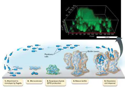
mucoid environmental strain of P. aeruginosa produces uneven, lumpy biofilms in a
continuous-flow cell. Cells in the biofilm were stained green with live/dead viability stain
(3D confocal laser scanning microscopy).
Source: O. E. Petrova and K. Sauer. 2011. J. Bacteriol. 193 : 6614–6628.
O. E. PETROVA AND K SAUER. 2011. J. BACTERIOL. 193 (23):6614–6628
25.3 Toxins Subvert Host Functionsnot assigned
Following attachment, many microbes secrete protein toxins (called exotoxins) and/or effector
proteins that kill or disable host cells to unlock their nutrients (because dead host cells
ultimately lyse). Secreted effector proteins will be discussed in Section 25.4. Here we will
focus on toxins. Bacterial pathogens have developed an impressive array of toxins that take
advantage of different key host proteins or structures. Gram-negative bacteria also possess a
toxic compound called endotoxin, which is an integral component of lipopolysaccharides (LPS).
Endotoxin can hyperactivate host immune systems to harmful levels.
Note: Do not confuse end otoxins from lipopolysaccharides in Gram-negative bacteria with
protein ex otoxins that can be secreted by Gram-negative and Gram-positive bacteria.
Categories of Microbial Exotoxins
Microbial exotoxins fall into several categories based on their mechanisms of action (Table
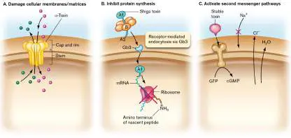
25.2). These classes are summarized here, and several are illustrated in Figure 25.16 .
Plasma membrane disruption. Toxins that damage membranes are exemplified by
alpha (α) toxin of Staphylococcus aureus. These toxins form pores in host cell membranes
and cause leakage of cell constituents (Fig. 25.16A ).
Cytoskeleton alterations. These toxins alter actin polymerization. Consequences include
altered host cell morphology and motility, disruption of tight junctions, and defects in
antigen presentation.
Protein synthesis disruption. Diphtheria and Shiga toxins target eukaryotic ribosomes
and destroy host protein synthesis (Fig. 25.16B ).
Cell cycle disruption (cyclomodulins). These toxins either stop (E. coli cytolethal
distending toxin, or CLDT) or stimulate (Pasteurella multocida toxin) host cell division.
Signal transduction disruption. These toxins alter host cell second messenger
pathways. E. coli ST (stable toxin), for instance, stimulates synthesis of cyclic guanosine
monophosphate (cGMP; Fig. 25.16C ) in target cells, which alters ion transport and fluid
movement.
Cell-cell adherence. These toxins cleave adhesion proteins that bind host cells together.
Exfoliative toxin of S. aureus, for example, breaks adhesion between dermis and
epidermis, mimicking the appearance of scalded skin.
Vesicle traffic. The major toxin in this class (VacA of Helicobacter pylori) has several
modes of action, depending on the host cell. The most visually striking effect is its ability
to cause vacuolization, which is the fusion of numerous intracellular vesicles.
Inhibit exocytosis. Tetanus and botulism toxins are proteases that prevent exocytosis of
neurotransmitters to cause spastic and flaccid paralysis, respectively.
Superantigens. These toxins, exemplified by toxic shock syndrome toxin (TSST), activate
the immune system without being processed by antigen-presenting cells (discussed in
Section 24.3).
TABLE a b 25.2 Characteristics of Bacterial Exotoxins,
Toxin Organism Mode of action Host target Disease
Damage membranes
Perfringolysin Clostridium Pore former Cholesterol Gas
O perfringens gangrene
c
Listeriolysin Listeria Pore former Cholesterol Food-borne
O monocytogenes systemic
illness,
meningiti
s
Alpha toxin Staphylococcus Pore former Plasma membrane Abscesses c
aureus
Panton- Staphylococcus Pore former Plasma membrane Abscesses,
Valentine aureus necrotizin
leukocidin g
pneumoni
a
Pneumolysin Streptococcus Pore former Cholesterol Pneumonia
pneumoniae c
Streptolysin Streptococcus Pore former Cholesterol Strep
S pyogenes throat,
scarlet
fever
Disrupt cytoskeletons
Iota toxin Clostridium ADP- Actin Gas
perfringens ribosyltransferas gangrene
e c
Inhibit protein synthesis
Diphtheria Corynebacterium ADP- Elongation factor 2 Diphtheria
toxin diphtheriae ribosyltransferas
e
Shiga toxins E. coli/Shigella N -glycosidase 28S rRNA HC and
dysenteriae HUS
Disrupt cell cycle
Pasteurella Pasteurella Mitogen (also Nucleus (encourages Wound
multocida multocida activates Rho cell division) infection
toxin GTPases)
Activate second messenger pathways
LT E. coli ADP- G proteins Diarrhea
ribosyltransferas
e
ST E. coli d Stimulates Guanylate cyclase Diarrhea
guanylate receptor
cyclase
Edema factor Bacillus anthracis Adenylyl cyclase ATP Anthrax
Pertussis Bordetella ADP- G protein(s) Pertussis
toxin pertussis ribosyltransferas (whoopin
e g cough)
Toxin A and Clostridioides Glucosyltransferase Rho G protein(s) Diarrhea/PC
B difficile
Cholera toxin Vibrio cholerae ADP- G protein(s) Cholera
ribosyltransferas
e
Lethal factor Bacillus anthracis Metalloprotease MAPKK1/MAPKK2 Anthrax
Disrupt cell-cell adherence
Exfoliative Staphylococcus Serine protease, Desmoglein; TCR, Scalded
toxins aureus superantigen and MHC II skin
(superantigen) syndrome
c
Alter vesicle traffic
VacA Helicobacter Large vacuole Receptor-like protein Gastric
pylori formation, tyrosine ulcers,
apoptosis phosphatase, gastric
sphingomyelin cancer
Block exocytosis
Neurotoxins Clostridium Zinc VAMP/synaptobrevin, Botulism
A–G botulinum metalloprotease SNAP-25 syntaxin
Tetanus toxin Clostridium tetani Zinc VAMP/synaptobrevin Tetanus
metalloprotease
Superantigens (activate immune response)
Enterotoxins Staphylococcus Superantigen TCR and MHC II, Food
aureus medullary emetic poisoning
center (vomit c
center)
Toxic shock Staphylococcus Superantigen TCR and MHC II Toxic shock
syndrome aureus syndrome
toxin c
Pyrogenic Streptococcus Superantigens TCR and MHC II Toxic shock
exotoxins pyogenes syndrome
, scarlet
fever
FIGURE 25.16 ■ Three classes of microbial exotoxins. These classes are defined
by mode of action. A. Pore-forming toxins assemble in target membranes and cause
leakage of compounds into and out of cells. B. Shiga toxin attaches to ganglioside Gb3,
enters the cell, and removes an adenine residue from 28S rRNA in eukaryotic ribosomes to
stop translation. C. Enterotoxigenic E. coli heat-stable toxin affects cGMP production. The
result is altered electrolyte transport: inhibition of Na + uptake and stimulation of Cl −
transport. In response to the resulting electrolyte imbalance, water leaves the cell.
Mechanisms of selected exotoxins are described in the following sections. We focus on
toxins that disrupt membranes and a set of exotoxins collectively called AB-subunit exotoxins
that target either protein synthesis or signal transduction. Superantigens are described in
Section 24.3, and exotoxins that affect exocytosis (tetanus and botulism toxins) are discussed
in Section 26.6.
Membrane Disruption
Toxins that disrupt membranes include pore-forming proteins that bind cholesterol and insert
themselves into target membranes and phospholipases that hydrolyze membrane
phospholipids into fatty acids. General descriptive terms for these toxins are hemolysins, which
lyse red blood cells (and other cells as well), and leukocidins, which more specifically lyse
white blood cells (leukocytes).
A classic example of a pore-forming exotoxin is the hemolytic alpha toxin produced by
Staphylococcus aureus, an organism that causes boils and blood infections. Alpha toxin forms
a transmembrane, oligomeric (seven-member) beta barrel pore in target cell plasma
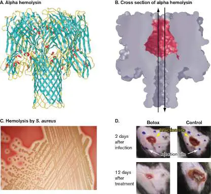
membranes (Fig. 25.17A and B ). It is easy to see how the resulting leakage of cell
constituents and influx of fluid cause the target cell to burst. Diagnostic microbiology
laboratories visualize hemolysins such as alpha toxin by inoculating bacteria onto agar plates
containing sheep red blood cells (Fig. 25.17C ). The clear, yellow zones around the S. aureus
colonies growing on blood agar indicate that the microbe secretes a hemolysin.
FIGURE 25.17 ■ Hemolysins of Staphylococcus aureus and Streptococcus
pyogenes. A. 3D image of the S. aureus alpha hemolysin pore complex, comprising
seven monomeric proteins. (PDB code: 7AHL) B. Cross section showing the hemolysin
channel. Arrows indicate movement of fluids through the pore. C. A blood agar plate
inoculated with S. aureus. The alpha toxin is secreted by the organism and diffuses away
from the producing colony. It forms pores in the red blood cells embedded in the agar,
causing the cells to lyse. D. Botox helps wound healing in a mouse model. Botulism toxin
(Botox) injected around a necrotizing fasciitis (NF) infection site (left column) prevented
neurons from releasing the streptolysin S–induced neurotransmitter that inhibits neutrophil
recruitment. NF wounds treated with Botox heal faster than without Botox. Infection site
and botulism injection sites are marked. Control wound received saline injections without
botulinum toxin.
BANOS/ALAMY STOCK PHOTO
MODIFIED FROM PINHO-RIBIERO ET AL. 2018. CELL 173 :1083–1097, FIG. 7E.
An example of a hemolysin that is also a leukocidin is streptolysin S, produced by
Streptococcus pyogenes. This pathogen can cause pharyngitis (sore throat) and necrotizing
fasciitis (NF), an aggressive wound infection. A surprising feature of streptolysin S is its role in
helping NF strains of S. pyogenes spread more quickly through tissue. Isaac Chiu from Harvard
University has shown that streptolysin S secreted from S. pyogenes will activate host
nociceptor neurons that detect pain. The activated neurons transmit the excruciating pain
signals of NF to the brain but also release a neuropeptide that suppresses neutrophil
recruitment. Fewer neutrophils at the site of infection enable S. pyogenes to spread through
tissue nearly unimpeded. Chiu’s group then wondered if botulism toxin, an agent that inhibits
neurotransmitter release, would counter the effect of streptolysin S (Fig. 25.17D ). When
botulism toxin was injected around the site of an NF infection in a mouse, the wound healed
even without antibiotics.
A particularly potent pore-forming leukocidin is produced by most strains of methicillin-
resistant Staphylococcus aureus (MRSA). Its name is Panton-Valentine toxin. This leukocidin
contributes to the formation of chronic staphylococcal infections by triggering neutrophils to
release neutrophil extracellular traps (NETs; Special Topic 25).
SPECIAL TOPIC 25 Chronic Staph Infections Work with a NET
Staphylococcus aureus is a versatile pathogen that can infect any human organ system. In
doing so, the organism deploys a vast array of virulence factors that subvert host
functions and disorient immune responses. S. aureus also builds tenacious biofilms that
repel innate immune mechanisms. Neutrophils, for example, can migrate to a biofilm
infection site but will fail to kill the pathogen. Such a failure in the innate immune system
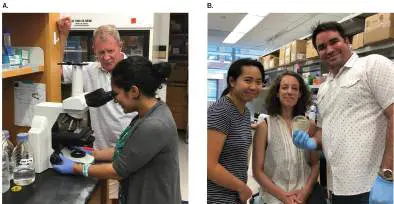
can produce a long-term (or chronic) infection. Daniel J. Wozniak (The Ohio State
University) and Victor Torres (New York University) and their colleagues (Fig. ST 25.1 )
have identified an important mechanism that S. aureus biofilms use to thwart neutrophil-
mediated killing while establishing a chronic infection.
FIGURE ST 25.1 ■ Wozniak and Torres laboratories. A. Daniel Wozniak
(back) and graduate student Mohini Bhattacharya. B. Victor Torres (right, holding a
Petri dish with MRSA), next to lab tech Evelien Berends (center) and postdoctoral
researcher Rita Chan (left).
COURTESY OF MATTHEW PESTRAK
COURTESY OF VICTOR J. TORRES
The scientists, armed with funds from the National Institutes of Health, the Cystic
Fibrosis Foundation, and the Burroughs Welcome Fund, began this line of study after
discovering that culture supernatants from S. aureus biofilms were better at killing
neutrophils than were supernatants generated by free-living planktonic cells (Fig. ST
25.2A ). Because proteinase K and heat (100°C) treatments of the supernatants
destroyed the neutrophil-killing activity, the researchers suspected that a protein secreted
by the biofilm was involved.
FIGURE ST 25.2 ■ Leukocidins PVL and HlgACB mediate biofilm-
dependent neutrophil killing. Cell-free supernatants (sup) were incubated
with neutrophils for 90 minutes. Neutrophil death was monitored by
LIVE/DEAD fluorescent staining. A. Comparison of biofilm and planktonic cell
supernatants. B. Biofilm supernatants used from various leukocidin mutants. Source:
Modified from Bhattacharya et al. 2018. PNAS 115 :7416–7421, figs. 1B (part A) and
2A (part B).
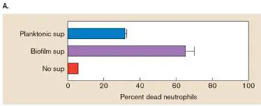
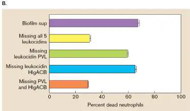
S. aureus secretes five different leukocidins that can form pores and damage
neutrophil cell membranes. Wondering whether one or more of these leukocidins was
involved in killing the neutrophils, the scientists prepared biofilm supernatants from S.
aureus mutants that lacked one or more of the enzymes, and they tested the fluids for
neutrophil-killing ability (Fig. ST 25.2B ). The results showed that eliminating two of the
leukocidins (PVL and HlgACB) prevented biofilm-specific neutrophil death.
The authors noticed that the leukocidin-containing supernatants also caused DNA
(chromatin) to be released from the dying neutrophils. This finding suggested that the
bacterial leukocidins were triggering a phenomenon called neutrophil extracellular traps
(NETs; shown in Fig. 23.21). NETosis is a form of neutrophil cell death in which the dying
cell spews a latticework of chromatin into the surrounding environment. Laced with
antimicrobial compounds, the NETs can trap and kill nearby pathogens. One hallmark of
NETosis is the presence of citrullinated histones that coat the released chromatin. To
prove that NETosis was being triggered, the scientists examined neutrophils incubated
with biofilm supernatants for the release of citrullinated histones. Neutrophils were added
to biofilm supernatants collected from various leukocidin mutants and then stained for
extracellular citrullinated histones using a fluorescently tagged antibody (Fig. ST 25.3 ).
The results confirmed that either PVL or HlgACB leukocidins were able to trigger NETosis.
FIGURE ST 25.3 ■ Biofilm culture supernatants containing leukocidins PVL
or HlgACB trigger NETosis. Chromatin released by neutrophils undergoing NETosis
(containing citrullinated histones) appears green. Scale bar = 10 μm.
MOHINI BHATTACHARYA, ET AL. 2018. PNAS 115 :7416–7421.
The researchers then asked whether neutrophils could even penetrate the leukocidin-
producing biofilms. Surprisingly, neutrophils penetrated biofilms erected by wild-type and
leukocidin-defective mutants (Fig. ST 25.4 ). However, the neutrophils invading the
wild-type, leukocidin-producing biofilms underwent NETosis and were no longer able to
effectively kill the bacteria. In contrast, the neutrophils that penetrated a leukocidin-
deficient mutant retained their nuclear structure and their ability to kill biofilm bacteria.
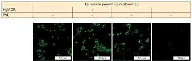
FIGURE ST 25.4 ■ Neutrophils penetrate biofilms of wild-type and
leukocidin-deficient S. aureus. Neutrophils prestained with CellTracker Blue dye
were added to 24-hour biofilms. After 1 hour, biofilm cross sections were stained for
viable bacteria (Syto-9 dye; small green cells) and for DNA of damaged or dead
neutrophils (ethidium homodimer-1 dye; red). A. Neutrophils penetrating the wild-
type biofilm underwent NETosis (seen as red and yellow cells). B. Neutrophils
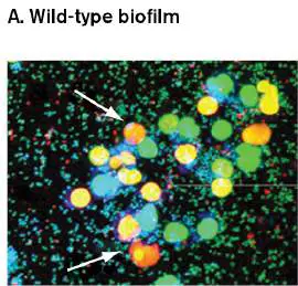
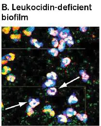
penetrating the leukocidin-deficient biofilm retained intact nuclei. Examples are
marked with white arrows.
BHATTACHARYA, ET AL. 2018. PNAS 115 :7416–7421, FIG. 4B.
BHATTACHARYA ET AL. 2018. PNAS 115 :7416–7421.
Why, then, don’t the chromatin NETs triggered by leukocidins kill S. aureus cells in the
biofilm? The authors propose that the combination of a known S. aureus –secreted DNA
nuclease and an adenosine synthase can degrade NET chromatin and destroy its
antimicrobial activity.
These remarkable findings suggest a possible new therapeutic strategy for treating
chronic S. aureus infections. By administering an anti-leukocidin antibody in combination
with antibiotic agents, clinicians could enhance a patient’s ability to eliminate biofilms—
the root cause of chronic infections.
RESEARCH QUESTION
How would you test whether the S. aureus nuclease NucA is required to protect
leukocidin-producing biofilms from the neutrophil extracellular traps that they trigger?
Mohini Bhattacharya, Evelien T. M. Berends, Rita Chan, Elizabeth Schwab, Sashwati Royd, et
al. 2018. Staphylococcus aureus biofilms release leukocidins to elicit extracellular trap formation and evade
neutrophil-mediated killing. Proceedings of the National Academy of Sciences USA 115 :7416–7421.
Phospholipase toxins are different from pore-forming toxins. For instance, phospholipase C
of Clostridium perfringens, a cause of gas gangrene, cleaves phosphatidylcholine in host
plasma membranes. At high concentrations the toxin causes membrane disruption, but at
sublethal concentrations the exotoxin will generate signaling molecules from membrane lipids
that can activate cytokine production. Some phospholipases, also called lecithinases, can
increase the permeability of capillaries to cause edema (fluid accumulation in tissues).
Another group of membrane-disrupting exotoxins specifically targets vacuolar membranes.
Many bacterial pathogens enter eukaryotic host cells by inducing phagocytosis and then end up
in a phagosome vacuole. Some of these intracellular pathogens need to break out of the
phagosome to grow in the cytoplasm. One example is Listeria monocytogenes, a
gastrointestinal pathogen that can also cause meningitis (described later). This Gram-positive
rod uses listeriolysin (a pore-forming exotoxin) and two phospholipases to escape the
phagosome to grow in the relative safety of the host cytoplasm.
Two-Subunit AB Exotoxins
As noted earlier, exotoxins target a variety of different host mechanisms. But despite the
diversity of their targets, many exotoxins share a common structure in that they have two
subunits, usually called A and B. These two-subunit complexes are called AB exotoxins. The
actual toxic activity in AB exotoxins resides within the A subunit. The role of the B subunit is to
bind host cell receptors and deliver the A subunit to the host cell. Many such toxins are called
AB5 exotoxins, because they have five identical B subunits arranged as a ring with a single A
subunit nestled in the center (Figs. 25.16B and 25.18A ).
FIGURE 25.18 ■ AB toxins. A. A typical AB toxin consists of an A subunit and a
pentameric B subunit joined noncovalently. B. Many AB toxins are ADP-ribosyltransferase
enzymes that modify protein structure and function.
One major subclass of AB exotoxins has ADP-ribosyltransferase enzymatic activity as part of
its toxic A subunits. These toxins, such as cholera toxin, transfer the ADP-ribose group from an
NAD molecule to an amino acid residue in a target host protein (Fig. 25.18B ). Sometimes
the function of the host protein is destroyed (for example, protein synthesis is destroyed by
diphtheria toxin); other times a targeted enzyme is locked into an active form (cholera toxin).
Cholera toxin. Vibrio cholerae (Fig. 25.19A ), a waterborne pathogen commonly found in
marine environments, produces a severe diarrheal disease called cholera that generally afflicts
malnourished people in developing countries like Bangladesh or countries where access to
clean water has been disrupted by natural disasters or war, as in recent years across central
and western Africa. After being ingested, V. cholerae colonizes the brush border of the victim’s
small intestine (Fig. 25.19B and C ) and secretes cholera enterotoxin (Fig. 25.19D ).
Cholera toxin causes diarrhea by reversing an important intestinal process. Normally, the
intestine absorbs NaCl and other ions (electrolytes), as well as water from food material
moving through the intestine. The result is well-formed feces with very little water and salt
content. Cholera toxin, however, reverses this process by causing intestinal cells to secrete
water and electrolytes into the intestinal lumen. How does cholera toxin accomplish this feat?
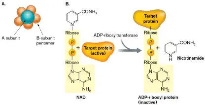
FIGURE 25.19 ■ Pathogenesis of cholera. A. Vibrio cholerae (SEM). Note the slight
curve of the cell and the presence of a single polar flagellum. B. Brush border of intestine
(TEM). V. cholerae binds to the fingerlike villi on the apical surface. C. V. cholerae, binding
to the surface of a host cell (SEM). Note that V. cholerae does not invade the host cell. D.
3D structure of cholera toxin, binding ganglioside GM1 on the intestinal cell surface. (PDB
code: 1S5F).
DR. GOPAL MURTI/SCIENCE SOURCE
DENNIS KUNKEL MICROSCOPY/SCIENCE SOURCE
KREBS, S. AND R. TAYLOR. 2011. JOURNAL OF BACTERIOLOGY 193 :5260–5270. © 2011 AMERICAN SOCIETY OF
MICROBIOLOGY.
Cholera toxin is an AB5 exotoxin. The B subunits attach to ganglioside GM1 on intestinal cell
membranes to trigger endocytosis and the formation of a toxin-containing vacuole (Fig.
25.20 , steps 1 and 2). The vacuole is then transported to the endoplasmic reticulum (ER)
and the A1 subunit containing ADP-ribosyltransferase activity is released into the ER. The A1
subunit is then exported from the ER into the cytoplasm (steps 3 and 4).
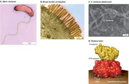
FIGURE 25.20 ■ Cholera toxin mode of action. Delivery of cholera toxin (CT) into
target cells and deregulation of adenylyl cyclase activity. NAm = nicotinamide.
The mission of the A1 peptide is to modify (that is, to ADP-ribosylate) an arginine residue in
a membrane-associated GTPase (or G factor) called G s. When bound to GTP, G s stimulates
host adenylyl cyclase to make cAMP (Fig. 25.20 , step 5). Normally, the GTPase activity in G s
quickly hydrolyzes the bound GTP, which halts stimulation and limits cAMP production. Disease
results when cholera toxin removes this control by ADP-ribosylating G s (step 5). The modified
G s factor does not hydrolyze GTP, so adenylyl cyclase is constantly stimulated and cAMP levels
sharply rise (step 6).
The sharp rise in cAMP stimulates a host protein kinase that activates various ion transport
channels, including the cystic fibrosis transmembrane conductance regulator (CFTR), so named
because a defect in this protein manifests as the lung disease cystic fibrosis. CFTR controls
chloride transport in several cell types, including intestinal epithelia (discussed in Section 23.4
). As a result of CFTR activation, chloride, sodium, and other ions leave the cell, and in an
attempt to equilibrate osmolarity, water leaves as well. Because the affected cells line the
intestine, the escaping water enters the intestinal lumen, leading to watery stools, or diarrhea
(Fig. 25.20 , step 6).
Note that some pathovars of Escherichia coli, called enterotoxigenic E. coli, also
make an enterotoxin close in sequence but identical in function to cholera toxin. The E.
coli enterotoxin is called labile toxin (LT) because it is easily destroyed by heat.
Bordetella pertussis, the cause of the lung disease whooping cough, produces a similar AB5
toxin that increases cAMP production in the lung.
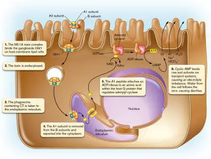
Thought Question
25.2 Figure 25.20 illustrates how cholera toxin works to cause diarrhea. To develop a
vaccine that generates protective antibodies, which subunit of cholera toxin should be used to
best protect a person from the toxin’s effects?
How does diarrhea benefit the pathogen? For one thing, diarrhea can decrease competition
with resident microbiota as the microbiota are swept away. For example, the vast majority of
organisms found in the diarrhea of cholera patients are V. cholerae bacteria. Few normal
microbiota are present. Diarrhea also distributes the pathogen throughout the environment,
thereby increasing the chance that another host will ingest the organism and perpetuate the
species.
Anthrax. A century ago, anthrax (caused by Bacillus anthracis; Fig. 25.21A ) was mainly a
disease of cattle and sheep. Humans acquired the disease only accidentally. Today we fear the
deliberate shipment of B. anthracis through the mail (as happened in 2001) or its dispersion
from the air ducts of heavily populated buildings. What makes this Gram-positive, spore-
forming microbe so dangerous? In large part, its lethality is due to the secretion of a plasmid-
encoded tripartite toxin (a variant of the AB exotoxin theme).
FIGURE 25.21 ■ Bacillus anthracis and anthrax toxin. A. B. anthracis (approx. 2
μm in length; SEM) in splenic tissue from a monkey. Spores inside the cell are not visible.
B. Single subunit and heptamer of protective antigen (PA). (PDB code: 1TZO) C.
Mechanism of toxin entry. EF = edema factor; LF = lethal factor.
SCOTT CAMAZIME/ALAMY STOCK PHOTO
The core subunit of the toxin is called protective antigen (PA) because immunity to this
protein protects hosts from disease. PA is really the B subunit of anthrax toxin. Protective
antigen binds to a host cell surface (there are multiple receptors), where a human protease
cleaves off a fragment (Fig. 25.21B ). The remaining part of PA autoassembles in the
membrane to form seven- and eight-membered pores. The other two components of anthrax
toxin—namely, edema factor (EF) and lethal factor (LF)—bind to the PA rings and are carried
into the cell (Fig. 25.21C ). EF and LF represent different A subunits of anthrax toxin. After
the complex is endocytosed, EF and LF are passed through the PA pore into the host
cytoplasm. Proton motive force across the vesicle membrane helps unfold the exotoxins and
powers their translocation.
Edema factor and lethal factor are enzymes that attack the signaling functions of the cell.
Edema factor is an adenylyl cyclase that remains inactive until entering the cytoplasm, where it
binds host calmodulin. Binding to calmodulin activates adenylyl cyclase, resulting in a huge
production of cAMP, and inactivates calmodulin from its normal function in the cell.
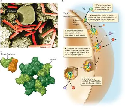
Lethal factor is a protease that cleaves several host protein kinase kinases, each of which is
part of a critical regulatory cascade affecting cell growth and proliferation. A protein kinase
kinase is an enzyme that phosphorylates and thereby activates another protein kinase that can
then phosphorylate one or more subsequent target proteins. One consequence of subverting
these phosphorylation cascades is a failure to produce signals that recruit immune cells to fight
the infection.
Thought Question
25.3 How might you experimentally determine whether a pathogen secretes an exotoxin? (
Hint: Where might you find the exotoxin in a culture tube growing the pathogen? How would
you determine if the material had toxic activity?)
AB Exotoxins That Target Protein Synthesis
Shiga toxin. Shigella dysenteriae and E. coli O157:H7 (also known as enterohemorrhagic E.
coli) cause food-borne diseases whose symptoms include bloody diarrhea. These organisms
produce an important exotoxin known as Shiga toxin (which is actually two toxins whose
primary differences are found in the B subunit). The genes (stx1 or stx2) encoding Shiga toxin
are part of prophage genomes integrated into the bacterial chromosome. The toxins are AB5
exotoxins with five B subunits for binding and one A subunit imbued with toxic activity. The A
subunit, upon entry, destroys protein synthesis by removing an adenine base from 28S rRNA in
eukaryotic ribosomes. Strains that produce high levels of this toxin are associated with acute
kidney failure, known as hemolytic uremic syndrome.
Note: Shigella dysenteriae is the only Shigella species that produces Shiga toxin. The O and
H designations for strains within a species of Enterobacteriaceae reflect antigenic differences in
LPS and flagella, respectively.
Shiga toxin is also an important virulence factor for E. coli O157:H7, a pathogen that has
emerged over the past 40 years. The organism can colonize cattle intestines without causing
bovine disease; as a result, undetected bacteria can easily contaminate meat products
following slaughter as well as pollute irrigation water from farms (see Chapter 28). The first
large U.S. outbreak of O157:H7 disease was associated with fast-food hamburgers served at a
Washington State Jack in the Box restaurant in 1993. Every year since, there have been
outbreaks of diarrhea caused by E. coli O157:H7. The most recent outbreak lasted from 2016 to
2019 and was linked to contaminated romaine lettuce from California.
Neither Shigella nor E. coli O157:H7 continually express Shiga toxins, so what activates stx
transcription? Iron availability is a key factor for inducing the expression of stx and many other
virulence genes in pathogens. The body holds its iron tightly in proteins such as lactoferrin and
transferrin. To an invading organism, the body is a very iron-poor environment. In the presence
of low iron, expression of Shiga toxin increases, the toxin kills host cells, and dead cells release
their iron. Shiga toxin, then, offers a way to rob the host of its iron stores. Another important
stx induction mechanism is tied to the induction of Stx prophages triggered by the SOS
response in stressed bacteria (described in Chapter 9).
An intriguing question often pondered by scientists is: What roles do virulence factors play
in the natural ecology of these bacteria? Surely, factors such as Shiga toxin did not evolve only
after Shigella started infecting humans. In fact, Shiga toxin is thought to be a natural defense
against Tetrahymena thermophila, a ciliated protist that grazes on bacteria.
Diphtheria toxin. The classic example of an exotoxin that targets protein synthesis is
diphtheria toxin, produced by Corynebacterium diphtheriae, the cause of the respiratory
disease diphtheria. The two-component diphtheria kills cells by ADP-ribosylating eukaryotic
protein synthesis elongation factor 2 (eEF-2), halting protein synthesis. The vaccine used to
prevent diphtheria (the “D” in the DTaP vaccine) is an inactivated form of this exotoxin that
retains its antigenicity (see Section 24.6). C. diphtheriae is another example of a pathogen
whose toxin gene (dtx) is regulated by iron availability and is part of a prophage genome
integrated into the bacterial chromosome.
Thought Question
25.4 Would patients with iron overload (excess free iron in the blood) be more susceptible to
infection?
We have examined only a few of the many protein exotoxins employed by pathogens. Some
of the others, including tetanus and botulism toxins, are described in the next chapter. What
should be apparent from our brief sampling is the evolutionary ingenuity that pathogens have
used to try to subdue their hosts.
Thought Question
25.5 Search the Internet to determine which other toxins are related to the cholera
enterotoxin A subunit. (Hint: Start by searching “Cholera enterotoxin subunit A protein
sequence” in a search engine. Use the FASTA version of the protein sequence to BLAST-search
the National Center for Biotechnology website for similar proteins.)
Endotoxin (LPS) Is Made Only by Gram-Negative Bacteria
Another important virulence factor common to all Gram-negative microorganisms is endotoxin
present in the outer membrane (discussed in Chapter 3). Endotoxins are important
contributors to inflammation. Not to be confused with secreted exotoxins, “endotoxin” is a
medical term sometimes used synonymously for “lipopolysaccharide” (LPS). LPS is really
composed of lipid A (the actual endotoxic factor), core glycolipid, and a repeating
polysaccharide chain known as the O antigen (Fig. 25.22 ). LPS molecules form the outer
leaflet of the Gram-negative outer membrane (discussed in Chapter 3). As bacteria die, they
release endotoxin in the form of LPS molecules. Endotoxin is a microbe-associated molecular
pattern (MAMP) molecule that can bind to certain Toll-like receptors (mainly TLR4) on
macrophages or B cells and trigger the release of TNF-alpha, interferon, IL-1, and other pro-
inflammatory cytokines (MAMPs, Toll-like receptors, and cytokines are discussed in Chapters 23
and 24). The release of these active agents causes a variety of symptoms, such as:
Fever
Activation of clotting factors, leading to disseminated intravascular coagulation
Activation of the alternative complement pathway
Vasodilation, leading to hypotension (low blood pressure)
Shock due to hypotension
Death, when other symptoms are severe
The major differences between exotoxins and endotoxins are summarized in Table 25.3.
FIGURE 25.22 ■ Endotoxin. A. Model of a lipopolysaccharide (LPS) membrane of
Pseudomonas aeruginosa, consisting of 16 lipopolysaccharide molecules (red) and 48
ethylamine phospholipid molecules (white). B. Basic structure of endotoxin, showing the
repeating O-antigen side chain that faces out from the microbe and the membrane
proximal core glycolipid and lipid A (contains endotoxic activity).
ROBERTO D. LINS AND T.P. STRAATSMA. 2001. BIOPHYSICAL JOURNAL 81 : 1037–1046.
TABLE Major Distinctions between Bacterial Exotoxins 25.3 and Endotoxins
Property Exotoxins Endotoxins
Producing Gram-positive or Gram-negative Gram-negative only
organism
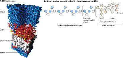
TABLE Major Distinctions between Bacterial Exotoxins 25.3 and Endotoxins
Property Exotoxins Endotoxins
Chemical Protein (size 50–1,000 kDa) Lipopolysaccharide (lipid A
moiety; size 10 kDa)
Denatured by Yes, if boiled long enough No
boiling
Mode of action Some exotoxins target specific Bind Toll-like receptor 4; activate
features of eukaryotic cells cytokine production
(membrane, protein synthesis,
signal transduction, etc.);
others are superantigens
Enzyme activity Often No
Toxicity High (1-μg quantities) Low (>100-μg quantities), but is
the primary cause of Gram-
negative sepsis (serious blood
infections)
Immunogenicity Highly antigenic Poorly antigenic
Vaccine Toxoids can be made for some Toxoids cannot be made
Fever Occasionally Yes
production
(pyrogenicity
)
The role of endotoxin in the disease process is obvious in infections with the Gram-negative
diplococcus Neisseria meningitidis (Fig. 25.23A ), a major cause of bacterial meningitis. N.
meningitidis has, as part of its pathogenesis, a septicemic phase in which the organism can
replicate to high numbers in the bloodstream. The large amount of endotoxin present causes a
massive depletion of clotting factors, which leads to internal bleeding, most prominently
displayed to a physician as small pinpoint hemorrhages called petechiae on the patient’s hands
and feet (Fig. 25.23B ). Capillary bleeding near the surface of the skin causes petechiae. One
danger of treating massive Gram-negative sepsis with antibiotics is that the enormous release
of endotoxin from dead bacteria could well kill the patient. Untreated Gram-negative sepsis is,
however, almost always fatal, so antibiotics are usually administered despite the risk.
FIGURE 25.23 ■ Effect of Neisseria meningitidis endotoxin. A. N. meningitidis
(cell 0.8–1 μm in diameter; SEM). B. Petechial rash caused by N. meningitidis.
DENNIS KUNKEL MICROSCOPY/SCIENCE SOURCE
MEDISCAN/VISUALS UNLIMITED
A proposed, but not yet approved, approach to prevent endotoxic shock revolves around the
knowledge that LPS must bind to Toll-like receptor TLR4 to cause endotoxic shock. Several
investigators have shown that injecting monoclonal antibody against TLR4 (anti-TLR4) into
infected mice can successfully block TLR4 and protect the mice from E. coli –induced septic
shock. However, some experts caution that interfering with TLR4 could have unforeseen and
possibly dangerous consequences for the patient.
To Summarize
There are nine categories of protein exotoxins based on mode of action. These
include toxins that disrupt membranes, inhibit protein synthesis, or alter the synthesis of
host cell signaling molecules, as well as toxins that are superantigens or target-specific
proteases.
Staphylococcus aureus alpha toxin forms pores in host cell membranes.
Many bacterial toxins are two-component, AB-subunit toxins. The B subunit
promotes penetration through host cell membranes, while the A subunit has toxic
activity.
Cholera toxin, E. coli labile toxin, and pertussis toxin are AB5 toxins that alter host
cAMP production by adding ADP-ribose groups to different G-factor proteins.
Anthrax toxin is a three-part AB toxin with one B subunit (protective antigen) and two
different A subunits that affect cAMP levels (edema factor) and cleave host protein
kinases (lethal factor).
Shiga toxin is an AB toxin that stops host protein synthesis by removing one adenine
base from host ribosome 28S rRNA.
Lipopolysaccharide (LPS) , also known as endotoxin, is an integral component of
Gram-negative outer membranes and an important virulence factor that triggers massive
release of cytokines from host cells. The indiscriminate release of cytokines can trigger
fever, shock, and death.
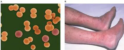
Glossary
exotoxin
A protein toxin, secreted by bacteria, that kills or damages host cells.
endotoxin
A lipopolysaccharide in the outer membrane of Gram-negative bacteria that becomes toxic
to the host after the bacterial cell has lysed.
hemolysin
A toxin that lyses red blood cells.
leukocidin
A toxin that lyses white blood cells.
ADP-ribosyltransferase
A bacterial toxin that enzymatically transfers the ADP-ribose group from NAD + to target
proteins, altering the target protein’s structure and function.
labile toxin (LT)
An Escherichia coli enterotoxin, destroyed by heat, that increases cellular cAMP
concentrations.
protective antigen (PA)
The core subunit of anthrax toxin, so called because immunity to this protein protects
against disease.
petechia pl. petechiae
A pinpoint capillary hemorrhage due to the absence of clotting factors. Petechiae may
indicate the presence of endotoxin.
Endnotes
1. Note a: Abbreviations: HC = hemorrhagic colitis; HUS = hemolytic uremic syndrome; LT
= heat-labile toxin; MAPKK = mitogen-activated protein kinase kinase; MHC II = major
histocompatibility complex class II; PC = antibiotic-associated pseudomembranous colitis;
SNAP-25 = synaptosomal-associated protein; ST = heat-stable toxin; TCR = T-cell
receptor; VAMP = vesicle-associated membrane protein. Return to reference a
2. Note b: All toxins have a known role in pathogenesis, as shown in an animal model or
appropriate cell culture. Return to reference b
3. Note c: Other diseases are also associated with the organism. Return to reference c
4. Note c: Other diseases are also associated with the organism. Return to reference c
5. Note c: Other diseases are also associated with the organism. Return to reference c
6. Note c: Other diseases are also associated with the organism. Return to reference c
7. Note c: Other diseases are also associated with the organism. Return to reference c
8. Note c: Other diseases are also associated with the organism. Return to reference c
9. Note c: Other diseases are also associated with the organism. Return to reference c
10. Note d: Toxin is also produced by other genera of bacteria. Return to reference d
25.4 Deploying Toxins and Effectorsnot assigned
A recurring theme among bacterial pathogens is the secretion of proteins that destroy, cripple, or subvert host target cells. The bacterial toxins described in the previous section are secreted into the surrounding environment, where they float randomly until chance intervenes and they hit a membrane-binding site. However, many pathogens attach to tissue cells and inject bacterial proteins (called effectors) directly into the host cell cytoplasm. The proteins may not kill the cell, but they redirect host signaling pathways in ways that benefit the microbe.
Protein secretion pathways were introduced in Section 8.5, which focused on ATP-binding cassette (ABC) proteins as a model. Additional secretion models are described here in their critical role of delivering pathogenicity proteins such as toxins. A particularly interesting aspect of these secretory systems is that many of them evolved from, and bear structural resemblance to, other cell structures that serve fundamental cell functions. The secretion systems and the molecular processes that share an evolutionary history include: Type II protein secretion (homologous to type IV pilus biogenesis)
Type III protein secretion (homologous to flagellar synthesis) Type IV protein secretion (homologous to DNA transfer by conjugation)
Type VI protein secretion (homologous to T4 phage tail structures)
Table 25.4 lists features of nine export systems of bacteria and examples of associated virulence effector proteins. We focus on secretion system types II, III, and IV as model delivery systems. TABLE Secretion Systems for Bacterial
25.4 Toxins a
Secretion Features Examples type I SecA dependent, one E. coli alpha effector per system hemolysin, Bordetella pertussis adenylyl cyclase II SecA dependent, Pseudomonas similar to type IV pili aeruginosa exotoxin A, elastase, cholera toxin III SecA independent, Yersinia Yop proteins, multiple effectors Salmonella Sip secreted, syringe proteins, mechanism injects enteropathogenic E. effectors into target coli (EPEC) EspA cells, related to proteins, TirA flagella IV Related to B. pertussis toxin, conjugational DNA Helicobacter CagA transfers, multiple effectors secreted, some systems inject effectors into target cells V Autotransporter, SecA Gonococcal and dependent to Haemophilus periplasm, self-influenzae IgA transport through proteases outer membrane, one effector per system VI Related to phage tails, Burkholderia and Vibrio single effector, cholerae VgrG harpoon mechanism VII Unrelated to other Mycobacterium systems tuberculosis Esx and Esp VIII Unrelated to other E. coli Curli pili systems subunits IX Unrelated to other Porphyromonas systems gingivalis (gingipain proteases)
Type II Secretion Resembles Type IV Pilus Assembly
Cholera toxin, discussed in Section 25.3, is a well-known example of a toxin secreted by a type II secretion system (T2SS). Type II secretion offers a clear example of how nature has modified the blueprints of one system to do a very different task. DNA sequence analysis revealed that the genes used for type IV pilus biogenesis (see Section 25.2) were duplicated at some point during evolution and repurposed to serve as a protein secretion mechanism. Type IV pili have the unusual ability to extend and retract from the outer membrane—a property that produces the gliding motility of Myxococcus (see Section 4.6) and the twitching motility of Neisseria and Pseudomonas. As you might guess, assembly/disassembly of these appendages is quite complex.
Type II protein secretion mechanisms mirror this complexity. Proteins to be secreted first make their way, via the SecA-dependent general secretion pathway, to the periplasm, where they are folded and then encounter the appropriate type II secretion system. Type II secretion systems cyclically assemble and disassemble the pilus-like structure (pseudopilus), using it as a piston to ram folded toxins or effector proteins through an outer membrane pore structure and into the surrounding void (Fig. 25.24).
FIGURE 25.24 ■ Vibrio cholerae type II secretion system, expelling cholera toxin. C, D, E, G, L, M, and N are protein components of the secretion system.
Type III Secretion Is an Injection Machine
Yersinia, Salmonella, and Shigella are the etiological agents of Black Death (Yersinia pestis) and various forms of diarrhea. These
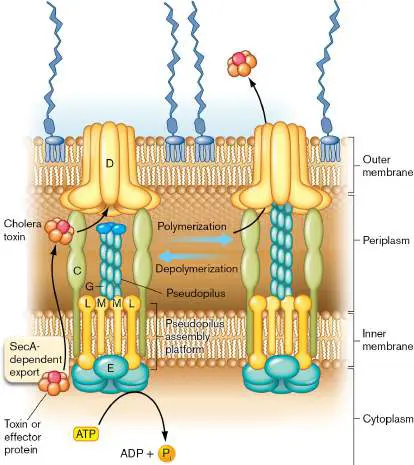
bacteria use a protein secretion system that takes bacterial virulence proteins (effector proteins) made in their cytoplasm and drives them directly into the eukaryotic cell cytoplasm without the proteins ever getting into the extracellular environment. Direct delivery is a good idea because it eliminates the dilution that happens when a toxin is secreted into an extracellular environment. Another advantage of this strategy is that it avoids the need to tailor the toxin to fit a preexisting host receptor.
These systems, collectively called type III secretion systems (T3SS), use tiny molecular syringes (injectisomes) embedded in their membranes to inject effector proteins directly into the host cytoplasm. Figure 25.25shows an electron micrograph of type III secretion needles and a model of the system spanning the cytoplasmic and outer bacterial membranes. Genes encoding T3SS systems evolved from flagellar genes, whose products export the flagellin proteins through the center of a growing flagellum (discussed in Section 3.6). It appears that a duplicated set of flagellar genes was evolutionarily reengineered to encode proteins that act more like molecular syringes (Fig. 25.25Aand B ). FIGURE 25.25 ■ The type III secretion complex from Salmonella enterica serovar Typhimurium type III injectisome. Unlike other secretion systems, the type III mechanism injects proteins directly from the bacterial cytoplasm into the host cytoplasm. The proteins in these systems are related to flagellar assembly proteins. A. Purified needle complexes (TEM) from S. Typhimurium. B. Schematic representation of the S. Typhimurium needle complex and its putative components. The image shows the translocon penetrating the host membrane and depositing a translocated effector protein. C. Shigella invades a host cell ruffle produced as a result of its type III secretion system. Shigella flexneri (approx. 2 μm) entering a HeLa cell ruffle (SEM) formed by host actin rearrangements. (HeLa cells are an immortal cancer cell line.)
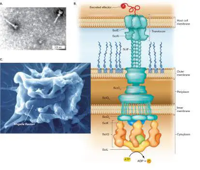
The ruffle engulfs the bacterium and eventually disassembles, internalizing the bacterium.
Source: Part B modified from Galán and Waksman. Cell 172 :1306–1318.
JORGE E. GALÁN AND ALAN COLLMER. 1999. SCIENCE 284 :1322–1328.
GALÁN, J. E. AND A. COLLMER. 2004. SCIENCE 304: 242.
The bacterial virulence proteins (effectors) secreted by type III systems subvert normal host cell signaling pathways, some of which cause dramatic rearrangements of host cytoskeleton at the cell membrane that lead to engulfment of the microbe (Fig. 25.25C ). The genes encoding type III systems in modern-day pathogens are usually located within pathogenicity islands inherited long ago via horizontal transfer from ancestral microbial sources. Many bacterial pathogens use this type of secretion system, including plant pathogens such as Pseudomonas syringae (the cause of blight, a disease of many plants in which leaves or stems develop brown spots). Secretion is normally triggered by cell-cell contact between host and bacterium.
E. coli pathogens use a T3SS to “inject” their own receptor into host cells. Enteropathogenic E. coli (EPEC) and enterohemorrhagic E. coli (EHEC) are two diarrhea-producing forms of E. coli. These pathogens use pili to initially bind to the host’s intestinal epithelial cells (Fig. 25.26A, step 1); however, the bacterium must establish a more intimate attachment to these cells to cause disease. The bacterial outer membrane protein called intimin mediates this intimate attachment. The only problem is that the host lacks a receptor for intimin. To solve this problem, EPEC and EHEC use a type III secretion system to insert their own receptor, Tir (for t ranslocated i ntimin r eceptor), into the target cells (Fig. 25.26A, step 2). The genes encoding intimin, Tir, and the secretion apparatus are all part of an EPEC pathogenicity island.
Note: Do not confuse bacterial intimin with the host protein
called integrin used by leukocytes for extravasation.
Once injected and placed in the host membrane, Tir binds intimin on the bacterial surface. Think of Tir as a wall anchor that you poke into a board in order to attach something to it. The result is a tighter, more intimate adherence between the bacterium and host cell surface, which is required for infection to proceed. In addition, host protein kinases phosphorylate Tir at tyrosine residue 474 (Fig. 25.26A, step 3). Phosphorylated Tir directly triggers a stunning reorganization of host cell cytoskeletal components (actin, alpha-actinin, ezrin, talin, and myosin light chain) such that a membrane “pedestal” is formed, raising the microbe up (Fig. 25.26A, step 4, and Fig. 25.26B ). The result of this attachment is the characteristic attaching and effacing (A/E) lesion, characterized by pedestal formation and destruction of surrounding microvilli. By placing itself on a “pedestal,” EPEC avoids engulfment and the perils of the phagolysosome. Because of their importance to virulence, type III secretion systems are being studied for ways to exploit them as potential drug targets.
FIGURE 25.26 ■ E. coli type III secretion and cell-cell interaction. A. Model of enteropathogenic E. coli (EPEC)
attachment and pedestal formation on intestinal epithelial cells. (1) EPEC attaches first, using type I pili. (2) Bound EPEC uses a T3SS to inject Tir protein into the host cell. Tir inserts into the host membrane and acts as a receptor for the EPEC surface protein intimin. (3) Tir also communicates through phosphorylation with other host factors that control actin filamentation and cytoskeleton formation. Actin polymerization raises the host membrane to produce a pedestal upon which EPEC sits (4). B. Pedestal formation (colorized SEM).
CELL HOST MICROBE. 2009. 5 (3), COVER. IMAGE COURTESY OF MANFRED ROSE
(HZI; BRAUNSCHWEIG, GERMANY).
Type IV Secretion Resembles Conjugation Systems
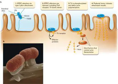
Many bacteria can transfer DNA from donor to recipient cells via a cell-cell contact system known as conjugation (see Section 9.3). The conjugation systems of some pathogens have been modified, through evolution, into new systems called type IV secretion systems (T4SS) that transport proteins, or proteins plus DNA, directly into target cells. Agrobacterium tumefaciens, for example, uses its Vir system to transfer the tumor-producing Ti plasmid and some effector proteins into plant cells. The result is a plant cancer called crown gall disease.
The Gram-negative bacterium that causes whooping cough in humans, Bordetella pertussis, also uses a type IV secretion system to export pertussis toxin, but it simply exports the toxin without injecting it into the host (Fig. 25.27). Pertussis toxin, similar to cholera toxin, sharply increases cAMP levels in lung epithelial cells. Water leaves the cells and enters the interstitium (causing edema). Type IV systems appear to recruit effector proteins directly from the cytoplasm, like pertussis toxin from Bordetella or CagA from Helicobacter, and pass them through the ATPases that drive the transport.
FIGURE 25.27 ■ Type IV secretion of pertussis toxin. Individual subunits of the toxin are transported to the periplasm (presumably via the Sec general secretion system) where the toxin is assembled. The type IV secretion system, which is evolutionarily related to conjugation systems, mediates the toxin’s exit from the cell. Effector proteins exit to the extracellular milieu through the pilus-like extension. (This
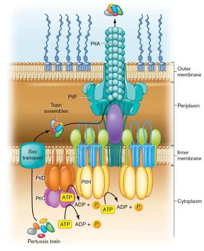
pseudopilus does not extend and retract, as happens in type II secretion.)
Type VI Secretion: Looks Like Phage, Acts Like a Blowgun
We end our discussion of secretion systems with a unique toxin delivery mechanism that resembles a harpoon or blowgun. The components of type VI secretion systems (T6SS) derive from the tail components of a T4 bacteriophage (see Chapter 6). But, unlike a phage tail, the tail-like contraction mechanism of the secretion system points outward from the cell and fires a rodlike core capped with a toxin protein into target cells, like a blowgun (Fig. 25.28). FIGURE 25.28 ■ Model of the type VI secretion mechanism. A Gram-negative bacterium is shown here using a
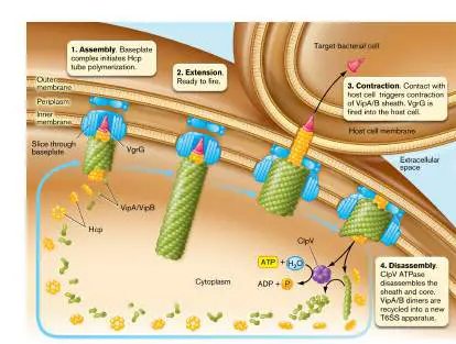
T6SS to attack another Gram-negative bacterium. A similar series of events takes place when a pathogen attacks a eukaryotic host cell.
The system is used primarily to kill bacterial competitors, but some systems, such as the one found in Vibrio cholerae, can deliver toxic effectors into eukaryotic cells. The system works by first assembling a baseplate in the cytoplasmic membrane from which the core and a surrounding sheath are assembled (Fig. 25.28, step 1). Once the sheath is complete (extended), the system is ready to fire (step 2). Firing happens when the sheath rapidly contracts, driving the core with its toxin cap outward and into a target cell (step 3). To reset the system, an ATPase chaperone (ClpV) disassembles the sheath and core proteins, and the structure reassembles with a new toxin molecule at the tip (step 4). With its role in virulence, its prevalence among Gram-negative bacterial genomes, and its predicted phage-like structure, the type VI secretion systems have become an exciting area of research. Identification of pathogen-specific T6SS components and substrates may reveal novel targets for treating bacterial disease.
Thought Question
25.6 Protein and DNA have very different structures. Why would a protein secretion system be derived from a DNA-pumping system? ( Hint: Review conjugation in Chapter 9.)
To Summarize
Many pathogens use specific protein secretion pathways to deliver toxins.
Type II secretion systems use a pilus-like extraction/retraction mechanism to push proteins out of the cell.
Type III secretion uses a molecular syringe to inject proteins from the bacterial cytoplasm into the host cytoplasm.
Type IV secretion utilizes a group of proteins homologous to conjugation machinery to secrete proteins from either the cytoplasm or the periplasm.
Type VI secretion is harpoon-like, shooting a rod capped with a toxin or effector protein into host cells or bacterial competitors.
Glossary
type II secretion system A bacterial protein secretion system that uses a type IV pilus– like extraction/retraction mechanism to push proteins out of the cell.
type III secretion system (T3SS)
A bacterial protein secretion system that uses a molecular syringe to inject bacterial proteins into the host cytoplasm. intimin A pathogenic Escherichia coli adhesion protein that binds tightly to an E. coli –produced receptor injected into host cells. type IV secretion system (T4SS)
Protein secretion system of Gram-negative bacteria whose components exhibit sequence homology with the components of conjugation systems.
type VI secretion system (T6SS)
A protein secretion system (derived from the tail components of a T4 bacteriophage) that propels a rod tipped with a toxin protein into adjacent bacterial or eukaryotic target cells.
Endnotes
1. Note a: Systems I–VI and VIII are found in Gram-negative bacteria; type VII is found in Gram-positive bacteria and Mycobacterium tuberculosis; type IX is restricted to Bacteroidetes. Return to reference a
25.5 Surviving within the Hostnot assigned
Once inside a host, how does a successful pathogen avoid detection and destruction? Many of the virulence factors in the pathogen’s arsenal help the microbe escape or resist innate immune mechanisms. Others are dedicated to stealth—that is, hiding from the immune system. But before discussing how these organisms survive in a host, we must ask how the pathogen knows it is in a host.
Where Am I?
A bacterial pathogen that can grow either outside or inside a host must adjust its physiology to match its whereabouts. Why make a type III secretion system if there are no host cells around? How do microbial pathogens know whether they are in a host or in a pond? And which bacterial genes are expressed exclusively while in a host? The same types of regulatory mechanisms that sense environmental conditions in a pond are used by the microbe to determine its whereabouts in a host. That is, various sensing systems act in concert to recognize a specific environmental niche. Two-component signal transduction systems, discussed in Section 10.1, are used to monitor magnesium concentrations, which are characteristically low in a host cell vacuole. Other regulators measure pH, which will be low (acidic) in the same vacuole, or osmolarity, which can range between 200 mOsm/liter (for intracellular fluid) to 800 mOsm/liter (for urine). The point is that there is no single system in bacterial pathogens that senses growth in a host. The various regulators collaborate to trigger the expression of virulence genes.
Note: Units of osmolarity are different from units of molarity.
Osmolarity is defined as the number of solute particles (measured in osmoles, Osm) per liter of a solution. Thus, a 1 mM solution of NaCl, which dissociates into Na + and Cl − ions, has an osmolarity of 2 mOsm. A 1 mM solution of MgCl, which dissociates to one Mg 2+
2
and two Cl − particles, has an osmolarity of 3 mOsm. Furthermore, multiple compounds may contribute to the osmolarity of a solution. Thus, a solution containing 1 mM NaCl and 1 mM MgCl 2 has an osmolarity of 5 mOsm.
Enterohemorrhagic E. coli uses positive and negative signals to determine where in a host it should express virulence genes. As mentioned earlier, EHEC uses a T3SS to inject effector proteins into gastrointestinal cells, so synthesis of the T3SS apparatus is best induced only in the intestine. One signal that induces T3SS synthesis is fucose. Fucose ends up in the intestine because fucosidases from the nonpathogenic gut microbe Bacteroides thetaiotaomicron cleave fucose from the host glycans present on intestinal epithelial cells. EHEC senses the fucose via a two-component system and responds by synthesizing the T3SS. However, other metabolites in the host can inhibit expression of the T3SS apparatus in unfavorable body sites. Andrew Roe and his team at the University of Glasgow found that the host metabolite D -serine can inhibit EHEC T3SS expression. D -Serine is found at high concentrations at extraintestinal sites such as the urinary tract and brain, two sites that EHEC does not infect. How D - serine inhibits T3SS gene expression is not clear.
Many bacterial pathogens regulate virulence genes by sensing the concentration of free iron, which is typically very low in the host. For instance, Corynebacterium diphtheriae sensing a low iron environment will induce synthesis of diphtheria toxin. The toxin kills host cells to release their iron, which the bacteria can now use. For other pathogens, iron concentration alone is usually not enough to provoke virulence. Regulators sensitive to other in vivo signals must be activated to achieve a successful infection.
Cell-cell communication is also important during infections. Pseudomonas aeruginosa, for example, has at least three quorum-sensing systems that detect secreted autoinducers (quorum sensing is discussed in Chapter 10). As the number of bacteria in a given space increases, so, too, does the concentration of the chemical autoinducer. When the autoinducer reaches a critical concentration, it diffuses back into the bacterium or binds to a surface receptor and triggers the expression of bacterial target genes. Genes included in the P. aeruginosa quorum-sensing regulons encode Pseudomonas exotoxin A and other secreted proteins, such as elastase, phospholipase, and alkaline protease.
Why would a pathogen employ quorum sensing to regulate virulence factors? One reason may be to prevent alerting the host that it is under attack before enough microbes can accumulate through replication. Tripping the host’s alarms too early would make eliminating infection easy. Waiting until a large number of bacteria have amassed before releasing toxins and proteases will increase the chance that the host can be overwhelmed.
Extracellular Immune Avoidance
The topic of extracellular immune avoidance was first covered in the discussions of host defense (see Chapter 23) and immunology (see Chapter 24). Many bacteria, such as Streptococcus pneumoniae and Neisseria meningitidis, produce a thick polysaccharide capsule that envelops the cell. Capsules hide the organism from Toll-like receptors on host cells, and they help the organism resist phagocytosis. Recall that phagocytes must recognize bacterial cell-surface structures or surface-bound C3b complement factor to begin phagocytosis (see Section 23.6). A capsule will cover bacterial cell wall components and mannose-containing carbohydrates that phagocytes normally use for attachment. The uniformity and slippery nature of capsule composition make it difficult for phagocytes to lock on to the bacterial cell.
But what about complement factor C3b, which can bind to the bacterial cell? Phagocytes have surface C3b receptors that latch on to C3b molecules fixed to an invading pathogen. Capsules, however, can envelop and hide any C3b complement factor that binds to the bacterial surface. Fortunately for us, immune defense mechanisms can eventually circumvent this avoidance strategy by producing opsonizing antibodies (IgG) against the capsule itself (see Section 24.2). The Fc regions of antibodies that bind to the capsule point away from the bacterium and are free to bind Fc receptors on phagocyte membranes. Binding of the antibody’s Fc region to the phagocyte’s Fc receptor triggers phagocytosis.
When should a pathogen make a capsule? Some pathogens make their capsules at all times, inside or outside of a host. But other organisms such as Salmonella enterica serotype Typhi (the cause of typhoid fever) make a capsule more economically, only when growing inside a human host. Because of its importance to virulence, the S. Typhi polysaccharide capsule is called virulence antigen (Vi antigen). Expression of the operon for making and exporting the Vi antigen is controlled by a transcriptional activator protein. The mRNA encoding the activator includes a stem-loop thermosensor region at its 5′ untranslated end that normally prevents translation at room temperature (see RNA thermometers, Chapter 10). When an infected person develops a fever, the thermosensor melts to expose the ribosome binding site needed to translate the activator. The result is production of the Vi antigen that will protect the pathogen during infection.
Pathogens can also use proteins on their cell surface to avoid phagocytosis. Staphylococcus aureus has a cell wall protein called protein A that binds to the Fc region of antibodies. Recall that phagocytes can easily phagocytize bacteria coated with antibody because phagocytes can grab the Fc regions of those antibodies. To counteract this mechanism, protein A on the S. aureus cell surface can bind to the Fc region of any antibody, hiding the Fc region from Fc binding sites on phagocyte membranes. When the Fc region of an antibody is bound to protein A, it cannot bind to phagocyte Fc receptors, thereby blocking phagocytosis.
Some extracellular microbes can trigger apoptosis in target host cells. Proteins made by the pathogen can slip into a host cell—a macrophage, for instance—and begin the process of destroying it (see Section 23.3). If the duped macrophage self-destructs, it cannot destroy the microbe.
Another immune avoidance strategy used by microorganisms, both the extracellular and intracellular types, is to change their antigenic structure. Genes encoding flagella, pili, and other surface proteins often use site-specific gene inversions to express alternative proteins (for example, Salmonella phase variation). Alternatively, some genes encoding cell-surface antigens have regions that can undergo slipped-strand mispairings during replication, a process that can randomly add or remove amino acid codons. The result is a functional but antigenically altered protein (a pilus, for instance). You can think of these processes as “shape-shifting” to avoid recognition.
Intracellular Immune Avoidance
In an effort to escape both innate and humoral immune mechanisms (see Chapters 23 and 24), many bacterial pathogens, called intracellular pathogens, seek refuge by invading host cells. (Viruses, by definition, are intracellular pathogens.) Hiding within a host cell temporarily provides the pathogen safe harbor from antibodies and phagocytic cells. Some bacteria dedicate their entire lifestyle to intracellular parasitism and are called obligate intracellular pathogens. Rickettsia, for example—for reasons unknown—will not grow outside a living eukaryotic cell. Other microbes, such as Salmonella and Shigella, are considered facultative intracellular pathogens because they can live either inside or outside of host cells. We have already discussed how intracellular pathogens get into cells, but how do they withstand intracellular attempts to kill them?
Once inside the phagosome, intracellular pathogens have three options to avoid being killed by a phagolysosome (Fig. 25.29). They can (1) prefer growth inside the phagolysosome, (2) prevent phagosome-lysosome fusion, or (3) simply escape the phagosome to grow in cytoplasm. We now explore each of these three fates. FIGURE 25.29 ■ Alternative fates of intracellular pathogens. Different pathogens have different strategies for surviving in a host cell. Some tolerate phagolysosome fusion (for example, Coxiella), others prevent phagolysosome fusion ( Salmonella and Legionella), and still others escape the phagosome to replicate in the cytoplasm (Shigella and Listeria). Salmonella enterica serovar Typhi, the cause of typhoid fever, invades intestinal cells, prevents phagolysosome fusion, exits the intestinal cell, and invades awaiting macrophages. The bacterium can survive inside the macrophage, which then shuttles the pathogen to lymph nodes and to the circulation.
Fate 1: Thriving under stress. In what could be called the “bring it on” strategy, some intracellular pathogens prefer the harsh
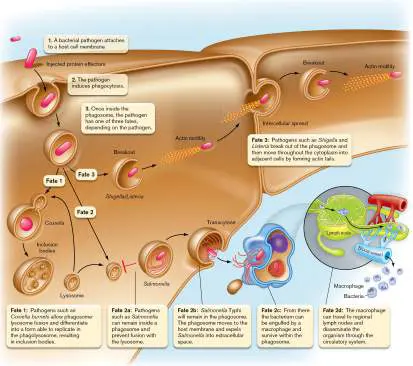
environment of the phagolysosome (Fig. 25.29, fate 1). Coxiella burnetii, for example, grows well in the very acidic phagolysosome environment (Fig. 25.30A). This obligate intracellular organism (an organism that grows only inside another living cell) causes a flu-like illness called Q fever (query fever). The symptoms of Q fever include sore throat, muscle aches, headache, and high fever. The illness has a mortality rate of about 1%, so most people recover to good health. The organism allows phagosome-lysosome fusion because the acidic environment that results is needed for it to survive and grow.
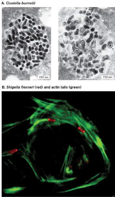
FIGURE 25.30 ■ Intracellular pathogens: Coxiella development and Shigella motility. A. A typical vacuole in J774A.1 mouse macrophage cells infected with Coxiella burnetii at 2 hours (left) and 6 hours (right) postinfection (TEM). The organism lives in an acidified vacuole and undergoes a form of differentiation that changes its shape and alters its interactions with the host cell. B. Intracellular Shigella flexneri (fluorescence microscopy), fluorescently stained red (1 μm in length), moves through the host cytoplasm propelled by actin tails, stained green.
HOWE ET AL. 2000. INFECT. IMMUN. 68 :3815.
GOLDBERG, M. 2001. MICROBIOL MOL BIO R 65 :595–626. © 2001, AMERICAN
SOCITY FOR MICROBIOLOGY.
Thought Question
25.7 How can you determine whether a bacterium is an intracellular pathogen?
Why some bacteria are obligate intracellular pathogens is unclear. One intracellular bacterium, Rickettsia prowazekii, a cause of epidemic typhus, appears to be an “energy parasite” that can transport ATP from the host cytoplasm and exchange it for spent ADP in the bacterium’s cytoplasm. But this does not explain its obligate intracellular status, since giving Rickettsia ATP outside a host does not allow the bacterium to grow. Other factors remain to be discovered.
Fate 2: Inhibiting phagosome-lysosome fusion. Some intracellular pathogens avoid lysosomal enzymes by preventing lysosomal fusion with the phagosome (Fig. 25.29, fate 2a).
Salmonella, Mycobacterium, Legionella, and Chlamydia are good examples. For instance, Legionella pneumophila grows inside alveolar macrophage phagosomes and produces the potentially fatal Legionnaires’ disease, so named for the veterans group that suffered the first recognized outbreak in 1976. Once inside a phagosome (called a Legionella -containing vacuole, or LCV), the organism uses a type IV secretion system (Dot/Icm) to secrete over 300 individual effector proteins through the vesicle membrane and into the cytoplasm.
The bacterial proteins secreted by Legionella interfere with host cell signaling pathways that cause phagosome-lysosome fusion. The bacterial effector proteins LegC7 and MavE, for instance, inhibit endosome trafficking that would lead to lysosome fusion. The result is that L. pneumophila can grow in a more habitable vesicle. Other effectors are E3 ligases that attach the small peptide ubiquitin to various host proteins as a way to manipulate host cell functions (discussed later). Interestingly, L. pneumophila is actually a soil and water microbe. Its ability to survive inside macrophages evolved from its ability to survive in amebas, which serve as a natural reservoir for this pathogen.
Salmonella Typhi is another pathogen that prevents phagosome-lysosome fusion, but this organism eventually leaves the host cell (via exocytosis) and enters extracellular tissue spaces where macrophages await (Fig. 25.29, fates 2b–d). The bacterium can be engulfed by a macrophage, or it can use T3SS mechanisms to enter by force. Either way, the pathogen will survive inside the macrophage phagosome. The infected macrophage travels to a regional lymph node, enters the bloodstream, and, like a Trojan horse, disseminates Salmonella throughout the body. Salmonella as a model of pathogenesis is discussed in more detail later.
Fate 3: Escaping the phagosome. The Gram-negative bacillus Shigella dysenteriae and the Gram-positive bacillus Listeria monocytogenes, both of which cause food-borne gastrointestinal disease, use hemolysins to break out of the phagosome vacuole before the vacuole can fuse with the lysosome (described earlier). By escaping the phagosome, the bacteria completely avoid lysosomal enzymes. Once free in the cytoplasm, they enjoy unrestricted growth. Yet even in the cytoplasm, these microbes have found a way to redirect host cell function to their own ends.
A fascinating aspect of escaping the phagosome involves motility. Shigella and Listeria are both nonmotile at 37°C in vitro; however, both move around inside the host cell, even though they have no flagella. How do they move? These species are equipped with a special device at one end of the cell that mediates host cell actin polymerization. The polymerizing actin, called a “rocket tail,” propels the organism forward through the cell (Fig. 25.30B ) until it reaches a membrane. The membrane is then pushed into an adjacent cell, where the organism once again ends up in a vacuole, this one with two membranes (Fig. 25.29, fate 3). This strategy enables the microbe to spread from cell to cell without ever encountering the extracellular environment, where it would be vulnerable to attack. Actin motility is also a feature of some species of Rickettsia (Fig. 25.31), Mycobacterium, and Burkholderia (a cause of serious lung infections).
FIGURE 25.31 ■ The obligate intracellular pathogen Rickettsia rickettsii. This SEM shows R. rickettsii (blue, approx. 0.7 μm in length), the cause of Rocky Mountain spotted fever, in association with host actin (gold). Several pathogens propel themselves through host cytoplasm by polymerizing host actin at one pole of the bacterial cell.
D. ELLISON ET AL. 2008. INFECT. IMMUN. 76 (2):542–550. COURTESY OF E. R.
FISCHER AND T. HACKSTADT.
Thought Questions
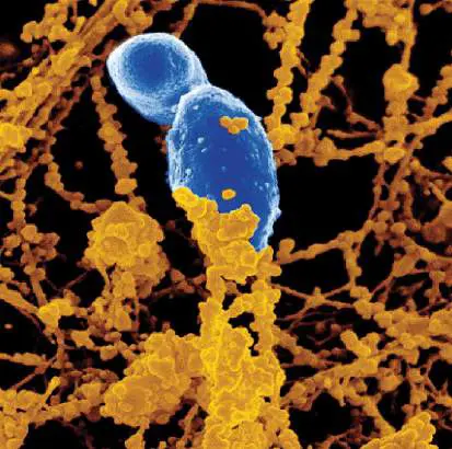
25.8 Figure 25.30B shows Shigella forming an actin tail at one pole. Why do organisms such as Shigella and Listeria assemble actin-polymerizing proteins at only one pole?
25.9 Why might killing a host be a bad strategy for a pathogen?
Sleeping with the Enemy
As just described, many bacterial and, of course, viral pathogens find safe haven by growing inside host cells. However, from our knowledge of innate and adaptive immune responses, it is not intuitively obvious why intracellular growth provides safety. After all, infected cells present microbial antigens on their class I or class II MHC receptors to alert the innate and adaptive immune systems that the infected host cell must be killed to resolve the infection. In addition, pieces of intracellular microbes (flagella, LPS, peptidoglycan) will bind pattern recognition receptors (Toll-like and NOD-like receptors—TLRs and NLRs) that activate intracellular inflammasomes. Inflammasomes trigger production of pro-inflammatory cytokines that mediate inflammation.
So, how do intracellular pathogens avoid destruction? It turns out that these invaders employ a variety of molecular tricks that misdirect the immune system much as a magician misdirects an audience. All of these strategies, summarized in Figure 25.32, buy the microbe more time to overwhelm the host.
FIGURE 25.32 ■ Summary of microbial strategies that misdirect the immune system. Bacteria and viruses produce different molecules that can mimic cytokines or transcriptional regulators, alter cytokine production, prevent programmed cell death, alter antigen presentation, or inhibit autophagy.
Molecular mimicry and subverting antigen presentation. A variety of bacteria and viruses use mimicry to confuse the immune system (mimicry is discussed in Section 24.7). In some cases, microbial proteins are made that look like cytokines or that bind to host cells and hitchhike via normal host trafficking to the nucleus, where the bacterial protein interferes with cytokine gene expression. These factors can manipulate the balance of helper T cells, for
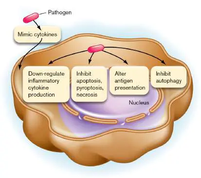
example, and send immunity down the wrong path for combating the microbe.
Microbes, especially viruses, can also interfere with antigen presentation on the surfaces of infected cells. Recall from Chapter 24 that proteins made by infectious agents growing in host cytoplasm are degraded and the pieces transported by the t ransporter of a ntigen p eptides (TAP) into the endoplasmic reticulum. The viral or bacterial antigens are loaded onto MHC I molecules found on the ER membrane, and the complexes are sent to the cell surface. Surface MHC I presents those peptide antigens to the TCRs of roaming CD8 cytotoxic T cells that will then kill the infected cell. Any scheme that interrupts MHC I presentation will spare the infected cell and its infectious cargo from destruction. So, how do pathogens derail MHC presentation?
Collectively, pathogens have four basic ways to subvert antigen presentation: (1) Make proteins that resist digestion by host proteasomes so that antigens are not processed. (2) Make a protein that blocks the TAP protein so that processed antigens cannot be loaded onto MHC I (herpesviruses such as HSV, CMV, and VZV are famous for this approach). (3) Make a protein that induces TAP degradation via ubiquitylation (Pseudomonas aeruginosa). (4) Induce degradation of MHC molecules via ubiquitylation.
Ubiquitylation strategies that coerce the host to degrade MHC proteins are described later.
Flipping cytokine profiles. Many pathogens defy death by growing inside macrophages that are “armed” with numerous antimicrobial weapons. Pathogens that can grow inside macrophages include bacteria such as Salmonella, Yersinia, Listeria, Mycobacterium tuberculosis, Francisella tularensis, and Chlamydia, as well as protozoa such as Leishmania and Trypanosoma cruzi. How do they survive? These and many other pathogens interrupt host cell signaling pathways that activate macrophage antimicrobial mechanisms. To perform this trick, most intracellular pathogens inhibit the production of pro-inflammatory cytokines, such as TNF-alpha, IL-8, IL-12, and IFN-gamma, but encourage the production of anti-inflammatory cytokines like IL-10, TGF-beta (transforming growth factor beta), and IL-4. Figure 25.33summarizes the general outcomes of these flipped profiles.
FIGURE 25.33 ■ General cytokine profiles for bacterial pathogens. The cytokine secretion profile during infection generally differs in extracellular pathogens (A) , compared to intracellular pathogens (B) . Not all cytokines are shown, and not all pathogens follow this scheme exactly. Extracellular pathogens generally permit high-inflammatory cytokine production, whereas intracellular pathogens promote anti-inflammatory cytokine production. Bars are not meant to be quantitative—only relative. CTL = cytotoxic T lymphocyte; MHC = major histocompatibility complex.
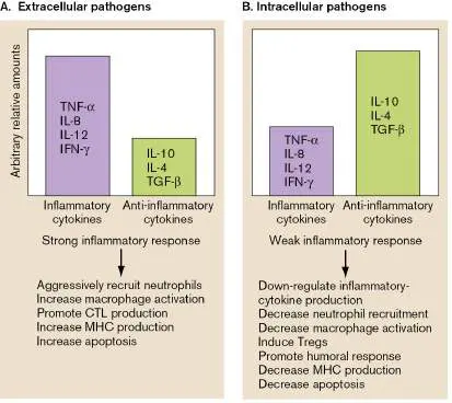
Mycobacterium, T. cruzi, and Leishmania also down-regulate the expression of host membrane receptors for IFN-gamma (thereby inhibiting inflammation) and interfere with downstream regulators that activate the production of MHC class I proteins, which dulls host immune mechanisms. Reduced MHC I production means less antigen presentation, fewer activated T H 1 helper cells, and thus fewer cytotoxic T cells to attack the infected cell.
Stopping programmed cell death. A macrophage that fails to eliminate infecting pathogens will eventually give up and try to kill itself and the infecting pathogens through one of three programmed-cell-death pathways (apoptosis, necrosis, or pyroptosis). This is a last resort to clear the infection. Apoptotic cells maintain membrane integrity during apoptotic death and are engulfed by nearby phagocytes. Inflammation is not provoked, because cytokines are not released. In contrast, necrosis and pyroptosis initiate rapid inflammatory responses by secreting inflammatory cytokines (see eResearch Activity 25). All of these mechanisms can kill the intracellular pathogen.
To “keep hope alive” and retain their intracellular niche, intracellular microbes can prevent host cell suicide by interfering with the molecular signals that initiate host death programs, or they can activate pro-survival mechanisms. Some pathogens even synthesize microbial mimics of host anti-apoptotic proteins. Yersinia enterocolitica and Mycobacterium tuberculosis, for instance, prevent pyroptotic cell death by inhibiting inflammasome formation and caspase-1 activation. Caspase-1 is a protease that initiates pyroptosis.
Some intracellular pathogens (Trypanosoma cruzi, Leishmania, and Yersinia pseudotuberculosis) actually tilt macrophage suicide pathways toward apoptosis to promote pathogen dissemination (recall that apoptotic cells are engulfed by other phagocytes). Some intracellular pathogens both inhibit and activate host cell death—just not at the same time. Early in infection, M. tuberculosis (the cause of tuberculosis) orchestrates the inhibition of host cell suicide pathways to enable the organism to grow, but then later it promotes suicide as a way to disseminate.
Neisseria gonorrhoeae uses a surprising strategy to activate apoptosis of macrophages. N. gonorrhoeae is mainly an extracellular pathogen that infects the mucosa of the genitourinary tract. The mucosa, however, is surveilled by resident tissue macrophages. N. gonorrhoeae triggers macrophage apoptosis to kill as many of these phagocytes as possible before the phagocytes engulf and kill the bacteria. The bacterial protein that triggers apoptosis is PorB, the major outer membrane porin of N. gonorrhoeae.
Until recently, no one knew how PorB gets from the bacterium to the mitochondria. Thomas Naderer (Fig. 25.34A) and colleagues at Monash University in Australia discovered that N. gonorrhoeae pinches off outer membrane vesicles (OMVs) containing PorB (Fig. 25.34B ), which then enter macrophages via endocytic pathways and ultimately deliver PorB into macrophage mitochondria (OMV formation and function are discussed in Chapter 3). After OMVs make contact with mitochondria, PorB appears to insert into the outer mitochondrial membrane (remember that mitochondria evolved from a Gram-negative bacterial ancestor) and then into the inner mitochondrial membrane, causing loss of membrane potential and leakage of cytochrome c into the cytosol, with the latter triggering apoptosis. Figure 25.34C shows the characteristic blebbing of apoptotic cells 15 hours after macrophages were mixed with N. gonorrhoeae OMVs. This is the first report of a membrane protein identified to target mitochondria, but others are believed to exist.
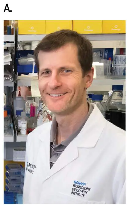
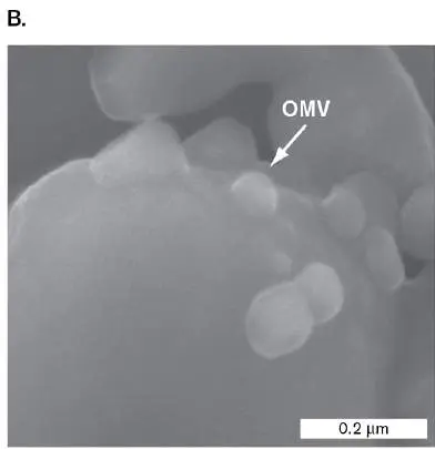
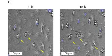
FIGURE 25.34 ■ Neisseria gonorrhoeae OMVs trigger macrophage apoptosis. A. Thomas Naderer studies the pathogenesis of N. gonorrhoeae. B. Scanning EM of OMV formation by N. gonorrhoeae. The white arrowhead points to a budding OMV. C. Macrophages undergo apoptotic cell death, noted by the blebbing (arrows), after 12–15 hours of contact with OMV (fluorescence microscopy).
COURTESY OF STEVE MORTON
DEO ET AL. 2018. PLOS PATH. 14 :E1006945, FIG. 8F.
DEO ET AL. 2018. PLOS PATH. 14 :E1006945, FIG. 1A.
Autophagy is a highly regulated internal surveillance mechanism by which eukaryotic cells form vesicles around damaged organelles to scavenge them for nutrients. One of the discoverers of autophagy, Yoshinori Ohsumi, won the 2016 Nobel Prize in Physiology or Medicine. Autophagy is also used as a universal innate defense mechanism to fight intracellular pathogens (Fig. 25.35A).
Autophagic vacuoles (autophagosomes) can encase these pathogens and deliver them to degradative lysosomes for destruction. Pathogen components are then sent to endosomes, where microbial structures are recognized by endosomal Toll-like receptors that trigger the innate immune system (see Chapter 23). The microbial components (antigens) are also sent to cell compartments rich in major histocompatibility complex II (MHC II) molecules. MHC II molecules then rise to the cell surface and present the microbial antigens to the adaptive immune system as described in Chapter 24.
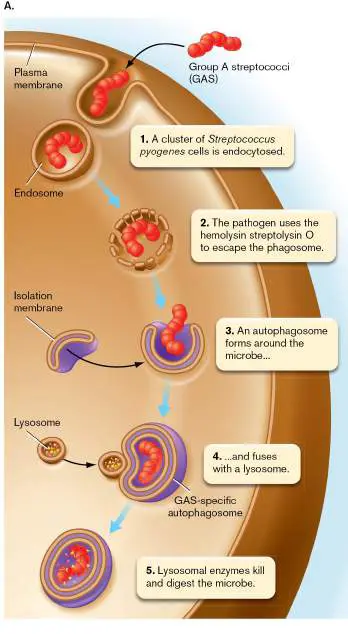
FIGURE 25.35 ■ Autophagy as an innate immune mechanism. A. When a pathogen escapes the phagosome, the host cell will try to form an intracellular vacuole (autophagosome) around the organism in a second attempt to kill it. B. Shigella may move in the host cytoplasm by actin tails to escape autophagy, or it can become trapped by a septin cage and succumb to autophagy.
SERGE MOSTOWY ET AL. 2010. CELL HOST MICROBE 8 (5):433–444.
Intracellular pathogens, however, have evolved mechanisms that can prevent autophagy and prolong their survival. For example, the Nef protein of human immunodeficiency virus (HIV) and protein M2 of influenza prevent autophagosome formation by targeting beclin 1, a protein central to autophagosome production. RNA viruses, as a group, encode proteins that can interact with 35% of autophagy-associated proteins, suggesting that autophagy is widely targeted by pathogens.
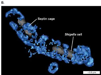
Shigella avoids autophagy by motoring through host cytoplasm using actin tails (described earlier). This “keep moving” tactic may sometimes work, but the host can stop the bacterium from making actin tails by wrapping Shigella in septin filaments, as shown in the 3D rendering in Figure 25.35B . Septin cages initially require actin to form, but then they inhibit actin polymerization. The septin-caged microbe is now trapped and marked for autophagy.
Redirecting host ubiquitylation signals. One important tool deployed by many pathogens forces the host to destroy or inactivate its own immune system regulators. The result is misdirection of the immune system. Some pathogens accomplish this misdirection by taking over a ubiquitous host protein modification system—namely, ubiquitylation (also called ubiquitination). Ubiquitin is a highly conserved, 76-amino-acid polypeptide in eukaryotes that can be covalently attached to other proteins. Attachment involves an enzymatic cascade of three enzymes: E1, E2, and E3. E1 and E2 activate ubiquitin and pass the peptide to many different E3 ubiquitin ligase enzymes, each of which recognizes a different host target protein. E3 ligases then directly attach the peptide to the appropriate target protein. Depending on where ubiquitin is ligated to the target, the ubiquitylated protein can be activated or, alternatively, delivered to the host proteasome for destruction. There are also deubiquitylation enzymes that can reverse the process.
In contrast to their eukaryotic hosts, viral and bacterial pathogens lack ubiquitylation systems but have evolved E3 ligases and deubiquitylases that effectively subvert normal host ubiquitylation pathways (Fig. 25.36). Like someone changing signs on a highway to misdirect drivers from their destination, the viral enzymes misdirect host signaling systems, causing the immune system to veer off course.
FIGURE 25.36 ■ Microbial E3 ligases and deubiquitylases alter innate immune systems. When a MAMP binds to a Toll-like receptor (TLR; 1A), a series of events, including a host E3 ligase–mediated ubiquitylation (1B and 1C), activates the transcriptional regulator NF-kappaB, which activates transcription of inflammatory cytokine genes. The cytokines are secreted and initiate inflammatory processes. Microbial E3 ligases (for example, rotavirus E3 ligase) can ubiquitylate other components of the NF-kappaB activation pathway and mark them for destruction (2). As a result, cytokine synthesis is inhibited and inflammation is limited. Salmonella makes a deubiquitylase that removes polyubiquitin from an inhibitor of NF-kappaB activation (3).
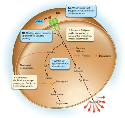
Deubiquitylation saves the inhibitor from destruction, enabling continued inhibition of NF-kappaB activation.
There are several ways that ubiquitylation normally directs the immune system. In the innate immune system, TLR pathway regulators become ubiquitylated and activated after a TLR encounters a MAMP. Simultaneously, proteins that inhibit the TLR pathway are marked by ubiquitin for destruction. The result is an activated signal pathway that leads to the formation of inflammatory cytokines (Fig. 25.36, step 1).
To subvert this system, some pathogens produce their own E3 ligases that divert the normal signal induction pathways. For example, rotavirus, a major cause of infant diarrhea, produces an E3 ligase that adds ubiquitin to activators of NF-kappaB, the transcription regulator that induces cytokine production (Fig. 25.36, step 2). The ubiquitylated activator proteins are destroyed, so NF-kappaB is not activated.
Pathogen deubiquitylases can also jumble the immune response. One example involves protein inhibitors of NF-kappaB that the host disposes of through ubiquitylation when cytokine synthesis is required. The bacterial pathogen Salmonella enterica produces a deubiquitylase that removes ubiquitin from a host inhibitor of NF-kappaB. Because the inhibitor is not degraded, NF-kappaB is not activated, and inflammatory cytokines are not made (Fig. 25.36, step 3). In both instances, inflammation is minimized, allowing the pathogen to survive.
Ubiquitylation is an important mechanism for adaptive immunity, too. For example, the process determines when cell-surface MHC class I and II molecules are expressed in dendritic cells (Fig. 25.37 ). Before dendritic cells mature, MHC molecules are polyubiquitylated and degraded, limiting their placement on cell surfaces. After maturation, however, the MHC molecules are no longer ubiquitylated, and as a result they are not degraded. The MHC molecules can now accumulate on the cell surface to present antigens to T cells. A number of viruses exploit this process by producing viral E3 ligases that polyubiquitylate MHC proteins, marking them for destruction ( Fig. 25.37). As a result, fewer MHC molecules remain to present viral proteins to the immune system.
FIGURE 25.37 ■ Microbial E3 ligases and deubiquitylases alter adaptive immune systems. MHC class I and class II molecules present antigens on host cell surfaces to helper T cells. Some viral E3 ligases can ubiquitylate the MHC molecules, marking them for degradation via endosomal pathways. Other E3 ligases can ubiquitylate MHC I molecules while in the endoplasmic reticulum (ER), leading to degradation of the MHC I receptors before they can be placed on the cell surface.
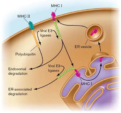
Some pathogens even make E3 ligases that ubiquitylate host proteins that are not normally tagged. One example is found in the interferon signal cascade. Interferons are secreted by virus-infected host cells as a signal to protect nearby uninfected cells from virus infection. Paramyxoviruses such as mumps and measles viruses produce E3 ligases that ubiquitylate key regulatory components (JAK-STAT) of the interferon signal cascade, which marks them for destruction. The host cell then becomes quite vulnerable to virus attack.
These examples demonstrate that ubiquitylation is a crucial part of immune regulation and a popular target of pathogens. Gaining an understanding of the ways that ubiquitylation directs the immune system and how pathogens influence that direction could lead to new ways of enhancing host defense.
Thought Question
25.10 Can antibodies against a viral E3 ligase stop an infection by that virus? Why or why not? What might be an alternative strategy that does not involve antibodies?
SARS-CoV-2 virus, the cytokine storm, and ISG15ylation.
COVID-19 is a deadly infection caused by the SARS-CoV-2 virus (see Section 6.5). A major reason this virus is so dangerous is its ability to trigger an unbalanced immune response. Inflammatory cytokines such as IL-6, TNF-alpha, and IL-1β can be wildly elevated. The elevated and unbalanced production of these and other cytokines is called a cytokine storm. One consequence of a cytokine storm is an aggressive inflammation that leads to an abnormal blood clotting condition called disseminated intravascular coagulation (DIC). As cells throughout the body are damaged by inflammation, they release a protein called tissue factor (TF) that initiates coagulation. Small clots forming in blood vessels block the flow of blood to organ systems, depriving them of oxygen. Ultimately, the organ systems fail (a condition called multi-organ system failure), and the patient dies. But how does SARS-CoV-2 cause a cytokine storm?
Sumana Sanyal (University of Oxford; Fig. 25.38B inset) and collaborators have probed this question. Their work has shown that SARS-CoV-2 hijacks the immune system by manipulating an interferon-stimulated gene product called ISG15 in macrophages (outlined in Fig. 25.38A). Interferon-stimulated genes (ISGs; see Section 23.5) are induced in response to viral infections; ISG15, a 17-kDa ubiquitin-like protein, is induced by interferon-alpha or -beta and is a central player in a person’s antiviral response (Fig. 25.38A, steps 1–4). ISG15 peptide can be conjugated to target host proteins using a pathway similar to that of ubiquitin, including E1-, E2-, and E3-like enzymes (step 5), and it can be removed from proteins by a host ISG15-specific protease.
FIGURE 25.38 ■ SARS-CoV-2 manipulation of host ISG15 response. A. Model summarizing how the virus manipulates free ISG15 levels. Blue type and arrows represent usual host responses to viral infection. Red type and arrows reflect viral manipulation of the host pathways. Virus interaction with host pattern recognition receptors in or on macrophages leads to interferon synthesis. Interferon will then trigger the induction of antiviral interferon-stimulated genes, including ISG15. Host proteins attach the ISG15 peptide to numerous other host proteins and, in the process, lower the intracellular concentration of free ISG15. However, a SARS-CoV-2 virus protease, PLpro, strips ISG15 from the ISGylated proteins, causing an increase in free ISG15 that stimulates the expression of pro-inflammatory cytokines. B. Evidence that free ISG15 triggers pro-inflammatory
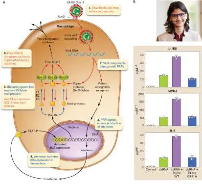
cytokine production. When stimulated with dsRNA, cells expressing viral PLpro (purple bars) produced higher levels of inflammatory cytokines than cells without the protease (green bar) or cells that expressed a defective protease (blue bar). Red bar represents control cells that were not given dsRNA. NS= no significant difference. Inset: Sumana Sanyal investigates SARS-CoV-2 effects on host immunity.
SUMANA SANYAL
The ISG15 peptide functions as a protein modifier of many intracellular host proteins, but unconjugated ISG15 itself is secreted and acts like a cytokine in several ways. It can stimulate IFN-gamma production, induce NK cell proliferation, and attract neutrophils. Sanyal and coworkers found that macrophages infected with SARS-CoV-2 decreased, instead of increased, the ISGylation of host proteins, down-regulated antigen-presenting MHC I and MHC II molecules, stimulated production of pro-inflammatory cytokines, and increased the secretion of free ISG15. Experiments with purified ISG15 then showed that the free peptide alone causes hyperproduction of pro-inflammatory cytokines (Fig. 25.38B ). The scientists discovered that the SARS-CoV-2 PLpro gene product, a papain-like protease, striped ISG15 from ISGylated proteins (mainly metabolic enzymes), thereby increasing the amount of free ISG15 in the cells (Fig. 25.38A, step 6). The authors propose that the combined effect of deISGylation of metabolic enzymes and the increased free form of ISG15 causes a hyperimmune response in macrophages during SARS-CoV-2 infection that includes a lethal cytokine storm. How free ISG15 manifests its hyperinflammatory effects is currently unknown.
Salmonella Is the Very Model of a Major GI Pathogen
When looking for a pathogen that employs the widest array of the virulence mechanisms just discussed, you can’t go wrong with the Gram-negative enteric pathogen Salmonella enterica serovar Typhimurium. Salmonella enterica (a facultative intracellular, Gram-negative pathogen) is currently the most common food-borne bacterial pathogen in the United States, causing approximately 1.2 million cases of diarrhea per year. Outbreaks in 2021 were linked to onions, prepackaged salads, poultry (chicken and turkey), frozen shrimp, cashew cheese, and even pet turtles and bearded dragons. Here we describe S. Typhimurium as a model of bacterial pathogenesis.
Following ingestion of contaminated food or water, S.
Typhimurium must survive passage through the acidic stomach and establish a presence in the small intestine. Attachment to intestinal epithelial cells involves numerous pili and nonpilus adhesins. As part of its pathogenesis, Salmonella uses type III secretion systems to invade the eukaryotic host cell and replicate intracellularly. The bacterium primarily attaches to and invades M cells that are interspersed along the wall of the small intestine. M cells are specialized intestinal epithelial cells (see Fig. 23.28B) that sample normal intestinal microbes and transfer pathogens across the epithelial barrier for recognition by the immune system. Salmonella subverts the normal function of M cells and causes an inflammatory response that leads to diarrhea.
During its evolutionary journey toward becoming a pathogen, Salmonella has acquired as many as 23 pathogenicity islands. Five of those islands can be found in all S. enterica serovars, but only two are discussed here. Salmonella pathogenicity island 1 (SPI-1) encodes a type III protein secretion system that delivers a cocktail of at least 13 different protein toxins (called effector proteins) directly into the cytosol of host epithelial cells in the gut (Fig. 25.39A, step 1). Inside epithelial cells, these effector proteins interfere with signal transduction cascades and modulate the host response. One mission of these effectors is to induce cytoskeletal actin rearrangements that cause ruffling of the eukaryotic membrane around the microbe (step 2; also see Fig. 25.25C and the chapter-opening images). The membrane ruffling starts the process of engulfment. Salmonella induces this response as a way to avoid the normal endocytic process.
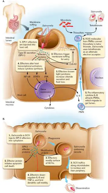
FIGURE 25.39 ■ Schematic overview of Salmonella pathogenesis. Effector proteins injected by Salmonella into a host M cell affect many aspects of host physiology. A. SPI-1 effector functions. B. SPI-2 effector functions. PMN = polymorphonuclear leukocyte; ROS = reactive oxygen species; SCV = Salmonella -containing vacuole; SPI = Salmonella pathogenicity island.
While engulfment is under way, some SPI-1 effectors act to loosen tight junctions that hold together adjacent epithelial cells (Fig. 25.39A, step 3). Uncontrolled chloride secretion (triggered by another effector) produces diarrhea as water leaves infected cells to compensate for an electrolyte imbalance. Other SPI-1 effectors activate transcription pathways that alter cytokine expression (step 4). One of the cytokines made, IL-8, helps lure phagocytic neutrophils (PMNs) to the area of infection, thereby initiating inflammation (step 5). Another effector is an E3 ligase that ubiquitylates and activates host proteins that induce interferon-beta expression, once again enhancing inflammation.
Initiating inflammation may seem counterintuitive, but Salmonella ’s strategy is to employ the neutrophils as “mercenaries” to kill competitors among the microbiota. The neutrophils squeeze between epithelial cells to reach the gut lumen, where they begin to engulf native members of the microbiome and produce reactive oxygen species (ROS) as part of their oxidative burst (see Chapter 23). Besides killing competitors, the oxidative burst converts thiosulfate, a compound made by gut microbiota, into tetrathionate, which Salmonella, but not its competitors, can use as an alternative electron acceptor (Fig. 25.39A, step 6). Tetrathionate provides a competitive advantage to Salmonella growing in the lumen.
Once Salmonella enters an epithelial cell or a macrophage, it finds itself in a vacuole called the Salmonella -containing vacuole, or SCV ( Fig. 25.39B , step 1). SPI-1 is down-regulated in this environment. In the normal course of events, an enzyme-packed lysosome would fuse with the phagosome and release its contents in an effort to kill the invader. Salmonella, however, possesses a second pathogenicity island, called SPI-2, which subverts this host response. As expression of the SPI-1 island powers down, a SPI-1 effector protein (SopE2) already secreted from Salmonella alters host cell metabolism in ways that trigger the induction of SPI-2. SPI-2 genes encode a separate type III secretion system that injects new effector proteins across the vacuole membrane. These proteins remodel the vesicle (step 2) and alter vesicle trafficking (step 3), thereby reducing phagosome-lysosome fusion so that the intravacuolar bacteria are spared. Some SPI-2 effectors, one of which is the bacterial deubiquitylase, down-regulate production of IL-8 and TNF-alpha to limit inflammation; other effectors inhibit migration of dendritic cells, which will subvert antigen presentation (Fig. 25.39B , step 4). Salmonella eventually escapes from the initial host cell when the SCV traffics to the cell periphery and fuses with the host membrane. In addition, some SPI-2 effectors will trigger host cell apoptosis (step 5), a programmed cell death that kills the host cell in a way that prevents further inflammation. Freed from the initial cell, Salmonella can now infect other cells, including macrophages. The combined effect of SPI-1 and SPI-2 effectors is the leakage of fluid and blood through a damaged intestinal lining and into the intestinal lumen to produce diarrhea that contains varying amounts of blood.
Salmonella enterica and SARS-CoV-2 are but two models of pathogenesis. Realize that every pathogen has a different but equally fascinating story to tell.
To Summarize
Two-component signal transduction systems can regulate virulence gene expression in response to the host environment.
Quorum sensing may prevent pathogens from releasing toxic compounds too early during infection.
Extracellular pathogens evade the immune system by hiding in capsules, by changing their surface proteins, or by triggering apoptosis.
Intracellular bacterial pathogens attempt to avoid the immune system by growing inside host cells. They use different mechanisms to avoid intracellular death.
Inhibiting phagosome-lysosome fusion is one way that pathogens can survive in phagosomes.
Hemolysins are used by certain pathogens to escape from the phagosome and grow in the host cytoplasm.
Actin tails are used by some microbes to move within and between host cells.
Molecular mechanisms for avoiding the immune system include molecular mimicry, altering cytokine profiles, stopping programmed host cell death, interfering with autophagy, and redirecting ubiquitylation and ISG15ylation signals. Specialized physiologies enable some organisms to survive in the normally hostile environment of fused phagolysosomes.
Glossary
capsule A slippery outer layer composed of polysaccharides that surrounds the cell envelope of some bacteria.
protein A A Staphylococcus aureus cell wall protein that binds to the Fc region of antibodies, hiding the S. aureus cells from phagocytes. intracellular pathogen A pathogen that lives within a host cell.
obligate intracellular pathogen A pathogen that can replicate only inside host cells.
facultative intracellular pathogen A pathogen that can replicate either inside host cells or outside host cells.
autophagy Eukaryotic cell function normally used to degrade damaged organelles. Also used to kill intracellular pathogens.
cytokine storm An exaggerated inflammatory immune response produced during certain infections and autoimmune diseases that triggers a rapid, unbalanced release of cytokines.
disseminated intravascular coagulation (DIC)
The uncontrolled formation of small clots in blood vessels that impedes blood flow to organs and results in multi-organ system failure.
Fig. 25.25C FIGURE 25.25 ■ The type III secretion complex from Salmonella enterica serovar Typhimurium type III injectisome. Unlike other secretion systems, the type III mechanism injects proteins directly from the bacterial
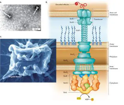
cytoplasm into the host cytoplasm. The proteins in these systems are related to flagellar assembly proteins. A. Purified needle complexes (TEM) from S. Typhimurium. B. Schematic representation of the S. Typhimurium needle complex and its putative components. The image shows the translocon penetrating the host membrane and depositing a translocated effector protein. C. Shigella invades a host cell ruffle produced as a result of its type III secretion system. Shigella flexneri (approx. 2 μm) entering a HeLa cell ruffle (SEM) formed by host actin rearrangements. (HeLa cells are an immortal cancer cell line.) The ruffle engulfs the bacterium and eventually disassembles, internalizing the bacterium.
Source: Part B modified from Galán and Waksman. Cell 172 :1306–1318.
JORGE E. GALÁN AND ALAN COLLMER. 1999. SCIENCE 284 :1322–1328.
GALÁN, J. E. AND A. COLLMER. 2004. SCIENCE 304: 242.
Fig. 23.28B
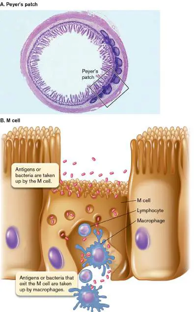
FIGURE 23.28 ■ Gut-associated lymphoid tissue (GALT). A. A Peyer’s patch located on the small intestine. B. Diagram of an M cell (microfold cell).
COURTESY OF DR. DEBORAH W. VAUGHAN
25.6 Tools Used to Probe Pathogenesisnot assigned
For any scientist studying pathogenic microbes, it is essential to identify virulence genes and determine what they do to undermine the host. As mentioned in Section 25.1, Stanley Falkow’s molecular Koch’s postulates argue that once a gene has been singled out as a possible virulence factor, proof requires knocking out the gene and observing a decrease in virulence, followed by restoring virulence by replacing the mutant gene. But how do you identify which genes to test? Also in Section 25.1, we discussed how scientists in years past used clever selection methods to identify virulence genes. Today’s methods, however, can more broadly and quickly reveal a microbe’s overall pathogenic strategy, illuminating new ways to prevent or cure an infection. In addition, we can now deeply probe how pathogens and hosts respond to each other during an infection.
Genomics
The sequencing of a pathogen’s genome, followed by bioinformatic analysis of the sequence (techniques described in earlier chapters), can yield valuable information about a pathogenic organism’s metabolism and can identify potential pathogenicity islands and virulence genes. Numerous genes encoding pili, secretion systems, toxins, and other effectors have been discovered in this way. We can also gain clues about potential virulence genes by comparing the sequences of virulent and naturally attenuated strains of a pathogen. For example, Leptospira species are spirochetes that cause a zoonotic renal disease in humans called leptospirosis. Dereck Fouts (J. Craig Venter Institute) and Joseph Vinetz (UC San Diego) compared the genome sequences of numerous Leptospira species classified as pathogenic, intermediate, or nonpathogenic. Several genes were found only in the pathogenic strains, suggesting their involvement in Leptospira pathogenesis or host adaptation. The pathogen-unique genes included, among others, a catalase, a protease able to degrade complement, a large family of virulence-modifying proteins of unknown function, and a CRISPR-Cas system. The results provided many new directions for research that probes leptospiral pathogenesis.
Transcriptomics
Other major goals of infectious disease research include learning how a pathogen causes an infection, how the host responds to the infection, and how the pathogen responds to the host’s response. The situation is much like watching two armies wage war, where the strategies of each side continually shift in response to each other. How can we map the cascading dynamics of attack and counterattack between pathogen and host?
RNAseq analysis is now used to monitor transcript modulation in pathogens during an infection and simultaneously view the host’s transcriptional response to the pathogen. Jörg Vogel (Fig. 25.40A ) at the University of Würzburg and colleagues used RNAseq to simultaneously examine host and pathogen transcriptomes during a Salmonella infection. The scientists infected human cells with GFP-labeled Salmonella and used flow cytometry (FACS; see Section 4.3 and Chapter 21) to select host cells at 4 hours (10 bacteria per cell) and 24 hours (75 bacteria per cell) postinfection. Next-generation RNAseq techniques were then used to quantify pathogen and host cell transcripts. At 4 hours postinfection, Salmonella SPI-1 transcripts decreased, while the transcript levels of SPI-2 genes increased (Fig. 25.40B ). On the host side, the scientists found that transcripts of host genes activated by NF-kappaB (a transcriptional activator of cytokine genes) were elevated at 24 hours postinfection (Fig. 25.40C ). The NF-kappaB result illustrates the host response to Salmonella invasion.
FIGURE 25.40 ■ Dual RNAseq captures the full transcript repertoire of Salmonella -infected human cells. A. Jörg Vogel uses dual RNAseq to study the interactions between pathogens and their hosts. B. Transcript profile of Salmonella during infection. Compared to extracellular Salmonella (0 hours), intracellular bacteria at 4 hours postinfection repress SPI-1 and induce SPI-2 effector genes. C. Transcript profile of infected host cells. Invaded (GFP +) host cells at 24 hours postinfection activate NF-kappaB-associated immunity genes relative to uninfected cells (GFP &minus). Source: Parts B and C modified from Alexander J. Westermann et al. 2016. Nature 529 :496–501, fig. 1A, C, E.
COURTESY OF JÖRG VOGEL
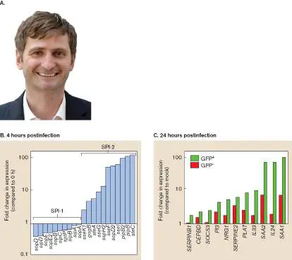
The scientists then identified Salmonella small RNA transcripts that were induced during infection. The most highly induced was the 80-nt sRNA called PinT (P hoP-induced sRNA in int racellular Salmonella). This sRNA was found to control the transition from SPI-1 to SPI-2 expression. By altering the timing of this transition, PinT influenced many aspects of the infected host cell’s physiology, including production of cytokine IL-8 and mitochondrial gene expression.
The simultaneous examination of host and pathogen transcriptional profiles by dual-RNAseq techniques promises to reveal unknown counteracting responses between pathogens and hosts during infection.
CRISPR-Cas9 Genome-Wide Screening
One of the innovative tools made possible by CRISPR-Cas9 technology (described in Section 12.3) is a method to screen host genomes for factors a pathogen needs to infect or replicate in that host. For instance, scientists can discover what host genes modulate the synthesis or membrane insertion of the ACE2 receptor for SARS-CoV-2 virus. The basic CRISPR whole-genome screening strategy involves the following steps: 1. Engineer host cells to make Cas9, the protein needed to introduce double-stranded breaks at targeted sites in the host genome.
2. Design and synthesize a pool of sgRNAs (single guide RNAs), each of which can direct Cas9 to target a different host gene. 3. Randomly clone the sgRNAs into separate lentivirus vectors ( Chapter 11). Each vector will express one sgRNA and a fluorescent GFP. A single sgRNA pool comprised of 70,000– 80,000 sgRNAs can target thousands of different host genes in one experiment.
4. Transduce the lentiviruses into a large number of host cells, such that no one host cell will be transfected with more than one sgRNA lentivirus. Recall that a lentivirus, which is a defective retrovirus, will randomly integrate as a DNA molecule into the host genome and express the sgRNA.
5. Use flow cytometry to collect only those host cells that fluoresce with GFP. Those cells were successfully transfected, and each will express one of the sgRNAs. The Cas9 protein also expressed in those cells will use the sgRNA to knock out the matching target gene.
6. Infect the pool of CRISPR-treated cells with the infectious agent under study. When a host gene required by the pathogen has been knocked out by CRISPR, that cell will survive the infection. 7. Because a lentivirus carrying the sgRNA gene has integrated into the host’s genome, PCR can be used to amplify whatever sgRNAs remain in the surviving pool of host cells. (All sgRNAs are designed to include identical flanking sequences suitable for PCR.)
8. Perform Illumina DNA sequencing (Chapter 7 and eAppendix 3 ) to identify those sgRNAs. The resulting sequences expose which host genes (designated proviral) were needed to promote pathogen infection or growth.
John Doench (Broad Institute of MIT and Harvard) and Craig Wilen (Yale School of Medicine) used this technique on African green monkey cells to reveal host factors critical for SARS-CoV-2 infection. The hope was to reveal novel therapeutic targets for treating severe acute COVID-19 disease (see Chapter 26). Numerous host genes required for virus infection were identified, the strongest of which was the ACE2 gene itself, encoding the receptor for the SARS-CoV-2 spike protein. Other host genes needed for virus infection included some involved with chromatin remodeling (SWI/SNF complex) and HMGB1, which turned out to be critical for ACE2 expression. The authors also tested whether known small-molecule inhibitors of SWI/SNF complex proteins could inhibit viral growth. Figure 25.41 shows the result of using PFI-3, which targets two members of the SWI/SNF complex likely involved in epigenetic control of ACE2 expression. The results indicate that the inhibitor, in a dose-dependent manner, significantly improved cell survival during SARS-CoV-2 infection. These findings may be applicable to emerging coronaviruses, and they may facilitate discovery of new host-directed therapies for infectious diseases (see Chapter 27). FIGURE 25.41 ■ Effect of a small-molecule inhibitor on SARS-CoV-2 infection. Vero-E6 cells were pretreated with the indicated concentrations of the inhibitor PFI-3 for 48 hours and then infected with SARS-CoV-2 at a multiplicity of infection of 2 viruses per 10 cells. Cell viability was measured at 3 days post infection (red bars) and compared with mock-infected controls (blue bars). The number of viable cells in culture was determined by quantitating the amount of ATP present, which indicates the presence of metabolically active cells. Values are percent viable relative to mock-infected cells. One, two, and three asterisks correspond to P values of 0.1, 0.01, and 0.001, respectively; ns = not significant.
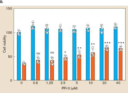
Cell Biology
You have seen numerous examples throughout this and previous chapters in which fluorescent stains, proteins (GFP), and antibodies combined with fluorescent microscopy were used to identify dramatic alterations in host cell structures, as well as the movement/location of bacterial or host cell proteins during the course of an infection. A powerful fluorescent technology has been developed that can identify which host cells during an infection have been targeted by translocated microbial effector proteins. The affected host cells fluoresce and can be sorted by a FACS machine for closer examination.
The technique starts with a translational fusion between a gene encoding the bacterial effector protein being studied and a beta-lactamase gene (Fig. 25.42A, step 1; fusion proteins are described in eAppendix 2). Beta-lactamase is an enzyme that cleaves the beta-lactam ring of the antibiotic penicillin (see Chapter 27). Once the bacterial effector–beta-lactamase fusion protein is expressed, the pathogen’s relevant secretion system (type III, IV, or VI) can translocate the fusion protein directly into specific host cells (step 2). Host cells that receive the effector protein can then be identified by addition of the fluorescent reporter CCF4-AM. FIGURE 25.42 ■ FRET detection method showing translocation of a bacterial effector protein and evidence that Legionella pneumophila translocates BlaM-RalF into mouse macrophages. A. The FRET detection method depends on a fluorescent reporter with two fluors. The emission wavelength of the donor fluorophore is blue, whereas the emission wavelength of the acceptor fluorophore is green. For the system to work, host cell esterases must de-acetylate the FRET reporter, changing it into a negatively charged complex that cannot leave the target cell. B. Sunny Shin studies how bacterial pathogens manipulate host defenses. C. In Shin’s work, macrophage monolayers were infected with dot + (T4SS-positive) and Δ dot (T4SS-negative) Legionella and treated with CCF4-AM fluorescent probe. Each image within the panels represents a single macrophage. Column 1: Uninfected cell scanned for uncleaved probe (green). Column 2: Infected host cell scanned for cleaved probe (blue). Only the T4SS-positive Legionella injected the BLA-RalF fusion, causing the macrophage
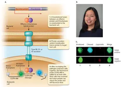
to fluoresce blue. Column 3: Infected cell immunostained for Legionella (red). The immunostain does not differentiate live from dead bacteria. Column 4: Merged images from columns 1, 2, and 3.
COURTESY OF ED HILLE
COPENHAVER, A. M., ET AL. 2014. INFECTION AND IMMUNOLOGY 82 :4325.
CCF4-AM is composed of two fluorescent parts (fluorophores) linked by a beta-lactam ring. One fluorophore can fluoresce blue when excited, but the blue emission wavelength is absorbed (or quenched) by the second fluorophore, which will fluoresce green ( Fig. 25.42A, step 3). The process is called fluorescence resonance energy transfer (FRET) . However, if the beta-lactam bond linking the two molecules is cleaved by beta-lactamase, blue fluorescence is no longer quenched and can be seen. CCF4-AM can enter all host cells, but fluorophore and quencher can be separated only by host cells that received the beta-lactamase–effector fusion protein (step 3). Fluorescence microscopy or FACS analysis can then identify those cells.
Sunny Shin (Fig. 25.42B ) and colleagues at the University of Pennsylvania used this technology to identify which host cells are targeted by the T4SS of Legionella pneumophila, the cause of the respiratory disease legionellosis (Fig. 25.42C) . This Gram-negative pathogen will invade host cells to inhabit a phagosome. From the phagosome it uses a T4SS to send effector proteins into the host cell cytoplasm. Shin’s group found that macrophages and neutrophils pulled from the airway space of infected mice were the primary recipients of T4SS-translocated effector RalF (fused to beta-lactamase) and that host cells receiving the fusion were the only cells harboring viable bacteria. Figure 25.42C (column 2) shows that the T4SS system (encoded by the dot genes) was required to translocate the BlaM-RalF fusion protein into the host cell. Only host cells infected by T4SS-positive Legionella fluoresced blue. FRET– beta-lactamase technology can also be used to recognize mutant bacteria defective in effector secretion or host factors that contribute to effector translocation.
Thought Question
25.11 How could you use the FRET–beta-lactamase technique to identify chemical inhibitors of type III, IV, or VI translocation?
To Summarize
Genome sequence analysis of pathogenic and nonpathogenic strains of a species can identify potential virulence genes.
DNA sequencing revealed growth requirements for the intestinal pathogen Tropheryma whipplei.
Dual RNA sequencing of pathogen and host transcripts from infected and uninfected host cells can expose a complex, yet orchestrated, response to infection.
Fluorescence resonance energy transfer (FRET)
approaches can identify host cells targeted by pathogens for type III, type IV, and type VI effector protein delivery.
Glossary
fluorescence resonance energy transfer (FRET)
The detectable transfer of fluorescent energy from one molecule to another. Because the participating molecules must be near each other, FRET can be used to monitor protein-protein interactions in cells and is also used in real-time PCR. eResearch Activity 25
Why Does Shigella “Prefer” Humans and Gorillas Over Other Mammals?
When studying any intracellular bacterial pathogen, scientists ask two important and related questions: How does a pathogen select a suitable host, and how does that bacterium evade the intracellular mechanisms meant to kill it? One such kill mechanism is pyroptosis, an inflammatory cell death program that kills the host cell and the pathogen. Pyroptosis is activated when bacterial PAMPs such as LPS are sensed by pattern recognition receptors in the cell. The process, outlined in Figure ERA 25.1 , activates the human proteases caspase −1, −4, and −5, which then cleave the host protein gasdermin D (GSDMD). The now released N-terminal fragments of GSDMD enter the host membrane to form pores that leak host cell contents (killing the cell) and facilitate the export of cytokines that trigger inflammation (killing the pathogen). This inflammatory cell death program is distinct from apoptosis, which dismantles the host cell without causing inflammation. Pyroptosis is critical to a host’s defense against intracellular infections.
FIGURE ERA 25.1 ■ Pyroptosis. In one form of inflammasome development, called noncanonical, LPS enters a host cell and activates proteases caspase-1, −4, and −5. The caspases cleave gasdermin D (shown as three differently colored domains), releasing an N-terminal pore-forming domain (pink) that enables leakage of cell contents, including inflammatory cytokines.
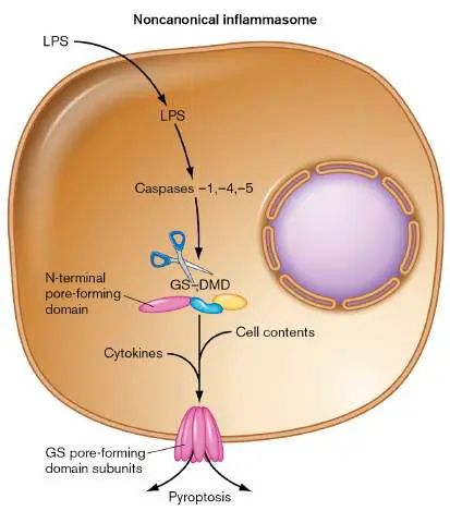
The Gram-negative genus Shigella is an intracellular pathogen that can produce life-threatening hemorrhagic dysentery in humans and nonhuman primates, such as gorillas. When infecting primates, Shigella species are highly infectious, needing as few as ten ingested organisms to cause disease. Mice, in contrast, remain undaunted at millionfold higher doses. Why are humans so vulnerable?
Vishva M. Dixit and colleagues from Genentech, with support from the Howard Hughes Medical Institute and the National Institutes of Health, tackled this question by screening cloned Shigella virulence factors for an ability to prevent LPS-induced pyroptosis. They introduced LPS into a series of human endothelial cell lines that expressed different Shigella flexneri effector proteins, each of which was controlled by a doxycycline-inducible promoter. One effector protein stood out: IpaH7.8. Figure ERA 25.2 illustrates that when human and mouse cells did not express the Shigella protein (purple bars), they both underwent pyroptosis when exposed to LPS. Pyroptosis was measured as the release of lactate dehydrogenase from the cytoplasm through gasdermin D pores into the surrounding medium. However, when IpaH7.8 was induced by adding doxycycline (green bars in Fig. ERA 25.2 ), LPS no longer triggered the leakage of LDH from human cells, suggesting that IpaH7.8 prevented the degradation of human gasdermin D. In contrast, expressing IpaH7.8 did not save mouse cells from LPS-induced pyroptosis. The finding that IpaH7.8 selectively prevented pyroptosis in human cells but not mouse cells can explain why mice are so resistant to Shigella infection.
FIGURE ERA 25.2 ■ Shigella effector protein IpaH7.8 blocks pyroptosis in human, but not in murine, cells. The human endothelial cell (Ea.hy926) and mouse macrophage cell (iMacs) lines harbored the IpaH7.8 gene under the control of a dox promoter. LPS was used to activate pyroptosis in these cells when the IpaH7.8 genes were turned off (−dox) and on (+dox). Membrane pores formed by cleaved gasdermin D were measured by the percent release of cytoplasmic LDH from the cell.
The next question was whether IpaH7.8 marked human gasdermin D or some other protein for degradation by the host proteasome. Without gasdermin D, pyroptosis doesn’t happen. This hypothesis was based on the knowledge that the Shigella IpaH7.8 effector protein is a ubiquitin ligase, a type of enzyme that, in eukaryotic cells, attaches a 76-amino-acid peptide (ubiquitin) to specific proteins, thereby marking them for degradation by the host proteasome (see Section 25.5). The authors wondered if the Shigella protein ubiquitylated gasdermin D, tagging it for
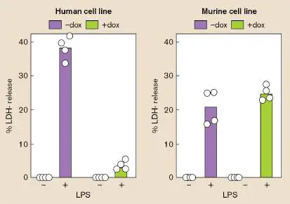
destruction. The researchers induced IpaH7.8 and used mass spectrometry (see Figure 8.30 and eAppendix 3) to follow the relative amounts of thousands of host cell proteins. As predicted, the level of GSDMD decreased dramatically, and three of its peptides became ubiquitylated prior to the molecule’s disappearance. Adding a chemical inhibitor of the host proteasome also prevented the IpaH7.8-dependent degradation of human GSDMD, supporting a model whereby IpaH7.8-dependent ubiquitylation of human GSDMD marked the protein for degradation by the proteasome, which would prevent pyroptosis. However, mouse GSDMD was not ubiquitylated by IpaH7.8 nor degraded after mouse cells were exposed to LPS, consistent with IpaH7.8 failing to prevent pyroptosis in mice. The project next addressed whether the N-terminal pore-forming domain (PFD) of human GSDMD was the actual target of the IpaH7.8 ubiquitin ligase. To test this, the team genetically swapped the mouse and human PFDs (Fig. ERA 25.3A ). By replacing the N-terminal pore-forming domain of mouse GSDMD with that of the human protein the scientists made a human-mouse chimeric GSDMD (H-M) that could be targeted by IpaH7.8 (Fig. ERA 25.3B , 1D4 immunoblot panel). In contrast, the Shigella ubiquitin ligase did not affect the opposite M-H chimera.
FIGURE ERA 25.3 ■ Shigella IpaH7.8 ubiquitin ligase targets the N-terminal pore-forming domain of human GSDMD. A. Schematic showing the PFD boundaries for human and mouse GSDMD. Numbers represent the ending residue of the PFDs. The colored PFD areas were genetically swapped to make chimeric GSDMD proteins M-H (mouse N-terminus PFD fused to human C-terminus) and H-M (human N-terminal PFD fused to mouse C-terminus). B. Immunoblots of 293T-cell (human kidney) extracts containing cloned wild-type or PFD-swapped GSDMD molecules. For immunoblotting purposes, the GSDMD and IpaH7.8 molecules were tagged with antigen 1D4 and IpaH7.8,
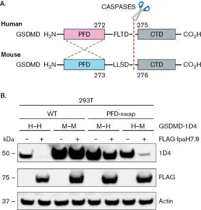
respectively. The actin panel was used to confirm that each lane was loaded equally.
G. LUCHETTI ET AL. 2021. CELL HOST MICROB. 29 :1521–1530
The scientists then proposed that mice lacking GSDMD should be hypersusceptible to Shigella disease. They used GSDMD-deficient mice to test this idea. After orally infecting these and control mice with S. flexneri, they collected stool samples and measured the ratios of wet weight to dry weight (the weight of stool remaining after drying); the higher the ratio, the more water there was in the stool, indicating a more significant diarrhea. Figure ERA 25.4 illustrates that double knockout (dKO) mice defective in both GSDMD and in NLRC4 (an inflammasome that controls a compensatory death pathway) produced more diarrhea than control mice or mice defective in only one of the test loci. Both genes had to be removed to see the GSDMD effect.
FIGURE ERA 25.4 ■ Mice lacking GSDMD produce diarrhea when infected with Shigella. Wet weight/dry weight ratios increased (indicating diarrhea) when GSDMD was deleted from NLRC4-defective mice (dKO = double knockout). NLRC4 controls a compensatory pyroptotic pathway. Each circle represents a separate mouse.
In sum, Shigella IpaH7.8 ubiquitin ligase is a virulence factor that specifically targets the PFD region of human gasdermin D. Ubiquitylation of the human PFD region directs gasdermin D to the proteasome. Without GSDMD in the cell, the caspase-1 protease
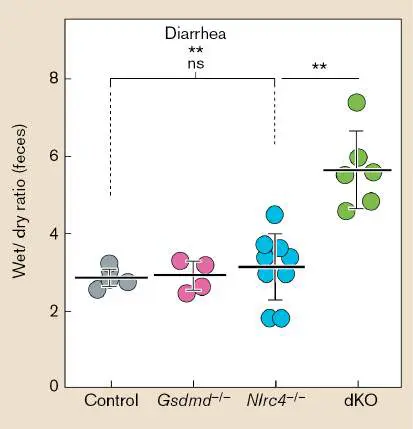
cannot generate the pore-forming domain needed for pyroptosis. Without pyroptosis and its associated inflammation, Shigella is free to grow intracellularly in human cells and cause disease.
Further Exploration
The authors also demonstrated that IpaH7.8 ubiquitin ligase was able to bind mouse PFD, but it could not ubiquitylate mouse PFD.
Formulate a hypothesis that would explain this apparent contradiction.
Source: Luchetti, Giovanni, Justin L. Roncaioli, Roberto A. Chavez, Alexander F. Schubert, Eric M. Kofoed, et al. 2021. Shigella ubiquitin ligase IpaH7.8 targets gasdermin D for degradation to prevent pyroptosis and enable infection. Cell Host and Microbe 29 :1521–1530.
https://doi.org/10.1016/j.chom.2021.08.010.
Glossary
Figure 8.30 FIGURE 8.30 ■ Identifying proteins directly from whole-cell extracts by mass spectrometry. Proteins extracted from a bacterial culture are digested into peptides with trypsin. The peptides are separated by column chromatography and analyzed by mass spectrometry (here by the Thermo Scientific Q Exactive hybrid quadrupole-Orbitrap mass spectrometer). In tandem mass spectrometry
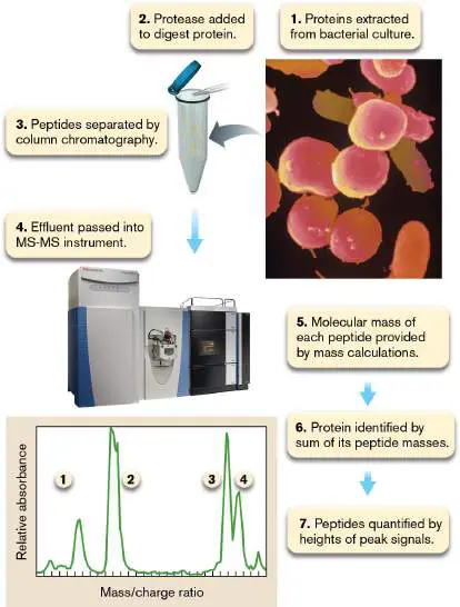
(MS-MS), the mass of each peptide is determined first (peaks 1−4 in the graph), and then selected peptides are subjected to additional fragmentation by ion spray (not shown). Each resulting peptide fragment will differ in size by one or more amino acids. Knowing the mass of each amino acid and the masses of the different peptide fragments enables extrapolation of the original peptide’s sequence.
COURTESY OF THERMO FISHER SCIENTIFIC
SIMKO/VISUALS UNLIMITED, INC.
CHAPTER REVIEW
Review Questions
1. Describe the differences between infection and disease; pathogenicity and virulence; LD 50 and ID 50.
2. What is meant by direct versus indirect routes of infection?
3. What are the characteristics of a good reservoir for an infectious agent?
4. Name the various portals of entry for infectious agents, and a disease associated with each.
5. Describe the basic features of a pathogenicity island. 6. Explain various ways in which bacteria can attach to host cell surfaces.
7. Describe the basic steps by which pili are assembled on the bacterial cell surface. How do type I and type IV pili differ?
8. Explain the nine broad categories of toxin mode of action.
9. What is ADP-ribosylation, and how does it contribute to pathogenesis?
10. Explain the differences between exotoxins and endotoxins.
11. Explain the mechanisms of secretion carried out by type II and type III protein secretion systems. What are the paralogous origins of these systems?
12. Describe the key features of Salmonella pathogenesis. 13. How can genomic approaches help identify pathogens in an infection?
14. What different mechanisms do intracellular pathogens use to survive within the infected host cell?
15. Describe different molecular strategies that microbes use to avoid the immune system.
16. How do bacteria determine whether they are in a host environment?
17. Discuss the relationships between ubiquitylation and intracellular pathogens.
Thought Questions
1. Using the Internet as an investigative tool, describe how you might interrupt the infection cycle of Zika virus without using chemical pesticides.
2. How would you determine whether a particular pilus on group A streptococci (GAS) is required for an organism’s pathogenesis? Use a tissue culture model.
3. Why have humans not developed resistance to microbial toxins?
4. You want to make a live oral vaccine for cholera. But, because Vibrio cholerae is an acid-sensitive organism, the person to be immunized would have to ingest a large number of organisms. E. coli, on the other hand, is very acid resistant and able to survive stomach acidity for long periods of time. You think that moving the acid resistance system from E. coli to V. cholerae will solve this problem. What ethical issue should you consider when trying to move the acid resistance system from E. coli into V. cholerae?
5. You have discovered a Gram-negative lung pathogen that uses a T3SS to inject an effector protein into a subset of lung cells. Design an in vivo experiment to identify which lung cells are targeted by the pathogen, and then determine the transcriptional responses of pathogen and host, but only among the infected cells.
Key Terms
adhesin (1061)
ADP-ribosyltransferase (1069)
airborne transmission (1054)
asymptomatic carrier (1056)
autophagy (1089)
capsule (1083)
cytokine storm (1091)
direct contact (1054)
disseminated intravascular coagulation (DIC) (1091) endotoxin (1066)
exotoxin (1066)
facultative intracellular pathogen (1084) fecal-oral route of transmission (1057) fimbria (1061)
fluorescence resonance energy transfer (FRET) (1097) fomite (1055)
genomic island (1058)
hemolysin (1068)
horizontal transmission (1054)
immunopathogenesis (1057)
indirect contact (1054)
infection (1053)
infection cycle (1054)
infectious dose 50% (ID 50) (1054) intimin (1079)
intracellular pathogen (1084)
labile toxin (LT) (1073)
latent state (1053)
lethal dose 50% (LD 50) (1054)
leukocidin (1068)
mechanical vector (1055)
obligate intracellular pathogen (1084) opportunistic pathogen (1053)
parasite (1053)
parenteral route (1057)
pathogen (1053)
pathogenesis (1053)
pathogenicity (1053)
pathogenicity island (1058)
petechia (1076)
pilus (1061)
portals of entry (1056)
primary pathogen (1053)
protective antigen (PA) (1074)
protein A (1084)
reservoir (1055)
transovarial transmission (1056)
transplacental transmission (1054) type II secretion system (T2SS) (1078) type III secretion system (T3SS) (1079) type IV secretion system (T4SS) (1080) type VI secretion system (T6SS) (1081) vector (1055)
vehicle transmission (1055)
vertical transmission (1054)
virulence (1053)
virulence factor (1057)
zoonotic disease (1056)
Recommended Reading
Aprianto, Rieza, Jelle Slager, Siger Holsappel, and Jan-Willem Veening. 2017. Time-resolved dual RNA-seq reveals extensive rewiring of lung epithelial and pneumococcal transcriptomes during early infection. Genome Biology 17 :198. https://doi.org/10.1186/s13059-016-1054-5.
Deng, Liwen, and Isaac M. Chiu. 2021. Microbes and pain. PLoS Pathogens 17 :e1009398.
https://doi.org/10.1371/journal.ppat.1009398.
Deo, Pankaj, Seong H. Chow, Iain D. Hay, Oded Kleifeld, Adam Costin, et al. 2018. Outer membrane vesicles from Neisseria gonorrhoeae target PorB to mitochondria and induce apoptosis. PLoS Pathogens 14 :e1006945.
https://doi.org/10.1371/journal.ppat.1006945.
Franklin, Tyler G., and Jonathan N. Pruneda. 2021. Bacteria make surgical strikes on host ubiquitin signaling. PLoS Pathogens 17 : e1009341.
https://doi.org/10.1371/journal.ppat.1009341.
Galán, Jorge E., and Gabriel Waksman. 2018. Protein-injection machines in bacteria. Cell 172 :1306–1318.
Golovkine, Guillaume, Emeline Reboud, and Philippe Huber. 2018. Pseudomonas aeruginosa takes a multi-target approach to achieve junction breach. Frontiers in Cellular and Infection Microbiology 7 :532. https://doi.org/10.3389/fcimb.2017.00532. Gussow, Ayal B., Noam Auslander, Guilhem Faure, Yuri I. Wolf, Feng Zhang, et al. 2020. Genomic determinants of pathogenicity in SARS-CoV-2 and other human coronaviruses. Proceedings of the National Academy of Sciences 117 :15193– 15199. https://doi/10.1073/pnas.2008176117.
Munnur, Deeksha, Qiwen Teo, Denzel Eggermont, Horace H. Y. Lee, Fabien Thery, et al. 2021. Altered ISGylation drives aberrant macrophage-dependent immune responses during SARS-CoV-2 infection. Nature Immunology 22 :1416–1427. https://doi.org/10.1038/s41590-021-01035-8.
Santos, José Carlos, and Jost Enninga. 2016. At the crossroads: Communication of bacteria-containing vacuoles with host organelles. Cellular Microbiology 18 :330–339.
https://doi.org/10.1111/cmi.12567.
Schroeder, Gunnar N., Jaclyn S. Pearson, and Teresa L. M. Thurston. 2021. Editorial: Bacterial effectors as drivers of uman disease: Models, methods, mechanisms. Frontiers in Cellular and Infection Biology 11 :708228.
https://doi.org/10.3389/fcimb.2021.708228.
Tripathi-Giesgen, Ishita, Christian Behrends, and Arno F. Alpi. 2021. The ubiquitin ligation machinery in the defense against bacterial pathogens. EMBO Reports 22 :e52864. https://doi.org/10.15252/embr.202152864.
Wei, Jin, Mia Madel Alfajaro, Peter C. DeWeirdt, Ruth E. Hanna, William J. Lu-Culligan, et al. 2021. Genome-wide CRISPR screens reveal host factors critical for SARS-CoV-2 infection. Cell 184 :76–91.
https://doi:10.1016/j.cell.2020.10.028.
Zhang, Wenchao, Xiaofeng Jiang, Jinghui Bao, Yi Wang, Huixing Liu, et al. 2018. Exosomes in pathogen infections: A bridge to deliver molecules and link functions. Frontiers in Immunology 9 :90. https://doi.org/10.3389/fimmu.2018.00090.
Glossary
pathogenesis The processes through which microbes cause disease in a host. parasite Any bacterium, virus, fungus, or protozoan (protist) that colonizes and harms its host; the term commonly refers to protozoa and to invertebrates.
pathogen A bacterial, viral, or fungal agent of disease.
infection The growth of a pathogen or parasite in or on a host.
primary pathogen A disease-causing microbe that can breach the defenses of a healthy host.
opportunistic pathogen A microbe that normally is not pathogenic but can cause infection or disease in an immunocompromised host organism. latent state A period of the infection process during which a pathogenic agent is dormant in the host and cannot be cultured.
pathogenicity The ability of a microorganism to cause disease.
virulence A measure of the severity of a disease caused by a pathogenic agent.
lethal dose 50% (LD 50)
A measure of virulence; the number of bacteria or virions required to kill 50% of an experimental group of hosts. infectious dose 50% (ID 50)
The number of bacteria or virions required to cause disease symptoms in 50% of an experimental group of hosts.
infection cycle The route a pathogen takes as it moves from one host into another.
horizontal transmission In disease, the transfer of a pathogen from one organism into another, nonprogeny organism.
vertical transmission In disease, the transfer of a pathogen from parent to offspring. See also transovarial transmission .
transplacental transmission The process by which certain pathogens in maternal blood can pass through the placenta to infect the fetus.
direct contact The process by which a disease-causing microbe is transmitted from an infected person to an uninfected person by direct physical contact with skin, blood, or body fluids.
airborne transmission In disease, the transfer of a pathogen via dust particles or on respiratory droplets produced when an infected person sneezes or coughs.
indirect contact The process by which a disease-causing microbe is transferred from an infected person to an inanimate object (fomite), food, or water and then to an uninfected person that touched or ingested the contaminated material.
fomite An inanimate object on which pathogens can be transmitted from one host to another.
vehicle transmission In disease, the transfer of a pathogen when an infected person deposits it on a surface or in food or drink that another person touches or consumes.
vector 1. An organism (e.g., insect) that can carry infectious agents from one animal to another. 2. In molecular biology, a molecule of DNA into which exogenous DNA can be inserted to be cloned; or an engineered virus that can clone a gene in its DNA or RNA genome.
reservoir 1. The major part of the biosphere that contains a significant amount of an element needed for life. 2. An organism that maintains a virus or bacterial pathogen in an area by serving as a high-titer host.
mechanical vector A vector that conveys pathogens to a susceptible individual or food without the pathogen needing to replicate in the vector; a housefly, for example.
zoonotic disease An infection that normally affects animals but can be transmitted to humans.
transovarial transmission The transfer of a pathogen from parent to offspring by infection of the egg cell. Typically seen in insects.
asymptomatic carrier Person or animal that lacks symptoms of a disease despite being infected and can unknowingly transmit that pathogen to others.
portals of entry Openings in the body, either natural (e.g., gastrointestinal and respiratory tracts) or caused by trauma (e.g., wound or injection), through which pathogens can gain entrance and cause disease.
fecal-oral route of transmission A method by which pathogens or parasites excreted in the fecal matter of an infected person are then indirectly ingested by an uninfected person.
parenteral route A method by which an infectious agent enters the body via injection into the bloodstream, often by a mosquito or other insect.
immunopathogenesis The process by which an immune response or the products of an immune response cause disease.
virulence factor A trait of a pathogen that enhances the pathogen’s disease-producing capability.
pathogenicity island A type of genomic island in which the stretch of DNA contains virulence factors and may have been transferred from another genome.
genomic island A region of DNA sequence whose properties indicate that it has been transferred from another genome. Genomic islands usually comprise a set of genes with shared function, such as pathogenicity or symbiosis support.
adhesin Any cell-surface factor that promotes attachment of an organism to a substrate.
pilus pl. pili Also called fimbria. A straight protein filament composed of a tube of protein monomers that extend from the bacterial cell envelope.
fimbria pl. fimbriae See pilus .
exotoxin A protein toxin, secreted by bacteria, that kills or damages host cells.
endotoxin A lipopolysaccharide in the outer membrane of Gram-negative bacteria that becomes toxic to the host after the bacterial cell has lysed.
hemolysin A toxin that lyses red blood cells.
leukocidin A toxin that lyses white blood cells.
ADP-ribosyltransferase A bacterial toxin that enzymatically transfers the ADP-ribose group from NAD + to target proteins, altering the target protein’s structure and function.
labile toxin (LT)
An Escherichia coli enterotoxin, destroyed by heat, that increases cellular cAMP concentrations.
protective antigen (PA)
The core subunit of anthrax toxin, so called because immunity to this protein protects against disease.
petechia pl. petechiae A pinpoint capillary hemorrhage due to the absence of clotting factors. Petechiae may indicate the presence of endotoxin. type II secretion system A bacterial protein secretion system that uses a type IV pilus– like extraction/retraction mechanism to push proteins out of the cell.
type III secretion system (T3SS)
A bacterial protein secretion system that uses a molecular syringe to inject bacterial proteins into the host cytoplasm. intimin A pathogenic Escherichia coli adhesion protein that binds tightly to an E. coli –produced receptor injected into host cells. type IV secretion system (T4SS)
Protein secretion system of Gram-negative bacteria whose components exhibit sequence homology with the components of conjugation systems.
type VI secretion system (T6SS)
A protein secretion system (derived from the tail components of a T4 bacteriophage) that propels a rod tipped with a toxin protein into adjacent bacterial or eukaryotic target cells. capsule A slippery outer layer composed of polysaccharides that surrounds the cell envelope of some bacteria.
protein A A Staphylococcus aureus cell wall protein that binds to the Fc region of antibodies, hiding the S. aureus cells from phagocytes.
intracellular pathogen A pathogen that lives within a host cell.
obligate intracellular pathogen A pathogen that can replicate only inside host cells.
facultative intracellular pathogen A pathogen that can replicate either inside host cells or outside host cells.
autophagy Eukaryotic cell function normally used to degrade damaged organelles. Also used to kill intracellular pathogens. cytokine storm An exaggerated inflammatory immune response produced during certain infections and autoimmune diseases that triggers a rapid, unbalanced release of cytokines.
disseminated intravascular coagulation (DIC)
The uncontrolled formation of small clots in blood vessels that impedes blood flow to organs and results in multi-organ system failure.
fluorescence resonance energy transfer (FRET)
The detectable transfer of fluorescent energy from one molecule to another. Because the participating molecules must be near each other, FRET can be used to monitor protein-protein interactions in cells and is also used in real-time PCR.