Chapter introduction
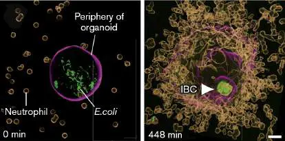
Urinary tract infections (UTIs) in miniature. Why do people, especially women, develop recurrent UTIs? Panels show a tiny (250-μm diameter), bladder organoid used to study UTIs. Escherichia coli (green) injected into the organoid grow in the luminal space, but a few bacteria enter bladder cells to form intracellular bacterial colonies (IBCs) protected from antibiotics and neutrophils (panel 1).
Neutrophils added outside an infected organoid will swarm into the organoid and kill lumen bacteria, but they cannot kill the IBC bacteria (panel 2, arrowhead). IBC bacteria can emerge later to reinfect the lumen. Scale bar = 20 μm.
K. SHARMA ET AL. 2021. CELL REP. 36
Infectious diseases have challenged human existence for millions of years. Even today, with our diagnostic acumen and myriad treatment choices, the impact of infectious disease on the world’s population is staggering. The number of infection-related deaths of immunocompetent individuals, combined with the number of people who are immunocompromised by conditions such as cancer, smoking, or drug abuse, makes infectious disease the leading contributor to death in the world.
Over 1,400 different pathogenic species of viruses, bacteria, fungi, and protozoa are known to infect humans. The latest major assault came from the novel coronavirus SARS-CoV-2, the cause of COVID-19, a disease that nearly brought humanity to its knees and tested our resolve beyond recent memory. Our tenacity as a species is due in large part not only to our immune system but also to our capacity to diagnose, treat, and prevent known infections as well as spot newly emerging ones.
In this chapter we introduce the art of diagnosis and explore the major types and etiologies of infections. We discuss a wide variety of infectious microbes by describing their routes of infection, the pathologies they cause, and the symptoms that result. We do not present an exhaustive compendium of microbial illnesses but instead a representative sampling of them that illustrates key aspects of microbial disease. The diseases selected, including COVID-19, are presented as a clinician views them: by the organ system affected, not by the taxonomy of the pathogen. The advantage of our strategy is diagnostic. When examining a sick patient, a clinician must first determine which organ system is affected and then mentally sort through the pathogens known to affect that system. This approach will help you better integrate your knowledge of microbiology and immunology within the framework of the practice of medicine.
Because patient histories are so helpful in diagnosing infectious diseases, we also use case histories throughout this chapter to segue into discussions of the various microbes that can infect each organ system. Key aspects of infectious diseases will emerge.
26.1 Basics of Diagnosis and Infections of Skin, Soft Tissue, and Bonenot assigned
How is a diagnosis made? It is not a simple matter of taking a specimen, ordering a test, and prescribing a drug. The clinician first needs to figure out which microbes, out of thousands, are possible causes. When you go to the clinic, what does a clinician first ask? “What brings you here today?” followed by “How long have you had these symptoms?” and perhaps “Where have you traveled recently?” This is not idle conversation; the clinician is collecting data by taking a patient history (Fig. 26.1).
FIGURE 26.1 ■ Clinician taking a patient history.
ROCKETCLIPS, INC./SHUTTERSTOCK
Because many infectious diseases display similar symptoms, a patient history can provide clues about the likely culprit. For example, Vibrio cholerae and enterotoxigenic E. coli both produce diarrheal diseases characterized by cramps, lethargy, and liters of watery stool each day, but cholera is not commonly seen in the United States. Nevertheless, a clinician might suspect cholera if the patient recently traveled to, or emigrated from, a part of the world where cholera is endemic (regularly observed). Clinicians learn this information by talking with the patient and asking about their recent travels.

Note: We recognize that gender identity is an important issue. Not everyone identifies as strictly male or female. However, the spectrum of recognized gender identities can make discussions of medical issues difficult. To simplify our explanations of infectious disease, we use the terms “male” and “female” solely to indicate reproductive anatomy, not gender.
Many questions the clinician asks while taking a patient’s social history can seem irrelevant or even intrusive to the patient, who only wants relief from the symptoms. For instance: Do you have any hobbies? What is your occupation? What foods have you eaten recently? Does your ill child attend day care? Has anyone in your family had similar symptoms? All of those questions address possible sources of the infectious agent. Other questions can reveal high-risk behavior that can lead to certain infections. Do you smoke, drink, or take recreational drugs? Have you had multiple sex partners? Do you use contraception? If a woman seeking help for abdominal pain reveals that she has multiple sex partners who don’t use condoms, then the differential, the list of possible causes of her symptoms, should include gonorrhea, syphilis, and chlamydia. A good patient history combined with a thorough physical exam helps the clinician decide which further tests to order and which procedures to perform.
However, different people infected with the same pathogen can display somewhat different symptoms. This variation can sometimes complicate diagnosis. For instance, some patients infected with measles virus develop muscle pain, whereas other patients do not. A small number of patients infected with measles virus develop life-threatening encephalitis, but most do not. Why do disease symptoms and severity differ among patients? Part of the explanation is immunocompetence. Those with more effective immune systems can better defend themselves against the pathogen. As a result, some infected people may not develop any symptoms. However, genetic differences in immunocompetence are only part of the story.
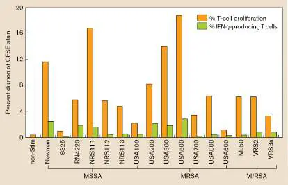
Uri Sela (Rockefeller University) and colleagues discovered that pangenomic gene differences between different strains of a pathogen will also alter a patient’s immune response (the concept of pangenome versus core genome was introduced in Chapter 17). In one experiment the scientists examined 16 different heat-killed Staphylococcus aureus strains (clinical isolates) for their abilities to stimulate T-cell proliferation and interferon-gamma (IFN-gamma) production in vitro using lymphocytes from a single volunteer (Fig. 26.2). T-cell proliferation and IFN-gamma production are markers of the immune response to infection (see Chapter 24). Even though the bacteria were dead, the different strains stimulated different levels of T-cell proliferation and IFN-gamma production. That is, a single host displayed a heterogeneous response to the different strains of S. aureus. This finding and other data indicate that the final immune response of a host is due as much to pathogen strain variability as it is to individual host variability. Both of these parameters can complicate diagnosis, treatment, and patient outcome. FIGURE 26.2 ■ The immune response of a host varies with pangenomic differences in Staphylococcus aureus strains.
Lymphocytes from a single host were tested in vitro for the ability to respond to 16 different heat-killed strains of S. aureus (designated by number or name along the x -axis). MSSA = methicillin-sensitive S. aureus; MRSA = methicillin-resistant S. aureus; VRSA = vancomycin-resistant S. aureus. T-cell proliferation was determined by staining T cells with CFSE (carboxyfluorescein succinimidyl ester) stain and monitoring dilution of the stain per cell as the cells proliferated. Every time a cell containing stain replicated, the stain was divided (diluted) into the two new cells. T cells that produced IFN-gamma were identified by fluorescent antibody followed by FACS analysis. non-Stim = T cells not stimulated by heat-killed S. aureus.
Source: Modified from Sela et al. 2018. PLoS Pathog. 14 :e1006726, fig. 1A.
Infections of Skin, Soft Tissue, and Bone Bones, skin, and soft tissues such as muscle and underlying connective tissues collectively shape and maintain body architecture. Because these structures are interconnected, infections that threaten the integrity of bones, skin, and soft tissues are discussed together. Infections that affect one of these tissues can also spread directly to an adjacent tissue.
Skin and soft-tissue infections range from simple boils to severe, complicated, so-called flesh-eating diseases that can be caused by a variety of bacteria, fungi, and viruses (see Table 26.1). Recall that the integrity of the skin, as well as the presence of normal skin microbiota, prevents most infections. However, even minor insults to the skin (such as a paper cut) can result in infections, most of which are caused by the Gram-positive pathogen Staphylococcus aureus. Healthy individuals develop infections of the skin only rarely, whereas people with underlying immunosuppressive diseases, such as diabetes, are at much higher risk.
Common Infectious Diseases of the TABLE 26.1 Skin Disease Symptoms Etiological Virulence agent(s) a factors Bacterial Folliculitis Boils Staphylococcus Coagulase, aureus (G+ protein A, cocci); fibrin wall TSST, around abscess leukocidin, renders it poorly exfoliative accessible to toxin antibiotics Scalded skin Peeling skin on S. aureus (G+ Disseminated syndrome infants, systemic cocci) exfoliation Common Infectious Diseases of the TABLE 26.1 Skin toxin exotoxin Impetigo Skin lesions on the S. aureus or Various, as for face, mostly in Streptococcus boils children pyogenes (G+ cocci)
Scarlet fever Sore throat, fever, S. pyogenes (G+ M-protein pili, rash cocci) C5a peptidase, hemolysin, pyrogenic toxins, others Erysipelas Skin lesions, usually S. pyogenes (G+ As for scarlet facial, that spread to cocci) fever (see cause systemic above)
infection Cellulitis Uncomplicated S. aureus, S. As for scarlet infection of the pyogenes fever (see dermis above)
Necrotizing Rapidly progressive S. aureus, S. As for scarlet fasciitis cellulitis pyogenes, fever (see Clostridium above); a perfringens (G+ variety of spore-forming toxins rod anaerobe)
Common Infectious Diseases of the TABLE 26.1 Skin Vibrio vulnificus (G − rod)
Viral Rubella b Discolored, pimply Rubella virus Envelope rash; mild disease [ssRNA(+)] proteins E1 unless congenital and E2 Measles b Severe disease, Rubeola virus V protein fever, conjunctivitis, [ssRNA(−)] (interferes cough, rash with interferon signaling)
Chickenpox b Generalized Varicella-zoster Glycoprotein B discolored lesions (dsDNA) (fusion of viral and cell membranes)
Shingles b Pain and skin lesions, Varicella-zoster Glycoprotein E usually on trunk in (dsDNA) (required for adults cell-cell fusion)
Smallpox c Raised, crusted skin Variola major SPICE rash, highly (dsDNA) (smallpox contagious inhibitor of Common Infectious Diseases of the TABLE 26.1 Skin complement enzymes)
Warts b Rapid growth of skin Papillomaviruses E6 and E7 cells (dsDNA) oncoproteins (see Section 26.5)
Fungal Dermatophytosis Dry, scaly lesions like Dermatophytes (Unclear athlete’s foot (tinea Epidermophyton, pedis) Tricophyton, Microsporum)
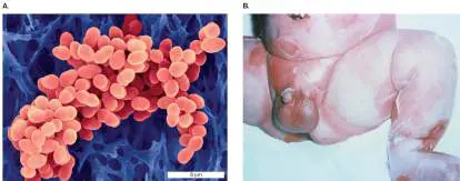
Sporotrichosis Granulomatous, pus-Sporothrix Melanin filled lesions; can schenckii disseminate to lungs or other organs Blastomycosis Granulomatous, pus-Blastomyces BAD1 filled lesions; can dermatitidis adherence disseminate to lungs or other organs Candidiasis Patchy inflammation Candida Proteinase, of mouth (thrush) or albicans; phospholipase, vagina; can Ssn6/Tup1 Common Infectious Diseases of the TABLE 26.1 Skin disseminate in Candida regulators, immunocompromised glabrata others patients Aspergillosis Infected wounds, Aspergillus spp. PacC/FOS1 burns, cornea, regulators, external ear gliotoxin Zygomycosis Oropharyngeal Mucor and Iron infections; affects Rhizopus spp. acquisition mainly diabetic (rhizoferrin); patients; can rapidly rhizoxin disseminate Boils Staphylococcus aureus (Fig. 26.3A) is a common cause of painful skin infections called boils or furuncles. This Gram-positive organism, often a normal inhabitant of the nares (nostrils), can infect a cut or gain access to the dermis via a hair follicle. It possesses a number of enzymes that contribute to disease, including coagulase, which helps coat the organism with fibrin, thereby walling off the infection from the immune system and antibiotics. As a result, boils generally require surgical drainage in addition to antibiotic therapy. FIGURE 26.3 ■ Staphylococcus aureus. A. S. aureus (colorized SEM). B. Exfoliative toxin from some strains of S. aureus causes scalded skin syndrome.
DENNIS KUNKEL MICROSCOPY/SCIENCE SOURCE
DR. KEN GREER/VISUALS UNLIMITED, INC.
As noted in Chapters 24 and 25, some strains of S. aureus also produce toxic shock syndrome toxin (TSST), a superantigen that can lead to serious systemic symptoms. Recall that a superantigen links and activates antigen-presenting cells and T cells by binding to the outside of MHC class II receptors and T-cell receptors. Antigen recognition is not required. As a result, many different T cells become activated to release a flood (or storm) of cytokines. A particularly dangerous strain of S. aureus is called methicillin-resistant S. aureus (MRSA). Today, infections suspected to be caused by S. aureus are treated with penicillin-like drugs, such as oxacillin (similar to methicillin). MRSA strains, however, have developed resistance to methicillin, oxacillin, and many other penicillin-like drugs through an altered penicillin-binding protein (PBP) involved in cell wall synthesis (see the discussion of MecA in Section 27.2). So, using oxacillin or amoxicillin to treat what turns out to be a MRSA infection will result in treatment failure that can become life-threatening. Once the mistake is discovered, immediate use of alternative drugs, such as vancomycin, is required.
MRSA strains, originally discovered causing hospital-acquired (nosocomial) infections, are now commonly found outside hospitals. MRSA and other infections can develop during a visit to any health care facility, such as hospitals, nursing homes, doctor offices, or rehabilitation facilities. These infections are now referred to as health care–associated infections (HAIs). Another category, called community-acquired infections (CAIs), especially MRSA, can develop in individuals who have not recently encountered health
care. HAI-MRSA and CAI-MRSA infections are occurring at an epidemic rate in the United States (an incidence of approximately 20 per 100,000 population). Seventy percent of staphylococcal skin infections are now caused by MRSA, and 30% of the U.S. population have their skin or nostrils colonized by MRSA. Thus, a physician can no longer assume that a patient walking into the office with a staphylococcal infection will respond to methicillin-like drugs such as oxacillin. The doctor must assume that the infection may be caused by MRSA. As a result, treatment regimens around the country and the world are being forced to change. Today, vancomycin and linezolid are initially used to treat serious infections where MRSA is a suspected cause (treatments will vary depending on antibiotic resistance patterns in the community). Once the clinical laboratory rules out MRSA, the antibiotic treatment is changed. Antibiotics are discussed in Chapter 27.
Other staphylococcal diseases are caused by toxin-producing strains in which the organism remains localized but the toxin disseminates. We have already mentioned TSST, but there are other toxins. For example, some strains of S. aureus produce a toxin called exfoliative toxin, which causes a blistering disease in children called staphylococcal scalded skin syndrome (Fig. 26.3B ). Like TSST, exfoliative toxin is a superantigen (described in Section 24.3). In addition, exfoliative toxin cleaves a skin cell adhesion molecule that, when severed, results in the epithelium separating from the underlying dermis (blisters).
Thought Question 26.1 Does Staphylococcus aureus have to disseminate through the circulation to produce the symptoms of scalded skin syndrome (SSS)? Explain why or why not.
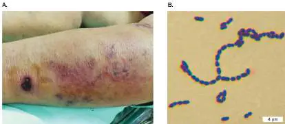
Case History: Necrotizing Fasciitis by “Flesh-Eating” Bacteria One weekend in June, Cassi was camping with her three children. She suffered a minor cut on her finger, which she bandaged properly. She also injured the left side of her body while playing sports with her kids. Not thinking much of either of her minor injuries, she went to bed. Two days later, Cassi was extremely ill. Her symptoms included vomiting, diarrhea, and a fever. She was also in severe pain where she had injured her side, and the area had begun to bruise (the skin was not broken). By the next day she could barely get out of bed, and by the end of that night she was having difficulty breathing and could not see. Within hours her side worsened and began to leak fluid and blood. Cassi was admitted to the hospital in septic shock, with no detectable blood pressure. An infectious disease specialist looking at the wound (similar to Fig. 26.4A) diagnosed the problem as necrotizing fasciitis. Cassi also developed sepsis, which led to dangerously low blood pressure. Because necrotizing fasciitis is often deadly, Cassi was rushed into surgery. In an effort to save her life, the surgeons removed about 7% of her body surface. The large wound infection in her side would need to resolve before a skin graft could be performed to repair it, so the hole in Cassi’s body was left wide open for days. After nearly 3 months and several operations, Cassi recovered.
What kind of organism can cause this type of devastating disease? The disease necrotizing fasciitis, also known incorrectly as “flesh-eating disease,” is rare and is often caused by the Gram-positive coccus Streptococcus pyogenes (Fig. 26.4B ), a microbe normally associated with throat infections (pharyngitis). Although sometimes described as a recently emerging infectious disease, necrotizing fasciitis was first discovered in 1783, in France.
FIGURE 26.4 ■ Flesh-eating Streptococcus pyogenes. A. Early-stage wound of necrotizing fasciitis. B. Gram stain of S. pyogenes. Each cell is approx. 1 μm in diameter.
ANDA PHOTO/SHUTTER STOCK
COURTESY OF DR. WILLIAM SCHWAN
In this case history, Cassi probably had this organism on her skin when the injury to her side occurred. The injured area probably suffered an invisible
microabrasion, providing a good growth environment for the organism, leading to the secretion of potent toxins and death of surrounding tissues. The bacteria will spread through subcutaneous tissue, destroying fat and fascia without initially harming the skin itself. Fascia is the sheath of thin, fibrous tissue that covers muscles and organs.
Rapid, aggressive surgical removal of affected tissue and immediate treatment with antibiotics are required in these extreme cases, even before the clinical microbiology lab has had time to identify the organism. In this approach to antibiotic treatment, called empiric therapy, one or more antibiotics are given to “cover” (kill) the most likely causative agents. Therapy can include clindamycin and metronidazole (both of which act against anaerobes and Gram-positive cocci) and gentamicin or piperacillin (drugs particularly effective against Gram-negative microbes). (Chapter 27 further discusses these and other antibiotics.) Often, however, antibiotic treatment of patients with necrotizing fasciitis is difficult because of insufficient blood supply to affected dead tissues.
Other bacteria can also cause necrotizing fasciitis, such as Staphylococcus aureus and the marine organism Vibrio vulnificus. In rare cases (the most recent was documented in 2018), Capnocytophaga canimorsus, a Gram-negative bacterium unique to the mouths of dogs and cats, has caused necrotizing fasciitis in people who have been licked or bitten by a dog. C. canimorsus infections are very rare, however, because those most at risk are immunocompromised in some way.
Cellulitis is another form of skin infection but is less aggressive than necrotizing fasciitis. Cellulitis does not involve the fascia or muscles but is characterized by localized pain, swelling, tenderness, erythema, and warmth. Streptococcus pyogenes is the most frequent cause of cellulitis in immunocompetent adults but a number of other bacteria, including Staphylococcus aureus, Gram-negative bacilli, and anaerobes, can also cause this skin infection. Cellulitis can progress to necrotizing fasciitis if left untreated.
Streptococcus pyogenes wields many different virulence factors, such as the M protein used for nonpilus attachment and inflammasome activation, various superantigen exotoxins, and secreted enzymes such as hyaluronidase and DNase (some of these are described in Chapter 25). Many established virulence factor genes in S. pyogenes are located on prophages (phage genomes) integrated into the bacterial genome. Prophages constitute approximately 10% of the organism’s genome. One study found that a soluble factor produced by human pharyngeal cells could facilitate activation of at least some of these phages and cause horizontal transfer of the associated virulence factors between strains of this pathogen. Prophage activation and phage production are discussed in Chapter 6.
Thought Question 26.2 Why would treatment of some infections require multiple antibiotics? Osteomyelitis Osteomyelitis is a bone infection with accompanying inflammation and bone destruction caused by bacteria. All bones can be infected, but the lower extremities and vertebrae are most commonly involved. Bone can be infected in several ways. Acute trauma or surgery can directly introduce organisms into affected bone, or organisms from an adjacent soft-tissue infection can spread to nearby bone. Organisms can also spread through the bloodstream from peripheral sites of infection (an abscess, for instance) to bone (called hematogenous seeding). Hematogenous seeding most commonly affects vertebrae because they are well vascularized.
The most common causes of osteomyelitis include staphylococci ( Staphylococcus aureus), streptococci (Streptococcus pyogenes), Gram-negative bacilli (Pseudomonas, Escherichia), and some anaerobes ( Bacteroides). Bone biopsy is usually needed to identify the organism. Host factors that contribute to osteomyelitis include diabetes (for instance, when a diabetic foot wound goes untreated), sickle-cell disease in children, joint prosthetics, and intravenous drug use. Antibiotic treatment of osteomyelitis is challenging because most bones are not well vascularized, so debridement (removal of damaged tissue) is also necessary.
Viral Diseases Causing Skin Rashes Several viruses can produce skin rashes, although their route of infection is usually through the respiratory tract. Measles, for example (see Section 6.1), is a highly contagious viral infection caused by a paramyxovirus, whose hallmark symptom is a maculopapular skin rash (see Fig. 6.3D). In a maculopapular rash, flat, red (macular) spots are intermixed with raised bumps (papules). The virus is transmitted by coughing and sneezing, but the first signs of measles, also known as rubeola, are fever, cough, runny nose, and red eyes occurring 9–12 days after exposure. A few days later, telltale spots (Koplik’s spots) appear in the mouth, along with a sore throat. Then a skin rash develops that typically starts on the face and spreads down the body. The virus replicates in the lymph nodes and spreads to the bloodstream (viremia), where it can infect endothelial cells of the blood vessels. The rash occurs when T cells begin to interact with these infected cells.
Although skin rash is the main symptom of measles, infection can also cause respiratory symptoms and serious complications, including pneumonia, bronchitis, croup, and even a fatal encephalitis in immunocompromised patients. Approximately one out of four cases of measles requires hospitalization. One out of 1,000 dies. In the United States, measles had been almost completely eliminated by the measles, mumps, and rubella (MMR) multivalent vaccine. Unfortunately, a disturbing, misinformed anti-vaccine movement over the past two decades has led to a decrease in vaccinations and an increase in U.S. measles cases. Case numbers in the United States for 1999 were 100 and by 2019 had risen to 1,282. In 2020, measles cases dropped dramatically to 13, as public health measures implemented for the COVID-19 pandemic curtailed the transmission of measles and many other airborne infectious diseases. As these restrictions are relaxed, however, experts expect cases of measles and other preventable infections will once again rise. Worldwide, however, measles has remained a serious problem, killing 207,000 people in 2020, including in places where vaccinations are routine.
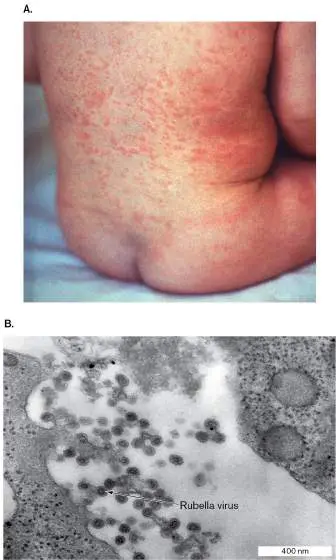
Rubella virus, a togavirus, causes a maculopapular rash known as German measles or 3-day measles. The rash is similar to but less red than that of measles (Fig. 26.5). German measles is an infection of primarily the skin and lymph nodes and is usually transmitted from person to person by aerosolization of respiratory secretions. It is not dangerous in adults or children; the virus can, however, cross the placenta in a pregnant woman and infect her fetus. If the virus crosses the placenta within the first trimester, the result is congenital rubella syndrome, which can cause death or serious congenital defects in the developing fetus.
FIGURE 26.5 ■ German measles. A. Skin rash caused by rubella virus.
B. Rubella virus budding from the host cell membrane to form an enveloped virus particle (approx. 50–70 nm; TEM).
CENTERS FOR DISEASE CONTROL AND PREVENTION/SCIENCE SOURCE
CDC/DR. FRED MURPHY; SYLVIA WHITFIELD
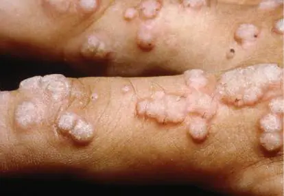
Human papillomavirus (HPV) is a common virus (over 100 types) that causes warts, abnormal skin growths on feet, hands, or genital organs (Fig. 26.6). The replication of HPV is discussed in Section 6.5. The virus enters the skin through small cuts or abrasions and then replicates in the basal epithelial cells. The virus causes excessive growth of the cells it infects, in part by triggering overexpression of host c-Myc protein, which is needed for mitosis. Excess c-Myc (an oncoprotein) causes uncontrolled cell division that produces warts or, in the worst-case scenario, cervical or penile cancer. After the initial infection has resolved, the virus may remain latent in tissues and can reactivate if immune system function is impaired. Cryosurgery with liquid nitrogen to freeze the growth will remove most common warts. Genital HPV is more dangerous than common warts, and it is a common sexually transmitted infection among college students.
FIGURE 26.6 ■ Noncancerous hand warts caused by human papillomavirus (HPV).
CLINICAL PHOTOGRAPHY, CENTRAL MANCHESTER UNIVERSITY HOSPITALS NHS FOUNDATION
TRUST, UK/SCIENCE SOURCE
Infection by any of several different types of HPV can produce cervical and penile cancers years after infection (see Fig. 6.31A). As with common warts, cryosurgery can be used to treat genital HPV growths, but preventing the
disease is more effective and less painful. Prevention is provided by Gardasil, a 9-valent vaccine that prevents infection by the most common cancer-causing types: HPV16, HPV18, and seven others. The U.S. Food and Drug Administration (FDA) and Centers for Disease Control and Prevention (CDC) recommend administering the vaccine to young people between ages 9 and 26, ideally before they become sexually active and potentially encounter the virus. Unfortunately, most kids in the United States do not receive it. Other viruses affecting the skin, such as chickenpox, the related disease shingles, and smallpox are included in Table 26.1. The vaccination schedule for preventing these diseases is provided in Table 24.5.
To Summarize Staphylococcus aureus and Streptococcus pyogenes are common bacterial causes of skin infections. The organisms usually infect through broken skin.
Methicillin-resistant Staphylococcus aureus (MRSA) has become an important cause of community-acquired staphylococcal infections (CAI-MRSA).
Necrotizing fasciitis is usually caused by Streptococcus pyogenes, but it can be the result of other infections.
Infections of the skin can disseminate via the bloodstream to other sites in the body.
Osteomyelitis , usually caused by Staphylococcus aureus, begins with direct bone trauma, hematogenous seeding, or contact with a nearby soft-tissue infection.
Rubeola and rubella viruses (RNA viruses) infect through the respiratory tract, but their main manifestation is the production of similar maculopapular skin rashes.
Human papillomaviruses (DNA viruses) enter through skin to cause warts on skin; some types can cause genital cancers.
Glossary
differential In disease, the list of possible causes of an infection.
nosocomial Hospital-acquired; commonly refers to an infectious agent.
health care–associated infection (HAI)
Any infection contracted by a patient while receiving treatment for a medical condition at a health care facility, such as hospitals, nursing homes, doctor offices, or rehabilitation centers.
necrotizing fasciitis Also known as flesh-eating disease. A severe skin infection usually caused by the Gram-positive coccus Streptococcus pyogenes.
empiric therapy An approach to treating infection before the infective organism is known, in which multiple antibiotics are administered in an effort to kill the most likely causative agents.
cellulitis A spreading infection (with inflammation) of connective tissue just below the skin.
Fig. 6.3D FIGURE 6.3 ■ Virus infections and disease. A. Bacteriophage T2 particles pack in an array within an Escherichia coli cell (TEM). B. Bacteriophage infection forms plaques of lysed cells on a lawn of bacteria. C. Measles virions bud out of human cells in tissue culture
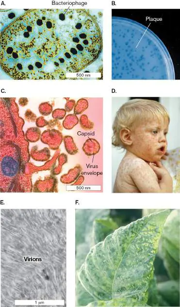
(TEM). D. Child infected with measles shows a rash of red spots. E. Tobacco leaf section is packed with tobacco mosaic virus (TMV) particles. F. Tomato leaf infected by TMV shows mottled appearance.
LEE D. SIMON/STAMMERS/SCIENCE SOURCE
DR. EDWARD CHAN/VISUALS UNLIMITED, INC.
NIBSC/SCIENCE SOURCE
LOWELL GEORGIA/SCIENCE SOURCE
B. ZECHMANN ET AL. 2011. J. VIS. EXP. 56:E2950
NORM THOMAS/SCIENCE SOURCE
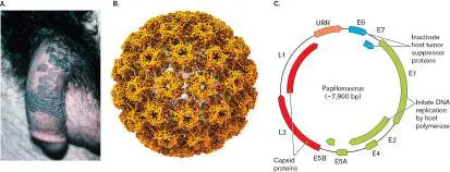
Fig. 6.31A FIGURE 6.31 ■ Human papillomavirus. A. Certain strains of human papillomavirus (HPV) cause warts on the genitals or anus. B. HPV virion model (cryo-EM). C. HPV genome.
KEN GREER/VISUALS UNLIMITED
Endnotes
1. Note a: G+ = Gram-positive. Return to reference a 2. Note b: Vaccine is available against the causative agent. Return to reference b 3. Note b: Vaccine is available against the causative agent. Return to reference b 4. Note b: Vaccine is available against the causative agent. Return to reference b 5. Note b: Vaccine is available against the causative agent. Return to reference b 6. Note b: Vaccine is available against the causative agent. Return to reference b
7. Note c: Vaccine is no longer in use because the disease has been eradicated. The exceptions are highly restricted laboratories. Return to reference c
26.2 Respiratory Tract Infectionsnot assigned
Lung and upper respiratory tract infections are among the most common diseases of humans. Many
different bacteria, viruses, and fungi are well adapted to grow in the lung. Successful lung pathogens
come equipped with appropriate attachment mechanisms and countermeasures to avoid various lung
defenses (such as alveolar macrophages). One reemerging bacterial pathogen, Bordetella pertussis,
the cause of whooping cough, inhibits the mucociliary escalator by binding to lung cilia (Fig. 26.7 ).
Although many microbes can infect the lung, most respiratory diseases are of viral origin, and most
viral respiratory infections (such as the common cold) do not spread beyond the lung. Viral diseases
are usually self-limiting and typically resolve within 2 weeks; however, the damage caused by a
primary viral infection can lead to secondary infections by bacteria. Other viral diseases, such as
influenza and COVID-19, can be deadly in their own right.
FIGURE 26.7 ■ Rabbit tracheal epithelial cells infected with Bordetella
bronchiseptica. Primary cultured rabbit tracheal epithelial cells, following 5 minutes of
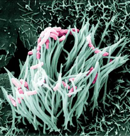
coincubation with virulent wild-type B. bronchiseptica. B. bronchiseptica is a close relative of B.
pertussis, the cause of human pertussis (whooping cough). Bacteria (approx. 1 μm in length)
have been pseudocolored pink for easier visualization (SEM).
JESSICA A. EDWARDS, ET AL. INFECTION AND IMMUNITY 2005. 73(6):3618–3626.
Bacterial infections of the lung, whether of primary or secondary etiology, require antibiotic
therapy. Before the advent of antibiotics, the only recourse was to insert a tube into the patient’s
back to drain fluid accumulating in the pleural cavity around the lung (a pathological process known
as pleural effusion). Unless released, the pressure on the lung will collapse the alveoli and make
breathing difficult.
Viral infections predispose patients to secondary bacterial infections in several ways. Viral lung
infections cause the patient to dehydrate, which increases mucus viscosity in the airways. Increased
mucus viscosity limits motility of the mucociliary escalator (described in Section 23.1), making it
harder to eliminate bacterial pathogens. To keep the escalator moving, cold sufferers are advised to
drink plenty of fluids to decrease mucus viscosity. Another factor leading to secondary bacterial
infections is that viruses can inhibit key aspects of lung innate immune mechanisms that prevent
bacterial growth. Note that many deaths resulting from viral influenza are caused by secondary
bacterial infections.
Case History: Bacterial Pneumonia
James, an 80-year-old resident of a New Jersey nursing home, had a fever accompanied by a
productive cough with brown sputum (mucous secretions of the lung that can be coughed up). He
reported to the attending physician that he had pain on the right side of his chest and suffered from
night sweats. Blood tests revealed that his white blood cell (WBC) count was 14,000 per microliter
(normal is 5,000–10,000), with a makeup of 77% segmented forms (polymorphonuclear leukocytes,
PMNs; normal range 40%–60%) and 20% bands (immature PMNs; normal range 0%–5%). The
chest radiograph revealed a right-upper-lobe infiltrate (Fig. 26.8A ). From this information, the
clinician diagnosed pneumonia. Microscopic examination of the patient’s sputum (Gram stain and
capsule stain) revealed Gram-positive cocci in pairs and short chains surrounded by a capsule (Fig.
26.8B ). Bacteriological culture of the patient’s sputum and blood yielded Streptococcus
pneumoniae.
FIGURE 26.8 ■ Pneumonia caused by Streptococcus pneumoniae. A. X-ray view of a
patient with lobar pneumonia. Infiltrate in the right upper lobe (RUL) is caused by S.
pneumoniae. The sharp lower border of the infiltrate marks the upper boundary of the right
middle lobe (arrow). B . Micrograph of S. pneumoniae. Gram-stained sputum sample showing
numerous PMNs and extracellular diplococci in pairs and short chains. Bacteria range from 0.5 to
1.2 μm in diameter. C. Relative incidence of pneumonia caused by various microorganisms.
TOMATHEART/SHUTTERSTOCK
LEBEAU/SCIENCE SOURCE
Pneumonia is a disease that can be caused by many different microbes (Table 26.2). The
pneumococcus Streptococcus pneumoniae accounts for about 25% of community-acquired cases of
pneumonia (worldwide), but pneumococcal pneumonia occurs mostly among the elderly and
immunocompromised, including smokers, diabetics, and alcoholics. A breakdown of pneumonia cases
by causative organism is shown in Figure 26.8C .
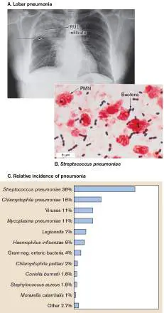
TABLE 26.2 Selected Respiratory Tract Infectious Diseases
Agent [disease] Key symptoms Virulence Source(s) Treatment
properties options (as of
2022)
Bacterial
Bacillus anthracis Hypotension, Peptide capsule; Soil/airborne Ciprofloxacin
[anthrax] respiratory failure PA, LF, and EF [vaccine (military)]
toxins
Corynebacterium Tracheal Diphtheria toxin Humans Penicillin (vaccine)
diphtheriae pseudomembrane
[diphtheria]
Streptococcus Fever, chills, Capsule, Humans Fluoroquinolones,
pneumoniae cough, chest pain pneumolysin ceftriaxone
[pneumonia] (vaccine)
Bordetella pertussis Violent cough, Adenylate cyclase Humans Azithromycin
[whooping cough] inhalation toxin, filamentous (vaccine)
“whoop” hemagglutinin
(adhesin)
Pseudomonas Infects cystic Exotoxin A, Water, soil Quinolones,
aeruginosa fibrosis patients phospholipase C, aminoglycosides,
[pneumonia] exopolysaccharide, carbapenems
others
Legionella Chest pain, Intracellular Water Azithromycin
pneumophila cough, muscle growth, towers/ (macrolide),
[Legionnaire’s pain, vomiting hemolysin, inhalation quinolones
disease] cytotoxin,
protease
Chlamydia a Sore throat, chest Obligate Humans Tetracycline,
pneumonia pain intracellular macrolides
[pneumonia] growth; prevents
TABLE 26.2 Selected Respiratory Tract Infectious Diseases
phagolysosome
fusion
Chlamydia a psittaci Sore throat, chest Obligate Bird Doxycycline,
[psittacosis] pain intracellular droppings/ erythromycin
growth; prevents dust
phagolysosome
fusion
Mycobacterium Cough, bloody Cord factor, wax Humans Rifampin, isoniazid,
tuberculosis sputum, fatigue, D, intracellular ethambutol,
[tuberculosis] weight loss growth pyrazinamide
[vaccine (BCG b)]
Mycoplasma Sore throat, Adhesin tip Humans Azithromycin,
pneumoniae nonproductive doxycycline
[pneumonia] cough
Viral
Cytomegalovirus Cough, chest pain Reduces MHC I Humans Ganciclovir,
(CMV) [CMV presentation valganciclovir
disease]
Respiratory Cough, chest pain Prevents T-cell Humans Treat
syncytial virus (RSV) activation symptoms/ribavirin,
[RSV disease] palivizumab c
Influenza virus Cough, chest pain Neuraminidase; Humans Zofluza,
[influenza] hemagglutinin oseltamivir,
zanamivir (vaccine)
Severe acute Cough, chest pain Papain-like Humans Treat symptoms/
respiratory protease (PLP) dexamethasone,
syndrome (SARS- inhibits type I monoclonal Ab to
CoV virus [SARS] interferon spike protein,
activities by
TABLE 26.2 Selected Respiratory Tract Infectious Diseases
and SARS-CoV-2 affecting host IRF- Paxlovid (protease
virus [COVID-19]) 3; inhibits inhibitor)
phosphorylation of,
and cleaves,
ubiquitin and
ubiquitin-like
ISG15 peptide
from IRF-3
Fungal
Aspergillus spp. Lungs, sinuses; Dimorphism; Environment Amphotericin B,
[aspergillosis] breathing gliotoxin voriconazole
difficulty
Histoplasma Flu-like Dimorphism; Bird, Amphotericin B,
capsulatum calcium-binding chicken, bat itraconazole
[histoplasmosis] protein droppings
Coccidioides immitis Flu-like Dimorphism; Environment Amphotericin B,
[coccidioidomycosis] arginase 1 fluconazole
Blastomyces Flu-like Dimorphism; BAD1 Environment Amphotericin B,
dermatitidis itraconazole
[blastomycosis]
Pneumocystis Chest pain, Unknown Environment Bactrim
jirovecii cough, skin lesion
[pneumocystosis]
The noses and throats of 30%–70% of a given population can contain S. pneumoniae. The
microbe can be spread from person to person by sneezing, coughing, or other close, personal
contact. Pneumococcal pneumonia may begin suddenly, with a severe shaking chill usually followed
by high fever, cough, shortness of breath, rapid breathing, and chest pains.
Pneumococcal lung infection begins when the pneumococcus is aspirated into the lung. Once in
the lung, the microbe grows in the nutrient-rich edema fluid of the alveolar spaces. Neutrophils and
alveolar macrophages then arrive to try to stop the infection. They are called into the area from the
circulation by chemoattractant chemokines released by damaged alveolar cells. The thick
polysaccharide capsule of the pneumococcus, however, makes phagocytosis very difficult (see Section
25.5). In an otherwise healthy adult, pneumococcal pneumonia usually involves one lobe of the
lungs; thus, it is sometimes called lobar pneumonia. The infiltration of PMNs and fluid leads to the
typical radiological findings of diffuse, cloudy areas. In contrast, infants, young children, and elderly
people more commonly develop an infection in other parts of the lungs, such as around the air
vessels (bronchi), causing bronchopneumonia.
The white blood cell count in the case history is telling. The patient had an elevated WBC count
(normal is 5,000–10,000 per microliter) and an elevated proportion of band cells (normal is 0%–5%).
These increases are indicative of a bacterial, not viral, infection. Neutrophils (PMNs), the front-line
combatants against infection, rise in response to bacterial infections and are first released from bone
marrow as immature band cells, whose presence is a sure sign of bacterial infection.
Outbreaks of pneumococcal pneumonia in nursing homes are not uncommon. Their residents’
susceptibility to this disease underscores the importance of elderly people receiving the
pneumococcal polysaccharide vaccine (PPSV23) as a hedge against infection. While there are over 80
antigenic types of pneumococcal capsular polysaccharides, the injected vaccine contains the 23 types
that are most often associated with disease (see Section 24.6). This type of vaccine is called
multivalent because it contains capsular antigens from multiple strains. The pneumococcal
polysaccharide vaccine is recommended for individuals over age 65, as well as for those who are
immunocompromised. The patient in this case history failed to receive the vaccine.
Children are also susceptible to S. pneumoniae and should be vaccinated, but children do not
have a vigorous T cell–independent antibody response to polysaccharide antigens. To facilitate T-cell
help, these patients are given a pneumococcal conjugate vaccine (PCV13), which contains capsular
antigens from 13 serotypes of S. pneumoniae conjugated to a nontoxic variant of diphtheria toxin
(see Chapter 24).
Thought Question
26.3 How does nontoxic diphtheria toxin (Dtx) conjugated to a capsular antigen facilitate T-cell
help?
In addition to causing serious infections of the lungs, S. pneumoniae can invade the bloodstream
(bacteremia) and the covering of the brain (meningitis). The death rates for these infections are
about one out of every 20 who get pneumococcal pneumonia, about four out of 20 who get
bacteremia, and six out of 20 who get meningitis. Individuals who are immunocompromised because
of liver disease, AIDS (caused by HIV), or organ transplants are even more likely to die from the
disease.
An emerging infectious disease problem throughout the United States and the world is the
increasing resistance of S. pneumoniae to antibiotics. At least 30% of the strains isolated today are
resistant to one or more antibiotics such as beta-lactams (penicillin) and macrolides (azithromycin).
However, one consequence of vaccination is that the incidence of antibiotic-resistant isolates has
been declining. Currently, amoxicillin can be used for penicillin-susceptible isolates, but third-
generation cephalosporins (ceftriaxone) or fluoroquinolones are typically used to treat infections by
antibiotic-resistant isolates. Chapter 27 discusses why antibiotic resistance is on the rise for this and
other microbes.
Case History: Fungal Lung Infection
A 40-year-old salesman named Jaylen presented to an Ohio hospital emergency department with
fever, cough, myalgias, and chest pain. One month earlier he had reported similar symptoms and had
been treated with azithromycin for suspected bacterial lung infection. His symptoms had not
improved, and he now reported increasing weakness, difficulty breathing, abdominal pain, and a
weight loss of 8 pounds in the previous month. While taking the patient’s history, the physician
assistant (PA) discovered that Jaylen is a weekend spelunker who frequently explores local caves and
that he is bisexual, having had two male and three female partners in the previous year. Physical
exam revealed enlarged cervical (neck) and axillary (armpit) lymph nodes (lymphadenopathy), lung
congestion and hepatosplenomegaly (enlarged liver and spleen), and red, bumpy lesions on his legs.
A complete blood count showed low numbers of red cells, white cells, and platelets (pancytopenia). A
chest X-ray (Fig. 26.9A ) showed small calcifications (opaque white areas) in both lungs.
Suspecting tuberculosis, the PA administered a tuberculin skin test. The patient’s symptoms and
sexual history also dictated that serological tests for syphilis; hepatitis A, B, and C; and HIV be
administered. Tests for tuberculosis, syphilis, and hepatitis were all negative, but Jaylen tested
positive for HIV. His blood was cultured for the presence of bacteria and fungi.
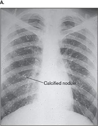
FIGURE 26.9 ■ Lung infection by Histoplasma capsulatum. A. X-ray of a patient with
histoplasmosis. B. Colonies of H. capsulatum.
SCIENCE PHOTO LIBRARY/GETTY IMAGES
GADO IMAGES/ALAMY STOCK PHOTO
This is a complicated though not unusual case. Health care providers often treat a patient for the
most likely cause of the chief complaint, which in this case was a suspected bacterial lung infection.
Azithromycin was prescribed, and the infection worsened. Antibiotic failure leading to a return visit
forced a closer look. The flu-like symptoms and calcifications seen on the lung X-ray then suggested
tuberculosis (another lung infection that can disseminate, discussed later). The negative skin test for
tuberculosis, however, ruled out that disease, leaving fungus as the probable cause, given the chronic
nature of the patient’s symptoms. The infection in this patient probably started in the lung (clued by
the cough), after which the organism spread throughout the body via the bloodstream to infect the
liver and spleen.
Cryptococcus, an encapsulated yeast, is a potential cause. It typically requires an
immunocompromised host to cause disease, and Jaylen’s positive HIV test indicated he might be
immunocompromised (he had a low CD4 T-cell count). Cryptococcus usually causes a brain infection,
meningoencephalitis, but the organism can also infect the lungs, prostate gland, urinary tract, eyes,
myocardium, bones, skin, and joints. The chest X-ray does not rule out Cryptococcus, because this
yeast can cause similar images when infecting the lung. However, the pancytopenia is not typical of
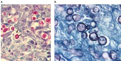
Cryptococcus, and a biopsy failed to find yeast cells with a thick capsule (example shown in Fig.
26.10A ).
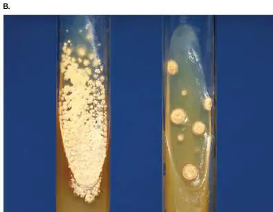
FIGURE 26.10 ■ Cryptococcus neoformans and Histoplasma capsulatum in lung
biopsies. A. C. neoformans in lung nodule. Periodic acid–Schiff stain. B. H. capsulatum. Silver
stain.
CDC/DR. EDWIN P. EWING, JR.
CDC/DR. LIBERO AJELLO
The most likely fungal causes of infection in this case history are the endemic mycoses, such as
coccidioidomycosis, blastomycosis, and histoplasmosis. This patient had never traveled to the
western United States, where coccidioidomycosis is endemic, so exposure to Coccidioides was ruled
out. Blastomyces dermatitidis is a soil fungus endemic to the Ohio and Mississippi river valleys and
the southeastern United States. The disease, blastomycosis, begins as a respiratory infection
following the inhalation of conidia. From the lung, the organism can disseminate to the skin, bone,
and genitourinary tract but rarely affects the liver or spleen, both of which were affected in Jaylen.
Histoplasmosis, on the other hand, most commonly presents as a flu-like pulmonary illness that can
progress to more serious pulmonary infection and can disseminate to the liver and spleen in severe
cases.
On the basis of Jaylen’s presentation and history of present illness, amphotericin B, a powerful
antifungal agent (discussed in Section 27.5), was administered intravenously. The patient’s fever
lowered almost immediately. Histoplasma antigen was identified in his urine, and a fungus was found
in the cultures of bronchoalveolar lavage fluid (lung washes; Fig. 26.9B ). The fungus was
definitively identified by a DNA probe and tissue biopsy as Histoplasmosis capsulatum (tissue biopsy
in Fig. 26.10B ), confirming the diagnosis of histoplasmosis.
Histoplasma is a dimorphic fungus that thrives in damp soil rich in organic material, especially the
droppings of birds and bats. Dimorphic fungi take on a mycelial form at 25°C but grow as budding
yeast at body temperature (37°C). Although Histoplasma is distributed worldwide, endemic areas in
the United States include the Ohio and Mississippi river valleys (where Jaylen lives). People like
Jaylen who explore caves populated by bats are at particular risk for contracting this disease,
sometimes called cave disease. The infectious forms of H. capsulatum are microconidia (fungal
spores) produced by differentiated mycelium. Jaylen did not wear a mask while spelunking and most
likely inhaled a good number of spores while crawling through the caves.
The incubation period for histoplasmosis ranges from 3 to 17 days. Three general forms are
recognized. In acute pulmonary histoplasmosis, immunocompetent patients are usually
asymptomatic unless they inhale a large number of spores. If symptoms develop, patients typically
display cold-like symptoms that can resolve within 3–4 weeks.
Chronic pulmonary histoplasmosis is a more serious manifestation, whose symptoms persist for at
least 3 months. Symptoms include cough, dyspnea (difficulty breathing), fever, weight loss, malaise,
and reddish, nodular skin lesions called erythema nodosum (a delayed-type hypersensitivity reaction
to antigens from various infectious agents, including Histoplasma). Jaylen displayed all of these
symptoms.
The most serious form of histoplasmosis, progressive disseminated histoplasmosis, includes
dissemination to the spleen and liver (enlarging both) and is generally seen in patients who are
immunocompromised because of corticosteroid use, organ transplant, or HIV infection. The
pancytopenia seen in Jaylen is also common in this form of the disease (70%–90%). Jaylen tested
positive for HIV and had a low CD4 T-cell count of 150 per microliter (normal is 500–1,500). Jaylen is
immunocompromised. Because of his HIV status, Jaylen was also treated with antiviral agents to
control his viral load. Lowering the viral load in his blood would allow his T-cell count to increase and
improve his immunocompetence.
Several critical features of this case help differentiate it from the preceding case of pneumococcal
pneumonia. First, the initial macrolide antibiotic, azithromycin, should have killed most bacterial
sources of infection. Second, the X-ray finding of calcified pulmonary nodules is more indicative of
fungal lung infection than of bacterial infection, which in a patient of this age would likely appear
uniformly dense and confined to one lobe (see Fig. 26.8A ). Tuberculosis would be an exception to
this rule. Jaylen’s blood count was also a clue. Fungal infections do not usually cause an increase in
WBCs or an increase in band cells, as happens with pneumococcal pneumonia. The pancytopenia,
however, is consistent with disseminated histoplasmosis. Note that many infectious diseases start out
as a localized infection but end up disseminating throughout the body to cause new sites of infection.
Thought Question
26.4 Why isn’t a dimorphic fungus like Histoplasma easily transmitted from person to person via
respiratory droplets?
Tuberculosis as a Reemerging Disease
Tuberculosis (TB), caused by the acid-fast bacillus Mycobacterium tuberculosis (see Fig. 28.5), was
the leading cause of death due to a single pathogen until the COVID-19 pandemic. Tuberculosis
claimed 1.5 million lives worldwide in 2020, whereas COVID-19 claimed about 2 million, according to
the World Health Organization. The death toll from tuberculosis is still shocking, considering that the
disease was nearly eradicated in the developed world going into the 1980s. What happened?
The explosion in tuberculosis cases started shortly after the HIV pandemic arrived in 1985.
Because HIV kills T cells, patients became immunocompromised and susceptible to many infections,
including TB. By 1991, inner-city hospitals were beginning to see highly infectious multidrug-resistant
(MDR) strains of M. tuberculosis that produced fulminant (rapid-onset) and fatal disease among
patients infected with HIV (time from TB exposure to death was 2–7 months). Alongside the
shocking rise in TB cases, U.S. health care workers exposed to TB patients had a tuberculin skin test
conversion rate of 50%. (The tuberculin test is described in the next section.)
Tuberculin conversion rate is the percentage of tuberculin-negative health care providers who
converted to tuberculin-positive over time, indicating exposure to M. tuberculosis. Today the rate is
less than 0.5%. New cases of TB in the United States have decreased over the years (from 26,283
cases in 1991 down to 8,920 cases in 2019), but approximately 1.5% of the new cases are caused by
MDR-TB. Despite progress reducing TB in developed countries, tuberculosis remains a leading cause
of global child mortality. Information about mycobacterial structure can be found in Section 18.3, and
mycobacterial pathogenesis is discussed in Section 25.5.
Tuberculin skin test. The tuberculin skin test (also known as the Mantoux test) is the primary
screen for tuberculosis. A mixture of Mycobacterium tuberculosis proteins (called purified protein
derivative, or PPD) is injected under the skin of the lower arm. A person who has been infected will
develop a localized delayed-type hypersensitivity reaction (a reddened area of skin with blisters)
within 48 hours (see Chapter 24). Note, however, that a positive tuberculin skin test does not signify
active disease, but only that the person was infected at one time. The bacterium may have been
killed by the immune system without having caused disease or may lie dormant, waiting to
reactivate. Figure 26.11 outlines the disease course of untreated tuberculosis.
FIGURE 26.11 ■ The disease progression of tuberculosis. Step 1: The disease begins
as primary tuberculosis. Step 2: Most patients never develop disease, or the organism becomes
latent within granulomas. Step 3: A few patients develop progressive primary tuberculosis
because the immune system failed to control the infection, leading to its spread to other lung
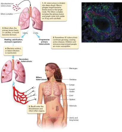
regions. Step 4: Secondary tuberculosis develops upon reinfection or reactivation of latent
bacilli. Step 5: More damage occurs in the lung, and bacilli disseminate to other organs
(including reseeding the lung) to produce miliary TB. Inset: TB granuloma in the lung of a
nonhuman primate, stained with antibodies for CD31 T cells (red) and CD681 macrophages
(green), surrounding a necrotic center (unstained). Cell nuclei are blue.
JOANNE L. FLYNN
Primary tuberculosis. Once inhaled into the lung, the bacilli deposit into alveoli and are subject to
three possible outcomes (Fig. 26.11 , steps 1 and 2): They can die, become latent, or produce
progressive primary tuberculosis. In all three cases the bacteria are initially phagocytosed by alveolar
macrophages, and a battle ensues. If the bacilli are not killed by the macrophages, the bacteria will
survive ensconced within modified phagolysosomes, where they multiply and kill the macrophage via
induced apoptosis. The bacilli are then free to infect other macrophages. The primary lesion is called
a Ghon focus. During the pitched battle, some infected macrophages from the Ghon focus will travel
to a regional lymph node of the lung. The Ghon focus and the infected lymph node are called the
Ghon complex. However, the Ghon complex cannot be seen on an X-ray at this early stage. A delayed
hypersensitivity develops, and these patients usually become tuberculin-positive.
The body reacts to bacilli in the lung by trying to wall them off in what is called a granuloma (
Fig. 26.11 inset ), a nodule (sometimes called a tubercle) containing macrophages (live and dead),
plus other white blood cells and bacteria (live and dead). The inside of the granuloma can become
necrotic with dead host and bacterial cells and develops the consistency of cheese—a process called
caseation. Over time, calcium is deposited and the nodules become calcified. At this point, the
hardened calcified nodules (Ghon complex) become visible on X-rays. Alternatively, the caseous
center of the granuloma can liquefy and the bacilli multiply to large numbers. The granuloma can
erode (cavitate) to release large numbers of bacilli into the bronchus. Coughing will expel the bacilli
into the air for another person to inhale.
Four to six weeks after infection, about 10% of infected people will develop progressive (or
active) primary disease in which bacilli in the primary focus grow and caseous material is
disseminated to other parts of the lung (Fig. 26.11 , step 3). Lung X-rays can show consolidation
similar to pneumonia in this form of tuberculosis. Symptoms include a productive cough that
generates sputum, fever, night sweats, and weight loss.
Secondary tuberculosis. As noted earlier, an alternative to primary disease is an asymptomatic
latent tuberculosis infection (LTBI). Here the bacilli remain hidden in granulomas and may even
become dormant, remaining so for many years. However, M. tuberculosis can sometimes overcome
dormancy and escape confinement imposed by the immune system (Fig. 26.11 , step 4). The
bacteria begin to multiply and cause secondary tuberculosis. Secondary TB due to reactivation
commonly occurs in immunocompromised people (such as HIV patients). Secondary tuberculosis can
also be caused by the inhalation of new bacilli. Because these patients already developed cell-
mediated immunity during primary infection, the symptoms of secondary TB are more serious than
those of primary TB and include severe coughing, greenish or bloody sputum, low-grade fever, night
sweats, and weight loss. The gradual wasting of the body is what led to the older name for
tuberculosis: “consumption.”
Extrapulmonary (miliary) TB. Mycobacterium tuberculosis bacilli in the lung can sometimes enter
the pulmonary vein (Fig. 26.11 , step 5). Once in the bloodstream, the organism will disseminate
to produce abscesses in many different organ systems. Extrapulmonary TB is sometimes called
miliary tuberculosis because the size of the infected nodules (1–5 mm), called tubercles,
approximates the size of millet seeds. Miliary TB can develop during primary or secondary TB
infections.
Treatment. Ten drugs are currently approved by the FDA for treatment of tuberculosis. Initial
treatment of active disease is aggressive and involves a four-drug regimen of what are called first-
line drugs (or drugs of choice)—namely, isoniazid, rifampin, pyrazinamide, and ethambutol—given
over a course of several months. MDR strains (about 1% of total TB cases in the United States) are
defined as being resistant to two or more first-line drugs. An MDR strain is treated with a regimen of
four to five drugs that do not include the first-line drugs to which it is resistant. Extensively drug-
resistant tuberculosis (XDR-TB) strains, as defined by the World Health Organization, are resistant to
two or more first-line drugs, plus any fluoroquinolone and at least one second-line drug (bedaquiline,
linezolid). XDR-TB strains are almost untreatable, although a recently approved drug called
pretomanid is effective against XDR-TB. Pretomanid inhibits the biosynthesis of mycolic acid, a
component of the M. tuberculosis cell wall. Although XDR-TB is rare in the United States, about 9% of
MDR-TB strains worldwide are XDR-TB.
Thought Question
26.5 Explain why patient noncompliance (failure to take drugs as directed) is thought to have led to
XDR-TB.
Case History: Viral Lung Infection
Felicia, a 57-year-old Black woman, presented to the emergency department in Tallahassee, Florida,
in severe distress. Her symptoms included a 103 ° F fever, a severe but dry cough, fatigue, and
dyspnea (difficulty breathing). Felicia’s daughter explained that a week ago her mother awoke with a
slight fever, headache, body aches, and a mild cough that had gotten progressively worse. Her
daughter also mentioned that Felicia could not taste food. On arrival, Felicia’s oxygen level by pulse
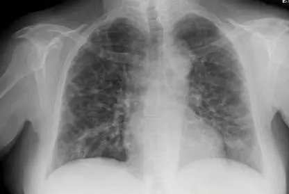
oximeter was 78% (normal is > 92%), and a lung X-ray showed bilateral ground-glass opacities (
Fig. 26.12), a sign of viral pneumonia. Felicia was immediately admitted to the COVID-19 wing of
the hospital. Her vaccination status was unknown.
FIGURE 26.12 ■ Felicia’s X-ray, showing inflammation in both lungs. The extensive
ground-glass opacities (hazy gray areas throughout all lobes, as opposed to the consolidation
observed in Fig. 26.8A) indicate a pneumonia that is typically (but not exclusively) of viral
origin, such as coronavirus, influenza, measles, or respiratory syncytial virus.
ZARRINTAJ ALIABADI
Several viral diseases of the lung can produce symptoms and X-ray findings similar to those of
Felicia. Examples include severe acute respiratory syndrome (SARS), Middle East respiratory
syndrome (MERS), and influenza. However, the sudden loss of smell or taste that Felicia experienced
is considered a hallmark of COVID-19, a disease caused by the SARS-CoV-2 virus (an enveloped,
single-stranded, positive-sense RNA virus). The structure and replication of this virus are described in
Section 6.5. Here we discuss clinical features of COVID-19, including diagnosis, disease course,
treatment, and prevention. Please recognize that as we write, the world was greeting 2022 mourning
the loss of 5.4 million family members to COVID-19, including 800,000 in the United States. Sadly, it
was not over.
Diagnosis. Diagnostic screening and confirmatory tests for COVID-19 include two nasal swab tests
that look for a virus-specific antigen or a virus-specific RNA sequence. The third test involves testing
blood for antiviral antibodies. The antigen test screens for the SARS-CoV-2 nucleocapsid [N] antigen,
which can be detected 2–4 days after symptoms begin. The second and more sensitive nucleic acid
test uses RT-PCR to look for N-gene RNA detectable at or just before symptoms start. The antibody
blood test uses immunochromatography to find antispike or antinucleocapsid antibodies that form
about a week after symptoms appear. Details of these kinds of tests are found in Chapter 28. Felicia
and her daughter tested positive by the antigen and the RT-PCR tests. The daughter, however, never
developed symptoms.
Because most people who feel well do not seek testing, no one really knows how many SARS-
CoV-2–infected but asymptomatic people there have been. However, estimates place the number at
about 80% of those infected. It is important to remember that although these people are not ill, they
still shed virus and can infect others. Why some infected people become seriously ill or die while
most are asymptomatic or develop only mild disease is not completely understood. However, people
who are immunocompromised by comorbidities such as obesity, hypertension, or diabetes are most
susceptible to developing severe COVID-19. People over age 65 account for 75% of COVID-19
deaths.
Clinical course. Transmission of the virus is mainly person to person via respiratory droplets or
aerosols. There is a risk that the virus can be transmitted indirectly via contaminated inanimate
objects, but the risk is slight. Early measures in 2020 to slow transmission included self-quarantining,
the use of masks, and avoiding crowds. Compliance, however, was not universal, so the virus spread
quickly. From January 19, 2020 (the first U.S. case), until October 2020, 7 million people in the United
States tested positive for the virus, and 205,000 died. As of November 2021, nearly 800,000
Americans had died, and the world as a whole had experienced 5.2 million deaths. By the time you
read this, many more will have succumbed.
Upon initial infection, one of the first tasks of the virus is to prevent the host from producing
interferon, which would limit viral replication. As a result, adults easily become infected by the virus.
Children, however, are more resistant to infection, probably because they are more efficient than
adults at producing protective interferons in their upper respiratory tract (see Section 23.5).
On average, symptoms appear about 5 days after infection. Clinical outcomes range from
symptomatic, as mentioned, to mildly or severely ill. The initial site of infection is the lung, primarily
via ciliated cells and alveolar epithelial cells that possess ACE2 receptors. Spike proteins of the virus
attach to ACE2 receptors on lung cells to gain entry. Replication is described in Chapter 6 (Figures
6.27 and 6.28). Once replicating in the lung, the virus can disseminate via the bloodstream and
infect ACE2-containing intestinal cells and cardiovascular endothelial cells—leading, respectively, to
diarrhea or cardiac injury in some patients. The liver and kidneys also contain ACE2 receptors and
can become damaged through direct infection.
In addition to direct infection, excessive inflammation triggered by a virus-induced cytokine storm
(see Section 24.3) will produce serious conditions such as acute respiratory distress syndrome
(ARDS), thromboembolism, heart arrhythmias, and renal failure. The most dangerous form of the
disease, severe acute COVID-19, can develop after a week of mild to moderate symptoms. Then, just
as the patient may think they are recovering, an unbalanced immune host response develops that
ruthlessly damages organ systems and can lead to death. Felicia, in our case, developed severe
acute COVID-19, was placed on a ventilator, and received antiviral monoclonal antibodies as well as
dexamethasone to combat inflammation (see below). Eventually, she recovered, whereas so many
others did not.
Multisystem inflammatory syndrome. Although children are less susceptible to developing
COVID-19, some who are infected develop a serious version called multisystem inflammatory
syndrome in children (MIS-C), in which a child with current or past infection (within 4 weeks of
onset) becomes feverish and shows signs of inflammation in various organ systems. Such signs
include chest pain or dizziness (heart), trouble breathing (lung), reduced urine output (kidney),
confusion (brain), rash (skin), bloodshot eyes, or diarrhea (GI). In rare cases, adults have developed
a similar condition referred to as MIS-A. With medical care, most adults and children with this
syndrome eventually recover.
Post-acute sequelae of SARS-CoV-2 infection. PASC, also known as long COVID, is defined as
having symptoms that linger for weeks or months after a patient begins to recover. About 10% of
people with COVID-19 get long COVID. Symptoms include “brain fog,” fatigue, loss of smell and
taste, cough, joint pain, or chest pain. The cause is unclear, but recent evidence indicates that
unusual levels of cytokines and autoantibodies may be involved.
Treatment. At first, treatment of COVID-19 was limited to supportive methods such as using IV
fluids or vasopressors to maintain blood pressure, ventilators to maintain airflow, and powerful,
broad-acting corticosteroids such as dexamethasone to limit inflammation in severely ill patients (still
used today). Effective antiviral drugs were unavailable. Fortunately, accelerated research produced
new, highly targeted, and effective treatments. For example, SARS-CoV-2 IgG monoclonal antibodies
(mAbs) were designed to target various sites on the viral spike protein to block virus attachment to
ACE2 receptors, essentially neutralizing the virus. The two monoclonal preparations, sotrovimab
(made by GlaxoSmithKlein) and REGEN-COV (made by Regeneron), were effective against early
forms of the virus and were used to treat mild to moderate disease. Unfortunately, these monoclonal
antibodies are no longer prescribed because they were ineffective against newer variants such as
Omicron or its subvariants (discussed below). Newer monoclonal antibodies that will neutralize those
variants are under investigation. Despite this setback, other monoclonal antibodies that act as
immunomodulators remain effective. Tocilizumab, for instance, binds to the receptor for the cytokine
interleukin-6 (IL-6), a major pro-inflammatory instigator of the vascular dysfunction leading to COVID
pneumonia.
New oral antivirals include a protease inhibitor (Paxlovid by Pfizer) and a cytosine nucleoside
analog (molnupiravir, originally developed by Merck to treat influenza). When used within 5 days of
the onset of symptoms, both drugs can significantly reduce the chances of high-risk patients
developing severe COVID-19: Paxlovid by 90%, molnupiravir by 30%. Paxlovid inhibits two viral
proteases needed to process viral proteins into their functional forms. Without this processing,
infectious virus cannot be formed. In contrast, a metabolite of molnupiravir causes the RNA-
dependent RNA polymerase of the virus to introduce so many mutations in the viral genome that the
progeny cannot survive. (These antiviral mechanisms are described in Chapter 27.) Both of these
drugs are effective against the variants of SARS-CoV-2, such as Omicron and its subvariants
(described next), that escape the immune responses elicited by the COVID-19 vaccines. Both drugs
received emergency use approval by the FDA in December 2021.
Prevention and SARS-CoV-2 variants. The remarkable mRNA vaccines developed by Moderna
and Pfizer (described in Section 16.6) significantly stemmed the COVID-19 pandemic in the United
States and throughout the world. Unfortunately, as of April 2022, only 66% of the U.S. population
had been fully vaccinated. As a result, new cases and deaths continued, primarily among the
unvaccinated. The data in Figure 26.13 illustrate how the pandemic in the United States
progressed in waves, where daily case numbers increased and waned, only to increase again. This
pandemic’s cyclical nature can be attributed to many reasons, including the loss of vaccine
effectiveness over time; careless adherence to public health measures such as mask wearing, social
distancing, and hand washing; and mutational changes taking place as the virus continues to
replicate and evolve in unvaccinated people (see eResearch Activity 25). However, evidence indicates
that the vaccines stimulate T-cell memory that can cross-recognize variants up through Omicron,
partially explaining why vaccinated individuals are less likely to develop serious COVID-19 symptoms
with those variants.
FIGURE 26.13 ■ COVID-19 pandemic cases in the United States. Data show positive
tests on each day.
The wave beginning in October 2020 was caused by lax adherence to public health measures as
people gathered indoors over the holidays. The number of cases dropped after the vaccine became
available in December 2020. But a subsequent rise starting in June 2021 developed once the SARS-
CoV-2 Delta variant arrived in the United States. The Delta variant, which originated in India, has
several mutations, two of which occur in the spike protein gene. The Delta variant was more
transmissible than the original and partially resistant to the available vaccines.
Late in 2021, a new, heavily mutated variant called Omicron emerged from Africa and spawned a
number of even more highly transmissible, but less virulent, subvariants such as BA.2. As with the
Delta variant, the available vaccines were not as effective against the Omicron variants. Efforts to
tailor the mRNA vaccines toward these new variants were underway as of April 2022.
Respiratory syncytial virus. Another important viral lung infection is respiratory syncytial disease,
caused by respiratory syncytial virus (RSV). A negative-sense, single-stranded RNA enveloped virus,
RSV is the most common cause of bronchiolitis and pneumonia among infants and children under 1
year of age (Fig. 26.14 ).
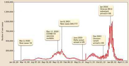
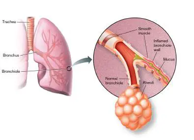
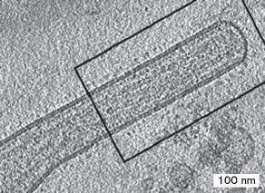
FIGURE 26.14 ■ Respiratory syncytial virus is the major cause of bronchiolitis in
children. Location of RSV infection in the lung. Inflammation and excess mucus impair
breathing. Inset photo: TEM of the linear form of RSV. Circular forms also occur.
KISS ET AL. 2014. J. VIROL. 88 :7602–7617, FIG. 1A.
Illness begins most frequently with fever, runny nose, cough, and sometimes wheezing. RSV is
spread from respiratory secretions through close contact with infected persons or by contact with
contaminated surfaces or objects. Infection can occur when the virus contacts mucous membranes of
the eyes, mouth, or nose and possibly through the inhalation of droplets generated by a sneeze or
cough. Unlike rubella or rubeola, which infect the respiratory tract and disseminate through the body,
RSV remains localized in the lung.
The majority of children hospitalized for RSV infection are under 6 months of age. RSV can cause
repeated infections throughout life, usually associated with moderate to severe cold-like symptoms.
Severe lower respiratory tract disease may occur at any age, especially among the elderly or people
with compromised cardiac, pulmonary, or immune systems. As yet, a vaccine to control this disease is
not available. However, a monoclonal antibody called palivizumab, which binds to an RSV epitope,
can prevent RSV infection in high-risk infants who were born either prematurely or with medical
problems such as congenital heart failure.
Table 26.2 presents many other bacterial, fungal, and viral microbes that can cause respiratory
tract infection. Be aware that very different diseases can produce similar symptoms. For instance,
people constantly confuse influenza (the flu) with the common cold. Symptomatically, they may start
out similarly, but there are telling differences. Influenza is characterized by fever, myalgia (muscle
aches), pharyngitis (sore throat), and headache (viral infection is discussed in Chapter 11). A runny
nose is not one of the symptoms. The common cold, however, manifests as a runny nose, nasal
congestion, sneezing, and throat irritation. No myalgia. The observant clinician will note the
difference.
To Summarize
Most lung infections are viral , but most deadly lung infections are caused by bacteria.
The mucociliary escalator is a primary defense mechanism used by the lung to avoid
infection.
An elevated white cell count in blood is an indicator of bacterial infection.
Pneumococcal vaccine should be administered to the elderly because they are often
immunocompromised.
Fungal agents commonly cause long-term, chronic infections.
Localized bacterial infections in the lung can disseminate via the bloodstream to form
lesions at other body sites.
Tuberculosis is an ancient bacterial disease with an increasing mortality rate resulting from
multidrug-resistant strains, the susceptibility of HIV patients, and an increasing indigent
population.
COVID-19, caused by SARS-CoV-2 virus , begins as a lung infection but can disseminate
via blood to other organ systems. The virus can initiate a life-threatening, out-of-control
inflammatory response (cytokine storm) capable of damaging many organ systems.
Respiratory syncytial virus is one of several viruses that can cause lung disease, but it
rarely spreads to other organs.
Glossary
granuloma
A thick lesion formed around a site of infection.
secondary tuberculosis
A new round of serious disease that is caused by Mycobacterium tuberculosis in patients with
latent tuberculosis who have become immunocompromised. Symptoms include severe cough,
blood sputum, night sweats, and weight loss.
multisystem inflammatory syndrome in children (MIS-C)
A systemic multi-organ inflammatory syndrome in children brought on by COVID-19 disease.
Fig. 28.5
FIGURE 28.5 ■ Acid-fast stain and growth of Mycobacterium tuberculosis. A.
Acid-fast Ziehl-Neelsen stain of M. tuberculosis. B. Auramine O fluorescent acid-fast bacilli
(AFB) stain. C. Löwenstein-Jensen medium enables growth of mycobacterial species, some
of which grow extremely slowly. The colonies have a “bread crumb–like” appearance.
CDC
SITTIPONG SREECHAT/© 123RF.COM
AGARWAL, S., ET AL. 2005. ANN CLIN MICROB ANTI 4 (18)
Figure 6.27
FIGURE 6.27 ■ SARS-CoV-2 virion structure and genome. ORFs (open reading
frames) and genes are shown staggered where translation begins in a different reading
frame (triplet start position) from that of the adjacent upstream gene. The (+) strand RNA
genome has a eukaryotic cap (7-methyl-Gppp) attached at the 5′ end (indicated as orange
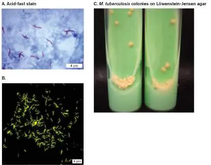
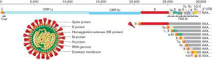
ball). Within the host cell, all genome copies and mRNA molecules have a cap attached by
the host cell’s capping enzyme. 3′ UTR = 3′ untranslated region; AAA = 5′ poly-A tail.
Figure 6.28
FIGURE 6.28 ■ SARS-CoV-2 replication cycle. The virion binds the ACE2 receptor. It
then undergoes endocytosis and uncoats in the host cytoplasm. The released RNA genome
is translated by host ribosomes to synthesize the RNA-dependent RNA polymerase
(replicase). The replicase synthesizes (−) strand RNA, which serves as a template for
transcription to mRNAs. Endoplasmic reticulum (ER) membrane becomes transformed into
viral factories (double-membrane vesicles) that organize virion assembly and transport to
the Golgi complex. From the Golgi, virions move to the cell membrane and bud out.
Endnotes
1. Note a: Formerly genus Chlamydophila but has been changed back to Chlamydia. Return to
reference a
2. Note a: Formerly genus Chlamydophila but has been changed back to Chlamydia. Return to
reference a
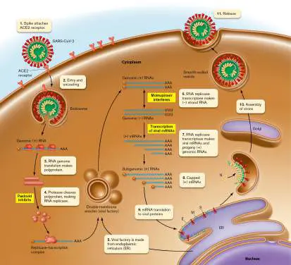
3. Note b: BCG = Bacille Calmette-Guérin (a weakened strain of the bovine tuberculosis strain).
Return to reference b
4. Note c: Palivizumab = monoclonal antibody to RSV protein. Return to reference c
26.3 Gastrointestinal Tract Infectionsnot assigned
Nearly everyone has experienced diarrhea, a condition characterized by frequent loose bowel
movements accompanied by abdominal cramps. Hundreds of millions of cases occur each year in the
United States and are a major cause of death in developing countries. As with respiratory tract
infections, most diarrheal disease is viral in origin, with rotavirus being the primary culprit. Among the
bacteria, the Gram-negative rod Salmonella enterica serovar Typhimurium and the spiral-shaped or
curved bacillus Campylobacter are the most frequent causes of self-limiting diarrheal disease.
You might wonder why some microbes, like Campylobacter jejuni (Fig. 26.15 ), prefer the chaos
of diarrhea to the relative stability of a nice commensal relationship. After all, isn’t finding a niche and
sticking to it the goal of every microbe? The simple answer is that diarrhea enables the dissemination
of microbes that might otherwise kill their host or be killed themselves by too aggressively provoking
the host’s immune system. Dissemination into the environment enables the microbe to reach new
hosts and proliferate. A diarrhea-causing microbe is rather like a serial bank robber fleeing from city to
city to avoid capture and find more banks to rob.
FIGURE 26.15 ■ Campylobacter jejuni (SEM). This Gram-negative, spiral-shaped bacterial
pathogen (cell length 5 μm) is a major cause of human diarrhea. Symptoms of C. jejuni infection
include cramps, fever, and bloody diarrhea. Animals, such as chickens and other birds raised for
meat and their eggs, are the usual reservoirs for this pathogen, so it is considered a zoonotic
disease. Transmission is typically via ingestion of contaminated water or undercooked poultry.
MEDISCAN/ALAMY STOCK PHOTO
Types of Diarrhea
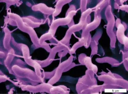
What causes diarrhea? Normally, intestinal mucosal cells absorb water (8–10 liters per day) from the
intestinal contents, essentially drying out stool. Diarrhea occurs when water is not absorbed or when it
actually leaves intestinal cells and enters the intestinal lumen. The excess water loosens stool, and
diarrhea results. There are several types of diarrhea:
Osmotic diarrhea happens when nonabsorbable substrates, such as lactulose, a synthetic sugar,
are used to treat constipation. Lactulose increases osmolarity in the intestine, causing water to
leave mucosal cells. Infectious organisms (such as rotavirus) that prevent nutrient absorption can
cause osmotic diarrhea.
Secretory diarrhea develops when microbes cause mucosal cells to increase ion secretion, as
seen with cholera toxin. Mucosal cells expel water to try to equilibrate the resulting electrolyte
imbalance.
Inflammatory diarrhea forms when an infectious agent triggers the production of inflammatory
cytokines that attract PMNs. Subsequent damage to the intestinal wall limits water and nutrient
absorption and causes red and white blood cells to enter the stool (bloody diarrhea or dysentery).
Shigella, Salmonella, and some strains of E. coli can cause inflammatory diarrhea.
Motility-related diarrhea can develop when enterotoxins made by pathogens such as rotavirus
cause intestinal hypermotility. Food moves through the intestine too fast for water or nutrients to
be absorbed.
Terms used to describe the inflammation of different parts of the GI tract include:
Gastritis (inflammation of the stomach lining—for instance, ulcers).
Gastroenteritis (nonspecific term for any inflammation along the gastrointestinal tract).
Enteritis (inflammation mainly of the small intestine).
Enterocolitis (inflammation of the colon and small intestine).
Colitis (inflammation of the large intestine—colon).
Rehydration therapy. Diarrhea and vomiting due to viral growth, bacterial growth, or toxin
production can cause large amounts of water to leave the intestinal cells and enter the intestinal
lumen. As a result, the patient can become dangerously dehydrated. Most deaths resulting from
infectious diarrhea are the result of dehydration. Consequently, the most important treatment for
diarrhea is rehydration therapy. But giving water alone is not enough. Water leaving tissues causes an
osmotic imbalance that compels electrolytes to exit too (as a way to reestablish balance). The
resulting electrolyte imbalance severely affects cardiovascular, respiratory, and renal systems, leading
to systemic failure, which is the real cause of death. Giving water alone will only make the electrolyte
imbalance worse. Therefore, rehydration solutions must also contain glucose, as well as sodium and
potassium salts in proper balance (for example, Pedialyte).
Antibiotics are often inappropriate when treating diarrhea. Although antibiotic treatment of
infectious gastroenteritis seems intuitive, it is rarely used and actually contraindicated. Most
gastrointestinal infections are viral (norovirus or rotavirus), so antibiotics are ineffective. Likewise,
gastroenteritis caused by bacteria usually resolves spontaneously without antibiotic treatment.
However, severe systemic disease stemming from gastroenteritis can develop, often in the young or
elderly. Diseases like bacillary dysentery (Shigella dysenteriae) respond well to antibiotics.
In some cases, antibiotic treatment can actually trigger gastrointestinal disease. For example,
many antibiotics used to treat infectious diseases can kill most normal intestinal bacteria, except the
naturally resistant Gram-positive, spore-forming anaerobe Clostridioides difficile (formerly Clostridium
difficile), the causative agent of pseudomembranous enterocolitis. Unrestrained by microbial
competition, C. difficile growing at the epithelial surface of the intestine will produce specific toxins
that damage and kill intestinal cells. The organism’s growth leads to inflammation and the formation of
exudative plaques along the intestinal wall (refer to Fig. 23.11D). The plaques eventually coalesce
into larger pseudomembrane structures that block the intestinal mucosa. The blockage causes the
malabsorption of nutrients and water, resulting in diarrhea. As the pseudomembrane enlarges, it
begins to slough off and pass into the stool. Diagnosis of this disease involves PCR identification of the
organism or immunological identification of the toxin in fecal samples.
Some patients suffer recurrent C. difficile infections. It is not clear why, but one suggestion
is that spores lodged in colon folds may escape clearance by peristalsis. Another hypothesis is
that patients with recurrent disease have an impaired response to the C. difficile toxins. How
can recurrences be prevented? In some instances, after vegetative C. difficile has been killed by
antibiotic treatment, a procedure known as a fecal transplant (see Section 23.2) can be used to
restore a healthy gastrointestinal microbiome that prevents the recurrence of C. difficile disease.
Staphylococcal Food Poisoning
We have all heard of a local church picnic or a restaurant where scores of people become violently ill
within hours of eating unrefrigerated potato salad or other food. Staphylococcus aureus is the usual
cause of these disasters, but it is not an infection. The culprit is an enterotoxin (an exotoxin that
affects the gastrointestinal tract) secreted by some strains of S. aureus into tainted foods such as pies,
turkey dressing, or potato salad. After ingestion, the toxin travels to the intestine, where it enters the
bloodstream and stimulates the vagus nerve leading to the vomit center in the brain.
Because the toxin is preformed, symptoms occur quickly after ingestion. Within 2–6 hours, the
poisoned patient will begin vomiting and may also experience diarrhea. The disease, though violent, is
not life-threatening and usually resolves spontaneously within 24–48 hours. In contrast, diarrhea
caused by infectious agents such as Salmonella enterica, which must first grow in the victim, does not
occur until 12–24 hours after ingestion and sometimes longer. A clinician noting quick onset of
symptoms in a patient will immediately suspect staphylococcal food poisoning. Obviously, antibiotic
treatment is not needed for staph food poisoning, but it may be indicated for other gastrointestinal
infections. Staphylococcal enterotoxins are also heat resistant, so simply heating a food already
containing enterotoxin will not destroy the toxic activity.
Case History: Enterohemorrhagic E. coli
Tammy, a 6-year-old girl from Montgomery County, Pennsylvania, arrived at the ER with bloody
diarrhea, a temperature of 39°C (102.2°F), abdominal cramping, and vomiting. She was admitted to
the hospital 5 days after a kindergarten field trip to the local dairy farm. When questioned about
Tammy’s activities during the trip, her parents said she had purchased a snack while at the farm. A
fecal smear analyzed in the laboratory was positive for leukocytes and, when cultured on a sorbitol
MacConkey agar plate, produced sorbitol-nonfermenting colonies suspected of being
enterohemorrhagic E. coli O157:H7. PCR tests confirmed this suspicion, identifying the presence of
genes for Shiga toxins 1 and 2 and the eae gene encoding intimin (see Chapter 25). Meanwhile,
Tammy had developed additional problems. Her face and hands had become puffy, her urine output
had decreased despite being given IV fluids (suggesting kidney damage), and she was beginning to
develop some neurological abnormalities. Laboratory analyses of blood samples revealed
thrombocytopenia (reduced blood platelet count) and confirmed hemolytic uremic syndrome (HUS;
renal failure). Tammy was treated by IV fluid and electrolyte replacement. Antibiotics were not
administered. She eventually recovered.
In this case history, the presence of leukocytes in a fecal smear is a sign that the intestinal pathogen
may have invaded the epithelial mucosa of the intestine (or severely damaged it). Breaching this
barrier sends out a chemical call (chemokine) to neutrophils, which then enter the area and, in an
effort to kill the pathogen, also damage the intestinal cells. Shigella dysenteriae, Salmonella enterica,
and enteroinvasive E. coli (EIEC) actually invade enterocytes and are considered intracellular
pathogens. Enterohemorrhagic E. coli (EHEC), which also produces leukocytes and blood in stools, is
not an intracellular parasite (it is not invasive) but causes damaging attachment and effacing lesions,
described in Section 25.4 that destroy the mucosal epithelium. The resulting inflammation, in
conjunction with damage caused by the Shiga toxin it produces, leads to blood and white cells in the
stool. E. coli O157:H7, the etiological agent in the case history, is a common serotype of EHEC.
There are at least seven different classes (pathovars) of pathogenic E. coli, differing in their
repertoire of pathogenicity islands, plasmids, and virulence factors. They include the enteroinvasive
(EIEC) and enterohemorrhagic (EHEC) pathovars just mentioned, plus the enterotoxigenic E. coli
(ETEC), neonatal meningitis-causing E. coli (NMEC), uropathogenic E. coli (UPEC), enteropathogenic E.
coli (EPEC), and enteroaggregative E. coli (EAEC). All but UPEC and NMEC cause gastrointestinal
disease. To distinguish these strains, each group has telltale O and H antigens that can be identified by
using serology or PCR to identify allelic differences in genes encoding LPS synthesis.
Note: “O antigen” is part of the bacterium’s LPS, while “H antigen” is a flagellar protein. Thus,
“O157:H7” denotes the specific versions of LPS (O157) and flagellar protein (H7) found on E. coli
O157:H7. Other pathogenic strains of E. coli have different O and H antigens.
Shiga toxin. Shigella and EHEC, the agent in the preceding case history, both produce toxins, called
Shiga toxins 1 and 2, that are encoded by genes of bacteriophage genomes (prophages) embedded in
the bacterial chromosome. The toxins, which are absorbed through the intestine and disseminated via
the bloodstream, are AB5 toxins (see Section 25.3). The A subunit, upon entry, destroys protein
synthesis by cleaving an adenine from 28S rRNA in eukaryotic ribosomes. These toxins inhibit host
protein synthesis and, in the process, damage endothelial cells in the intestine, kidney, and brain.
Shiga toxin–induced death of vascular endothelial cells in the intestine causes the breakdown of blood
vessel linings, followed by hemorrhage that manifests as bloody diarrhea. Shiga toxin 2 also triggers
the release of pro-inflammatory cytokines.
Endothelial damage initiates the formation of platelet-fibrin microthrombi (clots) that occlude blood
vessels in the various organs, leading to two major syndromes: hemolytic uremic syndrome (HUS) and
thrombotic thrombocytopenic purpura (TTP). HUS develops when the microthrombi are limited to the
kidney. The microclots clog the tiny blood vessels in this organ and cause decreased urine output,
ultimately leading to kidney failure and death. In TTP, the clots occur throughout the circulation,
causing reddish skin hemorrhages called petechiae and purpuras (discussed in Section 25.3).
Neurological symptoms (for example, confusion, severe headaches, and possibly coma) then arise from
microhemorrhages in the brain. The hemorrhaging occurs because platelets needed for normal clotting
have been removed from the circulation as they form the microthrombi. The decreased number of
platelets is called thrombocytopenia.
HUS is a common consequence of E. coli O157:H7 infection, as in the case history described.
Unfortunately, HUS can be treated only with supportive care, such as blood transfusions and dialysis
throughout the critical period until kidney function resumes. Antibiotic treatment can increase the
release of Shiga toxins from the organisms and actually trigger HUS. Thus, antimicrobial therapy is not
recommended.
Enterohemorrhagic E. coli (EHEC). E. coli O157:H7 is a recently emerged pathogen that can
colonize cattle intestines at the recto-anal junction without affecting the animal and, as a result, can
contaminate meat products following slaughter. Initially identified in 1982, the organism came to
national prominence during a large-scale U.S. outbreak in 1993, linked to a Washington State Jack in
the Box restaurant, in which 732 people were sickened.
E. coli O157:H7 rarely affects the health of the reservoir animal. But when an infected steer is
slaughtered, the carcass can become contaminated with EHEC-containing feces despite considerable
efforts by slaughterhouses to prevent it. Grinding the tainted meat into hamburger distributes the
microbe throughout. Cooking burgers to 160°C is essential to kill any existing EHEC. Cross-
contamination between foods is possible too. Using the same cutting board to prepare meat and salad
is a great way to contaminate the salad, which will not be cooked.
Despite EHEC’s common association with hamburger, vegetarians are not safe from this organism.
During heavy rains, waste from a cattle farm can easily wash into nearby vegetable fields unless
precautions are taken. If the cattle waste contains E. coli O157:H7, the crops become contaminated,
and the pathogen can enter the plant through stomates (respiration openings in the leaves; Fig.
26.16 ). One such outbreak occurred in 2018 when romaine lettuce grown in certain areas of Yuma,
Arizona, was contaminated with this pathogen. Lettuce shipped to 36 states infected 210 people. Of
those, 96 were hospitalized, 25 developed HUS, and 5 died. Whole-genome sequencing genetically
linked all of the isolated EHEC strains to various farms in Yuma and identified the likely source as water
from a canal in the Yuma growing area.
FIGURE 26.16 ■ E. coli entering a lettuce leaf. Escherichia coli individuals on a lettuce leaf
congregate at the stomates and enter the plant veins. Once inside the plant, the organisms resist
efforts to wash them out.
SCIMAT/SCIENCE SOURCE
Early on, the remarkably low infectious dose of E. coli O157:H7 mystified researchers. However, we
have since learned that E. coli has an impressive level of acid resistance, rivaling that of the gastric
pathogen Helicobacter pylori. Acid resistance mechanisms permit E. coli to survive in the acidic
stomach and enable a mere 10–100 individual organisms to cause disease.
In contrast to the case just described, many gastrointestinal infections do not produce fecal
leukocytes or blood in the stool. Diarrheal diseases caused by Vibrio cholerae (cholera) or
enterotoxigenic E. coli (ETEC), which produces a cholera-like disease, do not involve invasion of the
intestinal lining by the microbe, and they yield copious amounts of watery diarrhea. In these two toxin-
driven diseases, the bacteria attach to cells lining the intestine and secrete toxins that are imported
into the target cells (see Section 25.3).
How can we prevent disease caused by enterohemorrhagic E. coli? Industry approaches include
thoroughly washing carcasses before processing, maintaining cold temperatures, and testing for
possible contamination. In addition, the use of gamma irradiation to sterilize beef, spinach, and lettuce
has been approved. Outbreaks of EHEC disease caused by contaminated hamburger have declined
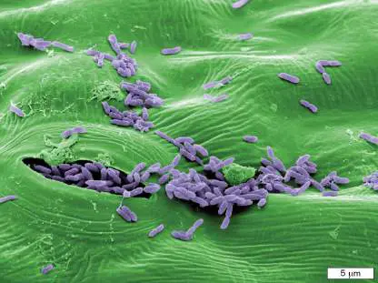
dramatically because of industry practices and USDA inspections. Irradiation (described in Chapter 5)
could eliminate the problem, but less than 5% of hamburger meat is currently irradiated in the United
States.
Rotavirus and Norovirus
Many people wrongly think that most cases of diarrhea are caused by a bacterial agent. Actually, two
viruses—namely, rotavirus and norovirus —cause more intestinal disease than do any bacterial species.
Rotavirus is a double-stranded RNA virus (Group III in Table 6.2); norovirus is a positive-sense, single-
stranded RNA virus (Group IV). Rotavirus is highly infectious, spreading by the fecal-oral route; it is
endemic around the globe, and it affects all age groups, although children between 6 and 24 months
are the most severely affected. It is estimated that by age 3, all children have had a rotavirus
infection.
The incubation period is approximately 2 days, after which the victim commonly suffers frequent
watery, dark green, explosive diarrhea. Accompanying symptoms may include nausea, vomiting, and
abdominal cramping. Severe dehydration and electrolyte loss due to the diarrhea will cause death
unless supportive measures, such as fluid replacement, are undertaken. There is no cure, but most
patients recover if rehydrated properly. Few deaths from rotavirus occur in the United States, but each
year 120,00 to 250,00 children worldwide die from this viral diarrhea (2021 estimate). The mortality
and incidence of this disease have decreased because of the introduction in 2006 of a safe and
effective vaccine (see Table 24.5). In Mexico alone, the vaccine resulted in a 50% decline in diarrheal
deaths.
With the success of the rotavirus vaccine, norovirus is set to become the most common worldwide
cause of nonbacterial gastroenteritis. Norovirus infections have already surpassed rotavirus in the
United States. The virus is perceived as the scourge of cruise ships and assisted-living facilities, but
norovirus can also spread quickly in hotels or anywhere there are many people in a small area. The
virus spreads by the fecal-oral route among children or adults, via contaminated food or person-to-
person contact. Within 24 hours of infection, the victim experiences sudden vomiting, stomach cramps,
and watery diarrhea that mercifully resolves within 12–24 hours. Treatment is similar to that for
rotavirus, but there is no vaccine. Death is rare, but when it does occur, it is most common among
infants and the elderly. Take notice that newly emerging single-stranded, positive-sense viruses in a
group called sapoviruses are now causing increasing cases of norovirus-like disease (1%–17% of
diarrhea episodes worldwide).
Note: Norovirus infection is sometimes called the “stomach flu,” but that is a misnomer. It is not the
flu and has nothing to do with influenza virus.
Diarrhea and the gut microbiome. An obvious question to ask about diarrhea is: How does it
affect the microbiome? A study headed by Shannon Manning (Michigan State University) found that the
composition of intestinal microbiota of patients with diarrhea differs significantly from those of their
healthy family members.
For one thing, the gut microbiomes of diarrhea patients who were not given antibiotics
were less diverse than those of their uninfected family members. Abundance of Bacteroidetes
and Firmicutes was higher in the healthy individuals, whereas Proteobacteria dominated the
patient microbiomes. Escherichia coli, for instance, predominated in all patients, regardless of the
pathogen causing the infection. The composition of diarrheal microbiomes also varied with the
bacterial cause of infection. As one example, the microbiome of Campylobacter -infected patients
differed from that of patients infected with Salmonella or Shigella.
How does the intestine restore its bacterial population after being decimated by diarrhea or
antibiotic treatment? Lawrence David (Duke University; Fig. 26.17A ) and Peter Turnbaugh (UC San
Francisco), along with their colleagues, used metagenomic procedures to characterize the stools of 41
people in Bangladesh (children and adults) who had diarrhea caused by E. coli or Vibrio cholerae.
Stools were monitored before, during, and after diarrhea episodes.
FIGURE 26.17 ■ Successive repopulation of the gut microbiome following Vibrio
cholerae infection. A. Lawrence David (pictured) and Peter Turnbaugh (not shown) unravel the
complexities of the human microbiome. B. Fecal samples were taken at 1, 7, and 30 days past
presentation (dpp) of diarrhea. Patients, identified by letters, were given a single dose of
azithromycin on the day of presentation, which nearly eliminated V. cholerae by 1 dpp. Healthy
contacts of each patient are identified by numbers and are shown on the left. For example, A-1,
A-2, A-3, and A-4 are the healthy contacts of patient A. Colored boxes reflect groups of different
genera that are prominent during the infection and afterward, during early, mid, and late stages
of repopulation. The x -axis values reflect relative abundances of genera. “All swab” microbes
were present only in rectal swab samples.
Source: Part B modified from Harvard Press Office figure.
COURTESY OF LAWRENCE DAVID
The researchers identified a consistent succession of repopulation events in nearly every case,
regardless of the cause of diarrhea (Fig. 26.17B ). After diarrhea clears out much of the microbiome
(either naturally or after antibiotic treatment), carbohydrates and oxygen accumulate in the gut.
Carbohydrates and oxygen would normally be metabolized by gut microbiota. During the early stage of
repopulation, facultative, oxygen-respiring, and carbohydrate-utilizing bacteria (especially those using
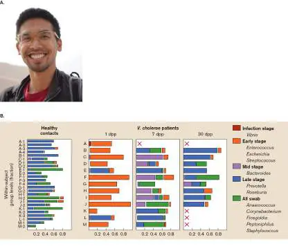
simple carbohydrates, such as Escherichia, Enterococcus, and Streptococcus) colonize the gut and
consume these nutrients. Midstage recovery begins when the lack of simple sugars and oxygen (as
well as increased phage predation) leads to a decline in the early-stage species. This decline enables
succession to anaerobic, complex carbohydrate-fermenting bacteria (Bacteroides). Finally, in late-
stage recovery, the gut microbiome once again resembles the complex community that existed prior to
infection—that is, the same composition seen in healthy contacts (Fig. 26.17B ). The entire process
takes about 30 days to complete but depends on a variety of factors, such as diet, antibiotic use, and
duration of diarrhea.
Repopulation occurs, in part, by microbes being reingested from food. But exciting research
suggests that the much maligned and trivialized appendix is, among other things, an important
reservoir of gut microbes that can seed the intestine and reestablish the microbiota. Once properly
reestablished, gut microbiota are capable of fending off pathogens such as Clostridioides difficile. As
evidence, researchers found that patients with appendectomies were more than twice as likely to
develop repetitive infections with C. difficile. eResearch Activity 26 presents additional intriguing
evidence that, during a transient infection, the gut microbiome “learns” how to better protect its host
against a subsequent GI (gastrointestinal) infection.
Case History: Ulcers—It’s Not What You Eat
Gary was a 34-year-old accountant who had immigrated to Nebraska from Poland 7 years earlier. Since
his teenage years, he had been bothered periodically by episodes of epigastric pain (pain around the
stomach), nausea, and heartburn. Antacids usually alleviated the symptoms. Over the years, he had
received several courses of treatment with Tagamet or Pepcid to reduce acid secretion and provide
relief. Recently, an upper-GI endoscopy had been performed, in which a long, thin tube tipped with a
camera and light source was inserted into Gary’s mouth and threaded down into his stomach. The view
through the endoscope showed some reddened areas in the antrum (bottom part) of the stomach. The
endoscope was also equipped with a small clawlike structure that obtained a small tissue sample from
the lining of Gary’s stomach. A urease test performed on the antral biopsy turned positive in 20
minutes. Histological examination of the biopsy confirmed moderate chronic active gastritis
(inflammation of the stomach lining) and revealed the presence of numerous spiral-shaped organisms.
Cultures of the antral biopsy were positive for Helicobacter pylori.
Painful and sometimes life-threatening gastric ulcers were for many years blamed on spicy foods and
stress. In the 1980s, after discovering odd, helical bacteria present in the biopsies of gastric ulcers,
Australians J. Robin Warren and Barry Marshall (a medical intern at the Royal Perth Hospital at the
time; Fig. 26.18A ) proposed that bacteria, not pepperoni, cause ulcers (Fig. 26.18B and C ).
FIGURE 26.18 ■ A bacterial cause of gastric ulcers. A. Physician Barry Marshall was so
sure he was right about the cause of stomach ulcers that he swallowed bacteria to prove his
point. B. Helicobacter pylori (SEM). Note the tuft of flagella at one pole. Cell length approx. 2 μm.
C. Helicobacter (arrows) attached to gastric mucosa.
XINHUA/ALAMY STOCK PHOTO
EYE OF SCIENCE/SCIENCE SOURCE
EYE OF SCIENCE/SCIENCE SOURCE
Their hypothesis was viewed with skepticism and derided as heresy by the established medical
community. Faced with disbelief bordering on ridicule, the young intern drank a vial of the helical
organisms and waited. A week later he began vomiting and suffered other painful symptoms of
gastritis. Barry Marshall could not have been happier. He had proved his point. We now know that this
curly microbe causes the vast majority of stomach ulcers and has colonized humans for at least
100,000 years.
The discovery of Helicobacter pylori and its association with gastric ulcer disease led to a major
shift in ulcer treatment, previously limited to suppressing acid production via proton pump inhibitors.
Therapy now includes antimicrobial treatment to kill the bacteria, coupled with acid suppression
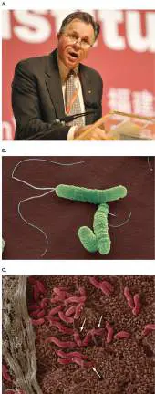
therapy to prevent further inflammation while the ulcer heals. Warren and Marshall, who recovered
from his gastritis, received the 2005 Nobel Prize in Physiology or Medicine for their groundbreaking
work.
H. pylori can be detected in about half of the world’s population, especially in impoverished
countries. Why most people colonized with H. pylori do not develop gastric ulcers is unclear. The exact
mechanism by which H. pylori causes gastric ulcers is not known, although a variety of virulence
factors have been identified. The basic scheme of Helicobacter pathogenesis is shown in Figure
26.19 . After the pathogen is ingested, H. pylori flagella propel the organism toward the mucosa,
driven by an ill-defined chemotaxis system that, in part, senses urea produced by the human body (
Fig. 26.19 , step 1). As it approaches the mucosa, the pathogen produces intracellular and
extracellular urease, an important virulence factor that converts urea to CO 2 and ammonia. The
ammonia neutralizes acid around Helicobacter, thereby enabling the organism to survive the extreme
acidity of the stomach (step 2).
Two other enzymes—namely, collagenase and mucinase—then soften the mucous lining, helping
the bacteria reach the stomach’s epithelial lining (Figs. 26.18C and 26.19 , step 3). The epithelial
lining is much less acidic than the lumen, so the organism can grow and divide. Once at the epithelium,
Helicobacter produces various adhesins, such as BabA or HpaA, to bind host cells (step 4). After the
organism has adhered, tissue damage develops with the release of vacuolating cytotoxin (VacA) and
neutrophil-activating protein (NAP). NAP activates neutrophils and mast cells to damage local tissue
(step 5). VacA forms a hexameric pore in the host membrane and induces apoptosis (programmed cell
death) by damaging mitochondria. Apoptotic cells decrease the immune response, which will stabilize
a chronic infection.
FIGURE 26.19 ■ Steps in Helicobacter pathogenesis.
Another protein, CagA, is injected (by a type IV secretion system; see Chapter 25) into host
epithelial cells, where it becomes phosphorylated. CagA then interacts with host signaling proteins and
activates host signal transduction pathways that can stimulate inflammation and growth possibly
leading to cancer (Fig. 26.19 , step 6). Gastric adenocarcinoma is the third leading cause of cancer
deaths worldwide, and Helicobacter is a leading cause of gastric cancer.
Evidence continues to suggest that H. pylori can affect diseases outside the stomach. CagA, for
example, has been found in patient serum, packaged within exosome vesicles generated by host
exocytosis. Helicobacter also sheds outer membrane vesicles (OMVs) that contain CagA. OMVs
containing CagA incubated with host cells produced changes in host cell gene expression and function.
Recent data suggests the CagA exosomes can damage endothelial cells of the cardiovascular system.
Tools useful for diagnosing H. pylori include a fecal antigen test, rapid urease testing, and serology
[for example, enzyme-linked immunosorbent assay (ELISA) to detect antibody to the CagA antigen].
ELISA is a common immunological tool used to detect the presence, in serum, of antibodies to a
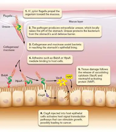
specific organism, which are an indication of infection. The ELISA test is described more fully in Section
28.3.
Protozoan Causes of Diarrheal Disease
As we learned in Chapter 20, some protozoa (also called protists) cause serious human diseases. For
instance, Entamoeba histolytica (Fig. 26.20A and Section 20.6) and Cryptosporidium (C. parvum
and C. hominis; Fig. 26.20B ) cause the diarrheal diseases amebic dysentery and cryptosporidiosis,
respectively. The CDC estimates (2018) that there are about 750,000 cases of cryptosporidiosis each
year in the United States but that less than 2% are reported because many cases are mild or
asymptomatic. Cryptosporidium infection begins with oocysts that contain four sporozoites (infectious
form of protozoan) being shed in the feces of infected hosts (human and nonhuman animals) to
contaminate water (drinking or recreational, such as in water parks). Following ingestion, the
sporozoites are released and parasitize intestinal epithelial cells. Asexual and sexual cycles then lead
to more oocysts. Treatment of immunocompetent patients usually involves only rehydration therapy,
although an antiparasitic drug (nitazoxanide) is available. HIV patients infected with C. parvum must
also receive antiretroviral therapy to improve CD4 T-cell count.
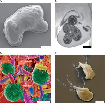
FIGURE 26.20 ■ Protists that cause diarrhea. A. Entamoeba histolytica trophozoite (SEM,
motile form). B. Cryptosporidium parvum oocyst encasing four infectious sporozoites (TEM). C.
Giardia duodenalis cysts (7–14 μm) present in fecal matter (colorized SEM). D. Trophozoite form
of G. duodenalis (5–15 μm in length; colorized SEM).
AVALOS-PADILLA ET AL. 2015. PLOS PATHOG. 11 :E1005079, FIG. 8A.
ALDEYARBI AND KARANIS. 2016. EUR. J. PROTISTOL. 52 :36–44, FIG. 1A.
DR. GARY GAUGLER/SCIENCE SOURCE
EYE OF SCIENCE/SCIENCE SOURCE
The flagellated protozoan Giardia duodenalis (formerly G. lamblia or intestinalis) is a major cause
of diarrhea throughout the world. In the United States alone, G. duodenalis is thought to cause over 1
million illnesses per year. G. duodenalis enters a human or other host as a cyst present in drinking
water contaminated by feces (Fig. 26.20C ). A cyst is a dormant form of a protist encased in a
protective wall (see Chapter 20). Aside from humans, G. duodenalis can be found in various rodents,
deer, cattle, and even household pets. It is very infectious. Ingestion of as few as 25 cysts can lead to
disease. Following ingestion, the hard outer coating of the cyst is dissolved by the action of digestive
juices to produce a trophozoite (Fig. 26.20D ), which attaches itself to the wall of the small
intestines and reproduces. Offspring quickly encyst and are excreted out of the host’s body.
Asymptomatic carriers of G. duodenalis are common; it has been estimated that anywhere from 1%
to 30% of children in U.S. day-care centers are carriers. Disease usually manifests as greasy stools
alternating between a watery diarrhea, loose stools, and constipation. However, some patients will
experience explosive diarrhea. Diagnosis usually comes from microscopically observing the cysts or
trophozoite forms of the protozoan in feces. Metronidazole is a drug often used to cure the disease. To
prevent it in the first place, proper treatment of community water supplies is essential.
Hepatitis Viruses Target the Liver
The liver is also considered part of the gastrointestinal system, because it provides bile to the intestine
and processes the nutrients absorbed from the intestine. Hepatitis is a general term meaning
“inflammation of the liver.” Although many pathogens can affect this organ, we focus on the major
infectious cause, an eclectic group of viruses collectively called the hepatitis viruses. They include a
single-stranded, negative-sense RNA virus (hepatitis A, HAV), a double-stranded DNA virus (hepatitis
B, HBV), and a single-stranded, positive-sense RNA virus (hepatitis C, HCV). Space does not permit
coverage of HDV or HEV.
Table 26.3 compares general features of these viruses. The disease symptoms of infectious
hepatitis, regardless of viral cause, include abdominal pain, fever, vomiting, and often dark urine, clay-
colored stools, and jaundice. Liver damage from hepatitis is marked by the detection of liver
transaminases in serum and an enlarged liver (hepatomegaly).
TABLE 26.3 Comparative Features of Hepatitis Viruses and Disease
Hepatitis A Hepatitis B Hepatitis C Hepatitis Hepatitis E
D
Type
ssRNA, DNA, ssRNA, ssRNA, ssRNA, positive
negative enveloped positive negative sense,
sense sense, nonenveloped
TABLE 26.3 Comparative Features of Hepatitis Viruses and Disease
sense, circular,
enveloped enveloped
Incubation
period 30 days 90 days 40 days 30 days; 50 days
requires
coinfection
with HBV
Route
Fecal-oral Parenteral, Parenteral, Parenteral, Fecal-oral
sexual, sexual sexual
perinatal
Viremia
Transient Persistent Persistent Uncommon Transient
Severity
Mild Severe Mild Mild to Mild
severe
Chronic
None 90% 50%–60% 80%–90% None
children
10% adults
Hepatitis A virus (HAV) is a single-stranded RNA picornavirus that causes an acute infection spread
person to person by the fecal-oral route, but hepatitis A can also be contracted by eating undercooked
shellfish collected from contaminated waters. The virus replicates in the intestinal endothelium and is
disseminated via the bloodstream to the liver. After replicating in hepatocytes, the progeny enter the
bile and are released into the small intestine, explaining why stools are so infectious. Though the virus
has an early viremic stage after leaving the intestine, it is rarely transmitted by transfusion, because
the viremic stage is transient and ends after liver symptoms develop. In contrast, hepatitis B and C
viruses produce persistent viremia and are readily transmitted by transfusion.
Many people who are infected with HAV are asymptomatic or exhibit very mild symptoms that
include nausea, vomiting, diarrhea, low-grade fever, and fatigue. As the virus attacks the liver, liver
transaminases are detected in serum, patients may become jaundiced (from the accumulation of
bilirubin in the skin), and their urine will turn dark brown. Diagnosis can be confirmed by PCR or the
detection of IgM antibody to HAV antigens. There is no specific treatment, but the disease usually lasts
for only a few months and then resolves without establishing a carrier state. Disease can be
prevented, however, by a vaccine containing inactivated virus (called HepA vaccine). The vaccine
should be administered starting at 1 year of age. For those not vaccinated, frequent handwashing is
important for preventing spread of the disease because it interrupts the fecal-oral cycle.
In contrast to HAV, hepatitis B virus (HBV) is a partially double-stranded circular DNA virus (family
Hepadnaviridae) that causes diseases of varying severity. These include acute and chronic hepatitis,
cirrhosis, and hepatocarcinoma. The virus is also wrapped in a membrane envelope when progeny
viruses are released from infected cells. The virion coat protein, a surface antigen, is called HBsAg. The
virus makes an excess amount of HBsAg, so it is sometimes extended as a tubular tail on one side of
the virus particle and is often found in the blood of infected individuals in the form of noninfectious
filamentous and spherical particles (Fig. 26.21 ). The presence of HBsAg in blood is an indicator of
HBV infection. There are also nucleic acid and serological tests for HBV RNA and antibodies,
respectively.
FIGURE 26.21 ■ Structure of hepatitis B virus. Hepatitis B is an enveloped, double-
stranded DNA virus in the Hepadnaviridae family (TEM). Also shown are the tubular structures
made from HepB surface antigen (HBsAg).
EYE OF SCIENCE/SCIENCE SOURCE
HBV is transferred primarily via parenteral routes such as blood transfusions, contaminated needles
shared by IV drug users, and any human body fluid, including saliva, semen, sweat, breast milk, tears,
urine, and feces. It can even be transferred transplacentally to a fetus and can be sexually transmitted.
Infection by HBV has two stages: a short-term acute phase and a long-term chronic phase that, if it
extends beyond 6 months, may never resolve (chronic infection). Symptoms resemble those of the flu
but with jaundice and brown urine.
Liver damage caused by HBV infection is due in large part to an efficient cell-mediated immune
response. Cytotoxic T cells and natural killer cells cause immune lysis of infected liver cells. Over the
long term, chronic hepatitis will lead to a scarred and hardened liver (cirrhosis), the only recourse
being a liver transplant. Fortunately, about 90% of people infected are able to fight off infection and
never proceed to the chronic stage. A HepB vaccine (made from recombinant HBsAg) is available. Its
administration is recommended after birth, followed by booster shots administered by 2 months and 18
months of age. There are also several antiviral agents used to treat acute and chronic HBV disease.
One is tenofovir, an inhibitor of HBV reverse transcriptase. Even though HBV is a DNA virus, it
replicates using reverse transcription of an RNA intermediate.
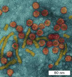
Hepatitis C virus (HCV) causes another form of hepatitis. HCV is a single-stranded, positive-sense,
linear RNA virus with a lipid coat and is a member of the Flaviviridae family. It is transmitted by blood
transfusions and causes 90% of transfusion-related cases of hepatitis. It can also be transmitted by
needle sticks, razor blades, tattooing, and less frequently by sex. Over 100 million people worldwide
are infected with HCV. Screening for HCV (serology or PCR) is recommended for anyone who exhibits
signs of hepatitis or practices the risky behaviors noted here. In addition, the CDC recommends that
anyone born between 1945 and 1965 (baby boomers) be tested because, for unknown reasons, baby
boomers are five times more likely to be infected than other adults.
Most HCV-infected individuals (80%) do not exhibit symptoms, and in those who do, symptoms
may not appear for 10–20 years. At least 75% of patients who exhibit symptoms ultimately progress to
chronic hepatitis requiring a liver transplant or possibly to liver cancer. Fortunately, infection can be
detected using ELISA. Liver biopsies of HCV patients are used to determine the extent of liver damage,
which in turn helps establish the stage of disease.
Prevention of HBV or HCV infection for health care personnel includes avoiding inadvertent needle
sticks. If such a stick should occur with HBV, anti-HBV immunoglobulin should be administered within 7
days. Currently, no effective post-exposure prophylaxis is recognized for HCV. Chronic hepatitis can be
treated with an antiviral protease inhibitor (glecaprevir), which prevents the proteolytic processing of
an important HCV polyprotein, and Harvoni, a combination of an RNA chain terminator with an inhibitor
of a virus phosphoprotein needed for replication. Though vaccines have been developed for HAV and
HBV (see Table 24.5), no vaccine is yet available for HCV.
Note: Because hepatitis viruses can be spread via contaminated blood products, all blood donations
collected by the Red Cross and other agencies are tested for the presence of these viruses, as well as
for HIV. Thus, the blood supply is safe.
We have examined in this section only a handful of the bacterial, viral, and protozoan microbes that
cause gastrointestinal infection. Others are listed in Table 26.4, and some of the protozoan pathogens
are described in Chapter 20.
TABLE 26.4 Selected Microbes That Cause Diseases of the Gastrointestinal Tract
Etiological agent Disease Symptoms Virulence Source(s) Treatm
a factors
Bacterial
Campylobacter Gastroenteritis Fever, muscle Cytotoxin, Poultry, Erythrom
jejuni (G−) pain, watery enterotoxin, unpasteurized
diarrhea, blood in adhesin milk
stool, headache
Clostridioides Pseudomembranous Fever, abdominal Cytotoxin, Animals, Vancom
difficile (G+) enterocolitis pain, diarrhea, antibiotic normal
resistance microbiota
TABLE 26.4 Selected Microbes That Cause Diseases of the Gastrointestinal Tract
pseudomembrane
in colon
Clostridium Botulism Fast onset of Neurotoxin Preformed Antiseru
botulinum (G+, symptoms; flaccid toxin in foods
anaerobe) paralysis
Clostridium Gastroenteritis Watery diarrhea, Alpha toxin Soil, food Self-limi
perfringens (G+) nausea
Enterohemorrhagic Gastroenteritis Bloody diarrhea, Intimin, Tir, Contaminated Oral
E. coli (G −) HUS type III foods rehydrat
secretion, (hamburger) antibioti
Shiga toxin and crops severe
Enterotoxigenic E. Traveler’s diarrhea Watery diarrhea Labile and Humans; Oral
coli (G −) stable food, water rehydrat
toxins
Helicobacter pylori Gastric ulcers Abdominal pain, Adhesin,? Triple dr
(G−) bleeding, urease, protocol
heartburn CagA, (omepra
vacuolating clarithro
toxin metroni
Salmonella Salmonellosis Symptoms after Type III Chickens, Oral
enterica (G−) 18 h; abdominal secretion, other rehydrat
pain, diarrhea; intracellular animals; antibioti
invade intestinal growth fecal-oral severe
M cells route
Salmonella Typhoid fever Headache, fever, Type III Human Quinolon
enterica serovar chills, abdominal secretion, carriers
Typhi (G−) pain, rash (rose intracellular (gallbladder
spots), growth, reservoir);
hypotension, PhoPQ food, water
regulators,
TABLE 26.4 Selected Microbes That Cause Diseases of the Gastrointestinal Tract
diarrhea in late Vi antigen
stages capsule
Shigella spp. (G−) Shigellosis Bloody diarrhea, Shiga toxin, Humans; Oral
HUS type III fecal-oral rehydrat
secretion, route antibioti
intracellular severe
growth,
actin-based
motility,
escape
phagosome
Staphylococcus Staphylococcal food Symptoms within Enterotoxin Preformed Support
aureus (G+) poisoning 4 h of ingestion; toxin in foods
nausea, vomiting,
diarrhea
Vibrio cholerae Cholera Watery diarrhea Cholera Human Oral
(G−) toxin, toxin- waste– rehydrat
coregulated contaminated antibioti
pili (TCPs), water
ToxR
regulator
Vibrio Gastroenteritis Diarrhea, blood in Enterotoxin Raw seafood Self-limi
parahaemolyticus stool
(G−)
Viral
Norovirus Stomach “flu” Nausea, VP1 Fecal-oral Oral
(Norwalk virus) vomiting, route rehydrat
diarrhea
Rotavirus (most Stomach “flu” Nausea, NSP4 Fecal-oral Oral
common cause) vomiting, route rehydrat
TABLE 26.4 Selected Microbes That Cause Diseases of the Gastrointestinal Tract
diarrhea
To Summarize
Diarrhea leads to dehydration , for which fluid replacement is a critical treatment. Antibiotic
treatment is usually not recommended.
Staphylococcal food poisoning is not an infection. It is a toxigenic disease.
Antibiotic treatments can sometimes cause gastrointestinal disease (for example,
pseudomembranous enterocolitis by Clostridioides difficile).
The presence of red and white blood cells in fecal contents is an indication of invasive
bacterial infection by intracellular pathogens such as Shigella, Salmonella, and EIEC.
Bacteria that do not invade intestinal cells usually produce watery diarrhea. EHEC is an
exception; the attachment and effacing lesions it produces result in bloody stools.
Bacterial toxins produced by bacterial enteric pathogens can cause systemic symptoms.
Rotavirus is still the single greatest cause of diarrhea worldwide. Increasing use of rotavirus
vaccine may eventually leave norovirus as the world’s leading cause of diarrhea.
The bacterium Helicobacter pylori , a common cause of gastric ulcers, is highly acid
resistant and lives in the stomach of half the world’s population. It is also a cause of gastric
cancer.
Giardia duodenalis and Cryptosporidium parvum are major protozoan causes of diarrhea
worldwide.
Hepatitis is caused by several unrelated viruses; among them, HAV, HBV, and HCV account for
most disease. HAV is transmitted by the fecal-oral route and does not establish chronic
infection. HBV and HCV can be transmitted by blood products (such as transfusions) and shared
hypodermic needles and can lead to chronic hepatitis. Vaccines for HAV and HBV, but not HCV,
are available.
Glossary
rehydration therapy
A medical treatment for dehydration, in which a liquid solution of salts and glucose is delivered
orally. Also called oral rehydration therapy (ORT) .
rotavirus
One of a group of nonenveloped dsRNA viruses that cause severe diarrhea in children.
norovirus
Also known as Norwalk virus. A nonenveloped ssRNA virus that causes severe diarrhea in children
and adults.
hepatitis
An inflammation of the liver, caused by infection or by exposure to a toxic substance.
hepatitis A virus (HAV)
A single-stranded RNA picornavirus that causes an acute infection of the liver spread person-to-
person by the fecal-oral route.
hepatitis B virus (HBV)
A partially double-stranded, circular-DNA hepadnavirus that causes diseases of the liver of varying
severity, including acute and chronic hepatitis, cirrhosis, and hepatocarcinoma.
hepatitis C virus (HCV)
A single-stranded, positive-sense, linear-RNA flavivirus that is transmitted by blood transfusions
and causes 90% of transfusion-related cases of hepatitis.
Fig. 23.11D
FIGURE 23.11 ■ Potential negative effects of intestinal dysbiosis. D. The firmicute
Clostridioides difficile is often a minor component of the normal gut microbiome whose numbers
are limited by competing gut microbiota. However, antibiotic treatment can kill off the competing
microbes, leaving the antibiotic-resistant C. difficile to grow unabated. Toxins made by C. difficile
will kill host cells, causing small exudative plaques to form on the intestinal wall. Plaques coalesce
to form a large, bloody, and painful pseudomembrane.
Endnotes
1. Note a: G+ = Gram-positive; G− = Gram-negative. Return to reference a
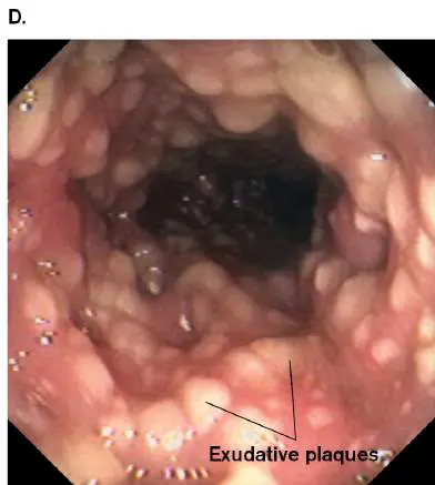
26.4 Genitourinary Tract Infectionsnot assigned
The genital and urinary tracts are certainly different organ systems. One is used for procreation,
whereas the other filters and excretes waste products from blood. However, their close association
in the body makes it useful to discuss their infections in the same section. Despite being close
neighbors, there are two interesting peculiarities to note about the agents that infect these
organs: First, viruses and bacteria can infect the genital tract, but viruses rarely cause urinary tract
infections. Second, very few pathogens can infect both organ systems.
Urinary Tract Infections
The urinary tract includes the kidneys, ureters, urinary bladder, and urethra. Infections anywhere
along this route are called urinary tract infections (UTIs). Figure 26.22 shows an SEM of a
bladder chronically infected with uropathogenic Escherichia coli. UTIs are the second most
common type of bacterial infection in humans, ranking in frequency just behind respiratory
infections such as bronchitis or pneumonia. In the United States, bladder infections and other UTIs
result in over 8 million patient visits annually, mostly by women. Estimates are that at least 60%
of women will experience a UTI during their lifetime, and 20%–40% of those develop recurrent
infections. UTIs result in 100,000 hospital admissions in the United States and over $1 billion in
medical expenses each year.
FIGURE 26.22 ■ Urinary tract infection. Bladder cells (blue) with adherent
uropathogenic E. coli (SEM). White blood cells (yellow) reach out with extracellular traps to
immobilize and kill the pathogen.
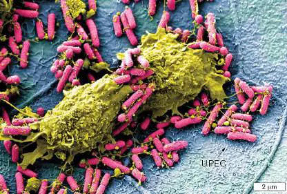
COURTESY OF SCOTT J. HULTGREN, VALERIE O’BRIEN, MATTHEW JONES, JAMES J.A. FITZPATRICK/WASHINGTON
UNIVERSITY, ST. LOUIS
Urine, as produced in the kidneys, is normally sterile and was thought to be sterile when stored
in the bladder. Studies now show that the bladder has a normal microbiome, although its role in
human health is unclear. Why didn’t we notice this earlier? All known bacterial causes of urinary
tract infections are facultative anaerobes that grow under aerobic conditions. The microbiota of our
urinary bladder are typically slow growing and/or anaerobes and do not grow when urine is plated
aerobically on blood agar for 24–48 hours, which is the normal procedure used to identify urinary
tract pathogens. The current thought is that bladder microbiota are not pathogenic, so the fact
that they do not grow under conditions used to identify uropathogens is convenient for the clinical
laboratory seeking pathogens.
Bacteria that cause UTIs are introduced into the bladder or kidney in one of four ways:
Infection from the urethra to the bladder. This is the most common route for bladder
infections (called cystitis). Bacteria residing along the urethra can ascend to the bladder. This
is a more common occurrence in women than in men. Uropathogenic bacteria colonizing the
urethra can also be introduced into the bladder by means of mechanical devices such as
catheters or cystoscopes that are passed through the urethra into the bladder.
Deposition of bacteria from the bloodstream to the kidney. Kidney infections (called
pyelonephritis) can arise when microorganisms from infections elsewhere in the body
disseminate via the bloodstream and arrive at the kidney.
Descending infection from the kidney to the bladder. Descending infection occurs when
bacteria from an infected kidney are shed into the ureters. The microbes are then carried by
urine into the bladder.
Ascending infection to the kidney. In ascending infection, bacteria from an established
infection in the bladder ascend along the ureter to infect the kidney. This is the most common
route for pyelonephritis.
Thought Question
26.6 Why do you think most urinary tract infections occur in women?
Urine is bacteriostatic to most of the commensal organisms inhabiting the perineum and
vagina, such as Lactobacillus, Corynebacterium, diphtheroids, and Staphylococcus epidermidis. In
contrast, many Gram-negative organisms thrive in urine. As a result, most urinary tract infections
are caused by facultative Gram-negative rods from the GI tract. The most common etiological
agents of UTIs are:
Certain serotypes of E. coli that comprise the uropathogenic E. coli (75% of all UTIs)
Klebsiella, Proteus, Pseudomonas aeruginosa, Enterobacter (20%)
Staphylococcus aureus, Enterococcus, Chlamydia, fungi, Staphylococcus saprophyticus,
other (5%)
Case History: Classic Urinary Tract Infection
Lashandra was 24 years old and had been experiencing back pain, increased frequency of
urination, and dysuria (painful or burning urination) over the previous 3 days. This was the first
time Lashandra had ever suffered from persisting dysuria. She consulted her general practitioner,
who requested a midstream specimen of urine. Upon microscopic examination, the urine was
found to contain more than 50 leukocytes per microliter (normal is fewer than 5) and 35 red blood
cells per microliter (normal is 3–20). No epithelial squamous cells (skin cells) were seen, indicating
a well-collected midstream catch. The urine culture plated on agar medium yielded more than 10 5
colonies per milliliter of urine of a facultative anaerobic Gram-negative bacillus capable of
fermenting lactose.
The first question to ask in this case is whether the patient had a significant UTI. The purpose of
the midstream urine collection is to provide laboratory data to make this determination. Even
though urine in the bladder is normally considered sterile (see the preceding discussion), urine
becomes contaminated with normal skin or GI microbiota that may adhere to the urethral wall. In
a midstream collection, the patient urinates briefly, stops to position a collection jar, and resumes
urinating to collect the sample. This procedure washes away organisms clinging to the urethra
before actually collecting the sample. Nevertheless, the collected sample will still contain low
numbers of organisms representing normal microbiota of the urethra. (see Section 28.1 for more
on urine collection.)
The number of bacteria per milliliter of urine does not have to reach 10 5, as happened in the
case history, to diagnose cystitis. A diagnosis of cystitis can be made in a symptomatic patient
when the number of bacteria (single colony type) in a sample is at least 1,000 per milliliter. In fact,
relying only on the number of bacteria in urine can be misleading. Some people (especially the
elderly) can be asymptomatic yet have bacterial counts of 10 5 per milliliter. These patients have
asymptomatic bacteriuria and are not usually treated with antibiotics, unless they are pregnant
women. The patient in the case history was symptomatic and had more than enough bacteria in
her urine to indicate a UTI.
The laboratory found the organism to be a Gram-negative bacillus that ferments lactose,
suggesting E. coli as the likely culprit. E. coli that can colonize the intestine and infect the urinary
tract are called uropathogenic E. coli (UPEC). Given that this was the first UTI suffered by the
patient, the infection was likely the result of an inadvertent introduction of the gut microbe into
the urethra. The organism makes its way up the urethra and into the bladder. As noted earlier,
another way the bladder can become infected is via a descending route from the kidney.
Gram-negative rods not only thrive in urine but are also equipped with specialized pili whose
terminal receptors bind to the glycolipids and glycoproteins present on urinary tract epithelial cells
(Fig. 26.22 ). UPEC strains of E. coli, for example, typically have type I pili whose tips attach to
mannose receptors on bladder epithelial cells, and P-type pili whose terminal receptor binds to P
antigens present more on kidney cells than on bladder cells. P antigen is a so-called blood group
marker expressed by approximately 75% of the population. Individuals with P antigen are
particularly susceptible to pyelonephritis.
Some patients, predominantly women, suffer with recurrent bladder infections. These infections
are thought to be caused in two ways: by a UPEC strain colonizing the intestine that is accidentally
reintroduced into the bladder or by UPEC already present in the bladder invading urinary tract
epithelial cells to form compact intracellular biofilms, called uropods. (The chapter-opening image
shows a tiny, laboratory-made bladder organoid used to examine how E. coli forms uropods.) E.
coli can emerge from these uropods to “reignite” a UTI. What triggers the emergence? Amanda
Lewis’s laboratory (Washington University School of Medicine in St. Louis; Fig. 26.23 ), using a
mouse model, found that exposing the bladder to Gardnerella vaginalis, a Gram-positive bacillus
that is part of the vaginal microbiome, can trigger the egress of E. coli from a uropod. Lewis has
proposed a new paradigm, called “covert pathogenesis,” in which transient exposure of a dormant
pathogen to some member of the human microbiome can drive the recurrence of disease.
FIGURE 26.23 ■ Amanda Lewis studies recurrent urinary tract infections.
COURTESY OF AMANDA LEWIS

FIGURE 26.24 ■ Uropathogenic E. coli. Distribution of pathogenicity islands (PAIs) in
uropathogenic E. coli. The location of each insert is given in map units within the circle
representing the genome. The 0–100 map units are called centisomes. Each centisome is
approx. 44 kb of DNA. Zero is arbitrarily placed at the thr (threonine) gene. The origin of
replication on this map is near 82 centisomes. A chromosomal gene flanking the insert is also
provided. The size of each island is shown above the insert. A key virulence gene for each
island is listed.
Urinary tract infections are frequently acquired during a hospital stay (also called nosocomial,
or hospital-acquired, infections). These infections are often precipitated by urinary catheters. In
these cases, the causal organism is less likely to be E. coli and more likely to be another Gram-
negative bacterium or Staphylococcus. Many UTIs resolve spontaneously, but others can progress
to pyelonephritis (destroying the kidney) or septicemia. As a result, antibiotic therapy is
recommended. In older patients, UTIs frequently show atypical symptoms, including delirium,
which disappears when the UTI is treated.
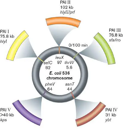
How can recurrent UTIs be managed? One way would be to prevent UPEC strains from
colonizing the intestine. A number of laboratories have found that d-mannose or a mannoside will
block type I pili on UPEC from binding to host cell mannose receptors and prevent UPEC strains
from colonizing the intestine and the bladder. The idea, still not tested, is that simultaneously
treating a patient with antibiotics to clear the UTI, along with the mannoside to prevent UPEC from
colonizing the intestine or recolonizing the bladder, will reduce the number of recurrent UTIs.
Thought Question
26.7 Urine samples collected from six hospital patients were placed on a table at the nurses’
station awaiting pickup from the microbiology lab. Several hours later, a courier retrieved the
samples and transported them to the lab. The next day, the lab reported that four of the six
patients had UTIs. Would you consider these results reliable? Would you start treatment based on
these results?
What makes uropathogenic E. coli different from other strains of E. coli? This is a question still
under investigation, but genomic analysis has exposed five pathogenicity islands unique to these
strains (Fig. 26.24 ). The functions of these pathogenicity islands are still under investigation.
Sexually Transmitted Infections
Sexually transmitted infections (STIs) are defined as infections transmitted primarily through
sexual contact. The organisms or viruses involved are generally very susceptible to drying and
require direct physical contact with mucous membranes for transmission. Because sex can take
many forms in addition to intercourse, these microbes can initiate disease in the urogenital tract,
rectum, or oral cavities. Condoms can prevent transmission but do so, of course, only when used
properly. Examples of common sexually transmitted infections are listed in Table 26.5.
TABLE Common Sexually Transmitted Infections 26.5
Disease Symptoms Etiological Virulence Treatment Reported
agent a factors cases
Gonorrhea Purulent Neisseria Type IV pili, Ceftriaxone plus 468,514 b
discharge, gonorrhoeae phase variation azithromycin
burning (G−)
urination;
can lead to
sterility
TABLE Common Sexually Transmitted Infections 26.5
Syphilis 1°: chancre; Treponema Motility Penicillin 88,042 b
2°: joint pallidum
pain, rash; (spirochete)
3°:
gummata,
aneurism,
central
nervous
system
damage
Nongonococcal Watery or Chlamydia Intracellular Azithromycin 1.6 million
urethritis mucoid trachomatis growth; b
urethral (Gram- prevents
discharge, negative–like) phagolysosome
burning fusion
urination
Trichomoniasis Vaginal Trichomonas Cytotoxin Metronidazole 2.3 million
itching, vaginalis c
painful (protozoan)
urination,
strawberry
cervix
Chancroid Painful Haemophilus? Erythromycin 7 b
genital ducreyi (G−)
lesion
HIV diagnoses For AIDS, HIV gp120, Rev, Azidothymidine 34,775 b
ranging from fever, Nef, and Tat (AZT), protease
asymptomatic diarrhea, proteins inhibitors,
to AIDS cough, night zidovudine
sweats,
fatigue,
opportunistic
infections
TABLE Common Sexually Transmitted Infections 26.5
Genital herpes Painful ulcer Herpes Cell fusion Acyclovir, 50 million
on external simplex 2 protein, iododeoxyuridine c
genitals, complement-
painful binding
urination protein,
latency
Genital warts Warts on Human E6, E7 proteins Vaccine now 50 million
external papillomavirus available c
genitals
Note: Because not all infections progress to diseases, the term “sexually transmitted infection ”
(STI) is preferred over the older phrase “sexually transmitted disease” (STD).
An important part of limiting the spread of any STI is to identify and treat all sexual partners
the patient has had in the previous 60 days. Either the patient (preferably) or the health care
provider can notify the affected people. In some instances, patients can be supplied with
medication to give directly to their partners. STIs are also reportable to state and federal health
agencies, such as the Centers for Disease Control and Prevention.
Case History: Secondary Syphilis
An 18-year-old pregnant woman came to the county urgent-care clinic with a low-grade fever,
malaise, and headache. She was sent home with a diagnosis of influenza. She again sought
treatment 7 days later, after she discovered a macular rash (flat, red) developing on her trunk,
arms, palms of her hands, and soles of her feet. When asked, the patient revealed that 1 year
earlier, she had had a painless ulcer on her vagina that healed spontaneously. She was diagnosed
with secondary syphilis—a diagnosis confirmed by a serological test. She was given a single
intramuscular injection of penicillin and told that her sexual partners had to be treated as well.
The vaginal ulcer, the long latent period, and the secondary development of a rash on the hands
and feet described in the case history are classic symptoms of syphilis. Christopher Columbus
and/or his crew are thought to have inadvertently delivered the treponeme that causes syphilis
from the Americas to Europe in the fifteenth century, but syphilis as a disease was not recognized
until the sixteenth century. The infectious agent, a spirochete named Treponema pallidum, was
finally discovered in 1905 (Fig. 26.25A ). (Chapter 18 describes spirochete structure.) But
another 113 years would pass before this anaerobe would be cultured in vitro. Steven Norris
(University of Texas Health Science Center at Houston) finally achieved long-term logarithmic
growth of T. pallidum in 2018, by periodically subculturing (6–7 days) and feeding the treponeme
in a microaerobic rabbit epithelial cell coincubation system.
FIGURE 26.25 ■ Syphilis. A. Treponema pallidum (dark-field microscopy). Organisms are
10–25 μm long. B. Chancre of primary syphilis. C. Rash of secondary syphilis.
CDC SUSAN LINDSAY
CDC/M. REIN, VD
CDC/M. REIN, VD
The disease syphilis has several stages. The incubation stage can last from 2 to 6 weeks after
transmission, during which time the organism multiplies and spreads throughout the body. Primary
syphilis is defined by an inflammatory reaction at the site of infection called a chancre (Fig.
26.25B ). About a centimeter in diameter, the chancre is painless and hard, and it contains
spirochetes. Patients are often too embarrassed to seek medical attention and, because it is
painless, hope it will just go away. It does go away after several weeks and without scarring. The
disease has now entered the primary latent stage. Over the next 5 years, symptoms may be
absent, but at any time, as described in the case history, the infected person can develop the rash
typical of secondary syphilis (Fig. 26.25C ).
The rash of secondary syphilis can be similar to rashes produced by many different diseases,
which contributes to syphilis’s nickname as the “great imitator.” In this stage the patient remains
contagious. The symptoms eventually resolve, and the patient reenters a latent phase of syphilis.
Some patients eventually progress over years to tertiary syphilis and develop many cardiovascular
and neurological symptoms. Neurological symptoms resulting from syphilis at any stage of the
disease are referred to as neurosyphilis. The patient can develop dementia and eventually die
from the disease.
The presence of T. pallidum in tissues can be detected with fluorescent antibody, but the initial
screen is usually serological (that is, patient serum is tested for antibodies). Antibiotics (penicillin
or doxycycline) are useful for eradicating the organism, but there is no vaccine, and cure does not
confer immunity.
The disease is particularly dangerous in pregnant women. The treponeme can cross the
placental barrier and infect the fetus to cause congenital syphilis. At birth, infected newborns will
have notched teeth (visible on X-rays), perforated palates, and other congenital defects. Women
should be screened for syphilis as part of their prenatal testing to prevent these congenital
infections. Nevertheless, cases of congenital syphilis have increased nearly threefold since 2015.
Crossing the placental barrier. Several viral and bacterial pathogens can, like Treponema
pallidum, cross the placental barrier. Known as the TORCH complex, these pathogens include T
oxoplasma, r ubella virus (German measles), c ytomegalovirus (CMV), and h erpes simplex 2. The
“O” stands for “others,” such as T. pallidum, Listeria monocytogenes, HIV, and varicella-zoster.
Bacteria that cross the barrier do so in one of two ways. They may move from the mother’s blood
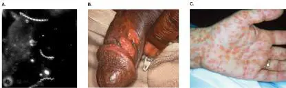
across the placental villus using intracellular cell-to-cell spread to reach and breach fetal
capillaries. Alternatively, the pathogen can initiate an acute inflammatory response that disrupts
the integrity of the placental barrier. In contrast, viral pathogens generally use white blood cells
(macrophages or lymphocytes) as cellular “Uber drivers” to carry them across the placenta.
The Tuskegee experiment. Unfortunately, much of what we know about untreated syphilis is
the result of the infamous Tuskegee experiment entitled “Untreated Syphilis in the Negro Male”
conducted in Alabama in the 1930s. Through dubious means and deception, a group of Black men
was enlisted in a study that promised treatment but whose real purpose was to observe how the
disease progressed without treatment. Today, such experiments are barred, thanks to strict
oversight by institutional review boards (IRBs) that require human subjects to sign informed
consent forms. A case study on the Tuskegee experiment can be found on the Internet at the
National Science Teaching Association website (search the site for “Bad Blood”).
Chlamydial Infections Are Often Silent
Chlamydia is the most frequently reported sexually transmitted infectious disease in the United
States (1.8 million cases in 2019), according to the Centers for Disease Control and Prevention, but
many people are unaware that they are infected. Three-fourths of infected women, for instance,
have no symptoms.
The chlamydias are unusual Gram-negative organisms with a unique developmental cycle. (
Chapter 18 describes chlamydial morphology.) They are obligate intracellular pathogens that start
as a small, nonreplicating, infectious elementary body that enters target eukaryotic cells. Once
inside vacuoles, they begin to enlarge into replicating reticulate bodies (Fig. 26.26 ). As the
vacuole fills, the reticulate bodies divide to become new nonreplicating elementary bodies.
Chlamydia trachomatis causes a sexually transmitted infection called chlamydia but also an eye
disease called trachoma, a major worldwide cause of blindness. Chlamydia (briefly Chlamydophila
) pneumoniae and Chlamydia psittaci are chlamydial species that cause pneumonia but not STIs.
FIGURE 26.26 ■ Replication cycle of Chlamydia. Inset: EM of a C. trachomatis –
containing vacuole in an infected cell, showing a reticulate body, infectious elementary
bodies, and intermediate forms.
DR. FRED HOSSLER/VISUALS UNLIMITED, INC.
People most at risk of developing genitourinary tract infections with chlamydia are young,
sexually active men and women; anybody who has recently changed sexual partners; and anybody
who has recently had another sexually transmitted infection. The astute clinician knows that when
one STI is discovered, others may also be present.
Left untreated, chlamydia can cause serious health problems. In female patients, the organism
can produce pelvic inflammatory disease, a damaging infection of the uterus and fallopian tubes
that can be caused by several different microbial species. The damage produced can lead to
infertility, tubal pregnancies, and chronic pelvic pain. Male patients left untreated can suffer
urethral and testicular infections and a serious form of arthritis.
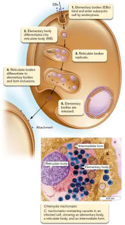
Case History: Gonorrhea
A 22-year-old mechanic saw his family doctor for treatment of painful urination and urethral
discharge. The patient was sexually active, with three regular and several “one time–good time”
partners. Physical examination was unremarkable except for prevalent urethral discharge. The
discharge was Gram-stained and sent for culture. The Gram stain revealed many pus cells, some of
which contained numerous phagocytosed Gram-negative diplococci (Fig. 26.27A ). Blood was
drawn for syphilis serology, which proved negative. The patient was given a single intramuscular
injection of ceftriaxone, and oral doxycycline was prescribed for 7 days. The bacteriology lab was
able to recover the bacteria seen in the Gram-stained smear of the urethral discharge. The
organism produced characteristic colonies on chocolate agar (agar plates containing heat-lysed red
blood cells that turn the medium chocolate brown; Fig. 26.27B ). The case was subsequently
reported to the state public health department. When the patient came back for his return visit, his
symptoms had resolved, and a repeat culture was negative.
The disease here is classic gonorrhea caused by Neisseria gonorrhoeae. A characteristic that
distinguishes Neisseria infections from Chlamydia infections is that bacterial cells are
microscopically visible in gonorrheal discharges but not in chlamydial discharges, even though they
are there (too small to see). Gonorrhea has been a problem for centuries and remains epidemic in
this country today (616,392 cases in 2019). Symptoms generally occur 2–7 days after infection, but
they can take as long as 30 days to develop. Most infected men (85%–90%) exhibit symptoms
that can include painful urination, yellowish white discharge from the penis, and in some cases,
swelling of the testicles and penis. The Greek physician Galen (CE 129–ca. 199) originally mistook
the discharge for semen. This mistake led to the name gonorrhea, which means “flow of seed.”
In contrast to men, most infected women (80%) do not exhibit symptoms and constitute the
major reservoir of the organism. If they are asymptomatic, they have no reason to seek treatment
and can unknowingly spread the disease. When symptoms are present, they are usually mild. Most
symptomatic women will experience a painful burning sensation when urinating and will notice
vaginal discharge that is yellow or occasionally bloody. They may also complain of cramps or pain
in the lower abdomen, sometimes with fever or nausea. As the infection spreads throughout the
reproductive organs (uterus and fallopian tubes), pelvic inflammatory disease occurs (see earlier
discussion of chlamydia). There is no serological test or vaccine for gonorrhea because the
organism frequently changes the structure of its surface antigens in a type of phase variation (see
Section 10.5).
Although N. gonorrhoeae is generally serum sensitive, owing to its sensitivity to complement
(see Section 23.6), certain serum-resistant strains can make their way to the bloodstream and
carry infection throughout the body. As a result, both sexes can develop purulent arthritis (joint
fluid containing pus), endocarditis, or meningitis. An infected mother can also infect her newborn
during parturition (birth), leading to a serious eye infection called ophthalmia neonatorum.
Because of this risk and because most infected women are asymptomatic, all newborns receive
antimicrobial eyedrops at birth.
Because adults engage in a variety of sexual practices, N. gonorrhoeae can also infect the anus
or the pharynx, where it can develop into a mild sore throat. These infections generally remain
unrecognized until a sex partner presents with a more typical form of genitourinary gonorrhea.
Because no lasting immunity is built up, reinfection with N. gonorrhoeae is possible. Reinfection
occurs, in part, because of phase variation in various surface antigens and because the organism
can apparently bind to CD4 + T cells, inhibiting their activation and proliferation to become
memory T cells (Fig. 26.27C ).
FIGURE 26.27 ■ Neisseria gonorrhoeae. A. Within pus-filled exudates, the Gram-
negative diplococci are found intracellularly inside PMNs. The intracellular bacteria in this case
are no longer viable, having been killed by the antimicrobial mechanisms of the white cell. B.
Colonies of N. gonorrhoeae growing on chocolate agar. C. N. gonorrhoeae binding to CD4 + T
cells, inhibiting T-cell activation and proliferation, which may explain the ease of reinfection
(colorized SEM).
DR. A. M. SIEGELMAN/VISUALS UNLIMITED
COURTESY OF JOHN W. FOSTER
NAT. IMMUNOL. 2002. 3 (3), COVER. PHOTO COURTESY OF DR. IAN C. BOULTON AND DR. GRAY-OWEN.
Over the decades, N. gonorrhoeae has incrementally developed resistance to many antibiotics
used in its treatment, but there has always been a new, effective drug ready to take the place of
the old drug. Soon, this may no longer be the case. To prevent treatment failures, the CDC
recommends dual antibiotic therapy that includes an intramuscular injection of ceftriaxone and oral
azithromycin or tetracycline. However, the incidence of ceftriaxone-resistant strains of N.
gonorrhoeae overseas is increasing. The alarm has been raised that we must develop new
antibiotics if we are to prevent an uncontrollable explosion of cases of this already epidemic
disease.
Thought Question
26.8 Aside from the CDC guidelines for treating gonorrhea, why else do you suppose the patient
in this case was treated with doxycycline (a derivative of tetracycline)?
HIV Causes AIDS, a Sexually Transmitted and Blood-Borne Disease
Though HIV (human immunodeficiency virus) is believed to have originated around the year 1900,
it was not discovered until 1981, when the virus caused the greatest pandemic of the late
twentieth century. HIV remains a serious problem today, especially in Africa, which is home to
nearly two-thirds of the people living with HIV worldwide. HIV has claimed the lives of almost 2
million people per year worldwide (about 14,000 per year in the United States). Women make up
about 19% of new AIDS cases per year in the United States, the majority resulting from
heterosexual sex. The molecular biology and virulence of HIV are discussed in Chapters 11 and 25
. This section focuses on the disease stages of HIV leading up to acquired immunodeficiency
syndrome (AIDS), the end-stage HIV disease.
HIV, a lentivirus in the retroviral family, is a prominent example of viruses that can be
transmitted either sexually (vaginally, orally, anally—homosexually or heterosexually) or through
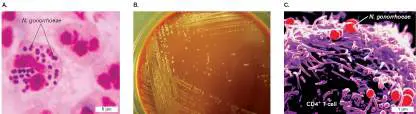
direct contact with body fluids, such as occurs with blood transfusion or the sharing of hypodermic
needles by intravenous drug users. HIV is not transmitted by kissing, tears, or mosquito bites. It
can, however, be transferred vertically from mother to fetus through the placenta (transplacental
transfer). Although the virus disseminates systemically throughout the host, we discuss HIV
infections in this section because sexual contact remains the major route of transmission.
Figure 26.28A shows the worldwide decrease in the number of newly infected HIV patients
and deaths due to AIDS. The number of people living with HIV has increased, however, because of
the development of more effective antiviral treatments.
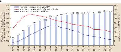
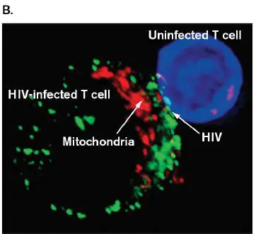
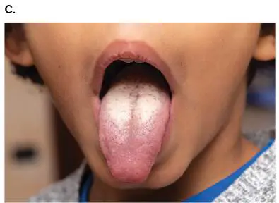
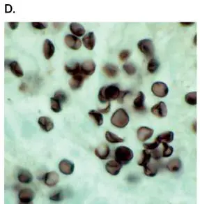
FIGURE 26.28 ■ Acquired immunodeficiency syndrome. A. HIV infections and AIDS
deaths worldwide. The number of people living with HIV continues to increase, yet the
number of people newly infected with HIV and the number of deaths due to AIDS have
decreased. B. HIV (green) can directly transfer from an infected to an uninfected T cell by
virological synapse. C. Oral candidiasis (thrush). The white patches are caused by secondary
infection by the yeast Candida albicans. D. Pneumocystis jirovecii infection of the lung. Note
the cuplike appearance of the fungus, almost like crushed Ping-Pong balls. Organisms range
from 2 to 6 μm in diameter. E. Kaposi’s sarcoma (oval spots). Source: Part A based on data
from the World Health Organization.
COVER. IMAGE COURTESY OF E. GROPPELLI ET AL. J. VIROL. 2015. 89 : DOI: 10.1128/JVI.02425–14
SRUILK/SHUTTERSTOCK
DR. F. C. SKVARA/VISUALS UNLIMITED
SPL/SCIENCE SOURCE
Once HIV enters the bloodstream, it infects CD4 + T cells and macrophages (which also have
CD4 on their surface). The virus replicates very rapidly, producing a billion particles per day, and it
can spread directly from cell to cell via virological synapses (Fig. 26.28B ). When infected and
uninfected T cells make contact, HIV virions and mitochondria (to supply energy) line up at the
contact interface where transmission occurs. Viral replication starts to kill the CD4 + T cells, which
progressively decrease in number.
HIV infection has four stages:
1. A primary stage beginning with seroconversion (finding antibodies to the virus)
2. Clinical latency (slow steady loss of T cells without symptoms or sometimes swollen lymph
nodes)
3. Early symptomatic disease (formerly called AIDS-related complex)
4. Acquired immunodeficiency syndrome (AIDS)
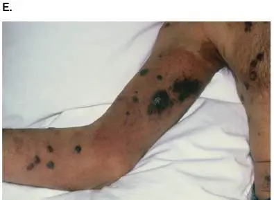
Symptoms in the early symptomatic stage can include fever, headache, macular rash, and
weight loss. The symptoms can manifest within a few months of infection, resolve within a few
weeks, and then recur. As T-cell numbers decline, the debilitated immune system leaves the victim
susceptible to secondary infections such as thrush (Fig. 26.28C ), caused by the yeast Candida
albicans (candidiasis) and related species.
Monitoring an HIV patient involves assaying blood for viral load using quantitative PCR to
detect HIV-specific genes, detecting anti-HIV antibodies, and determining the CD4 + T-cell count.
Remember, a person who is HIV-positive does not necessarily have AIDS, which may take years to
develop. The CDC defines a patient with AIDS as someone who is HIV-positive with a CD4 + cell
count of less than 200 cells per microliter or who has contracted an AIDS indicator disease. For
example, once the CD4 + T-cell population falls below 500 cells per microliter, opportunistic
infections start to arise. Opportunistic infections include pneumonia by Mycobacterium avium-
intracellulare or Pneumocystis jirovecii (Fig. 26.28D ), cryptococcal meningitis, Histoplasma
capsulatum infection, and tuberculosis. The presence of these diseases indicates that the patient
has AIDS, regardless of the CD4 + count.
Various cancers are also AIDS indicator diseases. AIDS patients are more susceptible to cancers
because their depressed immune systems cannot detect and destroy cancer cells generated by
secondary agents. Kaposi’s sarcoma (Fig. 26.28E ), for instance, is a common cancer seen in
AIDS patients that is caused by human herpesvirus type 8 (HHV8). Kaposi’s sarcoma originates in
endothelial or lymphatic cells, but the resulting tumors can develop anywhere—for example,
gastrointestinal tract, mouth, lungs, skin, or brain.
More than four decades after the HIV pandemic began, a vaccine for HIV may finally be close at
hand. Scientists at the National Institutes of Health (NIH) collaborating with Moderna, one of the
first companies to develop an mRNA vaccine for COVID-19, have devised a similar vaccine for HIV
that appears to be working. The HIV vaccine carries mRNA encoding two key proteins: Env from
HIV and Gag from the closely related simian immunodeficiency virus (SIV). Env protein is normally
cleaved by a host protease (furin) into gp120 and gp41, two proteins critical for HIV entry. Gp120
binds to CD4 and the coreceptor CXCR4. The gp41 protein then engineers fusion between the viral
and host cell membranes (described in Fig. 11.26). The end result is infection.
SIV Gag mRNA was included in the vaccine because Gag induces muscle cells of the test
animals, macaque monkeys, to produce and release virus-like particles (VLPs). The membrane
surfaces of these VLPs incorporate multiple Env glycoproteins when Env is coexpressed in the same
cell. The Env-Gag VLPs cannot cause disease because they lack the genes to do so, but they can
stimulate suitable immune responses. Importantly, the Env gene selected for the vaccine presents
epitopes that are immutable—that is, altering any of them in a natural HIV virus would destroy
infectivity. Thus, immune responses to these epitopes should prove protective against many
variants of HIV.
Even though a vaccine to prevent AIDS is not yet available, progression of the disease can be
controlled by antiretroviral drugs that inhibit several critical features of HIV biology: entry,
integration, reverse transcription, and the proteolytic processing of viral polyproteins. These
treatment regimens (antiretroviral therapy, or ART; see Chapter 27) have made HIV an
increasingly survivable infection. Consider this: In the past, nearly all HIV-infected individuals
eventually became ill and died from AIDS-related diseases, but in the United States at least half of,
if not most, HIV-positive people now die from diseases unrelated to HIV (for example, heart
attack). Because HIV infection is now considered a treatable disease, the CDC recommends
routine screening for HIV.
Can HIV infection be cured? Well, sort of. Only two people have been effectively “cured” of HIV,
by putting the virus in sustained remission. The first was Timothy Ray Brown, who was diagnosed
in 2006 with leukemia and HIV. In an attempt to eradicate both diseases, doctors destroyed
Brown’s white cells with chemotherapy and transplanted bone marrow from a donor who lacked
CCR5, which is the coreceptor needed for HIV to enter T cells. The transplant, performed in 2008,
successfully repopulated Brown’s blood with HIV-resistant T cells. Mr. Brown has not needed
antiretroviral therapy for more than 12 years, even though traces of the HIV genome remain.
Unfortunately, this approach repeatedly failed with other patients until 2016 when Adam Castillejo,
a patient from London with Hodgkin’s lymphoma, received a stem cell transplant from another
CCR5-deficient donor. Mr. Castillejo has been HIV-free for over five years without needing
antiretroviral therapy.
Because HIV DNA integrates into the genome of an infected cell, it has been impossible to
completely cure anyone of the infection. However, a study by Kamel Khalili proved that the
CRISPR-Cas9 system described in Chapter 12 can be engineered to “surgically” remove proviral
HIV DNA from infected T cells. In 2021, the U.S. Food and Drug Administration gave the startup
company Excision BioTherapeutics permission to use an adeno-associated virus and CRISPR
technology to excise HIV DNA from infected cells.
Thought Question
26.9 Like the cause of plague, HIV is a blood-borne pathogen. Why, then, do you think fleas and
mosquitoes fail to transmit HIV?
The Protozoan Trichomonas vaginalis Produces a Common Vaginal Infection
Trichomonas vaginalis is a flagellated protozoan (Fig. 26.29 ) that causes an unpleasant,
sexually transmitted vaginal disease called trichomoniasis. Approximately 2–3 million infections
occur each year in the United States. Both men and women can be infected; however, men are
usually asymptomatic. Even among infected women, 25%–50% are considered asymptomatic
carriers.
FIGURE 26.29 ■ Trichomonas vaginalis. T. vaginalis is a protozoan that causes a
common sexually transmitted infection (SEM).
DAVID M. PHILLIPS/VISUALS UNLIMITED
There is no cyst in the life cycle of T. vaginalis, so transmission is via the trophozoite stage
only (the form of a protozoan in the feeding stage). A female patient with trichomoniasis may
complain of vaginal itching and/or burning and a musty vaginal odor. An abnormal vaginal
discharge also may be present. A male patient may complain of painful urination (dysuria),
urethral or testicular pain, and lower abdominal pain.
Owing to colonization by lactobacilli (which produce large amounts of lactic acid), the normal,
healthy vagina usually, but not always, has a pH of less than 4.5. However, since T. vaginalis feeds
on bacteria, the pH of the vagina rises as the numbers of lactobacilli decrease. Definitive diagnosis
requires microscopically identifying the flagellated protozoan in vaginal secretions. PMNs, which
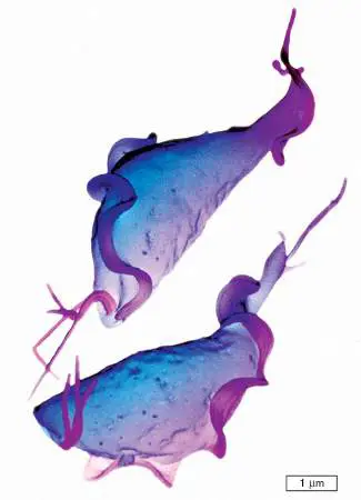
are the primary host defense against the organism, are also usually present. Like giardiasis, this
disease is treated with metronidazole.
To Summarize
Urinary tract infections (UTIs) include cystitis (bladder) and pyelonephritis (kidney). E.
coli is the most common cause of UTIs.
Cystitis can develop from bacteria ascending up the urethra (most common route) or
descending down a ureter from an infected kidney.
Pyelonephritis can develop from bacteria ascending along a ureter from an infected
bladder (most common route) or from bacteria in the bloodstream disseminating from an
infection elsewhere in the body.
Syphilis, chlamydia, and gonorrhea are the most common sexually transmitted
infections.
A patient with one STI often has another STI too.
Complement prevents bloodstream dissemination of Neisseria gonorrhoeae, which
lacks a carbohydrate capsule. Because the organism frequently changes the structure of its
surface antigens, no vaccine is available for N. gonorrhoeae.
HIV depletion of CD4 1 T cells results in lethal secondary infections and cancers.
Trichomonas vaginalis is a flagellated protozoan that causes a sexually transmitted
vaginal infection. The reservoirs for this organism are the male urethra and female vagina.
Glossary
cystitis
Bladder infection.
pyelonephritis
Kidney infection.
primary syphilis
The initial inflammatory reaction (chancre) at the site of infection by Treponema pallidum.
chancre
A painless, hard lesion due to an inflammatory reaction at the site of infection by Treponema
pallidum, the causative agent of syphilis.
secondary syphilis
A rash that may appear at some point after the primary latent stage of syphilis.
tertiary syphilis
A final stage of syphilis, manifested by cardiovascular and nervous system symptoms.
congenital syphilis
Syphilis contracted in utero.
chlamydia
1. A pathogenic bacterium lacking a cell wall; chlamydias (or chlamydiae) grow within human
or animal cells, transmitting as elementary bodies to other cells. 2. A disease caused by
chlamydia cells. The most frequently reported sexually transmitted disease in the United
States. Symptoms range from none, to a burning sensation upon urination, to sterility.
Fig. 11.26
FIGURE 11.26 ■ HIV-1 attachment to host cell. The SU (gp120) subunits of the
spike protein complex attach to the receptor (CD4 cell-surface protein) and to CCR5. The
fusion peptides contract, pulling the membranes together.
Endnotes
1. Note a: G+ = Gram-positive; G− = Gram-negative. Return to reference a
2. Note b: CDC reported cases for 2016. Return to reference b
3. Note b: CDC reported cases for 2016. Return to reference b
4. Note b: CDC reported cases for 2016. Return to reference b
5. Note b: CDC reported cases for 2016. Return to reference b
6. Note b: CDC reported cases for 2016. Return to reference b
7. Note c: Total estimated current cases. Nonreportable disease. Return to reference c
8. Note c: Total estimated current cases. Nonreportable disease. Return to reference c
9. Note c: Total estimated current cases. Nonreportable disease. Return to reference c
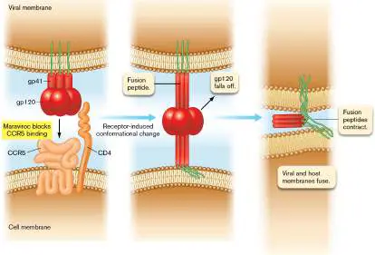
26.5 Cardiovascular and Systemic Infectionsnot assigned
The cardiovascular system, which includes the heart, arteries, veins, and capillaries, delivers oxygen,
nutrients, and immune system components to all tissues of the body. There are pathogens that can
directly infect the heart or the endothelial cells lining blood vessels. However, the blood itself can also
become infected or serve as a mass transit system to spread pathogens throughout the body.
Pathogens disseminated in this way can cause infections in many different organ systems. We examine
both types of infections in this section.
Infections of the cardiovascular system include septicemia, endocarditis (inflammation of the
heart’s inner lining (Fig. 26.30 ), pericarditis (inflammation of the heart’s outer lining), myocarditis
(inflammation of heart muscle), and, possibly, atherosclerosis (deposition of fatty substances along the
inner lining of arteries). These are all life-threatening diseases.
FIGURE 26.30 ■ View of bacterial endocarditis. Close-up of mitral valve endocarditis,
showing bacterial vegetation.
DR. E. WALKER/SCIENCE SOURCE
Septicemia is, by strict definition, the presence of bacteria or viruses in the blood. The presence of
viruses is a condition more specifically called viremia, while bacteria in the circulation is more
specifically called bacteremia. In practice, however, the terms “septicemia” and “bacteremia” are often
used interchangeably. Septicemia can develop from a local tissue infection situated anywhere in the
body. Once in the bloodstream, bacteria can travel to other organs and cause infections there. To
prevent septicemia, regional lymph nodes waiting to receive organisms from the initial infection site
recruit massive numbers of neutrophils to ambush the bacteria when they arrive (Special Topic 26).
Should some of the organisms escape the lymph node, blood factors such as complement can
nonspecifically kill many types of bacteria that enter the blood. Despite these failsafe mechanisms,
Gram-positives, Gram-negatives, aerobes, and anaerobes can all produce septicemia under the right
conditions.
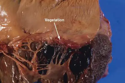
Endocarditis can be either viral or bacterial in origin. It can be a consequence of many bacterial
diseases, such as brucellosis, gonorrhea, psittacosis, staphylococcal and streptococcal infections,
candidiasis, and Q fever (Coxiella). Among the many viral causes are coxsackievirus, echovirus,
Epstein-Barr virus, and HIV. Bacterial infections of the heart are always serious. Viral infections,
although common, are rarely life-threatening in healthy individuals and are usually asymptomatic.
Case History: Bacterial Endocarditis
Elizabeth was 38 years old and had a history of mitral valve prolapse (a common congenital condition
in which a heart valve does not close properly). She was also on immunosuppressive therapy following
a kidney transplant. Recently, Elizabeth was admitted to the hospital complaining of fatigue,
intermittent fevers for 5 weeks, and headaches for 3 weeks—symptoms the physician recognized as
possible indications of endocarditis. Elizabeth reported having had a dental procedure a few weeks
prior to the onset of symptoms. A sample of her blood was cultured in a liquid bacteriological medium.
The culture grew Gram-positive cocci, which turned out to be Streptococcus mutans, a member of the
viridans streptococci. With the finding of bacteria in the bloodstream, the diagnosis of bacterial
endocarditis was confirmed. Elizabeth began a 1-month course of intravenous penicillin G and
gentamicin therapy and eventually recovered to normal health.
Endocarditis (inflammation of the inner lining of the heart) is traditionally classified as acute or
subacute, depending on the pathogenic organism involved and the speed of clinical presentation.
Subacute bacterial endocarditis (SBE) has a slow onset with vague symptoms. It is usually caused by
bacterial infection of a heart valve (Fig. 26.30 ). SBE infections are usually (but not always) caused
by a viridans streptococcus from the oral microbiota (for example, Streptococcus mutans, a common
cause of dental caries). “Viridans streptococci” is a general term used for streptococci whose colonies
produce green alpha hemolysis on blood agar (“viridans” is from the Greek viridis, “green”). Many
patients who develop infective endocarditis have mitral valve prolapse (20%) or mitral valve damage
from rheumatic fever (30%). Also at high risk are intravenous drug abusers or patients who develop
hospital-acquired (nosocomial) infections.
Subacute bacterial endocarditis can begin at the dentist’s office, as it did in the case history
presented here, although it very rarely does (our patient was also immunosuppressed). Following a
dental procedure (such as tooth restoration) or even while brushing your teeth, oral bacteria can
transiently enter the bloodstream and circulate. Streptococcus mutans, a member of the normal oral
microbiome, can become lodged onto damaged heart valves, grow as a biofilm, and secrete a thick
glycocalyx coating that encases the microbes and forms a vegetation on the valve, damaging it further.
If untreated, the condition can be fatal within 6 weeks to a year.
A rapidly progressive (acute) and highly destructive infection can develop when more virulent
organisms, such as Staphylococcus aureus, gain access to cardiac tissue. Symptoms of acute
endocarditis include fever, pronounced valvular regurgitation (backflow of blood through the valve),
and abscess formation.
Most patients with subacute bacterial endocarditis present with a low-grade fever that lasts several
weeks. They also complain of nonspecific symptoms, such as cough, shortness of breath, joint pain,
diarrhea, and abdominal or flank pain. Endocarditis is suspected in any patient who has a heart
murmur and an unexplained fever for at least a week. It should also be considered in an intravenous
drug abuser with a fever, even in the absence of a murmur. In either case, definitive diagnosis requires
blood cultures that grow bacteria. Blood cultures involve taking samples of a patient’s blood from two
different locations (such as two different arms). Once collected, the blood is added to liquid culture
medium (two bottles per site) and incubated at 37°C. One bottle should be incubated aerobically, the
other anaerobically. Growth of the same organism in cultures taken from two body sites rules out
inadvertent contamination with skin microbes, which would likely yield growth in only one culture.
Curing endocarditis is difficult because the microbes are usually ensconced in a nearly impenetrable
glycocalyx. Consequently, eradicating microorganisms from the vegetations almost always requires
hospitalization, where high doses of intravenous antibiotic therapy can be administered and monitored.
Antibiotic therapy usually continues for at least a month, and in extreme cases, surgery may be
necessary to repair or replace the damaged heart valve.
Patients with heart valve prolapse should not shy away from the dentist, however. As long as a
healthy immune system is in place, the risk of infection is low. In fact, maintaining good oral hygiene
can keep the risk of developing infectious endocarditis very low. Immunocompromised patients, on the
other hand, are at increased risk and should be treated prophylactically with oral amoxicillin or
azithromycin 1 hour before a procedure to kill oral bacteria that enter the bloodstream. Note, too, that
prosthetic heart valves can develop endocarditis (usually caused by Staphylococcus aureus). The
treatment of prosthetic valve endocarditis often requires valve replacement.
Viruses such as adenovirus and some enteroviruses can also cause endocarditis, as well as a
condition known as myocarditis, an inflammation of the heart muscle.
Thought Question
26.10 A patient presenting with high fever and in an extremely weakened state is suspected of
having septicemia. Two sets of blood cultures are taken from different arms. One bottle from each set
grows Staphylococcus aureus, yet the laboratory report states that the results are inconclusive. New
blood cultures are ordered. What would make these results inconclusive?
Malarial Parasites Target Red Blood Cells
Malaria is the most devastating infectious disease known. Each year, 300–500 million people develop
malaria worldwide, and 1–3 million of these people, mostly children, die. In the United States only
about a thousand cases occur annually, and almost all are acquired as a result of international travel to
endemic areas (Fig. 26.31A ). Once again, this correlation illustrates the diagnostic value of knowing
a patient’s travel history.
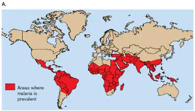
FIGURE 26.31 ■ Malaria is a major disease worldwide. A. Endemic areas of the world
where malaria is prevalent. B. Plasmodium falciparum schizont after completion of division
(colorized TEM). A residual body of the organism (yellow-green) is left over after division. The
erythrocyte has lysed and only a ghost cell remains; no cytoplasm is seen surrounding the
merozoites just being released. Free merozoites are seen outside the membrane.
DENNIS KUNKEL MICROSCOPY/SCIENCE SOURCE
SPECIAL TOPIC 26 How Neutrophils Ambush Staphylococcus aureus in a Lymph Node
Every day, bacteria breach the human skin barrier through cuts, abrasions, insect bites, and burns.
Yet in healthy people these pathogens rarely disseminate from the initial site of infection to the
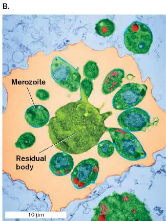
systemic circulation. This inability to spread seems odd, since bacteria always arrive at the site of
infection well before neutrophils do. Yet the bacteria rarely make it into the bloodstream to cause
sepsis or infect other sites. What stops them? Paul Kubes’s lab (University of Calgary), with
funding from the Canadian Institute of Health Research, found that macrophages, obvious
gatekeepers that reside at infection sites or in regional lymph nodes, can slow dissemination—but
not by much. Could there be an alternative intercept strategy?
One day, Ania Bogoslowski (Fig. ST 26.1 ), a novice graduate student in the Kubes
laboratory, infected a mouse’s footpad with Staphylococcus aureus and examined the lymph
nodes. She found that the organisms had traveled to the regional lymph nodes behind the knee
(popliteal lymph nodes, or popLNs) but no farther (Fig. ST 26.2A ). Why didn’t they break into
the circulation? Bogoslowski examined the nodes using in vivo microscopy (IVM) and found that,
within 5–6 hours of infection, neutrophils were streaming into the nodes, increasing in number
from 1,000 per node to over 250,000. IVM enables the observation of processes occurring in a live
animal. Figure ST 26.2B shows the increase in neutrophils after just 4 hours. The strategy
seems to be this: Get neutrophils to the lymph node quickly, and wait for lymph to bring the
microbes to them. This way the neutrophils can ambush, rather than chase, any pathogens
attempting to disseminate.
FIGURE ST 26.1 ■ Ania Bogoslowski (seated) and Paul Kubes study the innate
immune response to infection.
PHOTO BY CAITLYN MACQUEEN
FIGURE ST 26.2 ■ Neutrophil recruitment to popliteal lymph nodes (popLNs)
stops S. aureus dissemination. A. Bacterial dissemination from a footpad infection site.
Data represent counts of colony-forming units (CFUs) of Staphylococcus aureus in various
organ systems 24, 48, and 72 hours after initial infection (mean ± standard deviation; nd =
none detected). B. In vivo microscopy of neutrophils present in the lymph node before and 4
hours after footpad infection. Outline of node is marked with a dotted line. Steady-state
control shows neutrophils present in an uninfected node.
Source: Modified from Ania Bogoslowski et al. 2018. PNAS 115 :2449–2454, fig. 1A.
ANIA BOGOSLOWSKI ET AL. 2018. PNAS 115 :2449–2454, FIG. 1B.
Bogoslowski and colleagues showed that the neutrophils migrated into the nodes from the
bloodstream, passing through tiny balloon-like structures called high endothelial venules spaced
along capillaries situated within the node. But what called the neutrophils to the lymph node? It
wasn’t the bacteria; neutrophils swarmed to the node even when heat-killed bacteria were
injected into the footpad. Macrophages were not involved, since deleting them did not affect
neutrophil recruitment. The scientists then examined complement factor C5a, which can also act
as a chemoattractant for neutrophils (see Section 23.6). When they blocked C5a receptors
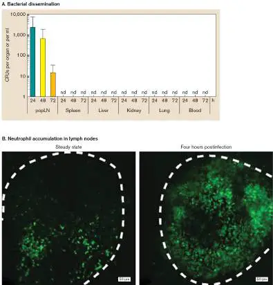
(C5aR) on neutrophils with anti-C5aR antibody, neutrophil recruitment to the lymph nodes
stopped (Fig. ST 26.3 ).
FIGURE ST 26.3 ■ Complement factor C5a recruits neutrophils to the regional
lymph node after infection. Mice were treated with phosphate-buffered saline (control) or
with blocking antibody to C5aR.
Source: Modified from Ania Bogoslowski et al. 2018. PNAS 115 :2449–2454, fig. 3E.
Bogoslowski proposes that a lot of C5a is formed at the site of infection (footpad). The edema
that forms in response to the infection causes fluid containing C5a to move from the footpad
through the lymphatic vessels to the regional lymph node. The complement factor arrives at the
node well before bacteria do and can form a chemotactic gradient that lures neutrophils into the
structure to await the arrival of bacteria. The strategy seems to hold also for other pathogens
known to cause neutrophil recruitment to regional lymph nodes (Pseudomonas aeruginosa and
Mycobacterium bovis, for instance).
In a follow-up study, the authors discovered that a subset of neutrophils, much like
lymphocytes, can recirculate through lymph nodes in a random hunt for pathogens. Those
patrolling neutrophils, combined with C5a recruitment, result in neutrophils swarming into a node
about to be inundated with bacteria.
RESEARCH QUESTION
How would you use flow cytometry to confirm the in vivo microscopy results? Search the Internet
for antigens you would use to screen for neutrophils and draw out (graphically) the results of the
flow cytometry experiment.
Bogoslowski, Ania, Eugene C. Butcher, and Paul Kubes. 2018. Neutrophils recruited through high endothelial
venules of the lymph nodes via PNAd intercept disseminating Staphylococcus aureus. Proceedings of the National
Academy of Sciences USA 115 :2449–2454.
Bogoslowski, Ania, Sathi Wijeyesinghe, Woo-Young Lee, Chien-Sin Chen, Samer Alanani, et al. 2020.
Neutrophils recirculate through lymph nodes to survey tissues for pathogens. The Journal of Immunology 204:2552–
2561. https://doi.org/10.4049/jimmunol.2000022.
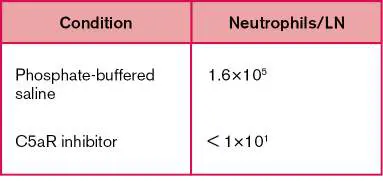
The disease is caused by four species of Plasmodium: P. falciparum (the most deadly), P. malariae
, P. vivax, and P. ovale. The life cycle of Plasmodium, discussed in detail in Chapter 20, is complex
and involves two cycles: an asexual erythrocytic cycle in the human and a sexual cycle in the mosquito
(see Fig. 20.39).
In the erythrocytic cycle, an infected female Anopheles mosquito bites the victim and injects a
small amount of saliva containing Plasmodium into the bloodstream. The haploid sporozoites go
directly to the liver and undergo asexual fission to produce merozoites. The merozoites attach to and
penetrate red blood cells, where Plasmodium consumes hemoglobin and enlarges into a trophozoite.
The protist nucleus divides so that the cell, now called a schizont, contains up to about 20 nuclei. The
schizont then divides to make the smaller, haploid merozoites (Fig. 26.31B ). The glutted red blood
cell eventually lyses, releasing merozoites that can infect new red blood cells (see Fig. 20.39).
Sudden, synchronized release of the merozoites and red blood cell debris triggers the telltale
symptoms of malaria: violent, shaking chills followed by high fever and sweating. The erythrocytic
cycle and, thus, the symptoms repeat every 48–72 hours. After several cycles, the patient goes into
remission lasting several weeks to months, after which there is a relapse.
Much of the research on malaria focuses on relapse. Why does the immune system fail to eliminate
the parasite after the first episode? When Plasmodium invades the red blood cells, it lines the blood
cells with the protein PfEMP1 that causes red blood cells (RBCs) to stick to blood vessels, removing the
parasite from circulation. But the protein cannot protect the parasite from patrolling macrophages,
which eventually detect the invader and recruit other immune cells to fight it. However, a fraction of
each generation of parasite expresses a different version of PfEMP1 that the immune system never
encountered. In its new disguise, Plasmodium can invade more red blood cells and cause another wave
of fever, headaches, nausea, and chills. The body now has to repeat its recognition and attack
responses all over again. The parasite has 60 of these cloaking genes, called var, that can be turned
on and off individually, changing the organisms’ antigenic structure, like a criminal repeatedly changing
disguises to elude police.
The var genes are regulated by chromosome packaging, which unwraps one gene for expression
while packing away inactive genes. DNA can be encased so securely that transcription proteins cannot
access the packed nucleic acid in a process known as epigenetic silencing. Epigenetic protein
modifications (acetylation or methylation) alter the affinity of histones toward different regions of the
plasmodial chromosomes. Becoming immune to all the types of malaria can take upwards of 5 years
and requires constant exposure; otherwise immunity is lost. Many children afflicted with malaria do not
live long enough to gain immunity to the disease in all its forms.
Diagnosis of malaria involves microscopic demonstration of the protist within erythrocytes (Wright
stain) or serology to identify antimalarial antibodies. Treatment regimens include artemisinin, whose
mechanism of action is unclear; chloroquine or mefloquine, which kills the organisms in their
erythrocytic asexual stages; and primaquine, effective in the exoerythrocytic stages by disrupting
Plasmodium mitochondria. These antimalarial drugs are also given prophylactically to persons traveling
to endemic areas. Which drug is given depends on the destination.
The chloroquine family of drugs prevents the detoxification of heme generated from hemoglobin
digestion. Malaria parasites accumulate hemoglobin released from red blood cells in plasmodial
lysosomes. Lysosomal enzymes digest the hemoglobin, and the parasite uses the amino acids to grow.
However, the heme released is toxic. The organism normally detoxifies heme via polymerization, which
produces a black pigment. Many antimalarial drugs, such as chloroquine, prevent polymerization by
binding to the heme. As a result, the increased iron level (from heme) kills the parasite. Unfortunately,
Plasmodium has been developing resistance to these drugs, forcing the development of new ones to
take their place.
As of 2021, a vaccine for malaria called RTS,S has finally been approved for use by the World
Health Organization. The vaccine is designed to defend against the first stages of the disease, when
sporozoites first enter the bloodstream before they invade the liver. RTS,S is a protein in which the
carboxyl terminus of the P. falciparum circumsporozoite protein (CS) is fused to the hepatitis B surface
antigen (HBsAg). Circumsporozoite protein is prevalent on the surface of sporozoites. When expressed,
the CS-HBsAg chimeric protein, mixed with free HBsAg, assembles into particulate structures; when
injected, these structures can induce anti-circumsporozoite antibodies. The antibodies bind to
sporozoites, after which serum complement attaches to the Fc regions of those antibodies, activates,
and kills the sporozoites. Although it is 50% effective against severe malaria during the first year, by
the fourth year the vaccine, unfortunately, has almost no effect.
While efforts continue toward making an effective, long-lasting vaccine, novel strategies to control
the spread of malaria are also being tested. Approaches include using anti-mosquito bacteria to
eradicate mosquito vectors, and a genetic approach, called gene drive, designed to eliminate female
mosquitoes. Female mosquitoes are targeted because only female mosquitoes take blood, which they
need to make eggs. In one gene drive approach, a CRISPR-Cas9 system would be introduced into the
mosquito Y chromosome. The guide RNAs of this system will direct the cleavage of sequences within
the X chromosome. As a result, sperm from these males should all contain the Y chromosome. No male
X chromosomes will be passed to eggs, so no female mosquitoes will be produced. Various gene drive
systems are currently being tested.
Bear in mind that we have described only selected organisms that cause cardiovascular infections;
there are many more (for example, Rickettsia typhi).
Note: Babesiosis is an emerging disease caused by a protozoan (Babesia microti) that, like
Plasmodium, infects red blood cells. B. microti is transmitted by the deer tick, the same insect that
transmits the agent for Lyme disease. Typically, babesiosis is a mild, flu-like disease, but in its severe
form it can present with symptoms similar to those of malaria.
Systemic Infections
Many pathogenic bacteria can produce septicemia as a way to disseminate throughout the body and
infect other organs. These organisms cause what are considered systemic infections.
Case History: The Plague
A 25-year-old New Mexico rancher was admitted to an El Paso hospital because of a 2-day history of
headache, chills, and fever (40°C; 104°F). The day before admission, he had begun vomiting. The day
of admission, an orange-sized, painful swelling in the right groin area was noted (Fig. 26.32A ). A
lymph node aspirate and a smear of peripheral blood were reported to contain Gram-negative rods
that exhibited bipolar staining (Fig. 26.32B ). The patient’s white blood cell count was 24,700 per
microliter (normal is 5,000–10,000), and his platelet count was 72,000 per microliter (normal is
130,000–400,000). In the 2 weeks prior to becoming ill, the patient had trapped, killed, and skinned
two prairie dogs, four coyotes, and one bobcat. The patient also mentioned that he had cut his left
hand shortly before skinning a prairie dog. The clinical laboratory isolated a Gram-negative rod from
blood cultures. PCR and biochemical testing identified the organism as Yersinia pestis, the bacterium
that causes plague. The severely ill rancher received an antibiotic cocktail of gentamicin and
tetracycline, and he eventually recovered after 6 weeks in intensive care.
FIGURE 26.32 ■ The plague. A. Classic bubo (swollen lymph node) of bubonic plague. B.
Yersinia pestis, bipolar staining (length 1–3 μm). C. Prairie dogs are often hosts to fleas that
carry plague bacilli. D. X-ray of pneumonic plague, showing pulmonary infection.
CDC
CDC
CRAIG K. LORENZ/SCIENCE SOURCE
CDC/SCIENCE SOURCE
Plague is caused by the bacterium Yersinia pestis, which can infect both humans and animals. During
the Middle Ages, the disease known as the Black Death decimated over a third of the population of
Europe. Such was the horror it evoked that invading armies would actually catapult dead plague
victims into embattled fortresses. This was probably the first reported case of biowarfare.
Y. pestis is present in the United States and is endemic in 17 western states. Typically, the bite of
infected fleas transmits the microbe between animals such as rats and even prairie dogs (Fig.
26.32C ). Figure 26.33 illustrates the various infection cycles of the plague bacillus. Humans are not
part of the natural infection cycle. In the absence of an animal host, however, the flea can take a blood
meal from humans and thereby transmit the disease to them. During the Middle Ages, urban rats
venturing back and forth to the countryside became infected by the fleas of wild rodents that served as
a reservoir. Upon returning to the city, the rat fleas passed the organism on to other rats, which then
died in droves. The rat fleas, deprived of their normal meal, were forced to feed on city dwellers,
passing the disease on to them.
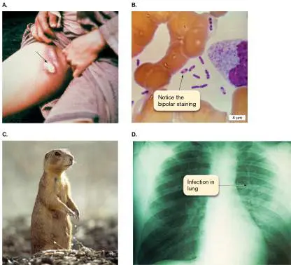
FIGURE 26.33 ■ The cycles of plague. The sylvatic cycle occurs in the wild, where fleas
transmit the organism between rodents. An accidental interaction with urban rats can trigger a
similar urban cycle. Humans can be infected through contact with infected fleas coming from
either cycle. Flea bite transmission initiates bubonic plague symptoms that can progress to
pneumonic plague. Because it is highly infectious, pneumonic plague can cause epidemic spread
of the disease.
Individuals bitten by an infected flea or accidentally infected through a cut while skinning an
infected animal will first exhibit the plague form known as bubonic plague. Bubonic plague develops
as the organism moves from the site of infection to the regional lymph nodes, producing
characteristically enlarged nodes called buboes (see Fig. 26.32A ). From the lymph nodes, the
pathogen can enter the bloodstream, causing septicemic plague. In this phase the patient can go into
shock (sudden drop in blood pressure) from the massive amount of endotoxin in the bloodstream. In
the case history, the rancher’s low platelet count (thrombocytopenia) was an indication that his
platelets were being consumed by the considerable clotting taking place. Neither bubonic nor
septicemic plague is passed from person to person. As the organism courses through the bloodstream,
however, it will invade the lungs and produce pneumonic plague (see Fig. 26.32D ), which can be
easily transmitted from person to person through aerosol droplets generated by coughing.
Pneumonic plague is the most dangerous form of the disease because it can kill quickly and spread
rapidly through a population. Pneumonic plague is so virulent that an untreated patient can die within
24–48 hours. The rapid spread of plague during the Middle Ages was most likely the result of person-
to-person transmission through respiratory aerosols.
Y. pestis has numerous virulence factors. For instance, YadA is a surface adhesin that binds
collagen. Another factor, the F1 protein capsule surface antigen, plays a part in blocking phagocytosis
in mammalian hosts. Certain biofilms formed by Y. pestis are also important. An extracellular matrix
synthesized by Y. pestis produces an adherent biofilm in the flea midgut that contributes to flea-to-
mammal transmission. The biofilm blocks the flea’s digestion, making the flea feel “starved” even after
a blood meal. Therefore, the flea jumps from host to host in a futile effort to feel full. As the flea tries
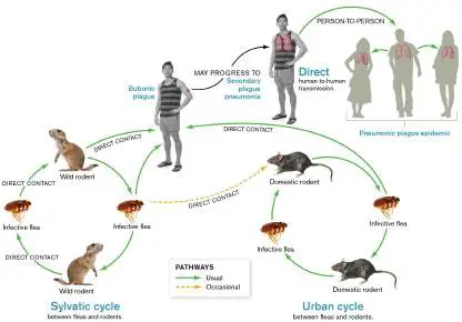
to take a blood meal, the blockage causes the insect to regurgitate bacteria into the wound. This
curious effect of Y. pestis on the insect vector is another unique aspect of how plague spreads so
quickly.
Y. pestis also uses type III secretion systems to inject virulence proteins (YopB and YopD) into host
cell membranes. Unlike Salmonella, which uses type III–secreted proteins to gain entrance into host
cells, Y. pestis is not primarily an intracellular pathogen, although it can survive in macrophages.
Injection of the Yop proteins disrupts the actin cytoskeleton, thereby helping the organism evade
phagocytosis. By evading phagocytosis, the organism avoids triggering an inflammatory response and
produces massive tissue colonization.
The last major outbreak of plague in Europe occurred in 1772. Why the disease disappeared from
Europe is unclear, but part of the reason was probably human intervention. Long before doctors
understood how germs could cause disease (1800s), Europeans recognized that plague was contagious
and could be carried from one area to another. Beginning in the 1600s, governments established a
medical boundary, or cordon sanitaire, between Europe and the areas to the east from which
epidemics came. Ships traveling west from the Ottoman Empire were forced to wait in quarantine
before passengers and cargo could be unloaded. Those who attempted to evade medical quarantine
were shot. We don’t shoot them anymore, but we do quarantine groups of people exposed to
dangerous infectious agents (Ebola virus, for example). Anyone who gets sick is then placed in
isolation. Because the SARS-CoV-2 virus was not as lethal as Ebola, protocols for quarantine and
isolation during the COVID-19 pandemic varied across the world. Some countries imposed strict
lockdown procedures; others, such as the United States, relied mostly on shelter-in-place or self-
imposed home quarantine for individuals exposed to the virus.
Sepsis and Toxic Shock
Many bacterial pathogens can, under the right circumstances, infect the bloodstream to cause
septicemia and ultimately sepsis—a life-threatening condition involving high fever, high white blood
cell counts, rapid heart rate and/or rapid breathing, and low platelet counts when severe. The
offending bacteria are typically found in the blood (see Chapter 28). Organisms that can cause sepsis
include Neisseria meningitidis, Escherichia coli, Enterococcus species, and many others, including
Yersinia pestis from the preceding case. In severe cases sepsis can progress to septic shock, in which
the patient’s blood pressure drops to dangerous levels.
Case History: Toxic Shock
In May 2017, a visibly ill 24-year-old woman was taken to the emergency department by her boyfriend,
where she complained of diarrhea, high fever (40°C; 104°F), and vomiting. She told the physician that
she had become ill 2 days earlier. On examination, the doctor noticed that the woman had very low
blood pressure (80/40 mm Hg; normal range 90–130/60–90), a rapid heart rate (122 beats per
minute; normal range 60–100), and an erythematous (red) rash on her trunk. These are all signs of
septic shock. Because of her deteriorating condition, the patient was admitted to the intensive care
unit, where she was immediately given intravenous fluids and broad-spectrum IV antibiotics. Blood
cultures taken when she first arrived were negative: No bacteria were found. The woman rallied and
left the hospital 4 days later. Patient history taken upon admission revealed that the woman had
started her menstrual period 4 days before becoming ill, providing an important clue as to the cause of
the disease.
This potential tragedy reflects a larger story that emerged in the late 1970s and early 1980s, when
women started dying from this dangerous, new (emerging) disease, now known as toxic shock
syndrome. We’ve since learned that it is caused by certain strains of Staphylococcus aureus and
Streptococcus pyogenes that produce a superantigen type of toxin (toxic shock syndrome toxin, or
TSST; see Section 24.3). Why wasn’t the disease recognized in previous decades? The answer turned
out to be the use of one brand of superabsorbent tampons (since removed from the market). The
tampons produced a rich growth environment for S. aureus. So, if the patient was colonized by a
TSST-producing strain, huge amounts of toxin were released to circulate in the bloodstream.
Even though the superabsorbent tampons are no longer available, toxic shock syndrome is still
sometimes associated with menstruation, as in the case history. Today, however, we recognize that
toxic shock syndrome can occur in both men and women and is a possible consequence of any S.
aureus infection involving a TSST-producing S. aureus. The toxin is secreted by S. aureus at the
infection site and spreads rapidly through the circulation to cause generalized, potentially lethal
symptoms. The bacteria usually do not circulate in blood, which explains the negative blood culture in
the case history.
Knowledge that a protein toxin was the cause of the disease led to a recommendation that
treatment of toxic shock syndrome include the antibiotic clindamycin. As you learn in Chapter 27,
clindamycin is an inhibitor of bacterial protein synthesis. By blocking protein synthesis, clindamycin
decreases the amount of TSST made, while other antibiotics (vancomycin, for instance) kill the
pathogen. In our case study, the combination saved the woman’s life. In Chapter 25 we discuss other
weapons that S. aureus uses to cause disease.
Case History: Lyme Disease
Brad, a 10-year-old from Connecticut, developed a fever and a large (8-cm) reddish rash with a clear
center (erythema migrans) on his trunk (Fig. 26.34A ). He also had some left-facial-nerve palsy
(partial paralysis of his face). Brad had returned a week previously from a Boy Scout camping trip to
the local woods, where he had done a lot of hiking. When asked by his physician, Brad recalled finding
a tick on his stomach while in the woods but thought little of it. The doctor ordered serological tests for
Borrelia burgdorferi (the organism that causes Lyme disease), Rickettsia rickettsii (which produces
Rocky Mountain spotted fever), and Ehrlichia equi (which causes ehrlichiosis). The ELISA test for B.
burgdorferi antibodies came back positive, confirming a diagnosis of Lyme disease. The boy was given
a 2-week regimen of doxycycline (a tetracycline derivative), which resolved the rash and palsy.
FIGURE 26.34 ■ Lyme disease. A. Erythema migrans rash. B. Borrelia burgdorferi, the
agent of Lyme disease (cell length 5–30 μm; colorized SEM microscopy). C. Ixodes vector (SEM).
D. Host associations of Ixodes scapularis. The life cycle of the tick from egg to adult takes two
years to complete. As the ticks develop, they are attracted to the barberry bush, from which
females can transfer to a variety of animals for a blood meal.
CDC
EYE OF SCIENCE/SCIENCE SOURCE
DAVID M. PHILLIPS/SCIENCE SOURCE
Lyme arthritis was first reported in Lyme, Connecticut, in the 1970s, but the causative organism,
Borrelia burgdorferi, was not identified until 1982. Since then, Lyme disease (a form of borreliosis)
has become the most common vector-borne illness in the United States (approximately 30,000–40,000
new cases per year). The main endemic areas are the northeastern coastal area from Massachusetts
to Maryland, Wisconsin and Minnesota, and northern California and Oregon, but the disease has been
spreading into the southern states. Lyme disease caused by different Borellia species is also common
in parts of Europe.
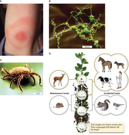
B. burgdorferi is a spirochete (Fig. 26.34B ) transmitted to humans by ixodid ticks (hard ticks;
Fig. 26.34C and D ). In the northeastern and central United States, where most cases occur, the deer
tick Ixodes scapularis transmits the spirochete, usually during the summer months. In the western
United States, I. pacificus is the tick vector.
During its nymphal stage (the stage after taking its first blood meal), I. scapularis is the size of a
poppy seed. Its bite is painless, so it is easily overlooked. Infection takes place when the tick feeds,
because the spirochete is regurgitated into the host. However, the organism grows in the tick’s
digestive tract and takes about 2 days to make its way to the tick’s salivary gland, so if the tick is
removed before that time, the patient will not be infected. Once the pathogen is transferred to the
human, the microbe can travel rapidly via the bloodstream to any area in the body, but it prefers to
grow in skin, nerve tissue, synovium (joint lining), and the conduction system of the heart.
Lyme disease has three stages. Similar to syphilis caused by the spirochete Treponema pallidum,
Lyme disease has three general stages. Stage 1 involves the localized spread of Borrelia burgdorferi
between 3 and 30 days after the initial exposure. Approximately 75% of patients experience an
erythema migrans rash, usually at the site of the tick bite. The appearance of the rash varies but is
classically erythematous with central clearing (“bull’s-eye” rash). This stage is often associated with
constitutional symptoms such as fever, myalgia (muscle pain), arthralgia (joint pain), and headache.
Stage 2 occurs weeks to months after the initial infection as B. burgdorferi spreads from blood to
other organs. In this stage, the patient can be quite ill with malaise, myalgia, and arthralgia, as well as
neurological or cardiac involvement. Common neurological manifestations include Bell’s palsy (facial
paralysis), inflammation of spinal nerve roots, and chronic meningitis. The most common cardiac
manifestation is an irregular heart rhythm.
Stage 3 borreliosis occurs months to years later and can involve the synovium, nervous system, and
skin. Arthritis occurs in the majority of previously untreated patients; it is usually intermittent, involves
the large joints, particularly the knee, and lasts from weeks to months in any given joint. Joint-fluid
analysis typically shows a WBC count of 10,000–30,000 per microliter (normal is fewer than 200). Late
neurological involvement may include peripheral neuropathy and encephalopathy, manifested as
memory, mood, and sleep disturbances.
Treatment with antibiotics (usually doxycycline) is recommended for all stages of Lyme disease but
is most effective in the early stages. Lyme arthritis is typically slow to respond to antibiotic therapy, so
treatment lasts from 2 to 4 weeks. However, the disease in some people does not resolve at all; why is
not known.
Curiously, 25% of people infected with B. burgdorferi never experience erythema migrans, and
many infected individuals are also unsure of tick bites. Hence, patients with Lyme disease may present
with arthritis as their first complaint. Although this makes diagnosis extremely difficult, knowing that
the patient lives in or recently traveled to an endemic area can provide a critical clue.
A startling example of how sinister Lyme disease can be is the case of Kris Kristofferson, a prolific
singer, songwriter, and actor, now in his eighties. Kristofferson suffered for years with severe memory
loss presumed to be a manifestation of Alzheimer’s disease until someone decided to test him for Lyme
disease. To his doctor’s surprise and relief, the singer tested positive. Kristofferson immediately began
antibiotic treatments and has since recovered much of his memory.
Many other bacteria can cause septicemia and systemic illness (Table 26.6). Gram-negative
organisms, such as E. coli, Salmonella Typhi, and Francisella, and Gram-positive microbes, such as
Staphylococcus aureus, Enterococcus, and Bacillus anthracis, can grow in the bloodstream if they can
gain entrance. Even anaerobes that are normal inhabitants of the intestine (for example, Bacteroides
fragilis) can be lethal if they escape the intestine and enter the blood, as might happen following
surgery. This is one reason surgical patients are given massive doses of antibiotics immediately before
and after surgery.
TABLE Selected Systemic Infectious Diseases 26.6
Etiological Virulence
Disease Symptoms agent a properties Source(s) Treatment
Lyme Stage 1: rash; stage Borrelia Antigenic Deer tick Penicillin,
disease 2: chills, headache, burgdorferi variation, doxycycline
malaise, systemic (spirochete) OspE (binds
involvement; stage complement)
3: neurological
changes
Brucellosis Fever, weakness, Brucella Intracellular, Animal Doxycycline
sweats, abortis (G− growth in products,
splenomegaly, rod) monocytes unpasteurized
osteomyelitis, milk
endocarditis, others
Leptospirosis Fever, photophobia, Leptospira Burrowing Urine of Penicillins,
headache, abdominal interrogans motility infected erythromycin
pain, skin rash, liver (spirochete) animals
involvement,
jaundice
Epidemic Chills, fever, Rickettsia Obligate Human louse, Tetracycline,
typhus headache, muscle prowazekii intracellular flying-squirrel chloramphenicol
pain, splenomegaly, (G− rod) growth, flea
coma escapes
phagosome
Tularemia Fever, chills, Francisella Intracellular Rabbits, Gentamicin,
headache, muscle tularensis rodents, streptomycin
pain, rash, (G− rod) insect vectors
bacteremia
TABLE Selected Systemic Infectious Diseases 26.6
Typhoid Septicemia, chills, Salmonella Type III Gallbladder of Ciprofloxacin,
fever fever, hypotension, Typhi (G− secretion, human carrier ceftriaxone
rash (rose spots) rod) intracellular
growth,
PhoPQ
regulators, Vi
antigen
capsule
Septicemia, chills, Salmonella Intracellular Animals, Ceftriaxone
fever, hypotension choleraesuis growth, poultry
(G− rod) invasin
Vibriosis Serious with Vibrio Cytolysin, Seawater, Tetracycline
immunocompromised vulnificus capsule raw oysters plus 3rd-
patients; fever, chills, (G− curved generation
multi-organ damage, rod) cephalosporin
death
Bubonic Buboes (swollen Yersinia Intracellular Rodents, Streptomycin or
plague lymph glands), high pestis (G− growth, type rodent fleas, tetracycline
fever, chills, rod) III secretion human
headache, cough, of YOPs (respiratory
pneumonia, Yersinia outer aerosol,
septicemia proteins), potential
phospholipase bioterrorism
D, toxin agent
Ebola—The Perfect Pathogen or Too Deadly for Its Own Good?
How would you define the perfect pathogen? Would it be an organism that can kill its host with
terrifying ease and quickness? If so, Ebola virus would fit the description. Ebola virus, a lipid-
enveloped, threadlike RNA virus (Filoviridae; see Fig. 25.4), was first associated with an outbreak of
318 cases of a hemorrhagic disease in Zaire (now the Democratic Republic of the Congo) in 1976. Of
the 318 people who contracted the disease, 280 died within days. The disease was characterized by
acute (rapid) onset of fever, headache, diarrhea, and severe muscle pains. Some patients exhibited
horrible bleeding (hemorrhaging) from multiple orifices (nose, mouth, anus, and vagina) and ultimately
died.
The Ebola virus has a frightening reputation. It spreads like wildfire through the body after
infection, causing severe hemorrhagic fever, and typically kills 20%–90% of its victims, depending on
the strain involved and the level of care received. Internal bleeding results in shock and acute
respiratory distress, leading to death. The major 2014–2016 epidemic of Ebola in western Africa
involved 28,683 cases, 40% of whom died (see Chapter 28). Smaller outbreaks have occurred since
then; the most recent (2021) was in the Democratic Republic of the Congo. The symptoms of Ebola
(and of a related disease caused by the Marburg virus) reflect subversion of the innate immune
system, coupled with uncontrolled viral replication, particularly in macrophages and dendritic cells.
Ebola virus infection of these cells enhances production of pro-inflammatory cytokines, such as TNF-
alpha, and inhibits stimulation of T-cell maturation by dendritic cells. Thus, Ebola infections not only
stimulate inflammatory processes leading to tissue damage, they also shut down early immune
responses and prevent activation of adaptive immune responses, thereby allowing unfettered viral
replication.
Ebola viral proteins and their locations in the virion are shown in Figure 26.35A . Ebola VP35
protein is a component of the viral RNA polymerase complex, but it is also a potent inhibitor of host
interferon (IFN) production. The cellular response to whichever IFN is made is inhibited by VP24, which
blocks the nuclear accumulation of a regulatory protein called STAT1. STAT1 is critical to IFN-stimulated
gene expression. These and other strategies enable rapid replication of the virus.
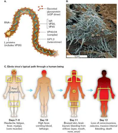
FIGURE 26.35 ■ Ebola virion. A. Composition of the virus. The ribonucleoprotein complex
consists of the nucleoprotein (NP), the structural proteins VP30 and VP35, and the virion-
associated RNA-dependent RNA polymerase (L proteins). The glycoprotein (GP-sGP) is an integral
membrane protein that can be secreted. B. Threadlike Ebola virions budding from a cell (center).
C. Progression of the disease.
Source: Part C modified from http://themindbodyshift.com/index.php/2014/10/16/when-the-threat-of-ebola-hits-home
.
KALETSKY, R. L., ET AL. 2009. PNAS 106 :2886.
After replicating, Ebola offspring sprout from the cell surface in a mass of tangled threads (Fig.
26.35B ). These new virions go on to attack new cells, riddling blood vessels and organs with damage
as they go. Rapid release of new virions involves subverting another host mechanism, tetherin,
designed to slow viral spread. Paul Bates and his colleagues at the University of Pennsylvania
discovered that the cell protein tetherin essentially tethers mature virus particles inside a cell so that
they are unable to spread. Tetherin is IFN induced and can restrict the spread of structurally diverse
enveloped viruses, including HIV (thus, it is part of the innate response). Ebola glycoprotein, however,
counteracts tetherin, so that nothing slows down viral spread. The result is rapid release of massive
numbers of virus particles that can then quickly spread infection to other organs and tissues.
The outlook for a patient infected with Ebola is typically dire (occasionally a person infected with
Ebola virus can develop antibodies but remain asymptomatic). The incubation period is 4–16 days, and
death occurs within 7–16 days (Fig. 26.35C ). With good supportive care, including the replacement
of coagulation factors, the mortality rate appears near 20%. In the absence of such care, mortality can
reach over 80%. For the latest outbreaks in Africa, the World Health Organization (WHO) has approved
use of three experimental treatments and a vaccine. The treatments include a recombinant
monoclonal antibody (ZMapp) that has Ebola virus–neutralizing activity, an RNA-dependent RNA
polymerase inhibitor, and an RNA chain terminator. The WHO-approved vaccine (ERVEBO) is an
attenuated vesicular stomatitis virus engineered to express a glycoprotein from the most lethal Ebola
virus (Zaire ebolavirus). Neutralizing antibody to this glycoprotein is produced after vaccination.
Vaccination efforts in Africa are widespread, but they principally focus on people living in areas ringing
an outbreak in order to limit spread of the disease.
Death comes very quickly with this disease, typically before the virus can be transmitted to very
many new hosts. Some scientists argue that efficient and quick killing is not the mark of a perfect
pathogen. The better pathogen lets its host linger to ensure a home for itself and more opportunity to
disseminate. So, why doesn’t Ebola die out? Where does it go when it is not infecting humans? The
natural ecology of these viruses is largely unknown, although an association with monkeys and/or bats
as possible reservoirs is suggested.
To Summarize
Blood cultures are useful in diagnosing septicemia and endocarditis.
Septicemia is caused by many Gram-positive and Gram-negative bacterial pathogens. It can
start with the bite of an infected insect, introduction via a wound, escape from an abscess, or
penetration of the mucosal epithelium by the pathogen (intestine or vagina) and can lead to
disseminated, systemic disease.
Endocarditis can have acute or subacute onsets. Subacute bacterial endocarditis is usually
an endogenous infection of a heart valve caused by Streptococcus mutans.
Malaria, caused by Plasmodium species , manifests as repeated episodes of chills, fever,
and sweating, owing to the organism’s ability to alter the antigenic appearance of its surface
proteins and evade the immune response.
Plague, caused by Yersinia pestis , has sylvatic and urban infection cycles involving
transmission by fleas. The bite of an infected flea leads to bubonic plague. Bubonic plague can
progress to septicemic and pneumonic stages. Pneumonic plague can be spread directly from
person to person (no insect vector) by aerosolized respiratory secretions .
Toxic shock syndrome is a sepsis-like, systemic disease caused by an exotoxin (toxic
shock syndrome toxin, TSST) secreted by some strains of Staphylococcus aureus and
Streptococcus pyogenes. It is caused by the circulatory spread of TSST made from bacteria that
infect or colonize a localized tissue.
Lyme disease is caused by the spirochete Borrelia burgdorferi, which is transmitted from
animal reservoirs to humans by the bite of Ixodes ticks. The three stages of Lyme disease are
characterized by a bull’s-eye rash, called erythema migrans (stage 1); joint, muscle, and nerve
pain (stage 2); and arthritis with WBCs in the joint fluid (stage 3).
Ebola virus spreads from human to human via body fluids and kills its victims quickly. Its viral
proteins alter cytokine production and facilitate virus release from infected cells.
Glossary
septicemia
An infection of the bloodstream.
viremia
The presence of large numbers of virions in the bloodstream.
bacteremia
A bacterial infection of the blood.
endocarditis
An inflammation of the heart’s inner lining.
bubonic plague
A disease caused by the bacterium Yersinia pestis; it is characterized by swollen lymph nodes that
often turn black.
septicemic plague
Infection of the bloodstream by Yersinia pestis.
pneumonic plague
A highly virulent and contagious Yersinia pestis lung infection.
erythema migrans
A bull’s-eye rash characteristic of borreliosis (Lyme disease).
Lyme disease
One form of borreliosis. A tick-borne disease caused by Borrelia burgdorferi, which may involve
skin lesions and arthritis.
borreliosis
Any of a variety of diseases caused by Borrelia species and transmitted by ticks or lice. Lyme
disease is a form of borreliosis.
Fig. 20.39
FIGURE 20.39 ■ Malaria: cycle of Plasmodium falciparum transmission between
mosquito and human.
Fig. 20.39
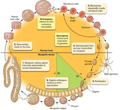
FIGURE 20.39 ■ Malaria: cycle of Plasmodium falciparum transmission between
mosquito and human.
Fig. 25.4
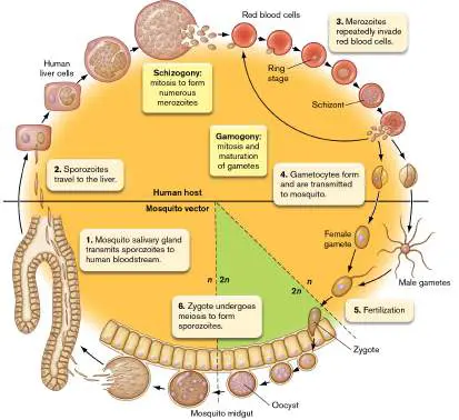
FIGURE 25.4 ■ Highly virulent viruses. A. Ebola virus (approx. 1 μm long; TEM). B. A
health care worker tends to an Ebola patient in Kenema, Sierra Leone. Ebola causes
hemorrhagic infections in which patients bleed from the mouth, nose, eyes, and other
orifices. The mortality rate for this disease is approximately 50%.
CDC/FREDERICK A. MURPHY
TOMMY TRENCHARD/ALAMY STOCK PHOTO
Endnotes
1. Note a: G+ = Gram-positive; G− = Gram-negative. Return to reference a
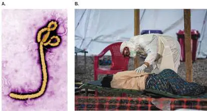
26.6 Central Nervous System Infectionsnot assigned
The brain and spinal cord are especially well protected against infection. Microbes cannot gain
easy access to the brain, in large measure because of the blood-brain barrier, a filter mechanism
that allows only selected substances into the brain. The blood-brain barrier works to our
advantage when harmful substances, such as bacteria, are prohibited from entering. However, it
works to our disadvantage when substances that we want to enter the brain, such as antibiotics,
are kept out. The barrier is not a single structure but a function of the way blood vessels,
especially capillaries, are organized in the brain. Furthermore, the endothelial cells in those
vessels have tight junctions that do not allow most compounds or microbes to cross. And yet,
brain infections do occur.
Case History: Meningitis
Laila, a 4-month-old infant from Saudi Arabia, was hospitalized with fever, tender neck, and
purplish spots (purpuric spots) on her trunk (Fig. 26.36A ). Suspecting meningitis, the clinician
took a cerebrospinal fluid (CSF) sample and examined it by Gram stain. The smear revealed
Gram-negative diplococci inside PMNs. The CSF was turbid with 900 leukocytes per microliter, and
Neisseria meningitidis was confirmed by culture. The child was treated with cefotaxime (a
cephalosporin antibiotic; see Chapter 27) and made a full recovery. Her father, Abdul, the person
who brought her in, was clinically well. However, the meningococcus was isolated from his
oropharynx, as well as from the throat of the patient’s 2-year-old brother. Isolates from the
patient, her father, and her brother were positive by agglutination with meningococcal
A/C/Y/W135 polyvalent reagent. Records showed that the father had previously received a
quadrivalent meningococcal vaccine. All three isolates were confirmed to be meningococcus
serogroup W135. DNA analysis found the three isolates to be indistinguishable, meaning that the
father and his children were infected with the same strain of N. meningitidis. Why did Laila’s
brother not have meningitis? And why was Laila’s vaccinated father colonized?
FIGURE 26.36 ■ Bacterial meningitis. A. Purpuric spots produced by local intravascular
coagulation due to Neisseria meningitidis endotoxin. The rash in meningitis typically has
petechial (small) and purpuric (large) components. B. N. meningitidis (diameter approx. 1
μm; SEM). C. Normal brain. D. Autopsy specimen of meningitis due to Streptococcus
pneumoniae. Note the greening of the brain, compared with the pink normal brain.
JOHN RADCLIFF HOSPITAL/SCIENCE SOURCE
EYE OF SCIENCE/SCIENCE SOURCE
DR. COLIN CHUMBLEY/SCIENCE SOURCE
CDC
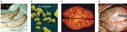
Meningitis is an inflammation of the meninges, the membranes that surround the brain and spinal
cord. Meningitis can be either bacterial or viral in origin. Sinus and ear infections can extend
directly to the meninges, whereas septicemic spread requires passage through the blood-brain
barrier. Viral meningitis is serious but rarely fatal in people with a normal immune system. The
symptoms generally persist for 7–10 days and then completely resolve. Bacterial meningitis is
usually caused by Streptococcus pneumoniae, Neisseria meningitidis, or Haemophilus influenzae
. Symptoms of bacterial meningitis can include sudden onset of fever, headache, pain or stiffness
in the neck, painful sensitivity to strong light (photophobia), vomiting, and irritability. Prompt
medical attention is extremely important because the disease can quickly progress to convulsions
and death.
The meningococcus N. meningitidis (Fig. 26.36B ) can colonize the human oropharynx,
where it causes mild, if any, disease. At any given time, 10%–20% of the healthy population can
be colonized and asymptomatic. The organism spreads directly by person-to-person contact or
indirectly via droplet nuclei from sneezing or fomites. The problem arises when this organism
enters the bloodstream. Unlike N. gonorrhoeae, the cause of gonorrhea, N. meningitidis is very
resistant to complement, owing to its production of a polysaccharide capsule. Protected by this
capsule, the microbe can produce a transient blood infection (bacteremia) and reach the blood-
brain barrier.
How does N. meningitidis enter the bloodstream from the nasopharynx and then cross the
blood-brain barrier? The organism initially uses type IV pili to adhere to and enter nasopharyngeal
endothelial cells (type IV pili are discussed in Section 25.2). The bacteria then cross the
endothelial cell layer by transcytosis, a process by which an internalized pathogen passes through
a host cell along microtubules to the opposite side. Ultimately, the pathogen passes into the
capillary lumen and is swept away to the brain. Once in the brain, type IV pili again adhere to
endothelial cells, but this time they trigger the recruitment of host proteins that destabilize
intercellular junctions. The bacteria then slip between the loosened junctions and enter the
cerebrospinal fluid (CSF). Once in the CSF, microbes can multiply almost at will. Figure 26.36C
and D shows the remarkable damage (greening) that N. meningitidis and other microbes, such as
S. pneumoniae, cause in the brain.
Several antigenic types of capsules, called type-specific capsules, are produced by different
strains of pathogenic N. meningitidis: types A, B, C, W135, and Y. Types A, C, Y, and W135 are
usually associated with epidemic infections seen among people kept in close proximity, such as
college students or military personnel. Type B meningococcus is typically involved in sporadic
infections, but it has also been known to cause large outbreaks like the ones that occurred at
Princeton University and UC Santa Barbara in 2013.
Antibodies to the capsular antigens are used to classify the capsular types of the organisms
causing an outbreak. Knowing the capsular type of organism involved in each case helps
determine whether the disease cases are related and where the infection may have started. In
Abdul’s case, a polyvalent reagent containing antibodies to the four main capsular types was used
to confirm N. meningitidis, and antisera to individual capsule types specifically identified the
strain as W135. Why was Abdul colonized but not ill? W135 likely colonized Abdul’s throat but
failed to spread because his previous vaccination provided circulating anti-W135 antibodies that
attacked the organism if it entered the bloodstream. Even unvaccinated people can become
colonized but remain asymptomatic if antibodies develop before the bacteria enter the
bloodstream. He was protected.
Meningococcal meningitis is highly communicable. As a result, close contacts, such as the
parents or siblings of any patient with meningococcal disease, should receive antimicrobial
prophylaxis within 24 hours of diagnosis. Current recommendation is a single dose of ceftriaxone
(a third-generation cephalosporin) for adults or 2 days of rifampicin for children. Highly
susceptible populations can be immunized with a vaccine containing four of the five capsular
structures. Because the type B capsule is not immunogenic, a separate vaccine containing outer
membrane vesicles and proteins from N. meningitidis type B (but not its capsule) is used.
Thought Question
26.11 Normal cerebrospinal fluid is usually low in protein and high in glucose. The protein and
glucose content does not change much during a case of viral meningitis, but bacterial infection
leads to greatly elevated protein and lowered glucose levels. What could account for this
difference?
Case History: Botulism—It Is What You Eat
A 47-year-old resident of Oklahoma was admitted to the hospital with rapid onset of progressive
dizziness, blurred vision, slurred speech, difficulty swallowing, and nausea. Findings on
examination included drooping eyelids, facial paralysis, and impaired gag reflex. He developed
breathing difficulties and required mechanical ventilation. The patient reported that during the 24
hours before the onset of symptoms, he had eaten home-canned green beans and a stew
containing roast beef and potatoes. Analysis of the patient’s stool detected botulinum type A
toxin, but no Clostridium botulinum organisms were found. The patient was hospitalized for 49
days, including 42 days on mechanical ventilation, before recovering and being discharged.
Imagine a disease that causes complete loss of muscle function. Two microbes cause such lethal
paralytic diseases by using secreted exotoxins (neurotoxins). In one instance, botulism, the victim
suffers a flaccid paralysis in which the muscles go limp, as in the case history just presented,
causing paralysis and respiratory difficulty. Botulism is typically a food-borne disease caused by an
anaerobic, Gram-positive, spore-forming bacillus, named Clostridium botulinum, that produces
botulism toxin.
In striking contrast to botulism is tetanus, a very painful disease in which muscles continually
and involuntarily contract (called tetany or spastic paralysis). Tetanus is caused by tetanospasmin
, a potent exotoxin made by another anaerobic, Gram-positive, spore-forming bacillus, called
Clostridium tetani (Fig. 26.37A ). Tetanospasmin alters neural transmission, but in contrast to
botulism toxin (which inhibits neurotransmission), tetanospasmin causes excessive nerve
signaling to muscles. The first symptom is usually spasms of the masseter muscles affecting
movement of the jaw, giving the disease its common name—lockjaw. Eventually, the toxin forces
the victim’s back to arch grotesquely while the arms flex and legs extend (Fig. 26.37B ). The
patient remains locked this way until death. Spasms can even be strong enough to fracture the
patient’s vertebrae. In both botulism and tetanus, death can result from asphyxiation.
FIGURE 26.37 ■ Tetanus and botulism toxins. A. Photomicrograph of Clostridium
tetani (cell length 4–8 μm). B. Painting from Charles Bell that depicts the spasms associated
with tetanus in a soldier dying from tetanus. C. Schematic diagram of tetanus and botulism
toxins. D. 3D representation of the tetanus neurotoxin, with the domains marked. (PDB
code: 3BTA)
DR. A. M. SIEGELMAN/VISUALS UNLIMITED
THE PICTURE ART COLLECTION/ALAMY STOCK PHOTO
Thought Question
26.12 Given the symptoms of tetanus, what kind of therapy would you use to treat the disease?
Toxin structure and function. Botulism and tetanus toxins share 30%–40% identity. They
have similar structures and nearly identical modes of action. Each is composed of two peptides—a
large, or heavy, fragment analogous to a B subunit of AB toxins; and a small, or light, fragment
analogous to an A subunit. Both toxins are initially made as single peptides (about 150 kDa) that
are cleaved by host proteases after secretion to form two fragments (the heavy and light chains)
that remain tethered to each other by a disulfide bond (Fig. 26.37C ). The heavy chains possess
binding domains for receptor molecules (gangliosides) on the nerve cell membrane and a
translocation domain that makes a pore in the nerve cell endosome through which the toxic light
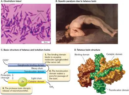
chains pass into the cytoplasm. The light chains are proteases that disrupt the movement of
exocytic vesicles containing neurotransmitters (Fig. 26.37D ).
Pathogenesis of botulism neurotoxin. Botulism is typically caused by ingesting preformed
toxin. Rarely, infected wounds or ingested spores can also produce disease. Normally, spores
germinate in contaminated food and the cells produce toxin, but an anaerobic environment is
required because the organism is an anaerobe. Home-canning processes sterilize the food but
also remove oxygen. If the sterilization is incomplete, surviving spores will germinate and the
bacteria will secrete toxin. The toxin is heat sensitive but will remain active in improperly cooked
food. After ingestion, the toxin is absorbed from the intestine. The organism is often absent from
stool samples, but the toxin can be detected in stool.
A rare form of botulism, called infant botulism or “floppy head syndrome,” can occur when
infants (not older children) are fed honey. Honey can harbor Clostridium botulinum spores that
can germinate in the gastrointestinal tract, after which the growing vegetative cells will secrete
toxin.
Figure 26.38A and B illustrates normal neurotransmission at a neuromuscular junction. A
signal sent from a motor neuron to the neuromuscular junction releases the neurotransmitter
molecule acetylcholine (ACh) from vesicles in the axon terminal (Fig. 26.38A ). Figure 26.38B
shows the proteins that enable the vesicle and plasma membranes to fuse. Once released, ACh
traverses the synapse and causes muscle contraction.
FIGURE 26.38 ■ Mechanism of action of botulism toxin. A. The neuromuscular
junction. The blowup shows vesicles filled with neurotransmitters. B. A series of proteins
within the nerve is needed to allow synaptic vesicles to bind to the nerve endings. Fusion of
the membranes releases acetylcholine into the neuromuscular junction. Botulism toxin types
A and E cleave SNAP-25. Botulism toxins B, D, F, and G cleave VAMP. Botulism toxin C1
cleaves syntaxin and SNAP-25. C, D. Toxin binds via the heavy chain and is endocytosed into
the nerve terminal. Once the toxin cleaves its target, the nerve terminal is no longer able to
release acetylcholine.
Botulism toxin enters a peripheral nerve by endocytosis (Fig. 26.38C ), and acidification of
the endosome reduces the disulfide bonds holding the heavy and light chains together. The heavy
chain assembles as a channel in the endosome membrane through which the proteolytic light
chain moves into the cytoplasm (Fig. 26.38D ). The light chain then cleaves key host proteins,
such as synaptobrevin (a vesicle-associated membrane protein, or VAMP), syntaxin, or
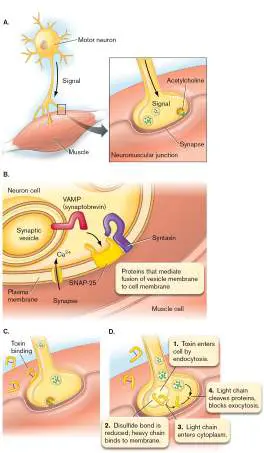
synaptosomal-associated protein (SNAP-25), involved in the exocytosis of vesicles containing
acetylcholine. Without acetylcholine to activate nerve transmission, muscles will not contract and
the patient is paralyzed. Currently, there is no FDA-approved vaccine for botulism. Treatment
involves a multivalent antitoxin that can neutralize all seven toxin serotypes, coupled with
supportive care.
Although botulism is now rare, the toxin is considered a select biological agent of potential use
to bioterrorists. As such, its use in laboratories is under strict governmental control. But botulism
toxin also has important medical uses. Because the toxin can safely relax muscles in a localized
area if injected in small doses, botulism toxin, or Botox, is used therapeutically by neurologists to
treat migraine headaches, excessive sweating, crossed eyes, and Bell’s palsy. Most commonly,
Botox is used cosmetically by plastic surgeons to reduce facial wrinkles.
Pathogenesis of tetanus neurotoxin. With such a high degree of structural and mechanistic
similarity, why do tetanus and botulism toxins have such drastically different effects? The answer
is based on where each toxin acts in the nervous system (Fig. 26.39 ). Tetanus, in contrast to
botulism, is not a food-borne disease. Clostridium tetani spores are introduced into the body by
trauma (such as by stepping on the wrong end of a nail). Necrotic tissue resulting from the
puncture provides the anaerobic environment required for germination. Growing bacteria release
tetanospasmin, which enters the peripheral nerve cells at the site of injury. But rather than
cleaving targets here, the toxin travels up axons in the direction opposite to nerve signal
transmission until it reaches the spinal column, where it becomes fixed at the presynaptic
inhibitory motor neuron (Fig. 26.39A ).
FIGURE 26.39 ■ Action of tetanus and botulism toxins. A. Tetanus toxin enters the
nervous system at the neuromuscular junction (A1) and travels retrogradely up the axons
until reaching an inhibitory neuron located in the central nervous system (A2). There it
cleaves VAMP protein (Fig. 26.38B) associated with the exocytosis of vesicles containing
inhibitory neurotransmitters (GABA). ACh = acetylcholine; GABA = gamma-aminobutyric (4-
aminobutanoic acid). B. Botulism toxin is absorbed by the small intestine, enters the
bloodstream, and acts at the neuromuscular junction to prevent release of acetylcholine.
There, the toxin also cleaves proteins like VAMP, but the vesicles in these nerves release
inhibitory neurotransmitters (GABA and glycine) that dampen nerve impulses. Tetanus toxin
blocks release of these inhibitors into the synaptic cleft, leaving nerve impulses unchecked. As a
result, impulses come too frequently and produce the generalized muscle spasms characteristic of
tetanus. A tetanus toxoid vaccine is available and is administered to children as part of the DTaP
(diphtheria, tetanus, and acellular pertussis) vaccine (see Section 24.6).
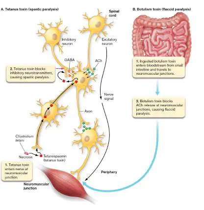
Thought Questions
26.13 Figure 26.39 demonstrates that tetanus toxin has a mode of action (spastic paralysis)
opposite to that of botulism toxin (flaccid paralysis). Since they have opposing modes of action,
can botulism toxin be used to save a patient with tetanus?
Case History: Eastern Equine Encephalitis
In August, Mr. C took his 21-year-old son, Rich, to a New Jersey emergency department. Rich
appeared dazed and had trouble responding to simple commands. When questioned about his
son’s activities over the previous few months, Mr. C told the physician that Rich had spent the
month of July relaxing and sunning himself on New Jersey beaches and visiting a pond in a
wooded area near a horse farm. On the afternoon prior to admission, Rich had become lethargic
and tired. He returned home and went to bed. That evening, his father woke him for supper, but
Rich was confused and had no appetite. By 11 p.m., Rich had a fever of 39.7°C (103.5°F) and
could not respond to questions. A few hours later, when his father had trouble rousing him, he
took Rich to the emergency department. Over the next week, Rich’s condition worsened to the
point where his limbs were paralyzed. Two weeks later, he died. Serum samples taken when he
entered the hospital and a few days before he died showed a sixfold rise in antibody titer to
eastern equine encephalitis (EEE) virus. Brain autopsy showed many small foci of necrosis in both
the gray and white matter.
The EEE virus, a member of the family Togaviridae, is transmitted from bird to bird by
mosquitoes. Horses contract the disease in the same way. Rich contracted it inadvertently while
visiting the pond. Most people who are infected never develop symptoms, but among those who
become ill, the disease is often fatal. The disease is rare, in part, because the species of mosquito
that transmits the virus between marsh birds does not prey on humans. Occasionally, a human-
specific mosquito will bite an infected bird and then transmit EEE virus to humans. Once in the
human body, the virus generally does not fare well because the immune system thwarts viral
replication.
An interesting point in the case history is that diagnosis relied on detecting an increase in
antibody titer to the virus. Typically, at the point when disease symptoms first appear, the body
has not had time to generate large amounts of specific antibodies. After a week or so, often when
the patient is nearing recovery (convalescence), antibody titers have risen manyfold. The rule of
thumb is that a greater-than-fourfold rise in IgG antibody titer between acute disease and
convalescence (or, in this case, death) indicates that the patient has had the disease. While this
knowledge could not help the patient in the case described here, it was valuable in terms of public
health and prevention strategies.
EEE virus is one of many so-called arboviruses, a general term for pathogenic viruses
transmitted to humans by arthropod insects. West Nile virus and yellow fever virus are two well-
known arboviruses that cause CNS (central nervous system) infections. Additional bacterial,
fungal, and viral causes of encephalitis and meningitis are listed in Table 26.7.
TABLE Selected Microbes That Cause Meningitis or 26.7 Encephalitis
Typical
initial
Type of Etiological Virulence infection or
disease agent(s) a factors source Treatment Vaccine
Bacterial
(septic) Streptococcus Capsule, Lung Ampicillin Multivalent,
meningitis pneumoniae pneumolysin capsule
(G+)
Neisseria Capsule, IgA Throat Cephalosporin Multivalent,
meningitidis protease, (3rd capsule
(G−) endotoxin generation),
ceftriaxone
Haemophilus Polyribitol Ear infection Cephalosporin Type b
influenzae type capsule, IgA (3rd polysaccharide
b (G−) protease, generation),
endotoxin ceftriaxone
Other G− Endotoxin Septicemia
bacilli
Group B Sialic acid Neonate Ampicillin Capsular
streptococci capsule, infected
(G+) streptolysin, during
inhibition of parturition
alternate
complement
path
Listeria Intracellular Mild GI Ampicillin plus
monocytogenes growth, PrfA disease of gentamicin
(G+) regulator, mother
actin-based
motility
TABLE Selected Microbes That Cause Meningitis or 26.7 Encephalitis
Mycobacterium Cord factor, Lung Combination BCG b
tuberculosis e wax D, therapy
(Gram- intracellular (rifampin,
positive–like) growth isoniazid,
ethambutol,
pyrazinamide)
Staphylococcus Coagulase, Septicemia Oxacillin,
aureus (G+) protein A, vancomycin
TSST,
leukocidin
Staphylococcus Biofilm, Complication Vancomycin,
epidermidis slime of surgical methicillin
(G+) procedure
Aseptic
meningitis c Fungi (e.g., Sinusitis, Ketoconazole,
Coccidioides, direct spread fluconazole
Cryptococcus) to meninges
Amebas (e.g., Swimming in Amphotericin
Naegleria) contaminated B
waters
Treponema Syphilis
pallidum
Mycoplasmas Adhesin tip Respiratory Erythromycin
Leptospira Burrowing Septicemia, Erythromycin Killed whole
motility water cell, animals
contaminated only
TABLE Selected Microbes That Cause Meningitis or 26.7 Encephalitis
with animal
urine
Viral
meningitis Viruses (90% Self-limiting
or caused by
encephalitis enteroviruses)
Eastern EEE virus? Mosquito bite None; often
equine fatal
encephalitis
West Nile West Nile virus? Mosquito bite Supportive
disease therapy d
Prions Are Infectious Proteins
Imagine slowly losing your mind and knowing there is nothing you can do about it. An unusual
infectious agent called the prion has been implicated as the cause of a series of relatively rare but
invariably fatal brain diseases (Table 26.8). Prions are infectious agents that do not have a
nucleic acid genome. The protein alone can mediate an infection. The prion is now recognized as
an infectious, misfolded protein that resists inactivation by procedures that destroy proteins.
TABLE 26.8 Prion Diseases
Susceptible Incubation Disease
Disease animal period characteristics
Creutzfeldt-Jakob Human Months (vCJD) Spongiform
[sporadic, familial, new to years encephalopathy
variant (vCJD)] (degenerative brain
disease)
TABLE 26.8 Prion Diseases
Kuru Human Months to years Spongiform
encephalopathy
Gerstmann-Straussler- Human Months to years Genetic
Scheinker syndrome neurodegenerative
disease
Fatal familial insomnia Human Months to years Genetic
neurodegenerative
disease with untreatable
insomnia
Mad cow disease Cattle 5 years Spongiform
encephalopathy
Wasting disease Deer Months to years Spongiform
encephalopathy
The discovery that proteins alone can transmit an infectious disease came as a surprise to the
scientific community. Diseases caused by prions are especially worrying, since prions resist
destruction by many chemical agents and remain active after heating at extremely high
temperatures. There have even been documented cases in which sterilized neurosurgical
instruments, originally used on a prion-infected person, still held infectious agent and transmitted
the disease to a subsequent surgical patient. Extremely rigorous decontamination procedures,
such as autoclaving surgical instruments immersed in 1-M sodium hydroxide, will destroy prions.
How can a nonliving entity without nucleic acid be an infectious agent? Prions associated with
human brain disease are thought to be aberrantly folded forms of a normal brain protein (PrP C).
The theory is that when a disease-related prion, designated PrP Sc or PrP D, is introduced into the
body and manages to enter the brain, it will cause normally folded forms of the protein to refold
incorrectly (Fig. 26.40A ). The improperly folded proteins fit together like Lego blocks to
produce damaging aggregated structures within brain cells.
FIGURE 26.40 ■ Spongiform encephalopathies. A. Refolding model of prion diseases.
PrP is a brain protein that can take two forms: the normal form (PrP C), which is a natural
brain protein; and the prion form (PrP Sc). B. Normal brain section. C. Section of brain taken
from a CJD victim. Note the Swiss-cheese appearance, indicating brain damage.
SCIENCE STOCK PHOTOGRAPHEY/SCIENCE SOURCE
CDC/TERESA HAMMETT
Prion diseases are often called spongiform encephalopathies because the postmortem
appearance of the brain includes large, spongy vacuoles in the cortex and cerebellum. These are
visible in a brain sample from a victim of one of these diseases, called Creutzfeldt-Jakob disease
(CJD; Fig. 26.40C ). Most mammalian species appear to be susceptible to these diseases.
Since 1996, mounting evidence has pointed to a causal relationship between outbreaks in
Europe of a disease in cattle called bovine spongiform encephalopathy (BSE, or “mad cow
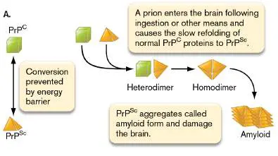
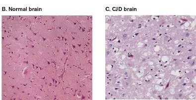
disease”), which is caused by prions, and a disease in humans called variant Creutzfeldt-Jakob
disease (vCJD). Both disorders are invariably fatal. For humans to contract disease, prions from
contaminated meat are ingested and penetrate the intestinal mucosa via the antigen-sampling M
cells in Peyer’s patches. The circulation then traffics the agent to the brain.
Between 1993 and 2018, only six cases of BSE in cattle were detected in the United States.
Because of aggressive surveillance efforts in the United States and Canada (where 20 cases have
been found since 1993), it is unlikely, but not impossible, that BSE will be a food-borne hazard to
humans in both countries. The CDC monitors the trends and current incidence of typical CJD
(approximately 560 cases in 2019) and variant CJD (4 cases total) in the United States. The
worldwide incidence of CJD is one per 1 million population.
There is no cure for CJD or vCJD. Two tests to detect PrP Sc in asymptomatic primates,
including humans, have been developed and are being used for human screening. One test is
called protein-mediated cyclic amplification (PMCA). The other is called the real-time, quaking-
induced conversion (RT-QuIC) assay. PMCA has successfully identified the presence of very tiny
amounts of PrP Sc in CSF, blood, or urine. RT-QuIC performs best for CSF. The PMCA process
involves adding large amounts of normal PrP (PrP C) to samples containing tiny amounts of PrP Sc
prions. The prions, if present in the sample, will seed the misfolding of PrP C to PrP Sc molecules
that aggregate. Sonication is then used to partially break up the newly formed PrP Sc aggregates
into pieces that can further seed the misfolding and aggregation of normal PrP. Repeated cycles of
sonication-incubation will exponentially amplify the amount of PrP Sc present in a sample. The PrP
Sc is then identified by western blot. RT-QuIC is similar, except it uses a recombinant PrPc and
shaking instead of sonication. Both tests have been used clinically since 2015 to diagnose CJD as
probable.
In this chapter we describe key concepts of infectious disease using just a small sample of
disease-causing pathogens. Other diseases are considered elsewhere in the book. In the next
chapter we describe how humans fight back against pathogens using pharmacology to sabotage
the physiology of the infecting microbes.
To Summarize
Neisseria meningitidis is resistant to serum complement because it produces a type-
specific capsule that enables the organism to reach and then cross the blood-brain barrier.
In contrast to Neisseria gonorrhoeae, a vaccine for N. meningitidis is available .
Botulism toxin causes flaccid paralysis.
Tetanospasmin causes spastic paralysis.
Serological diagnosis of many infectious diseases is possible if the specific pathogen
IgG antibody titer rises fourfold between the acute and convalescent stages of disease
(eastern equine encephalitis virus, for instance).
Spongiform encephalopathies are believed to be caused by nonliving proteins called
prions.
Glossary
blood-brain barrier
A selectively permeable membrane made up of tightly packed capillaries that supply blood to
the brain and spinal cord. Large molecules and most pathogens cannot permeate the narrow
spaces. Fat-soluble (lipophilic) molecules and oxygen can dissolve through the capillary cell
membranes and are absorbed into the brain.
transcytosis
The movement of a cell or substance from one side of a polarized cell to the other side, using
an intracellular route.
botulism
A food-borne disease caused by a Clostridium botulinum toxin, involving muscle paralysis.
tetanospasmin
The tetanus-causing potent exotoxin produced by Clostridium tetani.
prion
An infectious agent that causes propagation of misfolded host proteins; usually consists of a
defective version of the host protein.
spongiform encephalopathy
A brain-wasting disease caused by a prion.
Endnotes
1. Note a: G+ = Gram-positive; G− = Gram-negative. Return to reference a
2. Note b: BCG = Bacille Calmette-Guérin (a weakened strain of the bovine tuberculosis strain).
Return to reference b
3. Note c: For aseptic meningitis, bacterial sources cannot be isolated by ordinary means.
Return to reference c
4. Note d: Supportive therapy = hospitalization, intravenous fluids, airway management,
respiratory support, and prevention of secondary infections. Return to reference d
5. Note e: Mycobacteria do not stain by Gram stain, but they have a wall structure similar to
Gram-positive bacteria. Return to reference e
eResearch Activity 26
Can a Microbiome “Learn” from an Infection?
Human survival depends on a diverse microbiome that provides numerous advantages (see Section 23.1). One of the advantages is to stymie colonization by pathogens. The process is especially evident in the gut, where competition for mucosal binding sites and secretion of antimicrobials by native microbiota make colonization by bacterial pathogens difficult. However, Apollo Stacy and Yasmine Belkaid at the National Institutes of Health have wondered whether surviving an infection can actually “train” the host microbiome to be even more vigilant in preventing a future infection.
The scientists and their colleagues used several types of mice to test their hypothesis, but they mostly relied on two. They used germ-free mice completely lacking microbiota, gut or otherwise, and they also used specific pathogen–free (SPF) mice, which have a microbiome known to lack specific pathogens. Because SPF mice are maintained and bred in a controlled laboratory environment, their microbiomes differ from those of wild-type mice that have experienced pathogens.
To train the microbiomes of SPF mice, an attenuated strain of Yersinia pseudotuberculosis with a deletion in the virulence gene yopM (designated Δ yopM Yptb) was orally administered to SPF mice. Inflammation developed in the mouse gut, but the infection cleared within a couple of days. After the infection cleared, the scientists used fecal microbiome transplantation (FMT) techniques to introduce the Δ yopM Yptb - trained microbiome into germ-free mice. The control population was comprised of germ-free mice transplanted with the untrained microbiome of uninfected SPF mice (Fig. ERA 26.1 ).
FIGURE ERA 26.1 ■ Strategy for training the microbiome. SPF mice were orally infected with the attenuated Δ yopM Yersinia pseudotuberculosis (Yptb). After the infection
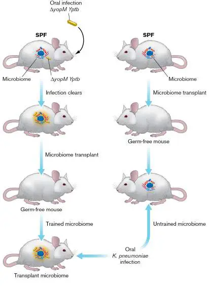
cleared (2 days), the trained microbiome was transplanted to a germ-free mouse. At the same time, an untrained microbiome from an uninfected SPF mouse was transplanted to another germ-free mouse. Both transplanted-germ-free mice were then orally infected with Klebsiella pneumoniae. Four weeks later, feces were assayed for colony-forming units of K. pneumoniae. In early experiments, the authors orally infected both sets of FMT mice with the hospital-associated pathogen Klebsiella pneumoniae (Kpn) carrying chloramphenicol resistance. One day later, they compared Kpn numbers (CFUs) in the feces of both sets as a measure of colonization (Fig. ERA 26.2 ). The results showed that the trained microbiota from postinfection Δ yopM Yptb mice conferred resistance to Kpn colonization.
FIGURE ERA 26.2 ■ Infection-trained microbiota enhance resistance to Kpn colonization. Controls (ctrl) are germ-free mice transplanted with SPF microbiota. Δ yopM are germ-free mice transplanted with microbiota from Δ yopM Yptb - infected SPF mice. Data points (y -axis) represent CFUs of Kpn
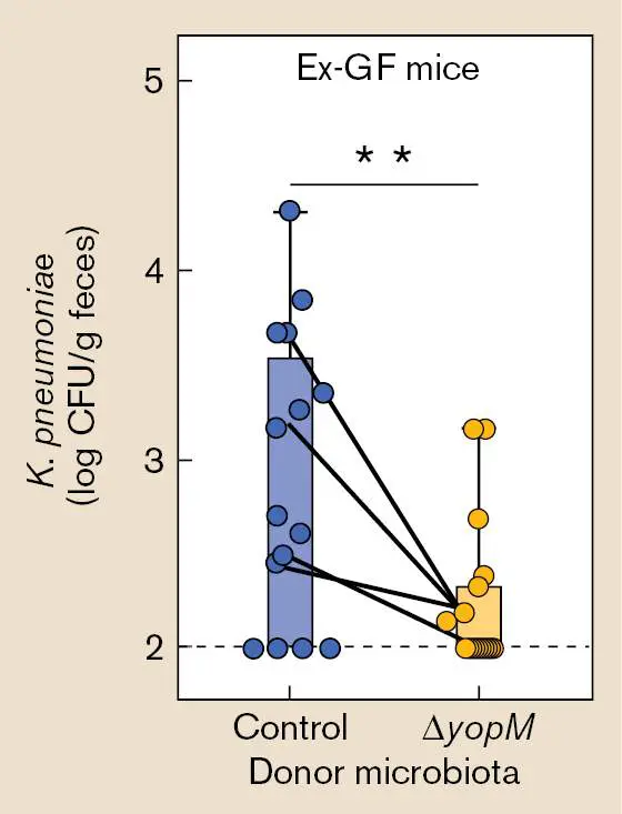
(chloramphenicol resistant) bacteria found per gram of feces per mouse one day after Kpn infection. ** p < 0.01. Each dot represents a different mouse.
After comparing the 16S ribosomal RNA profiles of microbiota from the post-Δ yopM Yptb-infected SPF mice to profiles of microbiota from uninfected SPF mice, the authors discovered that the most reproducibly enriched class in the trained microbiome were the Deltaproteobacteria (recently renamed Myxococcata). Shotgun metagenomes of microbiota from the post-Δ yopM Yptb-infected mice revealed that six enriched metabolic functions could be assigned to sulfur metabolism and menaquinone biosynthesis. A key feature of Deltaproteobacteria is that species in that class can collaborate with species from other phyla to respire anaerobically using sulfur-containing compounds such as taurine or sulfate. Desulfovibrio species, for instance, can convert taurine to sulfite via taurine-pyruvate aminotransferase (encoded by tpa). Bacteroides species can transform sulfate to sulfite using sulfate adenylyltransferase (sat). Then Desulfovibrio species can transform the sulfite to sulfide by dissimilatory sulfite reductase ( dsr) using electrons donated by menaquinone (Fig. ERA 26.3A ). The authors next found that the abundance of tpa genes (from Desulfovibrio) was about 4 times that of sat (from Bacteroides) in the Δ yopM Yptb -trained microbiome, suggesting that taurine rather than sulfate was responsible for the expansion of Deltaproteobacteria. Indeed, mass spectrometry confirmed that taurine was the most significantly increased metabolite in the cecum of post-Δ yopM Yptb-infected mice, even 15 weeks after infection ( Fig. ERA 26.3B ). The source of this taurine appeared to be increased production of taurine-conjugated bile acids in the livers of post-Δ yopM Yptb -infected mice.
FIGURE ERA 26.3 ■ The expansion of Deltaproteobacteria during infection correlates to increased taurine levels in the gut. A. Model for Deltaproteobacteria energy generation. MQ = oxidized menaquinone; MQH 2 = reduced menaquinone. B. Abundance of taurine in the fecal contents of control and post-Δ yopM Yptb mice at >15 weeks post-Δ yopM Yptb infection.
The authors then wondered if adding taurine alone to germ-free mice devoid of microbiota would provide resistance to Kpn colonization. It did not. However, transplanting microbiota harvested from taurine-treated SPF mice to germ-free mice did promote resistance to Kpn (Fig. ERA 26.4A ).
A major by-product of taurine metabolism resulting from the activity of Dsr (sulfite reductase), an enzyme mentioned earlier ( Fig. ERA 26.3A ), is the poisonous gas hydrogen sulfide (H 2 S). H 2 S at high doses can poison cytochrome oxidases used in aerobic electron transport chains to generate usable energy. The metagenomic data suggested that taurine-trained microbiota would produce more H 2 S than the control microbiota. Figure ERA 26.4B illustrates that taurine-trained microbiota produced significantly
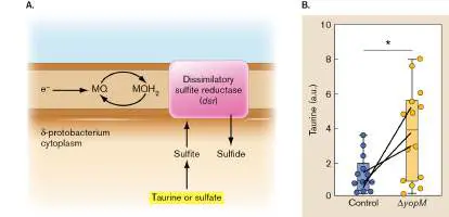
more H 2 S when grown in taurine medium than did control microbiota.
FIGURE ERA 26.4 ■ Taurine-trained microbiota enhance colonization resistance and hydrogen sulfide production.
A. Kpn in the feces of ex-germ-free (Ex-GF) mice that were transplanted with microbiota from vehicle-treated or taurine-treated SPF mice. Counts were determined one-day after Kpn infection. veh = vehicle. B. Ex vivo generation of H 2 S by microbiota taken from vehicle-treated SPF mice (green bars) and taurine-treated SPF mice (purple bars) grown in culture medium without taurine (+veh) or with taurine (+taurine).
The authors then proposed a model that pathogens equipped with cytochrome oxidases can use oxygen as a terminal electron acceptor to metabolize and grow on nonfermentable substrates as long as taurine levels are low. However, at high levels of taurine,
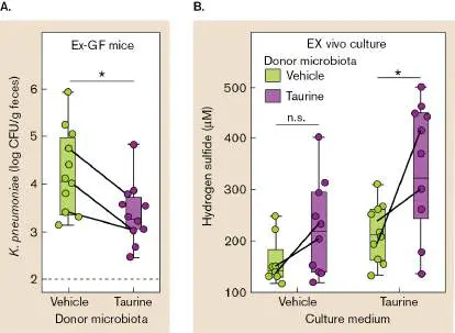
taurine-derived H 2 S would inhibit the cytochrome oxidases and prevent pathogens from using these substrates.
By screening transposon mutants in K. pneumoniae, the team discovered that the metabolism of 1,2-propanediol, a nonfermentable substrate, was required to bolster Kpn fitness (colonization) in control SPF mice (low taurine) but had no effect on colonization in post-Δ yopM Yptb -infected mice (high taurine). Kpn can metabolize 1,2-propanediol only in the presence of oxygen. Consistent with their model, Figure ERA 26.5 illustrates that sodium hydrosulfide prevented Kpn from growing in vitro on 1,2-propanediol under aerobic conditions, whereas the precursors to sulfide such as taurine and taurocholate (a taurine-conjugated bile acid) were not inhibitory, indicating the precursors must be converted to sulfide to be effective.
FIGURE ERA 26.5 ■ Sulfide added in vitro prevents aerobic growth of Kpn on 1,2-propanediol. Taurocholate (5
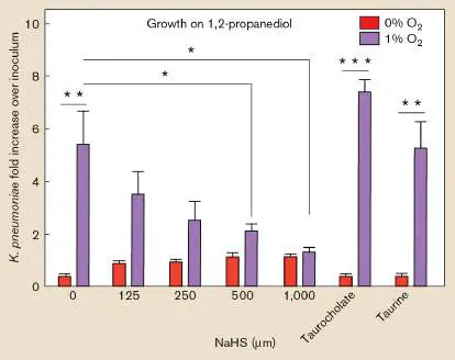
mM) and taurine (5 mM) precursors of sulfide did not affect growth on 1,2-propanediol.
The authors conclude that transient infection can establish long-term “metaorganism memory.” The model is summarized in Figure ERA 26.6 . Metaorganism memory relies on interdependent host and microbiome functions. In this case, the host responds to infection by increasing the production of bile acids. Some members of the microbiome, in turn, convert a bile acid metabolite (taurine) to the antimicrobial sulfide. Sulfide interferes with bacterial cytochrome oxidases, the consequence of which is to change microbiome composition and inhibit the growth of potentially pathogenic bacteria present in low numbers.
FIGURE ERA 26.6 ■ Model of metaorganism memory. A transient infection triggers host responses, including metabolites, that the microbiota use to alter their own composition and limit growth of newly introduced pathogens. In this model, sulfide produced from microbial metabolism of host-made taurine will poison a bacterial cytochrome oxidase needed by potential bacterial pathogens (step 6) and by certain members of the microbiome to aerobically metabolize nonfermentable carbohydrates.
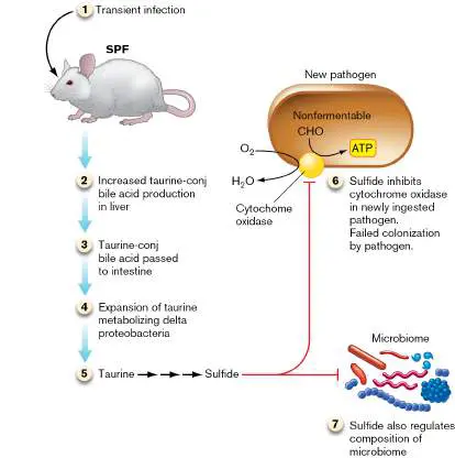
Note that some of these metabolites can also affect immune system memory and stimulate synthesis of antimicrobial peptides. The results of this paper, therefore, suggest that the immune system works in concert with the microbiome to promote colonization resistance against infection. So, the answer to our opening question is that a microbiome can “learn” from an infection! This innovative study was funded by numerous agencies, including the National Institutes of Health, the PEW Latin American Fellows Program, and Human Frontier Science Program.
Further Exploration
Design an experiment using bismuth, a compound that sequesters sulfide, to determine if the composition of resident microbiota will be altered by sulfide in the absence of infection. You can assume that there is a basal level of H 2 S produced even in the absence of an infection.
Apollo Stacy, Vinicius Andrade-Oliveira, John A. McCulloch, Benedikt Hild, and Ji Hoon Oh. 2021. Infection trains the host for microbiota-enhanced resistance to pathogens. Cell 184 :615–627.
https://doi.org/10.1016/j.cell.2020.12.011 CHAPTER REVIEW
Review Questions
1. Organize a list of pathogens by their mechanism of transmission (fecal-oral, aerosol, and so on).
2. Discuss some common skin infections.
3. What causes boils?
4. What are the symptoms of necrotizing fasciitis? 5. What is the difference between primary and secondary infections?
6. How do pneumococci avoid engulfment by phagocytes? 7. What causes the clouding seen in X-rays of infected lungs?
8. What are the key features of the pneumococcal vaccine? 9. Name the common fungal causes of lung disease.
10. What is the clinical course of COVID-19 disease? 11. What is the Ghon complex, and what distinguishes primary from secondary tuberculosis?
12. Why is diarrhea watery?
13. What is the most common microbial cause of diarrhea? 14. List common bacterial agents that cause diarrhea. 15. Would you suspect Salmonella infection in a cluster of nauseated patients rushed to the hospital directly from a church picnic? Why or why not?
16. What is the significance of finding leukocytes in stool? 17. What is one reservoir of E. coli O157:H7?
18. How is a UTI diagnosed?
19. What is an important virulence determinant of uropathogenic E. coli?
20. What is the most common sexually transmitted infection? 21. How is gonorrhea different in men and women?
22. Why will Neisseria gonorrhoeae not usually disseminate in the bloodstream, while N. meningitidis will?
23. Name the major causes of bacterial meningitis. What are the two routes of infection?
24. If tetanus and botulism toxins have the same mode of action, why do they cause opposite effects on muscles? 25. What are some virulence factors of Yersinia pestis? 26. Members of one class of infectious agent cannot be treated with antimicrobial chemotherapy. Explain why.
Thought Questions
1. Chickenpox is a disease of children and young adults caused by herpesvirus 3 (varicella). It is characterized by a rash of fluid-filled vesicles that eventually become crusty. The rash starts on the trunk and spreads to the extremities. Illness usually resolves in 7–10 days, and the patient becomes immune to the disease. However, some individuals later in life (usually over age 50) develop a painful disease, called shingles, that is caused by the same virus—even if they have never been reexposed to the virus. The lesions are tender, persistent vesicles that form on the skin. Propose a plausible explanation, considering the age of the shingles victims and the occurrence of severe pain in this illness, for how they contracted shingles and why the lesions are painful. 2. A 5-year-old boy was brought to the emergency room by his grandmother, who had found him on the floor of her apartment covered in bloody, loose feces. Patient history revealed that the boy attended day care regularly. The diagnostic laboratory determined the etiological agent to be a Gram-negative rod. This organism is a facultative intracellular pathogen that escapes the host cell vacuole and moves in and between cells by actin polymerization. From your reading of Chapters 25 and 26, what do you think are the most likely genus and species involved in this case? Why is the fact that the boy attended day care significant?
3. Why are urinary tract infections among the most commonly acquired nosocomial (hospital-acquired)
infections?
4. On the Internet, find the MMWR (Morbidity and Mortality Weekly Report) that provides the “notifiable infectious diseases” weekly tables (the URL for data as of this writing is http://wonder.cdc.gov/nndss/nndss_weekly_tables_menu.asp). Study the tables that summarize the weekly incidence of gonorrhea and West Nile disease. For the latter, you may need to view one of the arboviral tables to find “West Nile virus disease” or “WNV disease.” Explain the differences in the weekly incidence of these two diseases, and then view the incidence of those diseases in your state.
5. Why do new versions of swine and avian flu often originate in Asia?
Key Terms
bacteremia (1142)
blood-brain barrier (1154) borreliosis (1151)
botulism (1156)
bubonic plague (1148)
cellulitis (1107)
chancre (1135)
chlamydia (1136)
congenital syphilis (1136) cystitis (1132)
differential (1103)
empiric therapy (1106)
endocarditis (1142)
erythema migrans (1150)
granuloma (1115)
health care–associated infection (HAI) (1105) hepatitis (1128)
hepatitis A virus (HAV) (1129) hepatitis B virus (HBV) (1129) hepatitis C virus (HCV) (1130) Lyme disease (1150)
multisystem inflammatory syndrome in children (MIS-C) (1117 )
necrotizing fasciitis (1106) norovirus (1123)
nosocomial (1105)
pneumonic plague (1148)
primary syphilis (1135)
prion (1159)
pyelonephritis (1132)
rehydration therapy (1121) rotavirus (1123)
secondary syphilis (1136) secondary tuberculosis (1116) septicemia (1142)
septicemic plague (1148)
spongiform encephalopathy (1161) tertiary syphilis (1136)
tetanospasmin (1156)
transcytosis (1155)
viremia (1142)
Recommended Reading
Backert, Steffen, Matthias Neddermann, Gunter Maubach, and Michael Naumann. 2016. Pathogenesis of Helicobacter pylori infection. Helicobacter 21 (Suppl.1): 19–25.
Becker-Dreps, Sylvia, Fredman González, and Filemón Bucardo. 2020. Sapovirus: An emerging cause of childhood diarrhea. Current Opinion in Infectious Disease 33 :388–397. https://doi.org/10.1097/QCO.0000000000000671.
Bonneaud, Camille, and Ben Longdon. 2020. Emerging pathogen evolution. EMBO Reports 21 :e51374.
https://doi.org/10.15252/embr.202051374.
Coureuil, Mathieu, Hervé Lécuyer, Sandrine Bourdoulous, and Xavier Nassif. 2017. A journey into the brain: Insight into how bacterial pathogens cross blood-brain barriers. Nature Reviews. Microbiology 15 :149–159.
David, Lawrence A., Ana Weil, Edward T. Ryan, Stephen B. Calderwood, Jason B. Harris, et al. 2015. Gut microbial succession follows acute secretory diarrhea in humans. mBio 6:e00381-15.
Dong, Thi-Thu-Trang, and Katsuya Satoh. 2021. The latest research on RT-QuIC assays—a literature review. Pathogens 10:305. https://doi.org/10.3390/pathogens10030305.
Fleming-Davies, Arietta E., Paul D. Williams, André A. Dhondt, Andrew P. Dobson, Wesley M. Hochachk, et al.
2018. Incomplete host immunity favors the evolution of virulence in an emergent pathogen. Science 359 :1030–1033. https://doi.org/10.1126/science.aao2140.
Gilbert, Nicole M., Valerie P. O’Brien, and Amanda L. Lewis. 2017. Transient microbiota exposures activate dormant Escherichia coli infection in the bladder and drive severe outcomes of recurrent disease. PLoS Pathogens 13 :e1006238. https://doi.org/10.1371/journal.ppat.1006238.
Gilmore, William J., Natalie J. Bitto, and Maria Kaparakis-Liaskos. 2021. Pathogenesis mediated by bacterial membrane vesicles. Subcellular Biochemistry 97 :101–150.
https://doi.org/10.1007/978-3-030-67171-6_6.
Hammond, Andrew M., and Roberto Galizi. 2017. Gene drives to fight malaria: Current state and future directions. Pathogens and Global Health 111 :412–423.
Holmes, Edward C., Stephen A. Goldstein, Angela L.
Rasmussen, David L. Robertson, Alexander Crits-Christoph, et al. 2021. The origins of SARS-CoV-2: A critical review. Cell 184 :4848–4856.
https://doi.org/10.1016/j.cell.2021.08.017.
Kaminski, R., Y. Chen, T. Fischer, E. Tedaldi, A. Napoli, et al. 2016. Elimination of HIV-1 genomes from human T-lymphoid cells by CRISPR/Cas9 gene editing. Scientific Reports 6 :22555. Klein, Roger D., and Scott J. Hultgren. 2020. Urinary tract infections: Microbial pathogenesis, host-pathogen interactions and new treatment strategies. Nature Reviews Microbiology 18:211–226. https://doi.org/10.1038/s41579-020-0324-0.
Leung, Nancy H. L. 2021.Transmissibility and transmission of respiratory viruses. Nature Reviews Microbiology 19 :528–545. Merad, Miriam, Aruna Subramanian, and Taia T. Wang. 2021. An aberrant inflammatory response in severe COVID-19. Cell Host and Microbe 29 :1043-1047 (doi.org/10.1016/j.chom.2021.06.018)
Miroslaw, Jarzab, Gernot Posselt, Nicole Meisner-Kober, and Silja Wessler. 2020. Helicobacter pylori -derived outer membrane vesicles (OMVs): Role in bacterial pathogenesis? Microorganisms 8 :1328.
https://doi.org/10.3390/microorganisms8091328.
Ramírez-Flores, Carlos J., and Laura J. Knoll. 2021.
Breakthroughs in microbiology made possible with organoids. PLOS Pathogens 17 :e1010080.
https://doi.org/10.1371/journal.ppat.1010080.
Rhodes, Johanna. 2019. Rapid worldwide emergence of pathogenic fungi. Cell Host Microbe 26 :12–14.
Saag, Michael S. 2021. HIV infection—screening, diagnosis, and treatment. New England Journal of Medicine 384 : 2131–2143. https://doi.org/10.1056/NEJMcp1915826.
Sela, Uri, Chad W. Euler, Joel Correa da Rosa, and Vincent A. Fischetti. 2018. Strains of bacterial species induce a greatly varied acute adaptive immune response: The contribution of the accessory genome. PLoS Pathogens 14 :e1006726.
https://doi.org/10.1371/journal.ppat.1006726.
Torres, Antoni, Catia Cilloniz, Michael S. Niederman, Rosario Menéndez, James D. Chalmers, et al. 2021. Pneumonia.
Nature Reviews 7 :25. https://doi.org/10.1038/s41572-021-00259-0.
Glossary
differential In disease, the list of possible causes of an infection. nosocomial Hospital-acquired; commonly refers to an infectious agent. health care–associated infection (HAI)
Any infection contracted by a patient while receiving treatment for a medical condition at a health care facility, such as hospitals, nursing homes, doctor offices, or rehabilitation centers.
necrotizing fasciitis Also known as flesh-eating disease. A severe skin infection usually caused by the Gram-positive coccus Streptococcus pyogenes.
empiric therapy An approach to treating infection before the infective organism is known, in which multiple antibiotics are administered in an effort to kill the most likely causative agents.
cellulitis A spreading infection (with inflammation) of connective tissue just below the skin.
granuloma A thick lesion formed around a site of infection.
secondary tuberculosis A new round of serious disease that is caused by Mycobacterium tuberculosis in patients with latent tuberculosis who have become immunocompromised. Symptoms include severe cough, blood sputum, night sweats, and weight loss. multisystem inflammatory syndrome in children (MIS-C) A systemic multi-organ inflammatory syndrome in children brought on by COVID-19 disease.
rehydration therapy A medical treatment for dehydration, in which a liquid solution of salts and glucose is delivered orally. Also called oral rehydration therapy (ORT) .
rotavirus One of a group of nonenveloped dsRNA viruses that cause severe diarrhea in children.
norovirus Also known as Norwalk virus. A nonenveloped ssRNA virus that causes severe diarrhea in children and adults.
hepatitis An inflammation of the liver, caused by infection or by exposure to a toxic substance.
hepatitis A virus (HAV)
A single-stranded RNA picornavirus that causes an acute infection of the liver spread person-to-person by the fecal-oral route.
hepatitis B virus (HBV)
A partially double-stranded, circular-DNA hepadnavirus that causes diseases of the liver of varying severity, including acute and chronic hepatitis, cirrhosis, and hepatocarcinoma. hepatitis C virus (HCV)
A single-stranded, positive-sense, linear-RNA flavivirus that is transmitted by blood transfusions and causes 90% of transfusion-related cases of hepatitis.
cystitis Bladder infection.
pyelonephritis Kidney infection.
primary syphilis The initial inflammatory reaction (chancre) at the site of infection by Treponema pallidum.
chancre A painless, hard lesion due to an inflammatory reaction at the site of infection by Treponema pallidum, the causative agent of syphilis.
secondary syphilis A rash that may appear at some point after the primary latent stage of syphilis.
tertiary syphilis A final stage of syphilis, manifested by cardiovascular and nervous system symptoms.
congenital syphilis Syphilis contracted in utero.
chlamydia 1. A pathogenic bacterium lacking a cell wall; chlamydias (or chlamydiae) grow within human or animal cells, transmitting as elementary bodies to other cells. 2. A disease caused by chlamydia cells. The most frequently reported sexually transmitted disease in the United States. Symptoms range from none, to a burning sensation upon urination, to sterility. septicemia An infection of the bloodstream.
viremia The presence of large numbers of virions in the bloodstream. bacteremia A bacterial infection of the blood.
endocarditis An inflammation of the heart’s inner lining.
bubonic plague A disease caused by the bacterium Yersinia pestis; it is characterized by swollen lymph nodes that often turn black. septicemic plague Infection of the bloodstream by Yersinia pestis.
pneumonic plague A highly virulent and contagious Yersinia pestis lung infection. erythema migrans A bull’s-eye rash characteristic of borreliosis (Lyme disease). Lyme disease One form of borreliosis. A tick-borne disease caused by Borrelia burgdorferi, which may involve skin lesions and arthritis. borreliosis Any of a variety of diseases caused by Borrelia species and transmitted by ticks or lice. Lyme disease is a form of borreliosis.
blood-brain barrier A selectively permeable membrane made up of tightly packed capillaries that supply blood to the brain and spinal cord. Large molecules and most pathogens cannot permeate the narrow spaces. Fat-soluble (lipophilic) molecules and oxygen can dissolve through the capillary cell membranes and are absorbed into the brain.
transcytosis The movement of a cell or substance from one side of a polarized cell to the other side, using an intracellular route. botulism A food-borne disease caused by a Clostridium botulinum toxin, involving muscle paralysis.
tetanospasmin The tetanus-causing potent exotoxin produced by Clostridium tetani.
prion An infectious agent that causes propagation of misfolded host proteins; usually consists of a defective version of the host protein.
spongiform encephalopathy A brain-wasting disease caused by a prion.