Chapter introduction
Resistance of last resort. Most antibiotic resistance mechanisms stop drugs from finding or binding to their targets. However, a “last resort” form of resistance actually
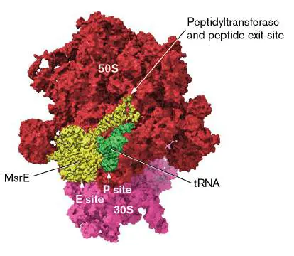
Many of you reading this book would not be alive today had antibiotics not saved you, or your ancestors, from infections. Infections that we consider minor today often killed their victims just 80 to 90 years ago. Now, imagine a world without antibiotics. Suddenly, simple infections would turn deadly, food supplies would be threatened, and human life expectancy would shorten— dramatically. This may sound like science fiction, but we face this prospect today. Many pathogens have become resistant to antibiotics because of indiscriminate antibiotic use in medicine as well as in farming to boost animal growth. For the moment we still have effective antibiotics, but their number is dwindling, and the discovery of new antibiotics has been exceedingly slow. One statistic underscores the seriousness of the situation: The number of patients who contract antibiotic-resistant infections in the United States—currently 2.8 million per year—has nearly tripled since 2002. Many of those patients have died.
We begin Chapter 27 discussing the golden age of antibiotic discovery (1940–1960) and explaining basic concepts of antimicrobial use. We describe how different classes of antibiotics work and then explore the ways pathogens develop resistance. In the process, we examine novel approaches that scientists use to search for new antibiotics. The urgency of this search cannot be overstated.
27.1 Fundamentals of Antimicrobial Therapynot assigned
Antibiotics (from the Greek meaning “against life”) are compounds produced by one species of microbe that can kill or inhibit the growth of other microbes. “Antimicrobials” is a broader term that includes antibiotics but also encompasses synthetic chemotherapeutic agents, such as sulfonamides, that are clinically useful but chemically made in the laboratory. Over time, the distinction between these words has blurred to where they are often used interchangeably.
We think of antibiotics as being a recent biotechnological development, but they have actually been used for centuries. Ancient remedies called for cloths soaked with organic material to be placed on wounds to help them heal faster. This organic material likely contained natural antibiotics that killed bacteria and prevented further infection. The medicinal properties of molds were also recognized for centuries. The ancient Chinese successfully treated boils with warm soil and molds scraped from cheeses, and in England a paste of moldy bread was a home remedy for wound infections until the beginning of the twentieth century.
The Golden Age of Antibiotic Discovery
The modern antibiotic revolution began with the discovery of penicillin in 1928 by Sir Alexander Fleming (1881–1955; Fig. 27.1C ). This discovery was actually a rediscovery, and it is arguably one of the greatest examples of serendipity in science. Although Fleming generally receives the credit for discovering penicillin, a French medical student, Ernest Duchesne (1874– 1912), originally discovered the antibiotic properties of Penicillium in 1896.
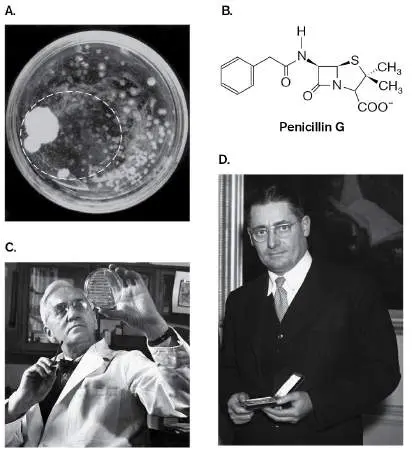
FIGURE 27.1 ■ The dawn of antibiotics. A. Alexander Fleming’s photo of the dish with bacteria and penicillin mold. The ring highlights the area of decreased growth of Staphylococcus aureus colonies. B. The
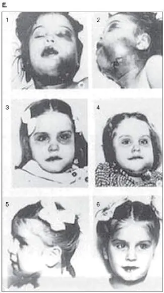
chemical structure of penicillin G. C. Alexander Fleming at work in his laboratory. D. Howard Florey. E. These pictures, taken in 1942, shortly after the introduction of penicillin, show the improvement in a child suffering from an infection 4 days (panel 2) and 9 days (panel 4) after treatment. Panels 5 and 6 show her fully recovered.
ST. MARY’S HOSPITAL MEDICAL SCHOOL/SCIENCE SOURCE
ALFRED EISENSTAEDT/PIX INC./THE LIFE PICTURE COLLECTION VIA GETTY IMAGES
J. A. HAMPTON/GETTY IMAGES
PHOTOGRAPHS USED WITH PERMISSION FROM MAYO CLINIC PROCEEDINGS
Duchesne observed that Arab stable boys at the nearby army hospital kept their saddles in a dark and damp room to encourage mold to grow on them. When asked why, they told him the mold helped heal saddle sores on the horses. Intrigued, Duchesne prepared a solution from the mold and injected it into guinea pigs infected with typhoid fever bacillus. All recovered.
Penicillium was forgotten in the scientific community until Fleming rediscovered it one day in the late 1920s. Petri dishes were glass in those days and could be rewashed and sterilized. Fleming was preparing to wash a pile of old Petri dishes he had used to grow the pathogen Staphylococcus aureus. He opened and examined each dish before tossing it into a cleaning solution. He noticed that one dish had grown contaminating mold, which in itself was not unusual in old plates, but all around the mold the staph bacteria had failed to grow (Fig. 27.1A).
Fleming took a sample of the mold and found that it was from the penicillium family; the sample was later identified as Penicillium notatum. The mold appeared to have synthesized a chemical, now known as penicillin (Fig. 27.1B ), which diffused through the agar, killing cells of S. aureus before they could form colonies. Fleming (Fig. 27.1C ) presented his findings in 1929, but they raised little interest, since penicillin appeared to be unstable and was not active in the body long enough to kill pathogens. As World War II began, Oxford professor Howard Florey (Fig. 27.1D ) and his colleague Ernst Chain rediscovered Fleming’s work, thought it held promise, and set about purifying penicillin. To their amazement, the purified penicillin cured mice infected with staphylococci or streptococci. Subsequent human trials proved successful (Fig. 27.1E ), and penicillin gained wide use, saving countless lives during the war. Fleming, Florey, and Chain received the 1945 Nobel Prize in Physiology or Medicine for their work. The next landmark discovery in antibiotics was made by Gerhard Domagk (1895–1964; Fig. 27.2A), a German physician at the Bayer Institute of Experimental Pathology and Bacteriology. In 1935, Domagk’s 6-year-old child developed a serious streptococcal infection induced by an innocent pinprick to the finger. The infection spread to her axillary (armpit) lymph nodes, and it became so severe that lancing and draining the pus 14 times did little to help. The only remaining alternative was to amputate the arm. Unfortunately, even this option would probably not save her life. FIGURE 27.2 ■ The discoverers of sulfanilamide and streptomycin. A. Gerhard Domagk discovered sulfanilamide. B.
Chemical structure of sulfanilamide, an analog of para -aminobenzoic
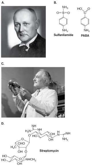
acid (PABA), a precursor of the vitamin folic acid, which is necessary for growth. Sulfanilamide inhibits one of the enzymes that converts PABA into folic acid. C. Selman Waksman discovered streptomycin in 1944. D. Chemical structure of streptomycin.
GEORGE RINHART/GETTY IMAGES
BETTMANN/GETTY IMAGES
Frustrated, Domagk took what would appear to be drastic measures. He gave his daughter a dose of a red dye (Prontosil) that he was investigating as an antimicrobial compound. On agar plates (the usual medium for testing antibiotics), Prontosil had shown absolutely no ability to inhibit the growth of streptococcus. Domagk, however, had tested the drug in animals, not agar plates, and found that it could cure animals of infection. Remarkably, Domagk’s daughter recovered completely.
Domagk discovered that Prontosil was metabolized by the body into another compound, sulfanilamide, which was clearly lethal to the streptococcus. This finding led to an entire class of drugs, called the sulfonamides, or sulfa drugs, that saved hundreds of thousands of lives. The take-home message of this story is that an antibiotic’s activity on a plate, or lack thereof, does not necessarily correlate with the drug’s activity in a patient.
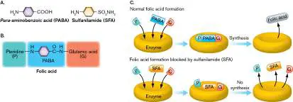
Sulfanilamide is an analog of para -aminobenzoic acid (PABA), a precursor of folic acid, a vitamin necessary for nucleic acid synthesis (Fig. 27.2B ). Sulfanilamide and other sulfa drugs bind to and inhibit the enzyme that converts PABA to folic acid. Without folic acid to make nucleic acid precursors, the pathogen stops growing. Sulfa drugs inhibit bacterial growth without affecting human cells because folic acid is not synthesized by humans (it is a dietary supplement instead) and because bacteria do not transport folic acid (they must make it themselves).
In a dark turn, Domagk’s possible participation in human experimentation with concentration camp prisoners during World War II (ordered by his German employer) was a source of controversy that haunted him long after the war ended. Yet his contributions to medicine continued; he also developed two effective chemotherapeutic agents for tuberculosis— namely, the thiosemicarbazones and isoniazid—that are still used today. During the same period of history, Selman Waksman (1888–1973; Fig. 27.2C ) at Rutgers University began screening 10,000 strains of soil bacteria and fungi for their ability to inhibit growth or kill bacteria. In 1944, this herculean effort paid off with the discovery of streptomycin, an antibiotic produced by the actinomycete Streptomyces griseus (Fig. 27.2D ). Waksman’s discovery of streptomycin triggered the antibiotic gold rush and earned him the 1952 Nobel Prize in Physiology or Medicine.
Antibiotics Exhibit Selective Toxicity
As early as 1904, the German physician Paul Ehrlich (1854–1915) realized that a successful antimicrobial compound would be a “magic bullet” that would selectively kill or inhibit the pathogen but not the host. This seemingly obvious premise was innovative at the time. Ehrlich made several discoveries based on this concept, the most celebrated of which was the arsenical compound known as Salvarsan. Salvarsan proved to be quite effective in killing the syphilis agent Treponema pallidum (this was long before penicillin was discovered). Syphilis, a sexually transmitted infection, had been untreatable and the source of considerable long-term suffering. Ehrlich’s “magic bullet” concept is now known as selective toxicity. Salvarsan, however, was not as selectively toxic as Ehrlich thought. This arsenical compound did harm the host, but usually it killed off the treponemes before killing the patient.
Selective toxicity is possible because key aspects of a microbe’s physiology are different from those of eukaryotes. For example, suitable antibiotic targets in bacteria include peptidoglycan, which eukaryotic cells lack, and ribosomes, which are structurally distinct between Bacteria and Eukarya. Thus, chemicals like penicillin, which prevents peptidoglycan synthesis, and tetracycline, which binds to bacterial 30S ribosomal subunits, inhibit bacterial growth but are essentially invisible to host cells because these drugs will not interact with host structures at the low doses used to treat infections.
Although their intended targets are bacterial cells, some antibiotics, particularly at high doses, can interact with elements of eukaryotic cells and cause side effects that harm the patient. For example, chloramphenicol, a drug that targets bacterial 50S ribosomal subunits, can interfere with the development of blood cells in bone marrow—a phenomenon that may result in aplastic anemia (failure to produce red blood cells). Fluoroquinolones like ciprofloxacin can cause tendon ruptures, and aminoglycosides such as gentamicin or kanamycin can damage hair cells in the ear, leading to hearing loss. Another problem caused by drugs is that of allergic reactions. For example, many people develop an extreme allergic sensitivity to penicillin, in which case the treatment of an infection may end up being worse than the infection itself. Physicians must be aware of these allergies and use alternative antibiotics to avoid harming their patients.
Note: As a student of microbiology, you must be able to properly
distinguish between the terms “drug susceptibility” and “drug sensitivity.” A microbe is susceptible to the drug’s action, but a human can develop an allergic sensitivity to the drug.
Spectrum of Activity
Because no single antimicrobial drug affects all microbes, antimicrobial drugs are classified by the types of organisms they affect. Thus, we have antifungal, antibacterial, antiprotozoan, and antiviral agents. The term “antibiotic” is usually reserved for compounds that affect bacteria. Even within a group, one agent might have a very narrow spectrum of activity, meaning that it affects only a few species, while another antibiotic inhibits many species. For instance, penicillin has a relatively narrow spectrum of activity, killing primarily Gram-positive bacteria. However, ampicillin is penicillin with an added amino group that enables the drug to more easily penetrate the Gram-negative outer membrane. As a result, ampicillin kills Gram-positive and Gram-negative organisms, giving it a broader spectrum of activity than penicillin has. There are antimicrobials as well that exhibit extremely narrow activities. One example is isoniazid, which is clinically useful only against Mycobacterium tuberculosis, the agent of tuberculosis. eResearch Activity 27 explores the discovery of a narrow-spectrum antibiotic, hygromycin A, that works best on the spirochetes that cause Lyme disease and syphilis.
Patients typically believe that all antibiotics kill their intended targets. This is a misconception. Many drugs simply prevent growth of the organism and let the body’s immune system dispatch the intruding microbe. Thus, antimicrobials are also classified on the basis of whether or not they kill the microbe. An antibiotic is bactericidal if it kills the target microbe but bacteriostatic if it merely prevents bacterial growth. A bacteriolytic antibiotic, such as penicillin, is bactericidal and also results in cell lysis.
Measuring Drug Susceptibility
One critical decision a clinician must make when treating an infection is which antibiotic to prescribe for the patient. There are several factors to consider, including: The relative effectiveness of different antibiotics on the organism causing the infection. The less effective a drug is at stopping growth, the less effective it will be at treating the infection. The average attainable tissue levels of each drug. An antibiotic may work on an agar plate, but the concentration at which it affects bacterial growth may be too high to be safe in the patient.
The route of administration. The easiest way to deliver an antibiotic is orally. However, some antibiotics are not absorbed well from the intestine. For instance, ceftriaxone, a third-generation cephalosporin, is not absorbed well in the gut but is effectively distributed to tissues when given intravenously (IV). Cefixime, another third-generation cephalosporin, can be taken orally and will distribute to tissues. Minimal inhibitory concentration. The in vitro effectiveness of an antimicrobial agent is determined by measuring how little of it is needed to stop growth. This amount is classically measured in terms of an antibiotic’s minimal inhibitory concentration (MIC), defined as the lowest concentration of the drug that will prevent the growth of an organism. But the MIC for any one drug will differ among different bacterial species. For example, the MIC of ampicillin needed to stop the growth of Staphylococcus aureus will be different from that needed to inhibit Shigella dysenteriae. The reasons that a drug may be more effective against one organism than another include the ease with which the drug penetrates the bacterial cell and the affinity of the drug for its molecular target.
So, how do we measure MIC? As shown in Figure 27.3A, an antibiotic is serially diluted along a row of test tubes containing nutrient broth. After dilution, the organism to be tested is inoculated at low, constant density into each tube, and the tubes are usually incubated overnight. Growth of the organism is seen as turbidity. In Figure 27.3A, the tubes with the highest concentration of drug are clear, indicating no growth. The tube containing the MIC is the tube with the lowest concentration of drug that shows no growth. Note, however, that the MIC does not indicate whether a drug is bacteriostatic or bactericidal. Today, clinical laboratories use microtiter plates read by automated systems to determine MICs (Fig. 27.3B and Chapter 28).
FIGURE 27.3 ■ Determining minimal inhibitory concentration (MIC). A. In this series of 5-ml tubes, tetracycline was diluted serially starting at 8.0 μg/ml. Each tube was then inoculated with an equal number of bacteria (Salmonella enterica). Turbidity indicates that the
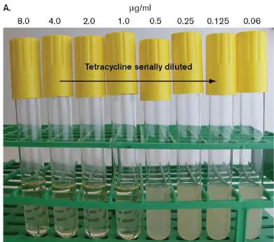
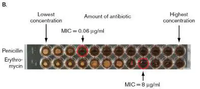
antibiotic concentration was insufficient to inhibit growth. The MIC in this example is 1.0 μg/ml. B. Microdilution, using a microtiter plate, to determine MIC. Pictured are two rows of a 96-well plate in which dilutions of penicillin (upper row) and erythromycin (lower row) were tested against group B streptococci. The principle is the same as in (A), except smaller volumes (200 μl) are used. The MIC in each row is circled.
COURTESY OF DR. JOHN W. FOSTER
COURTESY OF LESLEY MCGEE, CDC
Thought Questions
27.1 Figure 27.3 illustrates how MICs are determined. Test your understanding of how MICs are measured in the following example. The drug tobramycin is added to a concentration of 1,000 μg/ml in a tube of broth from which serial twofold dilutions are made. Including the initial tube (tube 1), there are ten tubes. Twenty-four hours after all the tubes are inoculated with Listeria monocytogenes, turbidity is observed in tubes 6– 10. What is the MIC?
27.2 What additional test performed on an MIC series of tubes will tell you whether a drug is bacteriostatic or bactericidal?
MIC determinations are very useful for estimating a single drug’s effectiveness against a single bacterial pathogen isolated from a patient, but they are not very practical to a technician trying to screen 20 or more different drugs. Dilutions take time—time that the technician, not to mention the patient, may not have. The time required to evaluate antibiotic effectiveness can be reduced by use of a strip test (like the ETEST shown in Fig. 27.4) that avoids the need for dilutions. The strip, containing a gradient of antibiotic, is placed on an agar plate freshly seeded with a dilute lawn of bacteria. While the bacteria are trying to grow, the drug diffuses out of the strip and into the media. The drug will diffuse at equal rates from all points along the strip. However, drug diffusing away from the most concentrated part of the strip will maintain a higher concentration in the agar per unit of time compared to that diffusing from less concentrated parts of the strip. Thus, the drug’s effect (killing or inhibiting the growth of cells) will extend farther away from the strip at locations of high concentration than at locations of lower concentration. The result is a zone of inhibition where the antibiotic has stopped bacterial growth. The MIC is the point at which the elliptical zone of inhibition intersects with the strip. FIGURE 27.4 ■ An MIC strip test. The ETEST (developed by AB Biodisk) is a commercially prepared strip that produces a gradient of antibiotic concentration (in μg/ml) when placed on an agar plate. The
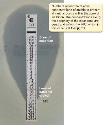
MIC corresponds to the point where bacterial growth crosses the numbered strip. C/T=ceftolozane/tazobactam.
COURTESY OF BIOMÉRIEUX
Kirby-Bauer disk susceptibility test. Although the strip test eliminates the time and effort needed to make dilutions, it would take 20 or more plates to test an equal number of antibiotics for just one bacterial isolate. Clinical labs can receive up to 100 or more isolates in one day, so individual MIC determinations are impractical. A simplified agar diffusion test, however, which can test 12 antibiotics on one plate, makes evaluating antibiotic susceptibility a manageable task.
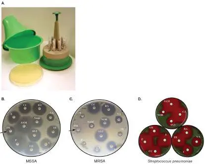
Named for its inventors, the Kirby-Bauer assay uses a series of round filter paper disks impregnated with different antibiotics. A dispenser (Fig. 27.5A) delivers up to 12 disks simultaneously to the surface of an agar plate covered by a bacterial lawn. Each disk is marked to indicate the drug used. During incubation, the drugs diffuse away from the disks into the surrounding agar and prevent growth of the lawn (Fig. 27.5B–D ). The zones of inhibition vary in width, depending on the antibiotic used, the concentration of the drug in the disk, and the susceptibility of the organism to the drug. The diameter of the zone correlates to the MIC of the antibiotic against the organism tested. Figure 27.5B and C show the results for methicillin-sensitive and methicillin-resistant Staphylococcus aureus (MSSA and MRSA, respectively). Note that oxacillin, an antibiotic structurally similar to methicillin, did not inhibit the growth of the MRSA strain (compare arrows in the two photos). Because oxacillin is used clinically to treat infections and methicillin is not, laboratories use oxacillin resistance to identify MRSA strains.
FIGURE 27.5 ■ The Kirby-Bauer disk susceptibility test. A. This device delivers up to 12 disks to the surface of a Mueller-Hinton plate. B–D. Disks impregnated with different antibiotics are placed on a freshly laid lawn of bacteria and incubated overnight. The clear zones around certain disks indicate growth inhibition. C, chloramphenicol; CC, clindamycin; CZ, cefazolin; E, erythromycin; NOR, norfloxacin; OX, oxacillin; P, penicillin; RA, rifampin; SAM, sulbactam-ampicillin; SXT, sulfa-trimethoprim; TE, tetracycline; VA, vancomycin. Results are shown for methicillin-sensitive Staphylococcus aureus (MSSA) (B) , methicillin-resistant S. aureus (MRSA) (C) , and Streptococcus pneumoniae (D) .
The arrow in (C) points out MRSA’s resistance to oxacillin, compared to the lack of resistance exhibited by MSSA in (B). The brownish tint of the blood agar plates outside the zones of bacterial inhibition is caused by a hemolysin secreted by the lawn of pneumococci.
COURTESY OF DR. JOHN W. FOSTER
COURTESY OF DR. JOHN W. FOSTER
COURTESY OF DR. JOHN W. FOSTER
COURTESY OF DR. JOHN W. FOSTER
Correlations between MIC values determined in broth and Kirby-Bauer zone sizes on agar are made empirically. Every disk containing a given antibiotic is impregnated with a standard concentration of that drug, but the standard concentration used may be different for each drug. The outermost ring of the no-growth zone in a Kirby-Bauer disk test must, by definition, contain the minimal concentration of drug needed to prevent growth on agar. Consequently, if species A and B have MIC values for penicillin of 4μg/ml and 40μg/ml, respectively, then species A will exhibit a proportionally larger zone of inhibition than species B in the disk test. A graph plotting MIC on one axis and zone diameter on the other provides the correlation.
After the agar plates are incubated, the diameters of the zones of inhibition around each disk are measured, and the results are compared with a table listing whether a zone is wide enough (meaning the MIC is low enough) to be clinically useful. Table 27.1 shows susceptibility data for S. aureus. The zone diameter (MIC) that indicates an organism will be clinically susceptible to the antibiotic correlates to the average attainable tissue level for that antibiotic (discussed later). For the antibiotic to remain effective in vivo, the tissue concentration of the drug must remain above the MIC; otherwise, the bacteria can grow, and spontaneous mutations providing drug resistance could develop.
TABLE Susceptibility Results for
27.1 Staphylococcus aureus
Zone of inhibition diameter (mm) Antibiotic Quantity Resistant Intermediate Susceptible in disk (μg)
Ampicillin <12 >13 10 12–13 Chloramphenicol <13 >17 30 13–17 TABLE Susceptibility Results for
27.1 Staphylococcus aureus
Erythromycin <14 >17 15 14–17 Gentamicin ≤12.5 >12.5 10 Streptomycin <12 >14 10 12–14 Tetracycline <15 >18 30 15–18 To ensure reproducibility, the Kirby-Bauer test was standardized over 60 years ago. Reproducibility means that results from a laboratory in California will match those in Alabama, Ohio, or any other location. The following are standardizations used to make the test reproducible and easier.
Size of the agar plate. 150 mm.
Depth of the media. 4 mm. Because antibiotics diffuse out of disks in three dimensions, the zone of inhibition measured on a thinly poured agar plate will be larger than the zone from a thick agar plate.
Media composition. Media should lack PABA. Sulfonamide antibiotics inhibit PABA synthesis in bacteria, so the presence of PABA in testing media bypasses any block imposed by the sulfonamides. The standardized medium used for the Kirby-Bauer test, called Mueller-Hinton agar, contains no PABA.
The number of organisms. The more organisms that are spread on an agar plate, the less time an antibiotic has to diffuse before visible growth develops. Consequently, the zone of inhibition will appear smaller than it should. To prevent this phenomenon, standard optical density solutions (0.06 at 600 nm) of each organism are prepared and a cotton swab is used to deliver organisms evenly over the entire agar surface. A commercially prepared suspension of latex particles (the new McFarland standards) is used to visually estimate the optical density of culture dilutions of the test organism.
Size of the disks. A standard diameter of 6 mm means that all antibiotics start diffusing into the agar at the same point.
Concentrations of antibiotics in the disks. The zone of inhibition for an antibiotic is proportional to the concentration of antibiotic in the disk. The higher the concentration of antibiotic in a disk, the farther the drug can diffuse and maintain a concentration above the MIC. To avoid differences between labs, the concentration of each drug impregnating a disk has been standardized.
Incubation temperature. Incubation temperature will not affect growth and diffusion equally. To avoid differences, a temperature of 37°C is standard (body temperature).
Automated MIC determinations. The methods just described to determine MIC are effective but take at least 24 hours to complete (the amount of time needed to see visible turbidity in broth or growth on agar). Modern clinical laboratories today are equipped with automated machines (described in Chapter 28) that can determine an MIC within 6 hours. This speed is critical to a clinician wanting to quickly treat a serious infection with the most effective drug. The instruments work by monitoring growth of the infectious agent in microtiter plate wells and extrapolating the MIC from growth curves recorded at different antibiotic concentrations.
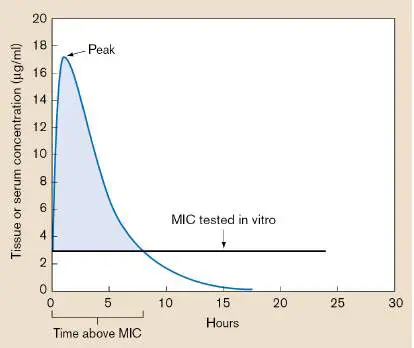
Correlating antibiotic MIC with tissue level. The average attainable tissue level for a drug depends on how quickly the antibiotic is cleared from the body via secretion by the kidney or destruction in the liver. It also depends on when side effects of the drug start to appear. The graph in Figure 27.6shows that as long as the concentration of the drug in tissue or blood remains higher than the MIC, the drug will be effective. The clinician can keep the concentration at sufficient levels either by initially administering a higher dose (running the risk of side effects) or by giving a second dose before the blood levels from the first dose decline below the MIC. This is why patients are told to take doses of some antibiotics four times a day and other antibiotics only once a day.
FIGURE 27.6 ■ Correlation between MIC and serum or tissue level of an antibiotic. This graph illustrates the serum level of ampicillin over time. The important consideration here is how long the serum level of the antibiotic remains higher than the MIC. Once the concentration falls below the MIC, owing to destruction of the drug in the liver or clearance through the kidneys and secretion, the infectious agent fails to be controlled by the drug—in this case, 7–8 hours after the initial dose. To maintain a serum level higher than the MIC, a second dose would be taken. The shaded area of the curve represents time above MIC.
Fluctuating serum levels of an antibiotic also explain why the patient should never miss a dose. Missing a dose allows the serum (or tissue) level to fall below the MIC for the pathogen. When that happens, any bacteria still living can once again grow and possibly develop spontaneous antibiotic resistance mutations. These antibiotic-resistant mutants will continue to
grow and cause disease even after the serum level of antibiotic is restored. The result is called treatment failure.
Thought Questions
27.3 A patient with a bacterial lung infection was given the antibiotic represented in Figure 27.6and was told to take one pill twice a day. The pathogen is susceptible to this drug. Will the prescribed treatment be effective? Explain your answer.
27.4 You are testing whether a new antibiotic will be a good treatment choice for a patient with a staph infection. The Kirby-Bauer test using the organism from the patient shows a zone of inhibition of 15 mm around the disk containing this drug. Clearly, the organism is being affected by the drug in vitro. But you conclude from other studies that the drug would be ineffective in the patient. What would make you draw this conclusion?
To Summarize
The importance of antimicrobials in treating disease was recognized in the early 1940s. Some antimicrobials are naturally produced by living organisms (antibiotics); others are synthetically made through chemical engineering.
Selective toxicity is the ability of an antibiotic to attack a unique component of microbial physiology that is missing or distinctly different from eukaryotic physiology. However, antibiotic side effects (host toxicity) can limit the clinical usefulness of an antimicrobial agent.
Antibiotic spectrum of activity is the range of microbes affected by a given drug.
Bactericidal antibiotics, or antimicrobials , kill microbes; bacteriostatic antibiotics inhibit microbial growth; and some antimicrobial agents are initially inactive until converted by the body to an active agent.
An antibiotic’s spectrum of activity and the infectious agent’s susceptibility to the antibiotic are critical points of information required before an antibiotic therapy is prescribed.
Minimal inhibitory concentration (MIC) of a drug, when correlated with average attainable tissue levels of the antibiotic, can predict the effectiveness of an antibiotic in treating disease. MIC is measured by tube dilution techniques, but it can be approximated by the Kirby-Bauer disk diffusion technique.
Glossary
antibiotic A molecule that can kill or inhibit the growth of selected microorganisms.
penicillin An antibiotic, produced by the Penicillium mold, containing a beta-lactam ring; it blocks cross-bridge formation during peptidoglycan synthesis.
sulfa drug An antibiotic that inhibits folic acid synthesis and, thus, nucleotide synthesis.
selective toxicity The ability of a drug, at a given dose, to harm the pathogen and not the host.
spectrum of activity The range of pathogens for which an antimicrobial agent is effective. bactericidal Having the ability to kill bacterial cells.
bacteriostatic Having the ability to inhibit the growth of bacterial cells. minimal inhibitory concentration (MIC)
The lowest concentration of a drug that will prevent the growth of an organism.
zone of inhibition A region of no bacterial growth on an agar plate that is due to the diffusion of a test antibiotic. Correlates to the minimal inhibitory concentration.
Kirby-Bauer assay A method for determining antibiotic susceptibility. Antibiotic-impregnated disks are placed on an agar plate whose surface has been confluently inoculated with a test organism. The antibiotic diffuses away from the disk and inhibits growth of susceptible bacteria. The width of the inhibitory zone is proportional to the susceptibility of the organism.
Mueller-Hinton agar A specialized, standardized, para -aminobenzoic acid–free medium used for the Kirby-Bauer assay.
27.2 Antibiotic Mechanisms of Actionnot assigned
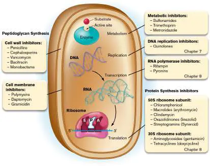
As noted in the preceding section, selective toxicity of an antibiotic depends on enzymes or structures unique to the bacterial target cell. The following aspects of a microbe’s physiology are classic targets: Cell wall synthesis Cell membrane integrity DNA synthesis RNA synthesis Protein synthesis Metabolism Table 27.2 summarizes the general targets of common antibiotics. Chapters 3, 7, and 8 describe these cell components and provide the basis for understanding how antibiotics work. Because the mechanisms of action for antibiotics affecting DNA, RNA, and protein synthesis are described in Chapters 7 and 8, they receive only brief mention here. Figure 27.7 summarizes the general mechanisms of action for these drugs.
Targets of Antimicrobial
TABLE 27.2
Agents
Target Antibiotic examples Cell wall synthesis Penicillins, cephalosporins, bacitracin, vancomycin
Targets of Antimicrobial
TABLE 27.2
Agents
Protein synthesis Chloramphenicol, tetracyclines, aminoglycosides, macrolides, lincosamides Cell membrane integrity Polymyxin, daptomycin, amphotericin, imidazoles (vs. fungi)
Nucleic acid function Nitroimidazoles, nitrofurans, quinolones, rifampin; some antiviral compounds, especially antimetabolites Intermediary Sulfonamides, trimethoprim metabolism FIGURE 27.7 ■ Summary of antimicrobial agents and their targets. Antibiotics that target DNA synthesis, RNA synthesis, and protein synthesis are described in Chapters 7 and 8.
Source: Modified from Foster et al. 2018. Microbiology: The Human Experience.
W. W. Norton.
Cell Wall Antibiotics
Bacterial cell walls are the basis of selective toxicity for some antibiotics because peptidoglycan does not exist in mammalian cells; thus, antibiotics that target the synthesis of these structures should selectively kill bacteria. The following case history illustrates the use of two cell wall–targeting antibiotics and also reveals how bacteria can evolve to escape destruction.
Case History: Meningitis
A 3-year-old crying child was brought to the emergency department with a stiff neck and high fever. Gram stain of cerebrospinal fluid revealed Gram-positive cocci, generally in pairs. The diagnosis was meningitis. The physician immediately prescribed intravenous ampicillin. Unfortunately, the child’s condition worsened, so antibiotic treatment was changed to a third-generation cephalosporin (which more easily crosses the blood-brain barrier). The patient began to improve within hours and was released after 2 days. A report from the clinical microbiology laboratory identified the organism as Streptococcus pneumoniae.
Both of the antibiotics used in this case kill bacteria by targeting cell wall synthesis (introduced in Section 3.3). To synthesize peptidoglycan, sugar molecules called N -acetylglucosamine (NAG) and N -acetylmuramic acid (NAM) are made by the cell and linked together by a transglycosylase enzyme into long chains assembled at the cell wall (Fig. 27.8). N -acetylmuramic acid contains a short side chain of amino acids that is assembled enzymatically, not by a ribosome. The side chains from adjacent strands are cross-linked to make the structure rigid. The enzyme transpeptidase (D -alanyl-D - alanine carboxypeptidase/transpeptidase) catalyzes the cross-link. Several antibiotics target various stages of this assembly process.
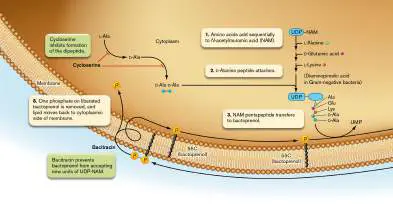
FIGURE 27.8 ■ Peptidoglycan synthesis in a Gram-positive bacterium, and the targets of antibiotics. Several small-molecular-weight compounds are sequentially joined to form a disaccharide unit that will be added to preexisting extracellular chains of this unit. Red lines indicate inhibition. Cycloserine inhibits ligation of the two D -alanines (step 2); bacitracin inhibits linking of the disaccharide units; vancomycin and the beta-lactams, such as penicillin, inhibit the peptide cross-linking of peptidoglycan side chains.
Peptidoglycan synthesis. Before we can explain how cell wall antibiotics work, we need to know how the cell wall is made. Synthesis of peptidoglycan starts in the cytoplasm with a uridine diphosphate (UDP)–NAM molecule. The amino acids L -alanine, D - glutamic acid, and L -lysine [or diaminopimelic acid (DAP) in Gram-negative organisms] are individually and sequentially added to NAM ( Fig. 27.8, step 1), and then a dipeptide of D -alanine is attached (step 2). Next, the NAM pentapeptide is transferred to a membrane-situated, 55-carbon lipid molecule called bactoprenol (step 3), releasing uridine monophosphate (UMP). This structure is also called
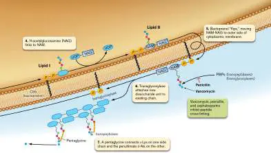
lipid I. Another sugar molecule, NAG, is then linked to NAM—once again through a UDP intermediate (step 4)—to make the bactoprenol structure called lipid II. All of this takes place on the cytoplasmic side of the membrane.
Bactoprenol then “flips,” moving NAM-NAG to the outer side of the cytoplasmic membrane (Fig. 27.8, step 5), where transpeptidases and transglycosylases (two penicillin-binding proteins, or PBPs) bind to the D -Ala-D -Ala part of the pentapeptide (the PBP complex is illustrated in Fig. 3.13). Transglycosylase attaches the new disaccharide unit to an existing peptidoglycan chain (step 6) and releases bactoprenol. Transpeptidase then links two peptide side chains from adjacent peptidoglycan molecules with a pentaglycine cross-link (in Staphylococcus aureus). The pentaglycine connects L - Lys on one side chain and the penultimate D -Ala on the other side chain (step 7). The terminal D -Ala is removed in the process. Other bacteria do not use a pentaglycine cross-link but directly form a peptide bond between L -Lys (or DAP in Gram-negative organisms) and the penultimate D -Ala (shown in Fig. 3.18). Cross-linking strengthens the cell wall. The bactoprenol liberated in step 6 loses one of its phosphates and recycles back to the cytoplasmic side of the membrane, ready to pick up and taxi another unit of peptidoglycan to the growing chain (Fig. 27.8, step 8).
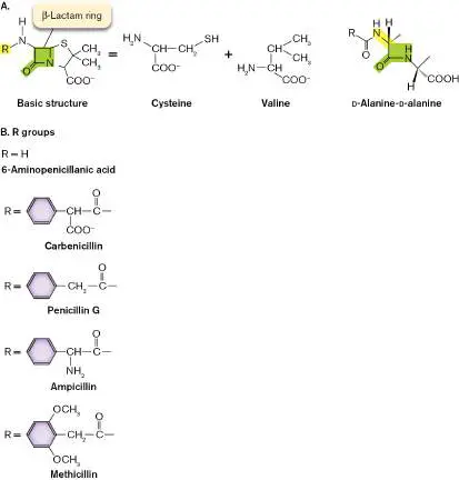
Beta-lactam antibiotics. Penicillin is an antibiotic derived from cysteine and valine, which are condensed by fungal enzymes to form the beta-lactam ring structure shown in Figure 27.9A. Different R groups can be added to the basic beta-lactam ring structure to change the antimicrobial spectrum and stability of the derivative penicillin ( Fig. 27.9B ). Note that the beta-lactam ring of penicillin chemically resembles the D -Ala-D -Ala piece of peptidoglycan, as highlighted by green shading in Figure 27.9A. This molecular mimicry enables penicillin and other beta-lactam antibiotics to target so-called penicillin-binding proteins, including transpeptidase and transglycosylase, that carry out cell wall synthesis and remodeling. FIGURE 27.9 ■ The structure of penicillins. A. Penicillanic acid (R group = H) is derived from cysteine and valine. Also shown is the D -alanine-D -alanine structure of peptidoglycan (far right), which is structurally similar to the beta-lactam ring of penicillins (green shading). B. The R group highlighted in (A) can be any one of a number of different groups, some of which are shown here. Modifying this group changes the pharmacological properties and antimicrobial spectrum of the drug.
Penicillins act by inhibiting transpeptidase-mediated cross-linking between adjacent peptidoglycan chains. This activity makes the cell
wall very weak. In addition, penicillins can somehow activate proteins in the cell wall that hydrolyze peptidoglycan. The consequence is a disaster for bacteria that are trying to grow larger and larger. Eventually, the growing cell bursts because the weakened cell wall cannot counter intracellular turgor pressure. Penicillin, then, is a bactericidal and bacteriolytic drug (unless the treated organism is suspended in an isotonic solution). Note that in addition to cell lysis, there is an alternative explanation for why penicillin and other bactericidal drugs kill bacteria, which we discuss at the end of this section.
Penicillin is more effective against Gram-positive than Gram-negative organisms because the drug has difficulty passing through the Gram-negative outer membrane. Ampicillin, which was used in the case history, is a modified version of penicillin (Fig. 27.9B ) that more easily penetrates this membrane and is more effective than penicillin against Gram-negative microbes. Thus, ampicillin has a broader spectrum of activity than penicillin. Ampicillin is poorly absorbed when ingested, so it is usually administered intravenously. However, amoxicillin, which is a derivative of ampicillin with an −OH group on carbon 4 of the benzene ring, survives stomach acidity and is absorbed well when taken orally. It is often used to treat pediatric ear or sinus infections.
Bacterial resistance to beta-lactam antibiotics. As noted earlier, antibiotic resistance is a growing problem throughout the world. Bacteria develop resistance to penicillin in two basic ways. The first is through inheritance of a gene encoding one of the beta-lactamase enzymes, which cleave the critical ring structure of this class of antibiotics. Beta-lactamase is transported out of the cell and into the surrounding medium (for Gram-positives) or the periplasm (for Gram-negatives), where it can destroy penicillin before the drug even gets to the cell. Bacteria that produce beta-lactamase are still susceptible to certain modified penicillins and cephalosporins engineered to be poor substrates for the enzyme. Methicillin, for example, works well against beta-lactamase-producing microbes. Note that chemicals, such as clavulanic acid, that inhibit beta-lactamase can be used in combination with beta-lactam antibiotics to treat patients (described in Section 27.3).
Unfortunately, a type of beta-lactamase called New Delhi metallo-beta-lactamase-1 (NDM-1) has emerged that confers resistance to almost all beta-lactam antibiotics. NDM-1-containing plasmids, which originated in India, are promiscuously transferred, being found in various enterobacterial species such as Klebsiella pneumoniae and E. coli, as well as the nonenteric pathogens Pseudomonas aeruginosa and Acinetobacter baumannii.
Aside from beta-lactamases, the second way a microbe can become resistant to beta-lactam antibiotics is by acquiring a gene that encodes an altered penicillin-binding protein that no longer binds penicillin. Methicillin-resistant Staphylococcus aureus (MRSA) uses this strategy. Resistance to methicillin in S. aureus is mediated by the mecA gene, which is part of a mobile genetic element called staphylococcal cassette chromosome mec (SCC mec). The mecA gene encodes an altered penicillin-binding protein (PBP2A or PBP2′) with low affinity for beta-lactam antibiotics. The low affinity provides resistance to all beta-lactam antibiotics, rendering them useless. Hospitals take special interest in MRSA because very few drugs can kill it.
One of the few remaining antibiotics effective against MRSA is vancomycin. Unfortunately, resistance is developing to this drug too. Vancomycin-resistant S. aureus strains are called VRSA. The penicillin-resistant Streptococcus pneumoniae in the preceding case history actually had an altered penicillin-binding protein. No beta-lactamase-producing S. pneumoniae has yet been found.
Thought Question
27.5 Clavulanic acid, tazobactam, and sulbactam are compounds that have no antibiotic activity but are sometimes used in combination with beta-lactam antibiotics. For example, Augmentin is a combination of ampicillin and clavulanic acid. Propose a hypothesis explaining why these mostly inert compounds are used.
Beta-lactamase-resistant antibiotics. Cephalosporins are another type of beta-lactam antibiotic originally discovered in nature but modified in the laboratory to fight microbes that are naturally resistant to penicillins (especially Pseudomonas aeruginosa). Over the years, the structure of cephalosporin has undergone a series of modifications to improve its effectiveness against penicillin-resistant pathogens. Each modification adds complexity and produces what is called a new “generation” of cephalosporins. There are currently five generations of this semisynthetic antibiotic (Fig. 27.10).
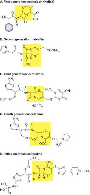
FIGURE 27.10 ■ Cephalosporin generations. Representative examples. With each successive generation, the side groups become more complex. Highlighted areas indicate the core structure of each of the cephalosporins, with beta-lactam rings.
Unfortunately, the microbial world continually adapts and eventually becomes resistant to new antibiotics. In the case of the cephalosporins, new beta-lactamases have evolved that can attack the sterically buried beta-lactam rings in these molecules. It is also important to note that because the core feature of these drugs is the beta-lactam ring, people who are allergic to penicillins may also suffer a hypersensitivity reaction to the lower-generation cephalosporins. Carbapenems (such as imipenem) are another subclass of beta-lactam antibiotics, called extended-spectrum penicillins. These antibiotics are resistant to most beta-lactamase enzymes made by pathogenic bacteria. To minimize the chance of developing resistance, carbapenems are reserved for severe or high-risk infections caused by multidrug-resistant, usually Gram-negative bacteria, including Pseudomonas. However, carbapenems such as piperacillin are often used empirically in seriously ill patients before the identity of the infectious agent is known. When used empirically, the carbapenem is typically combined with another drug, such as vancomycin (described later), that has broad activity against Gram-positive bacteria. Once the clinical lab identifies the actual organism, the carbapenem should be replaced with an antibiotic that has a narrower spectrum of activity (see the paragraph “Antibiotic stewardship” in Section 27.3). Treatment note: In the preceding case history, the infecting strain of Streptococcus pneumoniae was initially treated with ampicillin. Had the hospitalized patient been an adult, a macrolide (azithromycin) or a fluoroquinolone (see Section 7.2) might have been the initial treatment choices because their respective targets, the ribosome and a type II topoisomerase, are unrelated to cell wall synthesis. Quinolones are not recommended for children, however, because of potential side effects. Other beta-lactam antibiotics, such as the third-generation cephalosporins, may still work on penicillin-resistant Streptococcus pneumoniae, because the modified antibiotic often can still bind the altered PBP. Unfortunately, cephalosporin-resistant strains of S. pneumoniae are now common.
Note: Archaeal pseudopeptidoglycan contains talosaminuronic acid
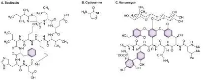
instead of muramic acid and lacks the D -amino acids found in bacterial peptidoglycan. Archaea are thus insensitive to penicillins, which interfere with bacterial transpeptidases. This natural resistance is not a problem because no archaea are known to be pathogens. Antibiotics that target other steps in peptidoglycan synthesis. Another antibiotic that affects cell wall synthesis is bacitracin, a large polypeptide molecule produced by Bacillus subtilis and Bacillus licheniformis (Fig. 27.11A). The antibiotic inhibits cell wall synthesis by binding to the bactoprenol lipid carrier molecule that normally transports monomeric units of peptidoglycan across the cell membrane to the growing chain (see Fig. 27.8). Bacitracin binds to and inhibits dephosphorylation of the carrier, which prevents the carrier from accepting a new unit of UDP-NAM. The inability to make peptidoglycan causes growth arrest. Resistance to bacitracin can evolve if the organism can rapidly recycle the phosphorylated lipid carrier molecule through dephosphorylation or if the organism possesses an efficient drug export system (discussed in Section 27.3). Normally, bacitracin is used only topically because of serious side effects, such as kidney damage, that can occur if bacitracin is ingested. For instance, the drug is often applied topically to skin that has been tattooed to prevent infection.
FIGURE 27.11 ■ Other antibiotics that affect peptidoglycan synthesis. A. Bacitracin is produced by Bacillus subtilis. It is generally used only topically to prevent infection. B. Cycloserine, an analog of D -alanine, is one of several drugs used to treat tuberculosis. C. Vancomycin is a cyclic polypeptide made by Amycolatopsis orientalis, previously classified as a streptomycete. These antibiotics, especially bacitracin and vancomycin, are synthesized by exceedingly complex biochemical pathways in the producing organisms. Me = methyl.
Lantibiotics are cyclic peptide antibiotics produced by some Gram-positive bacteria (such as Streptomyces). Members of the clinically useful subclass of lantibiotics, including Duramycin, interact with a membrane phospholipid (phosphotidylethanolamine) to impede lipid II function (see Fig. 27.8), resulting in inhibition of peptidoglycan synthesis.
Cycloserine (made by Streptomyces garyphalus) is one of several antimicrobials used to treat tuberculosis (Fig. 27.11B ). Relative to bacitracin, it acts at an even earlier step in peptidoglycan synthesis. Cycloserine inhibits the two enzymes that make the D -Ala-D -Ala dipeptide. As a result, the complete pentapeptide side chain on N - acetylmuramic acid cannot be made (see Fig. 27.8). Without these alanines, cross-linking cannot occur and peptidoglycan integrity is compromised.
Vancomycin, a very large and complex glycopeptide produced by Amycolatopsis orientalis (formerly Streptomyces orientalis; Fig.
27.11C ), binds to the D -Ala-D -Ala terminal end of the disaccharide unit and prevents the action of transglycosylases and transpeptidases (see Fig. 27.8). The mechanism of resistance is very different for vancomycin and penicillin, which makes vancomycin particularly useful against penicillin-resistant bacteria (notice that vancomycin does not contain a beta-lactam ring). To prevent the development and spread of vancomycin-resistant bacteria, this antibiotic is typically used only as a drug of last resort.
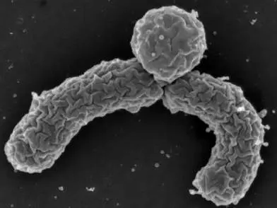
Resistance to vancomycin can develop when products from a cluster of van genes collaborate to make D -lactate and incorporate it into the ester D -Ala-D -lactate, to which vancomycin cannot bind. Another enzyme in the van gene cluster prevents the accumulation of D -Ala-D -Ala; as a result, D -Ala-D -lactate replaces D -Ala-D -Ala in peptidoglycan. Peptidoglycan containing D -Ala-D -lactate functions just fine, but the organism is resistant to the antibiotic because vancomycin cannot bind the D -lactate form. However, the antibiotic teixobactin, produced by the recently discovered species Eleftheria terrae (see Fig. 4.16), will bind to the D -Ala-D -lactate form and stop the growth of vancomycin-resistant Gram-positive bacteria.
Note that antibiotics targeting cell wall biosynthesis generally kill only growing cells. These drugs do not affect static or stationary-phase cells, because in this state the cell has no need for new peptidoglycan.
Thought Question
27.6 When treating a patient for an infection, why would combining a drug such as erythromycin with a form of penicillin be counterproductive? (Erythromycin is described in Section 8.3.)
Drugs That Affect Bacterial Membrane Integrity
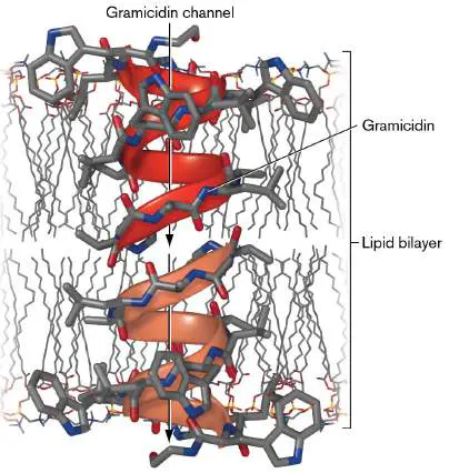
Poking holes in a bacterial cytoplasmic membrane is an effective way to kill bacteria. A few compounds are useful in this regard, among them a group called the peptide antibiotics, of which gramicidin is an example. Produced by Bacillus brevis, gramicidin is a cyclic peptide composed of 15 alternating D - and L -amino acids. It inserts into the membrane as a dimer, forming a cation channel that disrupts membrane polarity (Fig. 27.12). Polymyxin (from Bacillus polymyxa ), another polypeptide antibiotic, has a positively charged polypeptide ring that binds to the outer (lipid A) and inner membranes of bacteria, both of which are negatively charged. Its major lethal effect seems to be to destroy the inner membrane, much as a detergent does. Loss of the cell’s osmotic barrier causes the cell to lyse.
FIGURE 27.12 ■ Gramicidin is a peptide antibiotic that affects membrane integrity. As a dimer, gramicidin forms a cation channel across cell membranes through which H +, Na +, or K + can freely pass. There are two alternative structures proposed. The head-to-head dimer shown here and a side-by-side dimer arranged as double-stranded helices. (PDB code: 1GRM)
These peptide antibiotics are used only topically to treat or prevent infection. Because they can also form channels across human cell membranes, they should never be ingested. Polymyxin has been fused to some bandage materials used to treat burn patients, who are
particularly susceptible to Gram-negative bacterial infections (for example, Pseudomonas aeruginosa). Despite the drug’s toxicity, polymyxin or colistin can be injected as a drug of last resort to treat certain multidrug-resistant bacterial infections (for instance, those caused by Klebsiella pneumoniae).
Daptomycin is a lipopeptide made by Streptomyces roseosporus using nonribosomal peptide synthetases. The drug aggregates in the membranes of Gram-positive bacteria to form an ion channel that leaks potassium ions. The resulting membrane depolarization leads to cell death. This drug is very effective against MRSA.
Drugs That Affect DNA Synthesis and Integrity
Bacteria generally make and maintain their DNA using enzymes that closely resemble those of mammals. Thus, you might think it impossible to selectively target bacterial DNA synthesis, but it is possible, as you will see.
Case History: Pneumonia Due to a Gram-Negative Anaerobe
A 23-year-old woman arrived at the emergency room by ambulance with fever, chills, and severe muscle aches. She developed a nonproductive (dry) cough, had difficulty breathing, had pleuritic chest pain (stabbing pain when inhaling or exhaling), and became hypotensive (had low blood pressure). An X-ray showed lower-lobe infiltrate in the lungs, and the clinical laboratory reported the presence of the Gram-negative anaerobe Fusobacterium necrophorum in blood cultures. The patient was diagnosed with pneumonia and treated with metronidazole, a DNA-damaging agent specific for anaerobes. She fully recovered.
There are several classes of drugs, including sulfa drugs, quinolones, and metronidazole, that selectively affect the synthesis or integrity of DNA in microorganisms.
Sulfa drugs. The sulfa drugs belong to a group of drugs known as antimetabolites because they interfere with the synthesis of metabolic intermediates. Ultimately, sulfa drugs inhibit the synthesis of nucleic acids. Drugs such as sulfamethoxazole or sulfanilamide work at the metabolic level to prevent the synthesis of tetrahydrofolic acid (THF), an important cofactor in the synthesis of nucleic acid precursors (Fig. 27.13).
FIGURE 27.13 ■ Mode of action of sulfanilamides. A. The structures of PABA and sulfanilamide are very similar. B. PABA, pteridine, and glutamic acid combine to make the vitamin folic acid. C. Normal synthesis of folic acid requires that all three components engage the active site of the biosynthetic enzyme. The sulfa drugs replace PABA at the active site. The sulfur group, however, will not form a peptide bond with glutamic acid, and the size of sulfanilamide sterically hinders the binding of pteridine, so folic acid cannot be made.
All organisms use THF to synthesize nucleic acids, so why are the sulfa drugs selectively toxic to bacteria? The selectivity occurs because mammals do not synthesize folic acid, a precursor of THF. Higher mammals generally rely on bacteria and green leafy vegetables as sources of folic acid. Bacteria make folic acid from the combination of PABA, glutamic acid, and pteridine. Sulfanilamide (SFA), a structural analog of PABA, competes for one of the enzymes in the bacterial folic acid pathway and inhibits both folic acid and THF production (Fig. 27.13C ). Because humans lack that pathway, sulfa
drugs are selectively toxic toward bacteria. A commonly used drug called Bactrim combines sulfamethoxazole with trimethoprim to inactivate two different enzymes in the folic acid pathway—a strategy that limits the development of antibiotic resistance.
Quinolones. Another group of drugs inhibits DNA synthesis by targeting microbial topoisomerases such as DNA gyrase and topoisomerase IV (the mechanism is discussed in Section 7.2). Because these enzymes are structurally distinct from their mammalian counterparts, drugs can be designed to selectively interact with them while not interfering with mammalian DNA metabolism. One such drug, nalidixic acid, was discovered in 1963. The drug targets bacterial DNA gyrase but has a very narrow antimicrobial spectrum, covering only a few Gram-negative organisms. However, adding various chemical modifications to nalidixic acid, such as fluorine and amine groups, has increased its antimicrobial spectrum and its half-life in the bloodstream. The result is the class of drugs known as the quinolones. (The mode of action of quinolones and fluoroquinolones is discussed in Section 7.2.)
Mounting evidence, however, suggests that quinolones can also affect human mitochondria, which evolved from bacterial progenitors. Quinolones have been reported to increase mitochondrial production of reactive oxygen species, whose damaging effects can lead to muscle weakness, tendonitis, and other conditions. Some people are more prone to these effects than others.
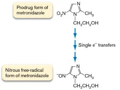
Metronidazole. Also known as Flagyl, metronidazole is an example of a prodrug—that is, a drug that is harmless until activated. Metronidazole is activated after it receives an electron (is reduced) from the microbial protein cofactors flavodoxin and ferredoxin, found in microaerophilic and anaerobic bacteria such as Bacteroides (Fig. 27.14). Once activated, the compound begins nicking DNA at random, thus killing the cell. Because the etiological agent in our case history was an anaerobe, metronidazole was an effective therapy. Metronidazole is also effective against protozoa such as Giardia, Trichomonas, and Entamoeba, all of which use ferredoxin and are anaerobes. Aerobic microbes also possess ferredoxin, but they are incapable of reducing metronidazole, presumably because oxygen is reduced in preference to metronidazole.
FIGURE 27.14 ■ Activation of metronidazole. Single-electron transfers are made by ferredoxin and flavodoxin from anaerobes. Ferredoxin and flavodoxin are reducing agents capable of reducing oxidized molecules in cells such as thioredoxin. They can also reduce the prodrug form of metronidazole.
RNA Synthesis Inhibitors
The mode of action of antibiotics that inhibit transcription, such as rifampin and actinomycin D (Fig. 27.15), is described in Chapter 8. These drugs are bactericidal and are most active against growing bacteria. The tricyclic ring of actinomycin D binds DNA from any source. As a result, it is not selectively toxic and not used to treat infections. Rifampin (also called rifampicin),
however, is selectively toxic for bacterial RNA polymerase and is often prescribed to treat tuberculosis or meningococcal meningitis. Rifampin binds to the exit tunnel of RNA polymerase. Binding stops transcription by blocking RNA from exiting the polymerase (Fig. 27.15B ; described in Chapter 8). Curiously, rifampin, which is reddish orange, turns bodily secretions, including breast milk, orange. The patient should be warned of this highly visible but harmless side effect to avoid unnecessary anxiety when the patient’s urine changes color.
FIGURE 27.15 ■ Antibiotics that inhibit transcription. A. Rifampin structure. B. Rifampin-binding site on the RNA polymerase beta subunit. Cyan = beta subunit; pink = beta-prime
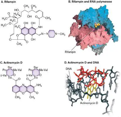
subunit; alpha subunits are behind the complex and not shown; the Mg 2+ ion chelated at the active site is shown as a magenta sphere. C. Actinomycin D structure. D. Actinomycin D (yellow and red) interacting with DNA. Covalent intercalation of actinomycin (yellow) between bases (gray) interferes with DNA synthesis and transcription. (PDB code: 1DSC)
CAMPBELL ET AL. 2001. CELL 104 :901–912, FIG. 3B.
Although rifampin is clinically useful, bacterial resistance to the drug has developed. Fortunately, two new classes of RNA polymerase–targeting antibiotics have been discovered. Pyronins, represented by myxopyronin (produced by Myxococcus fulvis), prevent RNA polymerase from ever starting polymerization. The pyronins bind to RNA polymerase at a site called the hinge region, which is needed to separate (melt) DNA strands—a requirement to begin transcription.
The second drug, lipiarmycin, is an unusual macrolide antibiotic made by the actinomycete bacterium Actinoplanes deccanensis.
Lipiarmycin binds to the same region as myxopyronin but completely stops RNA polymerase–DNA closed complexes from transitioning to the open forms. Rifampin-resistant RNA polymerase molecules are still sensitive to the new antibiotics because the binding site for rifampin is different from the binding site for the other two antibiotics. This is exciting because pathogens such as rifampin-resistant strains of Mycobacterium tuberculosis or Clostridioides difficile (formerly Clostridium difficile) can now be treated with a new drug that targets the same enzyme. Lipiarmycin has been approved by the FDA to treat C. difficile –associated diarrhea.
Thought Question
27.7 Given the mechanism by which rifampin stops transcription, what limitation does the drug have? Consider initiation versus elongation.
Protein Synthesis Inhibitors
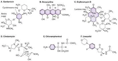
Antibiotics that specifically inhibit bacterial protein synthesis rely on the differences between prokaryotic and eukaryotic ribosomes for selective toxicity. How various antibiotics inhibit protein synthesis is discussed in Section 8.3. Recall that protein synthesis inhibitors can be classified into several groups based on structure and function (Fig. 27.16). Most of these antibiotics work by binding and interfering with the function of bacterial rRNA, which differs from the function of eukaryotic rRNA. Recall, too, that protein synthesis inhibitors are, by and large, bacteriostatic (not bactericidal). FIGURE 27.16 ■ Protein synthesis inhibitors. A. The aminoglycoside gentamicin. B. The tetracycline doxycycline. C. The macrolide erythromycin. D. The lincosamide clindamycin. E. Chloramphenicol. F. The oxazolidinone linezolid; F = fluorine. Purple shading highlights chemical groups common to different members within a given antibiotic class.
Case History: Erysipelas in a Penicillin-Sensitive Patient
Sixteen-year-old Jamal arrived at the emergency room after 2 days of fever, malaise, chills, and neck stiffness. His most notable symptom was a painful, red, rapidly spreading rash covering the right side of his face. The rash covered his entire cheek, which was swollen, and extended into his scalp. About 7 days earlier, Jamal had had a severe sore throat. Because it had subsided in 2 days, however, he had not been clinically evaluated. Throat cultures taken on admission revealed group A Streptococcus pyogenes, suggesting that the rash was a case of erysipelas caused by this organism. Although penicillin would be the drug of choice, Jamal was known to be allergic to this antibiotic. When a patient is known to be allergic to the usual drug of choice, it is best to prescribe a structurally different drug. Often that drug will be one that inhibits protein synthesis; in this case, the drug chosen was the macrolide azithromycin.
Targeting the 30S Subunit
Recall that bacterial 50S and 30S ribosomal subunits must assemble around mRNA to produce a functional 70S ribosome. Antibiotics affecting protein synthesis are classified by which ribosomal subunit is targeted. Thus, one class of antibiotics interferes with 30S subunit function, and the other impedes 50S subunit activities.
Aminoglycosides. Different aminoglycosides vary considerably in structure, but all contain a cyclohexane ring and amino sugars (Fig. 27.16A). The aminoglycosides are unusual among protein synthesis inhibitors in that they are bactericidal rather than bacteriostatic. Most of them bind 16S rRNA and cause translational misreading of mRNA, which is why these drugs are bactericidal (another possible explanation is presented later in the chapter). The resulting synthesis of jumbled or truncated peptides wreaks havoc with physiology and kills the cell.
Streptomycin and gentamicin (Fig. 27.16A) are two widely used drugs in this class. Ototoxicity (hearing damage) is a major, but uncommon, side effect of these antibiotics (approximately 0.5%–3% of patients treated with gentamicin suffer from this toxicity). Hearing is generally affected at frequencies above 4,000 Hz. The toxicity of aminoglycosides appears related to their ability to inhibit the function of mitochondrial ribosomes, which are evolutionarily related to bacterial ribosomes. Individuals with specific mutations in mitochondrial rRNA are more susceptible to aminoglycoside toxicity. Tetracyclines. Tetracycline antibiotics are characterized by a structure with four fused cyclic rings—hence, the name. Figure 27.16B shows one important, clinically used example, called doxycycline. Tetracyclines are bacteriostatic and act by binding to and distorting the ribosomal A site that accepts incoming charged tRNA molecules. Doxycycline is used to treat early stages of Lyme disease (caused by Borrelia burgdorferi), acne (Cutibacterium acnes), and other infections. An important adverse side effect of tetracyclines is that they can interfere with bone development in a fetus or young child. Tetracycline use by pregnant women will also cause yellow discoloration of the infant’s teeth. As a result, this drug is not recommended for pregnant women or nursing mothers.
Targeting the 50S Subunit
Five classes of drugs subvert translation by binding to the 50S ribosomal subunit. Most of these drugs are discussed in Chapter 8 and are recapped here only briefly.
Macrolides, all of which contain a 14-to 16-member lactone ring (Fig. 27.16C ), inhibit translocation of the growing peptide (bacteriostatic action). Commonly prescribed examples are erythromycin and azithromycin. Azithromycin was the antibiotic used to treat the Streptococcus pyogenes infection in our case history, although other drugs could have been used. Because it is structurally dissimilar to any of the beta-lactam antibiotics, such as penicillin, azithromycin can be used safely in patients who are penicillin sensitive.
Lincosamides (Fig. 27.16D ), such as clindamycin, are similar to macrolides in function but have a different structure.
Chloramphenicol (Fig. 27.16E ) inhibits peptidyltransferase activity (bacteriostatic). Bone marrow depression leading to aplastic anemia is the most common serious side effect and limits its clinical use.
Oxazolidinones (Fig. 27.16F ) are a class of synthetic antibiotics that are effective against many antibiotic-resistant microbes. Discovered in the 1990s, this was the first new class of antibiotics discovered since the “golden age” of antibiotic discovery ended over 35 years earlier. Oxazolidinones such as linezolid bind to the 23S rRNA in the 50S subunit of the prokaryotic ribosome and prevent formation of the protein synthesis 70S initiation complex. This is a novel mode of action; other protein synthesis inhibitors either block polypeptide extension or cause misreading of mRNA. Linezolid binds to the 50S subunit near where chloramphenicol binds, but it does not inhibit peptidyltransferase. Resistance is limited because most bacterial genomes have multiple operons encoding 23S rRNA. Usually more than one of these genes must mutate to confer high-level resistance. The more mutant 23S rRNA genes there are relative to native 23S genes, the more oxazolidinone-resistant ribosomes will be present. Oxazolidinones are useful primarily against Gram-positive bacteria. Gram-negative bacteria are intrinsically resistant because of multidrug efflux pumps (see Section 27.3) and decreased permeability due to the outer membrane.
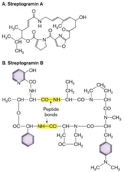
Streptogramins (Fig. 27.17), produced by some Streptomyces species, fall into two groups, designated A and B. Streptogramins belonging to group A have a large nonpeptide ring (Fig. 27.17A ), whereas streptogramin B members are cyclic peptides (Fig. 27.17B ). The two groups differ in their modes of action, although both inhibit bacterial protein synthesis by binding to the peptidyltransferase site. Group A streptogramins bind to the peptidyltransferase site and prevent binding of tRNA to the ribosome A site. In contrast, group B streptogramins are thought to narrow the peptide exit channel, preventing exit of the peptide and thereby blocking translocation. Natural streptogramins are produced as a mixture of A and B, the combination of which is more potent than either individual compound alone (an example of synergy). In tribute to this synergistic action, the drug combination is marketed under the name Synercid. Synergy between the two drugs occurs because the A-type streptogramin alters the binding site for the B-type drug, increasing its affinity. Bacteria can develop resistance through ribosomal modification (the modification in 23S rRNA is the same one that provides resistance to macrolides), via the production of inactivating enzymes, or by active efflux of the antibiotic.
FIGURE 27.17 ■ The streptogramins. A. Streptogramin A is a large nonpeptide ring structure. B. Streptogramin B is a cyclic peptide.
Targeting Aminoacyl-tRNA Synthetases
Aminoacyl-tRNA synthetases attach amino acids to the 3′ CCA end of tRNA molecules (described in Chapter 8). The catalytic domains of these enzymes include three distinct pockets; one recognizes the cognate amino acid, another recognizes adenylate, and the third binds to the cognate tRNA. An antibiotic called mupirocin (made by Pseudomonas fluorescens) is a structural mimic of isoleucyl-AMP, the intermediate used by bacterial isoleucyl-tRNAsynthetase to link isoleucine to isoleucyl-tRNA.
Mupirocin binds to the enzyme’s isoleucyl-AMP pocket and inhibits synthetase activity. As a result, protein synthesis stops. The drug is selective for bacterial isoleucyl-tRNA synthetase and does not affect the human form of the enzyme. Mupirocin (sold as Bactroban) is used topically in creams to treat skin infections caused by Gram-positive pathogens. Mupirocin cannot be used internally, because it is rapidly degraded in blood. Other aminoacyl-tRNA inhibitors exist, but none are used therapeutically.
To Kill or Not to Kill: What Makes a Bactericidal Drug?
Bactericidal antibiotics include quinolones that bind to DNA topoisomerases; aminoglycosides that bind to the 30S ribosome subunit; rifampin, which binds to RNA polymerase; and penicillins that inhibit peptidoglycan synthesis. While these antimicrobials do halt critical cellular processes, other antibiotics that inhibit some of these same processes are not bactericidal. For example, aminoglycosides and macrolides inhibit protein synthesis, but macrolides do not typically kill bacteria.
One theory proposes a general killing mechanism to explain why particular antibiotics are bactericidal. Put simply, bactericidal (but not bacteriostatic) antibiotics cause the generation of highly reactive hydroxyl radicals, which damage DNA, protein, and lipids, leading to cell death. Partial evidence for this model came when James J. Collins (Broad Institute of MIT and Harvard) and colleagues found that they could make cells more sensitive to killing by bactericidal antibiotics if they prevented induction of the SOS response that limits and repairs oxidative damage to DNA (see Section 9.2). Note that preventing the generation of reactive oxygen species (ROS) will limit the killing effect of bactericidal antibiotics, but not their bacteriostatic effect by, for instance, halting protein synthesis.
The proposed mechanism leading to ROS accumulation starts with drug-target interactions stimulating NADH oxidation via the electron transport system (ETS). How drug-target interactions stimulate the ETS varies with the antibiotic. Hyperactivation of the ETS induces formation of superoxide and hydrogen peroxide. The superoxide damages iron-sulfur clusters in proteins to release ferrous ions (Fe 2+ ). Fe 2+ then reacts with hydrogen peroxide via the Fenton reaction (see Fig. 5.18) to produce the highly reactive hydroxyl radicals that inflict lethal damage to other cell components. It should be noted that this model was initially met with considerable skepticism, but it has since gained qualified acceptance.
Conventional wisdom now holds that there is no single, unique mechanism by which antibiotics kill. A number of studies confirm that ROS have an important role, but their contribution to killing is synergistic with the immediate effect of the antibiotic on the primary target and on the physiological state of cells being stressed by the antibiotic.
You could ask, “Why do we care?” Is it not enough to know that an antibiotic can kill a pathogen? The answer is no, it is not enough. We care because knowing how and when different antibiotics kill bacteria can be useful in developing new antibiotics. What we learn can also be applied to existing drugs and the design of new treatment protocols. For example, we can learn which drugs or drug combinations will be most effective clinically. Detailed knowledge about how antibiotics kill can be used to prevent the rise of antibiotic resistance in pathogens and nonpathogenic microbiota. And, lastly, the information can be used to design new strategies or narrow-spectrum drugs that will minimize dysbiosis and its consequences.
Thought Question
27.8 Why might a combination therapy of an aminoglycoside antibiotic and cephalosporin be synergistic?
To Summarize
Antibiotic specificity for bacteria can be achieved by targeting a process present in bacteria but not host cells, by targeting small structural differences between components of a process shared by bacteria and hosts, or by exploiting a physiological growth condition unique to bacteria, such as anaerobiosis.
Antibiotic targets include cell wall synthesis, cell membrane integrity, DNA synthesis, RNA synthesis, protein synthesis, and metabolism.
Antibiotics targeting the cell wall bind to the transglycosylases, transpeptidases, and lipid carrier proteins involved with peptidoglycan synthesis and cross-linking. Antibiotics interfering with DNA include the antimetabolite sulfa drugs that inhibit nucleotide synthesis; quinolones that inhibit DNA topoisomerases; and the drug metronidazole that, when activated, randomly nicks the phosphodiester backbone. Inhibitors of RNA synthesis target RNA polymerase (rifampin and pyronins) or bind DNA and inhibit polymerase movement (actinomycin D).
Different protein synthesis inhibitors target prokaryotic ribosomes. Some target the 30S subunit to cause misreading of mRNA (aminoglycosides) or prevent tRNA binding (tetracyclines). Others target the 50S subunit to inhibit translocation (macrolides, lincosamides), peptidyltransferase activity (chloramphenicol), formation of the 70S complex (oxazolidinones), or peptide exit through the ribosome exit channel (streptogramins). Still others target aminoacyl-tRNA synthetases (mupirocin) to prevent charging of tRNAs.
The killing action of bactericidal antibiotics is a combination of the drug’s primary effect on a cellular target and its downstream secondary effects on cell physiology, such as the generation of lethal reactive oxygen species.
Glossary
transglycosylase An enzyme that condenses N -acetylglucosamine and N - acetylmuramic acid into chains during bacterial cell wall synthesis.
transpeptidase An enzyme that cross-links the side chains from adjacent peptidoglycan strands during bacterial cell wall synthesis. penicillin-binding protein (PBP)
A bacterial protein, involved in cell wall synthesis, that is the target of the antibiotic penicillin.
penicillin-binding protein (PBP)
A bacterial protein, involved in cell wall synthesis, that is the target of the antibiotic penicillin.
bacitracin A topical antibiotic that affects cell wall synthesis.
cycloserine A polypeptide antibiotic that inhibits peptidoglycan synthesis. vancomycin A glycopeptide antibiotic that binds the D-Ala-D-Ala end of the cell-wall peptide, thus inhibiting the transpeptidase enzyme and preventing crossbridge formation in the cell wall.
gramicidin A peptide antibiotic that acts as a channel for monovalent cations to cross the cell membrane, thus collapsing the transmembrane ion gradients.
daptomycin A lipopeptide antibiotic that forms ion channels in Gram-positive bacteria.
quinolone A type of antibiotic drug that inhibits DNA synthesis by targeting bacterial topoisomerases such as DNA gyrase.
macrolide Any of a group of antibiotics containing a large lactone ring (e.g., erythromycin).
lincosamide Any of a class of bacteriostatic antibiotics that include a pyrrolidine ring linked to a pyranose (e.g., clindamycin). chloramphenicol A bacteriostatic antibiotic that acts by inhibiting peptidyltransferase activity of the bacterial ribosome. oxazolidinone One of a class of synthetic antibiotics, containing an oxazole ring, that inhibit protein synthesis.
streptogramin An antibiotic that binds 23S rRNA and blocks elongation of protein synthesis in bacteria.
Fig. 3.13 FIGURE 3.13 ■ Peptidoglycan synthesis is organized by penicillin-binding proteins (PBP2, PBP1A) and by cytoskeletal proteins. Protein MreB guides the direction of synthesis in helical arcs around the cell.
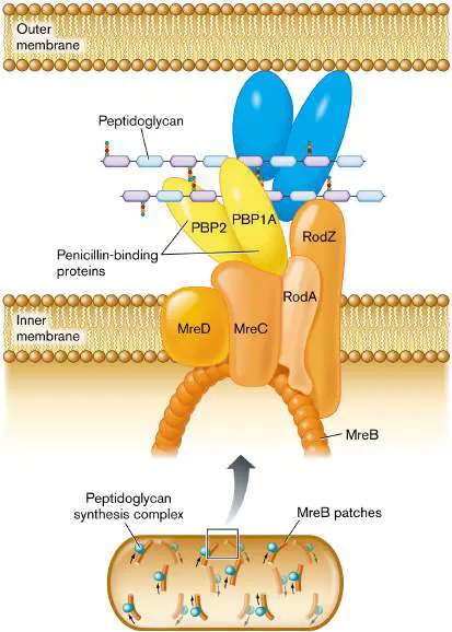
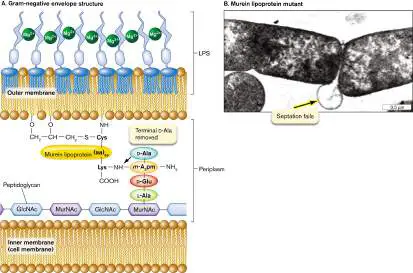
Fig. 3.18 FIGURE 3.18 ■ Gram-negative cell envelope (S-layer not shown). A. Murein lipoprotein has an N-terminal cysteine triglyceride inserted in the inward-facing leaflet of the outer membrane. The C-terminal lysine forms a peptide bond with the m -diaminopimelic acid (m -A 2 pm) of the peptidoglycan (murein) cell wall. Mg 2 ⁺ ions cross-link the LPS chains. B. Lack of murein lipoprotein in mutant Salmonella causes the outer membrane to balloon out (arrow) when the cell tries to divide (TEM).
JOAN FUNG ET AL. 1978. J. BACTERIOL. 133 :1467
Fig. 4.16 A
B C
D
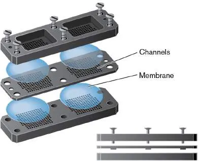
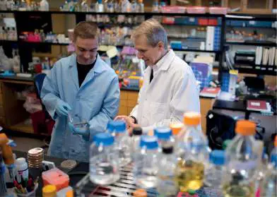
FIGURE 4.16 ■ In situ culturing of the uncultured. A. Kim Lewis (right), with postdoctoral researcher Brian Conlon. B. The recently discovered species Eleftheria terrae produces the novel antibiotic teixobactin. C. Schematic look at the iChip used to culture previously uncultured soil bacteria. D. The iChip being removed from soil.
Fig. 5.18

FIGURE 5.18 ■ Generation and destruction of reactive oxygen species (ROS). ROS are marked yellow.
The autooxidation of flavin adenine dinucleotide (FAD) and the Fenton reaction occur spontaneously to produce superoxide and hydroxyl radicals, respectively. The ferrous (Fe 2+) form of iron needed for the Fenton reaction comes from intracellular sources such as cytochromes. The other reactions require enzymes. FAD is a cofactor for a number of enzymes (for example, NADH dehydrogenase 2). Catalase and peroxidase detoxify hydrogen peroxide.
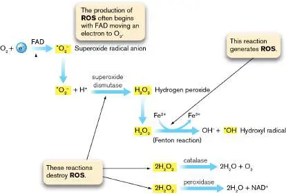
27.3 Challenges of Drug Resistance and Discoverynot assigned
Figure 27.18illustrates how long it has taken for bacterial pathogens to develop resistance to clinically used antibiotics. Resistance is often discovered in fewer than 10 years, and in some cases resistant mutants were discovered even before the drug was approved for clinical use. But antibiotic-producing microbes have existed for millions of years, so the rapidity with which antibiotic resistance develops to any clinically used antimicrobial drug raises intriguing questions: If antibiotic resistance is inevitable, as Figure 27.18suggests, why didn’t widespread resistance to all naturally occurring antibiotics develop long before humans ever discovered miracle drugs? What do microbes and humans do differently?
FIGURE 27.18 ■ The rapid journey of antibiotics from discovery to resistance (1905–2015). Each line represents the time from the year of first clinical use for an antibiotic (yellow arrowheads) to the first report of resistance (black bar). Cases in which resistance was discovered before clinical approval are indicated by a black bar preceding the yellow arrowhead. Cases in which clinical approval and resistance occurred in the same year are marked by a purple circle. Salvarsan was the name given to the arsenical compound used by Paul Ehrlich to treat syphilis.
To answer those two questions, we must first address two others: Why do microbes make antibiotics, and how do they avoid killing themselves in the process? The answers provide insight into
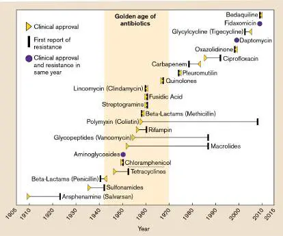
the origin of antibiotic-resistant pathogens and our fight to halt their spread throughout the world.
Risky Business: Why Do Microbes Make Antibiotics?
Antibiotics are considered secondary metabolites because they are not involved in the primary metabolism of the producing organism. But the complex pathways that assemble antibiotics (see Section 15.3) reflect the long evolutionary journeys microbes took to solve two ancient problems: survival in the midst of fierce competition, and interspecies cooperation. To accomplish these goals in nature, antibiotics produced by microbes do not need to accumulate to high concentrations, and they need to work only at tiny distances from the producer. This use of antibiotics is very different from the sustained, massive amounts of antibiotic a clinician uses to treat infections. In a natural environment like soil, an antibiotic diffuses away from secreting organisms and forms a concentration gradient that can inhibit the growth of antagonistic species and at the same time act as a signal molecule to coordinate activities of nearby symbiotic or mutualistic species. In essence, antibiotic-producing bacteria living in nature can orchestrate the assembly of cooperative societies, and they have done so since long before humans existed. Antibiotic production in native environments can also forge a mutualistic relationship between a microbe and its colonized host by protecting the host from deadly pathogens. A striking example is found in the leaf-cutter ant. These insects surgically cut, and dutifully carry, pieces of new leaves to their colony. The ants then use the leaves to carefully cultivate a particular fungus they use for food. However, a parasitic microfungus called Escovopsis can also feed on the fungus “garden,” which can devastate the colony. As defense, the ants grow a genus of actinobacteria called Pseudonocardia on their cuticles and spread the organism over the garden. The bacterium’s job is to protect the garden and, ultimately, the ant colony by secreting antibiotic variations of nystatin that kill the parasitic microfungus.
Given that microbes make antibiotics for good reasons, how does the producing microorganism avoid committing “suicide”? Fungi that make penicillin face no consequence for having done so, because the organism does not contain peptidoglycan. Actinomycetes that produce compounds such as streptomycin or chloramphenicol, however, could be susceptible to their own secondary metabolite. Ribosomes isolated from Streptomyces griseus, for example, are fully sensitive to the streptomycin that the organism itself produces. S. griseus avoids killing itself in two ways. First, the organism synthesizes 6-phosphorylstreptomycin, an inactive precursor of streptomycin that is secreted from the cell. Once outside the mycelium, the precursor is converted to active streptomycin by a specific phosphatase. In addition, this streptomycete has an enzyme that will inactivate any streptomycin that may leak back into the mycelium. Other organisms protect themselves by methylating key residues on their rRNA to prevent drug binding or by setting up permeability barriers that thwart reentry of the antibiotic. These and other strategies of self-preservation employed by antibiotic-producing microbes are clever. Unfortunately, these mechanisms can be shared via horizontal gene transfers (plasmids, transposons, transduction), as discussed in Sections 9.5 and 25.1. Now we can return to our original question: Why hasn’t antibiotic resistance become widespread in microbes inhabiting natural environments such as soil and water? In nature, a single microbe only needs to achieve an MIC of antibiotic in mere microliters of space around the cell. Even if the population of the antibiotic-producing species is large, the concentration of antibiotic made will be far lower than that used by clinicians to cure an infection. Thus, o ne reason antibiotic resistance did not become widespread in the natural environment is that organisms living there are rarely, if ever, subjected to high doses of antibiotic for long periods of time. Thus, selective pressure promoting resistance is weak. Medical practice, however, requires the use of large doses of antibiotics for extended periods of time to treat infected patients. This approach to save the patient will indiscriminately eradicate large numbers of antibiotic-susceptible pathogens and microbiota from the patient.
Simultaneously, this strategy may enable a small number of resistant bacteria to flourish and become a dominant presence. So, with antibiotic resistance becoming more and more widespread, what happens when a clinician unknowingly choses the wrong antibiotic to treat a serious infection?
Case History: Multidrug-Resistant Pneumonia
A 14-year-old boy with fever (39°C; 102.2°F), chills, and left-sided pleuritic chest pain was referred to a hospital emergency department by his general practitioner. A chest X-ray showed left-lower-lobe pneumonia. The boy reported that he was allergic to amoxicillin and cephalosporins (as a child he had developed a rash in response to these agents) and had been taking daily doxycycline (tetracycline) for the previous 3 months to treat mild acne. He was admitted to the hospital and treated with intravenous azithromycin (a macrolide antibiotic) because of his reported beta-lactam allergies, but he continued to feel sick. The day after admission, both sputum and blood cultures grew Streptococcus pneumoniae. After 48 hours, antibiotic susceptibility results indicated that the microbe was resistant to penicillin, azithromycin, and tetracycline. Armed with this information, the clinician immediately changed antibiotic treatment to vancomycin. The boy’s fever resolved during the next 12 hours, and he made a slow but full recovery over the next week.
Unfortunately, the scenario presented in this case is far too common and has become an extremely serious concern. Figure 27.19 shows how quickly penicillin-resistant clinical isolates of Streptococcus pneumoniae developed around the world. An extremely troubling instance of an emerging antibiotic-resistant pathogen is the Gram-negative rod Acinetobacter baumannii. Having first appeared in Europe and East Asia, A. baumannii has since spread throughout the world, including to the United States and Canada. It is increasingly seen as a dangerous cause of nosocomial infections that commonly colonizes hospitalized patients, particularly those in intensive care units. Patients hospitalized as a result of the COVID-19 pandemic have been especially vulnerable. Some estimates are that 50% of COVID-19 patients in intensive care units were coinfected with carbenicillin-resistant A. baumannii. As a result, the World Health Organization has named this pathogen a critical priority pathogen that poses a global threat to human health.
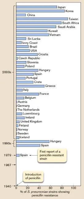
FIGURE 27.19 ■ The rise of penicillin-resistant Streptococcus pneumoniae throughout the world.
Numbers reflect the number of penicillin-resistant strains among clinical isolates (strains of disease-causing bacteria isolated from patients from different countries). No resistance among clinical isolates was noted until after 1967.
Before 1998, there were almost no cases of multidrug-resistant A. baumannii. The rate is now as high as 8%. The organism is resistant to drugs as diverse as ciprofloxacin (a quinolone), amikacin (an aminoglycoside), penicillins, third-generation cephalosporins, tetracycline, chloramphenicol, and imipenem, one of a relatively new class of beta-lactam drugs called carbapenems. Colistin and tigecycline remain the only antibiotics active against multidrug-resistant A. baumannii.
There are three basic antibiotic resistance strategies (Fig. 27.20). The resistant organism can keep the antibiotic out of the cell, prevent the antibiotic from binding to its target, or dislodge bound antibiotic from its target. The three strategies involve six antibiotic resistance mechanisms.
FIGURE 27.20 ■ Alternative mechanisms of antibiotic resistance. Antibiotic resistance genes can be plasmid-borne, or they can be part of the chromosome. Each specific antibiotic resistance gene product will use only one of the six mechanisms shown.
Strategy 1: Keep antibiotics out of the cell.
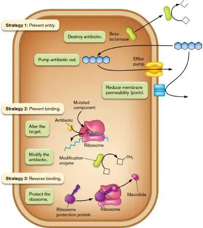
Destroy the antibiotic before it enters the cell. For example, the enzyme beta-lactamase (or penicillinase) is made exclusively to destroy penicillins (see Section 27.2). The sites of ring cleavage and the structure of the enzyme are illustrated in Figure 27.21.
Pump the antibiotic out of the cell via specific transporters (for example, tetracycline export). This strategy works because the pumps bail drugs out of the cell faster than the drugs can enter. Some transporters are single-component pumps present in the cytoplasmic membrane of Gram-negative and Gram-positive bacteria (for example, NorA in Staphylococcus aureus, PmrA in Streptococcus pneumoniae, and the TetA and B proteins in Gram-negative organisms).
Other drug efflux pumps are multicomponent systems present in Gram-negative bacteria only (discussed shortly). Efflux in either case is usually energized by proton motive force.
Decrease membrane permeability across the outer membrane. Many antibiotics, such as beta-lactams, tetracyclines, and fluoroquinolones, have to pass through outer-membrane porins to gain access to the target cell. Gram-negative bacteria, however, can express alternative porins with pores too narrow to allow penetration.
FIGURE 27.21 ■ Destroying penicillin. A. Beta-lactamase (or penicillinase) cleaves the beta-lactam ring of penicillins and cephalosporins. There are two types of penicillinases, based on where the enzyme attacks the ring. In either type, a serine hydroxyl group launches a nucleophilic attack on the ring. B. Structure of a beta-lactamase and location of the penicillin-binding site. (PDB code: 1XX2)
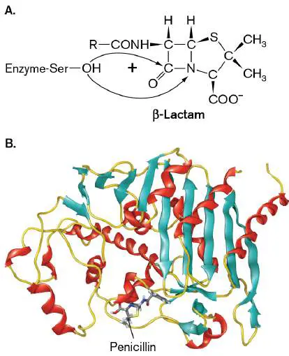
Strategy 2: Prevent antibiotics from binding the target.
Modify the target so that it no longer binds the antibiotic. Mutations in key penicillin-binding proteins and ribosomal proteins, for instance, can confer resistance to methicillin and streptomycin, respectively. These mutations occur spontaneously and are not typically transferred between organisms. An exception to this rule is MecA, the plasmid-borne, beta-lactam-resistant transpeptidase found in strains of Staphylococcus aureus described earlier.
Add modifying groups that inactivate the antibiotic. For instance, three classes of enzymes modify and inactivate aminoglycoside antibiotics. The results of these types of enzyme modifications are illustrated for kanamycin in Figure 27.22. The modifications decrease the antibiotic’s ability to bind to its target (increase the MIC).
FIGURE 27.22 ■ Aminoglycoside-inactivating enzymes. Different enzymes can inactivate aminoglycoside antibiotics. Strategy 3: Dislodge an antibiotic already bound to its target.
Ribosome protection (or rescue). As described in the chapter-opening image, Gram-positive organisms can produce proteins (for example, MsrE or TetO) that bind to ribosomes and dislodge antibiotics bound near the peptidyltransferase site.
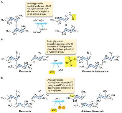
Thought Questions
27.9 Fusaric acid is a cation chelator that normally does not penetrate the E. coli membrane, which means E. coli is typically resistant to this compound. Curiously, cells that develop resistance to tetracycline become sensitive to fusaric acid. Resistance to tetracycline is usually the result of an integral membrane efflux pump that pumps tetracycline out of the cell. What might explain the development of fusaric acid sensitivity?
27.10 Mutations in the ribosomal protein S12 (encoded by rpsL) confer resistance to streptomycin. Would a cell containing both rpsL + and rpsL R genes be streptomycin resistant or sensitive? (Recall that genes encoding r ibosome p roteins for the s mall subunit are designated rps, and “+” indicates the wild-type allele, while “R” indicates a gene whose product is resistant to a certain drug.) A particularly dangerous type of drug resistance is mediated by what are called multidrug resistance (MDR) efflux pumps (Fig. 27.23). A single pump in this class can export many different kinds of antibiotics with little regard to structure. MDR pumps of Gram-negative microbes are similar to the ABC export systems described in Section 4.2. Each one includes three proteins: an inner-membrane pump protein fueled either by ATP or proton motive force, depending on the pump; an outer-membrane channel protein; and an accessory protein that links the two. For instance, the AcrB component of the AcrAB transporter (Fig. 27.24) has a promiscuous binding site located within a large central cavity that almost indiscriminately binds antibiotics. Proton motive force moves those compounds through the AcrB pore and out a funnel (AcrA) that connects to an outer-membrane channel, TolC. Efflux pumps similar to AcrAB are a major component of antimicrobial resistance of many Gram-negative pathogens, including Acinetobacter as described earlier. In contrast to Gram-negative bacteria, MDR pumps in Gram-positive bacteria such as Staphylococcus aureus are typically single-component transporters that span the cytoplasmic membrane. A single component is enough because the transport systems of Gram-positive organisms do not have to contend with an outer membrane.
FIGURE 27.23 ■ Basic structure of a multidrug resistance efflux pump in Gram-negative bacteria. These efflux systems have promiscuous binding sites that can bind and pump a wide range of drugs out of the bacterial cell. Some are driven by proton motive force, while others are driven by ATP hydrolysis (ABC transporters).
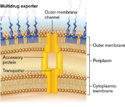
FIGURE 27.24 ■ Structure of the E. coli AcrAB multidrug resistance efflux pump. A. TolC (green ribbon) and AcrB and Z (red and blue ribbons) are homotrimers linked by six protomers of AcrA (yellow ribbons). Transport of antibiotics is driven by the proton motive force. B. A slice through the model shows the continuous conduit that runs from AcrB through the TolC porin domain, spanning the inner and outer membranes. AcrZ is a small peptide that affects substrate preference.
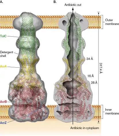
Source: Modified from Du et al. Nature 509 :512–515.
Antibiotic MDR efflux pumps contribute significantly to bacterial antibiotic resistance because of the broad variety of substrates they recognize. Strains of the pathogen Mycobacterium tuberculosis, for instance, have developed multidrug-resistant phenotypes in part because of MDR pumps. Approximately 2 million people die from tuberculosis annually, mostly in developing nations. What is even more alarming is that an increasing number of M. tuberculosis strains isolated from patients exhibit multidrug resistance. Although most of the antibiotic resistance in the majority of M. tuberculosis multidrug-resistant strains is due to the accumulation of independent mutations in several genes, MDR pumps are thought to increase the level of resistance. Chemists typically try to tweak the structure of an antibiotic to overcome a specific type of resistance mechanism, but the MDR pumps act on an exceptionally wide range of antibiotics.
Evolving, and Sharing, Drug Resistance Genes
As discussed in previous chapters, nature has engineered a certain degree of flexibility in the way genomes are replicated and passed from one generation to the next. DNA repair pathways that involve lesion bypass DNA polymerases (for example, UmuDC; see Section 9.2) are thought to contribute significantly to randomized adaptive and evolutionary processes. For instance, at some point during evolution, gene duplication and mutational reshaping produced a beta-lactamase gene, after which some lucky ancestral microbe became penicillin resistant.
However, every antibiotic-resistant species did not evolve its resistance de novo, via gene duplication and/or mutation. Why reinvent the wheel—or, in this case, drug resistance? Gene transfer mechanisms such as conjugation, described in Chapter 9, can move antibiotic resistance genes from one organism to another and from one species to another. In fact, several drug resistance genes found in pathogenic bacteria actually had their start in the chromosomes of antibiotic-producing organisms and were passed on through gene transfer. For instance, the bacterium Streptomyces clavuligerus produces penicillin but also makes a protective beta-lactamase (encoded by the bla gene) to prevent killing itself. These antibiotic resistance genes can be incorporated into plasmids that transfer into new species by transformation, conjugation, transduction, or nanotubes (see Chapter 3).
Enterococcus faecalis: a “pack rat” of antibiotic resistance systems. An interesting case study of antibiotic resistance is provided by the Gram-positive bacterium Enterococcus faecalis, a natural inhabitant of the mammalian gastrointestinal tract that can cause life-threatening disease if granted access to other body sites (as in subacute bacterial endocarditis; see Section 26.5). E. faecalis is naturally resistant to numerous antibiotics, making disease treatment particularly challenging.
Vancomycin is one of the last lines of defense for treating serious E. faecalis infections. Unfortunately, increasing numbers of vancomycin-resistant strains of Enterococcus (called VREs) have arisen in recent years. The completed genome sequence of one vancomycin-resistant strain illustrates the reason: the incorporation of numerous mobile genetic elements that encode drug resistance. About a quarter of the genome consists of mobile or exogenously acquired DNA, including 7 probable phages, 38 insertion elements, numerous transposons, and integrated plasmid genes. One such mobile element encodes vancomycin resistance.
Integrons: the “shopping cart” of drug resistance. Multidrug resistance in various microbes is also conferred by gene expression elements called integrons (Fig. 27.25). Present in plasmid, transposon, or chromosomal DNA, integrons can drive rapid adaptation in bacteria by capturing mobile genes called integron cassettes. The result is a “shopping cart” full of different antibiotic resistance gene cassettes to use later when needed.
FIGURE 27.25 ■ Integron function. A. An example of an integron that encodes resistance to aminoglycosides (aadB), quaternary ammonium compounds (qacE Δ), and sulfanilamide ( sul1). B. Capturing an integron cassette encoding a new resistance marker (orfX). Red arrows indicate a transcriptional promoter.
An integron cassette is a simple circular DNA molecule that contains a promoterless antibiotic resistance gene (or other gene) and a 50–100 bp cassette recombination site (attC; Fig. 27.25A). The origins of these cassettes are still something of a mystery, but they appear to have arisen from, and been shared between, a variety of bacterial taxons. The attC site of a cassette has homology
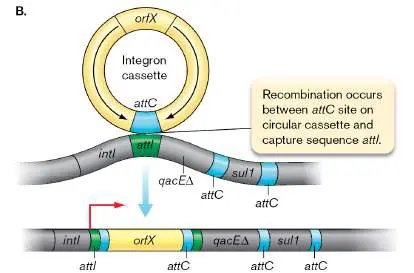
to the attI capture site on integrons (Fig. 27.25B ). An integrase ( intI) encoded by the integron catalyzes site-specific recombination between a cassette attC sequence and the integron attI site, enabling the integron to capture the cassette. As more cassettes are captured, the integron becomes longer and longer. Vibrio cholerae, for example, possesses a superintegron carrying more than 180 cassettes, most of whose functions are unknown.
A strong promoter next to attI in the integron will drive expression of adjacent, integrated antibiotic resistance cassette genes. Once captured, the cassette closest to the promoter will be expressed to the highest extent. However, the cassettes can also be “shuffled” in their position by excision and reintegration. Through this shuffling process, an integron containing several antibiotic resistance genes can, through trial and error, move the resistance gene that best combats the current antimicrobial challenge to a position where it will be more highly expressed.
What is the cost of antibiotic resistance? We should note that the development of antibiotic resistance is not without consequence to the bacterium. For example, the altered DNA gyrase that affords resistance to quinolones may not function as well as the “normal” gyrase does. Thus, when resistant and susceptible organisms cohabit the same environment, the wild-type (sensitive) strain may grow faster and eventually overwhelm the mutant strain—unless fluoroquinolone is present.
Antibiotic resistance within uncontacted communities.
Insight about the origins of antibiotic resistance in the human microbiome was provided in 2015 by Maria Dominguez-Bello (New York University and the University of Puerto Rico) and her collaborators, who studied the fecal, oral, and skin microbiomes of uncontacted Amerindians in the Amazon. The Amerindians had no previous contact with pharmaceutical antibiotics or with Westerners who could be a source of antibiotic-resistant microbes. The researchers predicted that the microbiomes of Amerindians would lack antibiotic resistance genes. They were wrong.
DNA sequencing revealed that the Amerindian microbiome did contain antibiotic resistance–like genes, possibly obtained by exchange with antibiotic-producing soil microbes. The scientists cloned the genes, placed them under the control of a constitutive plasmid promoter to ensure expression, and transferred the plasmids to E. coli. Many of these resistance-like genes conferred resistance to natural as well as synthetic antibiotics. The authors, however, suspect that many of these genes are naturally silenced in the Amerindian microbiome, but that exposure to antibiotics could readily select for regulatory mutations that would activate those genes.
It is also worth noting that the microbiomes of the Amerindians were unprecedented in their broad species diversity. The diversity is thought to have developed, at least in part, because of the more intimate contact that the Amerindians have with the natural environment (soils, animals, bodies of water) as compared to the contact that Westerners have.
The antibiotic resistome. In 1973, antibiotic resistance genes (ARGs) in the environment were shown to be genetically similar to clinical ARGs, suggesting that the clinical ARGs originated in the environment. This finding gave birth to the concept of an antibiotic resistome, which is now defined as all antibiotic resistance genes (ARGs) in microbes inhabiting humans, animals, and the environment. The variety of ARGs is further categorized as being acquired (vertically or horizontally transmitted), intrinsic (only vertically transmitted, taxa specific), silent or cryptic (functional but not expressed, such as those mentioned earlier among the Amerindian population), and protoresistant (having little to no activity until gaining one or more mutations).
The antibiotic resistome has several key features. It is ancient in origin. The environmental resistome is the primary source and reservoir of ARGs, but human activities can shape the content of the environmental resistome. It is also evident that mobile genetic elements are responsible for ARG transmission between species and for the flow of ARGs between humans, animals, and the environment.
Research has shown that wastewater is a hot spot for the proliferation of antibiotic-resistant bacteria and, because it is a major interface between humans and the environment, wastewater is key to the dissemination of ARGs. We have learned that the use of antibiotics in livestock animals (cattle and poultry, as mentioned earlier) and in aquaculture (fish farms) have contributed to the evolution and distribution of ARGs in the environment and their transmission to humans. It is also clear that ARGs are disseminated by human travel and also by migratory birds. In addition to the natural environment, dogs and cats serve as reservoirs for ARGs, as do the microbiomes of humans.
Future research, primarily metagenomic, will monitor resistomes at the interfaces between humans, animal, and natural environments. The data generated will define the dynamics of ARG transmission and expose factors that propel changes in the resistomes at these various interfaces—all with the goal of limiting human exposure to ARGs and antibiotic-resistant bacteria.
Methods to Identify Drug-Resistant Pathogens
The statistics are frightening. More than 2.8 million antibiotic-resistant infections occur each year in the United States and cause 35,000 deaths annually. Of the bacteria that cause infections in hospitals, 70% are resistant to at least one antibiotic. So, the faster a clinical laboratory can identify a pathogen’s antibiotic susceptibility pattern, the more quickly a clinician can prescribe an appropriate, and more narrow-spectrum, antibiotic. The traditional MIC method described in Section 27.1 can take 3 days—1 day to grow the organism on an agar plate from a clinical sample, 1 day to prep the MIC tubes, and another day for the organisms to grow and for the technician to read the results. Automated MIC determinations can cut 1 day from that timeline. Meanwhile, the seriously ill patient is subjected to empiric therapy in which very broad-spectrum antibiotics—sometimes two or three drugs—are used to “cover” as many pathogens as possible until the organism’s identity and antibiotic susceptibility pattern are known. Quickly replacing a broad-spectrum antibiotic with a narrow-spectrum antibiotic will slow the development of resistance to the broad-spectrum antibiotic.
Fortunately, more rapid tests that are able to provide answers in less than a day are now used in the clinical laboratory. For instance, multiplex PCR platforms are available that work directly from respiratory tract or stool samples. The technology can detect pathogen-specific or drug resistance gene DNA sequences within an hour. A miniature magnetic resonance machine has also been developed that can detect pathogens at concentrations as low as one organism per milliliter of blood within a few hours. Combining these technologies could mean that a sample brought to the lab at 8 a.m. could be ready by lunch. Such speed could quickly lead to more focused, pathogen-directed therapy. Unfortunately, we have a long way to go before realizing this ideal.
How Did We Get into This Mess?
Consider the following case: A grandmother takes her 4-year-old grandchild to the physician. The child is screaming because he has an extremely painful sore throat. Simply looking at the throat is not diagnostic. The raw tissue could mean the child is suffering from a bacterial infection, in which case antibiotics are needed.
Alternatively, a virus could be the cause—a situation in which antibiotics do nothing but pacify the grandparent or parent. Even today, a clinician will often prescribe an antibiotic without ever knowing the cause of disease. Sometimes when an infection is serious, it is necessary because time is of the essence. However, blindly administering an antibiotic for a minor infection, such as a sore throat, is inappropriate. The problem is this: The more an antibiotic is used, the more opportunities there are to select for an antibiotic-resistant organism. The extent of the problem was made clear by the Centers for Disease Control and Prevention, which reported in 2016 that nearly one in three antibiotic prescriptions in the United States is inappropriate. For acute respiratory infections, only half of the prescriptions for antibiotics were deemed appropriate.
Of course, the presence of a drug does not cause resistance, but it will kill off or inhibit the growth of competing bacteria that are sensitive, thereby allowing a resistant organism to grow to detectable numbers. A 2018 study, for instance, found that administering ceftriaxone (a third-generation cephalosporin) intravenously to hospitalized patients enabled the emergence of nonpathogenic intestinal Enterobacteriaceae that produce a powerful beta-lactamase (AmpC). AmpC can inactivate penicillins, as well as second-and third-generation cephalosporins. The danger, then, is that the gene imparting resistance might be horizontally transferred to other bacteria—some of them pathogens.
Oana Ciofu (Fig. 27.26) and colleagues at the University of Copenhagen have even shown that exposing biofilms of Pseudomonas aeruginosa to a subinhibitory concentration of ciprofloxacin can promote the development of resistance to ciprofloxacin and other antibiotics.
FIGURE 27.26 ■ Oana Ciofu studies bacterial evolution in biofilms.
COURTESY OF OANA CIOFU
Antibiotics and the microbiome. Antibiotics are good for treating infectious diseases, but our microbiomes pay the price. As discussed in several prior chapters, we are increasingly aware that our natural microbiota contribute in important ways to human health and development. Many studies are now exploring the impact of
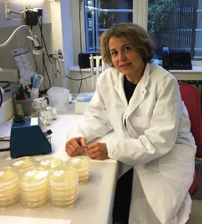
antibiotic use on host-microbiota interactions (discussed in Chapters 23 and 24). We have known for decades that antibiotics—especially broad-spectrum antibiotics—can destroy the ecological balance of bacterial species in the gut (as well as at other body sites) and lead to gastrointestinal disease. Pathology can result when one species resistant to the antibiotic gains a growth advantage over various drug-susceptible species that ordinarily keep the pathogen in check (see the discussion of Clostridioides difficile in Section 26.3). More recent research suggests that disturbing the microbial balance of power in the intestine with antibiotics can contribute to a vast range of diseases, such as irritable bowel disease, vitamin deficiency, obesity, and even asthma.
Antibiotic stewardship. When should antimicrobials be used? Certainly, in life-threatening situations where time is of the essence, antibiotics (usually broad-spectrum antibiotics) should be administered even before the cause of the infection is known (this is called empiric therapy). On the other hand, the most prudent course to take when a patient has a simple infection is to confirm a bacterial etiology and then prescribe a suitable drug. An exception may be in an elderly or otherwise immunocompromised individual, who may be more susceptible to secondary bacterial infections that can occur subsequent to viral disease.
Clinicians, pharmacists, and laboratory personnel are now being trained in antibiotic stewardship to slow, if not to eliminate, the development of new antibiotic-resistant pathogens. Antibiotic stewardship is defined as coordinated interventions that improve and measure antibiotic use. The goal is to cure patients of infections while minimizing drug toxicity and the selection of antibiotic-resistant strains. Proper antibiotic stewardship includes adhering to the following guidelines: Do not use antibiotics to treat what are most likely viral infections (for example, typical respiratory tract infections). Consider using antibiotics only if the patient worsens or does not improve.
Do not use an antibiotic to treat an infection if the patient’s microbiome includes strains that are already resistant to the antibiotic. Doing so can promote transfer of the resistance gene to the pathogen. This practice is difficult to achieve because it requires screening the patient’s microbiome before antibiotic treatment.
Know which antibiotic-resistant strains are prevalent in the community or hospital before prescribing an antibiotic regimen. Consider how long a patient needs to take the antibiotic. The chance of antibiotic resistance increases the longer the patient is being treated.
De-escalate antibiotic usage whenever possible. For instance, a patient being treated empirically with broad-spectrum antibiotics (sometimes involving two or more drugs) should be switched to a suitable, more narrow-spectrum antibiotic once an infectious agent and its resistance pattern are identified. In addition, discontinue any unnecessary antibiotics.
Thought Question
27.11 A clinician admits a seriously ill patient with sepsis to the hospital. She treats the patient empirically with piperacillin and vancomycin until the lab identifies the infectious agent. The next day the agent is identified as Escherichia coli sensitive to third-generation cephalosporins. What should the clinician do now? Why were piperacillin and vancomycin used initially?
Antibiotics in animal feed. Another proposed source of antibiotic resistance is the widespread practice of adding antibiotics to animal feed. Giving animals subtherapeutic doses of antibiotics in their food makes for larger and, therefore, more profitable animals. The reason antibiotics promote livestock growth is unclear, but the stimulation in growth may be the result of altering the diversity of gut microbiota. Some estimates suggest that 80% of all antibiotics used in the United States (up until 2017) were fed to healthy livestock. The consequence is that the animals may serve as incubators for the development of antibiotic resistance. Even if the resistance develops in nonpathogens, the antibiotic genes produced can be transferred to pathogens. Fortunately, in 2017 the FDA banned the use of medically important antibiotics for improving livestock weight gain. These drugs can still be used to treat animals for infectious diseases.
The continued use of nonmedically important antibiotics in cattle feed to promote growth causes other problems. While this practice does not select for resistance to therapeutic antibiotics, it can stimulate the spread of pathogenicity genes between bacteria. The antibiotics trigger SOS responses (see Chapter 9) that reactivate prophages embedded in bacterial chromosomes. If those phages carry toxin genes, the new phage can transfer toxin production to new strains or species that cohabit the intestine. For example, growth-promoting antibiotics such as carbadox or monensin (not used in humans) can reactivate prophages that carry Shiga toxin ( stx) and foster the distribution of stx to other strains. Bradley Bearson of the National Laboratory for Agriculture and the Environment (Ames, Iowa) showed that carbadox will induce prophage replication in pathogenic Salmonella and Shigella strains. The phage can subsequently transfer virulence or antibiotic resistance genes to other bacteria (Fig. 27.27).
FIGURE 27.27 ■ Carbadox induces prophage replication in enteric pathogens. A. Bradley Bearson showed that carbadox, an antibacterial compound formally used to promote growth in swine, activates prophage replication in several enteric bacteria. A phage example is shown on the screen behind him. B. Carbadox (0.5 μg/ml) was added to growing cultures of lysogens for 3 hours (closed symbols). At the indicated times, cells were killed with chloroform, and supernatants were titrated for phage on susceptible hosts. The data illustrate that carbadox-treated lysogens generated 100-to 1,000-fold higher titers of phage.
COURTESY OF BRAD BEARSON
Many of the situations that we have noted here have conspired to produce incredibly dangerous bacteria that are resistant to almost every antibiotic known. For example, a multidrug-resistant strain of Klebsiella pneumoniae was first isolated in 2008 in Örebro, Sweden, from a Swedish citizen returning from New Delhi, India (Fig. 27.28 ). This organism is resistant to most commonly used antibiotics, such as aminopenicillins, beta-lactam/beta-lactamase inhibitors, aminoglycosides, fluoroquinolones, cephalosporins, tigecycline (structurally similar to tetracycline), and carbapenems. It carries the NDM-1 plasmid noted earlier (see Section 27.2) and numerous
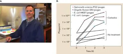
other antibiotic resistance genes. Despite attempted treatment with linezolid, the Swedish patient died.
FIGURE 27.28 ■ MacConkey agar plate containing a sputum culture of Klebsiella pneumoniae. K. pneumoniae carrying the antibiotic resistance gene bla NDM-1 is emerging as a dangerous, drug-resistant pathogen.
SIRIRAT SHUTTERSTOCK
The Infectious Diseases Society of America (IDSA) coined the term “ESKAPE pathogens” a decade ago, referring to the six bacterial species (including K. pneumoniae) that collectively cause about two-thirds of all U.S. nosocomial infections and are highly
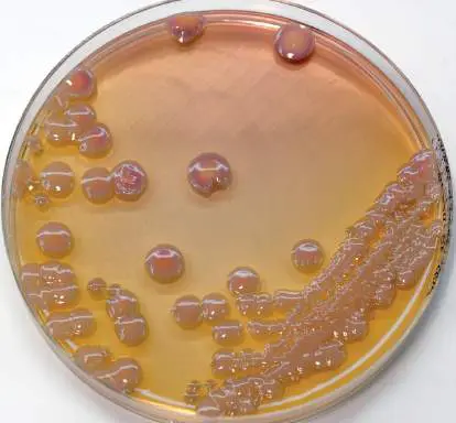
resistant to many existing drugs. These bacteria are E nterococcus faecium, S taphylococcus aureus, K lebsiella pneumoniae, A cinetobacter baumannii, P seudomonas aeruginosa, and E nterobacter species. During the COVID-19 pandemic, some hospitals reported that about 10% of COVID-19 patients developed sepsis from these pathogens. Clearly, the global threat posed by ESCAPE bacteria warrants a redoubling of efforts to identify new antibiotics capable of killing them.
Thought Questions
27.12 Figure 27.28 shows a colony with a distinctive morphology. Why are the colonies on this agar plate red and mucoid?
27.13 Could genomics ever predict the drug resistance phenotype of a microbe? If so, how?
Biofilms and the Mystery of Antibiotic Persisters
Why do some infections return after bactericidal antibiotic treatment is discontinued? At least part of the reason is a subpopulation of dormant organisms, called antibiotic persister cells, that arise within a population of antibiotic-susceptible bacteria. The stalled metabolism of persisters renders them tolerant to bactericidal antibiotics during treatment. Removing the drug allows persisters to grow and reestablish infection. The strategy is analogous to a hiker’s tactic, on accidentally encountering a bear in the woods, of “playing dead” to avoid being attacked.
Persistence is a long-recognized mystery of microbiology. Joseph Bigger in 1944 noticed that penicillin would lyse a growing culture of Staphylococcus aureus, but a small number of persister cells always survived. These persisters were not mutants made permanently resistant through mutation; they acted as though dormant. Persister cells that tolerate antibiotic treatment can be found in any biofilm or population of late-exponential-phase cells. In addition to causing antibiotic treatment failures, persistence may be the reason for latent bacterial infections such as recrudescent typhus or latent tuberculosis.
The mechanisms that cause persistence in the face of bactericidal antibiotics appear to be varied. The laboratory of Kim Lewis (Northeastern University), for instance, published evidence that stochastic depletion of ATP in cells of Staphylococcus aureus and other species underlies persister formation. In one example, the Lewis lab artificially increased ATP levels in Pseudomonas aeruginosa and nearly abolished persister formation. Lowering ATP levels, in contrast, increased the number of persisters.
Other laboratories have focused on a link between antibiotic persister cells and so-called toxin-antitoxin modules encoded by chromosomal genes. (More on toxin-antitoxin modules can be found in Section 5.5.) One example of a toxin-antitoxin module thought to be involved in persistence relies on the hipA and hipB genes in uropathogenic E. coli. HipA is a toxin that is neutralized by the antitoxin HipB (Fig. 27.29, step 1). A delicate counterbalance between HipA and HipB levels allows cells to grow normally. However, because HipB antitoxin is less stable than HipA, a portion of HipA toxin can become active if antitoxin degrades or its synthesis lags (step 2). When freed of antitoxin, HipA will phosphorylate and inactivate glutamyl-tRNA synthetase (step 3). The subsequent lack of charged glutamyl-tRNA stalls translation. The stalled ribosome synthesizes the guanosine tetra-and pentaphosphate [(p)ppGpp] signal molecules (see “The Stringent Response” in Section 10.4) that can alter the transcription of numerous genes. These molecules are very important to persistence: When researchers prevented (p)ppGpp synthesis, cell populations treated with antibiotic produced fewer persister cells.
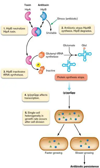
FIGURE 27.29 ■ HipA (toxin) and HipB (antitoxin) control over antibiotic persistence in Escherichia coli.
Current thinking is that the effects of (p)ppGpp on gene expression (Fig. 27.29, step 4), coupled with the heterogeneity in growth rates and gene expression by single cells within the population, can produce a few very slow-growing cells in a population. A subset of those cells can develop into persister cells. Heterogeneity in single-cell composition results from the unequal (epistatic) distribution of cell contents during cell division (step 5). Two daughter cells derived from the same parent cell will have differences in their mRNA content, levels of regulatory molecules, proteins, and signal molecules. As a result of this heterogeneity, some cells will grow more slowly than other cells. Add the effects imposed by heightened amounts of (p)ppGpp on gene expression, and the population develops a set of very slow-growing cells that can persist when antibiotics are added. Precise mechanisms are unclear, but most scientists in the field agree that there are multiple routes to persistence, most of which involve (p)ppGpp.
How might slow-growing cells explain antibiotic persistence? In what has been called the “hunkering down” model, persister cells that barely grow will have little or no cell wall synthesis, translation, DNA replication, or topoisomerase activity. Consequently, even if bactericidal antibiotics can bind to their targets, target function cannot be corrupted. Persistence, then, provides antibiotic resistance at the price of not growing. A bactericidal antibiotic will kill all susceptible bacteria in an infection, but the remaining persister cells serve as a source of population regrowth (and reinfection) once the antibiotic is removed.
Fighting Resistance and Finding New Drugs
The pervasive nature of bacterial resistance to antibiotics has led many to declare that we are in the “postantibiotic era.” But is humankind really doomed to a future in which antibiotics will no longer work? The hope is that the prudent use of current antibiotics and innovative strategies for finding new ones will enable us to continue to control evolving bacterial pathogens.
Directly countering drug resistance. In addition to antibiotic stewardship (discussed earlier), several strategies are being used to counter drug resistance. In some instances, dummy target compounds that bind to and inactivate resistance enzymes have been developed. Clavulanic acid, for example, is a compound used in combination with penicillins such as amoxicillin. Clavulanic acid is a beta-lactam compound with no antimicrobial effect. It is, however, a chemical decoy that competitively binds to beta-lactamases secreted from penicillin-resistant bacteria. Because the enzyme releases bound clavulanic acid very slowly, the amoxicillin remains free to enter and kill the bacterium. Note that bacteria have even developed resistance to beta-lactamase inhibitors, either by over producing the beta-lactamase or by altering the enzyme’s affinity for the inhibitor.
Another way to counter antibiotic resistance is to alter the structure of the antibiotic in a way that sterically hinders the access of bacterial modifying enzymes that could inactivate the drug. Figure 27.30illustrates how adding a side chain to gentamicin that converts it to amikacin blocks the activity of various aminoglycoside-modifying enzymes. Of course, there are now pathogens that are resistant to amikacin.
FIGURE 27.30 ■ Fighting drug resistance. A. Sites where gentamicin is vulnerable to enzymatic inactivation. AAC = aminoglycoside acetyltransferase; ANT = aminoglycoside adenylyltransferase; APH = aminoglycoside phosphotransferase. The inset shows the R groups for different gentamicin
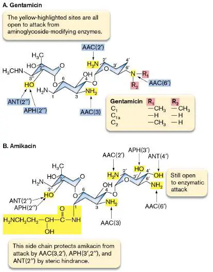
compounds: C 1, C 1a, and C 2. B. Gentamicin can be chemically modified at the highlighted sites to prevent loss of activity due to enzyme action. The side groups block access to enzyme active sites by steric hindrance (that is, the added groups prevent the active site from interacting with its target structure) but do not inactivate the antibiotic.
Finding new antibiotics. How do we find new antibiotics?
Certainly, the classic approach—in which microbes, plants, and even animals collected from around the world are screened for their abilities to make new antibiotics—is still valid and remains a fruitful source of new potential drugs. Even uncultured soil bacteria have been screened for new antibiotics, as described in Section 4.3. Note, too, that in Section 5.6 we describe how bacteriophages are being used in a strategy called phage therapy to prevent or circumvent antibiotic resistance in specific bacterial pathogens. Relatively recent brute-force screening techniques have revealed new classes of antibiotics. One example is platensimycin, made by Streptomyces platensis. The screening method, although laborious, was novel. Merck scientists screened 250,000 natural product extracts for an ability to specifically inhibit bacterial fatty acid biosynthesis. Fatty acid biosynthesis is an attractive target because the bacterial process is different from that of eukaryotes. Scientists engineered a strain of Staphylococcus aureus to contain a gene expressing an antisense RNA to fabF mRNA. The protein FabF is essential for bacterial fatty acid synthesis. When antisense RNA production was induced, the antisense RNA bound the ribosome-binding site of fabF mRNA and partially blocked its translation. As a result, the level of FabF protein in the cell decreased. The strain could still grow but would be exquisitely sensitive to any compound that targeted the remaining FabF protein. This novel screening method led to the discovery of platensimycin.
Platensimycin binds FabF and exhibits bacteriostatic, broad-spectrum activity. It is only the fourth entirely new class of antibiotic developed in the last four decades [lipopeptides (daptomycin), oxazolidinones (linezolid), and bedaquiline (discussed later) are the other three]. The novel chemical structure of platensimycin and its unique mode of action provide a great opportunity to develop a new class of critically needed antibiotics—that is, a class that selectively targets fatty acid biosynthesis. As of this writing, modifications of platensimycin that increase activity have been made but have not yet been approved by the FDA for clinical use. More information on fatty acid biosynthesis and inhibitors can be found in Chapter 15. Another success story paired brute-force screening with combinatorial chemistry to identify a novel antibiotic.
Mycobacterium tuberculosis is a reemergent, slow-growing pathogen that causes tuberculosis in 9 million people each year (see Chapter 26). What makes the situation even more desperate is that, of those 9 million cases, 500,000 are caused by multidrug-resistant (MDR) M. tuberculosis. Unfortunately, the antibiotics for this pathogen had not changed for 40 years.
To find new anti-tuberculosis agents, a team led by Belgian scientist Koen Adries screened 70,000 compounds for antimicrobial effects on Mycobacterium smegmatis, a fast-growing relative of M. tuberculosis. One compound that significantly affected growth was chemically modified to increase its efficacy. After exhaustive clinical trials, the new drug, now called bedaquiline, was approved by the FDA in 2013 to treat MDR-TB. The antibiotic selectively targets the organism’s energy-generating ATP synthase—a novel mode of action —and starves the pathogen of energy. The hope is that this new antibiotic can finally stem the rising tide of drug-resistant TB. Tempering that hope, however, is the knowledge that bedaquiline-resistant mutants of M. tuberculosis have already been isolated in the laboratory. eResearch Activity 27 describes how a more classic approach was used to find a narrow-spectrum antibiotic whose targets are the Borrelia and Treponema spirochetes of Lyme disease and syphilis.
Newer strategies of drug discovery center on genome sequence analysis to identify potential bacterial molecular targets. Once a target is identified, clever screening techniques are used to find natural antibiotics, and molecular modeling is used to synthetically design potential inhibitor molecules. Examples of genomic mining approaches to drug discovery are described in Section 15.3. High-throughput biochemical screens of large collections of synthetic chemicals have also been attempted. Although many promising drugs have been identified, unfortunately only a rare few have proved therapeutically useful. A recent and exciting development to the field involves artificial intelligence being used to screen large databases of chemical compounds for potential antimicrobial drugs ( Special Topic 27).
SPECIAL TOPIC 27 Artificial Intelligence Tackles the Superbug Threat
There is a desperate need to discover new antibiotics. The traditional method requires the painstaking screening of secondary metabolites made by soil microbes. Unfortunately, using old methods to find new drugs is becoming increasingly difficult. Without innovative efforts to find new antibiotics, deaths caused by antibiotic-resistant infections could reach 10 million per year by 2050.
To meet this need, antibiotic discovery programs now screen synthetic chemical libraries that can contain millions of compounds. But those libraries are costly to sort through and often fail to embody the chemistry of proven antibiotics. In fact, no new clinically useful antibiotics have been discovered using this random, large-scale approach. Advances in machine learning, however, may change all that. It is now possible to perform early drug discovery in silico.
As proof in point, scientists from American and Canadian institutions, led by MIT researchers Regina Barzilay (Fig. ST 27.1 ) and James Collins, recently used machine learning methods and artificial intelligence (AI) to discover a powerful new antibiotic they call halicin. The process began by training a deep neural network how to predict growth inhibition of E. coli. Researchers trained the AI on 2,560 molecules (2,335 structurally unique compounds) that had been tested for growth inhibition. Figure ST 27.2A shows the growth inhibition results of those molecules. Once trained, the AI model scanned a chemical library of 6,111 compounds for structures that the algorithm predicted would have activity against E. coli. Ninety-nine molecules were identified and tested for growth inhibition of E. coli (Fig. ST 27.2B ). Of those, 51 passed the inhibition cutoff of <0.2 OD 600. The 51 were then culled to one compound that was already in clinical testing, had structural similarity to molecules in the primary training set, and was predicted to have low toxicity by another program. That compound was c-Jun N-terminal kinase inhibitor SU-3327, renamed halicin, which was being studied as a treatment for diabetes. Halicin (Fig. ST 27.2C , inset) is structurally similar to nitro-containing antiparasitic compounds and the antibiotic metronidazole. Figure ST 27.2C presents growth inhibition results showing that the MIC for halicin is 2 μg/ml.
FIGURE ST 27.1 ■ Regina Barzilay from the Massachusetts Institute of Technology is a computational linguist who designed the artificial intelligence neural network used to discover new antibiotics.
REGINA BARZILAY

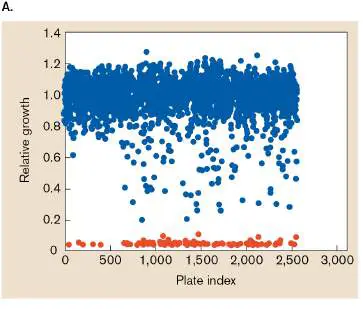
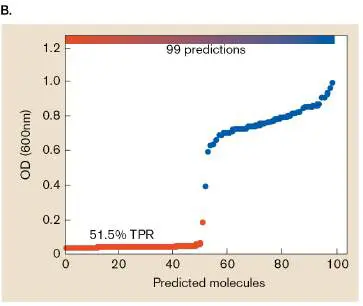
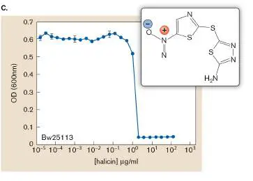
FIGURE ST 27.2 ■ Model training and the identification of halicin. A. Screening of 2,560 molecules for inhibition of E. coli growth. Red dots represent growth-inhibitory compounds; blue dots represent compounds that were deemed noninhibitory. Cells were diluted 1/10,000 into a nutrient broth and added to microtiter plate wells containing 50 μm of different compounds. The “Plate index” x -axis marks the identity of each microwell plate, and each dot represents the mean of two biological replicates within each plate. B. Growth-inhibitory performance of the top 99 artificial intelligence predictions scored from a 6,000-member chemical library (Drug Repurposing Hub). TPR= true positive results. C. Growth inhibition of E. coli BW25113 by halicin. Inset shows the structure of halicin. MIC = 2 μg/ml.
The next steps were to determine whether halicin was bactericidal or bacteriostatic and to screen for activity against different groups of bacteria. The drug is bactericidal (it lowered E. coli viable count by six orders of magnitude after only 4 hours incubation) and is broad spectrum, showing activity against Mycobacterium tuberculosis, clinical isolates of carbapenem-resistant Enterobacteriaceae, and Acinetobacter baumannii. These are pathogens most urgently in need of new treatments. Pseudomonas aeruginosa, unfortunately, was not very susceptible to this antibiotic.
Attempts to isolate halicin-resistant mutants were unsuccessful, so clues about the drug’s mechanism of action were gleaned from the transcriptomes of log-phase cells treated with a range of halicin concentrations. The researchers observed a down-regulation of genes involved in motility and an up-regulation of genes required for iron homeostasis. Given that dissipating transmembrane potential leads to decreased expression of flagellar genes, the authors wondered whether halicin killed bacteria by destroying proton motive force (PMF). The key experiment proving that halicin affected PMF involved the potentiometric fluorophore 3,3′-dipropyl thiadicarbocyanine iodide [DiSC 3 (5)]. This fluorophore accumulates in the cytoplasmic membrane in response to the Δψ component of PMF, and it self-quenches its own fluorescence as long as Δψ is intact. When Δψ is disrupted or the membrane is permeabilized, the probe is released into the extracellular fluid and fluoresces. In contrast, when ΔpH is disrupted, cells compensate by increasing Δψ, which boosts uptake of the fluorophore into the membrane and decreases fluorescence.
Adding increasing concentrations of halicin progressively decreased fluorescence (Fig. ST 27.3 ), meaning that halicin dissipated ΔpH, causing extracellular fluorophore to enter the membrane and self-quench. The vehicle control, DMSO, had no effect on fluorescence, whereas the polymyxin D control, which permeabilizes membranes, increased fluorescence.
FIGURE ST 27.3 ■ Halicin disrupts the ΔpH component of proton motive force and is effective as an antibiotic in vivo. DiSC 3 (5) fluorescence in E. coli upon exposure to polymyxin B (PMB), halicin, or DMSO.
Arrow indicates time of addition. RFU= relative fluorescence units.
The team then asked whether halicin could act as an antibiotic in vivo. They established an intestinal infection in mice using Clostridioides difficile spores administered by oral gavage. Beginning 24 hours after C. difficile infection, different groups of mice were orally treated for 5 days with halicin, metronidazole (the clinically used drug), or vehicle (10% polyethylene glycol). The scientists were excited to see that halicin cleared the infection by 6 days after the start of
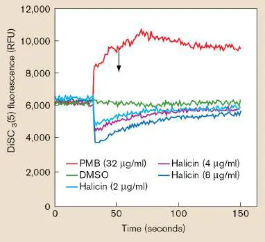
treatment, working better than metronidazole, one of the first-line treatments for C. difficile infection.
The research team went on to identify several other potentially useful antibiotics using the AI model. Their results suggest that a machine learning approach may eventually predict antimicrobials that not only have a narrow-spectrum activity against a specific pathogen but can also be administered systemically without damaging the host microbiota.
Research Question
If halicin acts by somehow dissipating the ΔpH component of PMF, what effect would changing external pH have on the MIC of the drug?
Stokes, Jonathan M., Kevin Yang, Kyle Swanson, Wengong Jin, Andres Cubillos-Ruiz, et al. 2020. A deep learning approach to antibiotic discovery. Cell 180 :688–702. https://doi.org/10.1016/j.cell.2020.01.021. Virulence genes and proteins of pathogens also hold great promise as targets for new drugs because these genes are required for a pathogen to grow in a host. However, these proteins are needed only during infection, not during in ex vivo growth outside of a host, the condition typically used to screen for antibiotics. Inhibitors that target virulence proteins will not inhibit growth in vitro (on nutrient agar, for example). So, how can we develop or screen for specific inhibitors of these proteins? Currently, it is easier to make monoclonal antibodies that target virulence proteins than it is to design and find chemical inhibitors. Synthetically engineered, bispecific antibodies (antibodies whose two antigen-binding sites target different surface virulence factors on the same pathogen) are now being tested as pathogen-specific antimicrobials.
Another intriguing idea involves using photosensitive chemicals that can penetrate the microorganism and generate toxic reactive oxygen species (such as superoxide) when exposed to specific wavelengths of visible light (obviously, good only for topical use). Interfering with the quorum-sensing mechanisms of pathogens is yet another clever approach. Finally, the promise and flexibility of synthetic-biology approaches discussed in Chapter 12 could revolutionize the process of antibiotic discovery and production. For instance, CRISPR-based strategies are being considered as pathogen-specific antimicrobials and as a tool to reverse antibiotic resistance in a pathogen by excising the resistance gene in vivo. Antipersister and antibiofilm approaches. Because persister cells and biofilms play prominent roles in recurrent infections and treatment failures, antimicrobials that target these features would be a major breakthrough in treating infections. Several approaches targeting persisters are possible. Some compounds can directly kill persisters—for example, compound HT61, a quinolone derivative that depolarizes the cell membrane and destroys the cell wall; or clofazimine, a compound that increases production of destructive reactive oxygen species. Other compounds can prevent persister formation by interfering with (p)ppGpp synthesis or reverse the persister state by stimulating the cell’s metabolism, once again making them susceptible to existing antibiotics.
What about biofilms? Biofilms not only harbor metabolically dormant persister cells, but also their sheer density and complex architecture provide their own level of antibiotic tolerance. There are two main antibiofilm approaches: to interfere with the synthesis and secretion of extracellular polymeric substances that coat biofilms and hold them together or to induce biofilm dispersal. Small molecule inhibitors that limit synthesis of cyclic-di-GMP, cyclic-di-AMP, and (p)ppGpp signaling molecules are being sought and tested (discussed in Chapters 4 and 10). These signaling molecules control several aspects of extracellular polymeric substance metabolism and biofilm dispersal.
Why has progress been so slow in finding new antimicrobial compounds? Part of the answer is that the road to FDA approval is long (8–10 years) and expensive, involving numerous animal and human trials. The process is necessary for safety reasons, but it discourages the pharmaceutical industry from investing in antibiotic discovery. However, because of the public health implications of a dwindling pipeline of new antibiotics, policy makers are trying to incentivize investment.
To Summarize
Certain microbes make antibiotics to eliminate competitors in the environment and prevent self-destruction by means of various antibiotic resistance mechanisms. Genes encoding some of these drug resistance mechanisms have been transferred to pathogens.
Antibiotic resistance can arise spontaneously through mutation, can be inherited by gene exchange mechanisms, or can arise de novo through gene duplication and mutational reengineering.
Antibiotic resistance involves three basic strategies. (1) Keep the antibiotic out of the cell by destroying the drug, reducing permeability, or pumping the drug out. (2) Prevent the antibiotic from binding to its target by altering the target or the drug. (3) Knock the drug off its target. Multidrug resistance efflux pumps use promiscuous binding sites to bind antibiotics of diverse structure.
Indiscriminate use of antibiotics has significantly contributed to the rise in antibiotic resistance.
Measures to counter antibiotic resistance include synthetically altering the antibiotic, using combination antibiotic therapy, and adding a chemical decoy.
Persister cells in a population have stopped actively growing, making these cells tolerant to bactericidal antibiotics.
Antibiotic resistomes comprise all the antibiotic resistance genes present in human, animal, and natural environments. The quest to discover novel antibiotics includes designing candidate antimicrobial compounds to interact with and inhibit the active site of a known microbial enzyme and screening previously uncultured microbes for new antibiotics.
Potential targets for new antimicrobials include persister cells, proteins expressed only in vivo (virulence proteins), and pathways contributing to biofilm formation and dispersal.
Glossary
secondary metabolite or secondary product An organic product of biosynthesis that does not have essential functions but enhances nutrient uptake under certain conditions or inhibits competing species (e.g., an antibiotic). Often produced during stationary phase.
multidrug resistance (MDR) efflux pump A transmembrane protein pump that can export many different kinds of antibiotics of diverse structure.
resistome The collection of antibiotic resistance genes present in all pathogenic and nonpathogenic microbes that inhabit humans, animals, or the environment.
persister cell Any of a subpopulation of dormant organisms that arise within a population of antibiotic-susceptible bacteria and are tolerant to bactericidal antibiotics during treatment. On removal of the drug, persisters can grow and reestablish infection.
27.4 Antiviral Agentsnot assigned
A father pleading with a physician to give his child antibiotics when the infant is suffering with a cold is an all-too-common dilemma faced by the general practitioner, but there is nothing of substance the physician can do. The common cold is caused by rhinoviruses, coronaviruses, and adenoviruses; no antibiotic designed for bacteria can touch them.
Why are there so few antiviral agents in the clinician’s arsenal? The reason is that it is much harder to apply the principle of selective toxicity for viruses than it is for bacteria. Viruses routinely commandeer host cell functions to make copies of themselves. Thus, a newly discovered compound that hurts the virus is likely to also harm the patient. Nevertheless, there are several useful antiviral agents for which selective viral targets have been found and exploited. The most recent and arguably heroic antiviral is Paxlovid by Pfizer, an oral protease inhibitor that attacks SARS-CoV-2, the virus responsible for the COVID-19 pandemic (Fig. 27.31).
FIGURE 27.31 ■ Structures of the main SARS-CoV-2 protease and of the antiviral protease inhibitor nirmatrelvir. A. The three-dimensional structure of the protease dimer. One monomer of the dimer is shown as a cartoon ribbon (orange), while the other monomer is shown as a space-filling model (teal). The catalytic site cavity is highlighted,
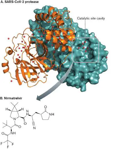
and water molecules are shown as red spheres. B. Nirmatrelvir, the active ingredient in Paxlovid, targets the catalytic site of the protease.
D. KNELLER ET AL. 2020. NAT COMMUN. 11 :3202
Some of the FDA-approved antiviral agents are listed in Table 27.3. Select examples are discussed in this section. All of the molecular mechanisms of viruses presented in Chapter 11 are studied as potential drug targets.
TABLE Examples of Antiviral Agents 27.3
Virus Agent Mechanism Result of action Influenza virus Amantadine Inhibits viral Prevents viral M2 protein uncoating Zanamivir Neuraminidase Prevents viral inhibitor release (nasal spray)
Oseltamivir Neuraminidase Prevents viral (Tamiflu) inhibitor (oral release prodrug)
TABLE Examples of Antiviral Agents 27.3
Baloxavir Inhibits cap-Prevents marboxil dependent transcription endonuclease and (oral prodrug) translation of viral mRNA Herpes simplex Acyclovir Guanosine Halts DNA virus and analog synthesis varicella-zoster virus (shingles)
Famciclovir Prodrug of Halts DNA penciclovir, a synthesis guanosine analog Cytomegalovirus Ganciclovir Similar to Halts DNA acyclovir synthesis Foscarnet Analog of Binds and inorganic inhibits virus-phosphate specific DNA polymerase
TABLE Examples of Antiviral Agents 27.3
Respiratory Ribavirin RNA virus Causes syncytial virus mutagen catastrophic and chronic replication hepatitis C virus errors Hepatitis C virus Sofosbuvir Analog Halts RNA (Sovaldi) inhibitor of synthesis HCV polymerase Simeprevir Protease Prevents viral (Olysio) inhibitor maturation HIV Zidovudine Nucleoside Inhibits (AZT) analog; reverse resembles transcriptase thymine Nevirapine Binds to Inhibits allosteric site reverse
TABLE Examples of Antiviral Agents 27.3
transcriptase Tenofovir Nucleotide Inhibits analog, reverse resembles transcriptases AMP of HIV and hepatitis B virus Nelfinavir Protease Prevents viral inhibitor maturation Raltegravir Integrase Prevents inhibitor integration into host genome Maraviroc CCR5 entry Prevents inhibitor virus entry into host cells
TABLE Examples of Antiviral Agents 27.3
SARS-CoV-2 Nirmatrelvir Protease Prevents viral inhibitor maturation
Target Virus Uncoating or Release
Membrane-coated viruses are vulnerable at two stages. The first is when the virus is invading the host cell. The second is after viral propagation, when the progeny viruses release from the host cell. The flu virus presents a good example of both.
Case History: Antiviral Treatment of Infant Influenza
A 9-month-old infant arrived at the Johns Hopkins Hospital with an acute onset of fever, cough, regurgitation from his gastrostomy feeding tube, and dehydration. This illness followed a series of chronic problems, including bronchiolitis (infection and inflammation of the bronchioles) caused by respiratory syncytial virus, and neonatal group B streptococcal sepsis. (Neonatal sepsis is often caused by Lancefield group B Streptococcus agalactiae; Lancefield group classification is described in Chapter 28.) Physical exam revealed fever, a severe cough resulting in respiratory distress, a rapid heart rate, and moderate dehydration. Nasopharyngeal aspirate was positive for influenza A antigen. The patient was treated with oseltamivir when influenza was diagnosed. He gradually improved and was discharged home 4 days after admission.
In 2017–2018, an unusually severe form of influenza (H3N2) spread across the United States. Flu-related hospitalizations in the United States were anticipated to exceed 700,000, and most states reported a higher-than-normal number of influenza-related deaths of children and young adults. Several factors, however, helped keep the outbreak from becoming an epidemic of larger proportions. The administration of flu vaccine afforded the population what is called herd immunity (discussed in Section 24.6). Herd immunity takes place when a majority of the population is immunized against a pathogen. The vaccinated individuals decrease the risk that an unvaccinated person will have direct contact with an infected person. At the very least, herd immunity slows the spread of infection throughout the population.
Note: It is impossible to immunize all humans against any given
disease. However, it is estimated that immunizing about 80% of a population for influenza can halt an epidemic by cutting off transmission. (See the discussion of herd immunity in Section 24.6.) Unfortunately, this level of immunization is rarely achieved. Another factor that limited the scope and severity of recent influenza epidemics was the availability of antiviral agents that limit the disease course. There are now three viral targets: hemagglutinin, required for virus entry; neuraminidase, needed for viral exit; and a viral endonuclease, the newest target, critical for viral replication.
Recall that influenza virus (200 nm) is encased in a membrane envelope acquired when the virion buds from an infected cell. As described in Section 11.2, the envelope contains the viral proteins neuraminidase (NA) and hemagglutinin (HA). Spikes of hemagglutinin bind to sialic acid receptors on the host cell and trigger receptor-mediated endocytosis. Once the virus is inside an endosome, proton pumps in the membrane acidify endosomal contents. The structure of hemagglutinin on the viral membrane changes as a result of acidification, which enables the protein to bind receptors on the endocytic membrane. The viral and endocytic membranes fuse, and the virion is released into the cytoplasm. For hemagglutinin structure to change, the interior of the enveloped virion must also be acidified. Acidification is mediated by a membrane channel formed by the virus-encoded M2 protein. The drug amantadine (Fig. 27.32A) is a specific inhibitor of the influenza M2 protein that prevents M2 channel formation, which, in turn, prevents viral uncoating. Unfortunately, amantadine-resistant strains of influenza have developed, in part because of the widespread use of amantadine by Chinese poultry farmers.
Consequently, the drug is no longer recommended as a treatment for influenza.
FIGURE 27.32 ■ Inhibitors of influenza proteins. A. Amantadine inhibits the M2 protein. B. Zanamivir inhibits neuraminidase. C. Neuraminidase without (left) and with (right) bound inhibitor. (PDB: 2HTQ)
The second target of the influenza virus is the envelope protein neuraminidase. The antiflu drugs zanamivir (Relenza) and oseltamivir (Tamiflu) are neuraminidase inhibitors that act against types A and B influenza strains (Fig. 27.32B and C ). Sialic acid residues on the host cell surface bind the hemagglutinin on flu viruses trying to exit the cell. Neuraminidase on the same viral envelope will cleave the cell-surface sialic acid and free the virus.
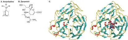
Unencumbered, the virus can leave the cell surface and infect another cell. Neuraminidase inhibitors, however, prevent sialic acid cleavage, causing the virus particles to aggregate at the cell surface and reduce the number of virus particles released.
The neuraminidase inhibitors, when used within 48 hours of disease onset, decrease shedding and reduce the duration of influenza symptoms by approximately one day. However, flu symptoms generally last only 3–10 days. While this does not sound like a substantial benefit, shortening the course of the flu in the elderly can minimize damage to the lungs, which in turn reduces the chance of developing life-threatening secondary bacterial infections such as pneumonia and bronchitis.
Note: RNA viruses, such as influenza, use RNA-dependent RNA
polymerases that lack proofreading capability. The consequence of an error-prone polymerase is a high mutation rate and the generation of antiviral-resistant virions. Widespread use of Tamiflu during the 2008 flu epidemic quickly led to Tamiflu resistance. Fortunately, the mutations that resulted in resistance to Tamiflu also slowed virus replication rate. Instituting more judicious use of the drug caused the resistant mutants to lose competitive advantage over the faster-replicating sensitive mutants, and the resistant mutants disappeared.
Target Virus Cap-Snatching
In 2018, the FDA approved a new antiviral agent that is effective against influenza and has a unique mode of action. Host mRNA molecules contain a 5′ cap composed of the modified base 7-methylguanosine, which protects host message from nucleases and is required for translation. As described in Chapter 11, influenza virus steals host 5′ caps (cap-snatching), along with 10–20 downstream nucleotides, using a cap-dependent endonuclease. Once the cap is stolen and attached to viral mRNA, it is used by RNA polymerase to initiate transcription of virus mRNA from the negative-sense RNA strand of the infecting flu virus. The stolen cap is also required for translation of the viral mRNA. The new drug, called baloxavir marboxil (sold as Xofluza) inhibits the activity of the influenza cap–dependent endonuclease, which will prevent cap-snatching and stop viral replication. Xofluza is effective against the growing number of flu viruses that are resistant to M2 and neuraminidase inhibitors, and it has been approved to treat influenza in adolescents and adults.
Targeting Viral DNA Synthesis
Most antiviral agents work by inhibiting viral DNA synthesis. These drugs chemically resemble normal DNA nucleosides in that they contain deoxyribose and analogs of adenine, guanine, cytosine, or thymine. Viral or sometimes host kinase enzymes then add phosphate groups to these deoxynucleoside analogs to form deoxynucleotide analogs. The deoxynucleotide analogs are then inserted into the growing viral DNA strand in place of a normal nucleotide. Once inserted, however, new nucleotides cannot attach to the nucleotide analogs, and DNA synthesis stops.
These DNA chain–terminating analogs (Fig. 27.33) are selectively toxic because viral polymerases are more prone to incorporate nucleotide analogs into their nucleic acid than are the more selective host cell polymerases. Antiviral DNA synthesis inhibitors work on DNA viruses or retroviruses, but not on viruses such as influenza, with its RNA genome.
FIGURE 27.33 ■ Antiviral inhibitors that prevent DNA synthesis. Zidovudine (AZT) (A) and acyclovir (B) are analogs of thymine and guanine nucleotides, respectively. Because the analogs have no 3′ OH to which another nucleotide can add, chain elongation ceases.
Antiretroviral Therapy
Retroviruses are RNA viruses that use viral reverse transcriptase to make DNA and then use viral integrase to insert that DNA into the eukaryotic host cell genome (see Chapter 11). The integrated provirus can then be activated to make retroviral RNA. The retroviral RNA travels to the cytoplasm and directs synthesis of more virus particles. One of the most devastating retroviruses is human
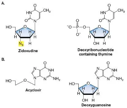
immunodeficiency virus (HIV), the cause of acquired immunodeficiency syndrome (AIDS; see Section 11.3).
Case History: Treatment of HIV
A married couple came to the community clinic for prenatal care. He was 20 years old. She was 19 and reportedly 2 months pregnant with her first child. She denied intravenous (IV) drug use or a history of other sexual partners, and she had no history of sexually transmitted infection; however, a routine prenatal HIV antibody screen was reported as positive for HIV-1. Careful questioning of the patient and her husband elicited from him a history of IV drug use 5 years earlier. An HIV antibody screen for him was also positive. The laboratory results indicated that the wife had a low viral load—that is, less than 1,000 copies per milliliter of blood—and a normal CD4 T-cell count. The husband had a higher HIV viral load (10,000 copies per milliliter) and a lower CD4 count (150 cells per microliter). Antiretroviral therapy was initiated for both husband and wife (even though her CD4 count was normal), but because she was pregnant, the wife’s regimen avoided drugs that could potentially harm the fetus. Their regimens included two nucleotide reverse transcriptase inhibitors plus a protease inhibitor.
Being diagnosed with HIV is no longer a death sentence. Advances in antiretroviral therapy (ART), especially over the past 10 years, have transformed HIV into a manageable chronic condition. At least half, if not most, of the HIV-positive people in the United States now live long enough to die from diseases of aging, such as heart attacks or strokes. Because it is such a treatable infection, the CDC recommends that everyone should be tested for HIV and further recommends that everyone with HIV, regardless of their CD4 T-cell count, receive antiretroviral therapy.
One reason for these recommendations is that many asymptomatic people do not know they are infected. So, identifying and treating an asymptomatic, HIV-positive person will reduce the risk of sexually transmitting the virus. Antiretroviral therapy can also prevent transplacental transmission from an asymptomatic, HIV-positive pregnant woman to her fetus, as in our case history. Because HIV transmission from the mother to the neonate can also occur at delivery or by breast-feeding, postdelivery treatment of the mother and the child is important for preventing transmission. The drugs described next are crucial components of effective treatment. Nucleoside, nucleotide, and nonnucleoside reverse transcriptase inhibitors. As an RNA retrovirus, HIV uses a reverse transcriptase to make DNA that then integrates into host nuclear DNA (discussed in Section 11.3). The antiretroviral drug zidovudine (abbreviated ZDV or AZT) is a nucleoside analog recognized by reverse transcriptase. Once incorporated into a replicating HIV DNA molecule, the DNA chain–terminating property of AZT (the lack of a 3′ hydroxyl group) prevents further DNA synthesis. Nucleoside inhibitors must undergo three successive phosphorylation steps inside the cell to become an active trinucleotide form of the drug. In contrast, nucleo tide inhibitors are essentially monophosphorylated analogs that require only two phosphorylation steps for activation. Tenofovir is a nucleotide analog (structurally similar to AMP) that is recognized by HIV reverse transcriptase and also by the reverse transcriptase of hepatitis B virus. Tenofovir is often given following needle stick accidents to prevent HIV or HBV transmission.
In addition to nucleoside inhibitors, there are nonnucleoside reverse transcriptase inhibitors. For example, the drug delavirdine binds directly to reverse transcriptase and allosterically inactivates the enzyme.
Protease inhibitors. To make optimal use of its limited provirus DNA sequence, HIV generates long, nonfunctional polypeptide chains that are proteolytically cleaved into the actual proteins and enzymes used to replicate and produce new virions. For example, the gag and pol genes reside next to each other in the HIV genome and are transcribed as a single mRNA molecule (see Section 11.3). The Gag and Pol open reading frames overlap but are offset by one base. This mRNA produces two polyproteins, called Gag and Gag-Pol, the latter being the result of a shift in reading frames that takes place during translation. Once made, both polyproteins are cleaved by HIV protease. The Gag protein is proteolytically cleaved to make different capsid components (p17, p24, and p15, the latter of which is further cleaved to make nucleocapsid protein p7; Fig. 27.34A). Gag-Pol is cleaved to make reverse transcriptase and integrase.
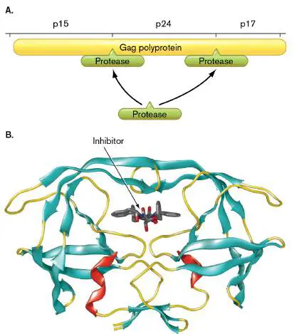
FIGURE 27.34 ■ HIV protease inhibitor. A. Representation of HIV protease cleavage of a single Gag polyprotein into multiple smaller proteins. B. The protease enzyme is shown here as a ribbon structure, while the protease inhibitor BEA 369 is shown as a stick model. (PDB code: 1EBY)
Protease inhibitors such as nelfinavir and lopinavir belong to a powerful class of drugs that block the HIV protease (Fig. 27.34B ). When the protease is inactivated, the polyproteins remain uncleaved and the virus cannot mature, even though new virus particles are made. Because immature HIV particles cannot infect other cells, progress of the disease stalls. Note that protease inhibitors do not cure AIDS; they can only decrease the number of infectious copies of HIV.
Protease inhibitors are also used to treat hepatitis C virus and SARS-CoV-2. The protease inhibitor used to treat SARS-CoV-2, nirmatrelvir (Fig. 27.31B ), is the active ingredient in Paxlovid (Pfizer), which also includes ritonavir. Ritonavir is another protease inhibitor, but its more important activity is to slow the metabolic destruction of nirmatrelvir by inhibiting the liver enzyme cytochrome P450. Ritonavir is also combined with HIV protease inhibitors for the same reason.
Entry inhibitors. Another way to stop HIV is to prevent the virus from infecting cells in the first place. Drugs called entry inhibitors do just that. Recall that for HIV to enter host cells, the HIV membrane protein gp120 (also known as SU) must first attach to the host membrane protein CD4 and to a coreceptor: either CCR5 or CXCR4, depending on the strain of HIV involved. Once the virus is attached, the HIV membrane protein gp41 mediates fusion between the host and viral membranes to enable virus entry (see Chapter 11 and Fig. 11.26 for details).
Three types of entry inhibitors interrupt the entry process. First, CCR5 inhibitors bind to the CCR5 coreceptor and block it from binding to gp120. As a result, the virus never attaches. The second type of entry inhibitor is fusion inhibitors. These chemicals do not prevent initial binding. Rather, the inhibitor binds to the gp41 fusion protein and stops HIV membranes from fusing with T-cell membranes. Viruses cannot enter the cell without their membranes fusing. Imagine that you are trying to enter a room through a door. A CCR5 inhibitor is like removing the doorknob from the door so that there is nothing for you to grab. Fusion inhibitors, however, are like gluing the door shut. You can grab the knob but still cannot open the door.
A newer member of the entry inhibitor class of HIV drugs is actually a postattachment inhibitor. The monoclonal antibody ibalizumab binds to CD4 but does not alter CD4 immune function. The antibody also allows CD4 to bind HIV gp120, but it prevents gp120 from binding to the CCR5 or CXCR4 coreceptors. So, the virus attaches, but it cannot enter. The FDA approved this drug in 2018 to treat patients with multidrug-resistant HIV.
Treatment regimens and HIV controllers. Because HIV can mutate rapidly and become resistant to single-drug therapies (see Section 11.3), HIV treatments today involve combinations of three or more antiretroviral drugs. This therapeutic strategy was originally called highly active antiretroviral therapy (HAART), but the name has been changed to simply antiretroviral therapy (ART). Current ART regimens include three drugs, usually two nucleoside reverse transcriptase inhibitors plus a protease inhibitor, a nonnucleoside reverse transcriptase inhibitor, or an integrase inhibitor. Integrase inhibitors block the enzyme needed to insert viral DNA into the host genome.
Although antiretroviral treatment of HIV has been very effective, only two patients seem to have eliminated all traces of HIV after treatment. You might wonder why. One reason is that HIV is a retrovirus whose cDNA genome has integrated into host genomes. But if ART prevents HIV from replicating and spreading to new cells, wouldn’t the remaining cells containing integrated HIV eventually die through senescence? In 2011, Timothy Schaker at the University of Minnesota, Twin Cities, examined patients undergoing ART who had undetectable blood levels of HIV. His group found evidence that the virus in these individuals still remained trapped in lymphatic tissues that were poorly penetrated by the drugs. So, even though ART can lower HIV to undetectable levels in blood, tissue pockets of HIV remain, able to reestablish infection if ART is stopped. New strategies that more effectively force drugs into tissues might provide the long-awaited cure.
Some patients originally thought to have been cured following early and aggressive ART have maintained very low viral loads (<400 per milliliter of blood) even without treatment. These people are called posttreatment controllers. Somehow their immune systems are able to keep the virus in check. Another group of individuals, called elite controllers, manage to prevent the progression of HIV in the absence of any treatment—possible mechanisms include the presence of newly discovered inhibitory receptors on their CD4 T cells, a more effective natural killer (NK) cell population, or the integration of provirus at chromosomal sites having limited transcriptional activity.
HIV treatment as prevention. As discussed, AIDS can be a devastating disease, but effective antivirals can prevent HIV replication after infection. We have also learned that treating at-risk populations with antivirals before exposure can be effective at preventing infection. This strategy is known as preexposure prophylaxis (PrEP). In fact, the FDA has approved the daily use of an HIV medicine, Truvada (tenofovir/emtricitabine), by healthy but high-risk people hoping to lower their risk of infection by a sexual partner. Both drugs are nucleoside analogs of adenosine and cytosine, respectively, and they are reverse transcriptase inhibitors. A new injectable PrEP drug, Apretude (cabotegravir, an HIV integrase inhibitor), was approved by the FDA in 2021. Although PrEP is an approved preventative strategy, it is controversial even among some physicians over the fear that the drug will encourage risky behavior.
Future Antivirals May Target Host Functions
As you can see from the preceding discussion, most antiviral drugs approved for clinical use target viral proteins (proteases, polymerases, entry proteins) because they afford some measure of selective toxicity. However, a perceived limitation of these direct-acting antivirals is their narrow spectrum of virus coverage. Narrow-spectrum antivirals cannot provide adequate protection against newer, rapidly emerging viral threats. Examples include the flavivirus dengue, coronaviruses SARS-CoV-2 and MERS-CoV, and the filovirus Ebola. Finding new broad-spectrum antivirals could address this need. But what do we target?
A novel approach is to ignore the virus and, instead, target host cell pathways that are required by multiple viruses for replication ( Fig. 27.35). For example, cyclophilin A is involved in host and viral protein folding. Cyclophilin A inhibitors such as alisporivir (DEB025) impair the folding of viral proteins and augment innate immune responses. These drugs affect a variety of DNA and RNA viruses (dengue, hepatitis C, HIV, SARS-CoV-2). Another potential target is the enzyme alpha-glucosidase in the endoplasmic reticulum. Many virus glycoproteins depend on host glucosidases for proper folding. The glucosidase inhibitor celgosivir has, indeed, proved effective against many unrelated viruses in vitro and in rodent models but not yet in humans. Nevertheless, the enzyme warrants further investigation. Host kinases that regulate intracellular virus trafficking are also potential drug targets.
FIGURE 27.35 ■ Potential host targets for broad-spectrum antivirals. Different stages of viral development are shown (entry, fusion, genome replication, assembly, and release). Examples of broad-spectrum compounds are connected
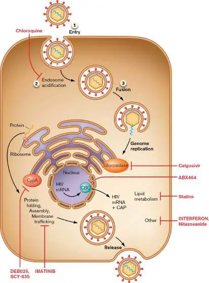
to the corresponding targeted proteins or pathways by blunt arrows. CypA = cyclophilin A.
A more pathogen-specific antiviral agent (ABX464) affects HIV replication by binding to the host cap-binding complex (CBC). (“Cap” refers to the 5′ 7-methylguanosine cap added to eukaryotic and viral mRNAs.) The CBC controls the export of host mRNA from the nucleus to the cytoplasm, where mRNA is translated to protein. The HIV Rev protein brings HIV mRNA to the CBC. ABX464 binds to the CBC and specifically blocks Rev-mediated export of viral RNA, while not interfering with host transcripts. Because the drug binds to a host protein rather than a viral protein, HIV is less likely to develop resistance. This antiviral agent is currently in clinical trials. Figure 27.35also shows some host-targeted drugs already approved for other purposes, such as chloroquine (antimalarial agent) and statins (anticholesterol metabolism). These drugs are now being evaluated for their usefulness against emerging viruses. Chloroquine inhibits endosome acidification, which is needed by some enveloped viruses to escape the vacuole and enter the host cytoplasm (dengue virus and Zika virus, for instance). Note that hydrochloroquine was not effective against the COVID-19 agent (SARS-CoV-2). Statins interfere with lipid metabolism needed for the life cycles of viruses such as hepatitis C virus. How therapeutically effective these drugs will be as antiviral agents is unclear. A particularly exciting strategy may provide broad antiviral protection. The idea is to stimulate a specific group of pattern recognition receptors (PRRs) called Rig-1-like receptors (RLRs). When stimulated, RLRs activate interferon production. Interferon, as described in Chapter 23, signals neighboring cells to express a series of host proteins that degrade viral nucleic acids and inhibit viral protein synthesis. A compound capable of modulating RLRs would be a broad-spectrum antiviral able to thwart replication of many different viruses. For example, short-nucleotide RNA molecules that retain the 5′ triphosphate end will activate RLRs and protect cells against vesicular stomatitis virus, vaccinia virus, dengue virus, and influenza virus. However, delivering these molecules to a host is challenging. Naked RNA molecules cannot be taken up by host cells, so they may have to be attached to nanoparticles or wrapped within lipid vesicles to be introduced into cells.
There are many challenges to the host-as-target approach to antiviral therapy. Host proteins function in a complex network of interactions, so elucidating drug mechanisms of action is difficult. Toxicity is another worry, although it may be possible to identify a therapeutic concentration window in which the drug inhibits virus replication but has minimal toxic effects on the patient.
CRISPR Technology May Lead to Virus-Specific Therapies
If there was one lesson to learn from the recent COVID-19 pandemic, it was that we were not prepared. During the first year of the disease, we were nearly powerless to protect ourselves. Of the 2.8 million people worldwide who died, 500,000 were from the United States. We were lucky to have tools to rapidly design and test novel mRNA vaccines, but it was a year before the vaccines were ready to put into arms. Then there were the viral variants able to slip past vaccine-derived immunity. The question we now ask ourselves is: “How can we make future pandemics less deadly?” One way is to devise a technology capable of quickly making narrow-spectrum antivirals for any virus we meet. Once made, the antiviral can be used to treat patients while we wait for a vaccine. CRISPR technology might be that technology.
CRISPR-Cas systems were described in Chapter 12 as mechanisms that bacteria use to combat their own viral infections and, now, a technique that scientists use to mutate, delete, or swap alleles in any bacterial or eukaryotic cell. Gene specificity for Cas endonuclease systems is provided by a guide RNA whose sequence supplies the address for the gene to be cleaved. Change the guide RNA sequence, and you change the gene targeted. Once scientists have the sequence of a new virus, they can design a guide RNA that can direct an attack against it, even before a vaccine can be made. Some CRISPR-Cas systems target DNA (Cas9), and other systems target single-stranded RNA (Cas13). Figure 27.36illustrates the ways that CRISPR systems can (1) target DNA viruses, (2) delete virus genomes (proviruses) integrated into the host chromosome, (3) destroy single-stranded RNA viruses, or (4) digest mRNA produced from a virus. Some of these techniques have been used experimentally, but certain issues still need to be resolved—such as what mechanism(s) to use for delivery (currently adeno-associated viruses), whether the Cas proteins will be antigenic and subject to host immunity, and how to eliminate potential off-target effects of the Cas endonuclease.
FIGURE 27.36 ■ Proposed CRISPR-based targeting of pathogenic viruses. Figure illustrates the ways CRISPR techniques can target the viral DNA or mRNA gene products of DNA viruses (1, 2, 4) versus RNA viruses (2, 3). Host DNA is black; guide RNAs are blue curves; black triangles mark cleavage. (1) Cas9 (orange) attacking a dsDNA viral genome
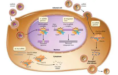
(purple), such as herpesvirus. (2) Cas9 (orange) attacking an integrated provirus (purple), such as HIV. (3) Cas13 (orange in cytoplasm) attacking a single-stranded RNA viral genome (dark blue), such as SARS-CoV-2. (4) Cas13 (orange in cytoplasm) attacking a DNA virus’ mRNA (gray).
Thought Question
27.14 The text states that cells and viruses would have difficulty developing resistance to antiviral drugs that target host proteins rather than viral proteins. Why would targeting a host protein decrease the likelihood of developing resistance? Propose a mechanism by which the drug could still become ineffective against a virus.
To Summarize:
Fewer antiviral agents than antibacterial agents are available because it is harder to identify viral targets that provide selective toxicity.
Preventing viral attachment to, or release from, host cells is a mechanism of action for antiviral agents, such as amantadine and zanamivir, used to treat influenza virus. Inhibiting DNA synthesis is the mode of action for most antiviral agents, although it works only for DNA viruses and retroviruses.
HIV treatments include reverse transcriptase inhibitors that prevent the synthesis of DNA, protease inhibitors that prevent the maturation of viral polyproteins into active forms, and entry inhibitors that either prevent HIV from binding to host membranes or inhibit fusion of the HIV envelope to the host cell membrane.
CRISPR technology is being used to design highly targeted (narrow-spectrum) antiviral therapies.
Future antiviral agents may target host proteins needed by the virus to replicate or stimulate interferon-dependent innate immune mechanisms that can repel multiple viral pathogens.
Glossary
neuraminidase inhibitor Any of a class of anti-influenza drugs that target neuraminidase on the viral envelope and decrease the number of virus particles produced.
antiretroviral therapy (ART)
A three-drug antiretroviral cocktail that is highly effective at inhibiting the replication of HIV in patients.
Fig. 11.26 FIGURE 11.26 ■ HIV-1 attachment to host cell. The SU (gp120) subunits of the spike protein complex attach to the receptor (CD4 cell-surface protein) and to CCR5. The fusion peptides contract, pulling the membranes together.
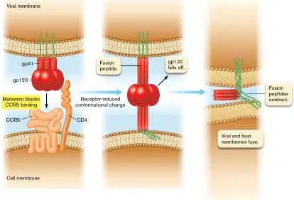
27.5 Antifungal Agentsnot assigned
Fungal infections are much more difficult to treat than bacterial infections, in part because fungal
physiology is more similar to that of humans than bacterial physiology is. The other reason is that fungi
have an efficient drug detoxification system that modifies and inactivates many antibiotics. Thus, to
have a fungistatic effect, repeated applications of antifungal agents are necessary to keep the level of
unmodified drug above MIC levels.
Case History: Blastomycosis
A 37-year-old man presented to the emergency department of a Florida hospital with persistent fever,
malaise, and a painful right-arm mass. He denied trauma to the arm. White blood cell count was
elevated at 27,000 per microliter (leukocytosis), and chest X-ray revealed a left-lung infiltrate.
Bronchoscopy revealed granulomatous inflammation containing a single yeastlike mass. Incision and
drainage were performed on the arm mass, and cultures were obtained. Serum cryptococcal antigen
tests were negative, as were tests for Bartonella henselae and Toxoplasma. Cultures from the right
arm grew out a fungal form similar to that identified from the bronchoscopy specimens. A tentative
diagnosis of Blastomyces dermatitidis was confirmed using PCR. The patient was placed on
amphotericin B (intravenous), and his fevers and leukocytosis subsequently subsided. His medication
was changed to oral fluconazole for a recommended duration of 6 months.
Superficial mycoses (fungal infections) such as athlete’s foot, and systemic mycoses such as
blastomycosis, require very different treatments. Imidazole-containing drugs (clotrimazole,
miconazole) are often used topically in creams for superficial mycoses (Fig. 27.37A ). Others, such as
itraconazole, are administered orally. Superficial mycoses include infections of the skin, hair, and nails,
as well as Candida infections of moist skin and mucous membranes (for example, vaginal yeast
infections). The imidazole-containing drugs appear to disrupt the fungal membrane by inhibiting sterol
synthesis. Lamisil (a terbinafine compound) is a different class of agent that selectively inhibits
ergosterol synthesis by fungi. Although the drug targets an enzyme (squalene epoxidase) also present
in mammals, the mammalian enzyme (used to make cholesterol, not ergosterol) is not significantly
affected. Humans do not make ergosterol or use it in their cell membranes. Lamisil is used only
topically to treat superficial mycoses.
FIGURE 27.37 ■ Examples of antifungal agents. A. Clotrimazole belongs to the group of
imidazole antifungals, so named because they all contain an imidazole ring. B. Griseofulvin is
produced by Penicillium griseofulvum. C. Nystatin is a polyene macrolide produced by
Streptomyces noursei. D. Amphotericin B is a polyene produced by Streptomyces nodosus.
More chronic dermatophytic infections typically require another antifungal agent, called griseofulvin
, produced by a Penicillium species (Fig. 27.37B ). Griseofulvin disrupts the mitotic spindle and
derails cell division (called metaphase arrest). This action does not kill the fungus, but as the hair, skin,
or nails grow and are replaced, the fungus is shed.
Vaginal yeast infections caused by Candida are often treated with nystatin, a polyene antifungal
agent synthesized by Streptomyces that forms membrane pores in yeast cells (Fig. 27.37C ). The
name “nystatin” came about because two of the people who discovered it worked for the laboratory of
the N ew Y ork Stat e Public Health Department (now the Wadsworth Center). In 2021, the FDA
approved ibrexafungerp, a member of a new class of antifungal, to treat vaginal yeast infections. As a
1-day oral therapy, ibrexafungerp inhibits glucan synthase, an enzyme that makes a crucial component
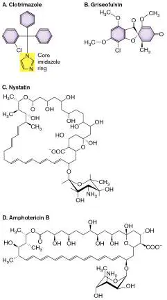
of the fungal cell wall. Echinocandins, a different class of antifungal drugs, also inhibit glucan synthase
but have a different structure and require intravenous administration.
The serious, sometimes fatal, consequences of systemic mycoses require more aggressive therapy.
The drugs used in these instances include amphotericin B (produced by Streptomyces; Fig. 27.37D )
and fluconazole. Amphotericin B binds to the sterols in fungal membranes and destroys membrane
integrity. It has a high affinity for ergosterol, which is prevalent in fungal but not mammalian
membranes. Fluconazole, on the other hand, inhibits the synthesis of ergosterol. Thus, fungal cells
grown in the presence of fluconazole make defective membranes. Typically, curing systemic fungal
infections requires long-term treatment to prevent disease relapse. Although resistance to fluconazole
is becoming a problem, the discovery of a compound called SWL-1 might change that. SWL-1 lowers
ATP levels in fluconazole-resistant C. albicans, a metabolic tweak thought to reduce the efficiency of
the ATP-dependent efflux pump responsible for resistance.
Another new class of antifungal drugs, called orotomides, was discovered in 2015. These drugs are
unique among antifungals because they stop pyrimidine biosynthesis in fungi. The drug specifically
inhibits fungal dihydroorotate dehydrogenase, an enzyme that converts dihydroorotate to orotate, a
precursor to pyrimidine. Orotomides are more effective than other antifungals, especially against
Aspergillus, which can cause life-threatening lung infections in immunocompromised patients. As of
this writing, one member of this class, F2G, is under development but not yet approved for clinical use.
Table 27.4 lists a number of other commonly used antifungal agents.
TABLE 27.4 Major Antifungal Agents and Their Common Uses
Systemic mycoses
Drug/Mode of Coccidioidomycosis Histoplasmosis Blastomycosis Paracoccidio
administration
Polyenes (fungicidal)
Amphotericin + + + +
B/Intravenous
Nystatin/Oral; topical − − − −
Natamycin/Topical; − − − −
eyedrops
Azoles (fungistatic): imidazoles
Clotrimazole/Topical − − − −
TABLE 27.4 Major Antifungal Agents and Their Common Uses
Miconazole − − − −
(Monistat)/Topical
Ketoconazole/Topical − − + −
Azoles (fungistatic): triazoles
Itraconazole/Oral + + + +
Fuconazole/Oral + + +?
Voriconazole/Oral − − − −
Allylamines (fungicidal)
Terbinafine − − − −
(Lamisil)/Topical or oral
Griseofulvin (fungistatic)
Griseofulvin/Oral − − − −
Echinocandins
Caspofungin/Intravenous − − − −
Triterpenoid
TABLE 27.4 Major Antifungal Agents and Their Common Uses
(fungicidal)
Ibrexafungerp/Oral − − − −
Antimetabolites (fungistatic or fungicidal)
5-Fluorocytosine d − − − −
(flucytosine)/Oral
By any measure, antibiotics have tamed all manner of infectious agents and greatly improved the
health and well-being of humans and animals over the past 80 years. Unfortunately, the remarkable
effectiveness of antibiotics has spawned complacency among patients and health care workers, leading
to the careless use of antimicrobials. This path unwittingly brought us to today’s antibiotic resistance
crisis. Important efforts encouraging antibiotic stewardship can slow, but will not stop, our march
toward a post-antibiotic future. Hope, however, comes in the form of exciting new approaches of
antimicrobial discovery that include artificial intelligence, innovative use of phage, and targeted
narrow-spectrum CRISPR therapies. With a little luck, and a whole lot of imagination, the human race
can remain one step ahead of the microbes.
To Summarize
Fungal infections are difficult to treat because of similarities in human and fungal
physiologies.
Imidazole-containing antifungal agents inhibit sterol synthesis.
Griseofulvin inhibits mitotic spindle formation.
Nystatin produces membrane pores.
Amphotericin B binds to membranes and destroys membrane integrity.
Glossary
griseofulvin
An antifungal antibiotic that inhibits cell division.
amphotericin B
An antifungal drug that binds the fungus-specific sterol ergosterol and destroys membrane
integrity.
Endnotes
1. Note a: + indicates that the drug inhibits growth of the disease-causing agent; − indicates that
the drug is not useful for the disease. Return to reference a
2. Note b: mc = mucocutaneous but not systemic candidiasis. Return to reference b
3. Note c: Insufficient data. Return to reference c
4. Note d: Used only in combination with amphotericin B. Return to reference d
eResearch Activity 27
Can a Selective, Narrow-Spectrum Antibiotic for Lyme Disease Be Found?
Many human infections are treated with broad-spectrum antibiotics such as doxycycline or ceftriaxone, but treatment comes at a cost. The broad-spectrum antibiotics disrupt the gut microbiome and select for antibiotic resistance in its members. Thus, the development of narrow-spectrum antibiotics to treat infections is highly desirable. Kim Lewis from Northeastern University led a team of scientists from several universities in a search for a narrow-spectrum antibiotic selective against the tick-borne spirochete Borrelia burgdorferi, the cause of Lyme disease (Chapter 26; see Fig. 26.34 for spirochete, tick vector, and animal reservoirs). Cases of Lyme disease have been increasing for years in the United States (now 500,000 per year) and expanding in geographic range.
Geographic spread and increased incidence have been caused by two factors: the expansion of cities into forested areas causing more frequent interactions between humans and animals, and climate change that has broadened the geographic habitat of the tick vector. The scientists reasoned that compounds acting selectively against spirochetes would have evolved in nature—probably in an actinomycete, a taxon famous for making antibiotics. From a screen of only 452 actinomycetes taken from soil, one, Streptomyces hygroscopicus, secreted a compound into culture medium that inhibited growth of B. burgdorferi but did not affect the growth of Staphylococcus aureus. A combination of HPLC, mass spectrometry, and NMR analysis identified the active compound as hygromycin A (HygA; Fig. ERA 27.1 ), a known antibiotic abandoned long ago because it had poor activity against Gram-negative and Gram-positive bacteria. Lewis’s group, however, found that the antibiotic was very effective against many spirochetes, including B. burgdorferi (MIC 0.12 μg/ml) and Treponema palidum (MIC 0.03 μg/ml). The minimal inhibitory concentrations against opportunistic pathogens like Pseudomonas aeruginosa and gut commensals, such as Bacteroides spp., ranged from 32-fold to over 500-fold higher than the MIC for B. burgdorferi, confirming that HygA selectively targets spirochetes.
FIGURE ERA 27.1 ■ Hygromycin A, the active compound.
The authors then demonstrated that HygA affected protein synthesis in Borrelia, using a strain that expressed GFP only when induced by anhydrotetracycline (ATC). The experiment (Fig. ERA 27.2 ) illustrates that ATC successfully induced GFP fluorescence (reflecting translation) both in antibiotic-free medium and in the presence of amoxicillin, an inhibitor of cell wall synthesis. However, ATC failed to induce GFP fluorescence when protein synthesis was inhibited by spectinomycin. HygA alone also prevented GFP expression, indicating that it, too, targeted translation in B. burgdorferi.
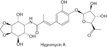
FIGURE ERA 27.2 ■ Hygromycin A inhibits protein synthesis by B. burgdorferi. A strain containing a GFP gene inducible by anhydrotetracycline (ATC) was used to determine if HygA inhibits protein synthesis. The first two bars (1 and 2) show that ATC does induce GFP. Adding amoxicillin (a form of ampicillin; bar 3) did not inhibit translation. However, adding the protein synthesis inhibitor spectinomycin (bar 4) prevented GFP synthesis, as did HygA (bar 5). Antibiotics were added at 2× MIC. Inset: Overlapping graphs that quantify the level and range of GFP fluorescence exhibited by 10 5 cells under each condition, as determined by flow cytometry. The color of each plot corresponds to the color of its matching bar in the main graph. The y -axis represents the number of cells at each level of fluorescence indicated on the x -axis.
Previous in vitro work using E. coli ribosomes demonstrated that HygA binds to 23S rRNA, most specifically to residues that form the
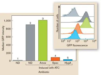
peptidyl transferase center of the ribosome (Fig. ERA 27.3 ). These residues are also present in B. burgdorferi 23S rRNA. The Figure ERA 27.3 inset shows that the drug causes residue A2062 to rotate and hydrogen-bond to residue m 2 A2503 (dashed arrow), a structural change that alters base pairing and arrests translation at the initiation codon of mRNAs.
FIGURE ERA 27.3 ■ View of the hygromycin A binding site in the T. thermophilus 70S ribosome. Hygromycin A (HygA) is shown binding to 23S rRNA residues that form the peptidyl transferase center.
In vitro translation studies revealed that E. coli ribosomes were extremely sensitive to HygA despite intact cells being quite resistant to the drug. This suggested that the high activity of HygA against spirochetes lies elsewhere, possibly in transport. To test whether transport was involved, the authors incubated B. burgdorferi and E. coli in increasing concentrations of the antibiotic for 1 min and 40 min, quickly filtered and lysed the cells, and then measured intracellular HygA concentrations using ultra-high-pressure liquid chromatography coupled to mass spectrometry (UHPLC/MS; Fig. ERA 27.4 ). Substantial amounts of HygA quickly entered B.
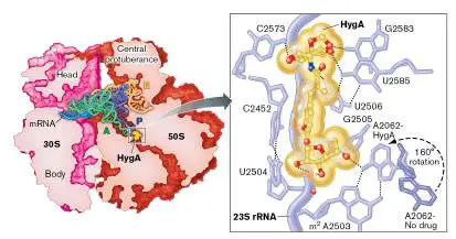
burgdorferi cells, but very little entered E. coli. The authors surmised that a transporter unique to spirochetes was responsible. FIGURE ERA 27.4 ■ Intracellular accumulation of hygromycin A in B. burgdorferi and E. coli. Cells were incubated in different concentrations of hygromycin A for 1 and 40 minutes and in intracellular concentrations of hygromycin A determined by UHPLC/MS.
The investigators then attempted to isolate B. burgdorferi mutants defective in transport by growing the organism in increasing concentrations of HygA. One mutant demonstrating stable resistance, KLEX-2, was impaired in transport. DNA sequencing revealed the strain had 21 mutations. Only one looked promising as a transporter, but knocking out that gene in B. burgdorferi did not affect HygA transport. The scientists then proposed that the HygA
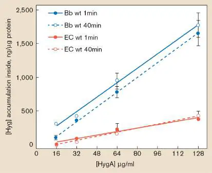
transporter relevant to resistance was also essential for growth (meaning the gene could not be knocked out). They further proposed that one of the 21 mutations possibly lowered expression of the unknown transporter just enough to cause HygA resistance. If true, down-regulation might be detected by transcriptome analysis. To test the hypothesis, they examined transcriptomes of the KLEX-2 mutant grown with HygA (1× MIC) and without the antibiotic. They predicted that growing the KLEX-2 mutant in media with the drug would somehow decrease expression of the transporter relative to growth without the drug, thereby providing resistance.
The RNAseq results, displayed on a volcano graph plotting the magnitude of change between two data sets (x -axis) against statistical significance (y -axis), showed that growth of KLEX-2 in HygA caused decreased expression (by more than threefold) of one transporter gene, bmpD (Fig. ERA 27.5A ). BmpD is a periplasmic substrate-binding protein for an ABC-type purine nucleoside transporter present in many spirochetes but not in other bacteria. BmpD-dependent transport of purine nucleosides is essential for B. burgdorferi because the organism cannot itself make purines. The researchers then asked whether overexpressing BmpD in B.
burgdorferi would make cells even more sensitive to HygA. The results in Figure ERA 27.5B show that overexpressing BmpD did make cells more susceptible to HygA, lowering the MIC fourfold, but had no effect on the MIC of a different antibiotic, ceftriaxone. The results indicate that HygA sneaks a ride on the Bmp nucleoside transporter to enter spirochetes, which explains the selectivity of the drug.
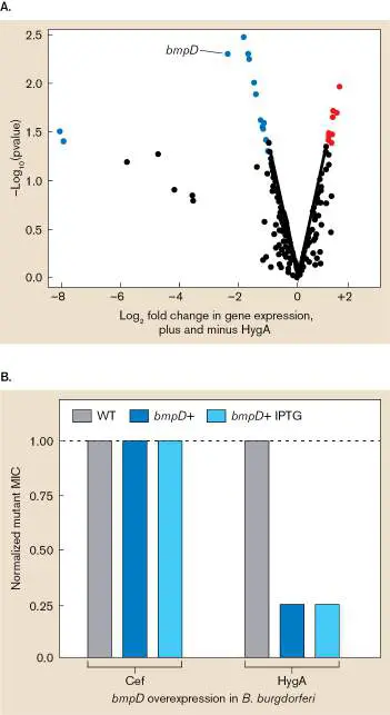
FIGURE ERA 27.5 ■ Basis for hygromycin A specificity. A. RNA sequencing (RNAseq) performed on the B. burgdorferi HygA-resistant mutant (KLEX-2) in the presence (4 μg/ml) and absence of hygromycin A. The volcano graph plots the log 2 of the fold changes in a gene’s expression in the presence relative to the absence of HygA (x-axis) versus the −log 10 of the p value denoting the significance of the difference (y -axis). Data points represent genes whose expression was, respectively, significantly diminished (blue) or significantly increased (red) in the presence of hygromycin A. B. Effect of overexpressing BmpD in B. burgdorferi on the MIC of ceftriaxone (Cef) and hygromycin A (HygA). The bmpD gene was cloned in a plasmid (pBAD)
downstream of a lac promoter inducible by IPTG (an analog of lactose). The 1.00 value on the normalized scale means the MIC of the strain containing a plasmid would equal the MIC of the strain without plasmid. The lack of effect by IPTG was attributed to a leaky intrinsic promoter in the construct.
The next question was whether hygromycin A could be used therapeutically. Mice were infected subcutaneously with B. burgdorferi. Once injected, the organisms replicate and spread from the injection site. After 3 weeks, HygA was administered by oral gavage twice a day for 5 days, after which skin samples were cultured and the presence of B. burgdorferi was confirmed by dark-field microscopy and 16S PCR analysis. Infected mice treated with saline remained infected for the duration, but all of the mice treated with HygA, doxycycline, or amoxicillin became culture-negative ( Fig. ERA 27.6 ). Importantly, the HygA-treated mice showed no signs of toxicity.
FIGURE ERA 27.6 ■ Therapeutic use of hygromycin A. Mice were infected subcutaneously with 10 5 B. burgdorferi cells (3 animals per treatment group). After 3 weeks, the mice were treated for 5 days with antibiotics by oral gavage. Daily doses were hygromycin A (100 mg/kg/day), amoxicillin (200 mg/kg/day), or doxycycline (100 mg/kg/day). Ear punches were collected after the last day of treatment and cultured. All protocols were approved by the Institute of Animal Care and Usage Committee.
In sum, with the goal of finding an antibiotic that was selectively active against the Lyme disease agent, the authors rediscovered HygA. They found that the selective activity of HygA was the result of a transport system in spirochetes that is absent from Gram-positive and Gram-negative bacteria. That transport system, Bmp, is normally used to move purine nucleosides into the cell but can also be used by HygA to gain entry and halt protein synthesis.
HygA has several features that make it an attractive agent for treating Lyme disease. First, B. burgdorferi is very susceptible to the drug, and oral administration of HygA was able to clear B.
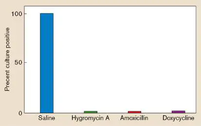
burgdorferi infections in mice. Beyond that, the development of resistance is very low, the drug is not cytotoxic against human cells, and the drug is unlikely to reach the bacteria-like 23S ribosomal RNA present in mitochondrial ribosomes.
This research has identified HygA as a potential oral therapeutic agent for Lyme disease and perhaps syphilis. In addition, the drug’s narrow spectrum of activity should protect the intestinal microbiome of patients from dysbiosis and the complications that can result. The drug also has the potential to eradicate B. burgdorferi from its natural reservoir (white-footed mice), possibly by spreading bait containing the drug in the mouse’s habitat. Eliminating the pathogen from mice would be a tremendous step toward eradicating Lyme disease or, at least, limiting its spread.
Further Exploration
How would you determine whether or not the narrow-spectrum antibiotic hygromycin A had an effect on the mouse gut microbiome? What result might you expect?
Leimer, Nadja, Xiaoqian Wu, Yu Imai, Madeleine Morrissette, Norman Pitt, et al. 2021. A selective antibiotic for Lyme disease. Cell 184 :1–14. https://doi.org/10.1016/j.cell.2021.09.011
Glossary
Fig. 26.34 FIGURE 26.34 ■ Lyme disease. A. Erythema migrans rash. B. Borrelia burgdorferi, the agent of Lyme disease (cell length 5–30 μm; colorized SEM microscopy). C. Ixodes vector (SEM). D. Host associations of Ixodes scapularis. The life cycle of the tick from egg to adult takes two years to complete. As the ticks develop, they are attracted to the barberry bush, from which females can transfer to a variety of animals for a blood meal.
CDC
EYE OF SCIENCE/SCIENCE SOURCE
DAVID M. PHILLIPS/SCIENCE SOURCE
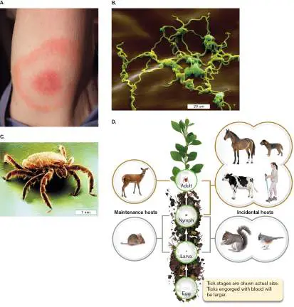
CHAPTER REVIEW
Review Questions
1. What is selective toxicity? Provide examples.
2. Explain the difference between antibiotic susceptibility and antibiotic sensitivity.
3. What does the term “spectrum of antibiotic activity” mean?
4. Provide examples of bacteriostatic and bactericidal antibiotics.
5. What is the Kirby-Bauer test? Does it indicate whether a drug is bacteriostatic or bactericidal?
6. Give examples of drugs that target each of the following: cell wall synthesis, RNA synthesis, protein synthesis, and DNA replication. What are their modes of action?
7. What mechanism do producing organisms use to synthesize peptide antibiotics?
8. How do antibiotic-producing microorganisms prevent “suicide”?
9. Why is antibiotic resistance a growing problem?
10. What are the four basic mechanisms of antibiotic resistance?
11. Explain the basic concept of an MDR efflux pump. 12. Discuss the current concepts of the origin of antibiotic resistance.
13. What are some mechanisms used to combat the development of drug resistance?
14. Why are there few antiviral agents available to treat disease?
15. What is herd immunity?
16. How does oseltamivir inhibit influenza?
17. Discuss the general modes of action of antifungal agents.
Thought Questions
1. A cephalosporin and clindamycin are often used to treat a patient with toxic shock syndrome caused by a TSST-producing Staphylococcus aureus. One agent is bactericidal; the other is bacteriostatic. Usually this combination is discouraged because the bacteriostatic agent dampens the effectiveness of the bactericidal agent, which kills only growing cells. Why would an exception to this rule be considered in the treatment of toxic shock?
2. A patient presented to the emergency room complaining of a nonproductive cough (no sputum) that had persisted for 6 weeks. The clinician prescribed a 7-day course of a cephalosporin. After day 7 the patient returned, no better than before he started the treatment. Laboratory tests later showed that the infection was caused by Mycoplasma pneumoniae. Explain why the antibiotic did not work.
3. How would you determine the MIC of an obligate intracellular pathogen such as Rickettsia prowazekii, the cause of typhus?
4. You already know that some antimicrobial compounds must be converted by human metabolism into an active drug. Imagine how else an antibiotic might be more effective in vivo than in vitro, even if you do not know of any specific examples of your hypothesis.
Key Terms
amphotericin B (1210)
antibiotic (1166)
antiretroviral therapy (ART) (1205) bacitracin (1178)
bactericidal (1169)
bacteriostatic (1169)
chloramphenicol (1183)
cycloserine (1179)
daptomycin (1179)
gramicidin (1179)
griseofulvin (1209)
Kirby-Bauer assay (1170)
lincosamide (1183)
macrolide (1183)
minimal inhibitory concentration (MIC) (1169) Mueller-Hinton agar (1172) multidrug resistance (MDR) efflux pump (1189) neuraminidase inhibitor (1203) oxazolidinone (1183)
penicillin (1166)
penicillin-binding protein (PBP) (1176) persister cell (1195)
quinolone (1180)
resistome (1192)
secondary metabolite (1186) selective toxicity (1168)
spectrum of activity (1169) streptogramin (1183)
sulfa drug (1167)
transglycosylase (1175)
transpeptidase (1175)
vancomycin (1179)
zone of inhibition (1170)
Recommended Reading
Ahmed, Marwa N., Ahmed Abdelsamad, Tina Wassermann, Andreas Porse, Janna Becker, et al. 2020. The evolutionary trajectories of P. aeruginosa in biofilm and planktonic growth modes exposed to ciprofloxacin beyond selection of antibiotic resistance. NPJ Biofilms and Microbiomes 6 (1):28.
https://doi.org/10.1038/s41522-020-00138-8.
Altamirano, Fernando Gordillo, John H. Forsyth, Ruzeen Patwa, Xenia Kostoulias, Michael Trim, et al. 2021.
Bacteriophage-resistant Acinetobacter baumannii are resensitized to antimicrobials. Nature Microbiology 6 :157–161. https://doi.org/10.1038/s41564-020-00830-7.
Baquero, Fernando, and Bruce R. Levin. 2020. Proximate and ultimate causes of the bactericidal action of antibiotics. Nature Reviews: Microbiology 19 :123–132.
https://doi.org/10.1038/s41579-020-00443-1.
Clemente, Jose C., Erica C. Pehrsson, Martin J. Blaser, Kuldip Sandhu, Zhan Gao, et al. 2015. The microbiome of uncontacted Amerindians. Science Advances 1 :e500183.
Crofts, Terence S., Andrew J. Gasparrini, and Gautam Dantas. 2017. Next-generation approaches to understand and combat the antibiotic resistome. Nature Reviews. Microbiology 15 :422–434.
Danko, David, Daniela Bezdan, Evan E. Afshin, Sofia Ahsanuddin, Chandrima Bhattacharya, et al. 2021. A global metagenomic map of urban microbiomes and antimicrobial resistance. Cell 184 :1–18.
https://doi.org/10.1016/j.cell.2021.05.002.
DiGiandomenico, Antonio, and Brett R. Sellman. 2015.
Antibacterial monoclonal antibodies: The next generation? Current Opinion in Microbiology 27 :78–85.
Fleming-Dutra, Katherine E., Adam L. Hersh, Daniel J. Shapiro, Monina Bartoces, Eva A. Enns, et al. 2016.
Prevalence of inappropriate antibiotic prescriptions among US ambulatory care visits, 2010–2011. Journal of the American Medical Association 315 :1864–1873.
Flentie, Kelly, Gregory A. Harrison, Hasan Tükenmez, Jonathan Livny, James A. D. Good, et al. 2019. Chemical disarming of isoniazid resistance in Mycobacterium tuberculosis. Proceedings of the National Academy of Sciences USA 116:10510–10517.
Freije, Catherine A., and Pardis C. Sabeti1. 2021. Detect and destroy: CRISPR-based technologies for the response against viruses. Cell Host and Microbe 29 :689–703.
https://doi.org/10.1016/j.chom.2021.04.003.
Ghaly, Timothy M., Jemma L. Geoghegan, Sasha G. Tetu, and Michael R. Gillings. 2020. The peril and promise of integrons: Beyond antibiotic resistance. Trends in Microbiology 28 :455– 464. https://doi.org/10.1016/j.tim.2019.12.002.
Gollan, Bridget, Grzegorz Grabe, Charlotte Michaux, and Sophie Helaine. 2019. Bacterial persisters and infection: Past, present, and progressing. Annual Review of Microbiology 73:359–385.
Hayden, Frederick G., Norio Sugaya, Nobuo Hirotsu, Nelson Lee, Menno D. de Jong, et al. 2018. Baloxavir marboxil for uncomplicated influenza in adults and adolescents. New England Journal of Medicine 379 :913–923.
Kim, Dae-Wi, and Chang-Jun Cha. 2021. Antibiotic resistome from the One-Health perspective: Understanding and controlling antimicrobial resistance transmission. Experimental & Molecular Medicine 53 :301–303. https://doi.org/10.1038/s12276-021-00569-z.
Kim, Wooseong, Wenpeng Zhu, Gabriel Lambert Hendricks, Daria Van Tyne, Andrew D. Steele, et al. 2018. A new class of synthetic retinoid antibiotics effective against bacterial persisters. Nature 556 :103–107.
Larsson, D. G. Joakim, and Carl-Fredrik Flach. 2022. Antibiotic resistance in the environment. Nature Reviews. Microbiology 20:257–269. https://doi.org/10.1038/s41579-021-00649-x. Lu, Lu, Shan Su, Haitao Yang, and Shibo Jiang. 2021.
Antivirals with common targets against highly pathogenic viruses. Cell 184: 1604–1620.
https://doi.org/10.1016/j.cell.2021.02.013.
Spagnolo, Fabrizio, Monica Trujillo, and John J. Dennehy. 2021 Why do antibiotics exist? mBio 12 :e01966-21.
Stokes, Jonathan M., Kevin Yang, Kyle Swanson, Wengong Jin, Andres Cubillos-Ruiz, et al. 2020. A deep learning approach to antibiotic discovery. Cell 180 :688–702.
https://doi.org/10.1016/j.cell.2020.01.021.
Urbaniec, J., Ye Xu, Yin Hu, Suzie Hingley-Wilson, and Johnjoe McFadden. 2021. Phenotypic heterogeneity in persisters: A novel “hunker” theory of persistence. FEMS Microbiology Reviews fuab042:1–16.
https://doi.org/10.1093/femsre/fuab042.
Van Acker, Heleen, and Tom Coenye. 2017. The role of reactive oxygen species in antibiotic-mediated killing of bacteria. Trends in Microbiology 25 :456–466.
Zipperer, Alexander, Martin C. Konnerth, Claudia Laux, Anne Berscheid, Daniela J. Anek, et al. 2016. Human commensals producing a novel antibiotic impair pathogen colonization. Nature 535 :511–516.
Glossary
antibiotic A molecule that can kill or inhibit the growth of selected microorganisms.
penicillin An antibiotic, produced by the Penicillium mold, containing a beta-lactam ring; it blocks cross-bridge formation during peptidoglycan synthesis.
sulfa drug An antibiotic that inhibits folic acid synthesis and, thus, nucleotide synthesis.
selective toxicity The ability of a drug, at a given dose, to harm the pathogen and not the host.
spectrum of activity The range of pathogens for which an antimicrobial agent is effective.
bactericidal Having the ability to kill bacterial cells.
bacteriostatic Having the ability to inhibit the growth of bacterial cells. minimal inhibitory concentration (MIC)
The lowest concentration of a drug that will prevent the growth of an organism.
zone of inhibition A region of no bacterial growth on an agar plate that is due to the diffusion of a test antibiotic. Correlates to the minimal inhibitory concentration.
Kirby-Bauer assay A method for determining antibiotic susceptibility. Antibiotic-impregnated disks are placed on an agar plate whose surface has been confluently inoculated with a test organism. The antibiotic diffuses away from the disk and inhibits growth of susceptible bacteria. The width of the inhibitory zone is proportional to the susceptibility of the organism.
Mueller-Hinton agar A specialized, standardized, para -aminobenzoic acid–free medium used for the Kirby-Bauer assay.
transglycosylase An enzyme that condenses N -acetylglucosamine and N - acetylmuramic acid into chains during bacterial cell wall synthesis.
transpeptidase An enzyme that cross-links the side chains from adjacent peptidoglycan strands during bacterial cell wall synthesis. penicillin-binding protein (PBP)
A bacterial protein, involved in cell wall synthesis, that is the target of the antibiotic penicillin.
bacitracin A topical antibiotic that affects cell wall synthesis. cycloserine A polypeptide antibiotic that inhibits peptidoglycan synthesis. vancomycin A glycopeptide antibiotic that binds the D-Ala-D-Ala end of the cell-wall peptide, thus inhibiting the transpeptidase enzyme and preventing crossbridge formation in the cell wall. gramicidin A peptide antibiotic that acts as a channel for monovalent cations to cross the cell membrane, thus collapsing the transmembrane ion gradients.
daptomycin A lipopeptide antibiotic that forms ion channels in Gram-positive bacteria.
quinolone A type of antibiotic drug that inhibits DNA synthesis by targeting bacterial topoisomerases such as DNA gyrase. macrolide Any of a group of antibiotics containing a large lactone ring (e.g., erythromycin).
lincosamide Any of a class of bacteriostatic antibiotics that include a pyrrolidine ring linked to a pyranose (e.g., clindamycin). chloramphenicol A bacteriostatic antibiotic that acts by inhibiting peptidyltransferase activity of the bacterial ribosome. oxazolidinone One of a class of synthetic antibiotics, containing an oxazole ring, that inhibit protein synthesis.
streptogramin An antibiotic that binds 23S rRNA and blocks elongation of protein synthesis in bacteria.
secondary metabolite or secondary product An organic product of biosynthesis that does not have essential functions but enhances nutrient uptake under certain conditions or inhibits competing species (e.g., an antibiotic). Often produced during stationary phase.
multidrug resistance (MDR) efflux pump A transmembrane protein pump that can export many different kinds of antibiotics of diverse structure.
resistome The collection of antibiotic resistance genes present in all pathogenic and nonpathogenic microbes that inhabit humans, animals, or the environment.
persister cell Any of a subpopulation of dormant organisms that arise within a population of antibiotic-susceptible bacteria and are tolerant to bactericidal antibiotics during treatment. On removal of the drug, persisters can grow and reestablish infection.
neuraminidase inhibitor Any of a class of anti-influenza drugs that target neuraminidase on the viral envelope and decrease the number of virus particles produced.
antiretroviral therapy (ART)
A three-drug antiretroviral cocktail that is highly effective at inhibiting the replication of HIV in patients.
griseofulvin An antifungal antibiotic that inhibits cell division. amphotericin B An antifungal drug that binds the fungus-specific sterol ergosterol and destroys membrane integrity.