Chapter introduction
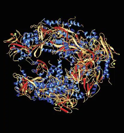
The enzyme RNA polymerase II (computer model).
The molecule comprises 12 subunits. This enzyme synthesizes a complementary messenger RNA (mRNA)
strand from a strand of DNA during a process called transcription. It recognizes a start sign on the DNA strand and then moves along the strand, building the mRNA until it reaches a stop sign. Messenger RNA is the intermediary between DNA and its protein product.
Source: Mark J. Winter/Photo Researchers, Inc.
Dr. Mark J. Winter/Science Source
Appendix Sections
A1.1 Elements, Bonding, and Water A1.2 Organic Molecules A1.3 Proteins A1.4 Carbohydrates A1.5 Nucleic Acids A1.6 Lipids A1.7 Biological Chemistry Here in eAppendix 1 we review concepts of chemistry that are typically covered in an introductory biology course. We first cover the chemical bonding principles needed to understand biological molecules and put an emphasis on the special properties of water. We then discuss organic molecules and pay particular attention to four important classes of organic biomolecules: proteins, carbohydrates, nucleic acids, and lipids. Finally, we explore common chemical principles, such as concentrations, thermodynamics, equilibrium, pH, and oxidation-reduction reactions.
A1.1 Elements, Bonding, and Waternot assigned
Living cells are remarkably complex machines, able to integrate and respond to multiple stimuli, to catalyze reactions, and to replicate themselves. Yet despite all the various tasks that cells perform, 98% of the mass of living organisms consists of only six chemical elements: hydrogen (H), oxygen (O), nitrogen (N), phosphorus (P), sulfur (S), and carbon (C), and 90% of the mass is accounted for by just C, H, and O. Of the compounds formed from these elements, the most abundant in cells is water. The remainder of the cell consists, for the most part, of just four different kinds of organic (carbon-based) macromolecules: proteins, nucleic acids, carbohydrates, and lipids. Cells can be thought of as compartments that orchestrate chemical reactions between these organic molecules.
It is clear that to understand life, we must understand the properties of water and organic molecules and of their building blocks, the atoms of various elements. An atom consists of a positively charged nucleus that contains protons and neutrons, surrounded by negatively charged electrons. The hydrogen nucleus consists of a single proton. Protons, neutrons, and electrons differ in their mass and charge as summarized in Table A1.1.
TABLE The Mass and Charge of Atomic
A1.1 Particles
Particle Mass * (atomic Charge (electronic mass unit) charge unit)
Proton 1 +1 TABLE The Mass and Charge of Atomic
A1.1 Particles
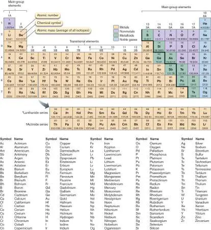
Neutron 1 0 Electron 0.0005 −1 The elements can be organized into a periodic table as in Figure A1.1, indicating each element’s atomic number (the number of protons) and atomic mass (the mass, in grams, of 1 mole of the element). The defining characteristic of an element is the atomic number. For example, all carbon atoms have six protons in their nucleus. To maintain neutrality, atoms have negatively charged electrons equal in number to the positively charged protons. FIGURE A1.1 ■ Periodic table of the elements. The atomic number (number of protons) and atomic mass are shown for each element.
The atomic mass is the sum of the number of protons and neutrons. The atomic mass is an average that takes into account the relative abundance of each isotope. Isotopes are atoms of an element that differ in the number of neutrons. For example, the most abundant isotope of carbon is carbon-12 (with six neutrons),
but there are naturally occurring isotopes of carbon-13 (seven neutrons) and carbon-14 (eight neutrons). Because carbon-13 and carbon-14 are rare, the average atomic mass is close to but not exactly 12. While some isotopes are stable, others decay at a known rate and give off radioactivity. Carbon-14 has a half-life (the amount of time it takes for half of a sample to decay) of 5,730 years and is used in radiocarbon dating to determine the age of organic material. Carbon-14 and shorter-lived isotopes, such as tritium (hydrogen with a mass of 3—one proton and two neutrons), are used by scientists as tracers to follow specific atoms in metabolic pathways.
Bonds between Atoms Form Molecules
Atoms combine by sharing electrons to form molecules. For example, two atoms of oxygen combine to form molecular oxygen, O 2. Molecules may also contain more than one kind of element; an example is water, H 2 O. The symbols O 2 and H 2 O are examples of molecular formulas, a shorthand notation indicating the number and types of atoms present in a molecule.
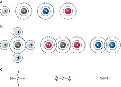
Each column (group) in the periodic table (Fig. A1.1) contains elements of similar reactivity as a result of the similarity in their electronic configurations, particularly of electrons in the outermost shell. The shell closest to the nucleus can hold a maximum of two electrons, and the next shell a maximum of eight electrons. Figure A1.2Ashows all the electrons for hydrogen, carbon, nitrogen, and oxygen. Each unpaired electron is capable of participating in a bond. In Figure A1.2Ait is clear that H can form one bond; C, four bonds; N, three bonds; and O, two bonds. For example, carbon has four unpaired electrons in its outermost shell; each can form a bond with an unpaired electron from another atom. The two atoms involved in this bond “share” the two electrons. This sharing of electrons is termed a covalent bond.
FIGURE A1.2 ■ Covalent bonding of hydrogen, carbon, nitrogen, and oxygen. A. The electrons present in hydrogen (H), carbon (C), nitrogen (N), and oxygen (O). B. Electron sharing in the four single bonds of methane, CH 4 (left); the two double bonds of carbon dioxide, CO 2 (middle); and the triple bond of diatomic nitrogen, N 2 (right). C. Structural formulas for methane, carbon dioxide, and diatomic nitrogen. Each line represents a covalent bond. Lone pairs of electrons in the outermost shell are represented by dots.
Covalent bonds are very strong and difficult to break. In methane (CH 4), carbon forms four covalent bonds with four hydrogen atoms (Fig. A1.2B , left). Methane is stable because both carbon and hydrogen have filled their outer shells. Atoms can also share more than one pair of electrons with another atom, forming double or triple bonds. In carbon dioxide (CO 2) (Fig. A1.2B , middle), each oxygen shares two pairs of electrons with carbon; and in diatomic
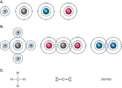
nitrogen (N 2) (Fig. A1.2B , right), the nitrogen atoms each share three pairs of electrons. The bonding in molecules can be represented in a shorthand representation by a structural formula, in which covalent bonds are shown as lines between two atoms ( Fig. A1.2C ).
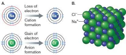
Another way atoms can obtain full outer shells is by gaining or losing electrons. A complete transfer of electrons can occur between two atoms that have a large difference in electronegativity, a measure of the affinity of an atom for electrons. A large electronegativity indicates a strong attraction for electrons. Of the elements listed in Table A1.2, oxygen has the greatest attraction for electrons, and sodium has the weakest. If two elements with greatly different electronegativities come into close contact, one element can “steal” an electron from the other. For example, sodium (Na) and chlorine (Cl) interact to form table salt, NaCl. The electronegative Cl strips an electron away from Na, and both Cl − and Na + now have full outer shells (Fig. A1.3A).
TABLE Electronegativities of Some
A1.2 Common Elements
Element Electronegativity Oxygen 3.44 Chlorine 3.16 Nitrogen 3.04 Sulfur 2.58 Carbon 2.55 TABLE Electronegativities of Some
A1.2 Common Elements
Hydrogen 2.10 Sodium 0.93 FIGURE A1.3 ■ Formation of ions and ionic crystals. A. Sodium atoms form sodium cations (Na +) by losing an electron; chlorine atoms form chloride anions (Cl −) by gaining an electron. B. Oppositely charged anions and cations—in this case, Cl − and Na + —are attracted to one another and form crystals of table salt (sodium chloride, or NaCl).
Both Cl − and Na + are charged atoms called ions, in which the number of electrons and protons are unequal. Anions are negatively charged ions, and cations are positively charged ions. Anions and cations can form ionic crystals held together by ionic bonds, electrostatic attractions between anions and cations (Fig. A1.3B ). In the absence of water, ionic crystals maintain their integrity, but in
the presence of water some kinds of ionic crystals are destabilized. To understand why, we need to understand the structure of water.
Water Is the Solvent of Life
Living organisms consist mostly of water, and water has many unique properties that render it particularly suitable for sustaining life. To appreciate these properties, we must understand the forces that hold water together: polar covalent bonds and hydrogen bonds. Molecules such as H 2 and O 2 have even charge distribution because both atoms in the molecule have the same electronegativity. Therefore, the electrons in the covalent bond are shared equally and form nonpolar covalent bonds. In contrast, the shared electrons in H 2 O spend more of their time around the highly electronegative oxygen than in the vicinity of the less electronegative hydrogen (Fig. A1.4A). A bond with unequal electron sharing is a polar covalent bond, so called because the molecule has partial positive and negative poles. Polar covalent bonds occur in individual water molecules, forming a partial charge separation within the molecule.
FIGURE A1.4 ■ Polar covalent and hydrogen bonds in water. A. The polar covalent bonds in an individual water
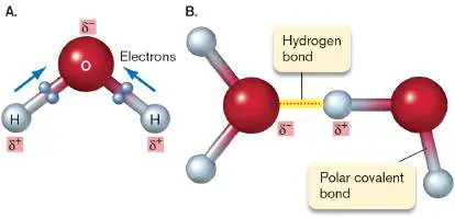
molecule. Displacement of electrons toward oxygen causes oxygen to have a partial negative charge (represented in this model as δ −) and the hydrogen atoms to have a partial positive charge (δ +). Hence, water is polar. B. A hydrogen bond between two water molecules.
Water molecules experience a strong electrostatic attraction for one another as a result of the charge separation present in individual molecules. This electrostatic attraction, known as a hydrogen bond, occurs between a hydrogen bonded to both an oxygen or nitrogen and a second oxygen or nitrogen, either in the same or a different molecule. Hydrogen bonds are short-lived and constantly break and re-form in liquid water. Figure A1.4B shows the polar covalent bonds and hydrogen bonds present in water. The hydrogen bonds in water contribute the unique properties that enable it to support life. Water is a liquid over a large temperature range because hydrogen bonds cause water molecules to associate, favoring the liquid state over the gas. The hydrogen bonds that keep water a liquid over a wide range of temperatures also endow water with a high specific heat, the amount of energy needed to raise the temperature of 1 gram (g) of a substance by 1°C. The high specific heat of water moderates the temperature of all aqueous environments, oceans and cells alike.
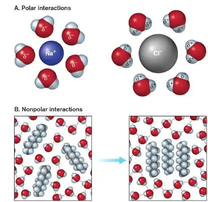
The polar nature of water defines its properties as a solvent. Compounds that are ionic or polar themselves tend to dissolve in water and are termed hydrophilic. For example, the ionic bonds in NaCl are very strong in the absence of water but are easily dissolved in the presence of water. The reason water is so good at dissolving substances like NaCl is that its polar nature enables it to surround and interact with the sodium and chloride ions and shield them from each other (Fig. A1.5A). Water also dissolves polar compounds. In this case, water does not break the polar covalent bonds; rather, individual, intact polar molecules are surrounded by water, owing to electrostatic interactions.
FIGURE A1.5 ■ Interactions between water and solutes. A. Water surrounds and interacts with individual ions or polar molecules, causing them to dissolve in water. B. Nonpolar molecules do not dissolve in water. Instead, to minimize the disruption of hydrogen bonding among water molecules, nonpolar molecules aggregate.
In contrast, compounds that are mostly nonpolar do not dissolve in water and are termed hydrophobic. Nonpolar molecules have no partial charges to attract water. Because water hydrogen-bonds with other water molecules, it tends to exclude nonpolar compounds, forcing them together (Fig. A1.5B ). The aggregated nonpolar
compounds can be further stabilized by van der Waals forces, weak, temporary, electrostatic attractions between molecules caused by random movements of their electron clouds. Table A1.3lists the bonds we have discussed and indicates the strength of each type of bond in water.
TABLE Bond Types and Strengths A1.3
Type of Description Bond strength in bond water (kJ/mol) a Covalent Sharing of electrons 210–418 Ionic Electrostatic attraction 12.5 between anion and cation Hydrogen Electrostatic attraction 4 between a hydrogen bonded to a nitrogen or oxygen and to a second nitrogen or oxygen Van der Electrostatic attraction 0.42 (per atom) Waals between temporary, shifting electron clouds
Glossary
atomic number The number of protons in an atom; it is unique for each element.
atomic mass The mass (in grams) of 1 mole of an element.
isotope An atom of an element with a specific number of neutrons. For example, carbon-12 (12 C), carbon-13 (13 C), and carbon-14 ( 14 C) are all isotopes of carbon.
half-life The amount of time it takes for one-half of a radioactive sample to decay.
molecular formula A notation indicating the number and type of atoms in a molecule. For example, H 2 O is the molecular formula for water.
covalent bond A chemical bond in which two atoms share a pair of electrons. structural formula A representation of molecular structure in which each covalent bond is shown as a line between atoms.
electronegativity The affinity of an atom for electrons. The greater the electronegativity, the stronger the attraction for electrons. ion An atom or molecule containing negative or positive charge; that is, a number of electrons, respectively, greater than or less than the number of protons.
anion A negatively charged ion.
cation A positively charged ion.
ionic bond A chemical bond between ions of positive and negative charge. nonpolar covalent bond A covalent bond in which the electrons in the bond are shared equally by the two atoms.
polar covalent bond A covalent bond in which the electrons in the bond are distributed unequally between two atoms.
hydrogen bond An electrostatic attraction between a hydrogen bonded to an oxygen or nitrogen and a second, nearby oxygen or nitrogen. hydrophilic Soluble in water; either ionic or polar.
hydrophobic Insoluble in water; nonpolar.
van der Waals force A weak, temporary electrostatic attraction between molecules caused by shifting electron clouds.
Endnotes
1. Note *: Values are approximate. Return to reference * 2. Note a: Bond strengths are given in water, which is similar to the environment in the aqueous cytoplasm. Anhydrous bond strengths differ from those listed. Return to reference a
A1.2 Organic Moleculesnot assigned
Now that we have discussed water, let’s take a look at some of the organic molecules found in
cells. Organic molecules are those that contain a carbon-carbon bond. The major
macromolecular components of cells are proteins, carbohydrates, nucleic acids, and lipids.
Although the macromolecules differ from each other in structure and cellular function, they all
share some common features.
As polymers, the macromolecules found in cells are composed of smaller units called
monomers. The monomers are joined to one another by a common type of reaction, called
condensation, that involves splitting out a molecule of water for each monomer unit added.
Conversely, the polymers can be broken apart into monomers by hydrolysis, the addition of a
molecule of water, as depicted in Figure A1.6 .
FIGURE A1.6 ■ Condensation and hydrolysis. Monomers can be covalently bonded
to form polymers through a condensation reaction that liberates a water molecule.
Polymers can be broken apart into monomers through hydrolysis reactions that split a
water molecule.
The 3D shape, or structure, of a molecule is critical for proper function. The structure is
determined by which atoms are present and how they are bonded together. Structural
formulas like those in Figure A1.2C represent the sequence of bonds in a molecule; however,
bond structure does not adequately convey information about the 3D shape of the molecule. It
is particularly important to understand the shapes of carbon-containing molecules because
carbon is the backbone for most cellular macromolecules. The bonding orbitals in the second
shell of carbon are arranged so that they point to the vertices of a tetrahedron; methane, the
simplest hydrocarbon, has the shape of a tetrahedron, with the hydrogen atoms at the vertices
(Fig. A1.7A ). Two different models are used to depict the 3D shape of molecules. The
space-filling model (Fig. A1.7B ) shows the volume filled by the outer shell of the atoms. The
stick model (Fig. A1.7C ) shows the length and orientation of interatomic bonds between the
nuclei of each pair of atoms.
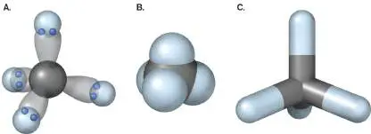
FIGURE A1.7 ■ Molecular models of methane. A. This model of a methane
molecule shows the tetrahedral arrangement of the electron orbitals in the second
(valence) shell of carbon. B. Space-filling model. C. Stick model.
An important feature of any organic molecule is the number and type of functional groups
present. Functional groups are small groups of atoms with characteristic bonding, shape, and
reactivity. A few of the more common ones are shown in Table A1.4 . Knowing which
functional groups are present in a molecule enables us to infer something about the structure
and reactivity of the molecule. Information about the functional groups present in a molecule
is often indicated by a molecule’s name; for example, amino acids contain an amino group and
a carboxyl (carboxylic acid) group.
TABLE Common Functional Groups A1.4
Functional General Example Comments
group structure
Aldehyde Can react with alcohols
Alkane Nonpolar, tends to make
molecules containing it
hydrophobic; nonreactive
Amino Acts as a base by binding a
proton; found in amino acids
TABLE Common Functional Groups A1.4
Carboxyl Acts as an acid by releasing a
proton; the ionized-form
name ends in “-ate” (e.g.,
“acetate”)
Ester Common linkage found in lipids
and nucleic acids
Hydroxyl Polar, makes compounds more
soluble through hydrogen
bonding; found in alcohols
and sugars
Ketone Found in many intermediates
of metabolism
Phosphate When two or more phosphoryl
groups are linked, a high-
energy bond forms because
the negative charges on the
oxygens repel each other
TABLE Common Functional Groups A1.4
We will discuss different classes of molecules individually, but in living cells, molecules of
different types are frequently found in combination. For example, sugars decorate some
proteins (glycoproteins) and lipids (glycolipids), and the ribosome is a complex of proteins and
ribonucleic acids. We now examine the structure of the fundamental biological macromolecules
in detail.
Glossary
organic molecule
A molecule that contains a carbon-carbon bond.
condensation
In biochemistry, the joining of two molecules to form a covalent bond, releasing a water
molecule.
hydrolysis
The cleaving of a bond by the addition of a water molecule.
space-filling model
A molecular model that represents the volume of the electron orbitals of the atoms,
usually to the limit of the van der Waals radii.
stick model
A molecular model in which stick lengths represent the distances between bonded pairs of
atomic nuclei.
functional group
A cluster of covalently bonded atoms that behaves with specific properties and functions
as a unit.
Figure A1.2C
FIGURE A1.2 ■ Covalent bonding of hydrogen, carbon, nitrogen, and
oxygen. A. The electrons present in hydrogen (H), carbon (C), nitrogen (N), and
oxygen (O). B. Electron sharing in the four single bonds of methane, CH 4 (left); the
two double bonds of carbon dioxide, CO 2 (middle); and the triple bond of diatomic
nitrogen, N 2 (right). C. Structural formulas for methane, carbon dioxide, and diatomic
nitrogen. Each line represents a covalent bond. Lone pairs of electrons in the
outermost shell are represented by dots.
A1.3 Proteinsnot assigned
Cells express thousands of different proteins and may contain more than 2 million protein molecules. Proteins perform many functions, such as catalyzing reactions, serving as receptors and transporters, providing structure, and aiding movement. Proteins can carry out such diverse functions because they can fold into a variety of 3D structures.
Amino Acids Are the Building Blocks of Proteins
Although different proteins can have vastly different structures, all proteins are composed of the same building blocks: amino acids. All 20 of the amino acids commonly occurring in nature have the same general structure. Each amino acid contains a central carbon atom (the alpha carbon) covalently bonded to four different moieties: a hydrogen atom, an amino group, a carboxyl group, and a side chain (R, residue) that is unique for each amino acid (Fig. A1.8A). FIGURE A1.8 ■ Amino acid structure and chiral carbons. A. All amino acids contain a central carbon (the alpha carbon) bonded to an amino group, a carboxyl group, a hydrogen, and a variable group or side chain, designated R. At cellular pH, the amino and carboxyl groups are ionized. Because the alpha carbon is bonded to four different molecules, it is a chiral carbon. B. Chiral molecules exist in two different forms that are mirror
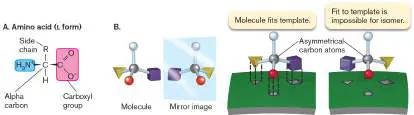
images of each other. The two forms cannot be superimposed, and only the correct isomer can interact with a template (for example, an enzyme).
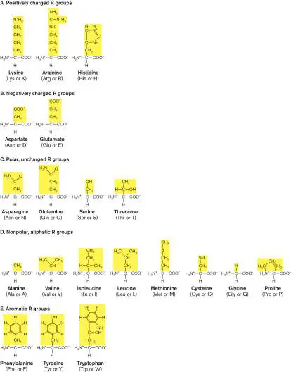
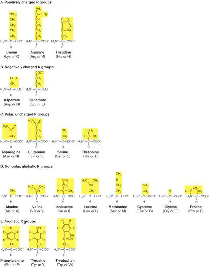
The alpha carbon of an amino acid is a chiral carbon, a carbon with four different groups attached to it. Chiral carbons exist in two different forms, called optical isomers, or enantiomers. Optical isomers have the same molecular formula and the same order of bonds within the molecules but a different arrangement of their atoms in space. Optical isomers are actually mirror images of each other, like your left and right hands, and the two forms cannot be superimposed (Fig. A1.8B ). Each of the amino acid isomers is designated L or D, depending on the configuration at the alpha carbon. The amino acids used to make proteins all have the L configuration at the alpha carbon (Fig. A1.8), and all of these amino acids can be derived by substituting the different R groups for one another. Some unusual D -amino acids, with the opposite configuration at the alpha carbon, are found in bacterial cell walls. The side chains of amino acids can be grouped according to their hydrophobicity, charge, or presence of specific functional groups. Figure A1.9depicts the 20 common amino acids, along with their three-letter abbreviations and single-letter codes. All are shown in the L conformation, except for glycine, which does not contain a chiral carbon and therefore does not have optical isomers. Proline has a ring structure that interrupts the regular geometry of the polypeptide chain. The side chain of cysteine is capable of forming intermolecular and intramolecular disulfide (−S−S−) bonds. FIGURE A1.9 ■ Twenty common amino acids. The grouping of amino acids is based on their side chains, highlighted in yellow.
Four Levels of Protein Organization
The first level of organization in proteins, the primary structure, is simply the linear sequence of amino acids. This sequence is genetically determined, specified by the DNA code. During protein synthesis, amino acids are connected together by a condensation reaction to form a covalent peptide bond. The peptide bond forms between the carboxyl group of one amino acid and the amino group of a second amino acid (Fig. A1.10A). The portion of the amino acid incorporated into the peptide after condensation is called an amino acid residue. Note that the peptide chain has an amino terminus (N terminus) and a carboxyl terminus (C terminus); numbering of the amino acid residues starts at the amino terminus ( Fig. A1.10B ). The bonds on either side of the alpha carbon can rotate, but there is no rotation around the peptide bond, so the backbone of a protein does not rotate freely.
FIGURE A1.10 ■ The peptide bond and primary protein structure. A. Formation of a peptide bond. Amino acids are joined by a condensation reaction between the carboxyl group of one amino acid and the amino group of another to form a covalent linkage called a peptide bond. B. The primary structure of a pentapeptide, a chain of five amino acids. This pentapeptide
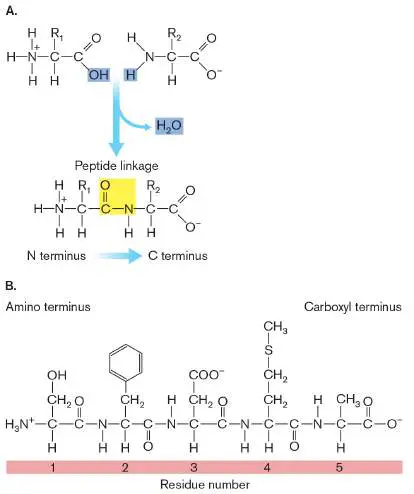
has the sequence SFDMA. A protein can be formed of hundreds or thousands of amino acids.
The secondary structures of proteins are regular patterns that repeat over short regions of the polypeptide chain. Figure A1.11A depicts two common secondary structures: the alpha helix and the beta sheet. Alpha helices are coiled structures formed by a series of hydrogen bonds between an oxygen in a carboxyl group and a hydrogen in an amino group of the fourth amino acid down the chain. Beta sheets form as a result of extensive hydrogen bonding between regions of the protein lying next to each other. Both alpha helix and beta sheet secondary structures are stabilized by hydrogen bonding between the oxygen and hydrogen atoms of the peptide bonds in the main chain; the side chains do not directly participate. Proline, however, acts as a helix breaker because its ring structure does not allow the proper rotation around the alpha carbon that is necessary to form an alpha helix.
FIGURE A1.11 ■ Secondary, tertiary, and quaternary structures of proteins. A. Secondary structures of proteins: alpha helix and beta sheet. B. Tertiary structure of a protein. C. Types of bonding that maintain protein tertiary structure. D. Ribbon representation of the tertiary structure of protein G of Streptococcus species. (PDB code: 2NMQ) E. Quaternary structure of the multisubunit enzyme phosphofructokinase (a key enzyme in glycolysis). Each subunit is shown in a different color. (PDB code: 3PFK)
The tertiary structure of a protein is its unique 3D shape. At the tertiary level of organization, regions distant in the primary structure may be brought close together. The nature and order of the side
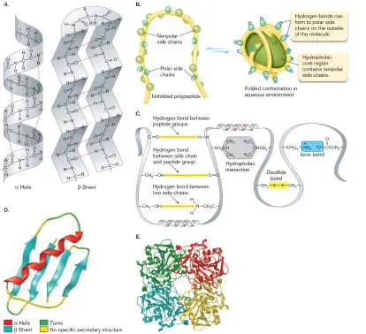
chains (the primary structure) determine how a protein folds into its final tertiary structure. The large number of amino acid side chains gives rise to the diversity of protein structures and, hence, functions. As proteins emerge from the ribosome, they are bound by chaperone proteins that help them fold into their functional shape, known as the native conformation. In soluble proteins, amino acids with hydrophobic side chains tend to cluster inside the protein to minimize reactions with water, while polar amino acids are exposed on the surface (Fig. A1.11B ).
Tertiary structure is stabilized by hydrogen bonding between side chains and between side chains and the main chain. Ionic interactions between acidic and basic side chains also contribute to tertiary structure. The one covalent interaction found stabilizing tertiary structure is disulfide bonding between two cysteine residues. The interactions that contribute to protein tertiary structure are shown in Figure A1.11C , and a ribbon representation of the 3D structure of a protein is illustrated in Figure A1.11D .
Some proteins form stable, functional complexes with other proteins. These multisubunit proteins exhibit the fourth level of protein organization: quaternary structure (Fig. A1.11E ). The forces that hold interacting polypeptide chains together are the same noncovalent interactions and disulfide bonds that determine protein tertiary structure.
Proteins are not static structures. The individually weak hydrogen bonds, ionic interactions, and van der Waals forces that contribute to protein structure are continually being broken and re-formed. Because these noncovalent bonds do not break all at once, the large number of such bonds that are present act collectively to maintain protein integrity. Some proteins are dynamic entities that may permute through a number of semi-stable states. Such conformational changes may be stochastic, random fluctuations due to the intrinsic thermal (kinetic) energy of the protein. Alternatively, the conformational changes may be influenced by external factors. For many proteins, alterations in conformation are critical for protein function.
Glossary
amino acid The monomer unit of proteins. Each amino acid contains a central carbon covalently bonded by a hydrogen, an amino group, a carboxyl group, and a side chain. An exception is proline, in which the side chain is cyclized with the central carbon.
chiral carbon A carbon bonded to four different types of functional groups. A molecule with a chiral carbon can thus take two different forms that exhibit mirror symmetry.
optical isomer Also called enantiomer. Either of two forms of a molecule that are mirror images of each other. Molecules that contain a chiral carbon can have optical isomers.
primary structure The first level of organization of polymers, consisting of the linear sequence of monomers; for example, the sequence of amino acids in a protein or of nucleotides in a nucleic acid. peptide bond The covalent bond that links two amino acid monomers.
secondary structure The second level of organization of polymers, consisting of regular patterns that repeat, such as the double helix in DNA or the beta sheet in proteins.
tertiary structure The third level of organization of polymers; the unique 3D shape of a polymer.
native conformation The fully folded, functional form of a protein.
quaternary structure The fourth (and highest) level of organization of proteins, in which multiple polypeptide chains interact and function together.
A1.4 Carbohydratesnot assigned
Carbohydrates are so named because their molecular formulas are multiples of CH 2 O (hydrated carbon). Polysaccharides are complex carbohydrates, composed of monosaccharide (simple-sugar)
monomers. Carbohydrates are a preferred energy source for many cells and also store energy. In addition, they play a structural role, as in cell walls. Moreover, when attached to proteins or lipids, they can serve an informational role.
Monosaccharides
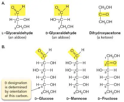
Monosaccharides differ from each other in the number of carbons present, in whether their carbonyl group (C⚌O) is an aldehyde or a ketone, and in the orientation of the hydroxyl groups. The smallest sugars, glyceraldehyde and dihydroxyacetone, have three carbons ( Fig. A1.12A). These two sugars have the same molecular formula, C 3 H 6 O 3, but because their atoms are arranged differently, they are structural isomers of each other. In addition, because glyceraldehyde contains a chiral carbon at C-2, it also has an optical isomer. In contrast to proteins, where the L -amino acids predominate, for sugars the D -forms (such as D -glucose) are of biological importance.
The six-carbon sugars (hexoses) are particularly important in the cell. Some important hexose sugars are D -glucose, D -mannose, and D -fructose (Fig. A1.12B ). Hexoses all have the molecular formula C 6 H 12 O 6 and are structural isomers of each other. While fructose is a keto sugar (ketose), mannose and glucose are both aldehydes (aldoses). The difference between mannose and glucose lies in the orientation of the hydroxyl group on one of the chiral carbons, C-2. The carbons in sugars are numbered starting with the carbon of the aldehyde group or the free carbon closest to the ketone group, so the aldehyde group is always C-1.
FIGURE A1.12 ■ Structural formulas for some monosaccharides. A. The three-carbon sugars glyceraldehyde and dihydroxyacetone. The aldehyde and ketone functional groups are shown in yellow. Glyceraldehyde contains a chiral carbon and has two optical isomers. B. The hexose sugars glucose, mannose, and fructose. The numbering of the carbons starts at the carbon of the carbonyl group (highlighted) or at the free carbon closest to the carbonyl. The D designation is determined from the orientation of the hydroxyl group on a chiral carbon farthest from the ketone or aldehyde functional group. In sugars of five or more carbons, one of the hydroxyl groups can react with the aldehyde or ketone group, forming a ring structure. The ring form predominates because it is more energetically favorable than the straight-chain sugar. The ring forms in two
different ways, as shown for D -glucose (Fig. A1.13). Depending on how the bond between C-1 and C-2 was oriented during formation of the ring, the cyclic form of D -glucose will be alpha-D - glucose or beta-D -glucose. The two forms interconvert rapidly in aqueous solution, and both forms are found as components of biological structures.
FIGURE A1.13 ■ Straight-chain and cyclic forms of D - glucose. The straight-chain form of D -glucose can cyclize to form two different ring structures: alpha-D -glucose and beta-D - glucose. In this process, a new chiral carbon is formed at C-1, the anomeric carbon.
The hydroxyl groups of sugars can react with other functional groups to form modified sugars. Examples include fructose 1,6-bisphosphate (an intermediate in glycolysis) and N - acetylglucosamine (a component of the peptidoglycan cell wall) ( Fig. A1.14).
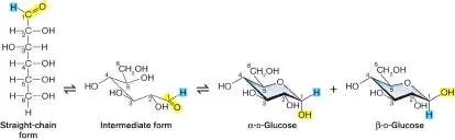
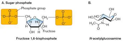
FIGURE A1.14 ■ Modified sugars. A. Phosphorylated sugars, such as fructose 1,6-bisphosphate, are intermediates in glycolysis. B. N -acetylglucosamine is a component of bacterial cell walls. Hydrogen atoms on the ring are not shown.
Two Monosaccharides Condense to Form a Disaccharide
Disaccharides form when two monosaccharides undergo a condensation reaction to form a covalent glycosidic bond. The glycosidic bond often occurs between the C-1 carbon of one monosaccharide and the C-4 carbon of another monosaccharide ( Fig. A1.15). The structure of the disaccharide varies, depending on which monosaccharides are involved and whether the C-1 carbon participating in the bond is in the alpha or beta form.
Oligosaccharides consist of a few monosaccharides linked together, while polysaccharides are much longer chains of monosaccharides. Polysaccharides are unbranched if all the linkages are 1,4. In branched polysaccharides, additional sugars are attached to other hydroxyl groups (for example, at C-6).
FIGURE A1.15 ■ Formation of the disaccharide maltose. Maltose forms by a condensation reaction between two molecules of D -glucose, so that the C-1 of one molecule of glucose is linked by an oxygen atom to the C-4 of a second molecule of glucose. The hydroxyl groups of the second glucose can be either alpha or beta (the beta form is shown). The covalent glycosidic bond is called an α-1,4-glycosidic linkage
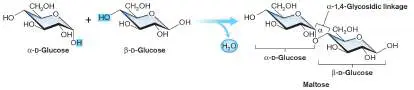
because the oxygen on the C-1 carbon of the glycosidic bond is in the alpha position.
Glossary
monosaccharide The monomer unit of sugars. Monosaccharides have a molecular formula of (CH 2 O) n.
structural isomer A molecule with the same molecular formula as a different molecule but a different arrangement of atoms.
A1.5 Nucleic Acidsnot assigned
Cells contain two kinds of nucleic acids: deoxyribonucleic acid (DNA) and ribonucleic acid (RNA). DNA is the hereditary material of the cell; it encodes the information to make proteins. Three different types of RNA play key roles in protein synthesis: Messenger RNA (mRNA) is transcribed from DNA and is the template for protein synthesis, transfer RNAs (tRNAs) bind amino acids and deliver them to the ribosome, and ribosomal RNA (rRNA) is a catalytic component of the ribosome.
A Chain of Nucleotides Forms the Primary Structure of Nucleic Acids
Nucleic acids are polymers of nucleotides. Nucleotides have three components: a pentose sugar (ribose in RNA, 2-deoxyribose in DNA), a nucleobase (also called a nitrogenous base), and a phosphate group (Fig. A1.16A). The nitrogenous base is attached to the C-1 of the sugar. Nucleobases come in two structural classes: purines (adenine, A; and guanine, G) and pyrimidines (cytosine, C, found in both DNA and RNA; thymine, T, found only in DNA; and uracil, U, found only in RNA). The phosphate group is esterified to the 5′ hydroxyl group of the sugar. (The prime indicates that the numbering refers to the sugar portion of the nucleotide; the base is numbered without primes.)
FIGURE A1.16 ■ Nucleotide and DNA structure. A. A nucleotide consists of a five-carbon sugar (ribose or deoxyribose), a phosphate group, and a nucleobase. B. A nucleotide may be a monophosphate, diphosphate, or triphosphate. The phosphate attached to the sugar is designated alpha, the beta phosphate is in the middle, and the gamma phosphate is distal to the sugar. C. The primary structure of DNA. The orientation of the strand is determined by the presence of a free phosphate on the 5′ end and a free hydroxyl on the 3′ end. The sequence of a nucleic acid is read from the 5′ end to the 3′ end, so for this portion of the DNA strand, the primary structure is GCTA. D. Ribbon structure of the DNA double helix, showing
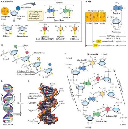
the purine-pyrimidine base pairs. E. Space-filling model of DNA. Note that the base pairs stack on top of each other like the rungs of a ladder. F. Hydrogen bonding between the base pairs. Two hydrogen bonds form between A and T; three hydrogen bonds form between G and C. Note the antiparallel orientation of the two strands.
A nucleoside is similar to a nucleotide, except that it has only two components: a sugar and a base (that is, it lacks a phosphate group). The names of the nucleosides are listed in Table A1.5. Nucleotides are formed when a phosphate group forms a covalent ester bond to the C-5 carbon of the sugar portion of a nucleoside. The nomenclature for nucleotides uses the abbreviation for the nitrogenous base and adds the abbreviation MP, DP, or TP to designate the phosphate group as a monophosphate, diphosphate, or triphosphate, respectively (Fig. A1.16B ).
TABLE A1.5 Bases and Nucleosides Base Nucleoside (base plus ribose or (abbreviation) deoxyribose)
a
Adenine (A) Adenosine Guanine (G) Guanosine Cytosine (C) Cytidine Thymine (T) Thymidine Uracil (U) Uridine In addition to being the precursors for nucleic acid synthesis, the ribose nucleotides have cellular functions of their own. For example, adenosine triphosphate (ATP) is an energy carrier in the cell, and guanosine triphosphate (GTP) and cyclic adenosine monophosphate (cAMP) are signaling molecules. A lowercase “d” in front of the abbreviated name of a nucleotide indicates that the sugar is 2-deoxyribose, the type of sugar found in DNA. For example, deoxycytosine triphosphate is dCTP, a molecule that can condense with DNA and extend the chain by one nucleotide.
The nucleoside triphosphate monomers (collectively designated NTPs or dNTPs) form nucleic acids through reactions catalyzed by nucleic acid polymerase enzymes. The phosphate at the C-5 position forms a diester linkage with the hydroxyl group at C-3, releasing pyrophosphate. At one end of the nucleic acid is a free phosphate group; at the other end is a free C-3 hydroxyl group. The terminus with a free phosphate is called the 5′ end, and the terminus with the free C-3 hydroxyl is called the 3′ end (Fig. A1.16C ). Repeating sugar-phosphate linkages form the “backbone” of the molecule, while the information content of nucleic acids resides in the sequence of bases attached to the backbone. This linear sequence of nucleotides, from the 5′ end to the 3′ end, is the primary structure of the nucleic acid.
The DNA Double Helix
Two strands of DNA, held together by hydrogen bonding between a purine and a pyrimidine on opposite strands, twist around each other to form a helix (Fig. A1.16D and E ). Two hydrogen bonds form between adenine and thymine, and three hydrogen bonds form between cytosine and guanine (Fig. A1.16F ). The sugar-phosphate backbone of DNA is on the outside of the helix. The base pairs are inside the helix, but portions of them are accessible to proteins such as transcription factors at two grooves in the helix: a wide major groove and a narrower minor groove. The double-stranded structure of DNA is critical to its function as the hereditary material. Each strand of DNA encodes the information to make a new double helix on the basis of strict base-pairing rules.
The Secondary Structure of RNA
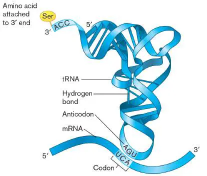
In contrast to the invariant double-helix structure of DNA, RNA molecules have a variety of secondary structures that allow RNA molecules to perform a number of different functions in the cell. Secondary and tertiary structures are possible because RNA is single-stranded and capable of complementary base pairing. Base pairing in RNA is similar to the base pairing exhibited by DNA, except that in RNA, uracil replaces thymine. Base pairing is used in making mRNA from a DNA template and also in protein synthesis, in which a tRNA charged with an amino acid matches its anticodon with the codon on the mRNA (Fig. A1.17). Because RNAs are single-stranded, they can undergo intramolecular as well as intermolecular base pairing. Base pairing within a single RNA molecule can lead to interesting 3D shapes that may allow the RNA to function as a catalyst. The activity of catalytic RNAs (ribozymes) may be enhanced by the reactive hydroxyl group at the C-2 of the ribose sugar, which is lacking in DNA.
FIGURE A1.17 ■ RNA secondary structure. A tRNA folds up into a secondary structure stabilized by the hydrogen bonds shown. The tRNA forms complementary base pairs with an mRNA.
Glossary
nucleotide The monomer unit of nucleic acids, consisting of a five-carbon sugar, a phosphoryl group, and a nitrogenous base.
nucleobase Also called nitrogenous base. A planar, heteroaromatic, nitrogen-containing base that forms a nucleotide of nucleic
acids; nucleobases determine the information content of DNA and RNA. There are five nucleobases: adenine, cytosine, guanine, thymine, and uracil.
nitrogenous base Also called nucleobase. A planar, heteroaromatic, nitrogen-containing base that forms a nucleotide of nucleic acids; nucleobases determine the information content of DNA and RNA. There are five nucleobases: adenine, cytosine, guanine, thymine, and uracil.
Endnotes
1. Note a: A, T, G, C, and U are abbreviations for the nucleobases; however, they are also used when referring to the nucleotides in a DNA or RNA strand to indicate which base is present in the primary structure. Return to reference a
A1.6 Lipidsnot assigned
Lipids are a structurally diverse class of molecules that have diverse functions. They are major structural components of cell membranes, they store energy, and they act as cellular signals. The common structural feature of lipid molecules is that they contain a substantial number of nonpolar C−H and C−C covalent bonds. Because lipids are nonpolar, they are hydrophobic and do not dissolve in water.
Lipids Are Either Hydrophobic or Amphipathic
An example of a hydrophobic lipid is isoprene (Fig. A1.18A), which functions as a building block for more complex cellular lipids, such as squalene. Other lipid building blocks include the fatty acids ( Fig. A1.18B ). In contrast to isoprene, the fatty acids have both a hydrophobic portion (the hydrocarbon tail) and a hydrophilic component (the carboxylic acid). Molecules with both hydrophobic and hydrophilic portions are described as amphipathic.
FIGURE A1.18 ■ Lipid building blocks. A. Isoprene is used to make squalene and cholesterol. B. Fatty acids are
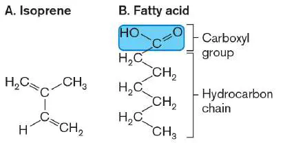
components of triglycerides and phospholipids.
Fatty Acids May Be Saturated or Unsaturated
The cell contains many different fatty acids, which differ in the number of carbons (usually even) and their saturation. In saturated fatty acids, the carbon-carbon bonds in the hydrocarbon tail are all single bonds, and the carbons are bonded to the maximum number of hydrogen atoms (Fig. A1.19A). Unsaturated fatty acids contain one or more double bonds between adjacent carbons in the hydrocarbon tail (Fig. A1.19B ). Monounsaturated fatty acids have one double bond; polyunsaturated fatty acids have multiple double bonds.
FIGURE A1.19 ■ Saturated and unsaturated fatty acids. A. Palmitic acid is a saturated fatty acid; there are no double bonds between the carbon atoms in the hydrocarbon tail. B. Linoleic acid is an example of a polyunsaturated fatty acid (it has more than one double bond). Note that the double bonds form kinks in the hydrocarbon tail.
Saturated fatty acids can pack together in an arrangement where they can be stabilized by van der Waals forces between adjacent hydrocarbon chains. Thus, saturated fatty acids (including animal fats such as lard) tend to be solids at room temperature. In
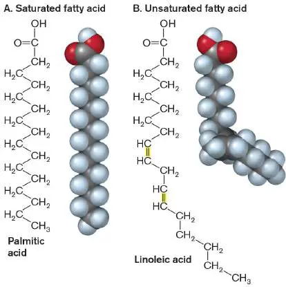
unsaturated fatty acids, the double bond causes a kink in the chain that prevents tight packing, so unsaturated fatty acids, such as vegetable oils, tend to remain fluid at room temperature.
Glycerol Is a Building Block of Lipids
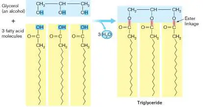
The three hydroxyl groups on glycerol can undergo a condensation reaction with the carboxylic acid portion of fatty acids to form triesters known as triglycerides (Fig. A1.20). Triglycerides are a compact energy source for cells. Phospholipids, key components of the cell membrane, contain fatty acids attached to two of the hydroxyl groups of glycerol and a phosphate covalently attached to the third hydroxyl. The structure of phospholipids and how they function in cell membranes are explored further in eAppendix 2, Section A2.1.
FIGURE A1.20 ■ Formation of a triglyceride. Condensation reactions form covalent ester linkages between the carboxyl groups of three fatty acids and the three hydroxyl groups of glycerol.
Glossary
amphipathic Having both hydrophilic and hydrophobic portions.
A1.7 Biological Chemistrynot assigned
Scientific Notation
Scientific notation is used to express very large numbers (such as the number of microorganisms in a
liter of seawater) or very small numbers (such as the diameter of a bacterium) with the help of
exponents. For example, a million, or 1,000,000, is often written as 1 × 10 6. The positive exponent 6
indicates how many decimal places to the right of the number 1 our number is. One-millionth, or
0.000001, is written as 1 × 10 − 6 to indicate six decimal places to the left of the number 1. Special
prefixes can be used to modify the magnitude of units. These are listed in Table A1.6 . For example, 1
× 10 −6 is designated by the symbol μ (the Greek letter mu, which stands for “micro”), so a length of 2
micrometers (μm) is equivalent to 2 × 10 −6 meter (m).
TABLE A1.6 Common Numerical Prefixes
Prefix Symbol Factor
kilo k 10 3
deci d 10 −1
centi c 10 −2
milli m 10 −3
micro μ 10 −6
nano n 10 −9
pico p 10 −12
femto f 10 −15
When scientists work with a set of numbers that range over many orders of magnitude, they often
use a logarithmic scale. For example, the pH scale uses base 10 logarithms. Base 10, or common,
logarithms are abbreviated “log,” as opposed to natural logarithms, abbreviated “ln.” Base 10
logarithms are defined as log 10 x > = x. For example, the log of 1 × 10 − 4 = −4, the log of 10 = 1,
and the log of 1 = 0.
Molarity Is the Unit of Concentration
Scientists often measure concentrations. “Concentration” refers to how much of something is present in
a given volume. A frequent way to report concentration is in units of molarity. Molarity is defined as the
number of moles of substance per liter of solution (usually water). One mole is Avogadro’s number
(6.02 × 10 23) of molecules. If you wanted to make a 1-molar solution of NaCl, it would be impossible
to know when you had added 6.02 × 10 23 molecules to a liter of water. It’s more convenient to weigh
out a mole of NaCl on a balance. The weight of a mole of a substance is equal to Avogadro’s number
multiplied by the weight of a molecule. For example, NaCl has a mass of 58.5 atomic mass units (amu)
—23 from sodium and 35.5 from chlorine (see Fig. A1.1). One molecule of NaCl weighs 58.5 amu
divided by 6.02 × 10 23 = 9.71 × 10 −23 g. A mole of NaCl then weighs 9.7 × 10 −23 g multiplied by
6.02 × 10 23 (Avogadro’s number) = 58.5 g. (Note that this weight is the same as just adding together
the atomic masses of Na and Cl and expressing the result in grams.) So, to make a 1-molar solution of
NaCl, you would put 58.5 g of NaCl in a container and add water up to a liter.
Change in Free Energy
Reactions in organisms, such as the synthesis of proteins from amino acids by condensation, can occur
only if they are energetically favorable. The study of energy and matter changes is called
thermodynamics. All systems (including living systems, such as cells) must obey the laws of
thermodynamics. The first law of thermodynamics states that energy is neither created nor destroyed.
In a closed system (a system that is not exchanging energy with its environment), the total amount of
energy remains constant. Although the amount of energy remains constant, energy can be converted
from one form to another. The second law of thermodynamics states that in energy transformations,
some energy becomes unavailable to do work and is lost as disorder, or entropy. In other words,
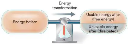
entropy tends to increase. The laws of thermodynamics are summarized in Figure A1.21 .
FIGURE A1.21 ■ The first and second laws of thermodynamics. The first law states that
in an energy transformation, the total amount of energy remains constant. Both sides of the
equation have the same amount of energy (the balance reads zero). The second law states that
in an energy transformation, the amount of usable energy, or free energy, decreases and the
amount of unusable energy (the product of temperature and entropy) increases.
The total energy in a system, called enthalpy, can be expressed in the following equation, where H
stands for enthalpy, G stands for free energy, T stands for temperature, and S stands for entropy:
H = G + T × S
Because scientists are interested in the amount of free energy—that is, the amount of energy
available, or free, to do work—the energy equation is usually written in terms of G:
Δ G = Δ H − T × Δ S
The triangle is the Greek letter delta, which signifies “change in.” Here, a change in energy is defined
as the energy of the products minus the energy of the reactants. Thus, Δ G means G products − G reactants
, and Δ S indicates S products − S reactants.
Systems Tend toward the Most Stable, Lowest Free Energy State
Reactions are favored when the free energy of the products is less than the free energy of the
reactants—that is, when Δ G is negative. Reactions with a negative Δ G are termed spontaneous, or
exergonic, because they are energetically favorable and can occur without an input of energy.
Reactions with a positive Δ G are endergonic; they are not energetically favorable, and energy needs
to be added for them to proceed. Reactions with a Δ G of zero are at equilibrium, and neither products
nor reactants are favored.
The sign of Δ G is determined by changes in both enthalpy and entropy. For example, the following
reaction—the oxidation, or “burning,” of methane to release carbon dioxide and water—is spontaneous
and has a negative change in free energy:
CH 4 + O 2 ⇌ CO 2 + H 2 O + energy
The bonds in methane and oxygen have more energy than the bonds in CO 2 and H 2 O. Because the
products CO 2 and H 2 O have less energy than the reactants, the change in enthalpy for this reaction,
Δ H (H products − H reactants), is negative and the reaction is said to be exothermic.
A reaction that absorbs heat from its surroundings (endothermic) can still have a negative value of
Δ G, if it has an increase in entropy, Δ S. Consider an ice cube placed at room temperature. The ice
cube will spontaneously melt, even though this reaction is endothermic, absorbing heat from the
environment. Ice melting is spontaneous because of the increase in entropy in going from a solid to a
liquid. Remember that entropy, S, is a measure of the disorder of a system. Gases are more
disordered than liquids, and liquids are more disordered than solids. Thus, the change in entropy, Δ S (
S products − S reactants), from solid water (ice) to liquid water is positive because the product, liquid
water, has more entropy than ice has. Positive changes in entropy may result in a negative change in
free energy, especially at high temperatures.
Chemical Equilibrium
To compare changes in free energy among different reactions, a standard change in free energy, Δ G °
(the circle is pronounced “naught”), is defined as the change in free energy when the concentrations of
all reactants and products are at 1 molar (M). Each molecule has a value of Δ G ° of formation from its
atoms in their standard state. For a given reaction, the overall Δ G ° can be calculated as the sum of
the Δ G ° of formation for all the products, minus the sum of the Δ G ° of formation for all the reactants
(as shown later).
Δ G ° is a constant and depends on the nature of the products and reactants. Another constant, Δ G
°′, refers to the standard change in free energy at pH 7. A standard free energy change can be related
to an equilibrium constant, K eq, that indicates whether products or reactants will be favored at
equilibrium. Reactions between biomolecules can be written as chemical equations. For example, in
the following reaction, reactants A and B form products C and D:
The double arrow indicates that the reaction is reversible; A and B can be forming C and D at the
same time that C and D are reverting to A and B. What determines whether the forward or reverse
reaction will predominate? The forward and reverse rates depend on rate constants k f and k r,
respectively. The rate constants depend on the nature of the interacting molecules and are an
indication of how easily they will react. The forward and reverse rates also depend on concentrations
of reactants and products:
Forward rate = [A][B] k f
Reverse rate = [C][D] k r
If the forward and reverse reactions are allowed to proceed, eventually equilibrium will be reached.
All chemical reactions have a preferred state called equilibrium (plural equilibria), where there is no
net change in the reaction. Equilibrium is not static; rather, at equilibrium the rate of the forward
reaction equals the rate of the reverse reaction:
[A][B] k f = [C][D] k r
For every reaction, there is an equilibrium constant, K eq, that indicates the relative ratios of products
and reactants at equilibrium:
K eq = k f / k r = [C][D]/[A][B]
A K eq of 1 means that neither products nor reactants are favored, a K eq of greater than 1 indicates
that products are favored at equilibrium, and a K eq of less than 1 means that reactants predominate. K
eq and Δ G ° are related as follows, where R is the gas constant [8.315 × 10 − 3 kJ/(mol · K)] and T is
the temperature in kelvins:
Δ G ° = − RT ln K eq
A value of 298 K (roughly 25°C) is often used for temperature. The relationship between K eq and Δ G °
is usually shown with the base 10 logarithm instead of the natural log:
Δ G ° = −2.303 RT log K eq
This equation defines a relationship between Δ G ° and K eq that is discussed in Chapter 13. When K
eq is 1 (neither products nor reactants favored), Δ G ° (defined as the change in free energy when
concentrations of all substances are at 1 M) is zero because the reaction is at equilibrium at that point.
A K eq of less than 1 (reactants favored) corresponds to a positive Δ G ° because at 1-M concentrations
of reactants and products, the reaction will move to the left. A K eq of more than 1 (products favored)
corresponds to a negative Δ G ° because at 1-M concentrations, the reaction will move to the right.
Free Energy in Cells and the Law of Mass Action
Although the standard changes in free energy are useful for comparing the energetics and equilibria of
different reactions, they do not reflect what is happening in the cell, where concentrations of
substances are probably not 1 M. While K eq and Δ G ° are constants and unique for a particular
reaction, Δ G depends on the actual concentrations of products and reactants as shown in the equation
relating Δ G to Δ G °:
Δ G = Δ G ° + 2.303 RT log ([C][D]/[A][B])
In this equation, the concentrations of products and reactants are not their equilibrium concentrations,
but the actual concentrations present at a given point in time. Because the concentrations of products
and reactants will change as a reaction proceeds, the value of Δ G will change over time.
At equilibrium, where the total energy on each side of the reaction is equivalent, Δ G is zero. For an
exergonic reaction at equilibrium, the total energy of reactants and products is equivalent because the
amount of products is greater than the amount of reactants. If, at equilibrium, reactants (A, B) are
added or products (C, D) are removed, Δ G will become negative. A negative Δ G causes the reaction
to move to the right to reestablish equilibrium. As A and B convert to C and D, Δ G becomes less and
less negative, until it reaches zero again at equilibrium. If, at equilibrium, products are added or
reactants are removed, Δ G will become positive and the reaction will proceed to the left to reestablish
equal energy on both sides. This law of mass action is the tendency of a reaction to reestablish
equilibrium after perturbations in the concentrations of products or reactants.
The law of mass action means that a reaction with a positive Δ G ° can be made spontaneous and
driven to the right by keeping the concentration of reactants high or the concentration of products low.
The cell employs this strategy in metabolic pathways, where the product of one reaction is constantly
removed by a subsequent reaction.
Calculating the Standard Free Energy Change, Δ G °, of Chemical Reactions
For a chemical reaction, the net change in free energy, Δ G, determines whether the reaction will go
forward. The value of Δ G at standard conditions of temperature (25°C), pressure (1 atmosphere, P a),
and concentration (each reactant at 1 mole/liter), is designated Δ G °. For a given reaction, how do we
find the value of Δ G °? The value is determined by summing the individual values for standard energy
of formation (Δ G f °) for all the products, and then subtracting the sum of the values for all the
reactants.
Consider the oxidation of glucose (C 6 H 12 O 6) to form water plus carbon dioxide:
C 6 H 12 O 6 + 6O 2 → 6CO 2 + 6H 2 O
We must assume that the glucose is present at 1-M concentration, that the O 2 and CO 2 are gases
at 1 atmosphere, and that the water is liquid at 25°C (298 K). The value of Δ G ° for this reaction is
given by:
Δ G ° = [(6 × Δ G f ° CO 2) + (6 × Δ G f ° H 2 O)] −[(Δ G f ° C 6 H 12 O 6) + (6 × Δ G f ° O 2)]
From Table A1.7 we can insert the values of Δ G f ° for each reactant and product. Note that one
reactant, O 2, has a value of zero, because oxygen gas is the defined standard state for oxygen:
Δ G ° = [(6 × −394.4 kJ/mol) + (6 × −237.1 kJ/mol)] −[(−910.4 kJ/mol) + (6 × 0 kJ/mol)]
Δ G ° = −2,879 kJ/mol
So, the oxidation of glucose has a negative value of Δ G at standard conditions, and the reaction
will go forward, releasing energy. The magnitude of the Δ G value suggests that enough energy could
be released to form a number of energy carriers, such as ATP, but many additional factors need to be
included before we know fully what a cell gains from this reaction.
TABLE Free Energies of Formation (Δ G f°), in Units of kJ/mol A1.7
Inorganic Organic
Inorganic compounds Organic compounds
compounds Δ G f° (continued) Δ G f° compounds Δ G f° (continued) Δ G
CH 4 −50.8 Mn 2+ −228.1 Acetaldehyde −127.6 Guanine +47
CO −137.2 MnCl 2 −490.8 Acetate −369.7 Lactate −51
CO 2 −394.4 MnSO 4 −972.8 Acetone −152.7 Lactose −1,
TABLE Free Energies of Formation (Δ G f°), in Units of kJ/mol A1.7
H 2 CO 3 −623.2 N 2 0 Arginine −240.5 Malate −84
HCO 3 − −586.9 NO +87.6 Aspartic acid −730.7 Methanol −16
CO 3 2− −527.9 NO 2 +51.3 Benzene +124.4 Methionine −50
HCOO − −351.0 NO 2 − −32.2 Benzoic acid −245.3 Methylamine +35
Cu + +50.0 NO 3 − −111.3 1-Butanol −162.5 Naphthalene +20
Cu 2+ +65.5 NH 3 −26.6 Butyrate +352.6 Oxalate −69
CuS −53.7 NH 4 + −79.3 Citrate −1,236.4 Oxaloacetate −79
Fe 2+ −78.9 N 2 O +103.7 Cysteine +339.8 2-Oxoglutarate +79
Fe 3+ −4.7 N 2 H 4 +149.3 Ethanol −174.8 Phenol −50
FeCO 3 −666.7 O 2 0 Ethylene +68.4 Propionate −36
FeS 2 −156.1 OH − −157.3 Fructose −915.4 Pyruvate +47
FeSO 4 −823.4 PO 4 3− −1,018.8 Fumarate −655.6 Ribose −75
H 2 0 S 0 0 Gluconate −1,128.3 Succinate −69
H + (pH 0) 0 SO 3 2− −486.5 Glucose −910.4 Sucrose −1,
H + (pH 7) −39.7 S 2 O 3 2− −522.5 Glutamic acid −731.3 Toluene +11
HCl −131.3 H 2 S −27.9 Glutamine −529.7 Trimethylamine +93
H 2 O −237.1 HS − +12.1 Glyceraldehyde +437.7 Tryptophan −11
H 2 O 2 −120.4 S 2− +85.8 Glycerol −477.0 Tyrosine −38
Sources: James G. Speight. 2005. Lange’s Handbook of Chemistry, 16th ed., McGraw-Hill, New York; WolframAlpha
(http://www.wolframalpha.com); Rudolf K. Thauer. 1977. Bacteriol. Rev. 41 :100.
The Rate of a Reaction Depends on the Activation Energy
It is important to realize that although a reaction with a large positive K eq and negative Δ G ° is
spontaneous, it may be slow. The value of Δ G ° says nothing about the rate of a reaction. An everyday
example of a spontaneous but slow reaction is the rusting of metal. A reaction will be slow, even
though it is spontaneous, if it must pass through an unstable, high-energy transition state on the way
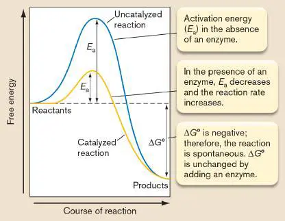
to forming products. The activation energy (E a) is the energy needed to reach this transition state (
Fig. A1.22 ). For reactions with low activation energies, random collisions between reactants may
provide enough energy to boost them up and over the E a. For reactions with high activation energies,
collisions between molecules may not provide enough energy for them to reach the transition state.
Enzymes are biological catalysts that can speed up reaction rates by stabilizing transition states
and lowering the activation energy. Most enzymes are proteins that specifically bind reactants and
provide an environment that facilitates product formation. Although enzymes increase reaction rates,
they do not change the Δ G ° or K eq of a reaction. Figure A1.22 illustrates how the difference in free
energy between products and reactants is unchanged even in the presence of an enzyme that lowers
the activation energy.
FIGURE A1.22 ■ Enzymes, activation energies, and reaction rates.
Biological Processes Depend on pH
Many biological processes occur within only a narrow range of hydrogen ion concentrations. Hydrogen
ions (H +) are also referred to as “protons” because they have lost their single electron, leaving a
proton. In water solution, the proton actually combines with a molecule of H 2 O, forming H 3 O +
(hydronium ion), but we generally simplify the notation as H +. Hydrogen ion concentration is reported
using a pH (power of hydrogen) scale, where pH is the negative logarithm of the hydrogen ion
concentration: pH = −log 10 [H +]. In pure water, which is considered neutral, [H +] is 1 × 10 − 7
molar (M)—a pH of 7.
Because the pH scale is logarithmic, every pH unit corresponds to a tenfold change in hydrogen ion
concentration. A solution with a pH of 6 has a hydrogen ion concentration of 1 × 10 − 6 M, ten times
the hydrogen ion concentration at pH 7. Hydrogen ion concentration [H +] multiplied by hydroxide ion
concentration [OH −] always equals 1 × 10 − 14. Hence, the concentrations of H + and OH − are
reciprocally related (if one goes up, the other goes down). If the pH is less than 7, the solution is acidic
(more H +, less OH −). If the pH is greater than 7, the solution is basic, or alkaline (less H +, more
OH −).
Acids (for example, carboxyl groups) release protons, and bases (for example, amino groups) bind
protons (Fig. A1.23 ). At intracellular pH (near 7), the carboxyl and amino groups of amino acids are
both ionized (charged), the carboxyl group carrying a negative charge and the amino group a positive
charge. The ionization of these groups can change if the pH changes. At lower pH (more acidic
solution), protons move back onto the ionized carboxylic acid. For example, the side chain of an acidic
amino acid such as glutamate (Fig. A1.9) is ionized at normal cell pH but may regain a proton and
become glutamic acid at a lower pH. At higher pH values (lower proton concentrations), amino groups
lose protons to become uncharged. Disturbances in the ionization state of carboxyl or amino groups on
the side chains of amino acid residues can disrupt ionic bonds between these groups and lead to
protein denaturation. This effect of pH on protein tertiary structure is one reason why cells can tolerate
only a narrow range of intracellular pH.
FIGURE A1.23 ■ Organic acids and bases.
Oxidation-Reduction (Redox) Reactions Transfer Energy
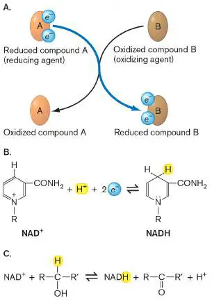
Biomolecules may undergo an important class of chemical reactions termed redox reactions. In redox
reactions, electrons are transferred from one molecule to another or from one atom to another. The
molecule that gains electrons becomes reduced, and the molecule that loses electrons becomes
oxidized (Fig. A1.24 ). (A useful mnemonic device to remember this is “LEO the lion says GER,”
where LEO stands for “lose electrons, oxidation” and GER stands for “gain electrons, reduction.”)
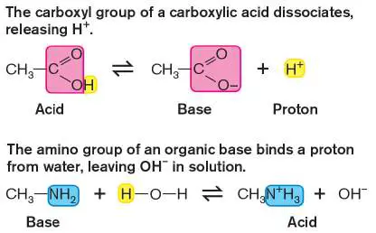
FIGURE A1.24 ■ Oxidation-reduction reactions. A. A reducing agent A donates electrons
to reduce compound B. Because A loses electrons, it becomes oxidized itself in the process. B is
the oxidizing agent. B. NADH is a common biological reducing agent. C. NAD + is reduced to
NADH as an alcohol is oxidized to an aldehyde.
Redox reactions are always coupled. If one atom loses electrons, another atom must gain
electrons. Oxygen, with its large electronegativity, usually gains electrons and becomes reduced in
redox reactions. Molecules that become reduced are called “oxidizing agents” because they cause
something else to become oxidized. In contrast, “reducing agents” can donate electrons, reduce other
molecules, and become oxidized themselves in the process. Common reducing agents (electron
donors) in the cell are NADH, NADPH, and FADH 2. These are all high-energy molecules that can
donate electrons.
In addition to achieving a complete transfer of electrons, redox reactions can also occur if electrons
are shifted toward or away from an atom. For example, in the burning (oxidation) of methane,
electrons move away from the carbon and toward the oxygen (Fig. A1.25 ). Oxygen usually acts as
an oxidizing agent (electron acceptor); thus, we expect it to become reduced (gain electrons). By
default, then, methane is oxidized to carbon dioxide. Indeed, while the bonding electrons are shared
fairly equally between C and H in methane, after oxidation they are close to the O in both carbon
dioxide and water and farther away from the C and H. Thus, in the oxidation of methane, methane is
oxidized and oxygen is reduced. This reaction releases energy because CO 2 and H 2 O are the most
stable forms available when carbon, hydrogen, and oxygen are combined.
FIGURE A1.25 ■ The oxidation of methane. Bonding electrons are shared equally by
carbon and hydrogen in methane, but they are closer to oxygen in carbon dioxide and water. In
this reaction, carbon and hydrogen are oxidized, and oxygen is reduced.
Glossary
molarity
A unit of concentration measured as the number of moles of solute per liter of solution.
entropy
A measure of the disorder in a system.
enthalpy
A measure of the heat energy in a system.
exergonic reaction
A spontaneous chemical reaction that releases free energy.
equilibrium pl. equilibria
A dynamic state in which there is no net change in a reaction.
equilibrium pl. equilibria
A dynamic state in which there is no net change in a reaction.
law of mass action
The rate of a chemical reaction is proportional to the substrate concentrations. At equilibrium, the
ratio of the concentrations of products to substrates is constant.
redox reaction
A chemical reaction in which one molecule or functional group becomes reduced and another
becomes oxidized.
Fig. A1.1
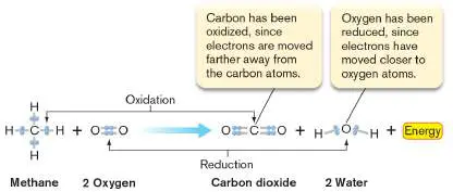
FIGURE A1.1 ■ Periodic table of the elements. The atomic number (number of
protons) and atomic mass are shown for each element.
Fig. A1.9
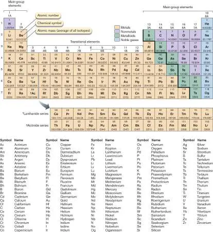
FIGURE A1.9 ■ Twenty common amino acids. The grouping of amino acids is based on
their side chains, highlighted in yellow.