Where the points are
440 points, none of them an exam. 130 come from weekly quizzes, 110 from lab skills, 190 from the cytotoxicity project. Concept Quizzes are cumulative, so the arithmetic below never stops being examinable — that is the single highest-yield thing to over-learn.
Bench math — the cumulative core
Cellular Biology (Johnson / Northam), Fall 2026 — built from the Week 1 lecture (IntroF23), the pipetting & calculations lab slides, the Pipet Exercise protocol, and BOTH practice sheets with their answer keys.
Why this matters now: the WEEK 1 Quiz is primarily calculations, “very similar to the practice sheet” (her words on the slide). Concept quizzes are cumulative, so this math never goes away — it is also the math of every later experiment and the final project.
1. The metric ladder (memorize cold)
- kilo (k) = 10³ · base = 1 · milli (m) = 10⁻³ · micro (µ) = 10⁻⁶ · nano (n) = 10⁻⁹ · pico (p) = 10⁻¹²
- Each step down the ladder is 1,000-fold. mM -> nM is 10⁶ (a million-fold); mM -> µM and µM -> nM are each 1,000-fold.
- Trick the practice sheet uses: 1 pmol/µl = 1 µM. (pico/micro = 10⁻¹²/10⁻⁶ = 10⁻⁶ mol per 10⁻⁶ L.)
2. Weights and molarity
- Atomic weight: amu (dalton): mass on the atomic scale; 1 amu ≡ 1 g/mol. C = 12.01, H = 1.01, O = 15.99.
- Molecular weight: sum of the atomic masses. Ethanol C₂H₆O = 24.02 + 6.06 + 15.99 = 46.07 amu -> molar mass 46.07 g/mol.
- Molarity (M): mol/L. 1 M ethanol = 46.07 g dissolved per liter. Grams needed = M × MW × liters.
- Normality (N): concentration of the Reactive species (e.g., H⁺): for HCl, 12 M = 12 N (one proton per molecule).
- % solutions: v/v unless told otherwise: 70% isopropanol in 15 ml = 0.70 × 15 = 10.5 ml isopropanol.
3. The four formulas that solve everything
- 1) Dilution: C₁V₁ = C₂V₂
- Units must match across the equation. Usually solve V₁ (how much stock).
- 2) Dilution factor: Solute volume = Final volume ÷ DF. A 1:10 dilution = 1 part solute + 9 parts diluent.
- 3) Step (serial) dilutions: Final DF = DF₁ × DF₂ × DF₃. Use when the pipettable volume would be too small (< 1 µl).
- 4) Mass from molarity: grams = M × MW (g/mol) × volume (L).
4. Worked problems — lecture slides
- Make 10 ml of 100 mM from a 1 M stock. C1V1=C2V2: 1000 mM x V1 = 100 mM x 10 ml -> V1 = 1 ml stock + 9 ml water.
- Make 300 µl of a 1:250 dilution. 300/250 = 1.2 µl solute into 298.8 µl diluent.
- Make 300 µl of a 1:1000 dilution when you cannot pipette < 1 µl. 300/1000 = 0.3 µl — too small! Step it: 1:10 then 1:100 (10×100 = 1000). D1: 1 µl + 9 µl water. D2: 1 µl of D1 + 999... (per slide: 1 µl of D1 into 99 µl for 1:100, then draw what you need).
- Primers: A = 20 mM, B = 35 mM; want 40 µl containing 5 nM of each. mM->nM = 1:10⁶. Two serial 1:1000 dilutions of A (1 µl + 999 µl, twice) -> 20 nM. Then 20 nM x V1 = 5 nM x 40 µl -> V1 = 10 µl. Same logic for B (35 nM working stock -> 5.7 µl). Water = fill to 40 µl.
- Fluconazole: MW 30.27, bottle has 100 mg. Water to add for a 1 M solution? A 1 M solution = 30.27 g/L. 100 mg / 30.27 g/mol = 3.3 mmol -> V = 3.3 mmol / 1 mol/L = 3.3 ml.
5. Worked problems — practice sheet (with the 2016/2017 key)
- KCl grams for 0.45 M in 1 L (K = 39.1, Cl = 35.43)? MW = 74.53. 74.53×0.45 = 33.5 g.
- Same, but only 100 ml? 10-fold less volume -> 3.35 g.
- Molarity of 3.5 g KCl in 300 ml? 3.5/0.3 = 11.67 g/L; / 74.53 = 0.16 M.
- 2 M KCl stock; need 5 ml of 400 mM. 2 M x V1 = 0.4 M x 5 ml -> V1 = 1 ml.
- 5 ml of 1 M NaCl; dilute to 542 mM. Water to ADD? V2 = 5/0.542 = 9.23 ml total -> add 4.23 ml.
- 4 g/L stock diluted to 5 L of 1.5 g/L. Stock used? 4 x V1 = 1.5×5 -> 1.875 L.
- 12 M HCl to make 100 ml of 0.01 N? 12 M = 12 N. V1 = (0.01×100)/12 = 0.083 ml.
- 10 N HCl to make 1000 ml of 0.1 N? V1 = 10 ml.
- 100% ethanol for 250 ml of 5%? 12.5 ml.
- mg of KOH in 25 ml of 0.25 M (MW 56.1)? 56.1×0.25 = 14.02 g/L x 0.025 L = 350.6 mg.
- Antibiotic at 10,000X; 1x in 100 ml media? V1 = 100/10,000 = 0.01 ml = 10 µl.
- 1x = 500 ng/ml, MW 334.5; make 50 ml of 2000X stock. Antibiotic needed? 500 ng/ml x 2000 = 1 mg/ml x 50 ml = 50 mg.
- Molarity of that stock? 1 g/L / 334.5 g/mol = 2.99 mM.
- Media with 10% serum, 500 ml media. Serum to add? The trick: adding serum raises total volume. Final volume = 500/0.9 = 555 ml -> add 55 ml serum (not 50!).
- Two primers, both 20 µM; 50 µl PCR needs TOTAL 5 pmol/µl, equal split. 1 pmol/µl = 1 µM -> 2.5 µM each. 20 x V1 = 2.5×50 -> 6.25 µl EACH.
- Two primers A = 20 µM, B = 100 µM; total 0.2 µM, equal split, 50 µl. 0.1 µM each: A -> 0.25 µl; B -> 0.05 µl.
- 25 µl reaction needs 50 ng DNA; stock reads 17 ng/µl. 50/17 = 2.94 µl.
- Fold difference: 500 mM vs 2.5 mM? 200-fold.
- 5 µl of stock into 5 ml to reach 2 µg/ml. Stock concentration? C1×0.005 ml = 2 µg/ml x 5 ml -> 2 mg/ml (a 1000X stock).
- 5000X stock -> 20 ml of 2000X. V1 = (2000×20)/5000 = 8 ml.
6. Worked problems — additional practice (Johnson/Judge KEY)
- KCl grams for 0.4 M in 500 ml? 74.53×0.4×0.5 = 14.9 g.
- Ethanol for 600 ml of 10%? 60 ml.
- 1x = 100 ng/ml, MW 74.5; 50 ml of 2000X stock? 100 ng/ml x 2000 = 0.2 mg/ml x 50 ml = 10 mg.
- 20,000 cells/ml -> need 2,500 cells/ml; media to ADD to 1 ml? V2 = 20,000/2,500 = 8 ml total -> add 7 ml.
- 400 ml of 4.5 M HCl -> dilute to 1 N. Solvent to add? V2 = 400×4.5 = 1800 ml -> ADD 1.4 L.
- 70,000 cells/ml x 7 ml, pellet, resuspend in 2 ml. New concentration? 490,000 cells / 2 ml = 245,000 cells/ml.
- 1.2 ml + 3 ml media: fold change of final vs initial? Final/Initial = 4.2/1.2 = 3.5-fold. (Key computes 4.2/3... the ratio to state: final over initial.)
- 100 mM -> 100 pM: dilution scheme? Three 1:1000 steps: 100 µM -> 100 nM -> 100 pM. (To reach 10 pM: one more 1:10.)
- Volume from stock for the first 1:1000 in 500 µl total? 500/1000 = 0.5 µl.
- Same in 300 µl total? 0.3 µl — below the 1-µl pipetting floor, which is exactly why step dilutions exist.
7. Pipetting — the skill being graded
- The set: P2 (0.2–2 µl), P20 (2–20 µl), P200 (20–200 µl), P1000 (100–1000 µl). Choose the smallest pipette that covers the volume.
- Press to the FIRST stop, immerse tip, release slowly to draw; press to first stop to dispense, Second stop to blow out the last drop. Pressing to the first stop Before drawing is correct — going to the second stop before drawing pulls up Too much liquid (that was the online-student question).
- Filtered tips: the filter blocks aerosols from contaminating the pipette barrel. P20 tips physically fit the P200, but the volume calibration and seal are wrong — never mix them (the other online question).
- Week 1 exercise: mixed solution (600 µl clear + 15 µl blue + 2 µl red), a cell dilution (2×10⁶ -> 1×10⁵ cells/ml in 2.5 ml: use 125 µl of stock), 10 M -> 500 mM NaCl (50 µl stock -> 1 ml total: add 950 µl), 20 µM -> 10 pM primer by serial dilution, KCl thought problem (0.5 M in 1 L = 37.3 g), and the parafilm drop challenge (12.5 / 25 / 50 / 100 µl rows).
8. Week 1 logistics worth remembering
- Quiz week 1 = primarily calculations, similar to the practice sheet; answers are on Canvas. Concept quizzes post Thursday, due Tuesday 10 AM, Respondus, Cumulative.
- Lab: AH408, Mon or Thu 1–3:50; BSL-1 but always gloves; biohazard waste goes in the Cell bio bins (non-sharp / sharp / glass sharp). Bring the lab notebook every time.
- Late work: -10% per day, 0 after 4 days. Extra credit 1 due Friday week 8; extra credit 2 Friday week 15.
- Dr. Johnson’s current interests (project seeds): p120-mutant cell motility with glucose/taurine, cytotoxicity of assembly-competent vs -incompetent Cx43 pancreatic cancer cells, kinase expression in communication-competent vs -incompetent cells, essential oils on wound healing. FUSE / Honors / biotech internships available.
Cellular Biology (Johnson / Northam), Fall 2026 — built from the Week 2 lecture deck “(wk2) Observing Cells and Aseptic technique”, both lab protocols, the Cell Observation Figure handout, and the four assigned primary papers.
This week is where the course gets hands-on: you learn to LOOK at cells (morphology, density, health, contamination, mitosis) and to work without contaminating them. The concept quiz covers lab math (cumulative from Week 1) plus these concepts; the paper quiz covers the assigned readings.
Part 1: Types of cells in culture — the three morphologies
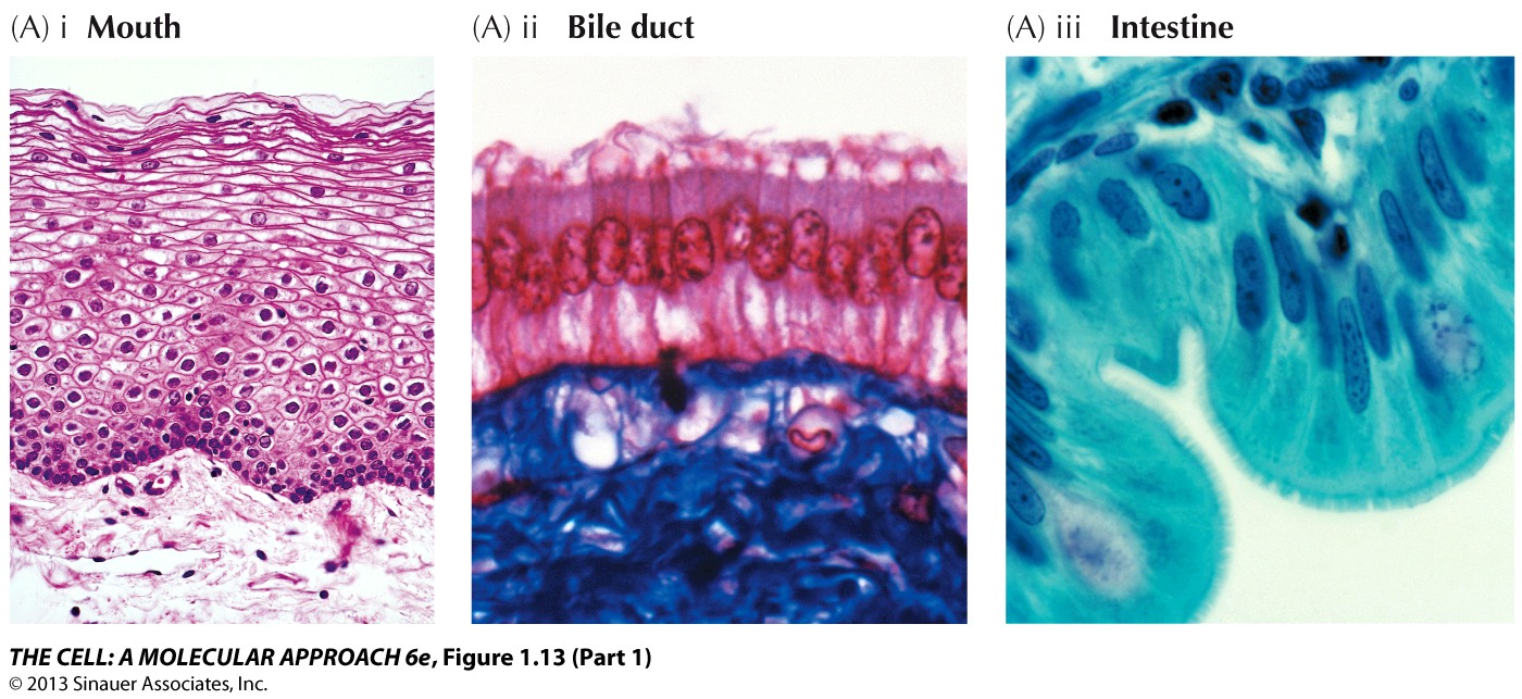
- Epithelial-like: flat, polygonal, pavement-like sheets that grow in discrete patches and stay tightly joined (they make cell-cell junctions — Dr. Johnson’s research area). From epithelium: skin, gut, kidney, cornea. Example this week: U2OS.
- Fibroblastic: bipolar or multipolar, elongated, spindle-shaped; grow attached to the substrate in swirling, parallel arrays. Example this week: HFF (human foreskin fibroblast).
- Lymphoblast-like: spherical cells that grow in Suspension without attaching — hematopoietic lines.
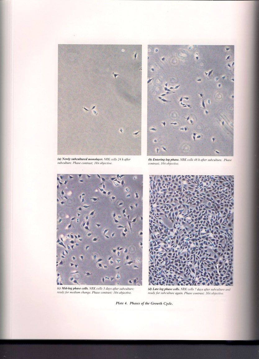
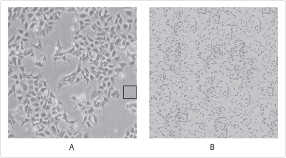
- The exam trap from the slide: at LOW cell density, epithelial cells can resemble fibroblasts. The correct morphology is most distinguishable in a Confluent dish — so judge morphology when the dish is full, not sparse.
The two cell lines you observed
- HFF: human foreskin Fibroblast — a normal, finite (non-transformed) diploid line. Expect spindle shapes, parallel/swirled patterning, contact inhibition (it stops at a monolayer).
- U2OS: derived from the bone tissue of a 15-year-old female with Osteosarcoma — a continuous, transformed line. Expect epithelial-like polygonal cells, loss of contact inhibition, piling up and random overgrowth.
- Protocol Thought Question 1 asks you to place each line in a morphologic category. Work it out from the descriptions above — the answer is in Appendix A at the end of this guide.
Part 2: What to look for at the scope (the protocol, step by step)
- Gloves on. Do NOT open a flask outside a hood — and this week you never open them at all.
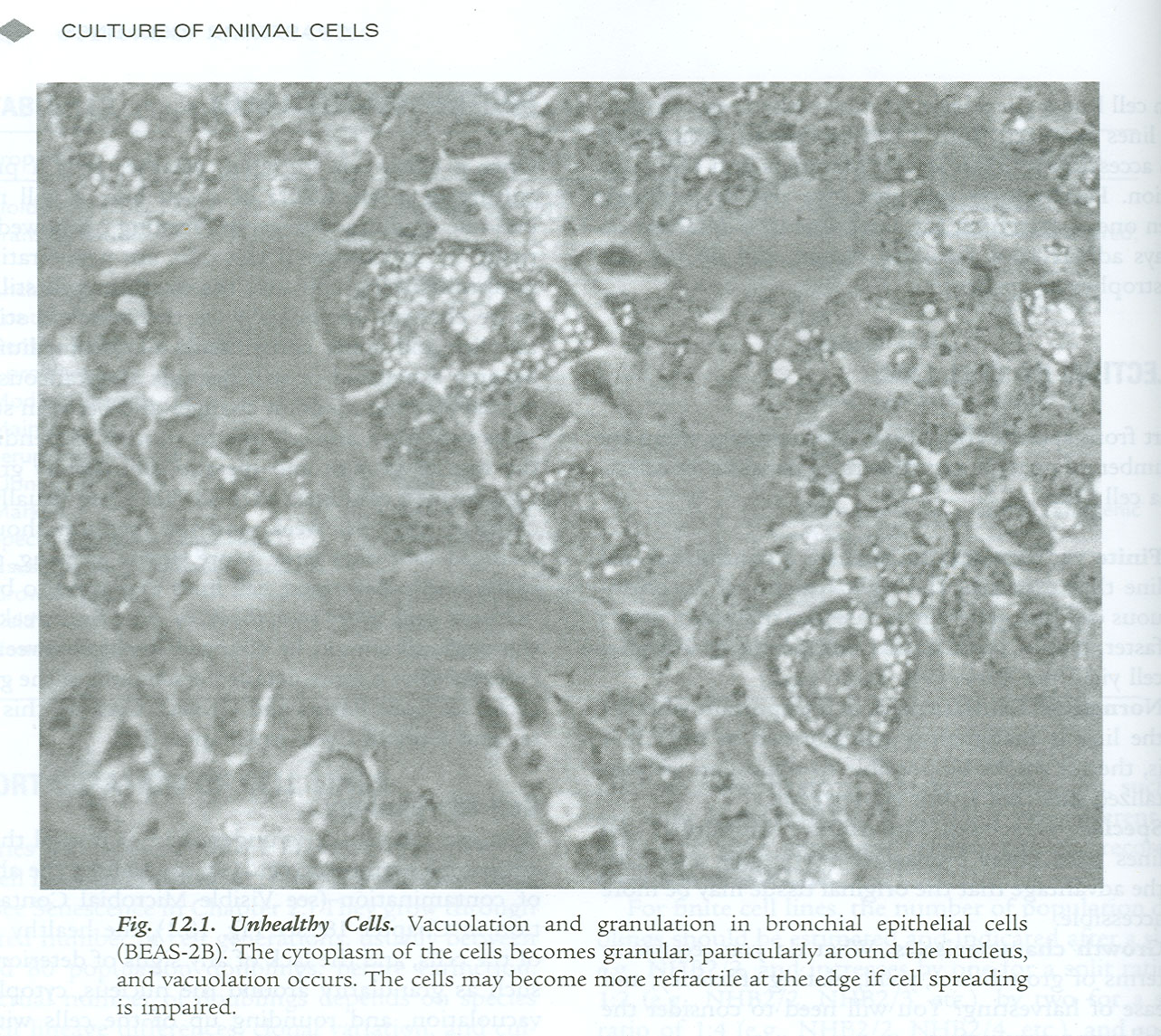
- EYE first: check the medium for turbidity, a fall in pH (phenol red turning yellow), granularity, or detached cells — before you ever touch the microscope.
- Inverted microscope, binocular, phase-contrast. 4x: cell Density, cell-cell interaction, aggregation, detachment.
- 10x and 20x: Health of individual cells — rounding up, contraction of the monolayer, detachment.
- Look for Patterning (normal fibroblastic growth) vs Piling up and random overgrowth (evidence of transformation).
- Check for microbial contamination. Then find Mitoses and estimate their frequency (number per high-power field) — a proxy for how actively the culture is growing.
- Record in the table: % confluency, cell appearance (morphology + health), and media color/clarity for each flask.
- Phenol red is your free pH meter: red-orange = healthy pH ≈7.4; Yellow = acidic (overgrowth or bacterial contamination); Purple/fuchsia = alkaline (mold, or a CO2 problem — incubator door left open, cap loose).
Identifying mitosis and motility
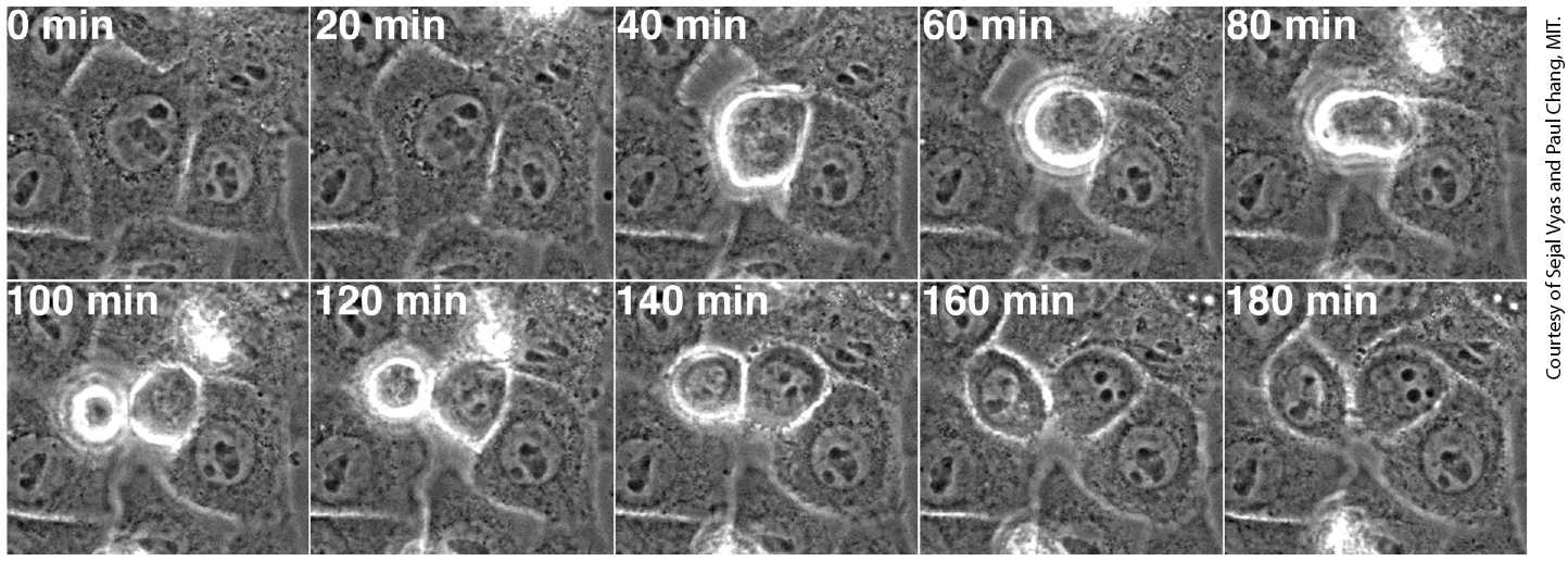
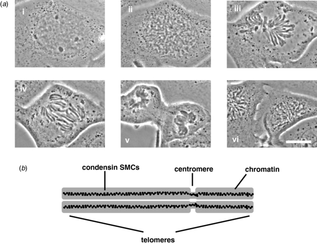
- Mitotic cells Round up and become bright/refractile under phase contrast — they detach partially from the substrate, so they look like shiny spheres sitting on the monolayer. Count these per high-power field.
- You may see the cleavage furrow or paired daughter cells still attached (“doublets”).
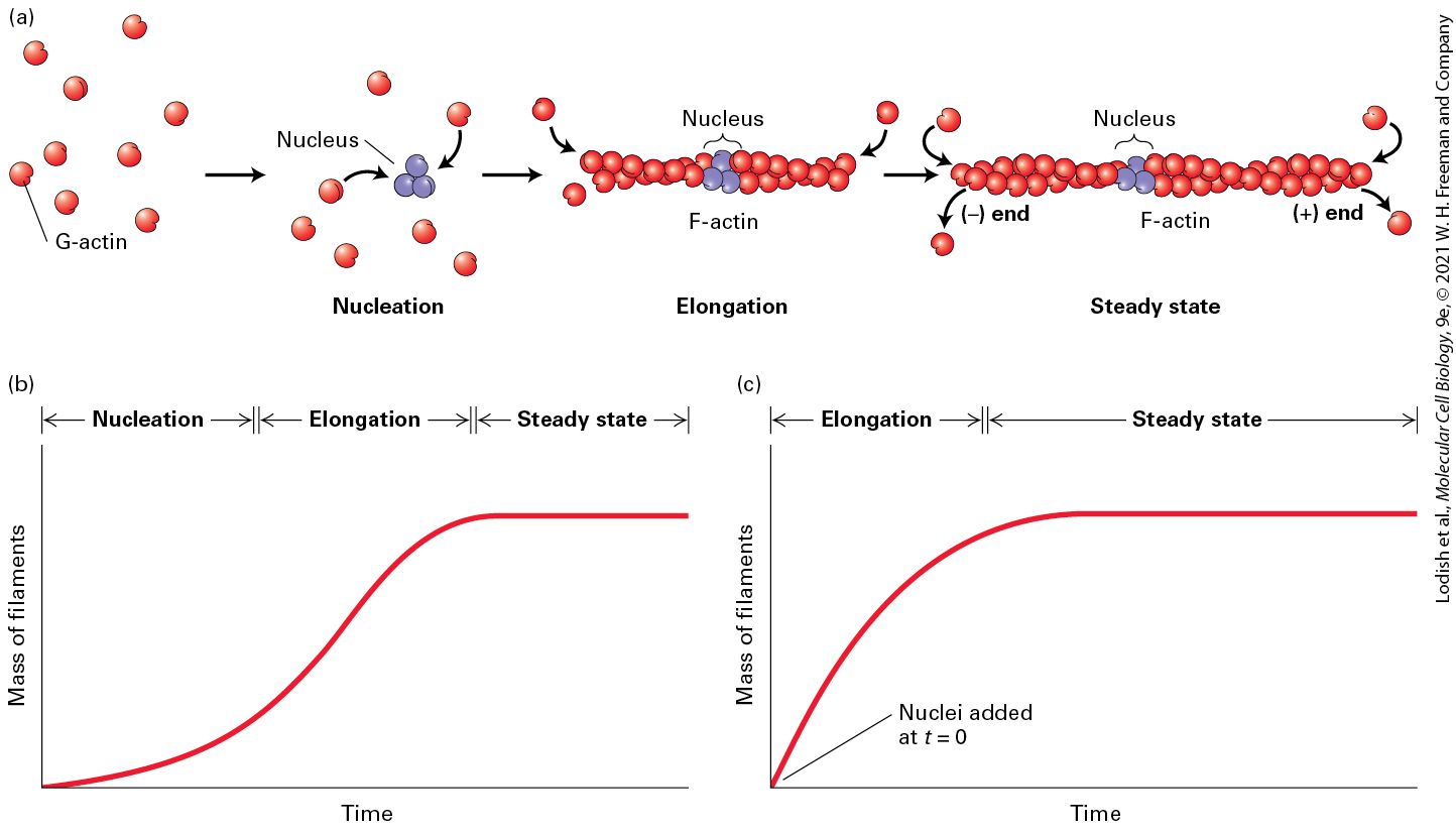
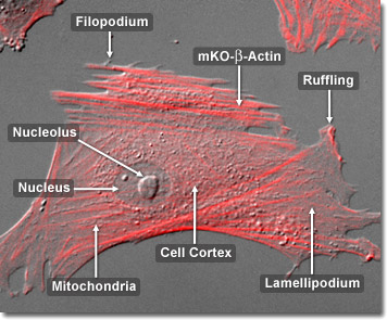
- Motility elements: lamellipodia (broad, sheet-like leading edges) and filopodia (thin spikes) driven by actin — the lecture linked to time-lapse of 3T3 cells and fluorescent actin movies.
Part 3: Identifying contamination — the table to memorize
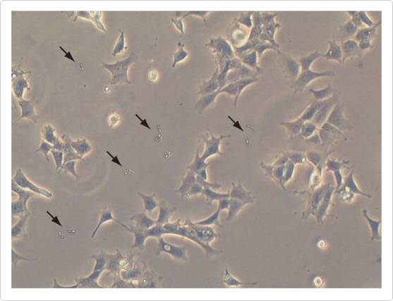
- The three-way memory hook: bacteria Acidify (yellow), mold Alkalinizes (purple), yeast clouds the medium Without the pH drop.
- Mycoplasma — the special case: a bacterial contaminant but <1 um — too small to see by phase contrast, so it hides. It does NOT usually kill the culture; instead it silently changes metabolism, growth rate, behavior, and can even cause Chromosomal aberrations in host cells. That is why it corrupts experiments invisibly. Detected only by fluorescent DNA staining (Hoechst), PCR, ELISA, or similar — and cultures must be tested Periodically.
- An overall decline in cell health can indicate contamination that is NOT visible under phase contrast (lecture slide 9) — if cells look unhappy with no visible cause, suspect mycoplasma.
- Size anchors from the slides: human cells ≈10 um, bacteria ≈1 um, mycoplasma <1 um, yeast larger than bacteria.
Part 4: Aseptic technique
- Definition (memorize): a task performed in a sterile environment in order to avoid introducing outside contamination.
The history the lecture used to frame it
- Two competing ideas had to be settled: Biogenesis (life comes from pre-existing life) vs Spontaneous generation / abiogenesis (life arises from non-living sources).
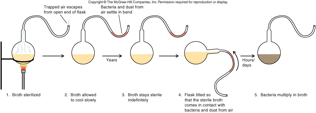
- Pasteur’s swan-neck flask experiment settled it: broth in a flask with an S-curved neck stayed sterile because airborne organisms were trapped in the bend, while air still reached the broth. TILT the flask so broth touches the neck — and it goes cloudy. That tilt is the positive control.
- Exam logic: the tilt is what makes it an experiment rather than a demonstration — it shows the broth was still capable of supporting growth, so sterility was due to exclusion of organisms, not some change in the broth.
- Common cell-culture contaminants named on the slide: bacteria, mycoplasma, yeast, fungal spores.
The two objectives of aseptic technique
- 1) Minimize risk of Contamination: from the operator, the atmosphere, work surfaces, solutions, and equipment. Contamination can be minor (one or two cultures) or catastrophic (wiping out an entire stock).
- 2) Reduce risk to Personnel: minimize exposure of personnel to potentially biohazardous materials.
The five elements of an aseptic environment
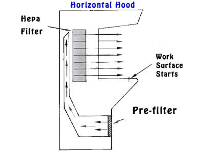
- 1. Laminar flow hood: establishes a “CLEAN-Air space” — it is NOT a sterile environment. Horizontal or vertical types. Keep the work area clear, clean spills immediately, follow sterility protocols strictly.
- The single most quotable line on the slide: anything Inside the hood must be CLEAN; anything Outside the hood is assumed DIRTY. And: “70% ethanol is your friend.”
- 2. Quiet area: minimize traffic, dust, and movement in the hood room.
- 3. Work surface: keep it clean and tidy; only the materials needed for the Current procedure go in the hood; arrange materials around the Periphery (so nothing passes over open vessels).
- 4. Personal hygiene: wash hands before and after; Always wear gloves; tie back long hair; lab coats recommended; wear a face mask if you have a cold (or let your lab partners do the aseptic steps).
- 5. Incubators: “a dangerously microbe-friendly place where we have to maintain our sterile cultures” — warm, humid, CO2-rich: perfect for contaminants too.
Sterile handling — the four habits
- Swabbing/spraying: swab all work surfaces and equipment with 70% alcohol before work; spray gloved hands and EVERY object entering the hood; clean spills immediately with 70% alcohol.
- Capping: all reagents, media, and cultures stay Capped when not in immediate use.
- Handling bottles and flasks: never pass your hands or equipment OVER an open bottle or flask; open sterile bottles only in the hood; minimize touch — “don’t use a bear claw.”
- Pipetting: all liquids transferred via a pipet — never poured.
- Why 70% and not 100% ethanol? 70% ethanol contains water, which lets it penetrate the cell wall/membrane and denature proteins throughout the organism. 100% ethanol dehydrates and fixes the outer surface too quickly, sealing the microbe and leaving the interior alive.
The two hood protocols (know which room you are in)
- Bench-top hood, room 408 (Protocol 1.1)
- Start: gloves on -> turn on fluorescent light + fan -> clear the surface -> spray ALL areas with BDD and Wait three minutes -> wipe with kimwipes -> spray gloves and surfaces with 70% EtOH and wipe. NOW the hood is sterile; everything entering must be washed in 70% EtOH.
- Finish: clear/store all materials, wipe spills -> if you aspirated, run BDD then 70% EtOH through the tubing -> spray + wipe all surfaces with 70% EtOH -> spray with BDD, wait three minutes, wipe -> if LAST group: empty the waste container (pour down the sink with running water), turn off the vacuum pump and the fluorescent light -> gloves off, wash hands.
- Biosafety cabinets, rooms 408 A and B (Protocol 1.2)
- Start: turn on the UV light with the sash DOWN, wait 10 Minutes, turn UV off -> gloves on -> lift the sash (turns on fluorescent light + fan) -> spray gloves and all surfaces with 70% EtOH, clear the area, wipe down. Hood is now sterile.
- Finish: clear/store, wipe spills -> run BDD then 70% EtOH through aspiration tubing -> spray all surfaces Including the uv bulb with 70% EtOH and wipe -> close the sash, UV on for 10 minutes, then off -> if LAST group: empty waste, turn off the vacuum pump -> gloves off, wash hands.
- The two contrasts that make good exam questions: BDD + 3-minute contact time (bench-top) vs UV 10 minutes with the sash down (BSC); and UV must be OFF while you work (it damages skin/eyes and is blocked by the sash anyway).
Part 5: This week’s lab — work these yourself first
- Answers to everything in this section are in Appendix A at the end of the guide. Try each one cold before you look — the point of the lab is to reason from what you saw at the scope, not to recognize an answer.
Lab A — Observation of cultured cells
- Materials: U2OS and HFF cultures (sterile); inverted microscope (nonsterile). Flasks stay Closed.
- Deliverable: Table 1 — Cellular Condition as Assessed by Microscopy, five flasks each of U2OS and HFF, recording density/appearance and media color/clarity.
Thought questions to answer
- Q1. You observed HFF (human foreskin fibroblast) and U2OS (from the bone tissue of a 15-year-old female with osteosarcoma). What morphologic category does each belong to — fibroblastic, epithelial-like, or lymphoblast-like? What visible feature would tip you off that one of them is transformed?
- Q2. Complete Table 1 with your observations of each flask: % confluency, cell appearance (morphology and health), and the appearance of the media (color and clarity).
- Q3. Which culture do you think is “unhealthy” and should be removed from future experiments? Which culture is ready to be subcultured — and what confluency should you passage at, and why not wait for 100%?
- Q4. Cells need a low-oxygen environment to prevent DNA damage, and these flasks sit out for more than 3 hours of lab. Will they still be healthy for future subculture and experiments? Name every variable that drifts while a flask is out of the incubator, and say what should be done differently.
- Space to work / your answers:
Lab B — Aseptic technique and media preparation
- You prepare a T25 flask of D10 media aseptically. A T25 flask holds 5 mL.
- The formula: D10 = 90% DMEM + 10% bovine serum (PBS substitutes for serum in this mock exercise).
- Q5. Calculate the volume of each component you will add to make 5 mL of D10 in the T25 — Before you go to the hood, as the protocol requires.
- Q6. Would your answer change if the protocol instead said “add 10% serum TO 5 mL of DMEM”? Show both numbers and explain the difference. (This is the Week 1 trap in disguise.)
- Then: prepare the BSC, make the flask aseptically, prepare the hood for the next student, label the flask properly, and place it in the incubator.
- The grading test: Dr. Johnson checks your flask in 2–5 days. There should be NO Growth if your aseptic technique was correct — your flask is literally a sterility control on your own hands. Growth = repeat the experiment Thursday during class.
- Space to work / your answers:
Figure conventions (Cell Observation Figure handout)
- You will write results assignments soon, and the Figure is a major part of the grade. From the handout, the four things to notice:
- 1) this is a Compound figure — many images under a single “Figure 1”.
- 2) the resolution in each image is clear.
- 3) each image is sized Without stretching it disproportionately.
- 4) the legend tells you WHAT each image is — without telling you how to Interpret it. Interpretation belongs in the results text, not the legend.
Part 6: The assigned primary papers (paper quiz)
- Paper quizzes are open-note and cover the assigned literature. For each paper: know the question, the model system, the key method, the main result, and the one figure that carries the argument.
1. Kitazawa et al. 2013 (IOVS) — Immortalizing a corneal epithelial cell line lacking TACSTD2 (GDLD model)
- Problem: gelatinous drop-like corneal dystrophy (GDLD) causes amyloid deposits and vision loss; it is caused by biallelic loss-of-function mutations in TACSTD2, and there is no fundamental treatment and no good model (the knockout mouse showed no corneal abnormality).
- Approach: take corneal epithelial cells from a GDLD patient (p.Gln118X nonsense mutation) and immortalize them by Lentiviral transduction of BOTH SV40 large T antigen AND hTERT.
- The immortalization logic — THE key concept for this course: normal human cells stop dividing at ≈40–60 population doublings against TWO barriers — M1 senescence (enforced by RB and p53) and M2 crisis (telomere shortening). SV40 large T binds and inactivates RB and p53 (bypassing M1); hTERT restores telomerase (bypassing M2). Using BOTH genes is far more reliable than either alone.
- Results — immortalization worked: transduced cells exceeded 100 cumulative PDs (nontransduced stopped at ≈19.7); stayed small and square while nontransduced cells became large and flattened; SA-β-gal senescence staining was negative in transduced, positive in nontransduced; TRAP assay showed telomerase laddering comparable to HeLa; stronger colony formation.
- Results — the disease phenotype was preserved: the immortalized GDLD cells had significantly LOWER trans-epithelial resistance (TER = barrier function) and reduced CLDN1 and CLDN7 (claudin) expression, with mislocalized dotted (rather than membrane-bound) staining — matching real GDLD corneas. Other tight-junction proteins (CLDN4, occludin, ZO-1) were normal.
- Gene-therapy pilot: re-introducing wild-type TACSTD2 (≈70% efficiency) restored CLDN1/CLDN7 levels and their membrane localization, and TACSTD2 was shown to bind CLDN1/CLDN7 by immunoprecipitation — but TER was NOT fully normalized (they attribute this to insufficient transduction efficiency).
- Why this paper is assigned in week 2: it is the molecular version of the “finite vs continuous cell line” distinction from Week 1 — Hayflick limit, senescence, telomerase, and the exact genes used to make a continuous line. Expect a quiz question on what SV40 large T does versus what hTERT does.
2. Wong et al. 2004 (BMC Cell Biology) — Second-hand smoke effects on fibroblasts
- Question: second-hand smoke causes abnormal tissue repair, but the Cellular mechanism was unclear. Fibroblasts drive repair by proliferating, migrating, and secreting ECM/cytokines.
- System: sidestream whole (SSW) smoke solutions on primary Chicken embryonic fibroblasts (they behave like wound fibroblasts). Doses were standardized by Nicotine as the biomarker — the 1:9 dilution (≈2.0 ug/mL nicotine) matches tissue levels in passive smokers. Note the paper’s own comparison: second-hand smoke has ≈2x the nicotine, tar, NO and CO of first-hand smoke.
- Dose matters: 1:9 SSW cells became elongated and separated (vs flat, contact-inhibited controls) but did NOT die — shown by recovery in fresh medium, high ATP, normal forward/side scatter by flow cytometry (staurosporine was the positive control for death), and no membrane/nuclear blebbing by acridine orange/ethidium bromide. At 1:4, cells rounded up and died.
- The central paradox and its resolution: cell Number was unchanged but BrdU incorporation FELL — fewer cells were dividing, yet the population held. Stress/survival proteins rose: cIL-8 (dose-dependent), PKB/Akt phosphorylated by 5 minutes, grp78 (ER stress), p53 and p21. Conclusion: SSW increases Survival while suppressing division.
- Cytoskeleton and migration: increased F-actin/stress fibers and MORE focal adhesion plaques (vinculin up by imaging and immunoblot) -> increased adhesion -> Decreased migration in the cloning-ring assay.
- Endomembrane system: vacuoles within 3–4 hours; TEM showed dilated, irregular ER while mitochondria and nucleus looked normal; DIOC6 showed fragmented perinuclear ER; β-COP staining showed Golgi breakdown; secreted cIL-8 was mislocalized. Critically, Mainstream smoke (MSW, “first-hand”) did NOT do this — the effect is specific to sidestream — and nicotine alone reproduced the endomembrane effect.
- Clinical logic: cells that survive but cannot migrate accumulate at the wound edge -> delayed healing, plus build-up of connective tissue -> fibrosis and excess scarring.
3. Hart, Fischer & Ullrich 2004 (Cancer Research) — Cannabinoids induce cancer cell Proliferation
- Question: cannabinoids are used to treat nausea/pain/appetite in cancer patients and were reported to KILL tumor cells — so do they help or hurt?
- System: THC, anandamide (AEA), HU-210, Win55,212–2 on NCI-H292 (lung), SCC-9 (squamous), 5637 (bladder), U373-MG (glioblastoma), 1321N1 (astrocytoma), A498 (kidney).
- The mechanism (the whole point): cannabinoid binds CB1/CB2 (Gi/o-coupled GPCRs) -> activates TACE/ADAM17 (a metalloprotease) -> sheds proAmphiregulin and/or proHB-EGF from the surface -> those ligands activate EGFR (and HER2/neu) -> ERK1/2 and Akt/PKB signaling -> Proliferation. This is “GPCR transactivation of a receptor tyrosine kinase.”
- Evidence: EGFR phosphorylation within 3 minutes; blocked by AG1478 (EGFR kinase inhibitor) or BB94/batimastat and TAPI (metalloprotease inhibitors); CB1-specific (ACEA) and CB2-specific (BML-190) agonists both worked; siRNA against TACE confirmed the protease.
- The dose-dependent punchline: Nanomolar thc (100–300 nM — comparable to patient serum levels) Increased proliferation by [3H]thymidine incorporation and MTT; only much Higher concentrations induce apoptosis. So the dose determines whether cannabinoids kill or feed the tumor.
- Method note for your own project: [3H]thymidine incorporation and MTT both measure proliferation/viability — MTT works by mitochondrial dehydrogenase reducing yellow tetrazolium to purple formazan, which is exactly the logic of the Alamar Blue assay you will run (resazurin -> resorufin).
4. Okamoto & Shikano 2017 (Mol Biol Cell) — Differential phosphorylation controls GPR15 endocytosis
- Question: GPR15 is an Orphan gpcr (an HIV/SIV co-receptor and a T-cell homing receptor for the colon; implicated in colitis). Cell-surface density of a GPCR sets signaling strength, so what controls its internalization?
- Key finding 1: GPR15 undergoes Constitutive endocytosis with NO ligand — ≈35% internalized by 60 min in HEK293 cells, versus only ≈8% for β-2 adrenergic receptor. Serum starvation did not change it (arguing against a serum-derived ligand). About half of internalized receptor Recycles back to the membrane.
- Key finding 2: the endocytosis is Clathrin-dependent and partially β-arrestin-dependent.
- Key finding 3 (the title): mutating distal C-terminal Arg-354 or Ser-357 to Ala markedly Reduced endocytosis, but the phosphomimetic S357D did NOT — so it is Phosphorylation of Ser-357 (by basophilic kinases such as PKA and PKC) that drives internalization. Activating those kinases pharmacologically increased both Ser-357 phosphorylation and endocytosis.
- Key finding 4: this Ser-357 route is Distinct from a proximal Ser/Thr cluster responsible for the β-arrestin/GRK-dependent route — two separable phosphorylation signals tuning surface density.
- Connect it to Dr. Johnson’s own work: her Cx43 paper showed phosphorylation at Ser-279/Ser-282 controls connexin43 Endocytosis and gap-junction assembly. Same principle, different protein: phosphorylation of specific C-terminal serines is a switch for membrane-protein internalization. Expect her to draw that parallel.
Condensed review — most likely to be tested
- Three morphologies: epithelial-like (polygonal sheets), fibroblastic (spindle, patterned), lymphoblast-like (spherical, suspension). Judge at Confluence — low density makes epithelial cells look fibroblastic.
- HFF = fibroblastic normal finite line; U2OS = epithelial-like transformed continuous line (osteosarcoma, 15-year-old female).
- Contamination table: bacteria cloudy + pH DROP; yeast cloudy, NO pH drop, budding; mold turbid + pH RISE, hyphae; virus nearly invisible and dangerous.
- Mycoplasma: <1 um, invisible by phase contrast, does not kill but alters metabolism/growth/behavior and causes chromosomal aberrations; detect by fluorescent DNA stain, PCR, or ELISA — test periodically.
- Aseptic technique = task performed in a sterile environment to avoid introducing outside contamination; two objectives = protect the culture AND protect the person.
- A laminar flow hood makes a CLEAN-Air space, not a sterile environment. Inside = clean; outside = dirty. Materials around the periphery; never reach over an open vessel.
- 70% ethanol beats 100% because water lets it penetrate and denature proteins instead of fixing the surface.
- Bench-top hood: BDD + 3 minutes. BSC (408 A/B): UV 10 minutes with the sash DOWN, before and after.
- D10 in a 5 mL T25 = 4.5 mL DMEM + 0.5 mL serum/PBS; no growth in 2–5 days = you passed the aseptic test.
- Passage at ≈80–90% confluence, not at 100%.
- SV40 large T inactivates RB and p53 (bypasses M1 senescence); hTERT restores telomerase (bypasses M2 crisis) — together they immortalize (Kitazawa).
- SSW smoke: survival UP (cIL-8, Akt, grp78, p53, p21), division DOWN (BrdU), adhesion UP (F-actin, vinculin), migration DOWN, ER/Golgi disrupted — and MSW does not do it (Wong).
- Cannabinoids: CB1/CB2 -> TACE/ADAM17 sheds proAR/proHB-EGF -> EGFR transactivation -> ERK + Akt -> proliferation at Nanomolar thc; apoptosis only at high dose (Hart).
- GPR15: constitutive, clathrin-dependent endocytosis; phosphorylation of Ser-357 (PKA/PKC) drives it; ≈half recycles (Okamoto) — the same phospho-switch logic as Johnson’s Cx43 Ser-279/282 work.
Mnemonic set
- “Confluent tells the truth” — morphology is only diagnostic in a full dish."
- “Bacteria sour it, mold sweetens it, yeast just clouds it” — pH direction by contaminant."
- “Mycoplasma: too small to see, big enough to ruin the data.”
- “Inside clean, outside dirty” — the hood rule."
- “70 beats 100” — ethanol needs water to work."
- “Large T kills the brakes, hTERT rewinds the clock” — SV40 large T (RB/p53) + hTERT (telomeres)."
- “Survive but do not move” — the second-hand smoke fibroblast phenotype."
- “Low dose feeds, high dose kills” — THC on cancer cells."
Self-test
- You view a flask at 20% confluence and the cells look spindle-shaped. Your partner calls them fibroblasts. Why should you wait?
- A flask has cloudy medium that has turned yellow. Name the most likely contaminant and the two observations that support it.
- A culture looks slightly unhealthy — slower growth, odd morphology — but the medium is clear and phase contrast shows nothing. What do you suspect, and how do you test it?
- Explain why the tilted flask is essential to Pasteur’s swan-neck experiment.
- You need 5 mL of D10 in a T25. Give the volumes, and then give the answer if instead you were told to add 10% serum TO 5 mL of DMEM.
- Dr. Johnson checks your practice flask 3 days later and it is cloudy. What does that mean and what happens next?
- A collaborator immortalizes cells with hTERT alone and it fails. Using Kitazawa’s reasoning, explain why, and what to add.
- SSW-smoke-treated fibroblasts show unchanged cell number but reduced BrdU incorporation. Resolve the apparent contradiction and name three proteins supporting your answer.
- Why does the smoke paper argue that decreased migration — not cell death — explains poor wound healing?
- A patient takes THC for chemotherapy-induced nausea. Using Hart et al., state the concern and the mechanism.
- How would you show experimentally that EGFR transactivation — not direct CB signaling — drives that proliferation?
- GPR15 internalizes without any ligand. Give the evidence and the phospho-switch that controls it.
Show answer key — try the questions first
- At low density epithelial cells can resemble fibroblasts (lecture slide 5) — morphology is most distinguishable in a Confluent dish. Re-evaluate near confluence or use another criterion (patterning, junction formation, origin of the line).
- Bacteria: cloudy/flocculated medium PLUS a drop in pH (phenol red to yellow) from fermentative acid production. Under the scope you would see ≈1 um motile dots between the ≈10 um cells; classify as cocci or rods.
- Mycoplasma (<1 um, invisible by phase contrast). It does not kill cells but alters metabolism, growth, behavior, and can cause chromosomal aberrations. Test by fluorescent DNA staining (Hoechst/DAPI), PCR, or ELISA — and test cultures periodically, not just when suspicious.
- It is the positive control: tilting lets the broth contact the trapped organisms in the neck bend, and the broth then goes cloudy. This proves the broth could still support growth, so sterility in the upright flask was due to physical exclusion of airborne organisms — not to something about the broth or the air.
- 5 mL of D10 = 4.5 mL DMEM + 0.5 mL serum (10% OF the total). If adding serum TO 5 mL DMEM: the DMEM is the 90%, so total = 5/0.9 = 5.56 mL and you add 0.56 mL serum — the Week 1 trap.
- Your aseptic technique failed — something non-sterile entered the flask (unsprayed object, reaching over an open vessel, uncapped reagent, or contact of the pipet with a nonsterile surface). Per the protocol you repeat the exercise Thursday during class.
- hTERT only bypasses M2 (telomere crisis). The M1 barrier — senescence enforced by RB and p53 — still stops the cells. Add SV40 large T antigen, which binds and inactivates RB and p53. Kitazawa used both precisely because starting cell numbers were limited and single-gene efficiency is low.
- Fewer cells are dividing, yet the population is maintained because Survival increased. Supporting: cIL-8 (dose-dependent stress response), phospho-PKB/Akt (survival, by 5 min), grp78 (ER stress), plus p53 and p21 (cell-cycle arrest with survival). Net phenotype: arrested but alive.
- At tissue-relevant doses (1:9) cells survive (ATP high, normal scatter, no blebbing, full recovery in fresh medium) but increase F-actin stress fibers and vinculin-positive focal adhesions, gluing them down; the cloning-ring assay shows reduced migration. Cells that cannot migrate pile at the wound edge, so granulation tissue never forms — and surviving, non-migrating fibroblasts deposit matrix, producing fibrosis and excess scarring.
- At nanomolar serum-relevant concentrations, Thc accelerates tumor cell proliferation: CB1/CB2 -> TACE/ADAM17 -> shedding of proAmphiregulin/proHB-EGF -> EGFR (and HER2/neu) transactivation -> ERK1/2 and Akt/PKB. Apoptosis occurs only at much higher concentrations — so the therapeutic dose may sit in the pro-proliferative window.
- Block the two required steps and lose the effect: AG1478 (EGFR kinase inhibitor) and batimastat/BB94 or TAPI (metalloprotease inhibitors) abolish EGFR phosphorylation, ERK/Akt activation, and thymidine/MTT proliferation; siRNA knockdown of TACE/ADAM17 does the same. Receptor-subtype agonists (ACEA for CB1, BML-190 for CB2) confirm the upstream input.
- Evidence: ≈35% internalized at 60 min vs ≈8% for beta2AR; serum starvation does not change the rate (no serum-derived ligand); it is clathrin-dependent and partly β-arrestin-dependent, with ≈half recycling. Switch: phosphorylation of C-terminal Ser-357 (by basophilic kinases including PKA/PKC) — S357A blocks endocytosis while phosphomimetic S357D does not, and activating the kinases increases both phosphorylation and internalization.
Appendix A — Worked answers to the Part 5 lab questions
- Do not read this until you have written your own answers. Compare, then note where your reasoning differed — that gap is the actual studying.
Lab A — Observation of cultured cells
- Q1 — morphologic category
- HFF = Fibroblastic: bipolar/multipolar spindle cells growing in swirled, parallel arrays, contact-inhibited at a monolayer (a normal, finite diploid line). U2OS = Epithelial-LIKE: flat polygonal cells in a cobblestone sheet (a continuous, transformed osteosarcoma line). The tip-off for transformation is Piling up and random overgrowth — loss of contact inhibition — instead of the orderly patterning a normal fibroblast culture shows. Remember the density caveat: at low confluency epithelial cells can look fibroblastic, so judge near confluence.
- Q2 — the table
- For each flask record: (a) % confluency — estimate the fraction of the growth surface covered at 4x; (b) morphology and health — spindle vs polygonal, patterned vs piled, plus any rounding up, granularity, monolayer contraction, or detachment; (c) media color and clarity — red-orange and clear is healthy; yellow means acidic; purple means alkaline; cloudy or flocculated means contamination. Also note mitoses per high-power field as a growth-activity proxy.
- Q3 — unhealthy vs ready to subculture
- The Unhealthy flask is the one with rounded and detached cells, granularity, a contracted monolayer, and turbid or yellowing medium — remove it from future experiments. The flask Ready to subculture is at roughly 80–90% confluence with healthy morphology and clear red-orange medium. You passage Before 100% because confluent cells stop dividing (contact inhibition), exhaust nutrients, acidify the medium, and begin detaching and dying — so a fully confluent flask gives you a stressed, partly dead population rather than a healthy exponential one.
- Q4 — three hours out of the incubator
- No, not reliably. Four variables drift the moment a flask leaves the incubator: (1) CO2 — the bicarbonate buffer loses CO2 to room air, so pH climbs and the medium turns purple/alkaline; (2) Temperature — it falls away from 37 °C, slowing metabolism and division; (3) Oxygen — room air is ≈21% O2 versus the lower-O2 environment used to limit DNA damage, so oxidative stress rises; (4) Evaporation and repeated handling/light exposure on the scope stage. What to do differently: shorter viewing shifts with flasks returned to the incubator between groups; prepare duplicate or sacrificial sets so no flask sits out the entire period; use HEPES-buffered medium (or properly sealed caps) to hold pH without CO2; keep flasks on a warmed stage; and simply do not re-use the observation flasks for downstream experiments.
Lab B — Aseptic technique and media preparation
- Q5 — D10 for a 5 mL T25
- 10% of 5 mL = 0.5 mL serum (PBS in this mock exercise); 90% of 5 mL = 4.5 mL DMEM. Total 5.0 mL.
- Q6 — would “add 10% serum TO 5 mL DMEM” change it?
- Yes. In that phrasing the 5 mL of DMEM is the 90% portion, so the FINAL volume is 5 / 0.9 = 5.56 mL and you add 0.56 mL of serum. The difference: taking 10% OF a specified total (0.5 mL) versus adding enough serum to make 10% of the resulting larger volume (0.56 mL). The Week 1 practice sheet used the second phrasing (500 mL media -> add 55 mL, not 50 mL) — read which volume is being specified before you calculate.
- Contaminant | Medium appearance | pH change | Microscopic ID
- Bacteria | Cloudy / flocculated | DROP in pH (yellow) | Tiny motile dots between cells (≈1 um vs human cells ≈10 um); distinguish cocci (spheres) vs rods
- Yeast | Cloudy | NO pH drop (until heavy) | Larger than bacteria; ovoid; characteristic Budding and chains/arrangement
- Mold (fungal) | Turbid | Increased pH (alkaline) | Filamentous hyphae, often visible to the naked eye as fuzzy mats/spores
- Virus | Often normal-looking | Variable | Very difficult to ID given size; if present, potentially Dangerous to the operator
This first lecture does three things at once: it defines what cell biology is and why the cell is the right unit of study, it hands you the century of cell-culture history that every later technique stands on, and it frames the course itself — culture technique, cytotoxicity, gene expression, and a final project you design. Do not treat the history as trivia: every date on the timeline is a capability the field gained, and the exam asks for capabilities.
Lecture 02 — The Class Session in Full (Aug 27)
- Everything Dr. Johnson said in class, reconstructed from the recording and matched to her slides. Lines marked PROF are the points she stressed out loud; Quiz alert marks what she explicitly said would be tested.
- Quiz alert — She said there are about TWO questions on Tuesday’s quiz drawn from the gap-junction / pancreatic cancer story in Part 12 below. That story is the single highest-value section of this lecture.
Part 8: Course logistics she covered first
Discussion post and groups
- She read every discussion post and used them to see where people’s interests are.
- PROF: What she is most excited about is students Replying to each other. If you have not replied to another student in that discussion post, do it by the end of this week.
- Lab groups are still forming. Monday lab groups may be Merged where groups are only two people, so everyone can finish and get to their next class or work.
- Eventually there will be group pages in Canvas and you will sign up BY GROUP — one person per group signs up for the whole group.
Hood appointments (this is how every week will run)
- Appointments are published in Canvas as a calendar; she opens them on Thursdays, then confirms groups afterward.
- First hood session: Monday the 31st, 1:00 PM until about 4:00 PM, in AH 408. Four time slots, roughly 45 minutes in the hood per group.
- ONE student per group signs up. It takes about five minutes to do.
- PROF: Signing up for a slot is about Accountability, not a lock-out: it does not mean you cannot come earlier — it means your group owns that window. She would like a buffer slot left open because groups run over.
- If a signup disappears after you make it, that is a device quirk — she opens the appointment details and confirms who is in each slot.
- PROF: Use Canvas in a Browser, not the phone app — she deleted the app from her own phone.
- Next week’s hood exercise is deliberately simple: “you are just moving liquids” — transferring a few mL from a tube to a flask and half a mL from another.
- PROF: Do the MATH before class. She will post a unit-conversion table, but the calculations are review of what you already learned — come ready.
Extra credit (two options, due by Week 8)
- Option 1 — a FIVE-Minute presentation teaching the class about something published within the last 10 years. It must be in a Cell system.
- Option 2 — a paper-discussion Podcast recorded by a small group on a paper from the last 10 years. Last year’s group used the library’s creative resources and sound booth, added theme music, and dramatized the conversation.
- PROF: Keep the language clean — she listens to these in the car and does not want her kids repeating them. A word or two is fine; a whole recording of it is not usable.
- She notes the due date for the extra-credit assignment was Updated in Canvas — check the new date.
Honors contract / graduate students, and how she wants papers read
- Honors-contract and graduate students: pick a topic you care about (her example: GLP-1), find 10–12 papers on it, write a summary and a Critique.
- PROF: A critique is NOT “I liked the paper.” It is connecting papers: “I understand why they did this, because Smith et al. showed the same thing in a different model system.”
- Her reading method for anyone: set aside a day, find five papers on one topic, read them all at once, then ask how they connect and who disagrees with whom.
- Then follow the citation trail BOTH directions: look at what a paper cited, and then ask PubMed “who cited this paper?” and move forward in time.
- PROF: Why publish and go to conferences: not to be cool — to get Answers. Arguments at conferences are unpleasant but they are how you find the right direction.
Part 9: The two questions that define cell biology
- PROF: Her PhD and postdoc mentors asked the same two questions about everything: What is the structure? What is the function? Any cell-biology topic boils down to those two.
- The smallest unit of life is the cell — but knowing that is useless unless you know the cell’s structure and how it is functioning.
- In this class you are not just learning cell biology, you are learning to be a cell biology Experimenter: using the cell model system to answer questions you have.
In vitro vs in vivo (she quizzed the room on this)
- in vitro literally means “in glass” (vitra = glass). We actually work in Plastic — so the real meaning is: Outside the body.
- in vivo means in the living organism.
- PROF: The intellectual honesty point she stressed: when you study a cell in culture you are making a LEAP — from cell, to tissue, to organ, to system, to disease. A good cell biologist stays aware they are making that leap.
- What culture buys you: you can see the cell’s structure, how it functions in that microenvironment, and how it responds to stimuli you design to mimic what the body would do to it.
Organelles, and what counts as “living”
- Organelles are Subcellular structures. On their own they are not living.
- Mitochondria and chloroplasts have their Own dna — which makes it tempting to call them alive — but they cannot live or reproduce themselves independently.
- PROF: Her number: roughly 70% of mitochondrial gene expression comes from the Nucleus’s DNA, even though the mitochondrion has its own genome.
- Her working definition of living (borrowed from how she teaches microbiology): it must be able to “breathe” — metabolize — AND it must carry the material/information to synthesize another version of itself.
- Viruses therefore are NOT living: the gene cassette is tiny (on the order of a couple of proteins), everything else comes from the host, and it has no energy of its own to build another one.
Part 10: How she told the history of cell culture
- We have been looking at cells in culture since about the 1960s — arguably earlier. (Her aside: “the 1900s, as my children say.”)
- 1960s :: fibroblasts and lung cells were used to understand Viruses and to make Vaccines — you cannot culture a virus without cells.
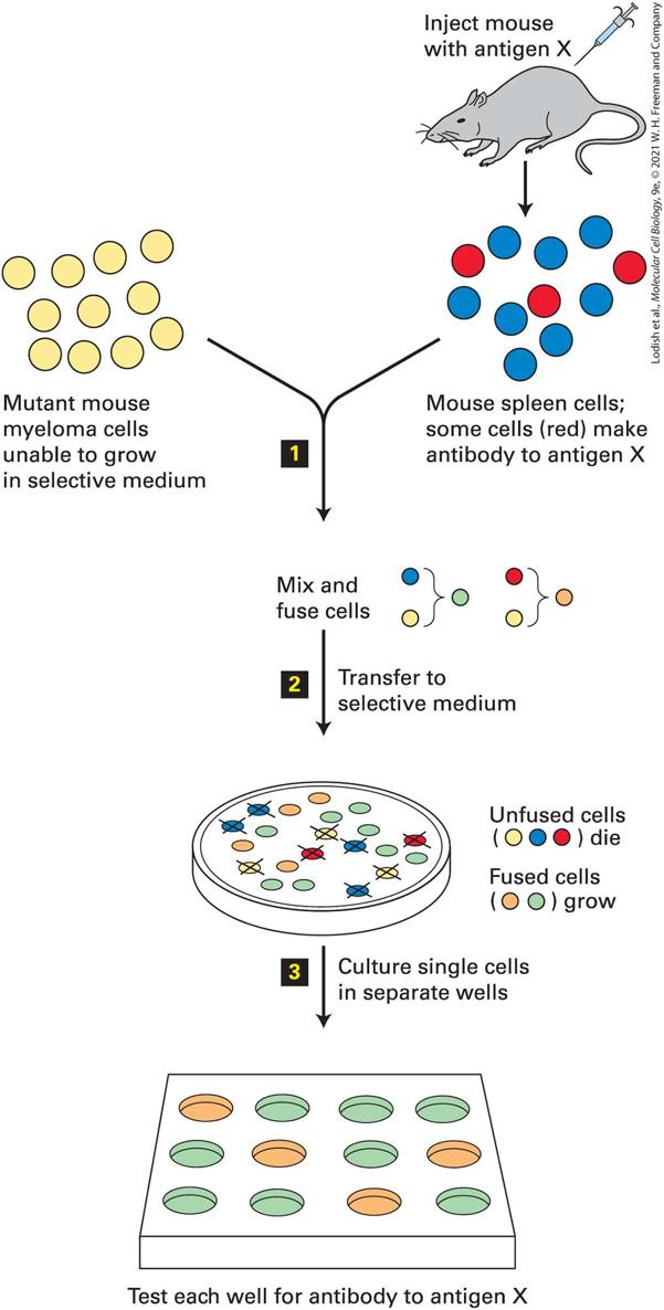
- 1970s :: Hybridomas — fuse an immune (B) cell with a cancer cell so it grows continuously and keeps producing antibody. Her postdoc adviser actually built a hybridoma facility.
- PROF: Monoclonal vs Polyclonal: monoclonal antibodies all target ONE antigen — one structure/epitope. Polyclonal antibodies target multiple antigens. That specificity is why pregnancy tests and diagnostics work.
- 1980s :: we discovered how to Immortalize cells so they grow continuously for years. She said there will be a whole talk on what immortalization means — and the Kitazawa paper you were assigned is exactly that.
- HeLa and The Immortal Life of Henrietta Lacks came up as the resource for that story — and she noted there are OTHER ways to immortalize a cell line besides being a tumor.
- What culture has let us do since: read gene sequences, locate proteins inside cells, harvest molecules, do subcellular fractionation — “really get a close picture of what is happening.”
Applications — her commentary on the list
- Cells as little Factories for biochemistry — the biotech application.
- PROF: Her example: people with growth-hormone deficiency once had to get growth hormone from the Pituitary glands of cadavers — somebody had to die for the product. Cell culture replaced that.
- PROF: Why not just use bacteria? Prokaryotes do not do post-Translational modification the same way. That is why the mammalian cell-culture system matters — you get authentic HUMAN products out of human cells.
- The same logic works for animals: a dog cell line gives you dog products.
- The full application list she ran through: model systems, toxicity testing, cancer research, virology, cell-based manufacturing, genetic counseling, genetic engineering, and drug screening.
- PROF: What she wants from you: not just to taste these applications, but to look for NEW applications cells could move toward — that is the spirit of the final project.
Part 10.5: Plain-English primer — read this before the story
- If the vocabulary in the next two sections is new, read this page first. Everything below is the same content in ordinary language, with the technical word attached so you can recognize it when she says it in class.
The cell as a factory (this analogy carries the whole lecture)
- Nucleus = the office that holds the master blueprints (your DNA). Blueprints never leave the office.
- Gene -> RNA = a photocopy of ONE blueprint page, sent out to the factory floor so the office keeps the original safe.
- Endoplasmic reticulum (ER) = the assembly-and-folding room. A protein is built as a chain and folded here into its 3-D shape. Think “the shape room.”
- Golgi apparatus = shipping and finishing. It decorates the protein (adds sugars) and puts an address label on it saying where in the cell it should go.
- Post-translational modification = the decorating step — changes made to a protein AFTER it is built (sugars, phosphate groups). Bacteria cannot do this the way we do, which is why human proteins are made in human cells.
- Vesicle = a delivery bubble — a little sphere of membrane that carries cargo from one place in the cell to another. A truck.
- Exocytosis = “exo” = out. The truck fuses with the outer wall and delivers its cargo to the cell surface. Delivery OUT.
- Endocytosis = “endo” = in. The outer wall pinches inward and swallows something into a bubble. Pickup IN. (This one word is the heart of her research story.)
- Lysosome = the incinerator — an acidic bag of enzymes that destroys worn-out material. Where big internalized junk gets sent.
- Proteasome = the paper shredder for single proteins — especially misfolded ones.
- Plasma membrane = the outer wall of the cell, made of a double layer of fat (lipid).
- Transmembrane protein = a machine or doorway built INTO that wall, so part of it sticks outside the cell and part sticks inside. Because it touches both sides, an event outside can change what happens inside.
- Cytoplasm = the goo filling the cell, where everything floats.
- Cytoskeleton = the internal scaffolding and rails. ACTIN is one kind — a rope that assembles and falls apart quickly, which is why things anchored to actin can change fast.
- Second messenger = a small signal molecule floating inside the cell that passes a message along — the internal “pass it on” note.
Gap junctions in plain English
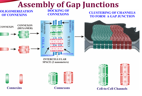
- Picture two neighboring houses (two cells) that want to talk. They build a Tunnel between them, and each house builds half of it.
- Connexin = one brick — a single protein shaped like a cylinder. (Cx43 just means “connexin, 43 kilodaltons in size” — the number is its weight, not a rank.)
- Connexon = half the tunnel — SIX connexins arranged in a ring with a hole down the middle. Also called a hemichannel (“hemi” = half).
- Docking = the two halves — one from each cell — meeting in the gap between the cells and locking together to make one continuous tunnel.
- Plaque = many tunnels parked side by side in one patch of membrane, like a cluster of straws.
- What travels through: small molecules under about 1500 daltons — ions and signal molecules. That is literally how neighboring cells “talk” and stay coordinated.
- Why cancer cares: if the tunnels never form, the cells stop coordinating — including signals like “stop dividing” or “time to die.” That is the whole point of her research story.
- Her naming joke, so it does not trip you up: the Cylinder is a connexIN and the TUBE is a connexON. One letter apart, on purpose, and yes it is confusing.
The lab techniques she mentions, in plain language
- Fixed and stained (immunofluorescence) = the cells are killed and preserved, then treated with antibodies that stick only to one chosen protein and carry a glow-in-the-dark dye. Under the microscope: blue is usually the nucleus (a DNA dye), and red or green is whichever protein they are hunting.
- Cell line = a population of cells taken from one person or animal, kept alive and dividing in plastic dishes. BxPC3 and Capan-1 are two pancreatic cancer cell lines from real patients.
- Site-directed mutagenesis = deliberately editing a few letters of a gene so the protein is built with specific amino acids changed or removed — a way to ask “what does THIS piece do?”
- Transient transfection = putting DNA into cells temporarily — it is not added to the cell’s own genome, so it fades out over a few days as cells divide.
- Retroviral infection = using a virus to permanently paste DNA into the cell’s own genome — so it stays, and every daughter cell inherits it.
- Soluble vs insoluble fraction = dissolve the cells with detergent. Loose, free-floating protein dissolves (soluble). Protein locked into an assembled junction does NOT dissolve (insoluble). So protein moving from the soluble to the insoluble pile is chemical proof that junctions actually assembled.
- Sucrose treatment (hypertonic sucrose) = sugar-loaded medium that prevents the clathrin cage from forming — a way to switch off one specific route of endocytosis.
- Clathrin = a protein that builds a soccer-ball-shaped cage around a patch of membrane to pull it inside. One of the two main “doors in.”
- Lipid raft = a thicker, cholesterol-rich patch of the membrane that acts as a different doorway in. The other main route.
- Transferrin / cholera toxin = known “tracer” molecules that each use ONE specific door in. If your block worked, the tracer gets stuck outside. They are controls that prove the experiment did what you claimed.
- Adapter protein = the middleman that reads an address label on the cargo and hands it to clathrin. No adapter, no pickup.
- Tyrosine motif (PY, YKLD) = a short address label written in the protein’s tail. “Y” is the one-letter code for the amino acid tyrosine. Adapters read this label as “bring me inside.”
- Kinase = an enzyme that sticks a phosphate group onto a protein — a common on/off switch. (This is why the papers on phosphorylation matter.)
- Apoptosis vs necrosis = apoptosis is the tidy, programmed suicide of a cell; necrosis is messy, uncontrolled death from injury. She wants you to be able to ask which one your treatment causes.
The one framing sentence to carry
- Structure = what a thing looks like and is made of. Function = the job it does. Her whole lecture is: the connexin’s structure (where its address labels sit in the tail) determines its function (whether it stays at the surface and lets cells talk).
Part 11: Junction architecture — the background for her story
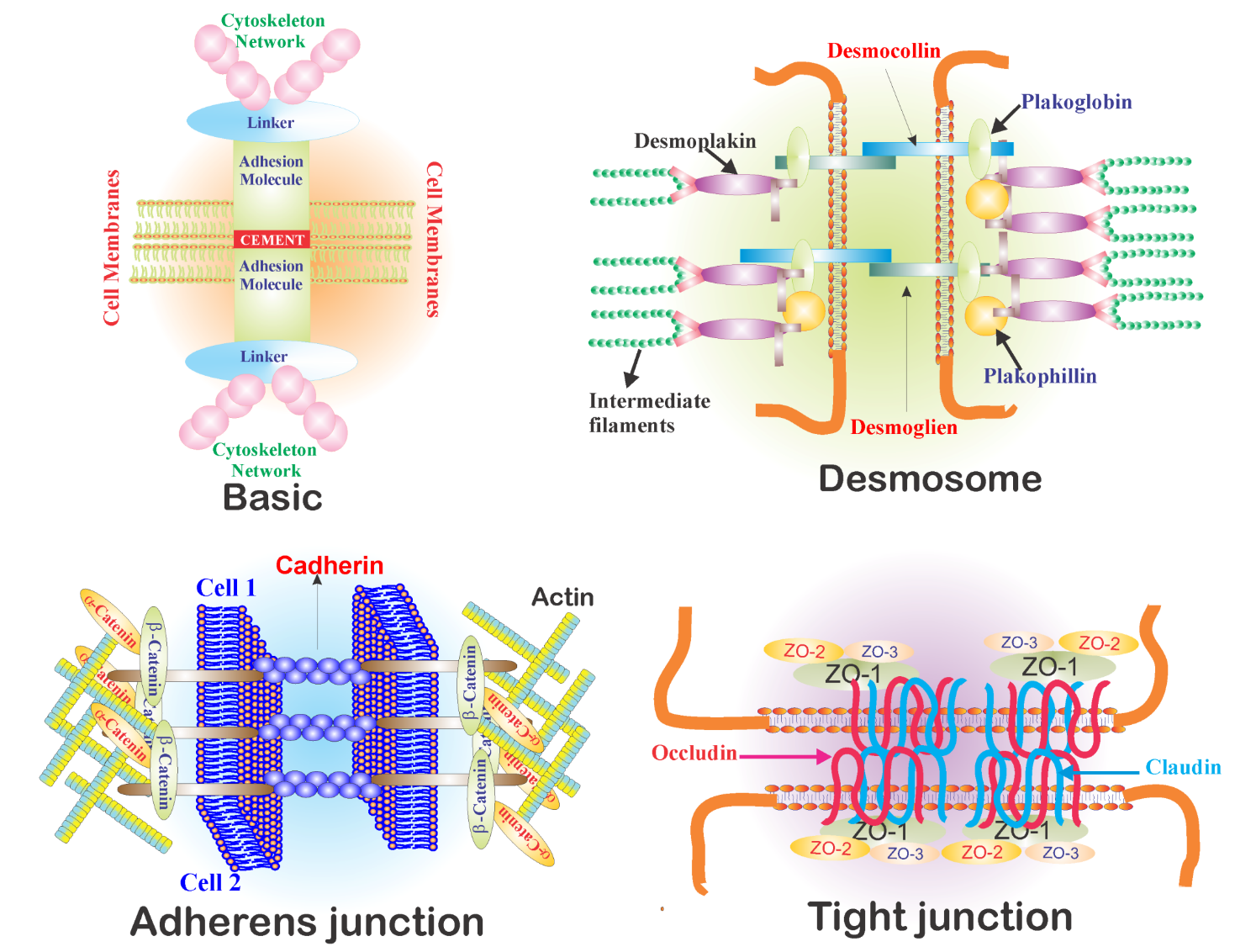
- The junction family she put on the board: TIGHT junctions, Adherens junctions, Desmosomes, GAP junctions, plus Hemidesmosomes.
- The universal architecture: two cells come together; there is an Integral membrane protein in each cell; for any junction to work those proteins must “cement” — make a Protein-Protein interaction on the Outside of the cell.
- PROF: Transmembrane proteins touch the outside AND the inside of the cell — and they never do nothing on the inside. Outside binding changes the conformation, and that change drives interactions inside the cell (receptor-mediated signaling), ending in big responses: a gene opens for expression, a lysosome acts, and so on.
- On the inside, junctions connect to the cytoskeleton through Linker proteins. ZO-1 turns up at essentially every junction type, though there are others.
- PROF: What makes a linker a linker: it connects the transmembrane protein to a Cytoskeletal element — something that can stabilize that protein or put it on the move.
- Adherens junctions attach ACTIN filaments. Actin is dynamic — it forms and falls apart quickly (globular subunits linking into a filament).
- PROF: Her point for the story: Gap junctions also mostly interact with actin — so quick, easy changes can happen to them.
Connexin structure — “we are becoming biochemists here”
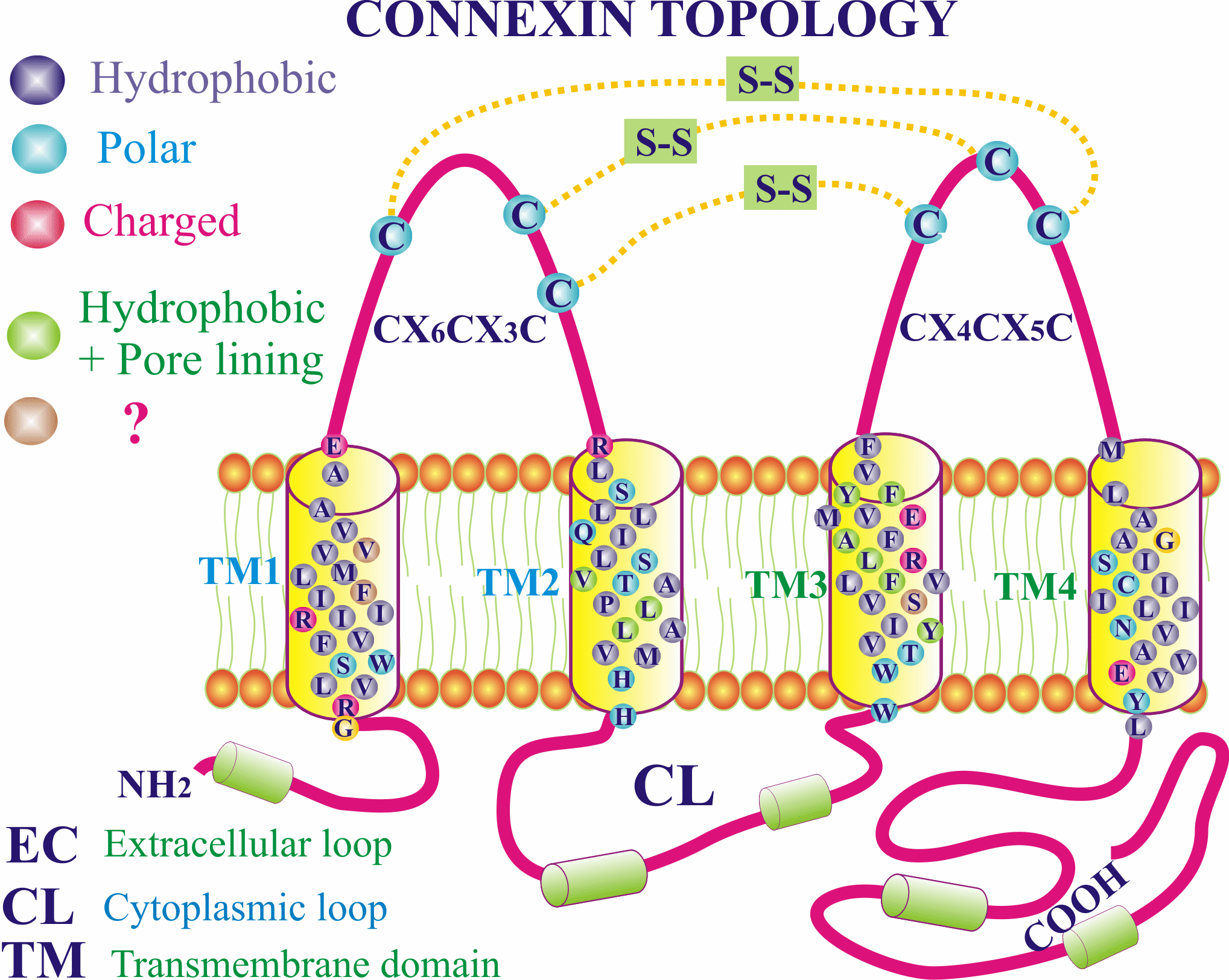
- Connexin topology, walked residue by residue on her slide: N-terminus -> transmembrane region 1 -> Extracellular loop -> back through the membrane -> Cytoplasmic loop -> out again -> second Extracellular loop -> back in -> a long C-Terminal domain tail.
- PROF: The one-letter amino-acid codes matter because folding in three dimensions depends on them: the Hydrophobic residues in the transmembrane domains sit on the Outside of the folded protein, facing the lipid.
- The protein folds into a Cylinder. Six cylinders come together and create a TUBE with a hole down the middle.
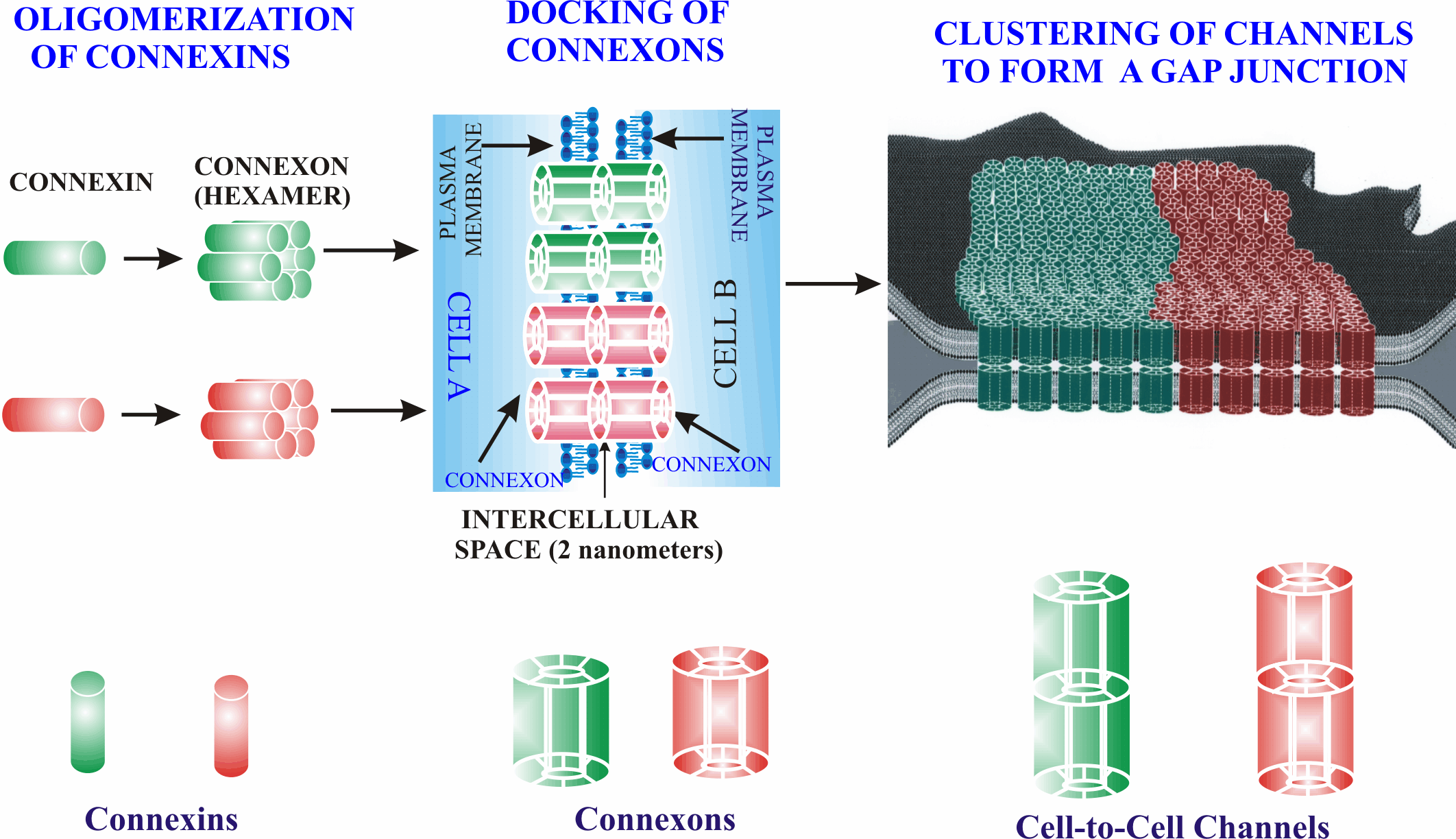
- PROF: Her nomenclature joke, worth memorizing because it is confusing on purpose: the Cylinder (the protein) is a Connexin, and the TUBE (the six-subunit hemichannel) is a Connexon — “one letter apart... very, very helpful when you are talking.”
- Two connexons — one from each cell — DOCK in the extracellular space. Connexons also interact Laterally with their neighbors in the membrane, which is what gathers them into a big Plaque.
- PROF: Function: gap junctions let the two Cytoplasms communicate — a free-floating second messenger passes straight from one cell to the next. That is what carries signals like apoptosis signaling, and in cardiac tissue it is what makes the tissue act as a unit.
The assembly pathway (she walked the whole diagram)
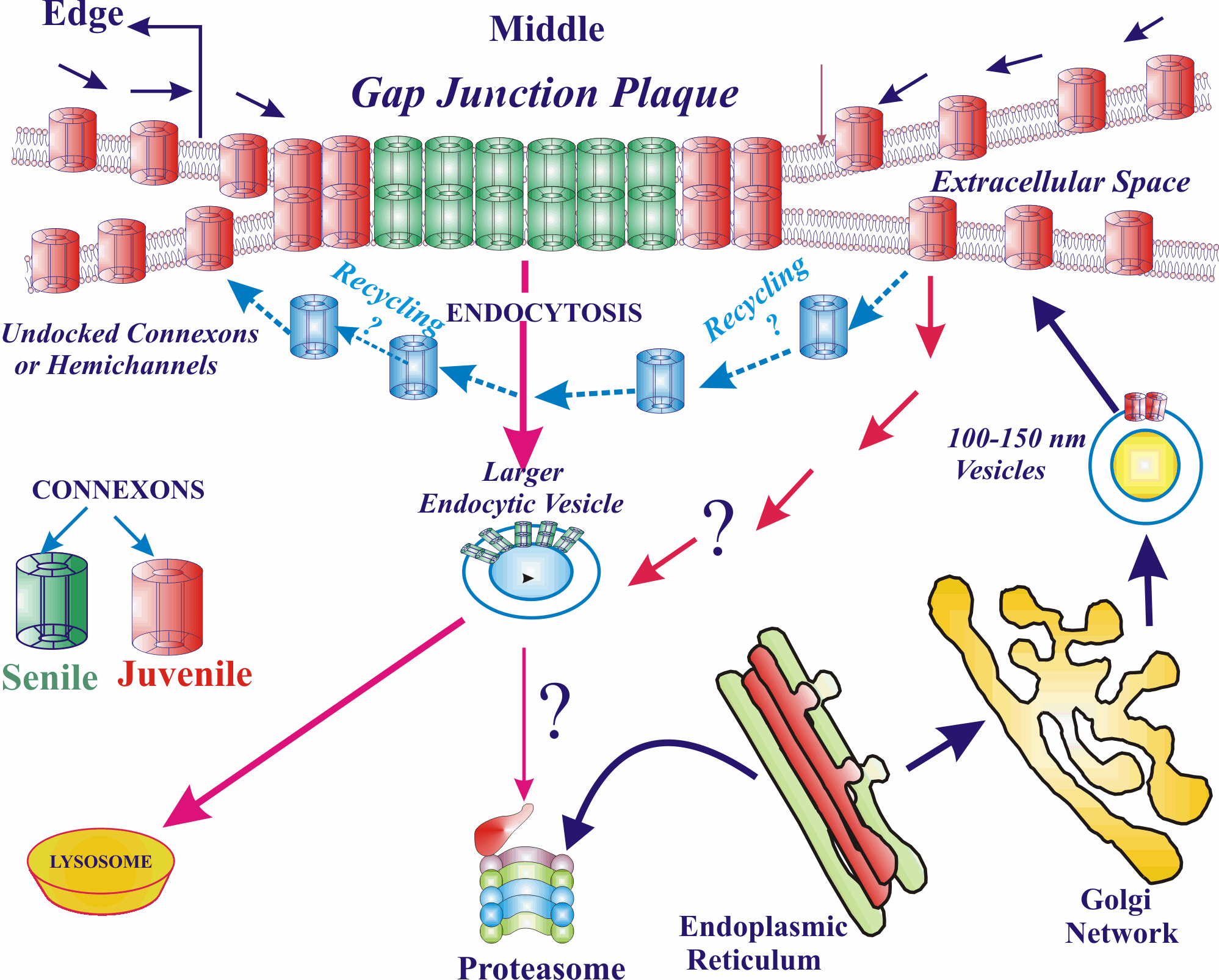
- Gene in the Nucleus -> RNA -> Endoplasmic reticulum, where the connexin cylinder is formed.
- ER -> GOLGI. What does the Golgi do? Transport, packaging, deciding destination — AND post-translational modification (sugars get attached).
- PROF: What is Unique about gap junctions: the half-channel (connexon) is assembled IN The golgi. For other channel-forming proteins — the ryanodine receptor and other ion channels — the subunits traffic first and assemble at their Final destination.
- PROF: Because the connexon is already built, the exocytic vesicles leaving the Golgi are HUGE — 100 to 150 nanometers — big enough that a microscopist can pick them out.
- Vesicle fuses with the plasma membrane -> half channel is now in the surface -> it docks with a connexon on the cell across from it -> lateral association builds the plaque.
- PROF: Attribution she insisted on: this diagram summarizes many years of work from her PhD lab — Dr. Parminder Mehta, who has since passed away.
The heart-attack aside (a favorite exam-style example)
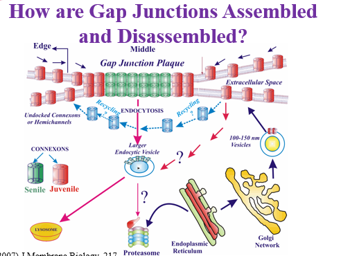
- During a heart attack, the damaged cell Acidifies — a large acidic event.
- PROF: To protect the neighboring tissue that is not damaged yet, the entire gap-junction Plaque is internalized into ONE of the two cells — one cell pushes it out, the other pulls it in — so the cytoplasms stop mixing. You do not want acid flowing into a healthy cell.
- That is the physiological reason internalization of whole plaques exists — and it is the same machinery that goes wrong in her cancer story.
Endocytosis routes and the fate of internalized channels
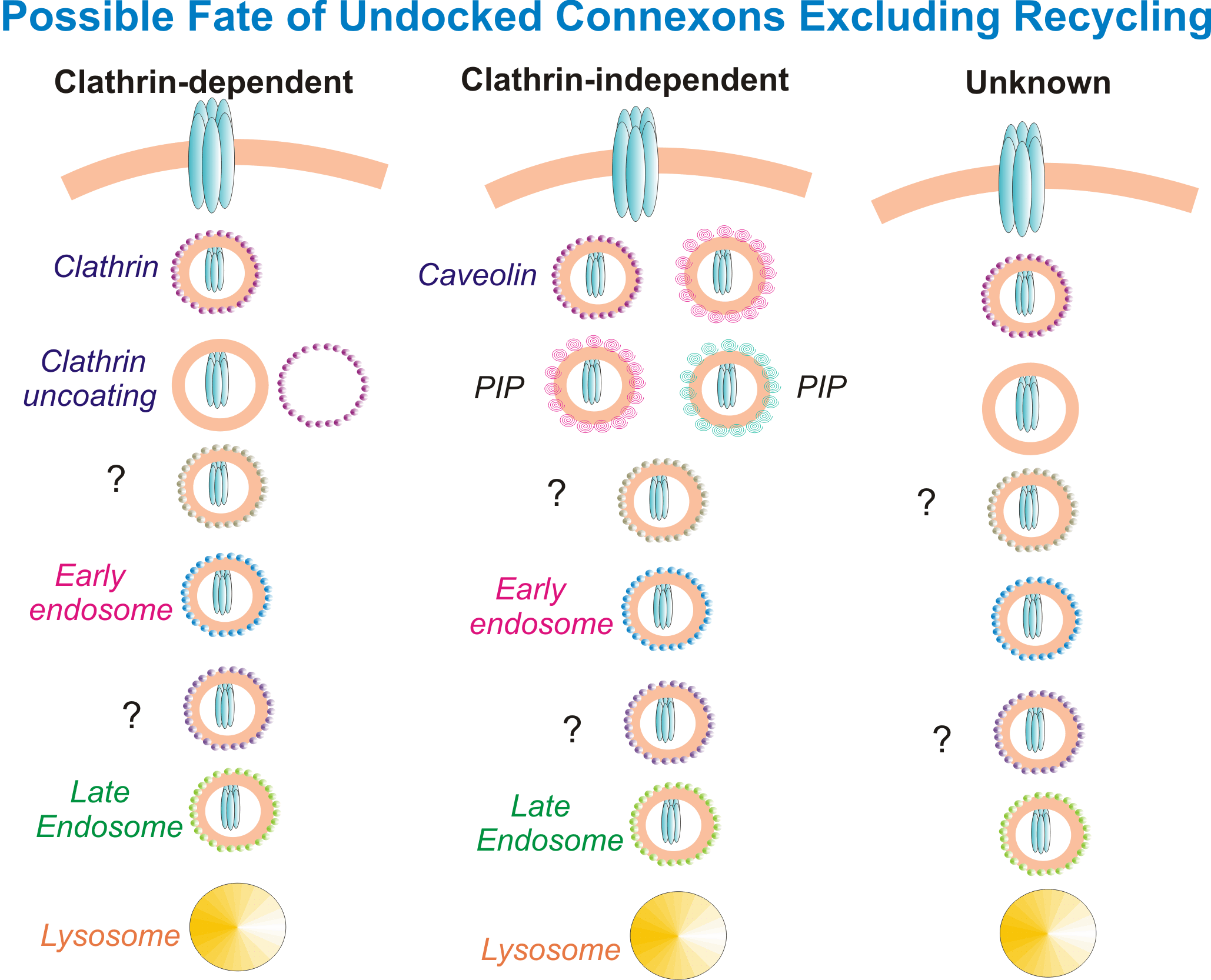
- Gap junctions and half-channels return into the cell by Clathrin-mediated endocytosis or by LIPID-RAFT-mediated endocytosis.
- The large endocytic vesicles can be targeted to the Lysosome (her lab has evidence for this going back to 2007).
- There is some evidence that smaller ones go to the Proteasome — and a Misfolded protein always goes to the proteasome.
- PROF: Her framing of why any of this matters: “it is very important to understand the trafficking — to understand where this thing is going.”
Part 12: Her pancreatic cancer research story (THE quiz material)
- Quiz alert — ≈2 questions on Tuesday come from this section.
The problem
- Pancreatic cancer cells from patients, fixed and stained: Nucleus in blue, Connexin in red.
- PROF: The cell-level problem in pancreatic cancer: The cells are not communicating with each other. They stop communicating.
- Across cancers generally, gap junctions fail in one of two ways: either the GENE gets turned off, or there is nothing wrong with the gene but the Proteins are all in the wrong place — inside the cell instead of at the membrane.
- PROF: In her pancreatic cancer system the gene sequence of the connexin is NOT mutated. It is the same sequence. The defect is everything else — trafficking.
The two cell lines
- BxPC3 :: a pancreatic cancer line obtained from a patient at an EARLY stage of disease.
- Capan-1 :: a LATE-stage pancreatic cancer line (metastatic disease).
- PROF: Comparing an early-stage line against a late-stage line is the whole experimental design — it lets you ask what changes as the cancer advances.
The connexin family and what each one did
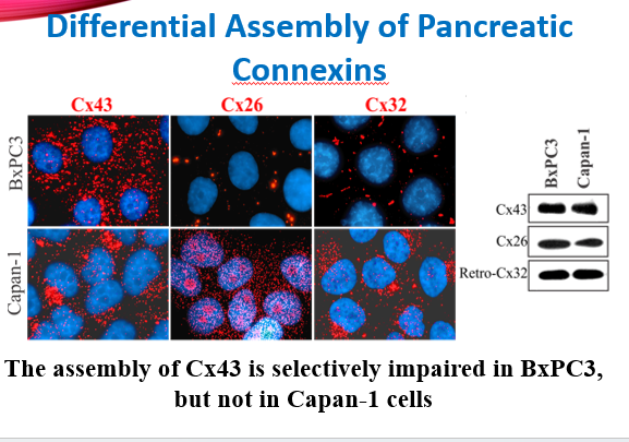
- Read the figure carefully: the slide’s own conclusion is that Cx43 assembly is impaired in BxPC3 but NOT in Capan-1, while Cx26 does the opposite (it forms junctions in BxPC3 and is trapped inside Capan-1). So it is not “Cx43 is always stuck” — each connexin fails in a Different cell line, which is precisely what the word “differential” in the title means. If she asks which connexin fails where, answer per connexin AND per cell line, not with one blanket statement.
- There are roughly 20 connexins in the gene family, with slightly different protein lengths and amino-acid sequences.
- PROF: Cx43 is expressed while we are developing as embryos. As adults we do NOT have Cx43 in every cell — but she found Cx43 in essentially EVERY cancer, from any part of the body, and wrote that in her dissertation.
- Cx43 in pancreatic cancer :: abundant in gene expression but NEVER in the membrane — it sits in an intracellular compartment.
- Cx26 (a pancreatic connexin, supposed to be there) :: in BxPC3 (EARLY stage) it reached the membrane and Formed junctions — those cells can still communicate. In Capan-1 (LATE stage) it is stuck inside the cell — no communication in the network.
- Cx32 (also supposed to be expressed in this tissue) :: LOST. They virally infected the cells with a Cx32-carrying virus, and in BxPC3 the cells formed gap junctions.
- PROF: She backed the cell lines up with Tissue sections from other people’s biopsies — and found the same pattern in all of them: Cx43 present in everybody’s tumor, but not in the junctions.
Why the obvious fix does not work
- Her mentor had shown that in cancers where Cx43 is LOST or strongly down-regulated, OVER-Expressing Cx43 can slow or stop tumor growth — prostate cancer, for example.
- PROF: But in pancreatic cancer there is already plenty of Cx43 — it is all stuck inside the cell. You cannot over-express your way out of a Trafficking problem. That is what pushed her to ask trafficking questions.
- Control experiment that proves the point: over-expressing wild-type connexin on top of what is already there did NOT form junctions — it just added more intracellular protein.
The experiments
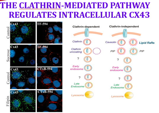
- First they tested Forward trafficking — experiments to halt delivery to the membrane. They still saw the same intracellular pattern.
- PROF: Her comment on that result: “a no is an answer to a question.” A negative result redirected the whole project toward endocytosis.
- Then they blocked the two endocytosis routes:
- • Clathrin-mediated endocytosis blocked with a Sucrose gradient (hypertonic sugar in the extracellular medium).
- • LIPID-RAFT-mediated endocytosis blocked by pulling the lipid rafts out of the membrane — dissolving the rafts, not the membrane.
- Cargo controls (the essential part of the design): Transferrin, the iron-carrying ligand, is the known Clathrin cargo; Cholera toxin is the known LIPID-RAFT cargo. Cx43 was imaged in green, the tracer in red.
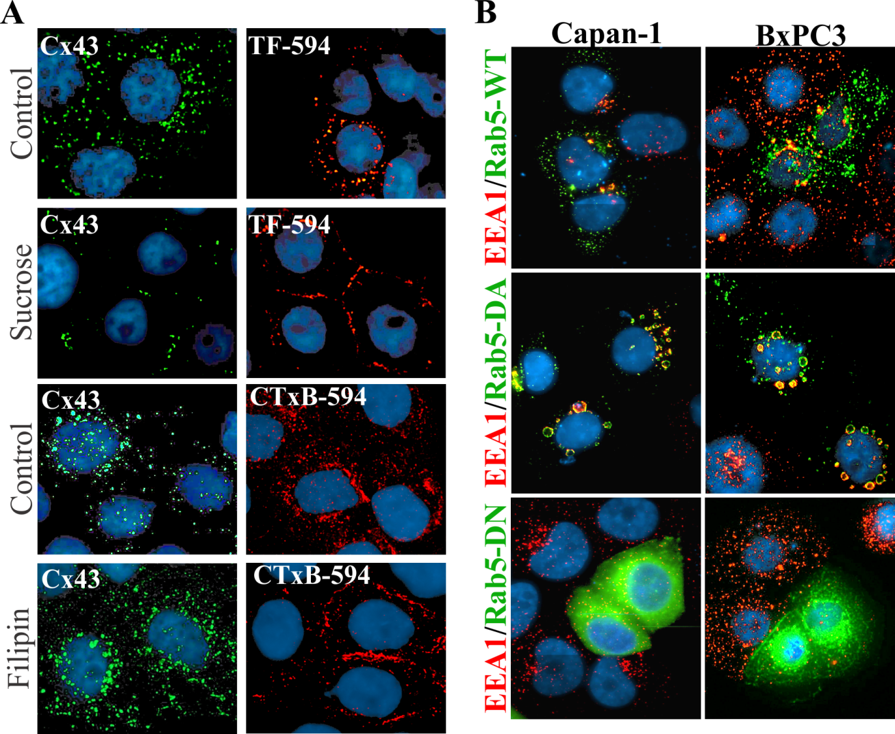
- PROF: The sucrose result: transferrin was Stuck at the membrane — proving the clathrin block worked — but Cx43 was STILL inside the cell. Blocking clathrin did not rescue it.
- The raft result was less clean — some cholera toxin still got in, some stayed at the membrane — and if anything there was even MORE Cx43 inside the cell.
The biochemistry that cracked it
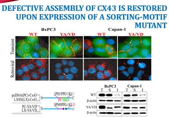
- Receptor-mediated endocytosis requires the CARGO to bind an Adapter protein, which then connects to clathrin to build the cage.
- So they read the amino-acid sequence of the C-Terminal domain and used bioinformatics to look for motifs that bind those adapters.
- PROF: They found Tyrosine-Based sorting motifs: a PY motif and a YKLD motif. Those are the “come inside” address labels.
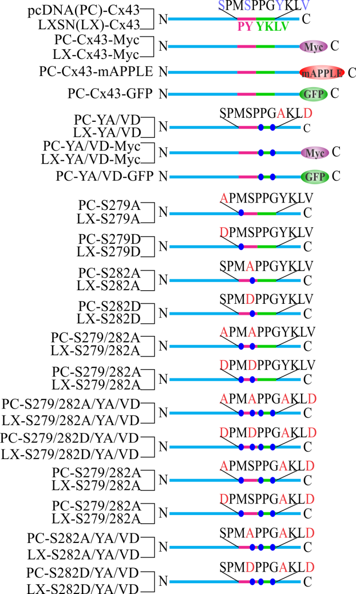
- They removed them by SITE-Directed mutagenesis — mutating out the tyrosines (and nearby serines/aspartates) — then put the mutant gene back into the cells by Transient transfection and by Retroviral infection.
- PROF: The result: junction plaques appeared. Fixed and stained, the cells finally built gap junctions — in BOTH the early-stage AND the late-stage cancer lines.
- The biochemical confirmation: protein shifted from the Soluble intracellular fraction into the Insoluble, gap-junction-associated fraction — the fractionation signature of assembled junctions.
- PROF: Her summary of the discovery: something makes the protein traffic to the membrane and then get Endocytosed before it can connect to the next cell and make communication happen. Delete the sorting motif and the junction forms.
Her open question and the follow-up
- PROF: A question she posed to the class and deliberately left open: What is the difference between transient transfection and retroviral infection? She said talk about it with each other, and that it will be answered when we cover immortalization.
- Preview of the answer to have ready: transient transfection introduces DNA that is NOT integrated — it is expressed for a few days and then diluted out as cells divide. Retroviral infection integrates the sequence into the host genome, giving stable, heritable expression in every daughter cell (the same integration logic used to immortalize cells with SV40 large T and hTERT in the Kitazawa paper).
- The field since: researchers after her found specific Kinases that are hyperactive in pancreatic cancer, and there are now drugs targeting those.
- PROF: The habit of mind she wants: be Mechanistic. For your own project ask — what is the mechanism? Which receptors might be impacted? Which surface proteins might be hurting? Is it Apoptosis or is it Necrosis?
Part 13: The lab-math review she ran at the end
- She will put a Unit conversion table on the board — millis, micros, nanos, and the rest of the scientific units.
- PROF: Her framing question for serial dilutions: do you want to do a bunch of 1:10 dilutions, or a bunch of 1:100 dilutions? Choose the scheme before you start pipetting.
- She teaches dimensional analysis as the “Train tracks” method — stack the conversions so units cancel, and flip a factor when you need the reciprocal.
- PROF: The unit discipline: whatever you are solving for, make sure your tracks end in the unit you actually want — GRAMS if it is a mass, Milliliters if it is a volume, moles/L if it is a concentration.
- This is the same math as Week 1 (C1V1 = C2V2, dilution factors, step dilutions) — see the Week 01 lab guide for worked examples.
Lecture 02 condensed review — what to have cold
- The two questions: What is the structure? What is the function?
- in vitro = “in glass” (really: outside the body, usually in plastic); in vivo = in the living organism. Studying a cell means knowingly making a leap to tissue, organ, system, disease.
- Organelles are subcellular structures and are not alive on their own; ≈70% of mitochondrial gene expression comes from nuclear DNA. Living = can metabolize AND carries the information to synthesize another of itself. Viruses fail both.
- History: 1960s lung fibroblasts -> vaccines/virology; 1970s hybridomas -> monoclonal antibodies; 1980s immortalization. Monoclonal = one antigen; polyclonal = many.
- Prokaryotes do not perform mammalian post-translational modification — which is why human cell culture is the biotech system for human products (growth hormone once came from cadaver pituitaries).
- Junctions: tight, adherens, desmosome, gap, hemidesmosome. All need protein-protein interaction Outside and a cytoskeletal Linker (often ZO-1) Inside. Adherens and gap junctions both work with actin.
- Connexin = the cylinder (one protein). Connexon = the tube (six connexins, a hemichannel). Two connexons dock; lateral association builds the Plaque.
- Connexons are assembled IN The golgi — unusual — so exocytic vesicles are huge, 100–150 nm. Ion channels like the ryanodine receptor assemble at their final destination instead.
- Heart attack: the damaged cell acidifies, so the whole plaque is internalized into ONE cell to stop cytoplasms mixing.
- Return routes: clathrin-mediated or lipid-raft-mediated endocytosis; large vesicles -> lysosome, smaller possibly -> proteasome; misfolded protein always -> proteasome.
- Pancreatic cancer: cells stop communicating. Cx43 gene sequence is Normal but the protein never reaches the membrane. Cx26 forms junctions in early-stage BxPC3 but is stuck inside late-stage Capan-1. Cx32 is lost; adding it back by virus restored junctions in BxPC3.
- You cannot fix a trafficking problem by over-expression — over-expressing wild-type connexin just added more intracellular protein.
- Blocking clathrin (sucrose) stopped transferrin at the membrane but did NOT rescue Cx43 — a negative result that redirected the project.
- The answer was in the C-terminal tail: tyrosine-based sorting motifs (PY and YKLD). Site-directed mutagenesis removing them, delivered by transient transfection and retroviral infection, Restored junction plaques in both early- and late-stage lines, and shifted protein from the soluble to the insoluble fraction.
- Open question to answer for yourself: transient transfection (non-integrated, temporary) vs retroviral infection (integrated, stable and heritable).
Lecture 02 self-test
Self-test
- Define in vitro literally and functionally, and state the caveat she insisted on.
- Why are organelles not “living,” given that mitochondria have their own DNA?
- What is the difference between a monoclonal and a polyclonal antibody, and what 1970s technology made monoclonals possible?
- Why can bacteria not simply be used to make every human protein?
- Distinguish a connexin from a connexon, and say where each is built.
- What makes a linker protein a linker, and which cytoskeletal element do gap junctions mostly use?
- Explain what happens to gap junction plaques during a heart attack and why.
- In her pancreatic cancer system, what was wrong with Cx43 — and what was NOT wrong with it?
- Contrast what Cx26 did in BxPC3 versus Capan-1, and what that tells you about disease stage.
- Why could over-expressing Cx43 not fix pancreatic cancer the way it helped in prostate cancer?
- The sucrose experiment blocked clathrin-mediated endocytosis. How did they know the block worked, and what did Cx43 do?
- What was the final mechanism, and what experiment proved it?
- Answer her open question: transient transfection vs retroviral infection.
Show answer key — try the questions first
- Literally “in glass” (vitra = glass); functionally it means outside the body — and we actually use plastic. The caveat: studying a cell in culture means you are making a leap from cell to tissue to organ to system to disease, and a good cell biologist stays aware of that leap.
- They cannot live independently or synthesize themselves: roughly 70% of mitochondrial gene expression comes from nuclear DNA. Living requires both metabolism (“breathing”) and carrying the information to build another version of itself — which is also why viruses are not living.
- Monoclonal antibodies all target ONE antigen/epitope; polyclonal antibodies target multiple. Hybridomas — fusing an immune B cell with a cancer cell — made immortal, single-specificity antibody factories possible.
- Prokaryotes do not carry out mammalian post-translational modification, so the product is not authentically human. That is why the mammalian cell-culture system is the biotech platform for human products — the growth-hormone example replaced harvesting from cadaver pituitary glands.
- A connexin is the single protein — the cylinder, with N-terminus, four transmembrane passes, two extracellular loops, a cytoplasmic loop, and a long C-terminal tail. A connexon is the hemichannel — six connexins forming a tube with a central pore. Unusually, the connexon is assembled IN The golgi, which is why its exocytic vesicles are 100–150 nm and visible by microscopy.
- It connects the transmembrane protein to a cytoskeletal element — stabilizing it or enabling movement (ZO-1 appears at essentially every junction type). Gap junctions, like adherens junctions, mostly interact with ACTIN, which is dynamic and allows quick changes.
- The damaged cell acidifies, so the entire plaque is internalized into one of the two cells — one pushes it out, the other pulls it in — which stops the cytoplasms from mixing and protects the undamaged neighbor from the acid.
- The gene sequence was NOT mutated and the gene was abundantly expressed. What was wrong is localization: the protein never reached the plasma membrane, sitting instead in an intracellular compartment — so the cells could not communicate.
- In BxPC3 (early stage) Cx26 reached the membrane and formed junctions, so those cells could still communicate. In Capan-1 (late stage) Cx26 was stuck inside the cell with no communication — so something about trafficking degrades as the cancer advances.
- In cancers where Cx43 is lost or down-regulated, adding more restores junctions. In pancreatic cancer Cx43 is already abundant — it is a Trafficking failure, not an expression failure — and their control experiment showed over-expressing wild-type connexin just produced more intracellular protein.
- Transferrin (the iron-carrying ligand and canonical clathrin cargo) was stuck at the plasma membrane, confirming the block. Cx43 nevertheless remained intracellular — a negative result that redirected the project toward the sorting sequence itself.
- Tyrosine-based sorting motifs (a PY motif and a YKLD motif) in the C-terminal tail were driving premature internalization. Site-directed mutagenesis removing them — delivered by transient transfection and retroviral infection — restored gap junction plaques in both early- and late-stage lines, and shifted protein from the soluble to the insoluble, junction-associated fraction.
- Transient transfection delivers DNA that is not integrated into the genome — expression lasts a few days and dilutes out as cells divide. Retroviral infection integrates the sequence into the host genome, so expression is stable and inherited by every daughter cell — the same integration principle used to immortalize cells with SV40 large T and hTERT.
Appendix B — Beginner’s glossary for this lecture
- Every term she used, in one place. Column 2 is the plain-English version; column 3 is why it matters in THIS lecture.
- Year | Milestone | Why it matters
- 1665 | Hooke names the “cell” (Micrographia, cork) | The word — from dead cell walls
- 1907 | Ross Granville Harrison: hanging-drop culture of embryonic frog neural tube grows nerve fibers | The birth of tissue culture — first cells grown outside a body
- 1907–50s | Alexis Carrel: rigorous aseptic technique, mammalian serum; the famous chicken-heart culture that “supposedly” survived 35 years | Long-term subculture becomes possible (and controversial)
- 1948 | L929 — first cloned cell strain, by capillary cloning from mouse L-cell fibroblasts (methylcholanthrene-treated) | A clone: continuous generations from a single cell. Enabled by antibiotics and trypsin. Still sold by ATCC
- 1949 | Enders et al. culture poliovirus in vitro | Leads to the Salk vaccine, produced in African-green-monkey kidney (Vero) cells with formalin inactivation
- 1952 | George Otto Gey establishes HeLa from the cervical carcinoma of Henrietta Lacks | First continuous human cell line; the consent and ownership controversy persists (The Immortal Life of Henrietta Lacks)
- 1950s | Defined media; wide use of antibiotics in media | Culture becomes routine; earlier work used plasma, serum, lymph
- 1961 | Enders & Peebles: measles vaccine from virus cultured in human kidney cells | Vaccine number two from culture
- 1964–69 | Rabies, mumps, and rubella vaccines developed in WI-38 human embryonic lung fibroblasts | Rubella grown at low temperature so it is inactivated at body temperature — attenuation
- 1967 | Stanley Gartler: 19 supposedly independent human lines are all HeLa (G6PD type A + PGM type 1 isoenzymes) | The cross-contamination problem — authenticate every line
- 1975 | Köhler & Milstein: hybridomas — B cell fused with myeloma | Monoclonal antibodies: ELISA, Western blot, immunofluorescence, cancer therapy, anti-TNF-α
- 1983 | Immortalization by SV40 | Engineered continuous lines
- 1980–87 | Development of many specialized cell lines | The modern catalog
- 1998 | Tissue-engineered cartilage (Aigner et al., 1998); first cultured human embryonic stem cells (Thomson) | Regenerative medicine opens
- 2000 | Human Genome Project (Dennis at al., 2001) | Sequence-era cell biology
- Model | Genome | Why use it
- E. coli | 4.6 Mbp · ≈4,300 genes | Divides every ≈20 min; simple nutrition; clonal populations. Source of core molecular biology: DNA replication, genetic code, gene expression, protein synthesis
- Yeast (S. cerevisiae) | 12 Mbp · ≈6,000 genes | Simplest eukaryote — nucleus, linear DNA, cytoskeleton, organelles; divides every ≈2 h
- C. elegans / Drosophila | — | Simple multicellular animals; models for development and cell differentiation
- Arabidopsis thaliana | 125 Mbp | The model plant; easily grown in the lab
- Xenopus laevis / zebrafish | — | Early vertebrate development; large eggs, watch and manipulate the embryo
- Mouse | sequenced | Knockout and transgenic animals — study mutations in a whole organism; often a great human model
- Human | ≈3 Gbp · 20,000–25,000 genes | Grow isolated cells in culture, including specialized types
- Type | Source | Effort | Characterization | Propagation | Homogeneity
- Organ culture | Embryonic organs, adult tissue fragments — in vivo architecture retained | High | Easy, by histology | Not possible | High inter-sample variation
- Primary culture | Tissue fragment dissociated by primary explant or enzymatic (trypsin, collagenase) or mechanical (sieving, syringing) means | Moderate | Cytology and markers | Possible from outgrowth | High inter-sample variation
- Cell culture | Dispersed cells from a primary culture or propagated line | Low | Biochemical, molecular, immunological, cytological assays | Standard; finite or continuous | Low inter-sample variation
- Organotypic / histotypic | Primary cultures or cell lines rebuilt in 3-D: multiple cell types on a scaffold (organotypic — skin, cartilage, the bioartificial lung) or one lineage in a 3-D matrix (histotypic) | Moderate | Histology, confocal microscopy, MRI | Only after dissociation | Low inter-sample variation
- Term | Plain English | Why it matters here
- In vitro | Literally “in glass” — outside the body (we use plastic). | What we do all semester: study cells in a dish.
- In vivo | In the living organism. | The thing culture is a stand-in for — remember you are making a leap.
- Organelle | A structure inside a cell that does one job. | Not alive on its own — the ER, Golgi, lysosome are all organelles.
- Nucleus | The office holding the DNA blueprints. | Blue in her immunofluorescence images.
- ER (endoplasmic reticulum) | Folding room where new proteins get their shape. | Where the connexin cylinder is formed.
- Golgi apparatus | Shipping + finishing: decorates and addresses proteins. | Unusually, gap junction half-channels are Assembled here.
- Vesicle | A membrane bubble that carries cargo. | Golgi vesicles carrying connexons are huge — 100–150 nm.
- Exocytosis | Delivering cargo OUT to the surface. | How the half-channel reaches the plasma membrane.
- Endocytosis | Bringing material IN by pinching the membrane inward. | The failure point in her cancer story — Cx43 gets pulled in too early.
- Clathrin | Protein cage that pulls a membrane patch inside. | One of the two endocytosis routes she blocked (with sucrose).
- Lipid raft | Cholesterol-rich membrane patch used as another way in. | The second endocytosis route she blocked.
- Lysosome | Acidic incinerator for worn-out material. | Destination of big internalized gap-junction vesicles.
- Proteasome | Shredder for individual (often misfolded) proteins. | Possible destination for smaller pieces.
- Plasma membrane | The cell’s outer fatty wall. | Where junctions must end up to work.
- Transmembrane protein | A protein built through the wall, touching inside and outside. | Connexins are transmembrane — which is why outside events change inside behavior.
- Cytoskeleton / actin | Internal scaffolding; actin is the fast-changing rope. | Gap junctions and adherens junctions both anchor to actin.
- Linker protein (e.g., ZO-1) | Connects a transmembrane protein to the cytoskeleton. | What makes a linker a linker — she asked this directly.
- Connexin (Cx) | One cylinder-shaped protein brick. Cx43 = the 43-kDa one. | The protein at the center of the whole story.
- Connexon / hemichannel | Six connexins forming half a tunnel. | Assembled in the Golgi — unusual and testable.
- Gap junction plaque | A patch of many docked tunnels between two cells. | What is missing in pancreatic cancer.
- Second messenger | Small signal molecule passed inside/between cells. | The cargo that travels through gap junctions.
- Tight / adherens / desmosome / hemidesmosome | Other junction types: seal, stick-with-actin, stick-with-strong-filaments, stick-to-the-surface-below. | The junction family she listed on the board.
- Post-translational modification | Changes made to a protein after it is built (sugars, phosphates). | Why bacteria cannot make authentic human proteins.
- Kinase / phosphorylation | Enzyme that adds a phosphate — a common on/off switch. | Later work found overactive kinases in pancreatic cancer.
- Immunofluorescence | Antibody + glowing dye used to see one protein under the microscope. | How all her red/green/blue images were made.
- Cell line | Cells from one donor kept dividing in the lab. | BxPC3 = early stage; Capan-1 = late stage pancreatic cancer.
- Site-directed mutagenesis | Deliberately editing specific letters of a gene. | How she deleted the PY and YKLD sorting motifs.
- Transient transfection | Temporary DNA delivery — fades in days. | One of two delivery methods she used (her open question).
- Retroviral infection | Virus permanently inserts DNA into the genome. | The other delivery method — stable and inherited.
- Soluble vs insoluble fraction | Free protein dissolves; assembled junction protein does not. | The biochemical proof that junctions really formed.
- Sorting motif (PY, YKLD) | A short address label in a protein’s tail. | The “bring me inside” label whose removal fixed the problem.
- Adapter protein | Middleman reading the address label and handing cargo to clathrin. | Why the motif matters at all.
- Transferrin / cholera toxin | Tracer cargoes that each use one known route in. | Controls proving each endocytosis block worked.
- Monoclonal vs polyclonal antibody | One target vs many targets. | Hybridoma technology from the 1970s.
- Hybridoma | Immune cell fused to a cancer cell = immortal antibody factory. | How monoclonal antibodies are made.
- Immortalization | Making cells divide indefinitely. | The 1980s advance — and the Kitazawa paper you were assigned.
- Apoptosis vs necrosis | Tidy programmed death vs messy injury death. | The question to ask about your own project treatment.
- The cell as a factory
- Nucleus = the office holding the master blueprints (DNA). Gene → RNA = a photocopy of one page sent to the floor. ER = the folding room where a new protein gets its 3-D shape. Golgi = shipping and finishing — decorates the protein (sugars) and puts an address label on it. Vesicle = a delivery bubble (a truck). Exocytosis = delivery OUT; endocytosis = pickup IN (this one word is the heart of her research story). Lysosome = the incinerator; proteasome = the shredder for single proteins.
- Gap junctions, in plain words
- Two neighbouring houses (cells) build a tunnel between them, each building half. One brick = a connexin (Cx43 just means the 43-kDa one — the number is its weight, not a rank). Six bricks in a ring = a connexon, half the tunnel. The two halves dock in the gap; many tunnels side by side = a plaque. Cargo: molecules under ~1500 Da — literally how neighbouring cells talk. Why cancer cares: no tunnels → no coordination → signals like “stop dividing” never arrive.
- Her techniques, translated
- Fixed and stained (immunofluorescence): cells killed and preserved, then antibodies that stick to one protein carry a glow-in-the-dark dye — blue is usually the nucleus, red/green the protein being hunted. Site-directed mutagenesis: deliberately editing a few letters of a gene to ask what one piece does. Transient transfection: DNA delivered temporarily, fades in days. Retroviral infection: virus pastes DNA permanently into the genome. Soluble vs insoluble fraction: loose protein dissolves in detergent, protein locked into an assembled junction does not — so a shift into the insoluble pile is chemical proof junctions formed.
- The blocking experiment, translated
- Clathrin = a protein that builds a cage around a patch of membrane to pull it inside (one door in). Lipid raft = a cholesterol-rich membrane patch used as the other door. Hypertonic sucrose switches off the clathrin door; filipin disrupts rafts. Transferrin and cholera toxin are tracer cargoes that each use one known door — if the block worked, the tracer gets stranded outside. That is why they are in the figure: they prove the experiment did what it claimed.
- The address-label idea
- An adapter protein is the middleman that reads a label on the cargo and hands it to clathrin. A tyrosine sorting motif (PY, YKLD — “Y” is the letter for tyrosine) is that label, written into the protein’s C-terminal tail, meaning “bring me inside.” Delete the label and the protein finally stays at the surface — which is the entire punchline of her story.
- One sentence to carry
- STRUCTURE = what a thing looks like and is made of. FUNCTION = the job it does. The whole lecture is: the connexin’s structure (where its address labels sit in the tail) determines its function (whether cells can talk).
- C₁V₁ = C₂V₂
- The dilution workhorse. Rearranged for the volume of stock you need: V₁ = C₂V₂ / C₁. Keep units identical on both sides before you touch the calculator — mixing mM with µM is the classic lost point.
- Dilution factor
- DF = final volume ÷ volume of stock. A "1:10 dilution" means 1 part stock + 9 parts diluent = 10 total, not 1 + 10. Serial dilutions multiply: three successive 1:10 steps give 1:1000.
- Working from a stock concentration
- To make 10 mL of 50 µM from a 10 mM stock: V₁ = (50 µM × 10 mL) / 10 000 µM = 0.05 mL = 50 µL stock + 9.95 mL medium. Always state the diluent volume, not just the stock volume.
- Hemocytometer count
- cells/mL = (average cells per 1 mm² large square) × 10⁴ × dilution factor. The 10⁴ comes from the chamber depth (0.1 mm) over a 1 mm² area = 0.1 µL per square. Count 4–5 squares and average; include cells touching two designated edges only.
- Total yield
- total cells = cells/mL × volume of the suspension in mL. The number you report for a flask is the yield, not the concentration — read the question carefully.
- Trypan blue viability
- % viable = live ÷ (live + dead) × 100. Trypan is a dye-exclusion stain: it enters only cells whose membrane has failed, so blue = dead. It scores membrane integrity, not metabolic health — a cell can be Trypan-negative and still dying.
- Seeding density
- Given a target of, say, 2 × 10⁵ cells per well in 2 mL, you need 1 × 10⁵ cells/mL. Volume of suspension to add = target cells ÷ measured cells/mL. Then top up to the working volume with medium.
- Split ratio
- A 1:4 split puts one flask's cells into four flasks' worth of volume. Converting between a split ratio and a seeding density is a favourite quiz move — go through cells/mL, never ratio-to-ratio.
- Population doubling time
- From a growth curve: td = t × log(2) / [log(N) − log(N₀)]. On a semi-log plot the exponential phase is the straight portion — fit only that region, not the lag or plateau.
- Growth curve phases
- Lag (attachment and recovery) → exponential/log (constant doubling time) → plateau/confluence (contact inhibition, nutrient depletion). Passage during log phase; cells split at plateau recover badly.
- alamarBlue (resazurin)
- Non-fluorescent blue resazurin is reduced by metabolically active cells to pink, fluorescent resorufin. Signal tracks metabolic activity, which is a proxy for viable cell number — so a compound that suppresses metabolism without killing reads as cytotoxic. Always run a no-cell (medium-only) blank and subtract it.
- Percent viability from an assay
- % of control = (signaltreated − blank) ÷ (signaluntreated control − blank) × 100. Normalise to the vehicle control, not to the untreated well, when the compound is dissolved in DMSO.
- IC₅₀
- The concentration giving 50% of the control response. Read it off a dose–response curve with concentration on a log axis; it is meaningless without stating the assay, cell line and exposure time.
- Vehicle control
- Cells receiving the solvent alone at the same final concentration. DMSO above roughly 0.5% is itself cytotoxic — if your top dose needs more than that, the experiment is confounded before it starts.
- qPCR Ct
- The cycle at which fluorescence crosses threshold. Lower Ct = more starting template. One cycle ≈ a doubling, so ΔCt of 1 ≈ 2-fold, of 3.3 ≈ 10-fold.
- ΔΔCt (fold change)
- ΔCt = Ct,target − Ct,reference gene. ΔΔCt = ΔCt,treated − ΔCt,control. Fold change = 2−ΔΔCt. The method assumes near-100% efficiency for both amplicons — check that before trusting it.
- Reference (housekeeping) gene
- GAPDH, ACTB, 18S. Chosen because expression should be unaffected by your treatment — verify that assumption, since many "housekeeping" genes shift under drug stress.
- Primer design rules of thumb
- 18–24 nt · 40–60% GC · Tm 58–62 °C and within ~2 °C of each other · avoid runs of 4+ identical bases · avoid 3′ complementarity (primer-dimers) · span an exon–exon junction so genomic DNA is not amplified · amplicon 70–200 bp for qPCR.
- RNA quality
- A260/280 ≈ 2.0 for clean RNA (≈1.8 indicates protein carryover); A260/230 ≈ 2.0–2.2 (low means guanidine or phenol carryover). Both must pass before cDNA synthesis is worth doing.
- Pipetting accuracy
- Use the smallest pipette that covers the volume — a P1000 set to 20 µL is far less accurate than a P20 set to 20 µL. Pre-wet the tip, aspirate to the first stop, dispense to the second.
Week 2 lecture companion — what the 1 September session actually covered
Two instructors ran this session at once. The lab thread walked the class through the Cell Observation Figure; the lecture thread ran quickly back over the Week 1 history and tissue-culture material. Everything below is what was said on the day, and the first four blocks are not in the slide deck the rest of this guide was built from.
Driving the scope, and the word she fished for
- Start at low power with the stage racked away from the objectives, find the object, then climb. This is the opening move of every activity for the rest of the semester, because you look at the cells before you do anything to them.
- The word she wanted the class to produce was parfocal: once the object is in focus at 4×, it stays in focus as you move to 10×, 20× and 100×, give or take a small refocus. If it does not, the stage was not set properly to begin with.
- Confluency is eyeballed. She wants “I think that is 50, maybe 80 percent”, then a conversation. It is a calibrated judgement built by arguing about it in front of a scope all semester, not a measurement.
- Lighting is a variable in your own figures. Her U2OS image took two weeks to get and she still apologises for the lighting on it. Treat illumination as something you control and report, not something that happens to you.
The yellow flask — the best exam-shaped reasoning of the day
- The control comes first. Every flask was fed at the same time, so a feeding schedule cannot explain why one is yellow and the rest are red. That is what turns the rest of this into an inference instead of a guess, and it is the move worth copying.
- Read phenol red as a free pH meter: red is roughly neutral, yellow is acidic, pink or fuchsia is basic.
- Yellow means something is acidifying the medium fast. Bacteria growing exponentially shift into fermentation, and the byproduct that matters here is lactic acid.
- Bacteria are faster fermenters than fungi, and fungi tend to push the medium basic. So yellow points at bacteria and pink points at fungi — the direction of the pH shift is itself the diagnostic.
- She corrected a student on the direction of causation, and the distinction is worth saying out loud: the pH is either creating an environment that suits another organism, or indicating that another organism is already there. Say which one you mean.
- At the scope, contamination at density looks like television static — a field of tiny circular structures far below the roughly 10 µm of a mammalian cell. A student named it before she did, which is the point: you recognise it by texture.
Vacuoles, apoptosis and autophagy
- The big circles inside a cell are vacuoles, not holes in the cell.
- Three readings are on the table: apoptosis, breakdown from within, or autophagy.
- Autophagy is not necessarily cell death. It is the cell digesting pieces of itself so it can scavenge and survive — and it may extend to consuming neighbouring cells.
- The reason her culture looked like that: she deliberately did not feed it for two weeks. The vacuoles are the visible signature of cells living off themselves.
- The second tell in the same image is very low density with one large, flat, thick cell. Low density had inhibited division — the opposite failure from the piling you look for in a transformed line.
Sizes, and why the scale bar matters
- Prokaryotes run about 1–10 µm; eukaryotic cells are larger and carry organelles. When you are hunting bacteria in a flask, hold the size difference in mind.
- The scale bar on her cheek-cell image is 100 µm, so a single cell is roughly a tenth of the bar. Reading a figure means reading its bar first.
- Cheek cells are squamous, so they are flat and spread and the cytoplasm is easy to see. Cultured epithelial cells stand taller, though the volume-to-membrane ratio is similar.
The Week 1 material she ran back over, with what was new on the day
- Ross Granville Harrison gets the credit for tissue culture: frog embryo nerve tissue on a coverslip in lymph fluid, in a chamber holding both air and liquid, kept humid. Nerve fibres grew out of the explant, which proved there was living, growing tissue in the preparation.
- Alexis Carrel, Harrison’s student, claimed a chicken-heart culture survived some thirty-five years. The word supposedly is doing the work: a normal line runs out of telomere buffer and stops dividing, so the claim cannot stand. Reading his procedure back, he was feeding in fresh tissue and lymph each time.
- Fifty is the magic number. A non-immortal line passages roughly 50 times and then stops. An immortal line is continuous.
- L929, 1948, a mouse L-cell fibroblast, was the first cloned cell strain: the first time a single cell was picked, nursed, and grown out into a population. A clone is a collection of genetically identical cells.
- Capillary cloning is how you pick that one cell — plate at low density, seat a small tube around a single cell under the scope using vacuum grease as the seal, then trypsinise just that cell up. Vacuum grease is a high-tension grease that seals two pieces together, sometimes with a robotic arm to place it.
- HeLa, from Henrietta Lacks in the 1950s, was the first human line, and the ethics around it are assigned reading. The 1950s also delivered defined media — amino acids and mitogenic signals in a bottle — which retired the practice of harvesting lymph fluid from animals.
- Why it is called tissue culture: adherent cells grow connected to one another as a monolayer of tissue. Rock the flask and you can watch that sheet slide off the bottom with your naked eye.
Advantages, and the caveat attached to each
- Control of the environment — pH, temperature, osmolality, gas. You decide when a stress is delivered, rather than the cells receiving a signal because the donor got frightened. Caveat: you cannot control everything, because we still cannot grow cells without serum, and serum is undefined. Hormone work therefore needs tested serum with stated estrogen and testosterone levels, since both change how a cancer behaves.
- Characterisation and homogeneity — morphology under the scope, then break the cells open for biochemistry (she named interleukins, the stress signalling molecules). Cloning is what buys the homogeneity. Here homogeneous means one cell type, epithelial rather than fibroblast, not identical genetics.
- Economy, scale and mechanisation — 5 mL of medium in the teaching T25 against litres of blood in a body, and robotics can take a useful culture down to about 100 µL. It answers questions far more cheaply than a clinical trial, and nobody has to be housed and paid for the duration.
- In vitro modelling of an in vivo condition — in vitro is literally “in glass”. Pulling cells out of their niche sounds like a fatal objection and is not: findings in culture have repeatedly matched what is seen in the animal, which is what makes the KRAS work she described interpretable.
Limitations, in her order
- Operator skill. Aseptic technique is the whole game, it takes about two months of salary to bring someone to the point of being trusted with stock, and a lapse costs vectors, transfection reagent and months. Her practical aside: cell culture on a CV makes you competitive for a research-tech post straight out of this class, and she has had at least ten students land one.
- Quantity. Structural biology needs micrograms of protein for NMR or crystallography, and you will not get that from a flask — not even from all of them.
- Misidentification. Lines look alike; a colon cancer line can resemble an osteosarcoma one. A paper showed that a set of widely used lung cancer lines were actually HeLa, and lung cancer researchers worldwide had to re-check and retract. This is why authentication is not optional.
- Genetic drift. Cultured cells barrel through the cell-cycle checkpoints, and poorly maintained cultures accumulate karyotype changes — visibly abnormal chromosomes on a karyotype — drifting away from whatever you thought you were studying.
- Loss of three-dimensional architecture. Flat culture costs you 3-D shape and cell polarity, so signals are sent and received differently. Cells polarise a little given the right cues, and 3-D systems recover some of it, but it is not the tissue.
- Loss of systemic regulation. No hormonal control, no nerve impulses.
- No pharmacokinetics. A drug is metabolised by the stomach, taken up by absorptive cells in the intestine, and arrives at a concentration that depends even on the time of day. None of that exists in a flask — it is why the second-hand smoke paper takes blood draws from patients.
The four types of tissue culture, as she distinguished them
- Organ culture — and note she said explicitly that organ culture is not organotypic. Tissue or whole organs are put into culture keeping most of their structure: every layer, inside and outside, epithelium and fibroblast together. It is not homogeneous, it cannot be propagated, and it is characterised by histology. Her example was skin at an air-liquid interface with the fluid fed from beneath, as it is in the body — used for wound-healing studies where the wound is made and watched to close.
- Primary culture — a tissue fragment taken directly from the body, then teased apart with enzymes that chew up the proteins holding the cells together, and laid down on plastic. Characterised by cytology rather than histology, because you stain the cells as they lie attached. High sample variability, and the cells that survive are the ones that suit the environment — the most fit, not a representative sample.
- Cell line — everything after the primary culture. Once you can grow them you can make a lot of them, so biochemical assays get better (fewer cell types, fewer confounding parameters) and the material stops being precious. Monitor karyotype and sample variation to catch drift.
- Organotypic or histotypic — different cell types deliberately layered back together on a scaffold. Her example was a bio-artificial lung that looks like a piece of supermarket chicken and will actually expand and exchange CO2 for O2 if you pull the diaphragm.
- The transwell filter is the workhorse version: grow an epithelial layer on the membrane, put macrophages in the chamber above or below, and watch whether they migrate across in response to cytokine signals.
Morphology in her own words
- Epithelial-like: a cobblestone appearance. In a dense culture you can see the border of one cell running into the border of the next, because they are stitched together.
- Fibroblast-like: elongated and more separate, making only a small connection here and there. You cannot pick out cell edges as cleanly, because extracellular matrix protein and cytoskeletal protein blur together in the stain.
- Lymphoblast-like: round, because they are not attached to the plastic. They have a cytoskeleton, but they take no shape from a substrate — they flow and move.
- Of the five human tissue types — epithelial, connective, blood, nervous, muscle — nerve and muscle are the hard ones to propagate without immortalising them first.
Logistics fixed on 1 September
- Concept quiz was due 11:30 that night, and it is mostly the math — work the practice calculations. Units are graded by hand, so a missing unit is fixed manually and is not worth panicking over; getting the number right is what matters.
- Paper Quiz 1 is an open-paper test with no Respondus, and it can be done digitally. Get comfortable navigating a paper by its four parts: introduction, methods, results, discussion.
- Group contract is due 11 September. Group name, each member’s strengths, what you expect of each other in preparation, how work is divided, and contact details so you can find each other. This is a heavy group class, run like a lab where nobody works alone.
- Quizzes are not taken as groups. The contract does not cover them.
- Group and course evaluations run every few weeks, and you evaluate the other members, so problems can be headed off early.
- Week 2 discussion post is the second-hand smoke paper, second in the list; talk it through with the class.
- Thursday is for breaking papers down, hood sign-ups are open, and the lab is open at 1 pm for anyone who wants to look at cells — including the Monday group.
Unit 1 · Basics in cell culture and maintenance
- Aseptic technique
- Work in a laminar-flow hood; nothing unsterile passes over an open vessel; wipe everything in with 70% ethanol; keep movements slow so the airflow curtain is not broken. Hood work outside your section requires an appointment.
- Contamination signatures
- Bacteria — sudden turbidity, pH crash (medium goes yellow), motile specks. Yeast — budding ovals, slower pH change. Fungi — visible filaments. Mycoplasma — invisible; only detectable by assay, and it quietly alters your data.
- Complete medium
- Basal medium + serum (usually 10% FBS) + supplements. Phenol red is a pH indicator, not a nutrient: red ≈ 7.4, yellow = acidic/overgrown, purple = alkaline.
- Passaging adherent cells
- Aspirate medium → wash with PBS (removes serum, which inhibits trypsin) → trypsin-EDTA → incubate briefly → neutralise with serum-containing medium → count → reseed. Over-trypsinisation damages surface proteins and lowers viability.
- Passage number
- Records how many times a line has been subcultured. Phenotype drifts with passage, so protocols specify a window — and your project data should record it.
- Confluence
- Percentage of the growth surface covered. Passage around 80–90%; letting cells hit 100% causes contact inhibition, detachment and irreproducible growth.
- Cryopreservation
- Freeze in medium with a cryoprotectant (typically 10% DMSO) at roughly −1 °C/min — DMSO limits intracellular ice. Freeze slowly, thaw fast, then dilute the DMSO out promptly because it is toxic at 37 °C.
- Cellular observation
- Phase-contrast lets you judge morphology, confluence, and health in live unstained cultures — rounding up, granularity and detachment are the early warnings.
Unit 2 · Cellular assays
- Fluorescence imaging
- Fluorophore absorbs at one wavelength, emits at a longer one; filter sets isolate the emission. DAPI/Hoechst stain nuclei; phalloidin marks F-actin; antibody conjugates localise a specific protein.
- Immunofluorescence workflow
- Fix (paraformaldehyde) → permeabilise (Triton X-100, only for intracellular targets) → block (serum/BSA) → primary antibody → labelled secondary → wash → mount. Skipping the block gives high background; skipping permeabilisation loses everything intracellular.
- Essential IF controls
- Secondary-only (tests non-specific binding) and unstained (tests autofluorescence). A pretty image without these proves nothing.
- Growth curve experiment
- Seed identically across wells, sample at intervals, count. Yields lag/log/plateau and the doubling time — and tells you which seeding density to use so a later assay reads during log phase.
- Viability vs proliferation vs cytotoxicity
- Three different questions. Viability — what fraction is alive now (Trypan). Proliferation — are they dividing (alamarBlue over time, growth curve). Cytotoxicity — does the compound kill or arrest. An assay answers only the question it measures; conflating them is a standard exam trap and a standard paper-critique point.
- Endpoint vs kinetic assay
- Trypan is destructive and endpoint. alamarBlue is non-destructive, so the same wells can be read repeatedly over time — which is why it suits dose–response work.
- Replicates
- Technical replicates (same sample, multiple wells) measure assay noise. Biological replicates (independent cultures/passages) measure real variation — only these support a claim.
Unit 3 · Cellular response & the cytotoxicity project
- Experimental design for the project
- One compound, a dose range spanning the expected IC₅₀, a vehicle control, and enough replicates to say anything. Decide the readout before you treat — the assay dictates the plate layout.
- Dose–response
- Log-spaced concentrations (e.g. 0.1, 1, 10, 100 µM) cover orders of magnitude; linear spacing wastes wells. Include an untreated and a vehicle control on every plate.
- RNA isolation
- Lyse in a guanidinium/phenol reagent or a column kit; RNA is fragile and RNases are everywhere — gloves, filtered tips, dedicated bench space. Isolate treated and untreated in parallel so processing is not a variable.
- DNase treatment
- Removes genomic DNA that would otherwise be amplified. The complementary safeguard is exon-spanning primers; the belt-and-braces control is a no-RT reaction.
- Reverse transcription
- RNA → cDNA using reverse transcriptase with oligo-dT, random hexamers, or gene-specific primers. Only cDNA is amplified in qPCR.
- qPCR controls
- No-template control (contamination) · no-RT control (genomic DNA carryover) · reference gene (normalisation) · melt curve (a single peak = one product; extra peaks mean primer-dimers or mis-priming).
- Linking cytotoxicity to expression
- The project's argument is: this compound reduces viability and shifts expression of genes plausibly connected to that death. Choose target genes with a stated rationale — that rationale is itself graded in the primer-selection assignment.
- Writing the results section
- From experiment 3 onward, data are organised into a figure or table plus a formal results section in the class format, due one week after collection. State what was measured, the n, and the control — the interpretation belongs in the discussion.
The five concept areas objective 1 names
- Cell adhesion → cell-cell junctions and ECM. Adherens junctions, cadherins, tight junctions, desmosomes, gap junctions, integrins.
- Proliferation → cell cycle. Cyclins/Cdks, checkpoints, contact inhibition — directly connected to your growth curves and alamarBlue assays.
- Differentiation → U12 and U14. Lineage commitment, stem cells, EMT.
- Cell signaling → U10. GPCRs, RTKs, MAPK, PI3K-AKT — the pathways a cytotoxic compound perturbs.
- Gene expression → U5. Transcription, regulation, and the qPCR readout you generate in Unit 3.
Background concepts
L01 · Cellular Biology: Experiments & Applications (Lecture 1)
This first lecture does three things at once: it defines what cell biology is and why the cell is the right unit of study, it hands you the century of cell-culture history that every later technique stands on, and it frames the course itself — culture technique, cytotoxicity, gene expression, and a final project you design. Do not treat the history as trivia: every date on the timeline is a capability the field gained, and the exam asks for capabilities.
Learning objectives
- Define cell biology and cell culture, and distinguish in vitro, ex vivo, and in vivo.
- Order the milestones of animal-cell culture from Hooke (1665) to the Human Genome Project (2000), and state what each made possible.
- Name the seven standard model systems and the reason each one is used.
- List the eight applications of cell culture with one concrete example each.
- State the four advantages and four limitations of tissue culture, and explain why culture is not the same as in vivo.
- Compare organ, primary, cell, and organotypic/histotypic culture on source, effort, characterization, propagation, and homogeneity.
- Describe the assays of the final project: Trypan blue exclusion, Alamar Blue, and qPCR of reverse-transcribed RNA.
Part 1: What cell biology is
- Cell biology is the scientific study of cell structure and function. It revolves around the concept that the cell is the fundamental unit of life: focusing on the cell permits a detailed understanding of the tissues and organisms that cells compose.
- Robert Hooke coined the word “cell” in 1665 (Micrographia), examining cork — he was actually looking at dead cell walls, but the term stuck. The classic cell theory followed: Schleiden and Schwann (1838–39) — all organisms are made of cells; Virchow (1855) — omnis cellula e cellula, all cells arise from pre-existing cells. Prokaryotes lack a nucleus and membrane organelles and carry small circular DNA; eukaryotes have a nucleus, linear DNA, a cytoskeleton, and organelles. The human body contains roughly 200 distinct cell types.
- Cell culture is the complex process by which cells are grown under controlled conditions, generally outside their natural environment. In vitro literally means “in glass”; ex vivo means tissue observed growing outside the body; in vivo means in the living organism.
Part 2: History of animal-cell culture — the timeline
Part 3: Cells as experimental models
Part 4: The eight applications of cell culture
- 1. Model systems — basic cell biology and biochemistry; interactions between disease-causing agents and cells.
- 2. Toxicity testing — effects of new drugs, cosmetics, chemicals; especially kidney and liver lines, because those organs metabolize and clear drugs (pharmacokinetics).
- 3. Cancer research — characteristics of cancer cells (oncogenes up-regulated, tumor suppressors down-regulated), mechanisms that turn normal cells cancerous, cancer-stem-cell theory.
- 4. Virology — viruses are obligate intracellular parasites, so culture is required for clinical detection, isolation, and vaccine development.
- 5. Cell-based manufacturing — vaccines; proteins (insulin, monoclonal antibodies, hormones); tissue engineering.
- 6. Genetic counseling — amniocentesis: amniotic fluid contains fetal cells that can be analyzed.
- 7. Genetic engineering — transfection of cultured cells to study gene expression or produce protein at scale (GMOs; insulin-secreting E. coli).
- 8. Drug screening and development — cytotoxicity assays and high-throughput screening. Plus: stem cells, IVF, in vitro inflammation assays, tissue transplantation.
Part 5: Advantages and limitations of tissue culture
Advantages
- Control of the environment — physiochemical factors (pH, temperature, osmolality, dissolved gases) are easily controlled; physiological factors (hormones, nutrients) are harder but improving with serum-free media and cloned matrix constituents.
- Characterization and homogeneity — cultures assume a homogeneous constitution; cloning adds more; assays run on virtually identical samples at each subculture.
- Economy, scale, and mechanization — reagent exposure without in vivo complications of metabolism and bioavailability; multi-well plates and robotics enable large studies cheaply.
- In vitro modeling of in vivo conditions — avoids much of the legal, moral, and ethical burden of animal experimentation; tight regulation of concentration, duration, metabolic state; histotypic and organotypic models keep improving realism.
Limitations
- Expertise — sterile handling and environmental control; the skill to recognize microbial contamination and cross-contamination (stock authentication).
- Quantity — expensive equipment and advanced training for a relatively small amount of tissue.
- Origin of cells — a long history of cross-contamination, stock-control errors, and overgrowth of undifferentiated cells producing misidentified lines; every line must be authenticated (today, STR profiling).
- Instability — genetic and phenotypic instability, largely from unstable aneuploid chromosomes, produces heterogeneity and passage-to-passage variability.
Culture is simply not the same as in vivo
- Three classes of loss: (1) dispersing cells destroys the three-dimensional structure and the mixed cell population, so tissue-specific cell interactions are lost; (2) the culture lacks systemic regulators — hormones, nervous influence, oxygen tension (low O2 with no hemoglobin pushes metabolism toward glycolysis and anaerobic pathways instead of the citric-acid cycle and oxidative phosphorylation); (3) an in vitro model cannot replicate pharmacokinetics, metabolism, or organ and systemic responses (inflammation, organ damage, pyrexia, teratogenic effects).
Part 6: The four types of tissue culture
- Organ culture is diffusion-limited — no blood vessels — and lacks nerve supply and hormonal stimulus; its gas–liquid interface balances gaseous exchange against surface-tension flattening. In a primary explant, cells migrate outward from the fragment into a loose monolayer. After several passages a culture becomes fairly homogeneous (fast-growing, quickly adherent cells dominate); if immortalized, it is a continuous cell line. Cultures grow adherent (monolayers — 3T3 fibroblasts, CPAE endothelium) or in suspension (hematopoietic cells, transformed lines, malignant tumors — L1210). Finite lines senesce at the Hayflick limit (≈50 divisions; WI-38 is the classic); continuous lines (HeLa, L929, SV40-transformed) divide indefinitely but are typically aneuploid. A clone is continuous generations of cells derived from a single original cell.
Part 7: The instructor’s science, and the final project
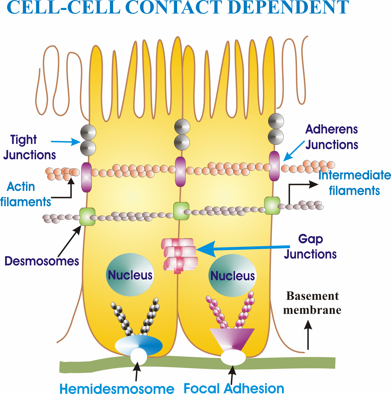
- Dr. Johnson studies how cells adhere and communicate in a contact-dependent manner. Gap junctions are clusters of cell-cell channels that permit leak-proof passage of molecules smaller than ≈1,500 Da; they are built from connexins. Lab findings to recognize: phosphorylation of connexin43 (Cx43) at Ser-279/Ser-282 regulates its endocytosis and gap-junction assembly in pancreatic cancer cells; E-cadherin and N-cadherin regulate Cx43 assembly differentially; the clathrin-mediated pathway and lipid rafts govern intracellular Cx43; and palmitoylation of plakophilin is required for desmosome assembly.
- The final project (from week 6): choose a natural product — tea tree oil, taurine, sodium fluoride, caffeine, manuka honey, juglone, peppermint oil, elderberry juice, glyphosate, phthalates, BPA, or your own idea — and test it. Why natural products? Drug discovery began with them (penicillin); motivations include antibiotic-resistant microbes, hypersensitivity reactions to synthetics, and disease links to synthetic products — while remembering most natural cures are not FDA-approved or regulated. Two experimental readouts: cytotoxicity by Trypan blue exclusion (dead cells with broken membranes stain blue) and Alamar Blue (live cells reduce blue resazurin to pink fluorescent resorufin), and gene-expression response by quantitative PCR of reverse-transcribed RNA. You plan everything: stock solutions, controls, disposal.
Condensed review — the eight things most likely to appear on the exam
- Cell biology = structure + function; the cell is the fundamental unit of life. Cell culture = growth under controlled conditions outside the natural environment.
- Harrison 1907 = birth of tissue culture; HeLa 1952 = first continuous human line; hybridomas 1975 = monoclonal antibodies.
- Gartler 1967: HeLa cross-contamination via G6PD/PGM isoenzymes — authenticate every line.
- Vaccines from culture: polio (Vero), measles, rubella/rabies/mumps (WI-38, cold-adapted = attenuated).
- E. coli for molecular fundamentals; yeast = simplest eukaryote; mouse = whole-animal genetics; ≈200 human cell types.
- Four advantages (control, homogeneity, scale, in vitro modeling) vs four limitations (expertise, quantity, origin, instability).
- Culture ≠ in vivo: no 3-D/mixed populations, no systemic regulation (low O2 → glycolysis), no pharmacokinetics.
- Four culture types: organ (architecture, no propagation) · primary (explant/enzymatic) · cell (finite vs continuous) · organotypic/histotypic (rebuilt 3-D).
Mnemonic set
- “Harrison Hung the drop, Carrel Kept it sterile, Gey Grew it forever.” — 1907 → 1950s arc.
- “HeLa: first, famous, and in everything” — continuous line + contamination story.
- “Kidney and liver clear the drug, so kidney and liver test the drug.”
- “Organ keeps the map, primary breaks the map, cell culture forgets the map, organotypic redraws it.”
- “Blue in = dead (Trypan); blue gone = alive (Alamar).”
Self-test
- Order these and give one sentence on each: HeLa, hybridomas, Harrison’s hanging drop, L929, Gartler’s isoenzyme study.
- Why must a rubella vaccine virus be grown at low temperature, and in which cells was it produced?
- A “novel liver line” carries G6PD type A and PGM type 1 despite records saying the donor was of European ancestry. Diagnose and prescribe.
- Give the three reasons a drug that kills cancer cells in a dish can fail in an animal.
- Why do cultured cells favor glycolysis even with functional mitochondria?
- Classify: (a) fetal skin fragment kept intact at a gas–liquid interface; (b) trypsin-dissociated fragment growing out; (c) 40th passage of dispersed cells; (d) two cell types combined on a scaffold.
- Which applications of culture are involved when insulin is produced from engineered cells and when amniotic fluid is analyzed for a fetus?
- What does each assay of the final project measure, mechanistically: Trypan blue, Alamar Blue, qPCR of RT-RNA?
- Define clone, passage, finite line, continuous line, adherent, suspension.
- What are gap junctions, and what did the Johnson lab show about Cx43 and about plakophilin?
Show answer key — try the questions first
- Harrison 1907 (birth of tissue culture, frog nerve fibers) → L929 1948 (first cloned strain, capillary cloning) → HeLa 1952 (first continuous human line, Gey/Lacks) → Gartler 1967 (19 lines were HeLa — G6PD-A/PGM-1) → hybridomas 1975 (Köhler & Milstein, monoclonal antibodies).
- Cold-grown virus replicates poorly at body temperature — attenuated, so it immunizes without disease. Produced in WI-38 human embryonic lung fibroblasts.
- Cross-contamination with HeLa — the exact signature Gartler used. Authenticate by STR profiling; discard or re-derive the stock; review sterile technique and stock control.
- Pharmacokinetics (metabolism, penetration, clearance are unmodeled), loss of 3-D architecture and mixed populations, and absent systemic responses (immune, hormonal, organ toxicity).
- Low oxygen tension in vitro — no hemoglobin delivers O2, so energy metabolism shifts to glycolysis/anaerobic pathways over oxidative phosphorylation.
- (a) organ culture; (b) primary culture; (c) cell culture — continuous line if immortalized; (d) organotypic culture.
- Insulin production = cell-based manufacturing + genetic engineering (transfection). Amniotic fluid = genetic counseling (amniocentesis).
- Trypan blue: membrane-integrity dye exclusion — dead cells stain. Alamar Blue: metabolic reduction of resazurin to fluorescent resorufin — signal ∝ viable cells. qPCR of reverse-transcribed RNA: transcript abundance — the gene-expression response to treatment.
- Clone: generations from one original cell. Passage: one subculture cycle. Finite: senesces at the Hayflick limit (≈50 divisions). Continuous: immortalized, indefinite. Adherent: grows attached as a monolayer. Suspension: grows floating (hematopoietic, transformed).
- Clusters of cell-cell channels passing molecules < 1,500 Da. Cx43: Ser-279/282 phosphorylation regulates endocytosis and assembly; E- vs N-cadherin regulate assembly differentially. Plakophilin: its palmitoylation is required for desmosome assembly.
- Lecture 02
U1 · Cells, model organisms & microscopy
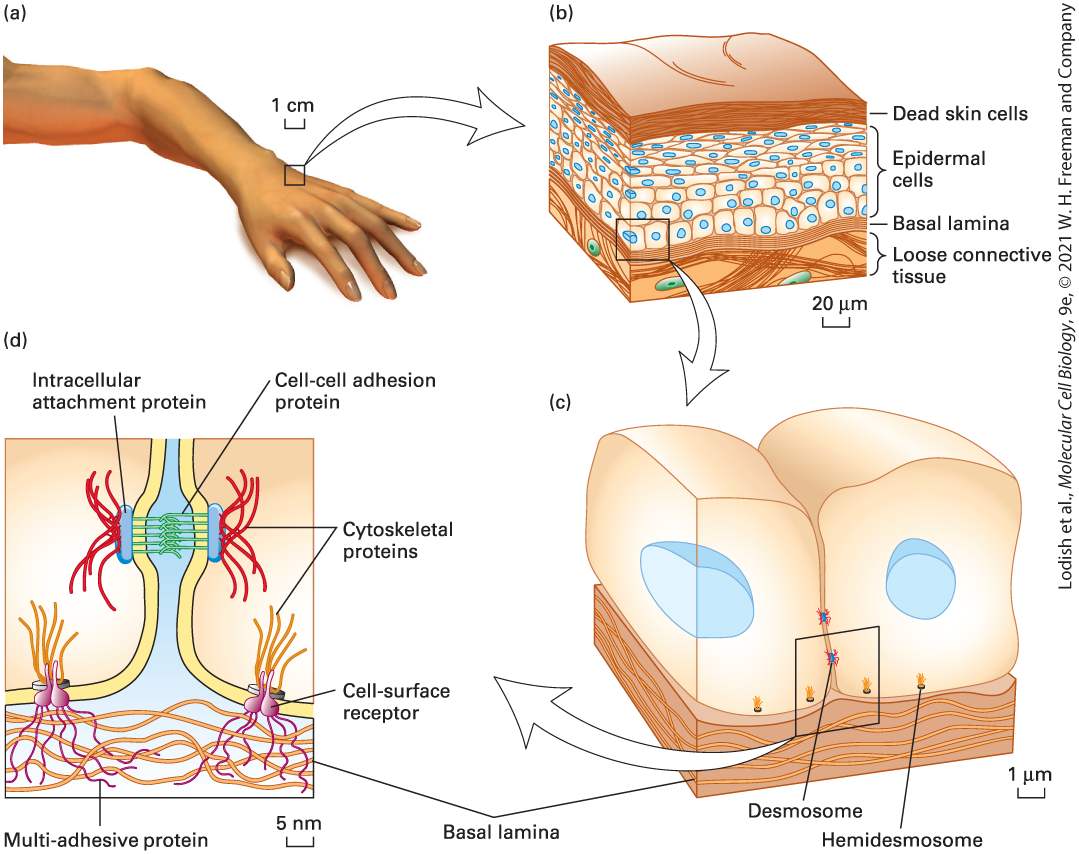
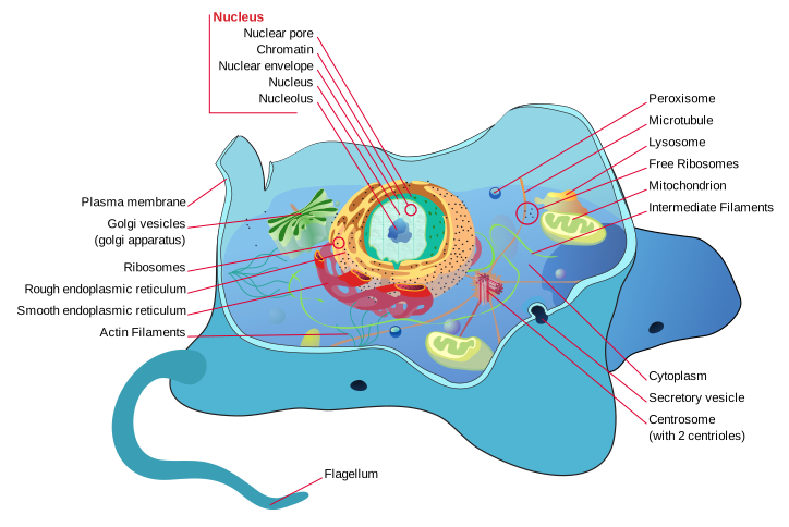
- Cell theory
- All organisms are made of cells; cells are the basic units of life; cells arise from preexisting cells (Schleiden, Schwann, Virchow).
- Prokaryotes vs eukaryotes
- Prokaryotes lack a nucleus and membrane organelles; eukaryotes have both. Eukaryotic cells originated by endosymbiosis (mitochondria from α-proteobacteria, chloroplasts from cyanobacteria).
- Model organisms
- E. coli (bacteria), S. cerevisiae (yeast), C. elegans (worm — Karen Kim Guisbert uses this), Drosophila, zebrafish, mouse, human cell culture lines (HeLa, HEK293).
- Light microscopy
- Resolution limit ~200 nm (Abbe). Bright-field, phase contrast, DIC, fluorescence (matters for Johnson's lab).
- Confocal microscopy
- Pinhole excludes out-of-focus light → optical sections of thick samples.
- Super-resolution microscopy
- STED, STORM, PALM beat the diffraction limit (~20-50 nm).
- Electron microscopy
- TEM (transmission, ~0.1 nm) and SEM (surface, ~1 nm). Requires fixation, sections.
- Immunofluorescence (IF)
- Primary antibody binds antigen; fluorescent secondary antibody binds primary. Used to localize proteins in cells.
U2 · Cell chemistry & biosynthesis
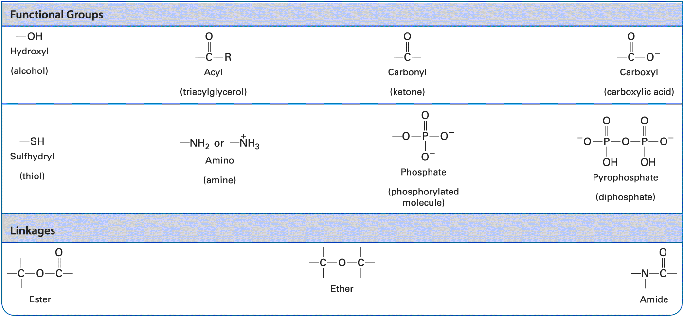
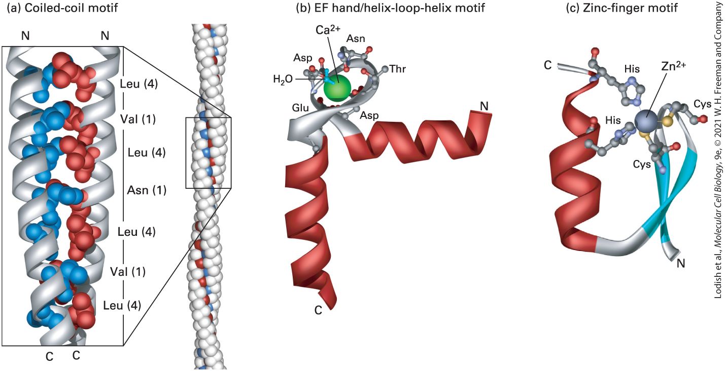
- Water properties
- Polar; H-bonds give high specific heat, surface tension, cohesion. Hydrophobic effect drives folding + membrane assembly.
- Macromolecules
- Proteins (AA), nucleic acids (nucleotides), polysaccharides (sugars), lipids (FAs + glycerol/sterol).
- ΔG (free energy change)
- ΔG < 0 = spontaneous (exergonic); ΔG > 0 = nonspontaneous (endergonic). Cells couple endergonic to exergonic via ATP hydrolysis.
- ATP
- Energy currency. Hydrolysis of γ-phosphate releases ~−7.3 kcal/mol under standard conditions; much more in cells.
- Enzyme catalysis
- Lower activation energy. Michaelis-Menten: v = Vmax[S] / (Km + [S]). Km = [S] at half Vmax.
- Allosteric regulation
- Effector binds non-active site, changes conformation + activity. Foundation of cell-signaling switches.
U3 · Proteins
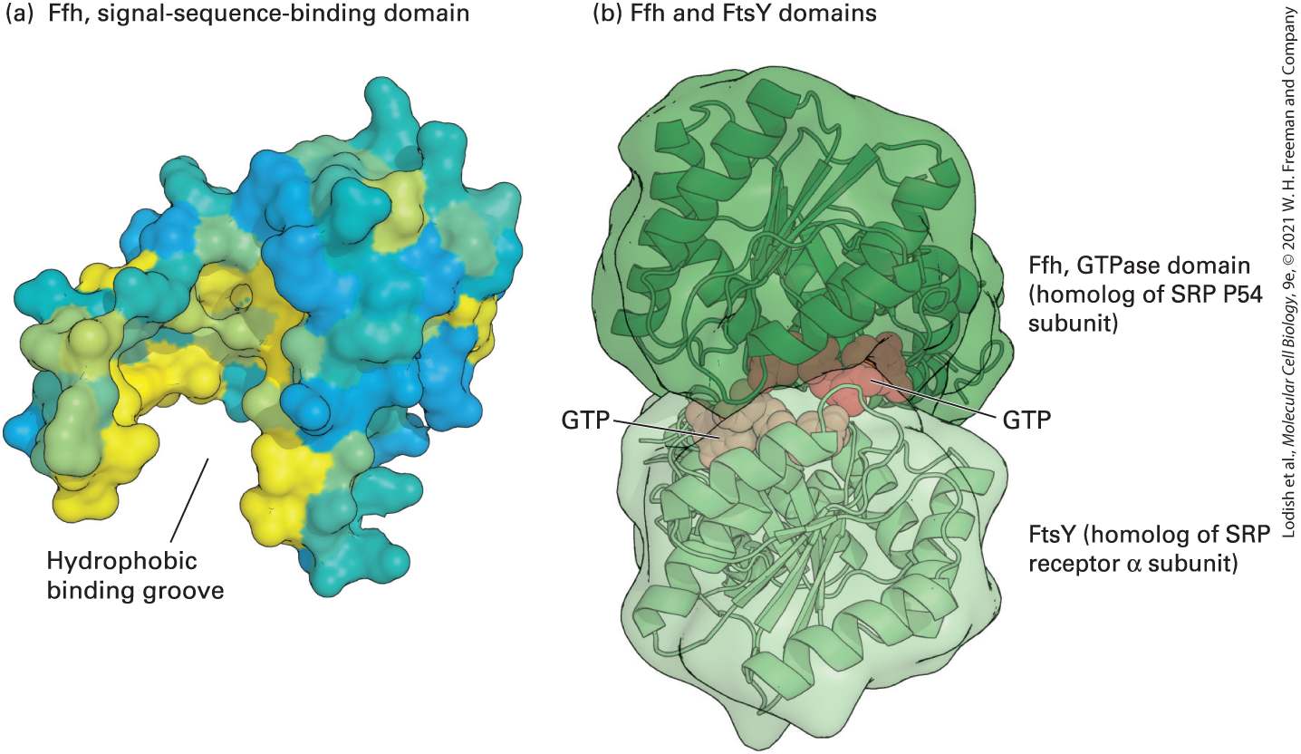
- Primary structure
- Amino acid sequence (peptide bonds).
- Secondary structure
- Local folds — α-helix, β-sheet — stabilized by backbone H-bonds.
- Tertiary structure
- 3D fold of one polypeptide; stabilized by H-bonds, ionic, hydrophobic interactions, disulfides.
- Quaternary structure
- Multi-subunit assembly (hemoglobin = 2α + 2β).
- Chaperone
- Hsp70, Hsp90, GroEL/ES — assist folding; rescue misfolded proteins.
- Ubiquitin-proteasome
- Tags damaged/regulated proteins with poly-Ub chain → 26S proteasome degrades.
- Motor proteins
- Myosin (actin), kinesin + dynein (microtubules); ATP-driven directional movement.
- Phosphorylation
- Kinase adds phosphate (Ser/Thr/Tyr), phosphatase removes. Most common reversible regulatory PTM.
U4 · DNA, chromosomes & replication
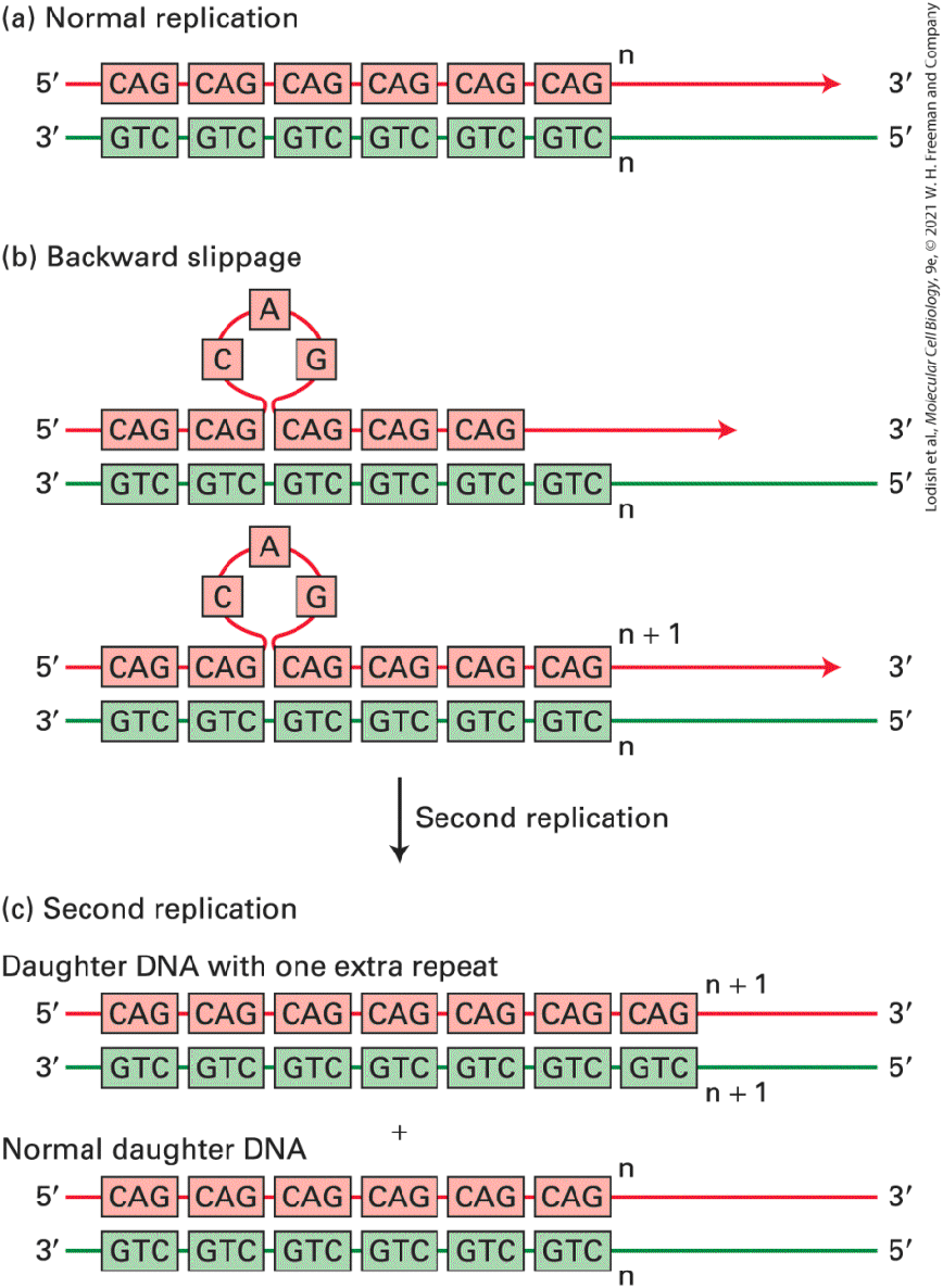
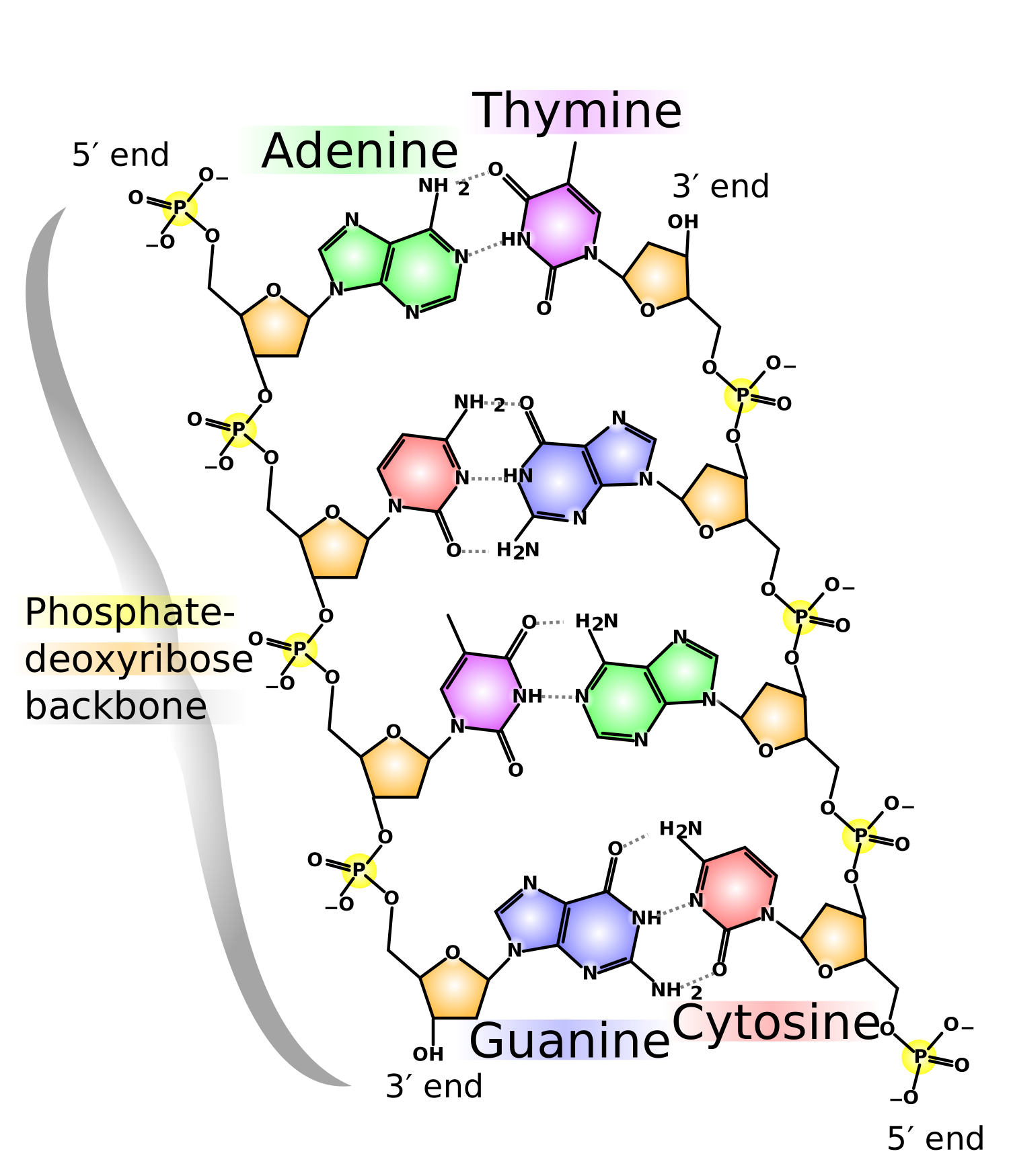
- DNA structure
- Antiparallel double helix; AT (2 H-bonds), GC (3). 10.5 bp/turn, major + minor grooves.
- DNA replication
- Semiconservative. Helicase unwinds, primase lays RNA primer, DNA pol III extends 5'→3'. Leading vs lagging strand (Okazaki fragments). Telomerase extends telomeres.
- DNA repair
- Mismatch repair (MMR), base excision (BER), nucleotide excision (NER), homologous recombination (HR), non-homologous end joining (NHEJ).
- Chromosome structure
- DNA + histones = nucleosome (~146 bp around 8 histones). 30-nm fiber → loops → chromatid in mitosis.
- Centromere / kinetochore
- Heterochromatic region where spindle attaches via kinetochore protein complex.
- Telomere
- (TTAGGG)n caps; shortens with each replication. Telomerase active in germ + stem + many cancer cells.
U5 · Gene expression
- Transcription
- RNA pol II makes mRNA from DNA template. Promoter (TATA, etc.) + general TFs + enhancers + activators.
- RNA processing
- 5' cap (m7G), poly-A tail, splicing (spliceosome removes introns; alternative splicing diversifies proteome).
- Translation
- Ribosome reads mRNA codons via tRNA anticodons. Initiation (Met-tRNA, eIFs) → elongation → termination (release factors at stop codon).
- Genetic code
- Triplet, redundant (degenerate), nearly universal. Wobble at 3rd codon position.
- Transcription factor (TF)
- DNA-binding protein that activates/represses transcription (e.g., p53, Myc, NF-κB).
- Epigenetic marks
- DNA methylation (CpG), histone modifications (H3K4me3 active, H3K27me3 repressive, H3K9ac active).
- miRNA
- ~22 nt; binds 3'UTR → translational repression or mRNA decay (RISC complex).
U6 · Membranes & transport
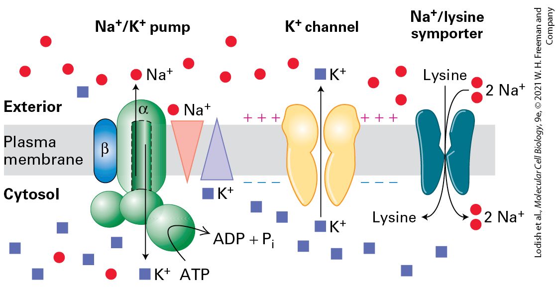
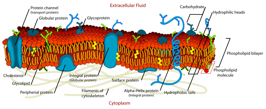
- Lipid bilayer
- Amphipathic phospholipids; fluid mosaic. Cholesterol modulates fluidity; sphingolipids cluster in lipid rafts.
- Membrane proteins
- Integral (transmembrane), peripheral (cytosolic-facing), lipid-anchored (GPI-anchored, prenylated).
- Passive transport
- Diffusion (down gradient, no ATP); facilitated diffusion through channels or carriers.
- Active transport
- Against gradient, requires ATP. Primary: Na⁺/K⁺-ATPase, Ca²⁺-ATPase, H⁺-ATPase. Secondary: symport/antiport coupled to ion gradient.
- Na⁺/K⁺-ATPase
- 3 Na⁺ out, 2 K⁺ in per ATP. Sets resting potential, drives secondary transport.
- Aquaporin
- Water-selective channel; key in kidney, RBCs.
- Action potential prep
- Voltage-gated channels open in response to depolarization. (Cross-reference NEUR 1520 U4!)
U7 · Internal compartments & sorting
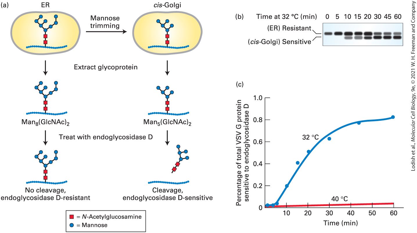
- ER (rough)
- Studded with ribosomes; site of secretory + membrane protein synthesis. Co-translational import via SRP + signal sequence.
- ER (smooth)
- Lipid synthesis, Ca²⁺ storage, detox (P450 enzymes in liver).
- Golgi apparatus
- Cis → medial → trans cisternae. Modifies glycoproteins, sorts to lysosomes / plasma / secretion.
- Lysosome
- Acidic (pH ~4.5) hydrolytic compartment; hydrolases tagged with mannose-6-phosphate.
- Peroxisome
- β-oxidation of very-long-chain fatty acids; H₂O₂ neutralized by catalase.
- Mitochondrion
- Double membrane; matrix has TCA cycle + mtDNA; inner membrane houses ETC + ATP synthase. Imports nuclear-encoded proteins post-translationally via TOM/TIM.
- Nuclear localization signal (NLS)
- Lys/Arg-rich sequence recognized by importin α/β; transport through nuclear pore.
U8 · Vesicle traffic
- COPII
- ER → Golgi anterograde traffic; Sar1 GTPase coordinates assembly.
- COPI
- Golgi → ER (retrograde) and intra-Golgi; Arf1 GTPase.
- Clathrin
- Plasma membrane endocytosis + Golgi → endosome. Adaptor proteins (AP1/2) link cargo to coat.
- SNARE
- v-SNARE (vesicle) + t-SNARE (target) zip into 4-helix bundle → membrane fusion. Specificity for compartment pairing.
- Endocytosis types
- Phagocytosis (large particles, immune cells), pinocytosis (fluid uptake), receptor-mediated endocytosis (LDL, transferrin).
- Exocytosis
- Constitutive (continuous) or regulated (Ca²⁺-triggered, e.g., insulin secretion, neurotransmitters).
U9 · Mitochondrial energy conversion
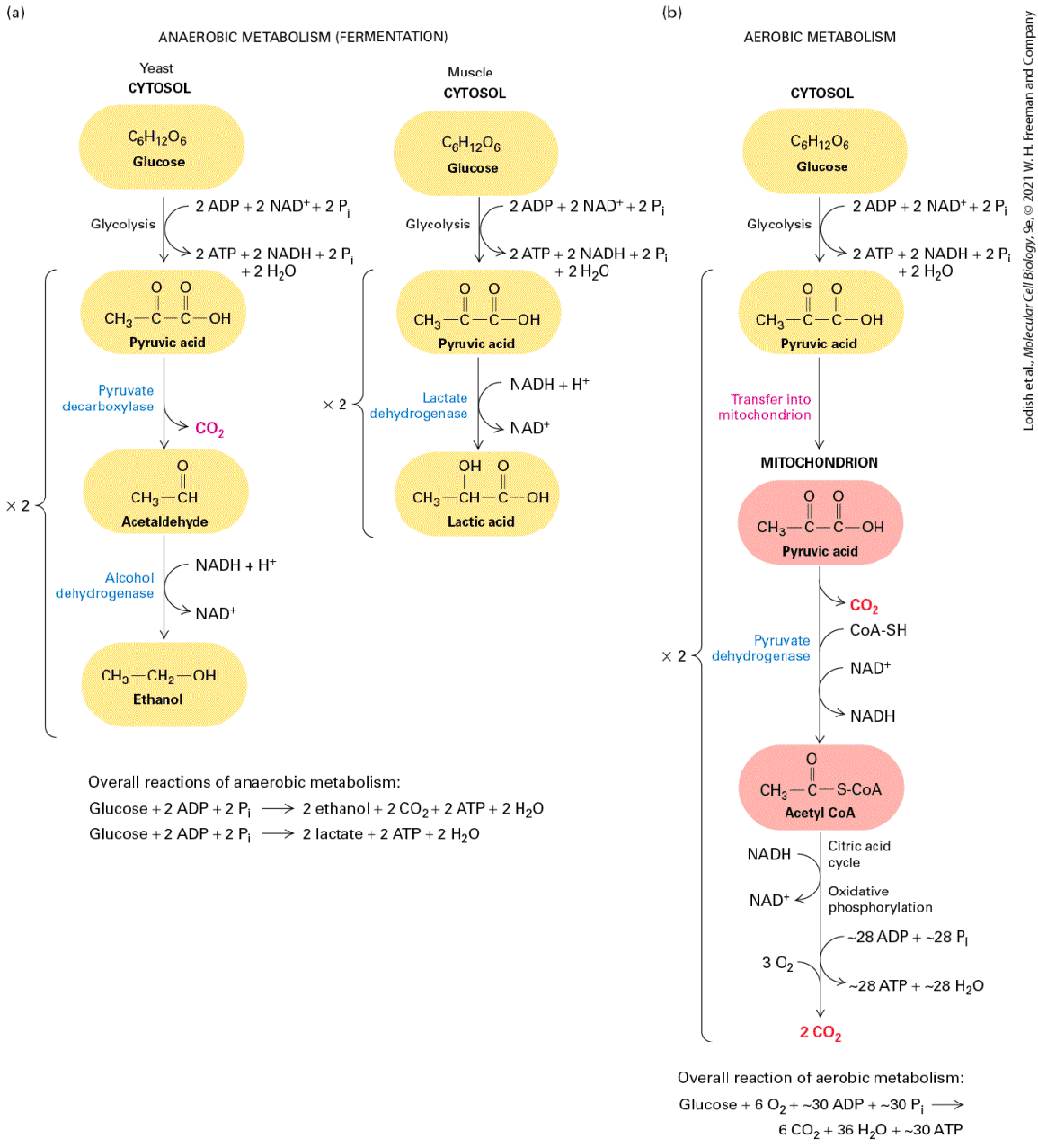
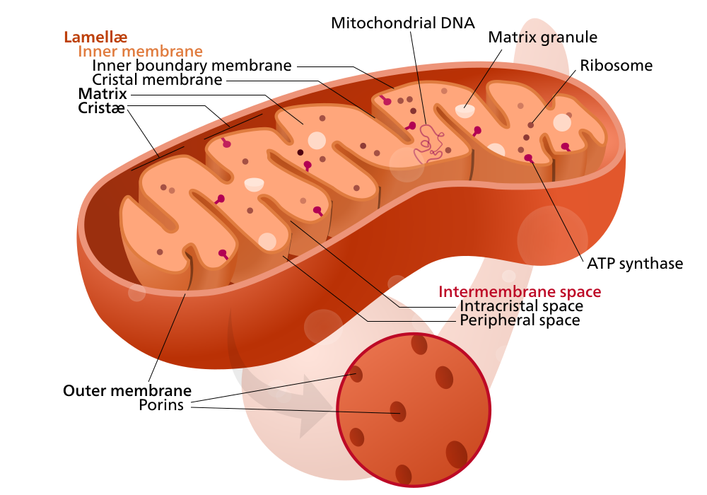
- Glycolysis
- Cytosolic, glucose → 2 pyruvate, net 2 ATP + 2 NADH.
- TCA / Krebs cycle
- Mitochondrial matrix. Acetyl-CoA → CO₂ + 3 NADH + 1 FADH₂ + 1 GTP per acetyl-CoA.
- Electron transport chain
- Complex I (NADH-DH), II (succinate-DH), III (cyt bc1), IV (cyt c oxidase). Electrons from NADH/FADH₂ → O₂ → H₂O. Pumps H⁺ to intermembrane space.
- Chemiosmosis
- Proton gradient drives ATP synthase (Complex V): F₀ rotor + F₁ catalytic head. ~32 ATP per glucose total.
- Uncoupling protein (UCP1)
- Brown adipose; dissipates proton gradient as heat (thermogenesis).
U10 · Cell signaling
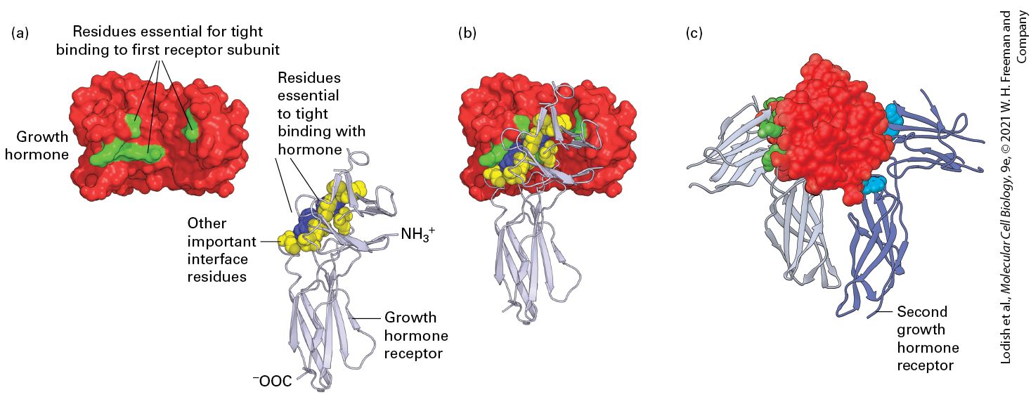
- Receptor classes
- Cell-surface: GPCR, RTK, ion-channel-coupled, integrins. Intracellular: nuclear receptors (steroid, thyroid).
- GPCR signaling
- Ligand → Gα-GTP → effectors (adenylyl cyclase → cAMP → PKA; PLCβ → IP₃/DAG → Ca²⁺/PKC). Desensitization by GRKs + arrestins.
- RTK signaling
- Ligand → dimerization → trans-autophosphorylation → SH2/PTB-domain adaptors (Grb2, Shc, PI3K). Examples: EGFR, insulin receptor, VEGFR.
- MAPK cascade
- Ras → Raf → MEK → ERK → nuclear TFs (Elk1, Fos). Drives proliferation. Mutated in many cancers.
- PI3K-AKT-mTOR
- Survival + growth. PIP₂ → PIP₃ recruits AKT; PTEN reverses. mTORC1 controls translation/autophagy.
- Notch
- Juxtacrine; intramembrane proteolysis releases NICD → nucleus → CSL → HES/HEY targets. Lateral inhibition.
- Wnt/β-catenin
- Wnt → Frizzled/LRP → Dishevelled → inhibits destruction complex (APC, GSK3, axin) → β-catenin to nucleus → TCF/LEF target genes.
- TGF-β / BMP
- Receptor Ser/Thr kinase phosphorylates SMADs → nucleus.
- Second messengers
- cAMP, cGMP, IP₃, DAG, Ca²⁺, NO.
U11 · Cytoskeleton
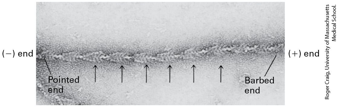
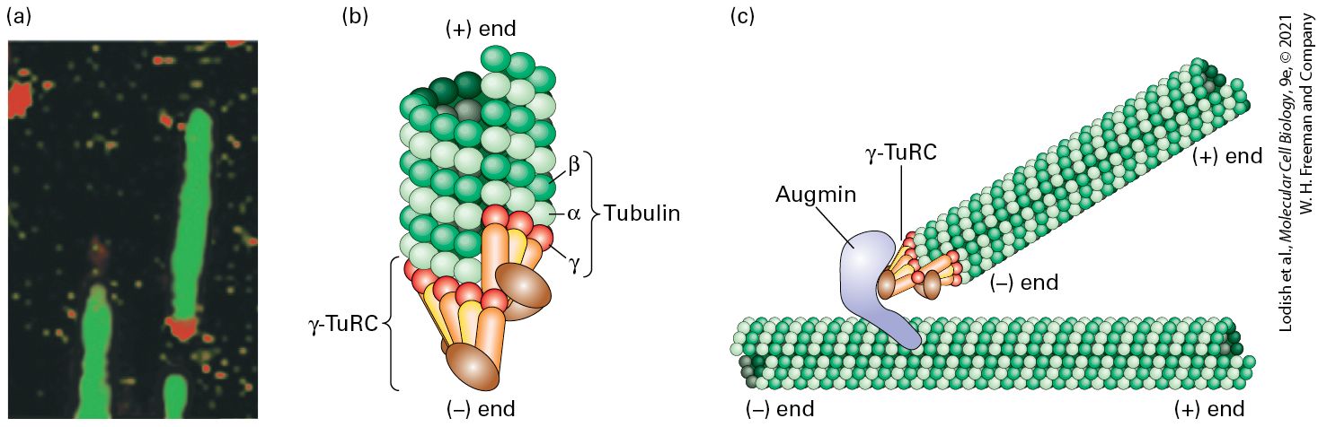
- Actin (microfilaments)
- ~7 nm; G-actin polymerizes (ATP-dependent) to F-actin. Polar (+/-). Regulators: profilin, cofilin, Arp2/3, formins. Cell shape, migration.
- Microtubules
- ~25 nm; α/β-tubulin dimers (GTP-dependent). 13 protofilaments. Polar — minus end at centrosome, plus end outward. Dynamic instability (catastrophe / rescue).
- Intermediate filaments
- ~10 nm; non-polar; coiled-coil dimers → tetramers → filaments. Mechanical support. Examples: keratins (epithelia), vimentin (mesenchymal), nuclear lamins.
- Myosin
- Actin-based motor. Myosin II = muscle + cytokinesis. Myosin V = vesicle transport.
- Kinesin
- MT + end-directed motor (anterograde transport, mitotic spindle).
- Dynein
- MT − end-directed (retrograde), also drives cilia/flagella.
- Cilia/flagella
- 9+2 axoneme. Beat by dynein-driven sliding of doublets.
U12 · Cell cycle & cell death
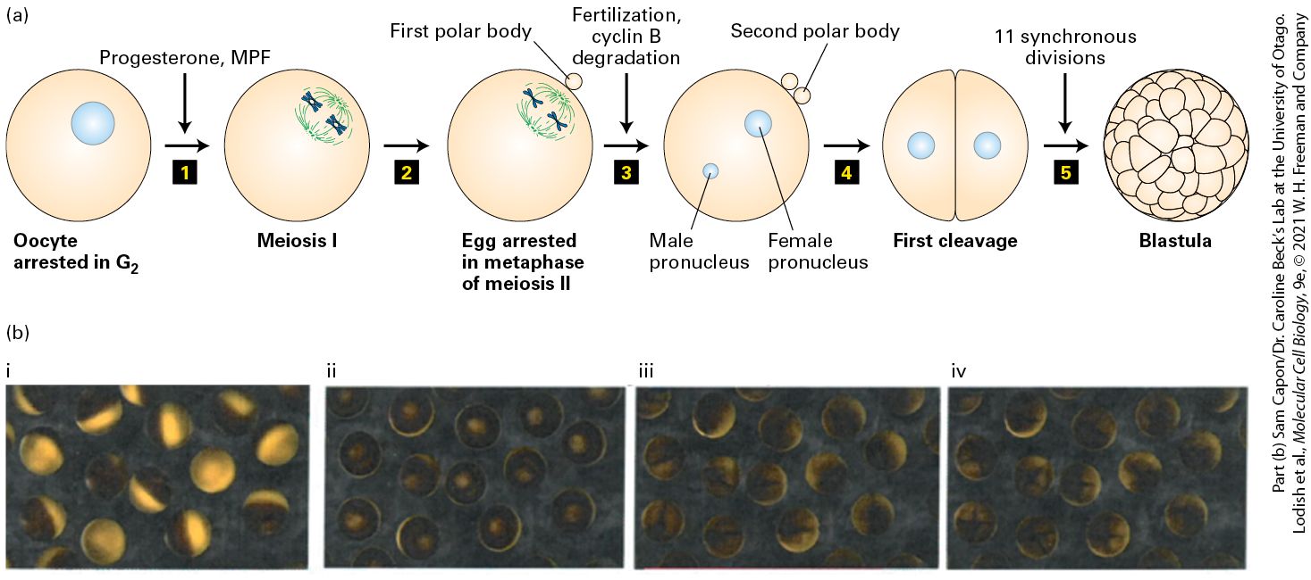
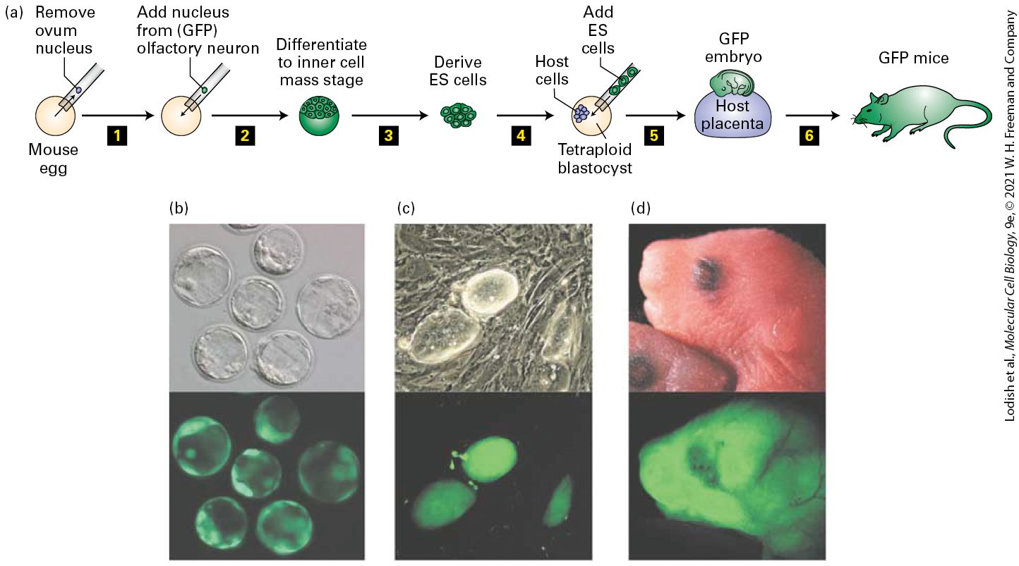
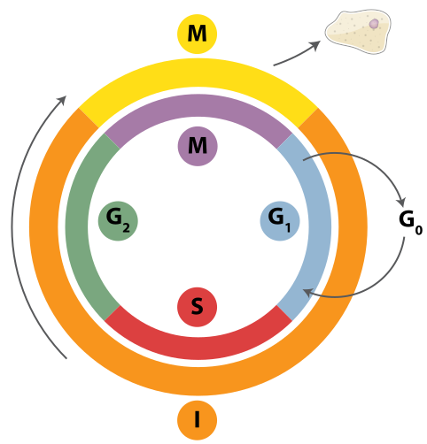
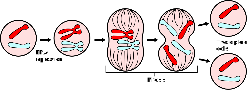
- Cell-cycle phases
- G1 → S (DNA replication) → G2 → M (mitosis + cytokinesis). G0 = quiescent.
- Cyclin/Cdk
- CDKs are kinases; cyclins are regulatory subunits, periodically expressed. G1: cyclin D + CDK4/6; S: cyclin E/A + CDK2; M: cyclin B + CDK1.
- Cell-cycle checkpoints
- G1/S (Restriction): DNA damage → p53 → p21 → CDK inhibition. G2/M: damage / unreplicated DNA. M (SAC): unattached kinetochores hold APC/C.
- Mitosis stages
- Prophase → prometaphase → metaphase (alignment) → anaphase (cohesin cleaved by separase) → telophase → cytokinesis.
- Apoptosis
- Programmed cell death. Intrinsic (mitochondrial cyt c → apoptosome → caspase-9 → effector caspase-3/7); extrinsic (death receptor Fas/TNFR → caspase-8). Bcl-2 family balance.
- Necroptosis / pyroptosis
- Lytic forms of cell death — RIPK + MLKL; gasdermin pores (matters for Chivero's NEUR 1520 inflammasome content!).
- Autophagy
- Self-degradation via autophagosome → lysosome. mTORC1 inhibits, ATG genes execute.
U13 · Cell-cell junctions — Johnson focus
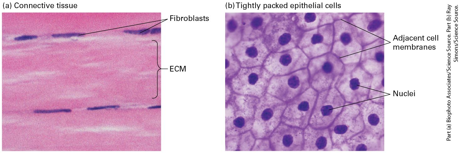
- Tight junctions (zonula occludens)
- Apical seal; claudins + occludin form paracellular barrier. Define apical/basolateral polarity.
- Adherens junctions
- E-cadherin (Ca²⁺-dependent) homophilic binding. Cytoplasmic tail binds β-catenin → α-catenin → actin. Johnson's specialty.
- Desmosomes
- Spot-weld between cells; desmocollins/desmogleins → plakoglobin/desmoplakin → intermediate filaments (keratin).
- Gap junctions
- Connexin hexamer = connexon; two connexons span the gap → channel for small molecules (<1 kDa: ions, cAMP, IP₃). Johnson's specialty.
- Hemidesmosomes
- Cell-to-ECM (basal lamina) via integrin α6β4 → keratin IFs.
- Focal adhesions
- Integrins + actin; mechanosensing; signal via FAK + Src.
- Cadherin switching in EMT
- E-cadherin ↓, N-cadherin ↑ during epithelial-to-mesenchymal transition (development + cancer metastasis).
- Catenins as signals
- β-catenin doubles as a Wnt-pathway TF when not sequestered at adherens junctions.
Week 1 · The pancreatic cancer story — Johnson's own research
This is one causal chain: the gene is fine, the protein is stuck, forward trafficking is fine, blocking clathrin does not rescue it, the answer is in the tail, delete the sorting motif and the junction forms. Roughly two quiz questions come out of it. The flashcards for it are on the study page under Ch 8 THE CHAIN, but do those after reading this, not instead of it.
QUIZ. She said roughly TWO questions on Tuesday come out of this chapter. Per hour spent, it is the highest-value section of the week after the math.
The image she opened with was pancreatic cancer cells taken from patients, fixed and stained, nucleus in blue and connexin in red. The cell-level problem she pointed at is one sentence long: the cells are not communicating with each other. They stop communicating. Across cancers generally, gap junctions fail in one of two ways. Either the gene gets turned off, or there is nothing whatsoever wrong with the gene and the proteins are simply all in the wrong place, sitting inside the cell instead of at the membrane. In her pancreatic cancer system the connexin gene sequence is not mutated. It is the same sequence. Everything that is wrong is downstream of the sequence, which is to say it is trafficking.
The experimental design rests on two cell lines from real patients. BxPC3 came from a patient at an early stage of disease. Capan-1 came from late-stage, metastatic disease. Comparing an early-stage line against a late-stage line is the whole design, because it lets you ask what specifically changes as the cancer advances rather than only asking what is broken.
WATCH OUT. The slide is titled "differential" for a reason. Cx43 assembly is impaired in BxPC3 but NOT in Capan-1, while Cx26 does the opposite. So it is not "Cx43 is always stuck." If she asks which connexin fails where, answer PER CONNEXIN and PER CELL LINE, never with one blanket statement.
Three connexins behave three different ways. Cx43 is abundant in gene expression but never appears in the membrane, sitting instead in an intracellular compartment. Cx26, a pancreatic connexin that is supposed to be there, reaches the membrane and forms junctions in BxPC3, the early-stage line, so those cells can still communicate, but in Capan-1, the late-stage line, it is stuck inside the cell and there is no communication anywhere in the network. Cx32, also supposed to be expressed in this tissue, is simply lost, and when they virally infected the cells with a Cx32-carrying virus the BxPC3 cells formed gap junctions. Her broader observation about Cx43 is worth carrying: Cx43 is expressed while we are developing as embryos, and as adults we do not have it in every cell, yet she found Cx43 in essentially every cancer she looked at, from any part of the body, and wrote that in her dissertation. She backed the cell lines up with tissue sections from other people's biopsies and found the same pattern in every one: Cx43 present in the tumor, but not in the junctions.
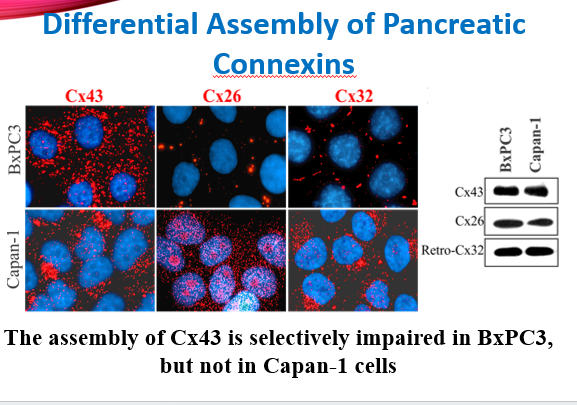
- Figure 7. Differential assembly of pancreatic connexins: Cx43, Cx26 and Cx32 compared in BxPC3 (early stage) versus Capan-1 (late stage), with matching Western blots. The slide's own conclusion is at the bottom.
The obvious therapeutic move does not work here, and understanding why is the hinge of the whole story. Her mentor had shown that in cancers where Cx43 is lost or strongly down-regulated, over-expressing Cx43 can slow or stop tumor growth, and prostate cancer is the standard example. But in pancreatic cancer there is already plenty of Cx43. It is all stuck inside the cell. You cannot over-express your way out of a trafficking problem, and that realization is what pushed her toward asking trafficking questions in the first place. They ran the control that proves it: over-expressing wild-type connexin on top of what was already there did not form junctions. It just produced more intracellular protein.
They started at the obvious end, testing forward trafficking with experiments designed to halt delivery to the membrane, and they still saw the same intracellular pattern. Her line about that outcome is the one to remember: "a no is an answer to a question." The negative result redirected the entire project away from delivery and toward endocytosis. So they blocked the two endocytosis routes. Clathrin-mediated endocytosis was blocked with a sucrose gradient, meaning hypertonic sugar in the extracellular medium, which prevents the clathrin cage from forming. Lipid-raft-mediated endocytosis was blocked with filipin, which pulls the cholesterol-rich rafts out of the membrane while leaving the membrane itself intact.
The essential part of that design is the cargo controls, because a block you cannot verify proves nothing. Transferrin, the iron-carrying ligand, is the known clathrin cargo, and cholera toxin is the known lipid-raft cargo. Cx43 was imaged in green and the tracer in red. The sucrose result was clean and disappointing in the productive way: transferrin was stuck at the membrane, which proved the clathrin block had worked, and Cx43 was still inside the cell anyway. Blocking clathrin did not rescue it. The raft result was messier. Some cholera toxin still got in and some stayed at the membrane, and if anything there was even more Cx43 inside the cell than before.
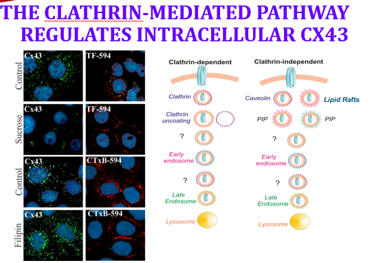
- Figure 8. The clathrin-mediated pathway regulates intracellular Cx43: immunofluorescence panels on the left beside the clathrin-dependent versus clathrin-independent route diagram on the right.
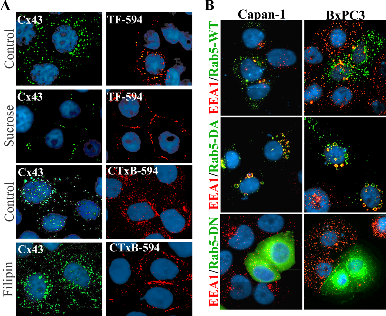
- Figure 9. The experiment itself. LEFT: Cx43 (green) paired with its tracer, TF-594 (transferrin, the CLATHRIN cargo) under Control versus SUCROSE, and CTxB-594 (cholera toxin B, the LIPID-RAFT cargo) under Control versus FILIPIN. Sucrose strands transferrin at the membrane, confirming the block, while Cx43 stays intracellular. RIGHT: EEA1 with Rab5-WT, Rab5-DA and Rab5-DN endosome mutants.
What cracked it was going back to the biochemistry of how endocytosis picks its cargo. Receptor-mediated endocytosis requires the cargo to bind an adapter protein, and the adapter then connects to clathrin, which builds a soccer-ball-shaped cage around a patch of membrane and pulls it inside. No adapter, no pickup. So they read the amino-acid sequence of the C-terminal domain, the long tail from chapter 7, and used bioinformatics to hunt for motifs that bind those adapters. They found tyrosine-based sorting motifs, a PY motif and a YKLD motif. Those are address labels, and what they say is "come inside."
They removed them by site-directed mutagenesis, mutating out the tyrosines along with nearby serines and aspartates, then put the mutant gene back into the cells two different ways, by transient transfection and by retroviral infection. The result is the payoff of the whole chapter: junction plaques appeared. Fixed and stained, the cells finally built gap junctions, and they did it in both the early-stage and the late-stage cancer lines. The biochemical confirmation came from fractionation, which works by dissolving the cells with detergent: free protein dissolves and goes into the soluble fraction, while protein locked into an assembled junction does not and stays in the insoluble fraction. Protein shifted out of the soluble intracellular fraction and into the insoluble, gap-junction-associated fraction. That shift is the chemical signature of assembled junctions, and it is the second independent piece of evidence. Her own summary of the discovery: something makes the protein traffic to the membrane and then get endocytosed before it can connect to the next cell and make communication happen. Delete the sorting motif and the junction forms.
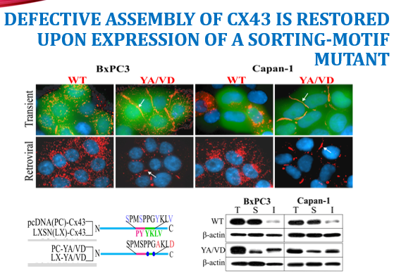
- Figure 10. THE PAYOFF. Defective assembly of Cx43 is restored by expressing a sorting-motif mutant: in both BxPC3 and Capan-1, and by both TRANSIENT transfection and RETROVIRAL infection, the YA/VD mutant forms junction plaques (white arrows) where wild-type Cx43 does not. The Western blots below show the shift from Soluble to Insoluble.
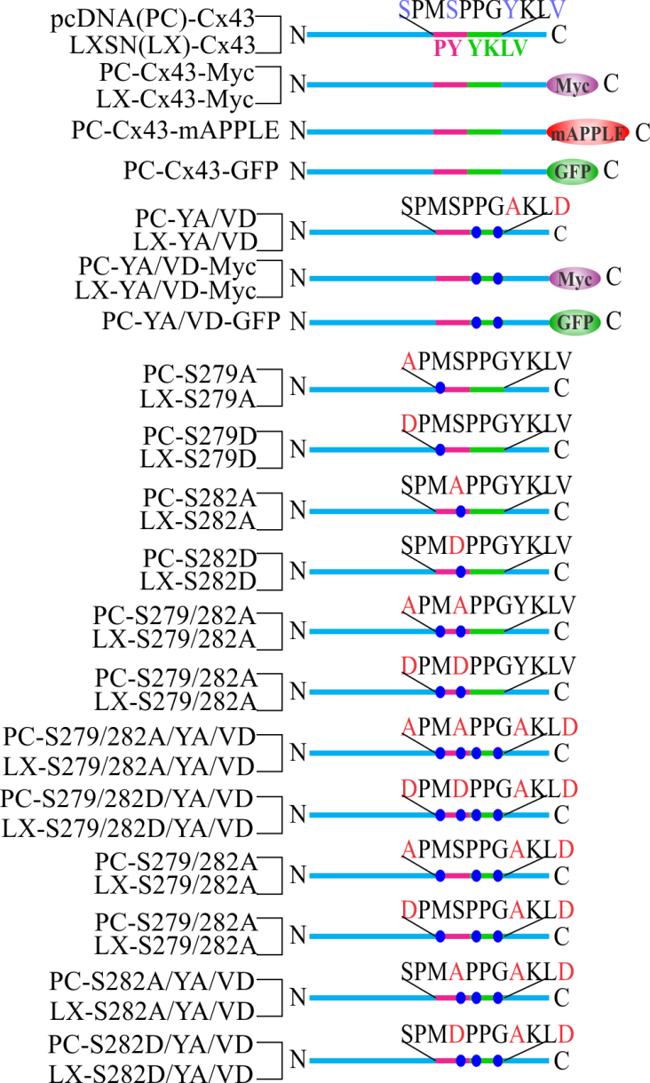
- Figure 11. The construct series: wild-type Cx43 carries SPMSPPGYKLV with the PY and YKLV motifs highlighted; the YA/VD mutants replace them, and the S279/S282 series tests the neighbouring serines. This is what "site-directed mutagenesis" physically means.
She closed with a question she deliberately left open and told the class to argue about: what is the difference between transient transfection and retroviral infection? She said it will be answered properly when the course covers immortalization, but the answer to have ready is that transient transfection introduces DNA that is not integrated into the genome, so it is expressed for a few days and then diluted out as the cells divide, while retroviral infection integrates the sequence into the host genome, giving stable, heritable expression in every daughter cell. That is the same integration logic used to immortalize cells with SV40 large T and hTERT in the assigned Kitazawa paper, which is why she is holding the answer until then.
As for where the field went, researchers after her identified specific kinases that are hyperactive in pancreatic cancer, and there are now drugs targeting those. Several related findings from her lab are worth recognizing by name if they appear: phosphorylation of Cx43 at Ser-279 and Ser-282 regulates its endocytosis and gap-junction assembly, a kinase being simply an enzyme that sticks a phosphate group onto a protein as an on/off switch; E-cadherin and N-cadherin regulate Cx43 assembly differentially; the clathrin-mediated pathway and lipid rafts govern intracellular Cx43; and palmitoylation of plakophilin is required for desmosome assembly. The habit of mind she is actually trying to install with all of this is to be mechanistic. For your own project, ask what the mechanism is, which receptors might be impacted, which surface proteins might be hurting, and whether what you are seeing is apoptosis or necrosis. Apoptosis is tidy, programmed cell suicide; necrosis is messy, uncontrolled death from injury, and she wants you to be able to say which one your treatment caused.
U14 · Cancer + tissues + ECM
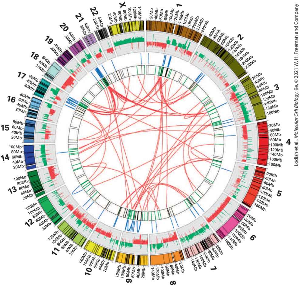
- Hanahan-Weinberg hallmarks
- Sustained proliferation, evasion of growth suppressors, resisting cell death, replicative immortality, angiogenesis, invasion + metastasis, deregulated metabolism, immune evasion + inflammation, genome instability.
- Oncogenes
- Gain-of-function mutations in proto-oncogenes (Ras, Myc, EGFR, BRAF). Dominant.
- Tumor suppressors
- Loss-of-function (p53, Rb, APC, PTEN, BRCA1/2). Recessive (Knudson 2-hit).
- Metastasis
- EMT → local invasion → intravasation → circulation → extravasation → colonization.
- ECM
- Collagen (most abundant), elastin, proteoglycans (heparan/chondroitin sulfate), fibronectin, laminin (basal lamina).
- Basal lamina
- Sheet of laminin + type IV collagen + perlecan. Separates epithelium from CT. Barrier disrupted in carcinoma invasion.
- Stem cells
- Self-renewal + multipotency. Niche-dependent. Adult (intestinal crypt, bone marrow, hair follicle).
U15 · Lab techniques (Northam's lab block)
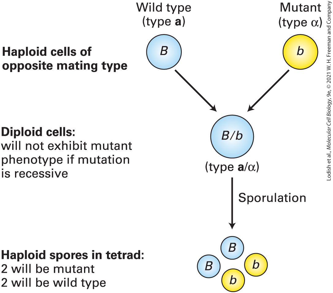
- Tissue culture
- Maintain mammalian cells in vitro: medium with FBS, sterile technique, passaging. Common lines: HeLa, HEK293, NIH-3T3, MCF-7.
- Trypsinization
- Protease detaches adherent cells for splitting/passaging.
- Transfection
- Introduce DNA/RNA: lipofection, electroporation, viral vectors (lentivirus).
- Immunofluorescence (IF)
- Fix → permeabilize → block → primary Ab → fluorescent secondary Ab → DAPI for nuclei → image. Localize proteins.
- Western blot
- SDS-PAGE → transfer → primary + secondary HRP Ab → ECL detection. Quantify protein.
- RT-qPCR
- RNA → cDNA (reverse transcriptase) → SYBR/TaqMan qPCR. Quantify mRNA. ΔΔCt analysis vs housekeeping gene.
- Flow cytometry / FACS
- Fluorescent markers + cytometer → analyze or sort cells by surface or DNA-content phenotypes.
- siRNA / shRNA / CRISPR
- Loss-of-function: siRNA transient, shRNA stable, CRISPR-Cas9 genomic edit.
- Live-cell imaging
- Fluorescent proteins (GFP/mCherry) + confocal/spinning disk; FRAP for diffusion; FRET for protein-protein interaction.
- High-throughput assays
- Multi-well plate readouts: viability (MTT, ATP), reporter, image-based phenotyping. Used in drug discovery.
Johnson + Northam exam tips
- Be prepared to diagram a junction (adherens, gap, tight, desmosome) labeled with proteins + linkages.
- Know EMT: cadherin switch, β-catenin re-localization, role in cancer.
- For lab: identify a microscopy technique from a sample image — bright-field vs phase vs IF vs confocal.
- Be ready to interpret RT-qPCR ΔΔCt data and a Western blot.
📚 Textbook companion · Lodish MCB 9e
Each unit above maps to chapters in the locally-OCR'd Lodish MCB 9e. Use the cards below as a quick visual jump into the embedded textbook reader — one figure per chapter, click to read the full chapter: